Introduction
Rheumatoid arthritis (RA) is a chronic systemic
autoimmune disorder characterized by persistent synovitis,
progressive cartilage degradation and bone erosion, which over time
leads to severe joint impairment and disability (1,2).
Although the age-adjusted prevalence of RA is ~1% worldwide, it
remains a major contributor to healthcare expenditure, particularly
in the context of global population aging (3,4). The
etiology of RA is multifactorial and has been attributed to a
combination of genetic susceptibility, environmental triggers and
dysregulated immune responses, which together lead to the
overactivation of various immune cells and, notably, drive
fibroblast-like synoviocytes (FLSs) toward an invasive and
anti-apoptotic phenotype, ultimately resulting in immune-mediated
joint destruction and dysfunction (5,6).
Current therapeutic strategies for RA primarily aim
to alleviate symptoms and slow disease progression. These
strategies include non-steroidal anti-inflammatory drugs,
glucocorticoids, and conventional synthetic or biologic
disease-modifying antirheumatic drugs (7,8).
Despite their clinical utility, a notable proportion of patients
exhibit inadequate responses to these therapies, and long-term use
is often associated with considerable adverse effects, including
infections, hepatic toxicity and cardiovascular complications
(9,10). Therefore, elucidating the intricate
molecular mechanisms underlying RA pathogenesis remains imperative
for identifying novel therapeutic targets, and developing more
effective and safer treatment modalities.
Among the numerous signaling pathways involved in
RA, the NF-κB pathway has been the most extensively studied because
of its role in mediating inflammation (11,12).
Nonetheless, accumulating evidence has highlighted the crucial role
of the PI3K/AKT signaling axis as a key inflammatory regulator in
RA, controlling not only inflammation, but also cell survival,
proliferation and metabolism (13). The PI3K/AKT signaling pathway is
activated by various cytokines and growth factors in the RA
synovial microenvironment. Once activated, this pathway exerts
profound effects on gene regulatory networks involved in
pathological processes; it modulates the activity of downstream
transcription factors, such as NF-κB, and regulates the expression
of pro-inflammatory cytokines (14). The present study hypothesizes that
pathological alterations in the PI3K/AKT pathway affect multiple
cellular functions, modulate immune responses, and contribute to
persistent synovitis and joint destruction by regulating the
expression of pro-inflammatory cytokines (for example, TNF-α, IL-1β
and IL-6), matrix metalloproteinases (MMPs) and anti-apoptotic
proteins (15,16).
Additionally, regulation of the PI3K/AKT pathway is
closely associated with non-coding RNAs (ncRNAs), which have been
recognized as important epigenetic regulators of gene expression in
RA (17–19). The interaction between ncRNAs and
signaling pathways adds a further layer of complexity to the
regulation of gene expression in RA.
Although the role of NF-κB in RA has been well
described, to the best of our knowledge, a systematic review
focusing on the PI3K/AKT signaling pathway and its complex
regulatory effects on gene expression in RA is still lacking.
Specifically, existing reviews (11,12)
have often discussed the PI3K/AKT pathway alongside other signaling
pathways without emphasizing its distinct role in gene expression
regulation, or they have failed to clearly distinguish between
general PI3K/AKT biology and RA-specific evidence. The present
review aimed to fill this gap by providing a focused synthesis of
how PI3K/AKT modulates gene expression in RA, with a clear
distinction between established findings in RA tissues and cells,
and indirect evidence derived from other disease models. To ensure
transparency and reproducibility, the present study conducted a
narrative review based on a systematic literature search of PubMed
(pubmed.ncbi.nlm.nih.gov) and Web of Science (https://www.webofscience.com) covering the period
January 2000-December 2025, using the key words ‘PI3K/AKT’,
‘rheumatoid arthritis’, ‘gene expression’ and ‘non-coding RNA’.
Studies were included if they addressed PI3K/AKT signaling in RA or
related inflammatory arthritis models; mechanistic studies from
non-RA contexts were used only as background support for general
pathway biology and were explicitly identified as such. The present
study aimed to summarize the PI3K/AKT signaling pathway and its
mechanisms of activation, its central role in regulating the
expression of genes involved in RA pathology, and its dynamic
crosstalk with other signaling pathways and ncRNAs. Finally, we
address the therapeutic potential of targeting the PI3K/AKT axis
and emerging ncRNA-based strategies for the future management of
RA.
PI3K/AKT signaling pathway in RA
The PI3K/AKT signaling pathway is a critical
intracellular signaling cascade that regulates a wide range of
cellular processes, including metabolism, proliferation, survival
and inflammation. Its aberrant activation is increasingly
recognized as a cornerstone of the pathogenic mechanisms underlying
RA (20,21).
Pathway components and activation
mechanisms
PI3Ks are heterodimeric enzymes composed of a
regulatory subunit and a catalytic (p110) subunit. They are
classified into different classes, among which class IA PI3Ks
(p110α, p110β and p110δ) are primarily activated by receptor
tyrosine kinases upon stimulation by growth factors and cytokines,
whereas class IB PI3K (p110γ) is activated by G-protein-coupled
receptors (GPCRs), such as chemokine receptors (22,23).
These general activation mechanisms have been well established in
various cell types; in the context of RA, they are mainly supported
by studies involving RA-derived FLSs and immune cells.
A key feature of PI3K activation is its potentiation
by Ras family GTPases. Once activated, PI3K phosphorylates
phosphatidylinositol lipids at the plasma membrane, thereby
generating docking sites for proteins containing pleckstrin
homology domains, most notably phosphoinositide-dependent kinase 1
(PDK1) and AKT (24). The tumor
suppressor PTEN serves as the primary negative regulator of this
pathway by dephosphorylating these lipid intermediates (25). AKT functions as the central node of
PI3K signaling, and its phosphorylation by PDK1 and mammalian
target of rapamycin complex (mTORC)2 results in full activation.
Downstream, AKT coordinates cellular responses by phosphorylating
and modulating the activity of numerous substrates, including
inhibitor of κB kinase (IKK), the pro-apoptotic protein Bad,
glycogen synthase kinase-3β (GSK-3β), mTOR and forkhead box O
(FoxO) transcription factors (26,27).
Aberrant PI3K/AKT activation in the RA
synovium
There is evidence for constitutive activation of the
PI3K/AKT axis in the RA synovial microenvironment (15). One line of evidence comes from the
reduced expression of PTEN mRNA in the invasive layer of the RA
synovium, a specific sublayer within the synovial lining, as well
as in FLSs invading cartilage in a murine arthritis model (28). Loss of this negative regulator
supports hyperactivation of the pathway. Consistent with this,
levels of phosphorylated-AKT are markedly increased in RA synovial
tissue and purified FLSs compared with specimens from patients with
osteoarthritis or healthy controls (29). Functionally, pro-inflammatory
cytokines, such as TNF-α, potently activate AKT in RA-FLSs in
vitro. Inhibition of PI3K by LY294002 or overexpression of PTEN
sensitizes these cells to TNF-α-induced apoptosis, demonstrating
that the PI3K/AKT pathway provides a survival signal that
contributes to the apoptosis-resistant phenotype of RA-FLSs
(30,31). Moreover, this pathway mediates
resistance to cell death induced by other cytokines, such as
TNF-related apoptosis-inducing ligand (32). These findings help explain the dual
role of the PI3K/AKT pathway in promoting both synovial hyperplasia
and inflammation. Most of these observations are derived from
direct studies of RA tissues and FLSs (28–32),
thus providing disease-specific evidence. The mechanistic details
regarding Ras-mediated PI3K activation are derived mainly from
studies non-RA systems (24,33).
As direct validation in RA tissues remains limited, these findings
should be interpreted as general PI3K/AKT pathway biology rather
than RA-specific evidence. Fig. 1
presents a comprehensive schematic diagram of the PI3K/AKT
signaling pathway in the pathogenesis of RA.
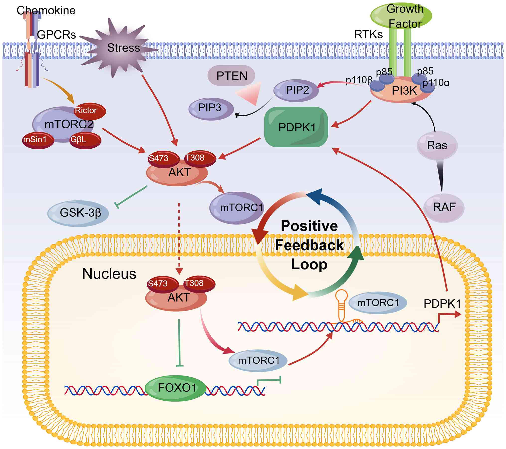 | Figure 1.Schematic diagram of the PI3K/AKT
signaling pathway in the pathogenesis of rheumatoid arthritis.
Upstream activators, including growth factors and chemokines, act
through RTKs and GPCRs to activate class IA and class IB PI3K,
leading to the conversion of PIP2 to PIP3, a process antagonized by
PTEN. Downstream activation of AKT regulates cell proliferation and
immune inflammation through multiple effectors, including the
Ras/RAF/MEK/ERK and mTOR pathways. FOXO1, Forkhead box O1; GPCR,
G-protein-coupled receptor; GSK-3β, Glycogen synthase kinase-3β;
mTORC1, Mammalian target of rapamycin complex 1; PDPK1,
3-phosphoinositide-dependent protein kinase 1; PIP2,
Phosphatidylinositol 4,5-bisphosphate; PIP3, Phosphatidylinositol
3,4,5-trisphosphate; RTK, Receptor tyrosine kinase. Created with
BioGDP.com (104). |
Regulation of gene expression via
PI3K/AKT
Gene expression in RA is regulated by the PI3K/AKT
pathway through a series of interconnected mechanisms, including,
but not limited to, inflammatory responses, synovial cell survival
and maintenance of joint structural integrity (13–15).
The present review distinguishes between evidence derived directly
from RA tissues and cells [for example, human RA synovium, RA-FLSs
and collagen-induced arthritis (CIA) models] and weaker or indirect
evidence from non-RA settings (for example, cancer, cardiac or
brain injury studies), which is used only to suggest plausible
mechanisms requiring further validation in RA.
Transcriptional regulation via
downstream effectors
Once activated, AKT phosphorylates a series of
transcription factors and regulatory proteins that modulate gene
expression profiles in RA synovial cells. Among these, the NF-κB
pathway represents one of the most important downstream effectors.
Phosphorylation and activation of IKK lead to the phosphorylation
of IκB, resulting in its degradation and the subsequent nuclear
translocation of NF-κB (34,35).
This, in turn, enhances the transcription of pro-inflammatory
genes, including TNF-α, IL-1β, IL-6 and IL-8, as well as MMPs, such
as MMP-3 and MMP-9, which have critical roles in synovitis and
cartilage degradation (36,37).
Moreover, AKT-mediated phosphorylation of FoxO transcription
factors leads to their cytoplasmic sequestration and inactivation,
thereby suppressing the expression of pro-apoptotic genes such as
Bim and FasL (Fig. 2). This
mechanism contributes to the apoptosis-resistant phenotype of FLSs
in RA (38,39). In addition, mTOR is another major
downstream substrate of AKT. Activation of mTORC1 positively
regulates mRNA translation and the expression of proteins involved
in cell proliferation and metabolism through phosphorylation of
ribosomal protein S6 kinase 1 and eukaryotic translation initiation
factor 4E-binding protein 1 (40–42).
This promotes the synthesis of cyclins, MMPs and inflammatory
mediators, thereby enhancing synovial proliferation and
inflammation. All of the aforementioned mechanisms have been
directly demonstrated in RA-FLSs or RA synovial tissues,
representing strong disease-specific evidence.
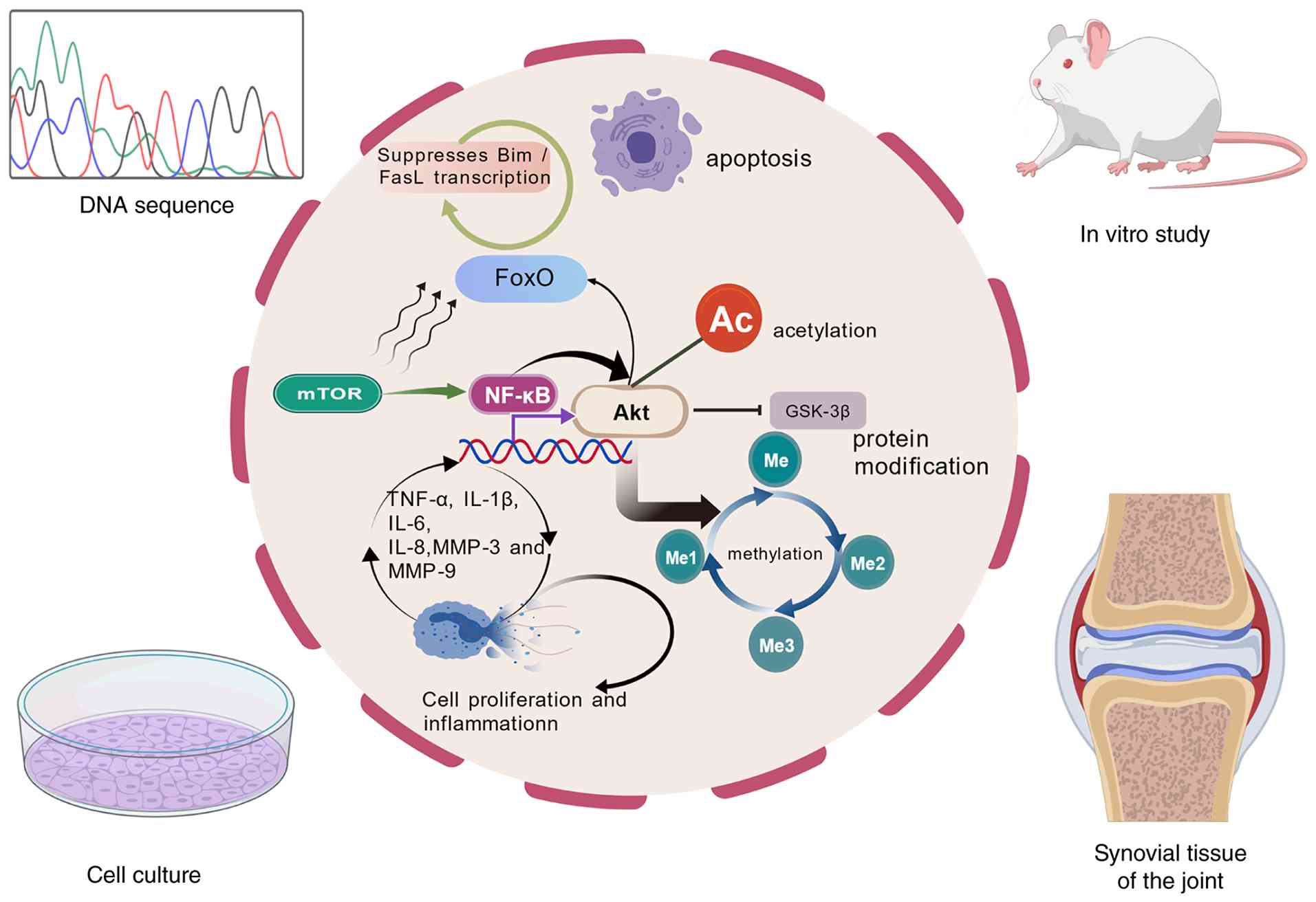 | Figure 2.Post-transcriptional and epigenetic
modification mechanisms of the PI3K/AKT pathway in rheumatoid
arthritis. This diagram illustrates how activated AKT modulates
transcription factors and downstream effectors, including NF-κB,
FoxO, mTOR and GSK-3β, to regulate the expression of
pro-inflammatory cytokines, such as TNF-α, IL-1β, IL-6 and IL-8, as
well as MMPs (MMP-3 and MMP-9), thereby promoting cell
proliferation and inflammation. FoxO, Forkhead box O; GSK-3β,
Glycogen synthase kinase-3β; Created with BioGDP.com (104). |
Post-transcriptional and epigenetic
mechanisms
In addition to its role in transcriptional control,
the PI3K/AKT pathway can influence gene expression through
post-transcriptional and epigenetic mechanisms. AKT activation may
stabilize specific mRNAs by inhibiting GSK-3β (Fig. 2), which is considered to mediate
mRNA decay (43). In addition,
PI3K/AKT signaling can induce epigenetic changes, such as histone
acetylation and DNA methylation, thereby remodeling chromatin and
altering gene expression in RA-FLSs (Fig. 2). For example, AKT activation has
been associated with increased histone H3 acetylation at the
promoters of inflammatory genes, thereby facilitating their
transcription (44,45). These epigenetic observations are
primarily based on in vitro studies of RA-FLSs; however, the
direct link between AKT and specific histone modifications in RA
synovium in vivo remains less well established and warrants
further investigation. Fig. 2
presents a schematic diagram of the transcriptional and epigenetic
regulation mediated by the PI3K/AKT signaling pathway.
Cross-talk with ncRNAs
A critical layer of regulation of PI3K/AKT signaling
is provided by ncRNAs. Multiple microRNAs (miRNAs/miRs) have been
shown both to regulate and to be regulated by PI3K/AKT signaling.
In particular, miR-124a directly targets the p110α catalytic
subunit of PI3K (encoded by PIK3CA), thereby inhibiting AKT
activation and downstream NF-κB signaling (Fig. 3). This leads to suppression of
RA-FLS proliferation and reduced production of inflammatory
cytokines (19). By contrast,
miR-21 is upregulated in the RA synovium and promotes PI3K/AKT
activation by suppressing PTEN (Fig.
3), thereby enhancing FLS survival and inflammation (46,47).
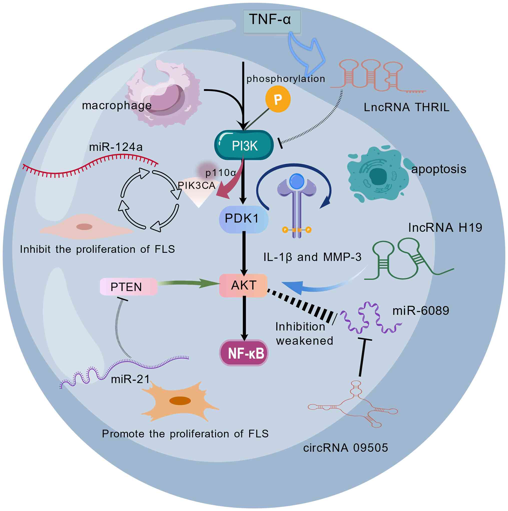 | Figure 3.Crosstalk between the PI3K/AKT
signaling pathway and ncRNAs in rheumatoid arthritis. ncRNAs,
including miR-124a, miR-21, lncRNA THRIL, lncRNA H19 and
circRNA_09505, modulate PI3K/AKT signaling at multiple levels.
miR-124a directly targets the PI3K p110α subunit (PIK3CA) to
inhibit FLS proliferation, whereas miR-21 suppresses PTEN to
promote FLS proliferation. lncRNA THRIL activates PI3K/AKT
signaling, thereby increasing IL-1β and MMP-3 expression levels;
lncRNA H19 functions as a competing endogenous RNA; and
circRNA_09505 sponges miR-6089 to enhance AKT1 expression in
macrophages. circRNA, circular RNA; FLS, fibroblast-like
synoviocyte; lncRNA, long ncRNA; miR, microRNA; PDK1,
phosphoinositide-dependent kinase 1.Created with BioGDP.com
(104). |
Notably, long ncRNAs (lncRNAs) are key modulators of
the PI3K/AKT pathway in RA. For example, the lncRNA THRIL is
upregulated in the serum of patients with RA, and knockdown of
THRIL in TNF-α-stimulated RA-FLSs reduces the phosphorylation of
PI3K and AKT, concomitantly decreases the production of IL-1β and
MMP-3 and promotes apoptosis (48)
(Fig. 3). These findings suggest
that THRIL exerts its pro-inflammatory and anti-apoptotic effects,
at least in part, through activation of the PI3K/AKT pathway.
Similarly, the lncRNA H19 has been shown to promote AKT
phosphorylation by serving as a competing endogenous RNA (ceRNA)
for miRNAs that target AKT regulators; for example, H19 acts as a
ceRNA to modulate the PI3K/AKT pathway (49,50),
thereby promoting inflammatory responses (Fig. 3).
Circular RNAs (circRNAs) also participate in this
regulatory network. For example, circRNA_09505 sponges miR-6089
(Fig. 3), thereby relieving its
suppression of AKT1 mRNA, which leads to enhanced AKT signaling and
aggravated inflammation in RA macrophages (51).
All ncRNA examples discussed in the present review
are derived from samples of patients with RA or RA-FLS models, thus
representing direct evidence in RA. Fig. 3 presents a schematic diagram of the
crosstalk between the PI3K/AKT signaling pathway and ncRNAs.
Crosstalk with other pathways
The pathogenesis of RA is not governed by isolated
signaling cascades, but rather by a highly interconnected network
of pathways that amplify and sustain inflammatory and destructive
processes. Although the PI3K/AKT pathway serves a central role, it
does not function in isolation; instead, it engages in extensive
crosstalk with other key signaling axes, thereby orchestrating a
synergistic amplification of pro-inflammatory gene expression and
synovial pathology.
Crosstalk with the NF-κB pathway
The interaction between PI3K/AKT and NF-κB is
arguably the most understood among these signaling networks. AKT
directly phosphorylates and activates IKK, thereby promoting IκB
degradation and the nuclear translocation of NF-κB. In this way, it
facilitates the increased expression of various pro-inflammatory
genes, including TNF-α, IL-1β, IL-6 and MMPs (52,53).
This mechanism has been repeatedly validated in RA-FLSs and animal
models of RA, providing strong direct evidence. Conversely, NF-κB
can further enhance PI3K/AKT signaling by upregulating cytokines
such as IL-6, which promote the activation of upstream receptor
tyrosine kinases and GPCRs, thereby creating a positive feedback
loop that sustains chronic inflammation and drives RA progression
(54).
Interaction with the JAK/STAT
pathway
The JAK/STAT pathway represents another important
inflammatory signaling pathway in RA. Cytokine receptors associated
with JAKs can also recruit and activate PI3K through phosphorylated
tyrosine residues, thereby leading to downstream AKT activation
(55,56). This interaction has been
demonstrated in RA-FLSs and synovial tissues. In addition, STAT3,
the principal transducer of IL-6 signaling, transcriptionally
upregulates genes involved in cell survival and inflammation that
are also modulated by AKT, such as Bcl-2 and MMP-9 (57,58).
This overlap suggests that combined inhibition of the JAK/STAT and
PI3K/AKT pathways may produce synergistic therapeutic effects, as
indicated by preclinical and clinical studies of dual-pathway
inhibition (59,60).
Interplay with MAPK signaling
In RA, the MAPK pathway, including ERK and p38,
frequently exhibits cross-activation with PI3K/AKT signaling. For
example, AKT antagonizes apoptosis signal-regulating kinase 1
(ASK1), an upstream activator of p38 and JNK, thereby influencing
stress-induced apoptosis and inflammation (61,62).
However, much of the evidence for the interaction between AKT and
ASK1 is derived from non-RA contexts, such as cancer and
neurotoxicity models (61,62), and direct validation in RA remains
limited. Meanwhile, MAPK activation can also influence PI3K
signaling through ribosomal S6 kinase-mediated phosphorylation of
AKT or by increasing the expression of growth factors that activate
PI3K (63,64). This bidirectional crosstalk further
underscores that targeting a single pathway in isolation may be
insufficient, and that multi-target therapeutic strategies may be
required.
Modulation by Notch signaling
Evidence indicates that Notch signaling modulates
PI3K/AKT activity in RA. The Notch intracellular domain (NICD) can
transcriptionally upregulate PIK3CA expression or inhibit PTEN,
thereby enhancing AKT phosphorylation (65,66).
These findings are supported by studies in RA synovial fibroblasts
and animal models (65,66). In turn, AKT can stabilize NICD and
enhance its transcriptional activity, thereby forming another
positive feedback loop that promotes synovial cell survival and
inflammatory cytokine production (67). Inhibition of Notch signaling has
been shown to reduce AKT activation and ameliorate arthritis in
animal models, suggesting that this pathway may represent a viable
therapeutic target (68). Notch
signaling via the NICD/CSL/Hey/DTX axis also promotes the invasive
migration of RA-FLSs, a key feature of synovial hyperplasia and
joint destruction (Fig. 4).
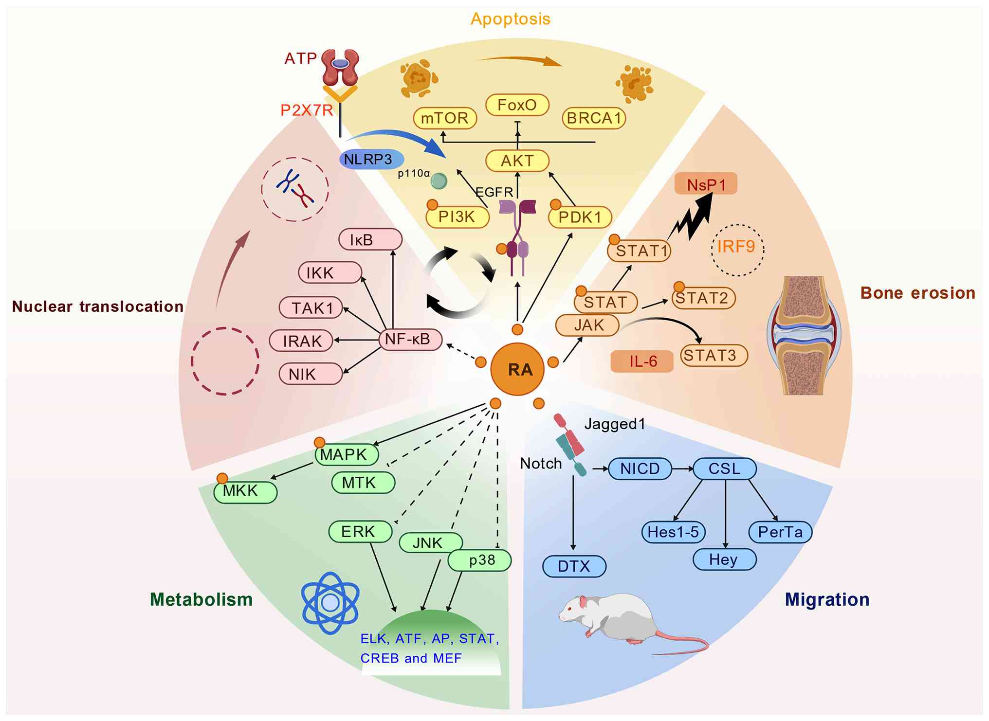 | Figure 4.Integrated crosstalk between the
PI3K/AKT signaling pathway and other key signaling pathways in RA.
Interactions between PI3K/AKT and other major signaling pathways in
RA, including apoptosis-related regulators (ATP/P2X7R, NLRP3, FoxO
and BRCA1), inflammatory pathways (NF-κB, JAK/STAT and
MAPK/ERK/JNK/p38), Notch signaling (NICD/CSL/Hey/DTX), and
transcriptional outputs (ELK, ATF, AP, STAT, CREB and MEF).
Together, these signaling networks contribute to bone erosion,
metabolic alterations and cell migration in RA. FoxO, forkhead box
O; IKK, inhibitor of κB kinase; NICD, Notch intracellular domain;
NLRP3, NOD-, LRR- and pyrin domain-containing protein 3; PDK1,
phosphoinositide-dependent kinase 1; RA, rheumatoid arthritis.
Created with BioGDP.com (104). |
Integration with purinergic and
inflammasome pathways
The P2X7R/NOD-, LRR- and pyrin domain-containing
protein 3 (NLRP3) inflammasome axis also intersects with PI3K/AKT
signaling. ATP-mediated activation of P2X7R can stimulate PI3K/AKT
signaling, which in turn enhances NLRP3 inflammasome assembly and
IL-1β maturation (69,70). These observations are primarily
derived from non-RA models, such as brain injury (69) and a colorectal cancer model
(70); however, recent studies in
RA have begun to explore this link (71,72).
Moreover, AKT-mediated activation of NF-κB primes NLRP3 expression,
thereby completing a pro-inflammatory circuit that links purinergic
signaling with cytokine release and synovitis (73,74).
Targeting P2X7R or NLRP3 may therefore indirectly attenuate
PI3K/AKT signaling. However, direct evidence for this crosstalk in
RA remains preliminary and requires further investigation.
Additionally, the PI3K/AKT pathway may contribute to apoptosis
resistance and inflammation through its interplay with DNA damage
response proteins. For example, DTX3L (also known as ARTD9), which
is involved in the DNA damage response, has been shown to promote
inflammation in RA-FLSs by increasing STAT1 translocation (75). This crosstalk represents a
promising area for future investigation into the mechanisms driving
the anti-apoptotic and pro-inflammatory phenotype of RA-FLSs
(Fig. 4).
Therapeutic implications
Pharmacological inhibition of
PI3K/AKT
Several small-molecule inhibitors targeting PI3K or
AKT have shown promise in preclinical models of RA. For example,
the PI3K inhibitor LY294002 and the AKT inhibitor MK-2206 have been
shown to induce apoptosis in RA-FLSs, and reduce the production of
pro-inflammatory cytokines and MMPs in vitro (76,77).
Isoform-specific inhibitors, particularly those targeting PI3Kδ and
PI3Kγ, are of particular interest because of their predominant
expression in leukocytes and their roles in immune cell activation.
Idelalisib, a PI3Kδ inhibitor approved for hematological
malignancies, has shown efficacy in reducing disease severity by
attenuating B-cell and macrophage activation (78,79).
Similarly, duvelisib, a dual PI3Kδ/γ inhibitor, has been reported
to suppress synovitis and bone erosion in animal models of RA by
impairing neutrophil and macrophage migration into the joints
(80).
Moreover, mTOR inhibitors such as rapamycin
(sirolimus) and everolimus, which target a downstream effector of
AKT, have also been evaluated in RA (81,82).
Rapamycin has been reported to reduce synovial hyperplasia and the
production of inflammatory cytokines in a review, suggesting that
it may serve as a potential therapeutic target for RA (83). However, the clinical translation of
these agents has been limited by systemic toxicity and off-target
effects, highlighting the need for more selective and
tissue-specific therapeutic strategies.
ncRNA-based therapeutic
strategies
The regulatory role of ncRNAs in the PI3K/AKT
pathway offers options for therapeutic intervention. For example,
miR-124a, which targets PIK3CA, is downregulated in the RA
synovium. Restoration of miR-124a expression through synthetic
mimics or nanoparticle-based delivery systems has been shown to
suppress PI3K/AKT/NF-κB signaling, inhibit FLS proliferation, and
reduce inflammatory cytokine production in vitro and in CIA
models (84). Similarly, a
clinical study showed that miR-21 was markedly positively
associated with RA disease activity, indicating its potential value
as a biomarker for future RA intervention (85). Targeting miR-21 may regulate the
PI3K/AKT signaling pathway and slow RA progression (86). Qu et al (87) reported that overexpression of
miR-126 in RA-FLSs inhibited PIK3R2 expression, while promoting
proliferation and suppressing apoptosis. This finding suggests that
inhibition of miR-126 may downregulate PI3K/AKT signaling and
provide therapeutic benefits in RA.
lncRNAs and circRNAs also represent promising
therapeutic targets. Silencing of lncRNA H19, which promotes AKT
activation, using small interfering RNA (siRNA)- or CRISPR-based
approaches has been shown to reduce inflammation and induce
apoptosis in RA-FLSs (88). Wang
and Liu (89) demonstrated through
integrated computational and experimental analyses that regulation
of the lncRNA DSCR9/RPLP2/PI3K/AKT axis may represent an important
mechanism by which Xinfeng capsule improves the inflammatory
response and hypercoagulable state in RA, and that lncRNA DSCR9 may
serve as a potential therapeutic target. Liu et al (90) demonstrated, using an RA-FLS-induced
human umbilical vein endothelial cell model, that lncRNA HOTAIR, as
a potential therapeutic target in RA, can activate the PI3K/AKT
pathway through the miR-126-3p/PIK3R2 regulatory axis, thereby
promoting angiogenesis in RA. Likewise, circRNA_09505, which
sponges miR-6089 and enhances AKT1 expression, promotes the
production of TNF-α, IL-6 and IL-12 through a ceRNA mechanism,
whereas knockdown of circRNA_09505 was shown to notably alleviate
arthritis and inflammation in a CIA mouse model (51). These findings highlight the
potential of RNA-based therapeutics to precisely modulate the
PI3K/AKT axis, potentially with fewer off-target effects than
broad-spectrum kinase inhibitors.
Combination therapies and pathway
crosstalk
Due to the extensive crosstalk between PI3K/AKT and
other signaling pathways, including NF-κB, JAK/STAT and MAPK,
combination therapies targeting these pathways may achieve
synergistic effects. For example, the co-administration of a PI3Kδ
inhibitor and a JAK inhibitor (for example, tofacitinib) has been
shown to suppress inflammatory cytokine production and synovial
hyperplasia more effectively than either agent alone (91). Likewise, combined inhibition of
PI3K and mTOR with agents such as dactolisib (BEZ235) has
demonstrated greater efficacy in suppressing inflammatory activity
(92).
Natural compounds and repurposed drugs also offer
multi-target potential. Curcumin, for example, has been shown to
inhibit both the PI3K/AKT and NF-κB pathways, and its combination
with methotrexate enhances anti-inflammatory effects in RA models
(93,94). Metformin, an AMPK activator,
indirectly suppresses PI3K/AKT signaling and has shown beneficial
effects in decreasing disease activity in patients with RA
(95,96). Wan et al (97) combined in vitro and in
vivo experiments to demonstrate that triptolide can
downregulate the expression of factors secreted by M1 macrophages,
and inhibit the NF-κB, PI3K/AKT and p38 MAPK signaling pathways,
thereby ameliorating immune imbalance, joint inflammation and
tissue damage in RA. Table I
summarizes the therapeutic strategies targeting the PI3K/AKT
pathway in RA.
 | Table I.Therapeutic strategies targeting the
PI3K/AKT pathway in RA. |
Table I.
Therapeutic strategies targeting the
PI3K/AKT pathway in RA.
| A, Pharmacological
inhibition |
|---|
|
|---|
| Agent/approach | Mechanism of
action | Experimental
model | Key effects | (Refs.) |
|---|
| LY294002 (PI3K
inhibitor) | Induces apoptosis
in RA-FLSs; reduces pro-inflammatory cytokines and MMPs | RA-FLSs | Anti-proliferation,
anti-inflammatory | (76,77) |
| MK-2206 (AKT
inhibitor) | Induces apoptosis
in RA-FLSs; reduces pro-inflammatory cytokines and MMPs | RA-FLSs | Anti-proliferation,
anti-inflammatory | (76,77) |
| Idelalisib (PI3Kδ
inhibitor) | Attenuates B cell
and macrophage activation; reduces disease severity | In vitro and
clinical trials | Immunosuppression,
reduces synovial inflammation | (78,79) |
| Duvelisib (PI3Kδ/γ
inhibitor) | Suppresses
neutrophil/macrophage migration; reduces synovitis and bone
erosion | RA animal
models | Anti-proliferation,
protects joint structure | (80) |
| Rapamycin (mTOR
inhibitor) | Reduces synovial
hyperplasia and cytokine production | - | Anti-proliferation,
anti-inflammatory | (81,82) |
| Metformin | AMPK activation;
indirectly suppresses the PI3K/AKT signaling pathway | RA-FLSs | Decreases disease
activity | (95,96) |
| Triptolide | Inhibits the NF-κB,
PI3K/AKT and p38 MAPK signaling pathways; modulates macrophage
polarization | Adjuvant arthritis
rat model | Improves immune
imbalance and joint inflammation | (97) |
|
| B, ncRNA-based
therapy |
|
|
Agent/approach | Mechanism of
action | Experimental
model | Key
effects | (Refs.) |
|
| miR-124a mimic | Targets PIK3CA;
suppresses the PI3K/AKT/NF-κB signaling pathway | RA-FLSs | Reduces FLS
proliferation and inflammation | (84) |
| Anti-miR-21
therapy | Regulates PI3K/AKT
signaling; associated with RA disease activity | In vitro and
clinical trials | Potential biomarker
and therapeutic target | (85,86) |
| Anti-miR-126
therapy | Inhibits PIK3R2;
promotes apoptosis, reduces proliferation | RA-FLSs | Pro-apoptotic,
anti-proliferation | (87) |
| siRNA against
lncRNA H19 | Reduces AKT
activation; induces apoptosis and reduces inflammation | RA-FLSs | Anti-inflammatory,
pro-apoptotic | (88) |
| Targeting lncRNA
DSCR9 | Regulates the
RPLP2/PI3K/AKT axis; improves inflammation and
hypercoagulability | RA-PBMCs +
RA-FLSs | Anti-inflammatory,
anti-thrombotic | (89) |
| Targeting lncRNA
HOTAIR | Activates PI3K/AKT
via miR-126-3p/PIK3R2; promotes angiogenesis | RA-FLS-induced
HUVEC model | Pro-angiogenic | (90) |
| siRNA against
circRNA_09505 | Sponges miR-6089;
reduces AKT1 expression and cytokine production | Macrophage models
and CIA models | Reduces arthritis
severity and inflammation | (51) |
|
| C, Combination
therapy |
|
|
Agent/approach | Mechanism of
action | Experimental
model | Key
effects | (Refs.) |
|
| PI3Kδ + JAK
inhibitor | Suppression of
cytokine production and synovial hyperplasia | - | Enhances
anti-inflammatory and anti-proliferative effects | (91) |
| PI3K + mTOR
inhibitor | Dual pathway
inhibition; reduces FLS survival and inflammation | In vitro
animal model | Enhances efficacy
in reducing inflammation | (92) |
| Curcumin +
methotrexate | Inhibits the
PI3K/AKT and NF-κB pathways; enhances anti-inflammatory
effects | RA-FLSs, MH7A and
CIA models | Synergistic
anti-inflammatory | (93,94) |
Discussion
The PI3K/AKT pathway is emerging as a key modulator
of gene expression in RA and is involved in multiple pathological
processes, ranging from inflammation to synovial hyperplasia and
joint destruction. The present review compiled up-to-date evidence
demonstrating the numerous, complex and occasionally conflicting
roles of PI3K/AKT in RA, from its activation and downstream effects
at the transcriptional and post-transcriptional levels to its
dynamic interplay with other signaling pathways and ncRNAs.
The present study demonstrated that dysregulation of
the PI3K/AKT pathway is a hallmark of the RA synovium, and is
closely associated with both genetic and epigenetic alterations.
Loss of PTEN, increased levels of phosphorylated-AKT and sustained
activation of downstream effectors, such as mTOR and FoxO
transcription factors, contribute to a pro-inflammatory and
anti-apoptotic microenvironment. These changes promote the
expression of key genes encoding cytokines, chemokines and
matrix-remodeling enzymes, thereby driving disease activity.
It is also important to note that PI3K/AKT and
ncRNAs form a complex, multilayered regulatory network. miR-124a
and miR-21 are two notable regulators of the PI3K/AKT pathway,
acting to suppress or enhance its activity, respectively. In
addition, lncRNAs and circRNAs also participate in this regulatory
system by functioning as molecular sponges or through direct
interactions. These observations not only provide deeper insight
into the pathogenesis of RA but also suggest new diagnostic and
therapeutic opportunities.
Additionally, the extensive crosstalk between
PI3K/AKT and other pathways, including NF-κB (52,54),
JAK/STAT (56), MAPK (63), Notch (65,66)
and inflammasome-related pathways (for example, nuclear pore
complex, inhibitor of apoptosis protein 1 and receptor-interacting
serine/threonine kinase 1) (69–72),
reflects the pathophysiological complexity of RA. However, for some
of these crosstalk mechanisms, particularly those involving MAPK
and inflammasome pathways, the direct evidence in RA is less robust
than that for NF-κB or JAK/STAT, and much of the current
understanding has been extrapolated from non-RA models.
Nevertheless, this extensive crosstalk argues against the
sufficiency of targeted monotherapy for comprehensive disease
control and suggests that combination strategies targeting multiple
nodal points should be considered.
Although preclinical studies of small-molecule
inhibitors and ncRNA-based therapeutics have shown considerable
promise, several major translational barriers must be addressed
before clinical application. First, toxicity remains a major
concern: Broad PI3K inhibitors (for example, LY294002) and pan-AKT
inhibitors are associated with notable systemic toxicities,
including hyperglycemia, rash and immunosuppression, owing to the
ubiquitous expression of PI3K/AKT in normal tissues (98). Isoform-selective inhibitors, such
as PI3Kδ/γ inhibitors, may reduce some off-target effects, but they
still carry risks of infection and hepatotoxicity (99,100). Second, selectivity remains a
challenge, as achieving specific inhibition of pathogenic PI3K/AKT
activity in synovial tissue without disrupting physiological
signaling is difficult (101).
Third, delivery represents a critical obstacle for ncRNA-based
therapeutics, such as miRNA mimics, siRNA and anti-lncRNA agents,
as efficient and stable delivery to inflamed joints with minimal
off-target accumulation in the liver and kidneys is required.
Current strategies include nanoparticle encapsulation,
exosome-based delivery and intra-articular administration, but none
has yet been approved for clinical use in RA (102,103). Fourth, heterogeneity in patient
responses must also be considered, as not all patients with RA
exhibit the same degree of PI3K/AKT activation (60). Biomarkers, such as PTEN loss,
phosphorylated-AKT levels and specific ncRNA signatures, are
therefore needed to identify those patients most likely to benefit
from PI3K/AKT-targeted therapies (84). These barriers are often
underemphasized in preclinical studies, and rigorous evaluation in
large-animal models and early-phase clinical trials is required
before clinical translation.
Overall, the current review hypothesizes that
therapeutic targeting of the PI3K/AKT axis holds considerable
promise, as supported by preclinical studies of small-molecule
inhibitors and ncRNA-based interventions. However, a number of
mechanistic details, such as certain crosstalk pathways and some
epigenetic modifications, are still based on indirect evidence from
non-RA settings, and direct validation in RA tissues or animal
models remains necessary. Future studies should therefore
prioritize RA-specific mechanistic validation, the development of
selective and safe inhibitors, and biomarker-driven clinical
trials. More selective inhibitors and improved RNA-based
therapeutics need to be developed, and biomarkers capable of
identifying patients most likely to benefit from PI3K/AKT-targeted
therapies should be established.
Overall, the PI3K/AKT pathway can be regarded as a
major regulator of gene expression in RA, linking diverse signaling
inputs to disease-relevant transcriptional programs. A
comprehensive understanding of this pathway will be essential for
the development of next-generation therapeutic strategies aimed at
achieving sustained remission in RA.
Acknowledgements
Not applicable.
Funding
The present study was supported by the following projects: The
High Level Key Disciplines of Traditional Chinese Medicine under
the State Administration of Traditional Chinese Medicine (grant no.
ZYYZDXK-2023100); the National Fund for the Inheritance and
Innovation of Traditional Chinese Medicine [Development and Reform
Commission Office Social Affairs (2022); grant no. 366]; the
Scientific Research Project of Higher Education Institutions in
Anhui Province (grant no. 2023AH050810); the Anhui Province
Clinical Medical Research Transformation Special Project (grant no.
202304295107020110); and the Open Fund Project of Xin'an Medical
Key Laboratory of Ministry of Education (grant no. 2020×ayx01).
Availability of data and materials
Not applicable.
Authors' contributions
CC was involved in conceptualization, constructed
figures and wrote, reviewed and edited the original draft. YW and
JL wrote the manuscript and supervised. CJ reviewed the manuscript.
Data authentication is not applicable. All authors read and
approved the final manuscript.
Ethics approval and consent to
participate
Not applicable.
Patient consent for publication
Not applicable.
Competing interests
The authors declare that they have no competing
interests.
References
|
1
|
Hu J, Wang X, Ge C, Qi W, Li Z, Wang Y,
Lai W, Ji W and Xu H: TSP-1-CD47-integrin α4β1 axis drives T cell
infiltration and synovial inflammation in rheumatoid arthritis.
Front Immunol. 16:15243042025. View Article : Google Scholar : PubMed/NCBI
|
|
2
|
Tong L, Qiu J, Xu Y, Lian S, Xu Y and Wu
X: Programmed cell death in rheumatoid arthritis. J Inflamm Res.
18:2377–2393. 2025. View Article : Google Scholar : PubMed/NCBI
|
|
3
|
Al-Baldawi S, Zúñiga Salazar G, Zúñiga D,
Balasubramanian S and Mehmood KT: interstitial lung disease in
rheumatoid arthritis: A review. Cureus. 16:e536322024.PubMed/NCBI
|
|
4
|
Kimbrough BA, Crowson CS, Lennon RJ, Davis
JM III, Strangfeld A and Myasoedova E: Multiple morbidities are
associated with serious infections in patients with rheumatoid
arthritis. Semin Arthritis Rheum. 65:1523862024. View Article : Google Scholar : PubMed/NCBI
|
|
5
|
Qin Y, Cai ML, Jin HZ, Huang W, Zhu C,
Bozec A, Huang J and Chen Z: Age-associated B cells contribute to
the pathogenesis of rheumatoid arthritis by inducing activation of
fibroblast-like synoviocytes via TNF-α-mediated ERK1/2 and
JAK-STAT1 pathways. Ann Rheum Dis. 81:1504–1514. 2022. View Article : Google Scholar : PubMed/NCBI
|
|
6
|
Takahashi S, Saegusa J, Sendo S, Okano T,
Akashi K, Irino Y and Morinobu A: Glutaminase 1 plays a key role in
the cell growth of fibroblast-like synoviocytes in rheumatoid
arthritis. Arthritis Res Ther. 19:762017. View Article : Google Scholar : PubMed/NCBI
|
|
7
|
Ozen G, Pedro S and Michaud K: Major
adverse cardiovascular events and mortality with opioids versus
NSAIDs initiation in patients with rheumatoid arthritis. Ann Rheum
Dis. 82:1487–1494. 2023. View Article : Google Scholar : PubMed/NCBI
|
|
8
|
Zhou Q, Li T, Fang G, Pang Y and Wang X:
Bioactive molecules against rheumatoid arthritis by suppressing
pyroptosis. Pharmaceuticals (Basel). 16:9522023. View Article : Google Scholar : PubMed/NCBI
|
|
9
|
Singh JA: Treatment guidelines in
rheumatoid arthritis. Rheum Dis Clin North Am. 48:679–689. 2022.
View Article : Google Scholar : PubMed/NCBI
|
|
10
|
Zhang A, Suzuki T, Adachi S, Yoshida E,
Sakaguchi S and Yamamoto M: Nrf2 activation improves experimental
rheumatoid arthritis. Free Radic Biol Med. 207:279–295. 2023.
View Article : Google Scholar : PubMed/NCBI
|
|
11
|
Cheng HH, Luo M, Jiang JR and Wang CX: In
rheumatoid arthritis, a review of ncRNAs related to NF-κB signaling
pathways. Curr Pharm Biotechnol. 26:319–327. 2025. View Article : Google Scholar : PubMed/NCBI
|
|
12
|
Xie B, Lin F, Bao W, Zhang Y, Liu Y, Li X,
Hou W and Zeng Q: Long noncoding RNA00324 is involved in the
inflammation of rheumatoid arthritis by targeting miR-10a-5p via
the NF-κB pathway. Immun Inflamm Dis. 11:e9062023. View Article : Google Scholar : PubMed/NCBI
|
|
13
|
Yang J, Liu J, Li J, Jing M, Zhang L, Sun
M, Wang Q, Sun H, Hou G, Wang C and Xin W: Celastrol inhibits
rheumatoid arthritis by inducing autophagy via inhibition of the
PI3K/AKT/mTOR signaling pathway. Int Immunopharmacol.
112:1092412022. View Article : Google Scholar : PubMed/NCBI
|
|
14
|
Zhang Y, Jin H, Jia W, Liu Y, Wang Y, Xue
S, Liu Y and Hao H: Ermiao San attenuating rheumatoid arthritis via
PI3K/AKT/mTOR signaling activate HIF-1α induced glycolysis. J
Ethnopharmacol. 345:1196152025. View Article : Google Scholar : PubMed/NCBI
|
|
15
|
Aihaiti Y, Tuerhong X, Zheng H, Cai Y,
Yang M and Xu P: Peroxiredoxin 4 regulates tumor-cell-like
characteristics of fibroblast-like synoviocytes in rheumatoid
arthritis through PI3k/Akt signaling pathway. Clin Immunol.
237:1089642022. View Article : Google Scholar : PubMed/NCBI
|
|
16
|
Qi W, Lin C, Fan K, Chen Z, Liu L, Feng X,
Zhang H, Shao Y, Fang H, Zhao C, et al: Hesperidin inhibits
synovial cell inflammation and macrophage polarization through
suppression of the PI3K/AKT pathway in complete Freund's
adjuvant-induced arthritis in mice. Chem Biol Interact. 306:19–28.
2019. View Article : Google Scholar : PubMed/NCBI
|
|
17
|
Chen Q, Li H, Liu Y and Zhao M: Epigenetic
regulation of immune and inflammatory responses in rheumatoid
arthritis. Front Immunol. 13:8811912022. View Article : Google Scholar : PubMed/NCBI
|
|
18
|
Shah P, Siddique A, Thakkar A, Gharat S,
Godad A, Kale P and Doshi G: An update on novel therapeutic
intervention in Rheumatoid arthritis. Int Immunopharmacol.
109:1087942022. View Article : Google Scholar : PubMed/NCBI
|
|
19
|
Yang B, Ge Y, Zhou Y, Wang J, Xie X, Li S,
Tang M, Xu L and Tian J: miR-124a inhibits the proliferation and
inflammation in rheumatoid arthritis fibroblast-like synoviocytes
via targeting PIK3/NF-κB pathway. Cell Biochem Funct. 37:208–215.
2019. View Article : Google Scholar : PubMed/NCBI
|
|
20
|
Yin J, Wang J, Su W, Tang R, Qin Z, Jia X,
Ma X and Gui S: Effects of TGMXD-208, a novel PI3K inhibitor, on
Adjuvant-induced arthritic rats by suppressing PI3K/AKT signaling
pathway. Endocr Metab Immune Disord Drug Targets. 22025.doi:
10.2174/0118715303373717250509063328 (Epub ahead of print).
PubMed/NCBI
|
|
21
|
Prajapati P and Doshi G: An update on the
emerging role of Wnt/β-catenin, SYK, PI3K/AKT, and GM-CSF signaling
pathways in rheumatoid arthritis. Curr Drug Targets. 24:1298–1316.
2023. View Article : Google Scholar : PubMed/NCBI
|
|
22
|
Zhao HF, Wang J, Jiang HR, Chen ZP and To
SS: PI3K p110β isoform synergizes with JNK in the regulation of
glioblastoma cell proliferation and migration through Akt and FAK
inhibition. J Exp Clin Cancer Res. 35:782016. View Article : Google Scholar : PubMed/NCBI
|
|
23
|
Lin RC, Weeks KL, Gao XM, Williams RB,
Bernardo BC, Kiriazis H, Matthews VB, Woodcock EA, Bouwman RD,
Mollica JP, et al: PI3K(p110 alpha) protects against myocardial
infarction-induced heart failure: Identification of PI3K-regulated
miRNA and mRNA. Arterioscler Thromb Vasc Biol. 30:724–732. 2010.
View Article : Google Scholar : PubMed/NCBI
|
|
24
|
Yang HW, Shin MG, Lee S, Kim JR, Park WS,
Cho KH, Meyer T and Heo WD: Cooperative activation of PI3K by Ras
and Rho family small GTPases. Mol Cell. 47:281–290. 2012.
View Article : Google Scholar : PubMed/NCBI
|
|
25
|
Xu X, Tang YY, Liang X, Luo W, Jiang DM
and Chen J: PTEN suppresses renal cell carcinoma proliferation and
migration via inhibition of the PI3K/AKT pathway. World J Surg
Oncol. 23:422025. View Article : Google Scholar : PubMed/NCBI
|
|
26
|
Zheng Y, Wei W, Wang Y, Li T, Wei Y and
Gao S: Gypenosides exert cardioprotective effects by promoting
mitophagy and activating PI3K/Akt/GSK-3β/Mcl-1 signaling. PeerJ.
12:e175382024. View Article : Google Scholar : PubMed/NCBI
|
|
27
|
Guo W, Yao X, Lan S, Zhang C, Li H, Chen
Z, Yu L, Liu G, Lin Y, Liu S and Chen H: Metabolomics and
integrated network pharmacology analysis reveal SNKAF decoction
suppresses cell proliferation and induced cell apoptisis in
hepatocellular carcinoma via PI3K/Akt/P53/FoxO signaling axis. Chin
Med. 17:762022. View Article : Google Scholar : PubMed/NCBI
|
|
28
|
Pap T, Franz JK, Hummel KM, Jeisy E, Gay R
and Gay S: Activation of synovial fibroblasts in rheumatoid
arthritis: Lack of Expression of the tumour suppressor PTEN at
sites of invasive growth and destruction. Arthritis Res. 2:59–64.
2000. View Article : Google Scholar : PubMed/NCBI
|
|
29
|
Zhang X, Zhao JX, Sun L and Liu XY:
Expression of CXCL16/CXCR6 in fibroblast-like synoviocytes in
rheumatoid arthritis and its role in synoviocyte proliferation.
Beijing Da Xue Xue Bao Yi Xue Ban. 49:663–668. 2017.(In Chinese).
PubMed/NCBI
|
|
30
|
Ji H, Ma J, Chen L, Chen T, Zhang S, Jia
J, Yang X, Guo C, Xiao Z and Niu P: Pyrroloquinoline quinine and
LY294002 changed cell cycle and apoptosis by regulating
PI3K-AKT-GSK3β pathway in SH-SY5Y cells. Neurotox Res. 38:266–273.
2020. View Article : Google Scholar : PubMed/NCBI
|
|
31
|
Tian J, Chen JW, Gao JS, Li L and Xie X:
Resveratrol inhibits TNF-α-induced IL-1β, MMP-3 production in human
rheumatoid arthritis fibroblast-like synoviocytes via modulation of
PI3kinase/Akt pathway. Rheumatol Int. 33:1829–1835. 2013.
View Article : Google Scholar : PubMed/NCBI
|
|
32
|
Morel J, Audo R, Hahne M and Combe B:
Tumor necrosis factor-related apoptosis-inducing ligand (TRAIL)
induces rheumatoid arthritis synovial fibroblast proliferation
through mitogen-activated protein kinases and phosphatidylinositol
3-kinase/Akt. J Biol Chem. 280:15709–15718. 2005. View Article : Google Scholar : PubMed/NCBI
|
|
33
|
Castellano E and Downward J: RAS
interaction with PI3K: More than just another effector pathway.
Genes Cancer. 2:261–274. 2011. View Article : Google Scholar : PubMed/NCBI
|
|
34
|
Deng C, Sun S, Zhang H, Liu S, Xu X, Hu Y,
Ma H and Xin P: Sappanone A attenuates rheumatoid arthritis via
inhibiting PI3K/AKT/NF-κB and JAK2/STAT3 signaling pathways in vivo
and in vitro. Int Immunopharmacol. 143:1135602024. View Article : Google Scholar : PubMed/NCBI
|
|
35
|
Yao Y, Wang J, Zhang H, Peng T, Sun Y,
Zhang R, Meng X, Lu X, Gao Y, Jin Y, et al: Ammopiptanthus nanus
(M. Pop.) Cheng f. stem ethanolic extract ameliorates rheumatoid
arthritis by inhibiting PI3K/AKT/NF-κB pathway-mediated macrophage
infiltration. J Ethnopharmacol. 338:1189742025. View Article : Google Scholar : PubMed/NCBI
|
|
36
|
Yang H, Liu C, Lin X, Li X, Zeng S, Gong
Z, Xu Q, Li D and Li N: Wogonin inhibits the migration and invasion
of fibroblast-like synoviocytes by targeting PI3K/AKT/NF-κB pathway
in rheumatoid arthritis. Arch Biochem Biophys. 755:1099652024.
View Article : Google Scholar : PubMed/NCBI
|
|
37
|
Jia Q, Cheng W, Yue Y, Hu Y, Zhang J, Pan
X, Xu Z and Zhang P: Cucurbitacin E inhibits TNF-α-induced
inflammatory cytokine production in human synoviocyte MH7A cells
via suppression of PI3K/Akt/NF-κB pathways. Int Immunopharmacol.
29:884–890. 2015. View Article : Google Scholar : PubMed/NCBI
|
|
38
|
Wu Z, Zhan W, Wu L, Yu L, Xie X, Yu F,
Kong W, Bi S, Liu S, Yin G and Zhou J: The roles of forkhead box
O3a (FOXO3a) in bone and cartilage Diseases-A narrative review.
Drug Des Devel Ther. 19:1357–1375. 2025. View Article : Google Scholar : PubMed/NCBI
|
|
39
|
Audo R, Calmon-Hamaty F, Combe B, Hahne M
and Morel J: Dual effects of soluble FasL and membrane bound FasL
on fibroblast-like synoviocytes cells from rheumatoid arthritis
patients. Ann Rheum Dis. 71 (Suppl 1):A862012. View Article : Google Scholar
|
|
40
|
Mi W, Ye Q, Liu S and She QB: AKT
inhibition overcomes rapamycin resistance by enhancing the
repressive function of PRAS40 on mTORC1/4E-BP1 axis. Oncotarget.
6:13962–13977. 2015. View Article : Google Scholar : PubMed/NCBI
|
|
41
|
Choi SH, Martinez TF, Kim S, Donaldson C,
Shokhirev MN, Saghatelian A and Jones KA: CDK12 phosphorylates
4E-BP1 to enable mTORC1-dependent translation and mitotic genome
stability. Genes Dev. 33:418–435. 2019. View Article : Google Scholar : PubMed/NCBI
|
|
42
|
Zhang S, Hu X, Su Q, Zhang H, Cheng T,
Wang J, Pei R, Li X, Zhang R, Shao H, et al: Rapamycin suppresses
rheumatoid arthritis fibroblast synovial cell proliferation and
induces apoptosis via the AKT/mTORC1 pathway. Rheumatol Autoimmun.
4:156–164. 2024. View Article : Google Scholar
|
|
43
|
Tang J, Qing MF, Li M and Gao Z:
Dexamethasone inhibits BMP7-induced osteogenic differentiation in
rat dental follicle cells via the PI3K/AKT/GSK-3β/β-catenin
pathway. Int J Med Sci. 17:2663–2672. 2020. View Article : Google Scholar : PubMed/NCBI
|
|
44
|
Wu J, Cang S, Liu C, Ochiai W and Chiao
JW: Development of human prostate cancer stem cells involves
epigenomic alteration and PI3K/AKT pathway activation. Exp Hematol
Oncol. 9:122020. View Article : Google Scholar : PubMed/NCBI
|
|
45
|
Wang J, Zhang W, Zou H, Lin Y, Lin K, Zhou
Z, Qiang J, Lin J, Chuka CM, Ge R, et al: 10-Hydroxy-2-decenoic
acid inhibiting the proliferation of fibroblast-like synoviocytes
by PI3K-AKT pathway. Int Immunopharmacol. 28:97–104. 2015.
View Article : Google Scholar : PubMed/NCBI
|
|
46
|
Wu S, Wang J, Li J and Li F: microRNA-21
aggravates Lipopolysaccharide-induced inflammation in MH7A cells
through targeting SNF5. Inflammation. 43:441–454. 2020. View Article : Google Scholar : PubMed/NCBI
|
|
47
|
Gong Z, Wang Y and Gai Y: Effects of
MiR-21 on proliferation and apoptosis of fibroblast-like
synoviocytes in rheumatoid arthritis through PTEN/PI3K/AKT
signaling pathway. Panminerva Med. Oct 24;doi:
10.23736/S0031-0808.19.03713-3 (Epub ahead of print).
|
|
48
|
Liang Y, Li H, Gong X and Ding C: Long
Non-coding RNA THRIL mediates cell growth and inflammatory response
of Fibroblast-Like synoviocytes by activating PI3K/AKT signals in
rheumatoid arthritis. Inflammation. 43:1044–1053. 2020. View Article : Google Scholar : PubMed/NCBI
|
|
49
|
Xu J, Xia Y, Zhang H, Guo H, Feng K and
Zhang C: Overexpression of long non-coding RNA H19 promotes
invasion and autophagy via the PI3K/AKT/mTOR pathways in
trophoblast cells. Biomed Pharmacother. 101:691–697. 2018.
View Article : Google Scholar : PubMed/NCBI
|
|
50
|
Xu H, Ding Y and Yang X: Overexpression of
long noncoding RNA H19 Downregulates miR-140-5p and activates
PI3K/AKT signaling pathway to promote invasion, migration and
Epithelial-Mesenchymal transition of ovarian cancer cells. Biomed
Res Int. 2021:66197302021. View Article : Google Scholar : PubMed/NCBI
|
|
51
|
Yang J, Cheng M, Gu B, Wang J, Yan S and
Xu D: CircRNA_09505 aggravates inflammation and joint damage in
collagen-induced arthritis mice via miR-6089/AKT1/NF-κB axis. Cell
Death Dis. 11:8332020. View Article : Google Scholar : PubMed/NCBI
|
|
52
|
Li N, Li X, Deng L, Yang H, Gong Z, Wang
Q, Pan D, Zeng S and Chen J: 6-Shogaol inhibits the proliferation,
apoptosis, and migration of rheumatoid arthritis fibroblast-like
synoviocytes via the PI3K/AKT/NF-κB pathway. Phytomedicine.
109:1545622023. View Article : Google Scholar : PubMed/NCBI
|
|
53
|
Zhou M, Tan W, Hasimu H, Liu J, Gu Z and
Zhao J: Euphorbium total triterpenes improve Freund's complete
adjuvant-induced arthritis through PI3K/AKT/Bax and NF-κB/NLRP3
signaling pathways. J Ethnopharmacol. 306:1161462023. View Article : Google Scholar : PubMed/NCBI
|
|
54
|
Lin W, Liu Y, Zhang S, Xu S, Qiu Q, Wang
C, Liu D, Shen C, Xu M, Shi M, et al: Schisandrin treatment
suppresses the proliferation, migration, invasion, and inflammatory
responses of fibroblast-like synoviocytes from rheumatoid arthritis
patients and attenuates synovial inflammation and joint destruction
in CIA mice. Int Immunopharmacol. 122:1105022023. View Article : Google Scholar : PubMed/NCBI
|
|
55
|
Malemud CJ: Intracellular signaling
pathways in rheumatoid arthritis. J Clin Cell Immunol. 4:1602013.
View Article : Google Scholar : PubMed/NCBI
|
|
56
|
Liu S, Ma H, Zhang H, Deng C and Xin P:
Recent advances on signaling pathways and their inhibitors in
rheumatoid arthritis. Clin Immunol. 230:1087932021. View Article : Google Scholar : PubMed/NCBI
|
|
57
|
Jia ZH, Jia Y, Guo FJ, Chen J, Zhang XW
and Cui MH: Phosphorylation of STAT3 at Tyr705 regulates MMP-9
production in epithelial ovarian cancer. PLoS One. 12:e01836222017.
View Article : Google Scholar : PubMed/NCBI
|
|
58
|
Chen X, Han K, Lin G, Liu C, Wang S, Shi
X, Hu Z, Wu C, Xu X and Hu C: Ctenopharyngodon Idella STAT3
alleviates autophagy by up-regulating BCL-2 expression. Fish
Shellfish Immunol. 91:194–201. 2019. View Article : Google Scholar : PubMed/NCBI
|
|
59
|
Guvenir Celik E and Eroglu O: Combined
treatment with ruxolitinib and MK-2206 inhibits the JAK2/STAT5 and
PI3K/AKT pathways via apoptosis in MDA-MB-231 breast cancer cell
line. Mol Biol Rep. 50:319–329. 2023. View Article : Google Scholar : PubMed/NCBI
|
|
60
|
Malemud CJ and Blumenthal DE: Protein
kinase small molecule inhibitors for rheumatoid arthritis:
Medicinal chemistry/clinical perspectives. World J Orthop.
5:496–503. 2014. View Article : Google Scholar : PubMed/NCBI
|
|
61
|
Pan J, Chang Q, Wang X, Son Y, Zhang Z,
Chen G, Luo J, Bi Y, Chen F and Shi X: Reactive oxygen
species-activated Akt/ASK1/p38 signaling pathway in nickel
compound-induced apoptosis in BEAS 2B cells. Chem Res Toxicol.
23:568–577. 2010. View Article : Google Scholar : PubMed/NCBI
|
|
62
|
Yuan ZQ, Feldman RI, Sussman GE, Coppola
D, Nicosia SV and Cheng JQ: AKT2 inhibition of cisplatin-induced
JNK/p38 and Bax activation by phosphorylation of ASK1: Implication
of AKT2 in chemoresistance. J Biol Chem. 278:23432–23440. 2003.
View Article : Google Scholar : PubMed/NCBI
|
|
63
|
Wang L, Iorio C, Yan K, Yang H, Takeshita
S, Kang S, Neel BG and Yang W: A ERK/RSK-mediated negative feedback
loop regulates M-CSF-evoked PI3K/AKT activation in macrophages.
FASEB J. 32:875–887. 2018. View Article : Google Scholar : PubMed/NCBI
|
|
64
|
Larrea MD, Hong F, Wander SA, da Silva TG,
Helfman D, Lannigan D, Smith JA and Slingerland JM: RSK1 drives
p27Kip1 phosphorylation at T198 to promote RhoA inhibition and
increase cell motility. Proc Natl Acad Sci USA. 106:9268–9273.
2009. View Article : Google Scholar : PubMed/NCBI
|
|
65
|
Zack SR, Alzoubi O, Satoeya N, Singh KP,
Deen S, Nijim W, Lewis MJ, Pitzalis C, Sweiss N, Ivashkiv LB and
Shahrara S: Another Notch in the belt of rheumatoid arthritis.
Arthritis Rheumatol. 76:1475–1487. 2024. View Article : Google Scholar : PubMed/NCBI
|
|
66
|
Ibrahim SSA and Huttunen KM: Orchestrated
modulation of rheumatoid arthritis via crosstalking intracellular
signaling pathways. Inflammopharmacology. 29:965–974. 2021.
View Article : Google Scholar : PubMed/NCBI
|
|
67
|
Boccalini G, Sassoli C, Bani D and Nistri
S: Relaxin induces up-regulation of ADAM10 metalloprotease in
RXFP1-expressing cells by PI3K/AKT signaling. Mol Cell Endocrinol.
472:80–86. 2018. View Article : Google Scholar : PubMed/NCBI
|
|
68
|
Park JS, Kim SH, Kim K, Jin CH, Choi KY,
Jang J, Choi Y, Gwon AR, Baik SH, Yun UJ, et al: Inhibition of
notch signalling ameliorates experimental inflammatory arthritis.
Ann Rheum Dis. 74:267–274. 2015. View Article : Google Scholar : PubMed/NCBI
|
|
69
|
Xu P, Xu Y, Hu B, Wang J, Pan R, Murugan
M, Wu LJ and Tang Y: Extracellular ATP enhances radiation-induced
brain injury through microglial activation and paracrine signaling
via P2X7 receptor. Brain Behav Immun. 50:87–100. 2015. View Article : Google Scholar : PubMed/NCBI
|
|
70
|
Zhang WJ, Luo C, Huang C, Pu FQ, Zhu JF
and Zhu ZM: PI3K/Akt/GSK-3β signal pathway is involved in P2X7
receptor-induced proliferation and EMT of colorectal cancer cells.
Eur J Pharmacol. 899:1740412021. View Article : Google Scholar : PubMed/NCBI
|
|
71
|
Cai L, Zhang K, Gao J, Xiao B, Li M, Meng
X, Chen Z, Chen X, Chen S and Li J: ACPA-induced ATP release and K+
efflux trigger NLRP3 inflammasome activation in rheumatoid
arthritis. Cell Commun Signal. 23:3022025. View Article : Google Scholar : PubMed/NCBI
|
|
72
|
Bobkova T, Bobkov A and Li Y:
Pharmacological Inhibition of the PI3K/AKT/mTOR pathway in
rheumatoid arthritis synoviocytes: A systematic review and
Meta-analysis (Preclinical). Pharmaceuticals (Basel). 18:11522025.
View Article : Google Scholar : PubMed/NCBI
|
|
73
|
Zhong R, Xia T, Wang Y, Ding Z, Li W, Chen
Y, Peng M, Li C, Zhang H and Shu Z: Physalin B ameliorates
inflammatory responses in lipopolysaccharide-induced acute lung
injury mice by inhibiting NF-κB and NLRP3 via the activation of the
PI3K/Akt pathway. J Ethnopharmacol. 284:1147772022. View Article : Google Scholar : PubMed/NCBI
|
|
74
|
Gong Y, Qiu J, Jiang T, Li Z, Zhang W,
Zheng X, He Z, Chen W, Wang Z, Feng X, et al: Maltol ameliorates
intervertebral disc degeneration through inhibiting PI3K/AKT/NF-κB
pathway and regulating NLRP3 inflammasome-mediated pyroptosis.
Inflammopharmacology. 31:369–384. 2023. View Article : Google Scholar : PubMed/NCBI
|
|
75
|
Hong R, Wang Y, Dong H and Geng R:
DTX3L/ARTD9 contributes to inflammation of fibroblast-like
synoviocytes by increasing STAT1 translocation. Tissue Cell.
64:1013392020. View Article : Google Scholar : PubMed/NCBI
|
|
76
|
Dinesh P and Rasool M: Berberine inhibits
IL-21/IL-21R mediated inflammatory proliferation of fibroblast-like
synoviocytes through the attenuation of PI3K/Akt signaling pathway
and ameliorates IL-21 mediated osteoclastogenesis. Cytokine.
106:54–66. 2018. View Article : Google Scholar : PubMed/NCBI
|
|
77
|
Fang L, Guo X and Pan Y: AB0116 Expression
of rictor in rheumatoid arthritis fibroblast-like synoviocytes. Ann
Rheum Dis. 72 (Suppl 3):A821.1-A821. 2014.
|
|
78
|
Herter S, Palazzo A, Bacac M, Grosmaire L,
Frey C, Pflanz S, Liu J, Tannheimer S, Umana P, Klein C, et al: The
PI3K delta selective inhibitor idelalisib minimally interferes with
immune effector function and B cell depletion mediated by
obinutuzumab (GA101) and rituximab. Blood. 124:33422014. View Article : Google Scholar
|
|
79
|
Nabergoj S, Markovič T, Avsec D, Gobec M,
Podgornik H, Jakopin Ž and Mlinarič-Raščan I: EP4 receptor agonist
L-902688 augments cytotoxic activities of ibrutinib, idelalisib,
and venetoclax against chronic lymphocytic leukemia cells. Biochem
Pharmacol. 183:1143522021. View Article : Google Scholar : PubMed/NCBI
|
|
80
|
Bhingarkar A, Wang Y, Hoshitsuki K,
Eichinger KM, Rathod S, Zhu Y, Lyu H, McNutt AT, Moreland LW,
McDermott L, et al: Duvelisib is a novel NFAT inhibitor that
mitigates adalimumab-induced immunogenicity. Front Pharmacol.
15:13979952025. View Article : Google Scholar : PubMed/NCBI
|
|
81
|
Yoon KH: Proliferation signal inhibitors
for the treatment of refractory autoimmune rheumatic diseases: A
new therapeutic option. Ann N Y Acad Sci. 1173:752–756. 2009.
View Article : Google Scholar : PubMed/NCBI
|
|
82
|
Soltani A, Bahreyni A, Boroumand N, Roshan
MK, Khazaei M, Ryzhikov M, Soleimanpour S, Avan A and Hassanian SM:
Therapeutic potency of mTOR signaling pharmacological inhibitors in
the treatment of proinflammatory diseases, current status, and
perspectives. J Cell Physiol. 233:4783–4790. 2017. View Article : Google Scholar : PubMed/NCBI
|
|
83
|
Zhang F, Cheng T and Zhang SX: Mechanistic
target of rapamycin (mTOR): A potential new therapeutic target for
rheumatoid arthritis. Arthritis Res Ther. 25:1872023. View Article : Google Scholar : PubMed/NCBI
|
|
84
|
Ge Y, Yang B, Xu S, Xie X, Li F and Tian
J: Effect of miR-124a on collagen-induced arthritis in mice and the
underlying mechanisms. Zhong Nan Da Xue Xue Bao Yi Xue Ban.
47:453–461. 2022.(In English, Chinese). PubMed/NCBI
|
|
85
|
Haroon MM, Hegazy GA, Hassanien MA, Shaker
OG, Labib S and Hussein WH: Expression of lncRNA NEAT1, miR-21, and
IL17 in rheumatoid arthritis patients. Biologics. 19:201–211.
2025.PubMed/NCBI
|
|
86
|
Wu S, Wang J, Li J and Li F: microRNA-21
aggravates lipopolysaccharide-induced inflammation in MH7A cells
through targeting SNF5. Inflammation. 43:441–454. 2020. View Article : Google Scholar : PubMed/NCBI
|
|
87
|
Qu Y, Wu J, Deng JX, Zhang YP, Liang WY,
Jiang ZL, Yu QH and Li J: MicroRNA-126 affects rheumatoid arthritis
synovial fibroblast proliferation and apoptosis by targeting PIK3R2
and regulating PI3K-AKT signal pathway. Oncotarget. 7:74217–74226.
2016. View Article : Google Scholar : PubMed/NCBI
|
|
88
|
Zhu X, Zhu Y, Ding C, Zhang W, Guan H, Li
C, Lin X, Zhang Y, Huang C, Zhang L, et al: LncRNA H19 regulates
macrophage polarization and promotes Freund's complete
adjuvant-induced arthritis by upregulating KDM6A. Int
Immunopharmacol. 93:1074022021. View Article : Google Scholar : PubMed/NCBI
|
|
89
|
Wang F and Liu J: Regulating the lncRNA
DSCR9/RPLP2/PI3K/AKT axis: An important mechanism of Xinfeng
capsules in improving rheumatoid arthritis. Front Immunol.
15:14654422024. View Article : Google Scholar : PubMed/NCBI
|
|
90
|
Liu F, Wang Y, Huang D and Sun Y: LncRNA
HOTAIR regulates the PI3K/AKT pathway via the miR-126-3p/PIK3R2
axis to participate in synovial angiogenesis in rheumatoid
arthritis. Immun Inflamm Dis. 11:e10642023. View Article : Google Scholar : PubMed/NCBI
|
|
91
|
Chen Y, Du Q and Xi M: Explor N Ther
Approaches Rheum Arthritis Based Basic Signal. Pathw HSET.
54:150–156. 2023. View Article : Google Scholar
|
|
92
|
Bahekar R, Dave B, Soman S, Patel D,
Chopade R, Funde R, Kumar J, Sachchidanand S, Giri P, Chatterjee A,
et al: Discovery of 1,3-dihydro-2H-imidazo[4,5-c]quinolin-2-ones
based novel, potent and PI3Kδ selective inhibitors. Bioorg Med Chem
Lett. 29:1313–1319. 2019. View Article : Google Scholar : PubMed/NCBI
|
|
93
|
Xu Z, Shang W, Zhao Z, Zhang B, Liu C and
Cai H: Curcumin alleviates rheumatoid arthritis progression through
the phosphatidylinositol 3-kinase/protein kinase B pathway: An in
vitro and in vivo study. Bioengineered. 13:12899–12911. 2022.
View Article : Google Scholar : PubMed/NCBI
|
|
94
|
Lang F, Li Y, Yao R and Jiang M:
Osteopontin in chronic inflammatory diseases: Mechanisms, biomarker
Potential, and therapeutic strategies. Biology (Basel).
14:4282025.PubMed/NCBI
|
|
95
|
Feng YY, Wang Z and Pang H: Role of
metformin in inflammation. Mol Biol Rep. 50:789–798. 2023.
View Article : Google Scholar : PubMed/NCBI
|
|
96
|
Chen K, Lin ZW, He SM, Wang CQ, Yang JC,
Lu Y, Xie XB and Li Q: Metformin inhibits the proliferation of
rheumatoid arthritis fibroblast-like synoviocytes through
IGF-IR/PI3K/AKT/m-TOR pathway. Biomed Pharmacother. 115:1088752019.
View Article : Google Scholar : PubMed/NCBI
|
|
97
|
Wan L, Liu J, Huang C, Wang K, Zhu Z and
Li F: A novel pharmaceutical preparation of Tripterygium wilfordii
Hook. f. regulates macrophage polarization to alleviate
inflammation in rheumatoid arthritis. J Pharm Pharmacol.
75:1442–1457. 2023. View Article : Google Scholar : PubMed/NCBI
|
|
98
|
Cheung YM, McDonnell M and Hamnvik OR: A
targeted approach to phosphoinositide-3-kinase/Akt/mammalian target
of rapamycin-induced hyperglycemia. J Clin Endocrinol Metab.
107:e4261–e4270. 2022.PubMed/NCBI
|
|
99
|
Hanlon A and Brander DM: Managing
toxicities of phosphatidylinositol-3-kinase (PI3K) inhibitors.
Hematology Am Soc Hematol Educ Program. 2020:346–356. 2020.
View Article : Google Scholar : PubMed/NCBI
|
|
100
|
Flinn IW, Miller CB, Ardeshna KM,
Tetreault S, Assouline SE, Mayer J, Merli M, Lunin SD, Pettitt AR,
Nagy Z, et al: DYNAMO: A phase II study of duvelisib (IPI-145) in
patients with refractory indolent non-Hodgkin lymphoma. J Clin
Oncol. 37:912–922. 2019. View Article : Google Scholar : PubMed/NCBI
|
|
101
|
Karaman MW, Herrgard S, Treiber DK,
Gallant P, Atteridge CE, Campbell BT, Chan KW, Ciceri P, Davis MI,
Edeen PT, et al: A quantitative analysis of kinase inhibitor
selectivity. Nat Biotechnol. 26:127–132. 2008. View Article : Google Scholar : PubMed/NCBI
|
|
102
|
Kumari A, Kaur A and Aggarwal G: The
emerging potential of siRNA nanotherapeutics in treatment of
arthritis. Asian J Pharm Sci. 18:1008452023.PubMed/NCBI
|
|
103
|
Lee ES, Ko H, Kim CH, Kim HC, Choi SK,
Jeong SW, Lee SG, Lee SJ, Na HK, Park JH and Shin JM:
Disease-microenvironment modulation by bare- or engineered-exosome
for rheumatoid arthritis treatment. Biomater Res. 27:812023.
View Article : Google Scholar : PubMed/NCBI
|
|
104
|
Jiang S, Li H, Zhang L, Mu W, Zhang Y,
Chen T, Wu J, Tang H, Zheng S, Liu Y, et al: Generic Diagramming
Platform (GDP): A comprehensive database of high-quality biomedical
graphics. Nucleic Acids Res. 53:D1670–D1676. 2025. View Article : Google Scholar : PubMed/NCBI
|


