Neuroprotection is a strategy that actively protects
the brain, spinal cord and peripheral nervous system from acute and
progressive neurodegenerative diseases (NDs) by preventing or
limiting damage to neurons and other components (1). NDs are a group of disorders
characterized by the progressive loss of neurons in the brain and
spinal cord. Their manifestations fall into two main categories:
One affects movement (for example, cerebellar ataxia) and the other
affects memory (for example, dementias) (2). NDs include Alzheimer's disease (AD),
amyotrophic lateral sclerosis (ALS), Huntington's disease (HD),
multiple sclerosis, Parkinson's disease (PD) and spinal muscular
atrophy (3,4). The nervous system is characterized by
high oxygen consumption, a high content of unsaturated fatty acids
and vulnerability to lipid peroxidation (5). Oxidative stress, resulting from an
imbalance between reactive oxygen species (ROS) and antioxidant
defenses, damages cellular structures and contributes to the
pathogenesis of NDs such as AD and PD (6). Oxidative stress also disrupts the
blood-brain barrier (BBB), permitting entry of neurotoxic plasma
components, blood cells and pathogens into the brain, leading to
amplified ROS production, mitochondrial dysfunction and
inflammation, ultimately driving neuronal apoptosis and the
progression of NDs (7).
Neuroprotection employs targeted biological and pharmacological
interventions to preserve neuronal function and network integrity
by mitigating neuronal damage, preventing cell death and
maintaining central nervous system (CNS) functionality (8).
Protocatechuic acid (PCA) is a natural phenolic
acid, widely found in plants and chemically defined as
3,4dihydroxybenzoic acid (9). PCA
occurs mainly in vegetables (10–12),
fruits (13), green tea (14) and walnuts (15), and is an active compound found in
several traditional Chinese medicines (such as Alpiniae
oxyphyllae Fructus) (16). PCA
shows a good neuroprotective effect by inhibiting oxidative stress,
regulating inflammatory response and promoting neuronal survival.
For instance, PCA reduces cyclophosphamide-induced neuronal
degeneration by regulating the NOD-, LRR- and pyrin
domain-containing protein 3 inflammasome, and sirtuin (SIRT)1,
thereby reducing the production of pro-inflammatory cytokines
(17). In addition, PCA enhances
the antioxidant capacity of nerve cells, promotes cell survival and
significantly improves scopolamine-induced learning and memory
impairment (10). PCA, melatonin
and hydroxytyrosol confer neuroprotection by inhibiting abnormal
α-synuclein (α-syn) assembly, reducing its toxicity, and
upregulating SIRT-2, Heme oxygenase-1 (HO-1) and 70-kDa heat shock
protein expression (18). The
present study provides a systematic summary of the role of PCA in
neuroprotection to offer novel mechanistic insights and a
theoretical foundation for developing novel therapeutic
strategies.
Neuroprotection after neural injury, which is
pivotal in neuroscience, involves mechanisms such as
anti-inflammation, antioxidation and anti-apoptosis, and is linked
to disorders such as neurodegenerative diseases, anxiety,
depression, ischemic/hemorrhagic stroke and drug-induced
neurotoxicity (19). Due to the
depletion of endogenous neurotrophic factors in neural injury,
neuronal repair requires sustained exogenous neurotrophic factor
supplementation to meet neuronal metabolic demands (20). Neuroprotective agents [such as
saffron, coenzyme Q10 and nerve growth factor (NGF)] may offer new
therapeutic benefits through anti-apoptotic mechanisms (21). In fact, NGF has been used
clinically to treat optic nerve-related diseases, but its short
half-life and poor bioavailability limit its efficacy (22).
The neurotransmitter system regulates the functions
of target organs by transmitting nerve impulses based on the types
of neurotransmitters it releases (including cholinergic,
glutamatergic, γ-aminobutyric acidergic, dopaminergic, serotonergic
and aminergic systems) (23). The
imbalance of neurotransmitters is closely associated with the
occurrence of various neurological disorders, especially in NDs,
where the abnormal metabolism of neurotransmitters and oxidative
stress are considered as important pathological mechanisms
(24). For instance, the gradual
reduction of dopaminergic neurons in the substantia nigra compacta
of the brain and the decrease in dopamine (DA) content are
important pathological features of PD (25). The addition of partial agonists of
DA and 5-hydroxytryptamine (5-HT) on top of norepinephrine
(NE)/5-HT reuptake inhibitors is often used to enhance the
antidepressant effect (26).
Moreover, the levels of acetylcholine and glutamate excitotoxicity
are related to AD (27).
Neural stem cells (NSCs)/precursor cells have
long-term potential for neural function recovery (28). Mesenchymal stem cell (MSC)
secretions exhibit neuroprotective effects in traumatic brain
injury (29). Through their
interaction with neuropeptides, MSC-derived extracellular vesicles
promote brain-derived neurotrophic factor (BDNF) expression and
neural repair, making them a promising therapeutic agent for
alleviating brain stroke damage (30). Recently, a growing amount of
research has highlighted the non-motor functions of the cerebellum,
such as cognitive, behavioural and emotional processing, which are
increasingly associated with mechanisms such as neurodegeneration,
neuroinflammation, oxidative stress and metabolic dysregulation via
multiple pathways (31–33).
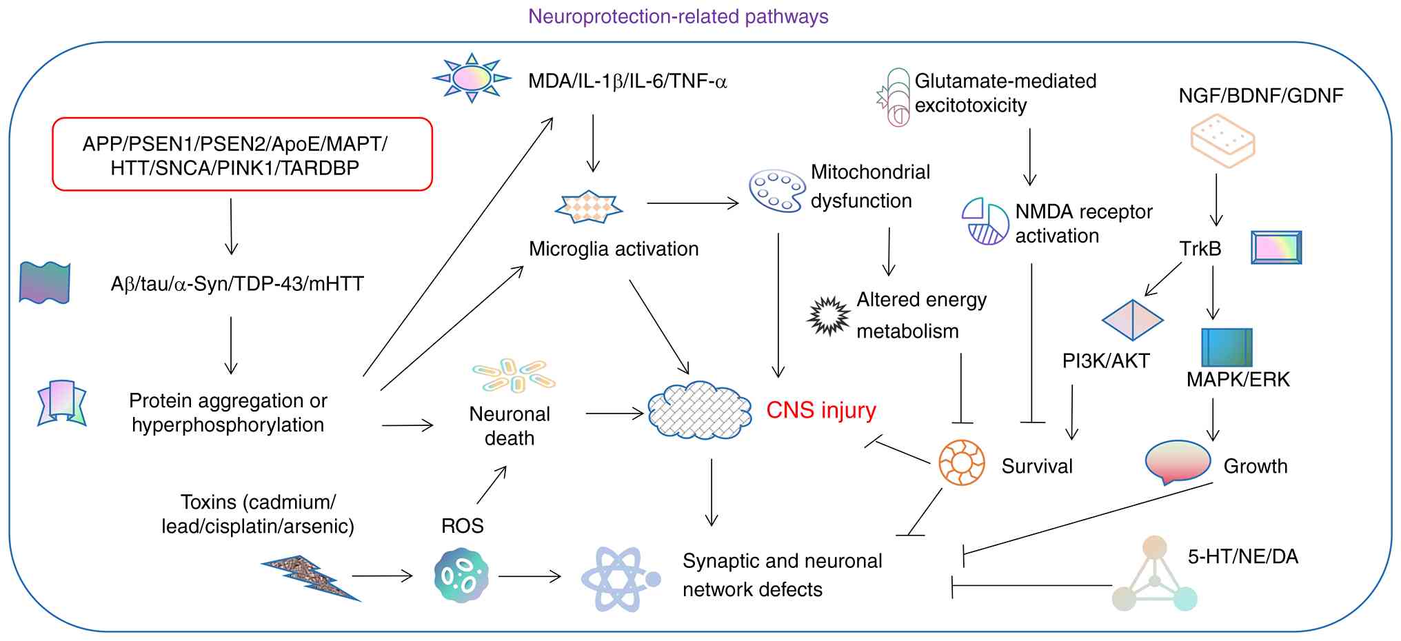
Overall, neuroprotective strategies target
neuroinflammation, oxidative stress and impaired neural repair to
promote functional recovery through pathways involving abnormal
protein aggregation, toxin-induced injury via redox modulation,
neurotrophic/TrkB/PI3K/Akt signaling, microglial activation and
mitochondrial dysfunction (Fig. 1)
(34). Neuroprotective agents
function through multifaceted mechanisms, including antioxidant
activity, anti-inflammatory effects via microglial suppression and
NF-κB inhibition, enhanced energy metabolism, mitochondrial
stabilization, apoptosis suppression through PI3K/Akt signaling,
clearance of pathological protein aggregates, and promotion of
neuronal repair and stem cell differentiation (35). In addition, various neuroprotective
or neurological disorders-related agents were summarized in
Table I (35–47).
Date palm fruits, rich in polyphenolic antioxidants,
including PCA, demonstrate neuroprotective properties in model
systems, suggesting potential to reduce AD risk, delay onset or
slow progression (48). PCA could
be responsible for the beneficial health effects of polyphenol-rich
foods, as they can easily cross the BBB (49). Bioavailable PCA extracted from
edible chicory has been shown to undergo partial glucosylation and
sulfation in human adults (50).
Among the four primary active components of Alpiniae oxyphyllae
fructus (nootkatone, tectochrysin, chrysin and PCA), PCA exhibits
high BBB permeability via passive diffusion, whereas lactoferrin
demonstrates relatively poor permeability (51).
PCA exhibits diverse pharmacological activities,
including antioxidant, anti-inflammatory, neuroprotective,
antibacterial, antiviral, anticancer, anti-osteoporotic, analgesic
and anti-aging effects, metabolic syndrome prevention, and
protection of liver, kidney and reproductive functions (52,53).
PCA also modulates neuroprotective factor expression, suppresses
apoptosis, activates the autophagy-lysosomal pathway, reduces
oxidative stress and inflammation, enhances synaptic plasticity,
inhibits amyloid-β (Aβ) accumulation, decreases amyloid precursor
protein (APP) processing, strengthens the cholinergic system and
mitigates neuronal excitotoxicity (54). Consequently, PCA, present in
various fruits, vegetables and grains, shows promise as a dietary
supplement for alleviating cognitive deficits associated with
NDs.
Oxidative stress, characterized by a systemic
oxidant/antioxidant imbalance, leads to excessive ROS production
that damages critical biomolecules (such as lipids, proteins and
DNA), resulting in neuronal dysfunction and ultimately cell death
in brain tissue (55,56). Due to its high metabolic oxygen
consumption characteristic, the brain is extremely sensitive to
oxidative stress, which is a key pathogenic factor for NDs
(57). Consequently, antioxidant
strategies, including phytochemical-rich dietary supplements,
combined with moderate exercise, may mitigate oxidative
stress-induced neurodegeneration (1).
As a natural antioxidant, PCA demonstrates broad
potential for neuroprotection and antioxidant therapy. Pretreatment
with Lycium barbarum polyphenols, such as PCA, attenuated
hydrogen peroxide (H2O2)-induced toxicity in
PC12 cells, a rat adrenal pheochromocytoma-derived neuroendocrine
cell line, by reducing ROS production, restoring mitochondrial
membrane potential and inhibiting apoptosis (58). In a global ischemia model in rats,
PCA significantly reduced cell death, oxidative stress, microglial
and astrocyte activation, and BBB disruption in degenerative
neurons, and increased glutathione concentration in hippocampal
neurons (59). PCA and chrysin
exerted synergistic neuroprotection in 6-hydroxydopamine-treated
PC12 cells by enhancing viability, reducing lactate dehydrogenase
release and modulating cellular redox status through upregulation
of key antioxidant enzymes (60).
Pre-treatment with PCA significantly reduced apoptosis,
inflammation and oxidative stress in the neonatal mouse hippocampus
following sevoflurane exposure (61).
In protein-misfolding disorders such as AD and PD,
neuroinflammatory pathway activation triggers concomitant oxidative
and nitrosative stress (66).
Inhibition of neuroinflammatory nitric oxide signaling can mitigate
functional neurodegeneration and reduce cellular stress associated
with aberrant nitrogen metabolism and protein glycosylation
(67). The neuroimmune axis
exhibits complex interdependencies, serving as the primary
pathogenic driver in multiple sclerosis with similarly amplified
involvement in other NDs, including AD, ALS and PD (68). Chronic innate immune cell
activation, a hallmark of age-related NDs, exacerbates
neurodegeneration by promoting Aβ plaque formation and τ
hyperphosphorylation, as exemplified in AD (69). A range of neurological conditions,
including NDs and COVID-19 neurological sequelae, share persistent
neuroinflammation, with mounting evidence implicating inflammasome
activation in driving their pathogenesis (70). Aggregated proteins linked to NDs,
such as Aβ, τ, α-syn and TAR DNA-binding protein 43, function as
damage-associated molecular patterns (DAMPs), activating innate
immune responses via multiple pattern recognition receptors,
including Toll-like receptors, NOD-like receptors, cytosolic DNA
sensors and other DAMP receptors (71). As resident immune cells of the CNS,
microglia play a protective role by phagocytosing pathological
protein aggregates, yet excessive phagocytosis can impair their
function, induce neuroinflammation and ultimately promote
neurodegeneration in various NDs (72).
PCA exerted anti-inflammatory effects in
lipopolysaccharide (LPS)-stimulated BV2 microglia by inhibiting the
Toll-like receptor 4-mediated NF-κB and MAPK signaling pathways
(73). PCA also inhibited the
immune response in LPS-activated BV2 microglia via the SIRT1/NF-κB
pathway and suppressed PC12 cell apoptosis induced by microglial
activation (74). In addition, PCA
promoted the M1/M2 phenotypic shift via m-TOR pathway inhibition,
thereby ameliorating inflammation in mouse models of brain
haemorrhage (75).
3,4-Dihydroxyphenylacetic acid, PCA and dihydrocaffeic acid, and
their conjugated forms, significantly attenuated neuroinflammation
by scavenging ROS, thereby protecting neuronal cells. Notably,
phenolic acid conjugates demonstrated superior efficacy in
mitigating oxidative stress and inflammatory damage to neuronal
SH-SY5Y cells stimulated by bacterial lipopolysaccharide and
tert-butyl hydroperoxide compared with their free forms (76).
Neural regeneration refers to the restoration of
neurological function through complex biological processes,
including neuronal regrowth, proliferation or differentiation of
NSCs, and participation of dual roles of glial cells (such as
microglia and astrocytes) (77).
Post-injury, damaged neurons release neurotrophic factors
(including BDNF and NGF) and cytokines that promote axonal
regeneration and synaptic reconnection (78). Neural regeneration also relies on
signal transmission between cells and microenvironmental
regulation, such as the influence of BDNF and NGF, which enhance
neuronal survival and promote neuronal growth and differentiation
(79). Concurrently, NSCs
contribute to repair via their self-renewal capacity and
multilineage differentiation potential, essential for maintaining
CNS homeostasis (80). MSCs also
play an important role in reconstructing neural networks and
restoring their functions with burgeoning preclinical evidence
(81–83).
PCA promotes neural regeneration, with its mechanism
potentially involving the insulin-like growth factor 1
receptor/PI3K/Akt signaling pathway (84). PCA increased the survival of
primary cultured cortical neurons in newborn rats and promoted
neurite growth in these neurons (85), and this neurotrophic protective
effect exerted by PCA was correlated to its regulation of
phosphorylated AKT expression (86). In addition, PCA promoted RSC96
Schwann cell migration, regeneration and peripheral nerve repair by
regulating MAPK, plasminogen activator and MMP signaling pathways
(87). When combined with fetal
bovine serum in vitro, PCA promoted neuronal
differentiation, induced neuronal maturation and enhanced neurite
growth in cultured neural stem and progenitor cells (88). PCA treatment significantly reduces
ROS levels, caspase 3 activity and apoptosis in NSCs (89). Therefore, PCA has also gained
increasing attention in neural regeneration and neurotrophic
protective effects.
Despite clinical differences, NDs share fundamental
pathological mechanisms, such as abnormal protein deposition,
intracellular calcium overload, mitochondrial dysfunction, REDOX
homeostasis imbalance and neuroinflammation (90). NDs are characterized by a suite of
interconnected hallmark features, such as pathological protein
aggregation, synaptic dysfunction, disrupted proteostasis,
cytoskeletal defects, metabolic imbalance, nucleic acid
alterations, neuroinflammation and neuronal loss, all of which
collectively drive disease onset and progression through complex
interactions modulated by genetic determinants and biochemical
pathways (91).
The pathological mechanisms of AD mainly include the
formation of amyloid plaques, abnormal phosphorylation of τ
protein, neuroinflammation and oxidative stress. As a primary
metabolite in blueberry extracts, PCA mitigates neuronal damage by
enhancing autophagy, supporting its potential for dietary AD
intervention (13). While okadaic
acid induces AD-like pathology, including τ hyperphosphorylation,
neurofibrillary tangle formation and Aβ deposition, PCA
counteracted this cytotoxicity in PC12 cells by regulating
Akt/glycogen synthase kinase-3β (GSK-3β)/myocyte-specific enhancer
factor 2D signaling and modulating autophagic activity, thereby
demonstrating neuroprotective efficacy (92). In a rat model of mild memory
impairment induced by long-term intragastric administration of
D-galactose, PCA improved learning and spatial memory abilities in
the Morris water maze test and restored dysregulated serotonergic
and dopaminergic activity (93).
The abnormal aggregation of Aβ and α-syn drives the
formation of amyloid plaques in AD and Lewy bodies in PD, with the
latter also characterized by progressive degeneration of
dopaminergic neurons in the substantia nigra. Treatment of amyloid
precursor protein (APP)/presenilin 1 (PS1) transgenic mice (a
double-transgenic mouse model co-expressing mutant human APP/PS1,
commonly used to model AD) with PCA significantly increased BDNF
levels in the hippocampus and cerebral cortex, reduced Aβ
deposition, decreased APP expression and inflammatory responses,
and improved learning and memory abilities (94). High doses of PCA (50 mg/kg)
alleviated symptoms in an AD mouse model induced by Aβ injection
into the hippocampus, potentially via the cholinergic synaptic
signaling pathway (95). By
downregulating inflammatory mediators in the brain of
Aβ25-35-injected AD model mice, particularly inducible
nitric oxide synthase and cyclooxygenase-2, PCA significantly
alleviated neuroinflammation and inhibited lipid peroxidation in
the brain, kidney and liver tissues (96). As the main metabolite of
anthocyanins, PCA, also known as anthocyanin 3-glucoside, inhibited
the aggregation of Aβ and α-syn, destabilizing their pre-formed
fibrils and preventing the PC12 cell death mediated by the toxicity
of the Aβ and α-syn (97).
PCA exerted neuroprotective effects in both
1-methyl-4-phenylpyridinium (MPP+)-treated PC12 cells and the
1-methyl-4-phenyl-1,2,3,6-tetrahydropyridine (MPTP)-induced PD
mouse model, and was associated with the inhibition of α-syn
oligomerization (98,99). Although PCA significantly increased
dopamine turnover in the striatum and improved cognitive function
in experimental memory impairment mice, it did not significantly
affect memory performance in healthy rats (100). The combination of PCA with
ginkgolide B significantly restored the motor ability of PD mice,
alleviated neuronal damage, boosted the activity of antioxidant
enzymes in brain tissue and increased the expression in the
midbrain substantia nigra (101).
The combination of honokiol with PCA reduced neuronal loss in
6-hydroxydopamine-treated zebrafish PD models (102). PCA demonstrated neuroprotective
efficacy in ALS transgenic mice by extending survival time,
suppressing spinal glial proliferation, preventing motor neuron
apoptosis, alleviating pathological manifestations and preserving
neuromuscular junction integrity, thereby countering key features
of this severe disease (103).
Arsenic and common heavy metals (including plumbum,
hydrargyrum, cadmium and manganese) exhibit neurotoxic effects,
with notable sex-specific differences observed in response to
exposure (104). Neurotoxic
compounds, such as rotenone, 6-hydroxydopamine, MPTP, MPP+,
paraquat and maneb, are commonly used in preclinical models of PD
(105). In anisodamine-induced
amnesia models, PCA administered orally could protect against
oxidative stress-related learning and memory deficits (10). PCA enhanced the antioxidant defense
system, suppressed inflammation and apoptosis, and thereby
counteracted cadmium-induced neurocortical toxicity (106). PCA could also exert protective
effects against cisplatin-induced neurotoxicity by inhibiting
neuroinflammation and restoring the oxidative/antioxidative balance
(107). As a toxic metalloid,
arsenic exposure increased pro-inflammatory cytokine levels (TNF-α
and IL-1β), upregulated apoptosis-related molecules (caspase-3 and
Bax), and reduced acetylcholinesterase activity and BDNF levels in
the mouse cerebral cortex. PCA pretreatment attenuated
arsenic-induced histopathological alterations in brain tissue
(108). PCA also markedly reduced
hippocampal neuronal death and microglial activation in a model of
intraperitoneal injection of pilocarpine-induced epilepsy in adult
male rats (109).
PCA prevented rotenone-induced apoptosis of PC12
cells by alleviating mitochondrial dysfunction (110). Additionally, PCA significantly
alleviated MPP(+)-induced mitochondrial dysfunction in these cells
(111). Although bromate, used as
a food additive, was shown to disrupt the CNS, PCA protected the
cells from bromate-induced gastric mucosal ulceration (112). PCA alleviated oxidative stress,
elevated neurotransmitter levels, and improved learning and memory
deficits in lead-exposed rats (113). Additionally, PCA prevented
cadmium-induced neurotoxicity by altering the activities of key
enzymes, such as Na+/K+-ATPase,
acetylcholinesterase, butylcholinesterase and endogenous
antioxidant enzymes (114).
Depression and anxiety are mental disorders
characterized by persistent dysregulation of emotional and
behavioral responses, which is associated with reduced levels of
5-HT, DA and NE in the CNS (115). Chronic corticosterone exposure
induces depressive-like behavior in mice, accompanied by oxidative
stress, neuroinflammation and medial prefrontal cortex synaptic
plasticity impairment, further supporting the pivotal role of
oxidative stress in depression pathogenesis (116).
Acute inhibitory stress triggers depression-like
behavior via oxidative neuronal damage in mice. Ethyl PCA mitigates
serum corticosterone elevation and lipid peroxidation induced by
acute inhibitory stress while restoring enzymatic antioxidant
levels in the cerebral cortex and hippocampus (117). In scopolamine-induced long-term
memory impairment of mice, following acute treatment, PCA induced
an anxiogenic effect, whereas repeated administration produced
anxiolytic effects and enhanced cognitive function in both acute
and chronic models, but their impact on long-term memory was
greater than on short-term memory (118). PCA not only reduced the
immobility time, serum corticosterone, cytokines TNF-α and IL-6,
and malondialdehyde (MDA) levels in mice exposed to chronic
unpredictable mild stress, but also improved sucrose preference and
restored BDNF levels (119). PCA
exhibited antidepressant-like effects by enhancing BDNF, 5-HT, DA
and NE levels in the hippocampus and cerebral cortex, while
reducing oxidative and inflammatory markers, including MDA, IL-6
and TNF-α (120).
PCA alleviated post-traumatic stress disorder-like
symptoms in rats induced by single prolonged stress, through
modulation of central monoaminergic systems, improved freezing
behavior and demonstrated antidepressant and anxiolytic properties
(121). PCA also markedly reduced
the biomarkers of inflammation and oxidative stress in the
hypothalamus, testis and epididymis (122). Furthermore, PCA improved the
hypothalamic-pituitary-gonadal axis function defect in rats exposed
to furan by inhibiting oxidative inflammatory stress and apoptosis
(123). Both hyperoside and PCA,
two polyphenolic compounds, were shown to mediate
antidepressant-like effects in mice by modulating the monoamine
system and upregulating BDNF levels (124).
CIRI, a major cause of adult disability and
mortality, refers to the secondary brain damage that results
following the restoration of blood flow to previously ischemic
brain regions (125). The
pathological mechanisms of ischemic stroke involve oxidative
stress, apoptosis, ferroptosis and mitochondrial dysfunction,
whereas N-butylphthalide with ligustrazine confer anti-ischemic
effects via the Kelch-like ECH-associated protein 1-nuclear factor
erythroid 2-related factor 2 (NRF2) pathway and isocitric rutinine
provides neuroprotection in CIRI mice through Nrf2
activation to alleviate oxidative stress and mitochondrial
impairment (125,126).
The protective effects of PCA against CIRI are
considered to be mediated by the upregulation of NRF2 expression
(127). PCA may have the
potential to prevent early reperfusion injury, restore the balance
between survival and death proteins, and serve as a cost-effective
adjunctive treatment for stroke (128). PCA could also reduce brain edema
and BBB damage caused by intracerebral hemorrhage via the Nrf2/HO-1
signaling pathway (129). In a
collagenase IV-induced mouse model of intracerebral hemorrhage, PCA
attenuated oxidative stress, inflammation and apoptosis through
downregulation of the p38/JNK-NF-κB pathway, thereby reducing
third-stage brain edema, improving neurological function and
decreasing TNF-α, IL-1β and IL-6 expression at both the protein and
gene levels (130). In a rat
model of global CIRI, both silymarin and ethyl PCA improved
cognitive and motor function, and reduced histopathological damage,
cerebral edema and infarct volume, with silymarin demonstrating
superior efficacy compared with piracetam and ethyl PCA (131). In a mouse model of intestinal
ischemia-reperfusion injury, PCA exerted protective effects on both
the local intestine and remote liver damage, which were mediated
through its anti-apoptotic and antioxidant properties (132).
Neuralgia, one of the most debilitating neurological
disorders, poses a major therapeutic challenge due to the complex
interplay of pathogenic mechanisms involving oxidative stress,
neuroinflammation and mitochondrial dysfunction (133). Trigeminal neuralgia is a severe
facial pain disorder primarily attributed to neurovascular
compression and demyelination of afferent fibers leading to
neuronal hyperexcitability, with carbamazepine and oxcarbazepine
serving as first-line pharmacotherapy (134). Furthermore, a longer duration and
a broader involvement of trigeminal neuralgia are associated with
more severe depression, anxiety and insomnia, while these emotional
disorders in turn can exacerbate the risk and manifestation of
neuralgia (135).
In a chronic constriction injury-induced neuropathy
rat model, PCA exhibited similar therapeutic effects as
carbamazepine and mitigated the adverse effects caused by
neurogenic pain drugs alone (136). PCA alleviated neuropathic pain in
rats with chronic constriction injury by inhibiting the
JNK/CXCL1/CXCR2 signaling pathway, which contributes to improved
oxidative stress (137). A
pharmaceutical co-crystal composed of pentoxifylline and PCA
effectively reduced allodynia in rats with complex regional pain
syndrome following chronic ischemia, through mechanisms involving
reduced peripheral tissue ischemia/hypoxia and suppression of
hypoxia-induced mitochondrial dysfunction (138).
Additionally, various other neurological diseases
are associated with neuroprotection and PCA. PCA prevents
blood-spinal cord barrier disruption and hemorrhage by
downregulating sulfonylurea receptor 1/transient receptor potential
melastatin 4 and matrix metalloproteinases, thereby enhancing
functional recovery following spinal cord injury (139). Administration of PCA reduced
elevated levels of ROS, protein carbonyls, carboxymethyl lysine and
methylglyoxal in the brains of D-galactose-treated mice, indicating
its potential to delay or prevent age-related changes (140). PCA regulated blood glucose
levels, alleviated cerebral mitochondrial dysfunction and prevented
oxidative stress in the brains of streptozotocin-induced diabetic
rats (141).
In a rat model of thiamine deficiency, PCA not only
ameliorated systemic rigidity and improved motor coordination but
also enhanced cognitive function, specifically memory consolidation
and retrieval, while restoring normal alanine and glutamate
concentrations in the medulla oblongata, which are dysregulated due
to the deficiency (142). In a
rat model of chronic intermittent hypoxia, which mimics the
hallmark cognitive impairment of obstructive sleep apnea, PCA
mitigated cognitive dysfunction by reducing cerebral IL-1β levels,
upregulating BDNF and synapsin expression, attenuating oxidative
stress, apoptosis and reactive gliosis, and ultimately improving
learning and memory (143).
Beyond neurological effects, PCA exhibited organoprotective
properties, demonstrating cardioprotective and lipid-lowering
activity in rats with high-fat/high-fructose-induced coronary
artery disease (144).
Neuronal death is a common feature of neurological
diseases, and protecting neurons and rebuilding damaged neural
networks are key to treating NDs such as HD (145). In fact, neuronal injury and death
across various NDs converge on shared pathological mechanisms,
including oxidative stress, neuroinflammation, ion dyshomeostasis
and proteotoxicity (146,147). PCA has exerted broad protective
effects across multiple ND models, not by targeting a specific
disease but by modulating these fundamental, shared pathological
pathways. The most prominent and conserved mechanisms of PCA in NDs
include inhibiting abnormal protein aggregation (such as Aβ and
α-syn), attenuating neuroinflammation (for example, suppressing the
NF-κB pathway), enhancing antioxidant defenses, and modulating
autophagy and cell survival signaling (for example, Akt/GSK-3β).
These mechanisms often work in concert, ultimately promoting
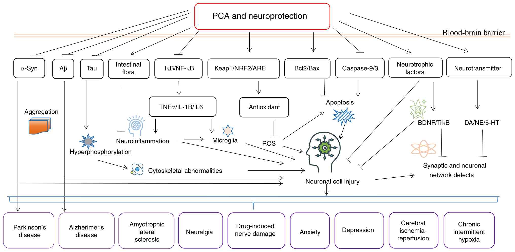
neuronal survival and function (Table
II). The roles and mechanisms of PCA related to neuroprotection
are summarized in Fig. 2,
including antioxidant activity, anti-inflammatory modulation,
anti-apoptotic regulation, mitochondrial protection and homeostasis
maintenance.
To establish the efficacy and safe dosage of PCA,
conducting rigorous human clinical trials, including
pharmacokinetic, dose-finding and efficacy studies as a single
compound, is essential. In a mouse study, PCA appeared to be
rapidly absorbed, achieving a peak plasma concentration of 73.6 µM
at 5 min, with an initial elimination half-life of ~3 min and a
terminal half-life of 16 min, and remaining detectable for up to 8
h (148). A pH-responsive,
rapidly adjustable hydrogel based on PCA enabled sustained and
controlled drug release, demonstrating excellent clinical potential
for NDs (149). However, clinical
observations indicate a higher incidence of stroke onset during the
human active phase (daytime), whereas most rodent models are
conducted during the animals' inactive phase, introducing a
chronobiological discrepancy that may limit translational
predictability (150). Currently,
there are limited direct clinical studies on PCA; it is more
frequently investigated as a metabolite, and since it derives from
various natural sources often containing other compounds, detected
doses across studies may vary substantially (151). As a phenolic acid metabolite from
anthocyanin degradation, PCA is a key urinary bioactive compound
whose increased excretion is associated with reduced serum oxidant
status, indirectly supporting its role in boosting antioxidant
defences (152).
While PCA exhibited neuroprotective properties in
animal models by mitigating oxidative stress and neuronal
apoptosis, a 9-week clinical trial using PCA-rich juices (such as
cranberry or red grape) in elderly men (age ≥67 years, n=30) with
memory deficits showed no notable improvement in choice memory
scores, but led to a reduction in biomarkers of inflammation and
tissue damage (153). This
finding is significant, indicating successful engagement of the
intended therapeutic targets, namely, the suppression of chronic
inflammatory and oxidative stress pathways. The negative primary
cognitive outcome may be attributed to the short intervention
duration, insufficient dosage, limited sample size or heterogeneity
within the study population. Therefore, the reduction in
inflammatory markers should be regarded as a positive
pharmacodynamic signal, suggesting potential for cognitive benefit
with optimized or longer-term intervention strategies.
PCA, a natural compound with multiple biological
activities, exhibits considerable neuroprotective potential in
diverse neurological disorders, including AD, PD and cerebral
ischemia, primarily through mechanisms such as antioxidation,
anti-inflammation and the promotion of neuronal survival. Although
preclinical studies have underscored its broad efficacy, clinical
translation remains limited by a scarcity of large-scale human
trials and inconsistent outcomes attributable to its multi-target
nature, which complicates mechanistic clarity and reproducibility.
Future clinical studies to elucidate the disease-specific
mechanisms of PCA, optimize its dosing and delivery strategies, and
evaluate long-term safety and efficacy for neurological disorders
are anticipated.
Not applicable.
This study was supported by the Natural Science Foundation of
Jiangxi Province (grant no. 20242BAB25585) and the 2022 Ganzhou
Municipal Science and Technology Project (grant no.
2022-YB1414).
Not applicable.
XYX, YCL, SSS, LPW and LFW contributed to the
manuscript conception and design. The first draft of the manuscript
was written by XYX and LFW. XYX, YCL and LFW contributed to
reference investigations and figure visualization. YCL, SSS and LPW
commented and critically revised previous versions of the
manuscript. All authors have read and approved the final version of
the manuscript. Data authentication is not applicable.
Not applicable.
Not applicable.
The authors declare that they have no competing
interests.
|
1
|
Dash UC, Bhol NK, Swain SK, Samal RR,
Nayak PK, Raina V, Panda SK, Kerry RG, Duttaroy AK and Jena AB:
Oxidative stress and inflammation in the pathogenesis of
neurological disorders: Mechanisms and implications. Acta Pharm Sin
B. 15:15–34. 2025. View Article : Google Scholar : PubMed/NCBI
|
|
2
|
Colwell CS: Defining circadian disruption
in neurodegenerative disorders. J Clin Invest. 131:e1482882021.
View Article : Google Scholar : PubMed/NCBI
|
|
3
|
Bawari S, Tewari D, Arguelles S, Sah AN,
Nabavi SF, Xu S, Vacca RA, Nabavi SM and Shirooie S: Targeting BDNF
signaling by natural products: Novel synaptic repair therapeutics
for neurodegeneration and behavior disorders. Pharmacol Res.
148:1044582019. View Article : Google Scholar : PubMed/NCBI
|
|
4
|
Iskusnykh IY, Zakharova AA, Kryl'Skii ED
and Popova TN: Aging, neurodegenerative disorders, and cerebellum.
Int J Mol Sci. 25:10182024. View Article : Google Scholar : PubMed/NCBI
|
|
5
|
Singh A, Kukreti R, Saso L and Kukreti S:
Oxidative stress: A key modulator in neurodegenerative diseases.
Molecules. 24:15832019. View Article : Google Scholar : PubMed/NCBI
|
|
6
|
Ravi Singh J: Redox imbalance and
hypoxia-inducible factors: A multifaceted crosstalk. FEBS J.
292:3833–3848. 2025. View Article : Google Scholar : PubMed/NCBI
|
|
7
|
Kim S, Jung UJ and Kim SR: Role of
oxidative stress in blood-brain barrier disruption and
neurodegenerative diseases. Antioxidants (Basel). 13:14622024.
View Article : Google Scholar : PubMed/NCBI
|
|
8
|
Hajialyani M, Hosein Farzaei M, Echeverria
J, Nabavi SM, Uriarte E and Sobarzo-Sanchez E: Hesperidin as a
neuroprotective agent: A review of animal and clinical evidence.
Molecules. 24:6482019. View Article : Google Scholar : PubMed/NCBI
|
|
9
|
Kogure T, Suda M, Hiraga K and Inui M:
Protocatechuate overproduction by Corynebacterium glutamicum via
simultaneous engineering of native and heterologous biosynthetic
pathways. Metab Eng. 65:232–242. 2021. View Article : Google Scholar : PubMed/NCBI
|
|
10
|
Kim Y, Cho M, Lee JS, Oh J and Lim J:
Protocatechuic acid from euonymus alatus mitigates
scopolamine-induced memory impairment in mice. Foods. 13:26642024.
View Article : Google Scholar : PubMed/NCBI
|
|
11
|
Bozinou E, Georgiadou NT, Chalastara MS,
Makrygiannis I, Mantiniotou M, Athanasiadis V, Chatzilazarou A and
Lalas SI: Recovery of natural antioxidants from onion solid waste
via pressurized liquid extraction: Encapsulation and application
into a food system. Foods. 14:35832025. View Article : Google Scholar : PubMed/NCBI
|
|
12
|
Hamdi A, Jaramillo-Carmona S,
Rodriguez-Arcos R, Jimenez-Araujo A, Karray Bouraoui N and
Guillen-Bejarano R: Phytochemical profile and in vitro
bioactivities of wild asparagus stipularis. Molecules. 29:8172024.
View Article : Google Scholar : PubMed/NCBI
|
|
13
|
Li H, Zheng T, Lian F, Xu T, Yin W and
Jiang Y: Anthocyanin-rich blueberry extracts and anthocyanin
metabolite protocatechuic acid promote autophagy-lysosomal pathway
and alleviate neurons damage in in vivo and in vitro models of
Alzheimer's disease. Nutrition. 93:1114732022. View Article : Google Scholar : PubMed/NCBI
|
|
14
|
Lee SH, Choi BY, Lee SH, Kho AR, Jeong JH,
Hong DK and Suh SW: Administration of protocatechuic acid reduces
traumatic brain injury-induced neuronal death. Int J Mol Sci.
18:25102017. View Article : Google Scholar : PubMed/NCBI
|
|
15
|
Ghasemzadeh Rahbardar M and Hosseinzadeh
H: Neuroprotective effects of walnut (Juglans regia L.) in nervous
system disorders: A comprehensive review. Iran J Basic Med Sci.
27:1492–1505. 2024.PubMed/NCBI
|
|
16
|
Li R, Wang L, Zhang Q, Duan H, Qian D,
Yang F and Xia J: Alpiniae oxyphyllae fructus possesses
neuroprotective effects on H(2)O(2) stimulated PC12 cells via
regulation of the PI3K/Akt signaling pathway. Front Pharmacol.
13:9663482022. View Article : Google Scholar : PubMed/NCBI
|
|
17
|
Salama A, Elgohary R, Amin MM and Elwahab
SA: Immunomodulatory effect of protocatechuic acid on
cyclophosphamide induced brain injury in rat: Modulation of
inflammosomes NLRP3 and SIRT1. Eur J Pharmacol. 932:1752172022.
View Article : Google Scholar : PubMed/NCBI
|
|
18
|
Gallardo-Fernandez M, Hornedo-Ortega R,
Cerezo AB, Troncoso AM and Garcia-Parrilla MC: Melatonin,
protocatechuic acid and hydroxytyrosol effects on vitagenes system
against alpha-synuclein toxicity. Food Chem Toxicol.
134:1108172019. View Article : Google Scholar : PubMed/NCBI
|
|
19
|
Zhao H, Wang L, Zhang L and Zhao H:
Phytochemicals targeting lncRNAs: A novel direction for
neuroprotection in neurological disorders. Biomed Pharmacother.
162:1146922023. View Article : Google Scholar : PubMed/NCBI
|
|
20
|
Zhu H, Zhou L, Tang J, Xu Y, Wang W, Shi
W, Li Z, Zhang L, Ding Z, Xi K, et al: Reactive oxygen
species-responsive composite fibers regulate oxidative metabolism
through internal and external factors to promote the recovery of
nerve function. Small. 20:e24012412024. View Article : Google Scholar : PubMed/NCBI
|
|
21
|
Hill D, Compagnoni C and Cordeiro MF:
Investigational neuroprotective compounds in clinical trials for
retinal disease. Expert Opin Investig Drugs. 30:571–577. 2021.
View Article : Google Scholar : PubMed/NCBI
|
|
22
|
Jiang W, Xiao D, Wu C, Yang J, Peng X,
Chen L, Zhang J, Zha G, Li W, Ju R, et al: Circular RNA-based
therapy provides sustained and robust neuroprotection for retinal
ganglion cells. Mol Ther Nucleic Acids. 35:1022582024. View Article : Google Scholar : PubMed/NCBI
|
|
23
|
Nimgampalle M, Chakravarthy H, Sharma S,
Shree S, Bhat AR, Pradeepkiran JA and Devanathan V:
Neurotransmitter systems in the etiology of major neurological
disorders: Emerging insights and therapeutic implications. Ageing
Res Rev. 89:1019942023. View Article : Google Scholar : PubMed/NCBI
|
|
24
|
Rebas E, Rzajew J, Radzik T and Zylinska
L: Neuroprotective polyphenols: A modulatory action on
neurotransmitter pathways. Curr Neuropharmacol. 18:431–445. 2020.
View Article : Google Scholar : PubMed/NCBI
|
|
25
|
Zhou ZD, Yi LX, Wang DQ, Lim TM and Tan
EK: Role of dopamine in the pathophysiology of Parkinson's disease.
Transl Neurodegener. 12:442023. View Article : Google Scholar : PubMed/NCBI
|
|
26
|
Daniels S, El Mansari M and Blier P: AMPA
receptors modulate enhanced dopamine neuronal activity induced by
the combined administration of venlafaxine and brexpiprazole.
Neuropsychopharmacol. 49:2042–2051. 2024. View Article : Google Scholar : PubMed/NCBI
|
|
27
|
Soni U, Singh K, Jain D and Pujari R:
Exploring Alzheimer's disease treatment: Established therapies and
novel strategies for future care. Eur J Pharmacol. 998:1775202025.
View Article : Google Scholar : PubMed/NCBI
|
|
28
|
Imai R, Tamura R, Yo M, Sato M, Fukumura
M, Takahara K, Kase Y, Okano H and Toda M: Neuroprotective effects
of genome-edited human iPS cell-derived neural stem/progenitor
cells on traumatic brain injury. Stem Cells. 41:603–616. 2023.
View Article : Google Scholar : PubMed/NCBI
|
|
29
|
Pischiutta F, Tribuzio F, Magatti M, De
Simone G, Moro F, Nattino G, Signorini F, Loose L, Caruso E,
Bertani C, et al: Mesenchymal stromal cell secretome and its key
bioactive metabolites induce long-term neuroprotection after
traumatic brain injury in mice. Adv Sci (Weinh). 12:e155082025.
View Article : Google Scholar : PubMed/NCBI
|
|
30
|
Kim JE, Ji YE, Hwang HJ, Go GE, Lim HJ,
Yoo J, Kim J, Park D, Kim EH, Kim D and Bang OY: Engineered MSC-EVs
loaded with BDNF-enhancing neuropeptides via a non-disruptive
method enhance post-stroke neuroregeneration via intranasal
delivery. J Nanobiotechnology. 23:5942025. View Article : Google Scholar : PubMed/NCBI
|
|
31
|
Liu G, Yang C, Wang X, Chen X, Cai H and
Le W: Cerebellum in neurodegenerative diseases: Advances,
challenges, and prospects. iScience. 27:1111942024. View Article : Google Scholar : PubMed/NCBI
|
|
32
|
Rudolph S, Badura A, Lutzu S, Pathak SS,
Thieme A, Verpeut JL, Wagner MJ, Yang YM and Fioravante D:
Cognitive-affective functions of the cerebellum. J Neurosci.
43:7554–7564. 2023. View Article : Google Scholar : PubMed/NCBI
|
|
33
|
Beeraka NM, Nikolenko VN, Khaidarovich ZF,
Valikovna OM, Aliagayevna RN, Arturovna ZL, Alexandrovich KA,
Mikhaleva LM and Sinelnikov MY: Recent investigations on the
functional role of cerebellar neural networks in motor functions
& nonmotor functions -neurodegeneration. Curr Neuropharmacol.
20:1865–1878. 2022. View Article : Google Scholar : PubMed/NCBI
|
|
34
|
Tong L, Li MD, Nie PY, Chen Y, Chen YL and
Ji LL: miR-132 downregulation alleviates behavioral impairment of
rats exposed to single prolonged stress, reduces the level of
apoptosis in PFC, and upregulates the expression of MeCP2 and BDNF.
Neurobiol Stress. 14:1003112021. View Article : Google Scholar : PubMed/NCBI
|
|
35
|
Zhang X, Yang G, Liang C, Li Y, Gao L, Liu
Y, Wang Y, Li J, Zhou Y, Han Z and Ren J: Nodakenin attenuates
cerebral ischemia-reperfusion injury by modulating the
PI3K/AKT/NF-κB signaling pathway. Immunopharm Immunot. 48:272–281.
2026. View Article : Google Scholar
|
|
36
|
Zhang T, Liu N, Cao H, Wei W, Ma L and Li
H: Different doses of pharmacological treatments for mild to
moderate alzheimer's disease: A bayesian network meta-analysis.
Front Pharmacol. 11:7782020. View Article : Google Scholar : PubMed/NCBI
|
|
37
|
Wang J, Lin S, Bai C, Zhang H, Liu H, Wang
M and Guo R: Exploring the risk of adverse drug events in
combination with antiparkinsonics and antipsychotics-a two-decade
real-world pharmacovigilance analysis based on the FAERS database.
Int J Neuropsychopharmacol. 28:pyaf332025. View Article : Google Scholar
|
|
38
|
Wen X, Lan T, Su W, Cao B, Wang Y and Chen
Y: Latest progress and challenges in drug development for
degenerative motor neuron diseases. Neural Regen Res. 21:1849–1863.
2026. View Article : Google Scholar : PubMed/NCBI
|
|
39
|
Dodson K, Livezey S, Denson B, Choi L,
DeClercq J, Zuckerman AD and Johnson K: Deutetrabenazine treatment
outcomes with doses above U.S. Food and Drug Administration maximum
approved doses in Huntington's disease chorea: A dual-site
analysis. J Huntingtons Dis. 14:140–148. 2025. View Article : Google Scholar : PubMed/NCBI
|
|
40
|
Bayas A, Christ M, Faissner S, Klehmet J,
Pul R, Skripuletz T and Meuth SG: Disease-modifying therapies for
relapsing/active secondary progressive multiple sclerosis-a review
of population-specific evidence from randomized clinical trials.
Ther Adv Neurol Disord. 16:175628642211468362023. View Article : Google Scholar : PubMed/NCBI
|
|
41
|
Dang C, Wang Q, Zhuang Y, Li Q, Lu Y,
Xiong Y and Feng L: Synergistic effects of neuroprotective drugs
with intravenous recombinant tissue plasminogen activator in acute
ischemic stroke: A Bayesian network meta-analysis. PLoS One.
19:e3112312024. View Article : Google Scholar
|
|
42
|
French KF, White J and Hoesch RE:
Treatment of intracerebral hemorrhage with tranexamic acid after
thrombolysis with tissue plasminogen activator. Neurocrit Care.
17:107–111. 2012. View Article : Google Scholar : PubMed/NCBI
|
|
43
|
Hua Y, Keep RF, Hoff JT and Xi G:
Deferoxamine therapy for intracerebral hemorrhage. Acta Neurochir
Suppl. 105:3–6. 2008. View Article : Google Scholar : PubMed/NCBI
|
|
44
|
Huerta MÁ, Mayo-Moldes M, Garcia MM,
García-Parra B, Matute M, López-Tofiño Y, Paniagua N,
Hernández-Secorún M, Soler D, Salmerón M, et al: Prescription
trends and clinical decision-making in neuropathic pain
pharmacological treatment: Results from a cross-sectional survey by
the Spanish pain society. Eur J Pain. 30:e702462026. View Article : Google Scholar : PubMed/NCBI
|
|
45
|
Chauvet-Gélinier JC: Efficacy of
escitalopram vs paroxetine on severe depression with associated
anxiety: Data from the ‘Boulenger’ study. Encephale. 36:425–432.
2010.(In French). View Article : Google Scholar : PubMed/NCBI
|
|
46
|
Fava M, Dunner DL, Greist JH, Preskorn SH,
Trivedi MH, Zajecka J and Cohen M: Efficacy and safety of
mirtazapine in major depressive disorder patients after SSRI
treatment failure: an open-label trial. J Clin Psychiatry.
62:413–420. 2001. View Article : Google Scholar : PubMed/NCBI
|
|
47
|
Jesus Palma AC, Antunes Júnior CR, Barreto
ESR, Alencar VB, Souza AKDN, Mathias CMC, Lins-Kusterer LEF, Azi
LMTA and Kraychete DC: Pharmacological treatment of
chemotherapy-induced neuropathy: A systematic review of randomized
clinical trials. Pain Manag Nurs. 26:249–263. 2025. View Article : Google Scholar : PubMed/NCBI
|
|
48
|
Subash S, Essa MM, Braidy N, Awlad-Thani
K, Vaishnav R, Al-Adawi S, Al-Asmi A and Guillemin GJ: Diet rich in
date palm fruits improves memory, learning and reduces beta amyloid
in transgenic mouse model of Alzheimer's disease. J Ayurveda Integr
Med. 6:111–120. 2015. View Article : Google Scholar : PubMed/NCBI
|
|
49
|
Krzysztoforska K, Mirowska-Guzel D and
Widy-Tyszkiewicz E: Pharmacological effects of protocatechuic acid
and its therapeutic potential in neurodegenerative diseases: Review
on the basis of in vitro and in vivo studies in rodents and humans.
Nutr Neurosci. 22:72–82. 2019. View Article : Google Scholar : PubMed/NCBI
|
|
50
|
Zheng J, Xiong H, Li Q, He L, Weng H, Ling
W and Wang D: Protocatechuic acid from chicory is bioavailable and
undergoes partial glucuronidation and sulfation in healthy humans.
Food Sci Nutr. 7:3071–3080. 2019. View Article : Google Scholar : PubMed/NCBI
|
|
51
|
Xiao T, Pan M, Wang Y, Huang Y, Tsunoda M,
Zhang Y, Wang R, Hu W, Yang H, Li LS and Song Y: In vitro
bloodbrain barrier permeability study of four main active
ingredients from Alpiniae oxyphyllae fructus. J Pharm Biomed Anal.
235:1156372023. View Article : Google Scholar : PubMed/NCBI
|
|
52
|
Song J, He Y, Luo C, Feng B, Ran F, Xu H,
Ci Z, Xu R, Han L and Zhang D: New progress in the pharmacology of
protocatechuic acid: A compound ingested in daily foods and herbs
frequently and heavily. Pharmacol Res. 161:1051092020. View Article : Google Scholar : PubMed/NCBI
|
|
53
|
Khan AK, Rashid R, Fatima N, Mahmood S,
Mir S, Khan S, Jabeen N and Murtaza G: Pharmacological activities
of protocatechuic acid. Acta Pol Pharm. 72:643–650. 2015.PubMed/NCBI
|
|
54
|
Liang S, Zhao Z, Liu L, Zhang Y and Liu X:
Research progress on the mechanisms of protocatechuic acid in the
treatment of cognitive impairment. Molecules. 29:47242024.
View Article : Google Scholar : PubMed/NCBI
|
|
55
|
Munteanu C, Galaction AI, Turnea M,
Blendea CD, Rotariu M and Postaru M: Redox homeostasis, gut
microbiota, and epigenetics in neurodegenerative diseases: A
Systematic review. Antioxidants (Basel). 13:10622024. View Article : Google Scholar : PubMed/NCBI
|
|
56
|
Zhang Z, Yang J, Zhou Q, Zhong S, Luo J,
Chai X, Liu J, Zhang X, Chang X and Wang H: The role and mechanism
of the cGAS-STING pathway-mediated ROS in apoptosis and ferroptosis
induced by manganese exposure. Redox Biol. 85:1037612025.
View Article : Google Scholar : PubMed/NCBI
|
|
57
|
Moren C, deSouza RM, Giraldo DM and Uff C:
Antioxidant therapeutic strategies in neurodegenerative diseases.
Int J Mol Sci. 23:93282022. View Article : Google Scholar : PubMed/NCBI
|
|
58
|
Gao H, Yuan X, Wang Z, Gao Q and Yang J:
Profiles and neuroprotective effects of Lycium ruthenicum
polyphenols against oxidative stress-induced cytotoxicity in PC12
cells. J Food Biochem. 44:e131122020. View Article : Google Scholar : PubMed/NCBI
|
|
59
|
Kho AR, Choi BY, Lee SH, Hong DK, Lee SH,
Jeong JH, Park KH, Song HK, Choi HC and Suh SW: Effects of
protocatechuic Acid (PCA) on global cerebral ischemia-induced
hippocampal neuronal death. Int J Mol Sci. 19:14202018. View Article : Google Scholar : PubMed/NCBI
|
|
60
|
Zhang Z, Li G, Szeto SSW, Chong CM, Quan
Q, Huang C, Cui W, Guo B, Wang Y, Han Y, et al: Examining the
neuroprotective effects of protocatechuic acid and chrysin on in
vitro and in vivo models of Parkinson disease. Free Radic Biol Med.
84:331–343. 2015. View Article : Google Scholar : PubMed/NCBI
|
|
61
|
Gao Y, Ma L, Han T, Wang M, Zhang D and
Wang Y: Protective role of protocatechuic acid in
sevoflurane-induced neuron apoptosis, inflammation and oxidative
stress in mice. Restor Neurol Neurosci. 38:323–331. 2020.PubMed/NCBI
|
|
62
|
Winter AN, Brenner MC, Punessen N,
Snodgrass M, Byars C, Arora Y and Linseman DA: Comparison of the
neuroprotective and anti-inflammatory effects of the anthocyanin
metabolites, protocatechuic acid and 4-hydroxybenzoic acid. Oxid
Med Cell Longev. 2017:62970802017. View Article : Google Scholar : PubMed/NCBI
|
|
63
|
Shui Guan, Bao YM, Bo Jiang and An LJ:
Protective effect of protocatechuic acid from Alpinia oxyphylla on
hydrogen peroxide-induced oxidative PC12 cell death. Eur J
Pharmacol. 538:73–79. 2006. View Article : Google Scholar
|
|
64
|
An LJ, Guan S, Shi GF, Bao YM, Duan YL and
Jiang B: Protocatechuic acid from Alpinia oxyphylla against
MPP+-induced neurotoxicity in PC12 cells. Food Chem Toxicol.
44:436–443. 2006. View Article : Google Scholar : PubMed/NCBI
|
|
65
|
Lanigan SM and O'Connor JJ: The hypoxia
mimetic protocatechuic acid ethyl ester inhibits synaptic signaling
and plasticity in the rat hippocampus. Neuroscience. 369:168–182.
2018. View Article : Google Scholar : PubMed/NCBI
|
|
66
|
Umeno A, Biju V and Yoshida Y: In vivo ROS
production and use of oxidative stress-derived biomarkers to detect
the onset of diseases such as Alzheimer's disease, Parkinson's
disease, and diabetes. Free Radic Res. 51:413–427. 2017. View Article : Google Scholar : PubMed/NCBI
|
|
67
|
Bourgognon JM, Spiers JG, Robinson SW,
Scheiblich H, Glynn P, Ortori C, Bradley SJ, Tobin AB and Steinert
JR: Inhibition of neuroinflammatory nitric oxide signaling
suppresses glycation and prevents neuronal dysfunction in mouse
prion disease. Proc Natl Acad Sci USA. 118:e20095791182021.
View Article : Google Scholar : PubMed/NCBI
|
|
68
|
Weiner HL: Immune mechanisms and shared
immune targets in neurodegenerative diseases. Nat Rev Neurol.
21:67–85. 2025. View Article : Google Scholar : PubMed/NCBI
|
|
69
|
Abadin X, de Dios C, Zubillaga M, Ivars E,
Puigròs M, Marí M, Morales A, Vizuete M, Vitorica J, Trullas R, et
al: Neuroinflammation in age-related neurodegenerative diseases:
Role of mitochondrial oxidative stress. Antioxidants (Basel).
13:14402024. View Article : Google Scholar : PubMed/NCBI
|
|
70
|
Ravichandran KA and Heneka MT:
Inflammasomes in neurological disorders-mechanisms and therapeutic
potential. Nat Rev Neurol. 20:67–83. 2024. View Article : Google Scholar : PubMed/NCBI
|
|
71
|
Castro-Gomez S and Heneka MT: Innate
immune activation in neurodegenerative diseases. Immunity.
57:790–814. 2024. View Article : Google Scholar : PubMed/NCBI
|
|
72
|
Gao C, Jiang J, Tan Y and Chen S:
Microglia in neurodegenerative diseases: Mechanism and potential
therapeutic targets. Signal Transduct Target Ther. 8:3592023.
View Article : Google Scholar : PubMed/NCBI
|
|
73
|
Wang HY, Wang H, Wang JH, Wang Q, Ma QF
and Chen YY: Protocatechuic acid inhibits inflammatory responses in
LPS-Stimulated BV2 Microglia via NF-kappaB and MAPKs signaling
pathways. Neurochem Res. 40:1655–1660. 2015. View Article : Google Scholar : PubMed/NCBI
|
|
74
|
Kaewmool C, Kongtawelert P, Phitak T,
Pothacharoen P and Udomruk S: Protocatechuic acid inhibits
inflammatory responses in LPS-activated BV2 microglia via
regulating SIRT1/NF-kappaB pathway contributed to the suppression
of microglial activation-induced PC12 cell apoptosis. J
Neuroimmunol. 341:5771642020. View Article : Google Scholar : PubMed/NCBI
|
|
75
|
Xi Z, Xu C, Chen X, Wang B, Zhong Z, Sun
Q, Sun Y and Bian L: Protocatechuic acid suppresses microglia
activation and facilitates M1 to M2 phenotype switching in
intracerebral hemorrhage mice. J Stroke Cerebrovasc Dis.
30:1057652021. View Article : Google Scholar : PubMed/NCBI
|
|
76
|
González de Llano D, Roldan M, Parro L,
Bartolome B and Moreno-Arribas MV: Activity of microbial-derived
phenolic acids and their conjugates against LPS-induced damage in
neuroblastoma cells and macrophages. Metabolites. 13:1082023.
View Article : Google Scholar : PubMed/NCBI
|
|
77
|
Gluck L, Gerstein B and Kaunzner UW:
Repair mechanisms of the central nervous system: From axon
sprouting to remyelination. Neurotherapeutics. 22:e5832025.
View Article : Google Scholar : PubMed/NCBI
|
|
78
|
Nicoletti VG, Pajer K, Calcagno D, Pajenda
G and Nogradi A: The role of metals in the neuroregenerative action
of BDNF, GDNF, NGF and other neurotrophic factors. Biomolecules.
12:10152022. View Article : Google Scholar : PubMed/NCBI
|
|
79
|
Guo W, Liu K, Wang Y, Ge X, Ma Y, Qin J,
Zhang C, Zhao Y and Shi C: Neurotrophins and neural stem cells in
posttraumatic brain injury repair. Animal Model Exp Med. 7:12–23.
2024. View Article : Google Scholar : PubMed/NCBI
|
|
80
|
Wang K, Wang H, Wang J, Xie Y, Chen J, Yan
H, Liu Z and Wen T: System approaches reveal the molecular networks
involved in neural stem cell differentiation. Protein Cell.
3:213–224. 2012. View Article : Google Scholar : PubMed/NCBI
|
|
81
|
Zhang WJ and Chen D: Mesenchymal stem cell
transplantation plays a role in relieving cancer pain. Front
Pharmacol. 15:14837162024. View Article : Google Scholar : PubMed/NCBI
|
|
82
|
Jiang J, Dai C, Liu X, Dai L, Li R, Ma K,
Xu H, Zhao F, Zhang Z, He T, et al: Implantation of regenerative
complexes in traumatic brain injury canine models enhances the
reconstruction of neural networks and motor function recovery.
Theranostics. 11:768–788. 2021. View Article : Google Scholar : PubMed/NCBI
|
|
83
|
Wei S, Dong J, Hu Q, Bai J, Gao X, Shan H,
Sheng L, Dai J, Tao L, Yan B and Zhou X: Advances in mesenchymal
stem cells and their derivatives for promoting peripheral nerve
regeneration. Burns Trauma. 13:tkaf272025. View Article : Google Scholar : PubMed/NCBI
|
|
84
|
Ju DT, Liao HE, Shibu MA, Ho TJ, Padma VV,
Tsai FJ, Chung LC, Day CH, Lin CC and Huang CY: Nerve regeneration
potential of protocatechuic acid in RSC96 schwann cells by
induction of cellular proliferation and migration through
IGF-IR-PI3K-Akt signaling. Chin J Physiol. 58:412–419. 2015.
View Article : Google Scholar : PubMed/NCBI
|
|
85
|
Xue XY, Lin LF, Xiao F, Pi T, Lai YC and
Luo HM: Neurotrophic effects of protocatechuic acid on neurite
outgrowth and survival in cultured cerebral cortical neurons of
newborn rat. Zhong Yao Cai. 34:567–572. 2011.(In Chinese).
PubMed/NCBI
|
|
86
|
Xue XY, Liao MJ, Lin LF, Zhang Z, Zhou XW,
Zhou X and Luo HM: Phosphorylation of Akt is involved in
protocatechuic acid-induced neurotrophic activity. Neurol Res.
34:901–907. 2012. View Article : Google Scholar : PubMed/NCBI
|
|
87
|
Ju DT, Kuo WW, Ho TJ, Paul CR, Kuo CH,
Viswanadha VP, Lin CC, Chen YS, Chang YM and Huang CY:
Protocatechuic acid from alpinia oxyphylla induces schwann cell
migration via ERK1/2, JNK and p38 activation. Am J Chin Med.
43:653–665. 2015. View Article : Google Scholar : PubMed/NCBI
|
|
88
|
Guan S, Zhang XL, Ge D, Liu TQ, Ma XH and
Cui ZF: Protocatechuic acid promotes the neuronal differentiation
and facilitates survival of phenotypes differentiated from cultured
neural stem and progenitor cells. Eur J Pharmacol. 670:471–478.
2011. View Article : Google Scholar : PubMed/NCBI
|
|
89
|
Guan S, Ge D, Liu TQ, Ma XH and Cui ZF:
Protocatechuic acid promotes cell proliferation and reduces basal
apoptosis in cultured neural stem cells. Toxicol In Vitro.
23:201–208. 2009. View Article : Google Scholar : PubMed/NCBI
|
|
90
|
Solana-Manrique C, Sanz FJ,
Martinez-Carrion G and Paricio N: Antioxidant and neuroprotective
effects of carnosine: Therapeutic implications in neurodegenerative
diseases. Antioxidants (Basel). 11:8482022. View Article : Google Scholar : PubMed/NCBI
|
|
91
|
Wilson DM III, Cookson MR, Van Den Bosch
L, Zetterberg H, Holtzman DM and Dewachter I: Hallmarks of
neurodegenerative diseases. Cell. 186:693–714. 2023. View Article : Google Scholar : PubMed/NCBI
|
|
92
|
Huang L, Zhong X, Qin S and Deng M:
Protocatechuic acid attenuates βsecretase activity and okadaic
acidinduced autophagy via the Akt/GSK3β/MEF2D pathway in PC12
cells. Mol Med Rep. 21:1328–1335. 2020.PubMed/NCBI
|
|
93
|
Krzysztoforska K, Piechal A, Blecharz-Klin
K, Pyrzanowska J, Joniec-Maciejak I, Mirowska-Guzel D and
Widy-Tyszkiewicz E: Administration of protocatechuic acid affects
memory and restores hippocampal and cortical serotonin turnover in
rat model of oral D-galactose-induced memory impairment. Behav
Brain Res. 368:1118962019. View Article : Google Scholar : PubMed/NCBI
|
|
94
|
Song Y, Cui T, Xie N, Zhang X, Qian Z and
Liu J: Protocatechuic acid improves cognitive deficits and
attenuates amyloid deposits, inflammatory response in aged AβPP/PS1
double transgenic mice. Int Immunopharmacol. 20:276–281. 2014.
View Article : Google Scholar : PubMed/NCBI
|
|
95
|
Li S, Li S, Semde R, Teng H, Shi M, Huang
L, Lou X, Jia B, Zhu H and Zhao Y: Protocatechuic acid improves
Alzheimer's disease by regulating the cholinergic synaptic
signaling pathway. Chem Biodivers. 22:e2024027712025. View Article : Google Scholar : PubMed/NCBI
|
|
96
|
Choi JR, Kim JH, Lee S, Cho EJ and Kim HY:
Protective effects of protocatechuic acid against cognitive
impairment in an amyloid beta-induced Alzheimer's disease mouse
model. Food Chem Toxicol. 144:1115712020. View Article : Google Scholar : PubMed/NCBI
|
|
97
|
Hornedo-Ortega R, Alvarez-Fernandez MA,
Cerezo AB, Richard T, Troncoso A and Garcia-Parrilla M:
Protocatechuic acid: Inhibition of fibril formation,
destabilization of preformed fibrils of amyloid-β and α-synuclein,
and neuroprotection. J Agric Food Chem. 64:7722–7732. 2016.
View Article : Google Scholar : PubMed/NCBI
|
|
98
|
Zhang HN, An CN, Xu M, Guo DA, Li M and Pu
XP: Protocatechuic acid inhibits rat pheochromocytoma cell damage
induced by a dopaminergic neurotoxin. Biol Pharm Bull.
32:1866–1869. 2009. View Article : Google Scholar : PubMed/NCBI
|
|
99
|
Zhang HN, An CN, Zhang HN and Pu XP:
Protocatechuic acid inhibits neurotoxicity induced by MPTP in vivo.
Neurosci Lett. 474:99–103. 2010. View Article : Google Scholar : PubMed/NCBI
|
|
100
|
Krzysztoforska K, Piechal A, Blecharz-Klin
K, Pyrzanowska J, Joniec-Maciejak I, Mirowska-Guzel D and
Widy-Tyszkiewicz E: Effect of protocatechuic acid on cognitive
processes and central nervous system neuromodulators in the
hippocampus, prefrontal cortex, and striatum of healthy rats. Nutr
Neurosci. 25:1362–1373. 2022. View Article : Google Scholar : PubMed/NCBI
|
|
101
|
Wu T, Fang X, Xu J, Jiang Y, Cao F and
Zhao L: Synergistic effects of ginkgolide B and protocatechuic acid
on the treatment of Parkinson's disease. Molecules. 25:39762020.
View Article : Google Scholar : PubMed/NCBI
|
|
102
|
Angelopoulou E, Pyrgelis ES and Piperi C:
Neuroprotective potential of chrysin in Parkinson's disease:
Molecular mechanisms and clinical implications. Neurochem Int.
132:1046122020. View Article : Google Scholar : PubMed/NCBI
|
|
103
|
Koza LA, Winter AN, Holsopple J,
Baybayon-Grandgeorge AN, Pena C, Olson JR, Mazzarino RC, Patterson
D and Linseman DA: Protocatechuic acid extends survival, improves
motor function, diminishes gliosis, and sustains neuromuscular
junctions in the hSOD1(G93A) mouse model of amyotrophic lateral
sclerosis. Nutrients. 12:18242020. View Article : Google Scholar : PubMed/NCBI
|
|
104
|
Gade M, Comfort N and Re DB: Sex-specific
neurotoxic effects of heavy metal pollutants: Epidemiological,
experimental evidence and candidate mechanisms. Environ Res.
201:1115582021. View Article : Google Scholar : PubMed/NCBI
|
|
105
|
Hassani S and Esmaeili A: The
neuroprotective effects of ferulic acid in toxin-induced models of
Parkinson's disease: A review. Ageing Res Rev. 97:1022992024.
View Article : Google Scholar : PubMed/NCBI
|
|
106
|
Al Olayan EM, Aloufi AS, AlAmri OD,
El-Habit OH and Abdel Moneim AE: Protocatechuic acid mitigates
cadmium-induced neurotoxicity in rats: Role of oxidative stress,
inflammation and apoptosis. Sci Total Environ. 723:1379692020.
View Article : Google Scholar : PubMed/NCBI
|
|
107
|
Mert H, Kerem O, Mis L, Yildirim S and
Mert N: Effects of protocatechuic acid against cisplatin-induced
neurotoxicity in rat brains: An experimental study. Int J Neurosci.
134:725–734. 2024. View Article : Google Scholar : PubMed/NCBI
|
|
108
|
Li Z, Liu Y, Wang F, Gao Z, Elhefny MA,
Habotta OA, Abdel Moneim AE and Kassab RB: Neuroprotective effects
of protocatechuic acid on sodium arsenate induced toxicity in mice:
Role of oxidative stress, inflammation, and apoptosis. Chem Biol
Interact. 337:1093922021. View Article : Google Scholar : PubMed/NCBI
|
|
109
|
Lee SH, Choi BY, Kho AR, Jeong JH, Hong
DK, Lee SH, Lee SY, Lee MW, Song HK, Choi HC and Suh SW: Protective
effects of protocatechuic acid on seizure-induced neuronal death.
Int J Mol Sci. 19:1872018. View Article : Google Scholar : PubMed/NCBI
|
|
110
|
Liu YM, Jiang B, Bao YM and An LJ:
Protocatechuic acid inhibits apoptosis by mitochondrial dysfunction
in rotenone-induced PC12 cells. Toxicol In Vitro. 22:430–437. 2008.
View Article : Google Scholar : PubMed/NCBI
|
|
111
|
Guan S, Jiang B, Bao YM and An LJ:
Protocatechuic acid suppresses MPP+ -induced mitochondrial
dysfunction and apoptotic cell death in PC12 cells. Food Chem
Toxicol. 44:1659–1666. 2006. View Article : Google Scholar : PubMed/NCBI
|
|
112
|
Salami AT, Adebimpe MA, Olagoke OC, Iyiola
TO and Olaleye SB: Potassium bromate cytotoxicity in the Wister rat
model of chronic gastric ulcers: Possible reversal by
protocatechuic acid. J Food Biochem. 44:e135012020. View Article : Google Scholar : PubMed/NCBI
|
|
113
|
Chen Y, Sun L, Shi H, Mao G, Zhao T, Feng
W, Yang L and Wu X: Protective effect of protocatechuic acid on
oxidative damage and cognitive impairment in Pb-Induced rats. Biol
Trace Elem Res. 202:5556–5571. 2024. View Article : Google Scholar : PubMed/NCBI
|
|
114
|
Adefegha SA, Oboh G, Omojokun OS and
Adefegha OM: Alterations of Na(+)/K(+)-ATPase, cholinergic and
antioxidant enzymes activity by protocatechuic acid in
cadmium-induced neurotoxicity and oxidative stress in Wistar rats.
Biomed Pharmacother. 83:559–568. 2016. View Article : Google Scholar : PubMed/NCBI
|
|
115
|
Singh N, Hazari PP, Mittal P, Yadav SK,
Kumar N, Mishra G, Dahiya S and Mishra AK: Role of selective
serotonin reuptake inhibitors, serotonin-norepinephrine reuptake
inhibitors and psychedelics in the treatment of major depressive
disorder: A perspective on mechanistic insight and current status.
Eur J Pharmacol. 1001:1777372025. View Article : Google Scholar : PubMed/NCBI
|
|
116
|
Zhang K, Zhao Y, Chen X, Li Y, Lan T,
Chang M, Wang W, Wang C, Zhuang X, Zhang B and Yu S: p53 promote
oxidative stress, neuroinflammation and behavioral disorders via
DDIT4-NF-ĸB signaling pathway. Redox Biol. 86:1038362025.
View Article : Google Scholar : PubMed/NCBI
|
|
117
|
Thakare VN, Dhakane VD and Patel BM:
Attenuation of acute restraint stress-induced depressive like
behavior and hippocampal alterations with protocatechuic acid
treatment in mice. Metab Brain Dis. 32:401–413. 2017. View Article : Google Scholar : PubMed/NCBI
|
|
118
|
Orzelska-Gorka J, Dos Santos Szewczyk K,
Gawronska-Grzywacz M, Herbet M, Lesniak A, Bielenica A,
Bujalska-Zadrożny M and Biała G: Procognitive, anxiolytic, and
antidepressant-like properties of hyperoside and protocatechuic
acid corresponding with the increase in serum serotonin level after
prolonged treatment in mice. Pharmaceuticals (Basel). 16:16912023.
View Article : Google Scholar : PubMed/NCBI
|
|
119
|
Thakare VN, Lakade SH, Mahajan MP,
Kulkarni YP, Dhakane VD, Harde MT and Patel BM: Protocatechuic acid
attenuates chronic unpredictable mild stress induced-behavioral and
biochemical alterations in mice. Eur J Pharmacol. 898:1739922021.
View Article : Google Scholar : PubMed/NCBI
|
|
120
|
Thakare VN, Patil RR, Suralkar AA, Dhakane
VD and Patel BM: Protocatechuic acid attenuate depressive-like
behavior in olfactory bulbectomized rat model: Behavioral and
neurobiochemical investigations. Metab Brain Dis. 34:775–787. 2019.
View Article : Google Scholar : PubMed/NCBI
|
|
121
|
Sur B, Kwon S, Hahm DH and Lee B: The
anxiolytic-like effects of protocatechuic acid in an animal model
of post-traumatic stress disorder. J Med Food. 25:495–502. 2022.
View Article : Google Scholar : PubMed/NCBI
|
|
122
|
Adedara IA, Omole O, Okpara ES, Fasina OB,
Ayeni MF, Ajayi OM, Busari EO and Farombi EO: Impact of prepubertal
exposure to dietary protocatechuic acid on the
hypothalamic-pituitary-testicular axis in rats. Chem Biol Interact.
290:99–109. 2018. View Article : Google Scholar : PubMed/NCBI
|
|
123
|
Owumi SE, Adedara IA, Farombi EO and
Oyelere AK: Protocatechuic acid modulates reproductive dysfunction
linked to furan exposure in rats. Toxicology. 442:1525562020.
View Article : Google Scholar : PubMed/NCBI
|
|
124
|
Orzelska-Gorka J, Szewczyk K,
Gawronska-Grzywacz M, Kędzierska E, Głowacka E, Herbet M, Dudka J
and Biała G: Monoaminergic system is implicated in the
antidepressant-like effect of hyperoside and protocatechuic acid
isolated from Impatiens glandulifera Royle in mice. Neurochem Int.
128:206–214. 2019. View Article : Google Scholar : PubMed/NCBI
|
|
125
|
Lan X, Wang Q, Liu Y, You Q, Wei W, Zhu C,
Hai D, Cai Z, Yu J, Zhang J and Liu N: Isoliquiritigenin alleviates
cerebral ischemia-reperfusion injury by reducing oxidative stress
and ameliorating mitochondrial dysfunction via activating the Nrf2
pathway. Redox Biol. 77:1034062024. View Article : Google Scholar : PubMed/NCBI
|
|
126
|
Li G, Xiao H, Zuo C, Xie H, Wang X, Wang
J, Liu Y, Hou Q, Sun G and Tian Y: N-butylphthalide (NBP) and
ligustrazine (TMP) triazole hybrids target the KEAP1-NRF2 pathway
to inhibit ferroptosis and exert brain neuroprotectivity. Redox
Biol. 86:1038352025. View Article : Google Scholar : PubMed/NCBI
|
|
127
|
Khan H, Grewal AK, Kumar M and Singh TG:
Pharmacological postconditioning by protocatechuic acid attenuates
brain injury in ischemia-reperfusion (I/R) Mice model: implications
of nuclear factor erythroid-2-related factor pathway. Neuroscience.
491:23–31. 2022. View Article : Google Scholar : PubMed/NCBI
|
|
128
|
Kale S, Sarode LP, Kharat A, Ambulkar S,
Prakash A, Sakharkar AJ and Ugale RR: Protocatechuic acid prevents
early hour ischemic reperfusion brain damage by restoring imbalance
of neuronal cell death and survival proteins. J Stroke Cerebrovasc
Dis. 30:1055072021. View Article : Google Scholar : PubMed/NCBI
|
|
129
|
Xi Z, Chen X, Xu C, Wang B, Zhong Z, Sun
Q, Sun Y and Bian L: Protocatechuic acid attenuates brain edema and
blood-brain barrier disruption after intracerebral hemorrhage in
mice by promoting Nrf2/HO-1 pathway. Neuroreport. 31:1274–1282.
2020. View Article : Google Scholar : PubMed/NCBI
|
|
130
|
Xi Z, Hu X, Chen X, Yang Y, Ren J, Wang B,
Zhong Z, Sun Y, Yang GY, Sun Q and Bian L: Protocatechuic acid
exerts protective effects via suppression of the P38/JNK-NF-ĸB
signalling pathway in an experimental mouse model of intracerebral
haemorrhage. Eur J Pharmacol. 854:128–138. 2019. View Article : Google Scholar : PubMed/NCBI
|
|
131
|
Muley MM, Thakare VN, Patil RR, Bafna PA
and Naik SR: Amelioration of cognitive, motor and endogenous
defense functions with silymarin, piracetam and protocatechuic acid
in the cerebral global ischemic rat model. Life Sci. 93:51–57.
2013. View Article : Google Scholar : PubMed/NCBI
|
|
132
|
Ma L, Wang G, Chen Z, Li Z, Yao J, Zhao H,
Wang S, Ma Z, Chang H and Tian X: Modulating the p66shc signaling
pathway with protocatechuic acid protects the intestine from
ischemia-reperfusion injury and alleviates secondary liver damage.
ScientificWorldJournal. 2014:3876402014. View Article : Google Scholar : PubMed/NCBI
|
|
133
|
Mohsin M, Shams F, Li H, Alam A, Xia C,
Fan L, Cao Y, Jiang W, Nasir A, Khan S and Bai Q: Nanozymes in
neuropathic pain: strategies bridging oxidative stress,
mitochondrial repair, and neuroimmune modulation for targeted
therapy. J Neuroinflammation. 22:1562025. View Article : Google Scholar : PubMed/NCBI
|
|
134
|
Ashina S, Robertson CE, Srikiatkhachorn A,
Di Stefano G, Donnet A, Hodaie M, Obermann M, Romero-Reyes M, Park
YS, Cruccu G and Bendtsen L: Trigeminal neuralgia. Nat Rev Dis
Primers. 10:392024. View Article : Google Scholar : PubMed/NCBI
|
|
135
|
Wang J, Li M, Zhang Z, Duan Y, Zhang Z,
Liu H, Yang K and Liu J: Association between mental disorders and
trigeminal neuralgia: A cohort study and Mendelian randomization
analysis. J Headache Pain. 26:742025. View Article : Google Scholar : PubMed/NCBI
|
|
136
|
Cici MO and Bektas N: The effect of
protocatechuic acid on neuropathic pain and possible mechanism.
Indian J Pharmacol. 55:315–321. 2023. View Article : Google Scholar : PubMed/NCBI
|
|
137
|
Chang HX and Zhao YF: Protocatechuic acid
as an inhibitor of the JNK/CXCL1/CXCR2 pathway relieves neuropathic
pain in CCI rats. Bosn J Basic Med Sci. 22:217–228. 2022.PubMed/NCBI
|
|
138
|
Fulas OA, Laferriere A and Coderre TJ:
Novel Co-crystal of pentoxifylline and protocatechuic acid relieves
allodynia in rat models of peripheral neuropathic pain and CRPS by
alleviating local tissue hypoxia. Acs Chem Neurosci. 12:3855–3863.
2021. View Article : Google Scholar : PubMed/NCBI
|
|
139
|
Park CS, Lee JY, Choi HY, Ju BG, Youn I
and Yune TY: Protocatechuic acid improves functional recovery after
spinal cord injury by attenuating blood-spinal cord barrier
disruption and hemorrhage in rats. Neurochem Int. 124:181–192.
2019. View Article : Google Scholar : PubMed/NCBI
|
|
140
|
Tsai SJ and Yin MC: Anti-glycative and
anti-inflammatory effects of protocatechuic acid in brain of mice
treated by D-galactose. Food Chem Toxicol. 50:3198–3205. 2012.
View Article : Google Scholar : PubMed/NCBI
|
|
141
|
Semaming Y, Sripetchwandee J, Sa-Nguanmoo
P, Pintana H, Pannangpetch P, Chattipakorn N and Chattipakorn SC:
Protocatechuic acid protects brain mitochondrial function in
streptozotocin-induced diabetic rats. Appl Physiol Nutr Metab.
40:1078–1081. 2015. View Article : Google Scholar : PubMed/NCBI
|
|
142
|
Krzysztoforska K, Piechal A, Wojnar E,
Blecharz-Klin K, Pyrzanowska J, Joniec-Maciejak I, Krzysztoforski J
and Widy-Tyszkiewicz E: Protocatechuic acid prevents some of the
memory-related behavioural and neurotransmitter changes in a
pyrithiamine-induced thiamine deficiency model of
wernicke-korsakoff syndrome in rats. Nutrients. 15:6252023.
View Article : Google Scholar : PubMed/NCBI
|
|
143
|
Yin X, Zhang X, Lv C, Li C, Yu Y, Wang X
and Han F: Protocatechuic acid ameliorates neurocognitive functions
impairment induced by chronic intermittent hypoxia. Sci Rep.
5:145072015. View Article : Google Scholar : PubMed/NCBI
|
|
144
|
Li L, Liu S, Tang H, Song S, Lu L, Zhang P
and Li X: Effects of protocatechuic acid on ameliorating lipid
profiles and cardio-protection against coronary artery disease in
high fat and fructose diet fed in rats. J Vet Med Sci.
82:1387–1394. 2020. View Article : Google Scholar : PubMed/NCBI
|
|
145
|
Monk R and Connor B: Cell replacement
therapy for Huntington's disease. Adv Exp Med Biol. 1266:57–69.
2020. View Article : Google Scholar : PubMed/NCBI
|
|
146
|
Khezri MR and Ghasemnejad-Berenji M:
Icariin: A potential neuroprotective agent in Alzheimer's disease
and Parkinson's disease. Neurochem Res. 47:2954–2962. 2022.
View Article : Google Scholar : PubMed/NCBI
|
|
147
|
Fani G, Mannini B, Vecchi G, Cascella R,
Cecchi C, Dobson CM, Vendruscolo M and Chiti F: Abeta oligomers
dysregulate calcium homeostasis by mechanosensitive activation of
AMPA and NMDA receptors. ACS Chem Neurosci. 12:766–781. 2021.
View Article : Google Scholar : PubMed/NCBI
|
|
148
|
Chen W, Wang D, Wang L, Bei D, Wang J, See
WA, Mallery SR, Stoner GD and Liu Z: Pharmacokinetics of
protocatechuic acid in mouse and its quantification in human plasma
using LC-tandem mass spectrometry. J Chromatogr B Analyt Technol
Biomed Life Sci. 908:39–44. 2012. View Article : Google Scholar : PubMed/NCBI
|
|
149
|
Tang J, Wen C, Zheng S, Sun C, Wang F and
Guan S: Xanthan gum-based protocatechuic acid grafted carboxymethyl
chitosan hydrogel with injectable, spraying, self-healing, and
enhanced antioxidant properties. Biomed Mater. 20:0450062025.
View Article : Google Scholar
|
|
150
|
Esposito E, Li W, T Mandeville E, Park JH,
Şencan I, Guo S, Shi J, Lan J, Lee J, Hayakawa K, et al: Potential
circadian effects on translational failure for neuroprotection.
Nature. 582:395–398. 2020. View Article : Google Scholar : PubMed/NCBI
|
|
151
|
Vazquez-Flores AA, Mu Oz-Bernal ÓA,
Alvarez-Parrilla E, Rodriguez-Tadeo A, Martínez-Ruiz NDR and de la
Rosa LA: Identification of amino acids and polyphenolic metabolites
in human plasma by UHPLC-ESI-QTOF-MS/MS, after the chronic intake
of a functional meal in an elderly population. Foods. 13:24712024.
View Article : Google Scholar : PubMed/NCBI
|
|
152
|
Cardoso AL, Teixeira LL, Hassimotto NMA,
Baptista SL, Copetti CLK, Rieger DK, Vieira FGK, Micke GA, Vitali
L, Assis MAA, et al: Kinetic profile of urine metabolites after
acute intake of a phenolic compounds-rich juice of Ju?ara (Euterpe
edulis Mart.) and antioxidant capacity in serum and erythrocytes: A
human study. Int J Mol Sci. 24:95552023. View Article : Google Scholar : PubMed/NCBI
|
|
153
|
Bohn SK, Myhrstad MCW, Thoresen M, Erlund
I, Vasstrand AK, Marciuch A, Carlsen MH, Bastani NE, Engedal K,
Flekkøy KM and Blomhoff R: Bilberry/red grape juice decreases
plasma biomarkers of inflammation and tissue damage in aged men
with subjective memory impairment -a randomized clinical trial. BMC
Nutr. 7:752021. View Article : Google Scholar : PubMed/NCBI
|
|
154
|
Shin S, Cho SH, Park D and Jung E:
Anti-skin aging properties of protocatechuic acid in vitro and in
vivo. J Cosmet Dermatol. 19:977–984. 2020. View Article : Google Scholar : PubMed/NCBI
|
|
155
|
Daré RG, Oliveira MM, Truiti MCT, Nakamura
CV, Ximenes VF and Lautenschlager SOS: Abilities of protocatechuic
acid and its alkyl esters, ethyl and heptyl protocatechuates, to
counteract UVB-induced oxidative injuries and photoaging in
fibroblasts L929 cell line. J Photochem Photobiol B.
203:1117712020. View Article : Google Scholar : PubMed/NCBI
|