Introduction
PANoptosis is a distinct pathway of programmed cell
death (PCD), which was initially proposed in 2019 by Kanneganti
(1). PANoptosis is initiated by
specific triggers, such as cellular stress or pathogenic
infections, which activate designated sensors, including
Z-DNA-binding protein 1 (ZBP1), absence in melanoma 2 (AIM2),
receptor-interacting protein kinase (RIPK)1 and NLR family pyrin
domain containing (NLRP)12, to initiate the assembly of the
PANoptosome complex, which subsequently mediates the simultaneous
occurrence of apoptosis, pyroptosis and necroptosis. The core
mechanism of PANoptosis lies in crosstalk among the pathways
involved in these three types of PCD, which exerts regulatory
effects on cell death and inflammatory signaling (2). With developing exploration and
improved understanding, PANoptosis has been validated in multiple
organs; however, direct experimental evidence in gastric tissues
and gastric disease models remains limited. Most current mechanisms
are extrapolated from other systemic diseases, including acute lung
injury (3), cardiovascular disease
(4), neurological disorders
(5), fungal keratitis (6), metabolic diseases (7), immune disorders (8) and tumors [e.g. esophageal cancer
(9), colorectal adenocarcinoma
(10), gastric cancer (11), glioma (12) and lung cancer (13)]. In addition, the emergence of
PANoptosis has been suggested to aid the host in eliminating
infected cells and overcoming pathogen immune evasion (14). As a pivotal component of the
PANoptosis immune response, the PANoptosome has emerged as a novel
therapeutic target for disease prevention and treatment (Table I) (15–29).
Relevant references are listed in the table.
 | Table I.Diseases associated with
PANoptosis. |
Table I.
Diseases associated with
PANoptosis.
| Disease/disease
category | Main PANoptosome
complex/ key member | Regulatory
mechanisms | (Refs.) |
|---|
| Viral infection
(e.g., IAV, COVID-19) |
ZBP1-PANoptosome | IRF1 upregulates
ZBP1, ZBP1 detects viral RNA, resulting in the recruitment of RIPK3
and CASP-8, and promoted PANoptosome formation | (15,16) |
| Bacterial infection
(for example, Yersinia) |
RIPK1-PANoptosome | TAK1 mediates
PANoptosis | (17) |
| Tumors (such as
breast cancer, thyroid cancer and hepatocellular carcinoma) | Various regulatory
factors, including ZBP1-RIPK3-CASP-6, ADAR1 and TAK1 | Composition of the
PANoptosome varies among different types of cancer, involving ZBP1,
AIM2, RIPK1 and NLRP12 | (15,18) |
| Neurodegenerative
diseases (Alzheimer's disease, Parkinson's disease, stroke and
glioma) | ZBP1, AIM2, RIPK1,
NLRP12 and NINJ1 are representative related factors |
PANoptosome-mediated cell death can
exacerbate neuroinflammation | (12,19,21) |
| Autoimmune diseases
(such as systemic lupus erythematosus and rheumatoid
arthritis) | AIM2-ASC-CASP-1,
IRF1-ZBP1, TAK1-RIPK1, are representative related factors | PANoptosome
cross-regulates through inflammasomes and death complexes | (22,23) |
| Inflammatory
diseases (inflammatory bowel disease, vasculitis and diabetic
retinopathy) |
NLRP12-PANoptosome | PANoptosis can be
regulated by adjusting adaptors or inhibitors | (24–26) |
| Metabolic diseases
(obesity and metabolic syndrome) | IRF1-ZBP1 and
JAK/STAT-IRF1 | Inflammatory
signals drive PANoptosome formation | (26,27) |
| Other infections
(fungal and parasitic) | Multiple sensors
(ZBP1, AIM2, NLRP12) combine to form a specific PANoptosome | Type of pathogen
determines differences in the composition of the PANoptosome | (28,29) |
The gastrointestinal mucosa serves as a defensive
barrier against numerous pathogens and immunogens, maintaining
local immune homeostasis through dynamic regulation by the mucosal
barrier and immune system (30).
Imbalances in gastric mucosal immunity trigger various mucosal
disorders, including focal mucosal lesions (such as gastritis and
gastric ulcers) and diffuse mucosal damage [including spasmolytic
polypeptide-expressing metaplasia (SPEM) and gastric carcinoma]
(30,31). PCD is a key pathological factor
mediating gastric mucosal disorders (32); however, interventions targeting
only pyroptosis, apoptosis or necroptosis have demonstrated limited
efficacy in ameliorating gastric mucosal diseases (30). Consequently, investigating the role
of PANoptosis in gastric mucosal disorders may yield more effective
therapeutic targets.
Notably, previous studies (29,33–35)
have revealed that key sensors of PANoptosis, including ZBP1, AIM2,
NLRP12 and RIPK1, are activated during the development of gastric
mucosal diseases. The crosstalk between distinct cellular PCD
pathways resembling PANoptosis may represent a potential
therapeutic target for gastric mucosal disorders, potentially
aiding in the prevention and suppression of disease progression.
The present review briefly outlines the characteristics and
regulatory mechanisms of PANoptosis; summarizes its role in acute
gastric mucosal injury, infectious gastritis, autoimmune gastritis,
SPEM, gastric cancer and gastric-associated lymphoma; and explores
its potential as a therapeutic target. The current study aims to
provide novel research perspectives for understanding the
pathogenesis and treatment of gastric mucosal diseases.
PANoptosis
PANoptosis is initiated by innate immune sensors,
and constitutes an inflammatory, lytic cell death pathway driven by
caspases (CASPs) and RIPKs (36).
Its fundamental mechanism involves the assembly and regulation of
the PANoptosome complex, and it exhibits characteristics of
apoptosis, pyroptosis and necroptosis without being reducible to
any single pathway. While it serves an important role in cancer and
infectious diseases, its function in gastric mucosal disorders
remains to be elucidated. Therefore, investigating PANoptosome
assembly, and the crosstalk between pyroptosis, apoptosis and
necroptotic cell death signals, may facilitate the identification
of regulatory targets for PANoptosis in gastric diseases.
PANoptosome and its assembly
The PANoptosome constitutes a regulatory platform
formed by inflammasomes associated with apoptosis, pyroptosis and
necroptotic cell death (37),
simultaneously mediating the occurrence of PANoptosis. The
activation and assembly of the PANoptosome are crucial for
PANoptosis. Its assembly domain comprises three components: i)
Recognition of pathogen-associated molecular patterns (PAMPs),
damage-associated molecular patterns (DAMPs) and sensors (for
example, ZBP1, AIM2, NLRP3 and NLRP12); ii) sensor-adapter complex
formation [e.g., apoptosis-associated Speck-like protein containing
a CARD (ASC) and Fas-associated death domain protein (FADD)]; and
iii) recruitment of catalytic effectors (such as RIPK3, RIPK1,
CASP-1, CASP-8 and NLRP3) (14).
Variations in specific sensors activate distinct PANoptosome
assemblies. There are currently four known primary types of
PANoptosomes: ZBP1, AIM2, RIPK1 and NLRP12 PANoptosomes (38). Furthermore, studies have indicated
that activation of the gastric epithelial cell pattern recognition
receptor (PRR) NLR family CARD domain containing 4 (NLRC4) promotes
the activation of factors including ASC, NLRP3, CASP-8, RIPK1,
RIPK3 and ZBP1, Caspase-1 and FADD (39), thereby inducing inflammasome
assembly.
The PANoptosome, which functions as a molecular
scaffold, is driven primarily by CASPs and RIPKs, subsequently
promoting the activation of downstream executioner molecules. It
triggers pyroptosis via the NLRP3-ASC-CASP-1-gasdermin (GSDM)D
pathway (40,41), whereas CASP-8 activates the
effector CASPs CASP-3/6/7 to cleave BID and induce its
mitochondrial translocation, inducing cytochrome c release
(42,43), and concurrently activating RIPK1
and RIPK3 to induce mixed lineage kinase domain-like protein (MLKL)
phosphorylation, thereby mediating necroptosis (44). Given the close association of
PANoptosis with inflammatory diseases and tumorigenesis, the
PANoptosome and its assembly may serve pivotal roles in these
processes. Consequently, the PANoptosome is a key therapeutic
target.
Signal crosstalk among pyroptosis,
apoptosis and necroptosis
Apoptosis is a noninflammatory form of cell death
characterized by intact cell membranes, whereas necroptosis and
pyroptosis represent soluble and inflammatory forms of cell death
(2), respectively. Complex signal
crosstalk occurs among these three pathways and serves a notable
role in disease progression (45,46).
CASP-8 serves as a pivotal factor in signal crosstalk between
apoptosis, pyroptosis and necroptosis, and can inhibit RIPK3- and
MLKL-mediated necroptosis (47).
However, CASP-8 has distinct functions in different states. In its
activated form, CASP-8 mediates pyroptosis by cleaving GSDMD and
GSDME via CASP-1 and CASP-3, respectively. Inactivated CASP-8
promotes ASC (inflammasome component) and CASP-1 activation
(48), facilitating pyroptosis and
the secretion of proinflammatory cytokines (such as IL-1β and
IL-18). CASP-1, in turn, induces apoptosis via the
BID-CASP-9-CASP-3 axis (BID being a BCL-2 family member) or by
directly activating CASP-6 and CASP-3 (41,49,50).
However, in the presence of inactivated CASP-8, TNF-α activates
RIPK1 to bind to CASP-8, shifting the apoptotic pathway toward
necroptosis via the RIPK1-RIPK3-MLKL axis (51,52).
Stimulation of Fas, TNF-related apoptosis-inducing ligand receptor,
Toll-like receptor (TLR)3 and TLR4 also induces necroptosis
(53). The concept of PANoptosis
was initially proposed to offer mutually alternative and
complementary pathways for PCD, thus expanding the understanding of
cell death and the pathogenesis of inflammatory diseases,
potentially aiding disease diagnosis, treatment and prevention.
Experimental criteria for identifying
PANoptosis and distinguishing it from parallel cell death
activation
A major challenge in PANoptosis research is to
distinguish PANoptosome-dependent integrated PANoptosis from the
parallel, independent activation of apoptosis, pyroptosis and
necroptosis. To ensure conceptual rigor and avoid
overinterpretation in gastric mucosal studies, the following clear
and practical experimental criteria are proposed:
i) Molecular criterion: Direct demonstration of
PANoptosome assembly. The gold standard for PANoptosis is the
formation of a physical PANoptosome complex. This requires evidence
such as co-immunoprecipitation, proximity ligation assay (54) or immunofluorescence colocalization
(55) confirming the interaction
of core sensors (ZBP1/AIM2/RIPK1/NLRP12) (56), adaptors (ASC/FADD) and effectors
(RIPK3, CASP-1, CASP-8) in gastric cells or tissues (37).
ii) Functional criterion: Simultaneous blockade of
all three death pathways. Inhibition or knockout of a core
PANoptosome component [e.g., ZBP1, AIM2 or IFN regulatory factor 1
(IRF1)] (29,57) must simultaneously suppress
apoptosis, pyroptosis and necroptosis. By contrast, inhibition of a
single pathway (such as CASP-3, GSDMD or MLKL) should only
partially reduce cell death, indicating PANoptosis rather than
parallel activation.
iii) Phenotypic criterion: Concurrent biochemical
and morphological features. PANoptotic cells simultaneously display
(54): Apoptotic markers, cleaved
CASP-3/7 and PARP; pyroptotic markers, cleaved GSDMD/GSDME; and
necroptotic markers: Phosphorylated (p)-RIPK3 and p-MLKL. They
should also exhibit a combined morphology: Cell shrinkage, nuclear
fragmentation, membrane swelling and rupture (58).
Notably, the mere coexistence of apoptotic,
pyroptotic and necroptotic markers does not confirm PANoptosis.
Only when cell death is driven by a unified PANoptosome complex can
it be defined as PANoptosis. This distinction is critical for
mechanistic studies in gastric mucosal diseases.
Role of PANoptosis in gastric mucosal immune
homeostasis
Under normal physiological conditions, the
maintenance of gastric mucosal cell homeostasis relies upon a
dynamic equilibrium between cell proliferation and cell death.
PANoptosis, a novel form of PCD, serves a crucial regulatory role
in gastric mucosal homeostasis through the integration of the core
molecular mechanisms of apoptosis, pyroptosis and necroptosis. By
eliminating damaged or dysfunctional gastric epithelial cells,
PANoptosis prevents their accumulation and subsequent tissue
damage, thereby preserving the integrity of the gastric mucosal
barrier. For example, upon minor injury or infection of gastric
epithelial cells, PANoptosis activates sensors such as ZBP1 or AIM2
(16), promoting PANoptosome
assembly and the rapid initiation of PANoptosis. This process
restricts intracellular pathogen replication, stimulates immune
responses and the release of multiple cytokines (for example,
IL-1β, IL-18, IL-6, TNF-α and TGF-β) to induce clearance of damaged
cells and pathogens. This prevents excessive inflammatory
activation from damaging the gastric mucosa while accelerating
tissue repair. In healthy gastric mucosa, senescent or functionally
deteriorated cells are promptly eliminated via PANoptosis (59), making space for new cells and thus
maintaining dynamic equilibrium. In summary, PANoptosis is
indispensable in maintaining gastric mucosal homeostasis, barrier
function and immune equilibrium through the precise regulation of
the cell death process.
Role of PANoptosis in gastric mucosal
diseases
The discovery of PANoptosis offers a novel
perspective on the mechanisms of PCD, providing new therapeutic
targets for disease pathogenesis and immunotherapy. As a distinct
form of inflammatory cell death (38), PANoptosis has a dual role in
mucosal injury disorders: It regulates damage to and repair of the
mucosal barrier, while also regulating the activation of multiple
inflammasomes and cellular death pathways. This multidimensional
involvement renders it a crucial entry point for investigating the
pathological mechanisms of gastric mucosal injury. Consequently,
examining the role of PANoptosis in both focal and diffuse gastric
mucosal damage may provide a theoretical foundation for exploring
novel, effective therapeutic strategies for gastric mucosal
diseases (Table II).
| Table II.Evidence of PANoptosis in gastric
mucosal diseases. |
Table II.
Evidence of PANoptosis in gastric
mucosal diseases.
| Disease | Direct gastric
evidence |
Indirect/extrapolated evidence | Unproven
hypothesis | (Refs.) |
|---|
| Acute gastric
mucosal injury | Confirmed (cells
and mice) | None | None | (38,65) |
| Helicobacter
pylori-induced gastritis | Confirmed (humans
and mice) | Other bacterial
infections | None | (32,69) |
| Viral
gastritis | HSV-1 | Other viral
systems | EBV-PANoptosis | (67,68) |
| Autoimmune
gastritis | Partial | Systemic
autoimmunity | Full mechanism | (77,78) |
| SPEM | Indirect | Macrophage
studies | Direct PANoptosome
(directly initiated and assembled PANoptosome) | (83,84) |
| Gastric cancer | Cell lines | Tumor
immunology | In vivo
models | (11,100) |
| MALT lymphoma | Partial | Lymphoma
studies | Clinical
validation | (107,108) |
Role of PANoptosis in acute gastric
mucosal injury
Common diseases causing acute gastric mucosal injury
include acute gastritis and gastric ulceration. Previous studies
have revealed that their pathogenesis is associated with PANoptosis
(Fig. 1) (60,61).
Previous studies have indicated that the gastric mucosa, under
various stimuli, such as heavy alcohol consumption, nonsteroidal
anti-inflammatory drug use and Helicobacter pylori (HP)
infection, exhibits mitochondrial damage and dysfunction. This
leads to increased release of DAMPs, including mitochondrial
reactive oxygen species (mROS) and mitochondrial DNA (mtDNA). Upon
recognition by TLRs in gastric epithelial cells, mtDNA binds to
ZBP1 and AIM2, regulating inflammation via the TANK-binding kinase
1 (TBK1)/IRF pathway (38,62,63).
This process stimulates the activation of multiple inflammasomes,
including NLRP3, NLRP12 and NLRC4 (60,64),
which are key regulatory factors in PANoptosis in gastric
epithelial cells. For example, ethanol activates the NLRP3
inflammasome via the nuclear factor erythroid 2-related factor 2
(NRF2)-heme oxygenase-1 (HO-1)-NF-κB signaling pathway (65), which combines with calcium ion
channel proteins to promote the release of mtDNA and mROS. The
release of mtDNA and mROS activates the cyclic GMP-AMP synthase
(cGAS)-stimulator of IFN genes (STING) pathway, upregulating the
expression of ZBP1 and AIM2 and facilitating their binding
(62,66). Concurrently, it recruits RIPK1,
RIPK3, CASP-1 and CASP-8 in gastric glandular parietal cells
(33,64). These proteins participate in the
assembly of the AIM2-ZBP1-PANoptosome complex, thereby inducing
PANoptosis. Activation of PANoptosis leads to the excessive release
of proinflammatory cytokines, including IL-1β, IL-18 and TNF-α,
amplifying the immune response, and exacerbating the onset and
progression of acute gastric mucosal injury.
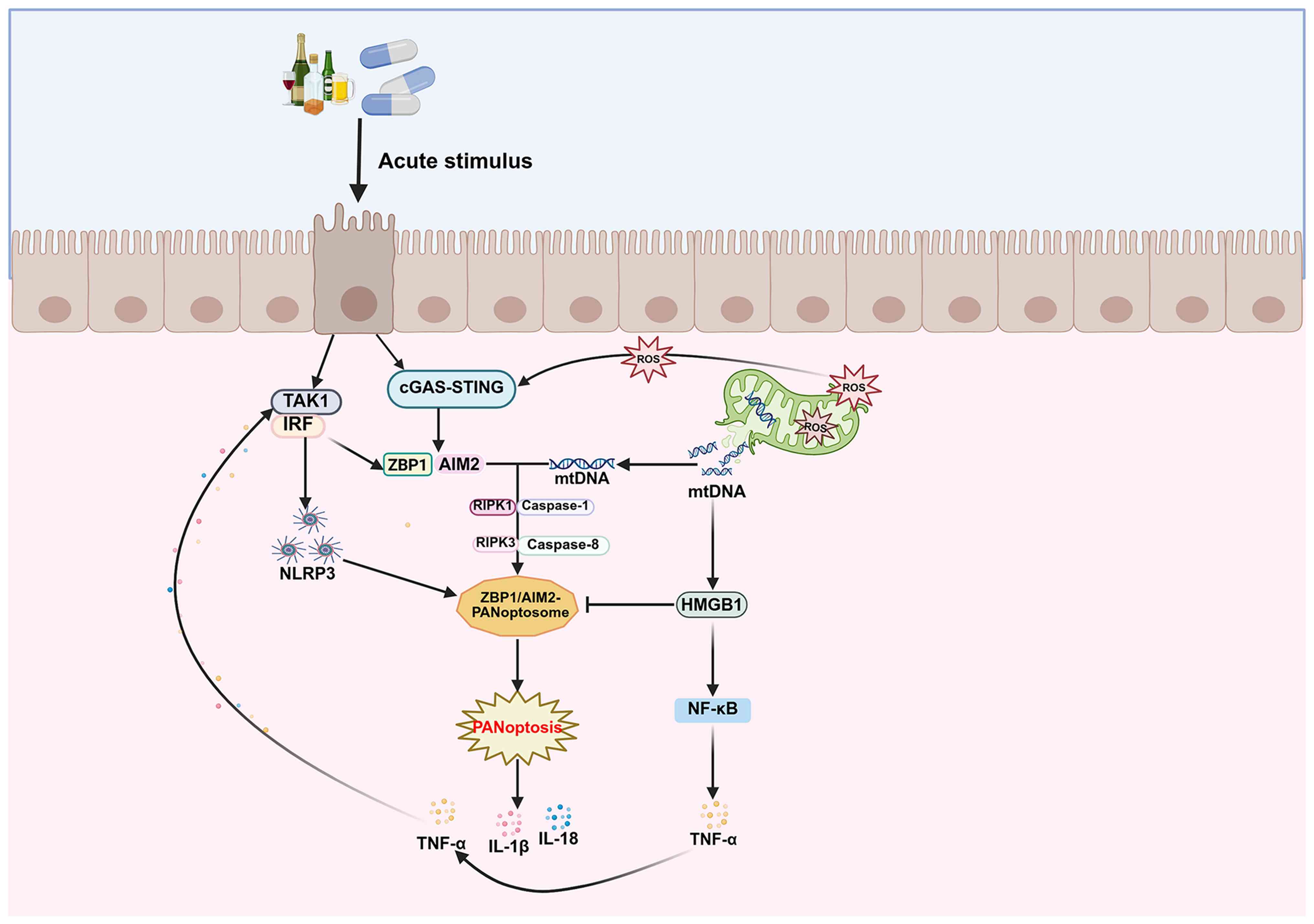 | Figure 1.Regulation of PANoptosis in acute
gastric mucosal diseases. Created with BioRender.com. AIM2, absence
in melanoma 2; cGAS, cyclic GMP-AMP synthase; HMGB1, high mobility
group box 1; IRF, IFN regulatory factor; mtDNA, mitochondrial DNA;
NLRP3, NLR family pyrin domain containing 3; RIPK,
receptor-interacting protein kinase; ROS, reactive oxygen species;
STING, stimulator of IFN genes; TAK1, TGF-β activating kinase 1;
ZBP1, Z-DNA-binding protein 1. |
Notably, substantial amounts of high mobility group
box 1 (HMGB1) released during gastric mucosal injury can bind to
mtDNA (66), thereby limiting
excessive AIM2 activation and competitively inhibiting PANoptosis.
However, as a DAMP, HMGB1 binds to TLR4 and receptor for advanced
glycation end products (61),
activating the NF-κB pathway to increase TNF-α production. This
subsequently induces PANoptosis in gastric mucosal cells, thereby
contributing to the development and progression of gastric ulcers.
Thus, in acute gastric mucosal injury, the complex regulation of
local gastric immunity leads to excessive activation of PANoptosis
during the mucosal immune response, promoting the progression of
mucosal damage. Moderate activation of generalized apoptosis may be
a therapeutic target for gastric mucosal injury diseases.
Role of PANoptosis in infectious
diseases of the gastric mucosa
Infectious diseases of gastric epithelial cells
include viral gastritis, bacterial gastritis and fungal gastritis,
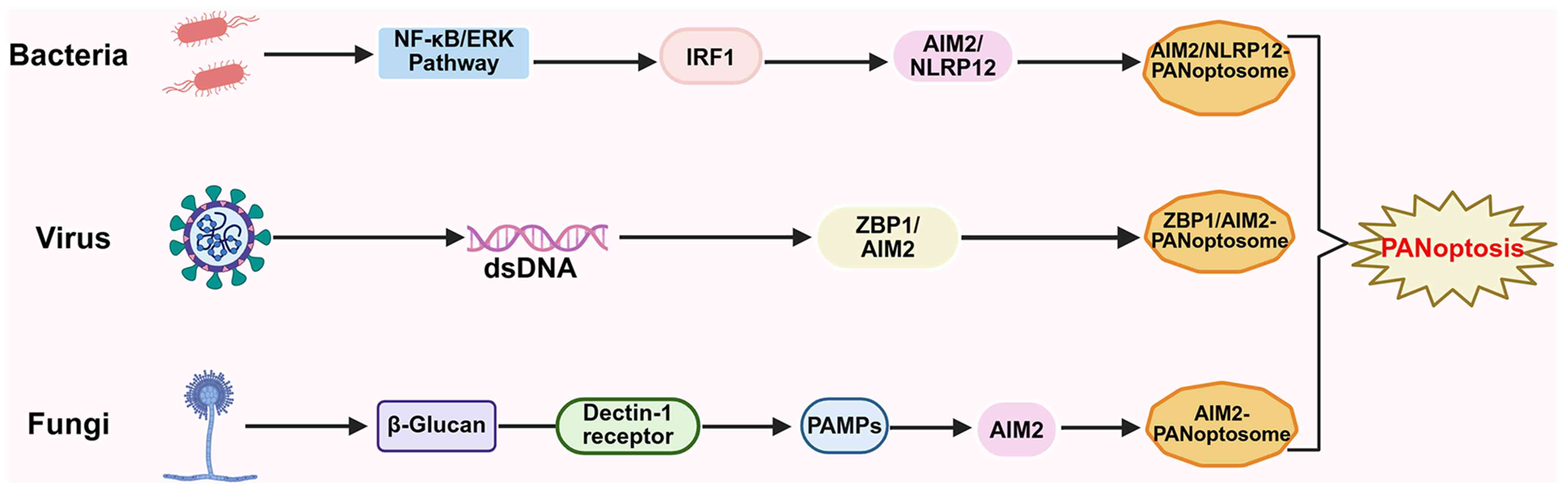
among others. Numerous studies have indicated that during the
initial phase of infection by various pathogens, including viruses,
bacteria and fungi, PAMPs can limit intracellular pathogen
replication by initiating PANoptosis (38,67,68),
a form of PCD, thereby facilitating host recovery. However,
persistent infection leads to excessive activation of PANoptosis,
resulting in the massive release of inflammatory cytokines; this
triggers an inflammatory storm, exacerbating damage to gastric
mucosal tissue (Fig. 2).
Viral gastritis
PANoptosis was initially identified and proposed in
the context of influenza A virus infection (38). In addition, gastric herpes simplex
virus type 1 (HSV-1) infection is regulated by PANoptosis. HSV-1 is
a double-stranded DNA (dsDNA) virus (67), and when it invades the gastric
mucosal epithelium, it promotes the activation of AIM2, pyrin,
ZBP1, ASC, RIPK3, RIPK1, FADD, CASP-1 and CASP-8 to assemble
AIM2-PANoptosomes and ZBP1-PANoptosomes (16), thereby mediating PANoptosis to
inhibit viral replication. However, when viral infection stimulates
excessive activation of immune cells and massive cytokine release,
the release of TNF-α and IFN-γ jointly enhances PANoptosome
assembly, driving PANoptosis to exacerbate damage to the gastric
mucosal epithelium (38).
Similarly, although no studies have yet demonstrated that the
ability of the dsDNA virus Epstein-Barr virus (EBV) (68) to infect gastric epithelial cells is
linked to PANoptosis, its viral structural properties suggest that
this may be possible; however, direct evidence in gastric
epithelial cells is lacking. EBV may potentially activate
ZBP1/AIM2, but this remains to be verified in gastric models. The
mechanisms underlying viral gastric mucosal infection remain
incompletely understood; however, PANoptosis in early infection
confers protection to a certain degree, offering novel therapeutic
perspectives for viral gastritis.
Bacterial gastritis
HP, Francisella and Yersinia species
are common pathogens involved in gastric mucosal cell bacterial
infections. As PAMPs, they activate TLRs, which are primary PRRs in
the gastrointestinal tract that respond to pathogenic infection.
PAMPs (e.g., β-glucan, chitin, mannans and fungal nucleic acids)
bind to hemoglobin, stimulating specific TLRs (TLR2 and TLR4) on
the gastric mucosa to activate the NF-κB and ERK pathways (61,69).
This increases the expression of IRF1, leading to the upregulation
of AIM2 and NLRP12 expression (34,70).
This in turn induces the assembly of AIM2-PANoptosomes and
NLRP12-PANoptosomes, mediating widespread PANoptosis in immune
cells and promoting inflammatory damage to the gastric mucosa. By
contrast, Yersinia-induced gastritis occurs via the
Yersinia outer protein J, which inhibits TGF-β activating
kinase 1 (17), thereby promoting
RIPK1-mediated assembly of the RIPK1-ASC-CASP-1-FADD complex,
CASP-8 and FADD, leading to the formation of the RIPK1-PANoptosome
and thereby inducing PANoptosis to promote the onset and
progression of gastritis.
For common HP infectious gastritis, mitochondrial
damage is induced through the virulence factors CagA and VacA,
promoting the accumulation and release of large amounts of mtDNA
and ROS, which induces PANoptosis in gastric epithelial cells,
driving cell death and inflammatory responses (32). Traditional anti-HP treatments
primarily focus on eradicating the pathogen and alleviating mucosal
inflammation, but they cannot reverse already formed mucosal
damage, chronic inflammation or infection-induced cascades of cell
death (71). By contrast,
inhibiting PANoptosis can directly block the vicious cycle of
inflammation and cell death, more effectively alleviating mucosal
damage by reducing the excessive release of IL-1β, IL-18 and TNF-α
(32,72); when combined with HP eradication
therapy, it may help accelerate mucosal repair and reduce the risk
of inflammation-related precancerous lesions. These findings
demonstrate that PANoptosis serves a role in the development of
bacterial gastritis and may represent a novel therapeutic
target.
Fungal gastritis
Fungal infections commonly occur in
immunocompromised individuals, with Candida albicans and
Aspergillus species being the most prevalent causative
agents of gastric mucosal fungal infections (16,28).
Research has indicated that ZBP1 serves as an apical sensor for
fungal infection (28). Upon
infection by C. albicans or Aspergillus fumigatus,
substantial amounts of PAMPs (e.g., fungal cell wall components,
nucleic acids and glycans) are secreted, activating ZBP1 to promote
ZBP1-PANoptosome assembly. Furthermore, another study has revealed
that during Aspergillus invasion (6,73),
the fungus is recognized by host cells via TLRs, specifically TLR2,
TLR4, TLR9 and dectin-1. Dectin-1 specifically recognizes β-glucans
in fungal cell walls (74),
triggering the release of substantial PAMPs that activate the
cytoplasmic sensor AIM2. This activity mediates AIM2-PANoptosome
assembly, inducing PANoptosis. This pathway may underlie the
pathogenesis of fungal gastritis.
In the early stages of infection by pathogens such
as viruses, bacteria and fungi in the gastric mucosal epithelium,
the activation of PANoptosis aids in preventing pathogen
replication and invasion. However, excessive and sustained
activation of PANoptosis may promote the progression of
inflammation and exacerbate damage to gastric mucosal epithelial
tissue. Therefore, achieving a balanced activation of PANoptosis is
crucial for the early prevention of infectious diseases of the
gastric mucosa. For pathogenic infection-mediated chronic mucosal
inflammation and epithelial cell damage, compared with traditional
anti-infective treatments, targeted inhibition of PANoptosis may
block the vicious cycle of inflammation and cell death, and promote
repair of the mucosal barrier.
Role of PANoptosis in autoimmune
gastritis
Autoimmune gastritis occurs when the autoantigen
H+-K+ adenosine triphosphatase on parietal
cells is recognized by autoreactive CD4+ T cells
(75), leading to spontaneous
inflammatory infiltration and atrophy of the gastric mucosa. This
suggested mechanism has been extrapolated from autoimmune disease
models; direct in vivo evidence in gastric tissues is still
insufficient. TNF-α and IFN-γ may act via JAK/STAT1/IRF1 (36) to drive inducible nitric oxide (NO)
synthase to produce NO. This activates FADD/RIPK1/RIPK3-mediated
CASP-8/Fas-associated protein ZBP1 and AIM2-PANoptosome assembly,
inducing gastric epithelial cell PANoptosis to promote autoimmune
gastritis. Moreover, TNF-α binds to the TNF receptor, stimulating
RIPK1 to interact on CASP-8 via FADD and induce apoptosis (76). Simultaneously, RIPK1 binds to RIPK3
through the RIPK homotypic interaction motif (RHIM) domain,
inducing necroptosis (77).
Furthermore, activated ZBP1 promotes IL-17 secretion by
CD4+ T helper 17 cells, which directly act on gastric
parietal cells. This activates CASP-3-mediated apoptosis via the
p53 and PI3K/Akt signaling pathways (78) or drives pyroptosis, thereby
advancing autoimmune gastritis progression (Fig. 3).
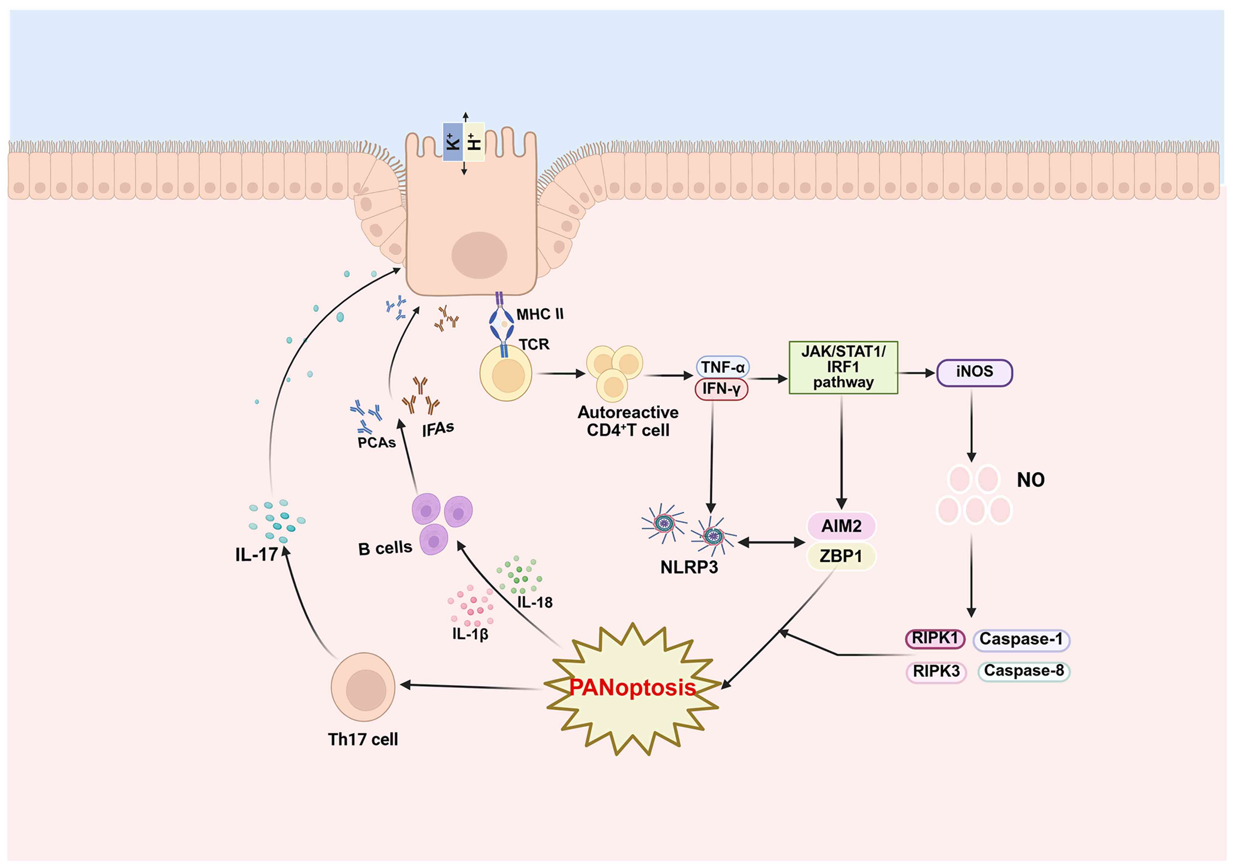 | Figure 3.Regulation of PANoptosis in
autoimmune gastritis. Created with BioRender.com. AIM2, absence in
melanoma 2; IFAs, intrinsic factor antibodies; iNOS, inducible NO
synthase; MHC II, major histocompatibility complex class II; NLRP3,
NLR family pyrin domain containing 3; NO, nitric oxide; PCAs,
parietal cell antibodies; RIPK, receptor-interacting protein
kinase; TCR, T-cell receptor; Th17, T helper 17; ZBP1,
Z-DNA-binding protein 1. |
In summary, PANoptosis may be an important
regulatory factor exacerbating the progression of autoimmune
gastritis, and appropriately blocking its activation represents an
effective therapeutic strategy for this condition. Previous studies
have confirmed that IRF1 is a key upstream regulator of PANoptosis
mediated by factors including ZBP1, AIM2 and NLRP12 (70,79),
and IRF1 deficiency reduces the activation of PANoptotic molecules.
Conversely, mutations or loss of function in adenosine deaminase 1
generates endogenous Z-RNA, activating ZBP1-mediated PANoptosis
(36). This mechanism presents a
potential therapeutic target for autoimmune gastritis.
Role of PANoptosis in SPEM
SPEM constitutes an important mucosal repair lineage
capable of progressing to intestinal metaplasia, thereby promoting
gastric carcinogenesis. Evidence has clearly indicated that IL-13
acts as an initiating factor in SPEM development (80), whereas IL-33 acts as an
upregulating factor (81),
inducing SPEM through stimulating M2 macrophage activation.
Notably, SPEM development is closely associated with PANoptosis.
Gastric AIM2 is produced primarily by gastric B220+
IgM+ immune cells (82). During gastric mucosal injury,
elevated AIM2 expression activates ZBP1 through the sensing of
endogenous dsDNA or mtDNA (83),
which participate in AIM2-PANoptosome assembly to mediate gastric
mucosal cell PANoptosis. Subsequently, CASP-6 is activated as a
downstream component and interacts with IL-4 to promote macrophage
activation via nonapoptotic pathways (84). Concurrently, IL-10 secretion
inhibits M1 macrophage activation (85) while enhancing M2 macrophage
activation and expression, thereby promoting the development of
SPEM in the gastric mucosa (Fig.
4). However, most evidence comes from gastric injury models;
direct evidence of PANoptosome assembly in SPEM-specific lineages
is lacking.
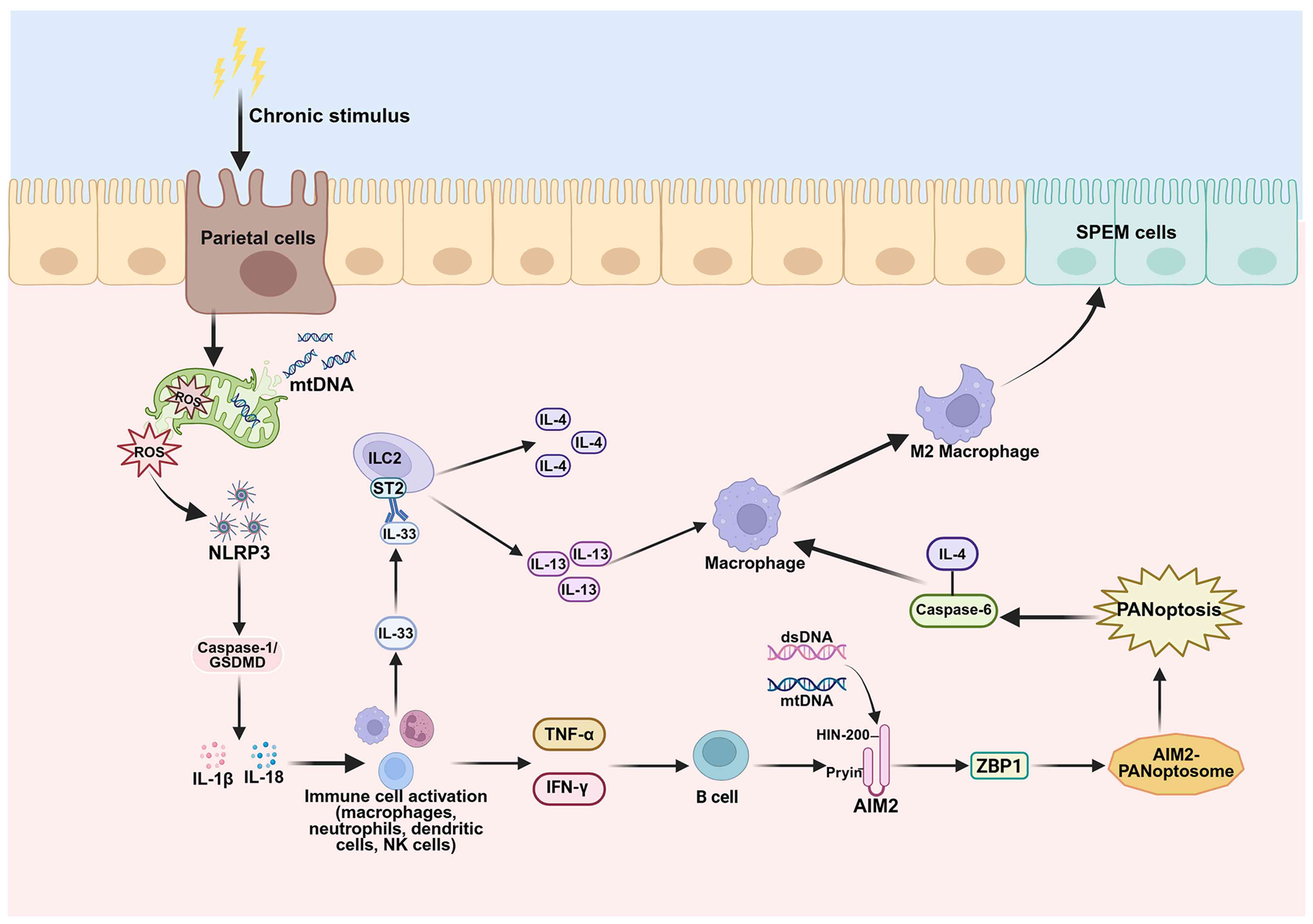 | Figure 4.Regulation of PANoptosis in SPEM.
Long-term chronic stimulation participates in the immune
microenvironment regulation of gastric mucosal SPEM through
PANoptosis. Created with BioRender.com. AIM2, absence in melanoma
2; dsDNA, double-stranded DNA; GSDMD, gasdermin D; ILC2, type II
innate lymphoid cells; mtDNA, mitochondrial DNA; NK, natural
killer; NLRP3, NLR family pyrin domain containing 3; ROS, reactive
oxygen species; SPEM, spasmolytic polypeptide-expressing
metaplasia; ZBP1, Z-DNA-binding protein 1. |
Furthermore, M2 macrophage activation induces CASP-6
upregulation, the release of N-terminal GSDME, the phosphorylation
of MLKL (86), and the binding of
RIPK3 to the RHIM domain of ZBP1 to stimulate ZBP1-PANoptosome
assembly (87), thereby increasing
PANoptotic activity. However, the noninflammasome AIM2 can inhibit
the progression of SPEM by restricting CD8+ T-lymphocyte
accumulation in chronic gastritis through the suppression of CXCL16
(produced by gastric B cells) (82).
Research has revealed that the absence of GRIM-19 in
parietal cell mitochondria can promote NLRP3/IL-33 activation via
the ROS-NRF2-HO-1-NF-κB pathway, thereby mediating the development
of SPEM (88). Following gastric
mucosal injury, NLRP3 upregulation is induced, promoting M2
macrophage activation and CD8+ T-cell recruitment. This
contributes to gastric SPEM development by increasing IFN-γ
production (89). The mechanism by
which NLRP3 induces M2 macrophage activation remains unclear. On
the basis of these findings, it may be hypothesized that gastric
mucosal injury increases NLRP3 expression, which, via the CASP-1
pathway, triggers the massive release and secretion of inflammatory
cytokines. This enhances the activity of dendritic cells,
neutrophils, gastric B cells and natural killer cells (90); promotes the upregulation of TNF-α
and IRF1; and induces activation of the AIM2 inflammasome through
guanine nucleotide-binding protein (66). Stimulation of AIM2-PANoptosome
assembly mediates SPEM development. Transcriptomic analysis has
further revealed that these immune cells express PANoptosis-related
genes (PANoDEGs), such as IL1B, IL18, TNF, CASP1 and MLKL,
indicating that the aforementioned immune cells are involved in the
pathogenesis of SPEM (66). This
stimulates AIM2-PANoptosome assembly, mediating SPEM via
PANoptosis. Transcriptomic analysis has revealed strong
associations between these immune cells and PANoDEGs (91). Furthermore, heightened inflammatory
cell activity increases the secretion of the gastric mucosal injury
alarmins IL-33 and HMGB1 (92).
These DAMPs may activate inflammasomes such as ZBP1 and AIM2,
mediating the recruitment of NLRP3, RIPK3, RIPK1 and CASP-8 to
assemble PANoptosomes. This PANoptosis pathway thereby promotes
SPEM progression. Current direct evidence of PANoptosome assembly
in SPEM-specific epithelial cells is still lacking; therefore,
PANoptosis is a potential regulatory factor in SPEM, and its core
role requires further verification.
Role of PANoptosis in gastric
cancer
The incidence and mortality rates of malignant
gastric cancer remain high, ranking fifth in cancer incidence and
fourth in cancer-related mortality globally (93,94).
The emergence of PANoptosis, a novel form of cell death, offers
fresh therapeutic options for tumor management. In gastric cancer,
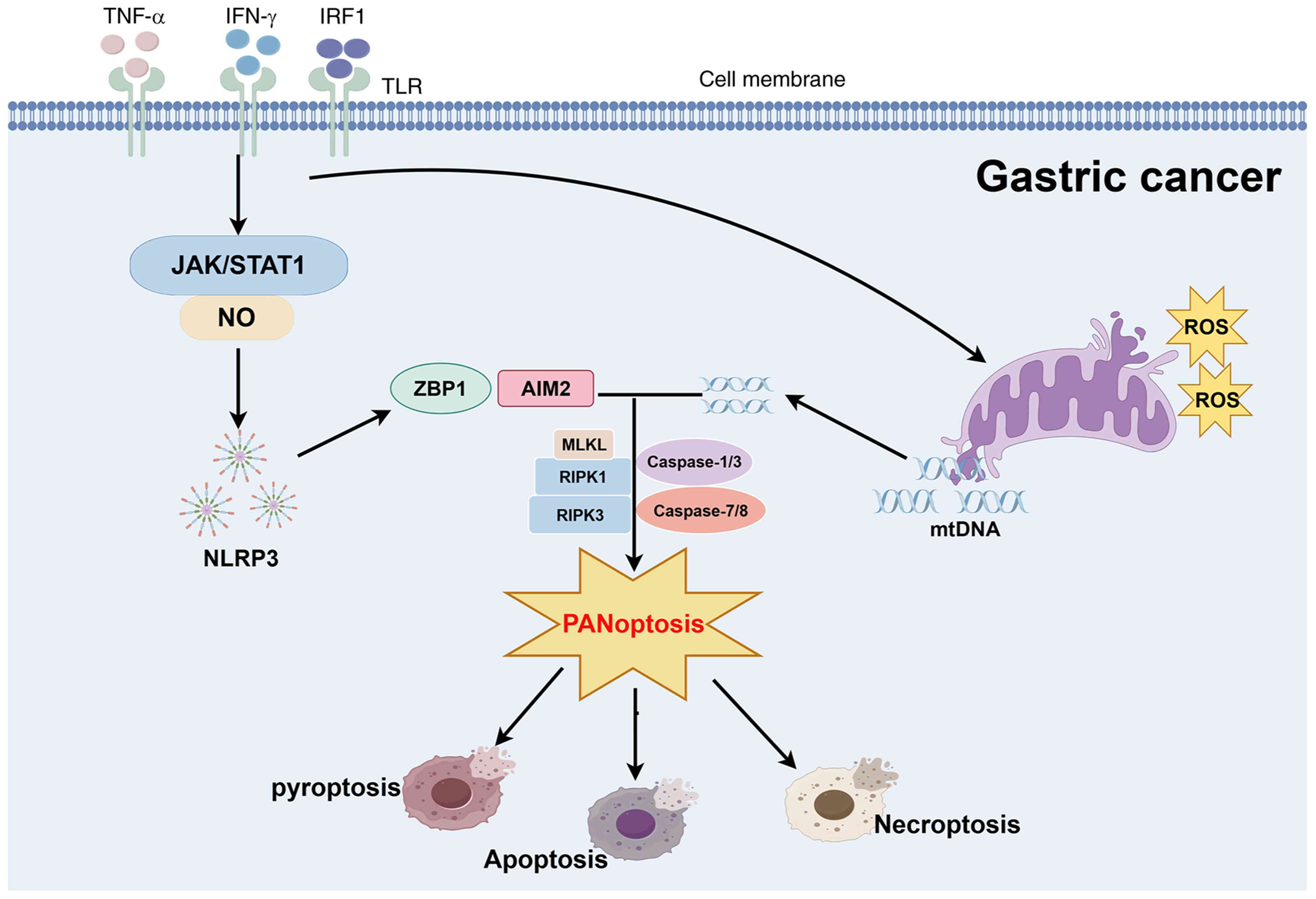
AIM2 upregulation is induced (35), thereby initiating the assembly of
AIM2-PANoptosomes within tumor cells. IRF1, TNF-α and IFN-γ have
been demonstrated to induce PANoptosis to prevent tumorigenesis
(29,95,96).
As an upstream regulator of PANoptosis, IRF1 produces NO to
modulate TNF-α and IFN-γ expression via the JAK/STAT1 signaling
pathway (90,97). The interaction of TNF-α and IFN-γ
activates GSDME, CASP-8/3/7 and MLKL phosphorylation, thereby
inducing PANoptosome assembly. Furthermore, the combination of IFN
with a nuclear export inhibitor (98) enhances ZBP1-PANoptosome assembly,
inducing robust PANoptosis in tumor cells and inhibiting their
proliferation (Fig. 5).
The therapeutic effect of cisplatin on gastric
cancer cell is mediated by the activation of CASP-8/9 and
downstream CASP-3/7 to induce apoptosis (99). This occurs because CASP-1
stimulates increased CASP-3 expression, lysing GSDMD/E to mediate
pyroptosis (100) and activating
RIPK3 to induce MLKL phosphorylation and necroptosis (101). It has been hypothesized that
cisplatin treatment in gastric cancer may increase chemotherapeutic
sensitivity and inhibit tumor growth by inducing PANoptosis in
cancer cells. However, this requires in vivo validation in
clinical samples. Furthermore, another study indicated that the use
of a PANoptosis score may aid in predicting gastric cancer
prognosis and survival rates, as well as immunotherapy outcomes
(11). PANoptosis may inhibit
tumor growth and progression by stimulating inflammatory factor
activity within tumor cells, thereby exacerbating PCD and
inhibiting tumor cell immune evasion.
Role of PANoptosis in
mucosa-associated lymphoid tissue (MALT) lymphoma
MALT lymphoma is a rare gastric tumor that
originates as an indolent B-cell non-Hodgkin lymphoma within the
marginal zone of the gastric lymphoid tissue (102). MALT is frequently induced by HP
infection and chronic stimulation by autoantigens, and is primarily
manifested as abnormal proliferation of small B lymphocytes within
the gastric mucosa (103). NF-κB
serves as a crucial mediator of immune responses and was first
described in B lymphocytes. Activation of NF-κB stimulates
unlimited B-cell proliferation and the transcription of
antiapoptotic genes, ultimately contributing to lymphoma
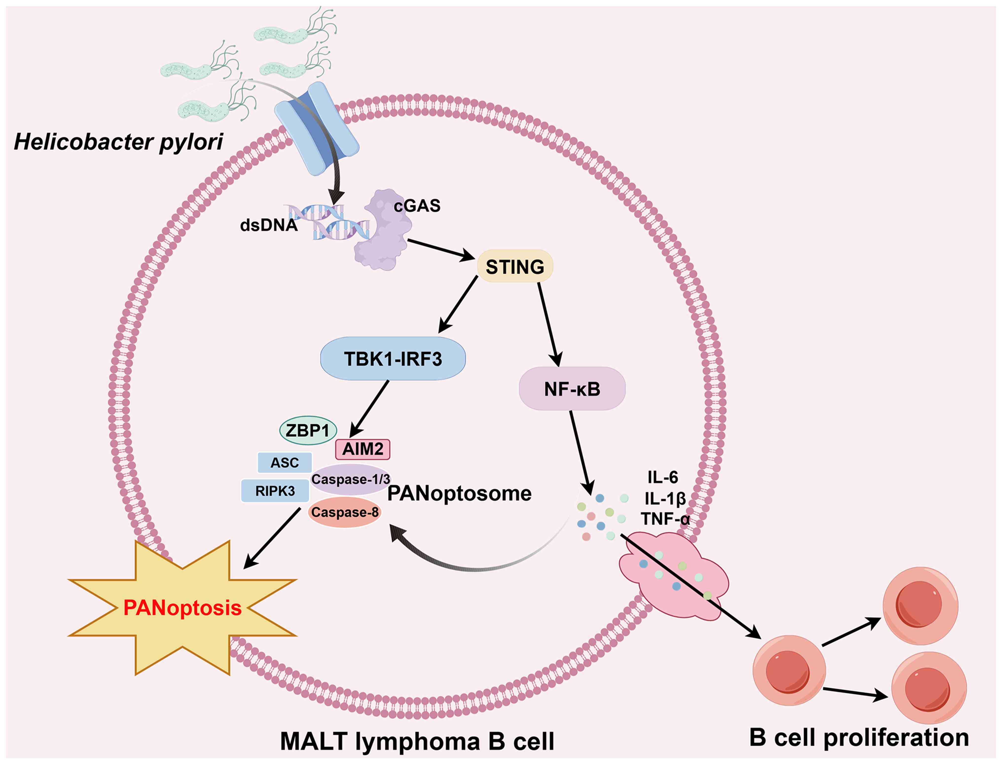
development (104). During
chronic HP-induced inflammatory infection, released bacterial dsDNA
is recognized by the cytoplasmic DNA sensor cGAS (3), triggering a DNA damage response that
activates the STING-TBK1-IRF3 signaling pathway (105), consequently promoting formation
of the ZBP1/CASP-8/RIPK3/ASC multiprotein complex. This further
induces MLKL phosphorylation, CASP-3 cleavage and GSDME cleavage
(106), thereby promoting gastric
lymphoma cell death and inhibiting tumor cell proliferation. These
findings demonstrate that STING suppresses MALT tumor formation by
mediating PANoptosis. However, STING activation may simultaneously
promote NF-κB activation and production of the proinflammatory
cytokines TNF-α and IL-6 (84). On
the one hand, this induces B-cell proliferation (Fig. 6), potentially driving MALT
progression; however, it may also amplify the effects of PANoptosis
within MALT, enhancing antitumor immune responses. Traditional
anti-HP targeted therapy has a notable effect on early HP-infection
type MALT lymphoma (107), but
its efficacy is poor for advanced non-HP-infection type MALT
lymphoma (108). However,
PANoptosis-targeted therapy can induce lymphoma cell-specific death
while modulating the immunosuppressive state of the tumor
microenvironment. When combined with HP eradication therapy, it may
improve the cure rate of early lesions and reduce the risk of
recurrence. Consequently, understanding the activation of
PANoptosis and investigating its balanced targets may hold key
therapeutic potential for MALT.
Conclusion
Through investigations into the interconnections and
crosstalk between multiple cell death pathways, PANoptosis has
emerged as a novel form of cell death that has garnered attention.
In gastric epithelial cells during early injury or infection,
PANoptosis has a protective effect; in immune cells or epithelial
cells during chronic or excessive activation, it has a pathological
effect; and in tumor cells, it exerts an antitumor effect through
activation. Currently, PANoptosis research is particularly advanced
in infectious and neoplastic diseases, offering novel perspectives
for advancing human disease prevention and treatment. However,
global inhibition of PANoptosis may weaken host defense, impair
mucosal repair and increase infection risk; and non-specific
activation may damage normal gastric epithelial cells and
exacerbate inflammation. Therefore, achieving a balanced activation
of PANoptosis is crucial for the early prevention of infectious
diseases of the gastric mucosa.
Currently, research on PANoptosis has a number of
unavoidable limitations, such as the lack of stomach-specific in
vivo models that can directly demonstrate the assembly and
activation of the PANoptosome in gastric mucosal injury (20). Most of the current evidence comes
from in vitro cell systems or models derived from other
organs (109), which cannot
adequately reflect the unique microenvironment, intercellular
interactions and regulatory characteristics of the stomach.
Moreover, accurately distinguishing the concurrent activation of
PANoptosis from apoptosis, pyroptosis and necroptosis in the
gastric mucosal microenvironment poses notable technical challenges
(110). Existing detection
methods lack specificity, making it difficult to avoid misjudgments
caused by signal overlap. In addition, multiple inflammatory
signaling pathways (including NF-κB, TLR4 and JAK/STAT) converge on
the same key molecular targets of PANoptosis (110), which may result in potential
confounding effects in mechanistic interpretations.
Although there are numerous unknowns regarding the
‘sensors’ that trigger PANoptosis, and the regulatory mechanisms in
gastric mucosal diseases lack relevant experimental validation, the
involvement of PANoptosis in the regulation of gastric mucosal
diseases may become a potential clinical intervention target.
Research addressing these limitations is essential. In the future,
focusing on establishing gastric-specific PANoptosis research
models, developing specific detection and intervention tools, and
elucidating precise regulatory mechanisms will provide new
theoretical bases and potential therapeutic targets for the
prevention and treatment of gastric mucosal diseases.
Acknowledgements
Not applicable.
Funding
The present study was supported by the National Natural Science
Foundation of China (grant nos. 82470540, 32460215 and 82160505);
the Guizhou International Science & Technology Cooperation Base
for Gastroenterology [grant no. Qian Ke Supplementary Platform
Talents-GHJD (2025) 003]; the Major Project of the Guizhou Province
Basic Research Program [grant no. Qian Ke He Basic Research-ZK
(2023) Major Project 059]; the General Project of the Guizhou
Province Basic Research Program [grant no. Qian Ke He Basic
Research-ZK (2022) General 659]; the Guizhou Province High-level
Innovative Talent Selection and Training Plan (Hundred-level Talent
Program) [grant no. Qian Ke He Platform Talents-GCC (2023) 043];
the Guizhou Innovative Talent Team on Ion Channels and Malignant
Tumors of Epithelial Origin [grant no. Qian Ke He Platform
Talents-CXTD (2023) 001]; the Guizhou Clinical Research Center for
Digestive Diseases [grant no. Qian Ke He Platform-LCZX (2025) 001];
the Zunyi City Breast Cancer Prevention and Treatment Basic and
Clinical Research Technology Innovation Talent Team [grant no. Zun
Shi Ke Talent (2023) No. 4]; the Zunyi City Science and Technology
Cooperation Plan Project grant no. Zun Shi Ke He HZ Zi (2023) No.
224]; the Wu Jieping Medical Foundation [grant no. Zun Yi He Zi
(2023) No. 28]; the Joint Medical Scientific Research Fund for
High-quality Development of Health in Guizhou Province (2024)
(grant no. 2024GZYXKYJJXM0019); the Guizhou Provincial Department
of Science and Technology 2025 Basic Research Program Youth
Guidance Project [Qiankehe Basic QN (2025) 152 to ZM]; and the 2026
Guizhou Provincial Basic Research Program (General Program)
[Qiankehe Basic MS (2026) 969 to ZM].
Availability of data and materials
Not applicable.
Authors' contributions
LW, ZM BT, TL and XL conceived the study and were
involved in its conception and design. LW drafted the manuscript.
SL and ST were also involved in the design and conception of the
study. BJ performed systematic literature retrieval, screened all
relevant studies on PANoptosis and gastric mucosal diseases, and
completed collation and comparative analysis of the data included
in this review. ZM assisted in the preparation of the figures. ST
reviewed the mechanistic framework and revised the manuscript for
intellectual content. BT, TL and XL edited and revised the
manuscript. Data authentication is not applicable. All authors read
and approved the final manuscript.
Ethics approval and consent to
participate
Not applicable.
Patient consent for publication
Not applicable.
Competing interests
The authors declare that they have no competing
interests.
Glossary
Abbreviations
Abbreviations:
|
AIM2
|
absence in melanoma 2
|
|
Akt
|
protein kinase B
|
|
ADAR1
|
adenosine deaminase 1
|
|
CASP
|
caspase
|
|
DAMP
|
damage-associated molecular
pattern
|
|
dsDNA
|
double-stranded DNA
|
|
GSDM
|
gasdermin
|
|
cGAS
|
cyclic GMP-AMP synthase
|
|
HSV-1
|
herpes simplex virus type 1
|
|
HO-1
|
heme oxygenase-1
|
|
HMGB1
|
high mobility group box 1
|
|
IRF1
|
IFN regulatory factor 1
|
|
IAV
|
influenza A virus
|
|
iNOS
|
inducible NO synthase
|
|
mROS
|
mitochondrial reactive oxygen
species
|
|
mtDNA
|
mitochondrial DNA
|
|
MALT
|
mucosa-associated lymphoid tissue
|
|
NLRP
|
NLR family pyrin domain
containing
|
|
NLRC4
|
NLR family CARD domain containing
4
|
|
NRF2
|
nuclear factor erythroid 2-related
factor 2
|
|
NO
|
nitric oxide
|
|
PCD
|
programmed cell death
|
|
PAMP
|
pathogen-associated molecular
pattern
|
|
PRR
|
pattern recognition receptor
|
|
PANoDEG
|
PANoptosis-related gene
|
|
RIPK
|
receptor-interacting protein
kinase
|
|
RHIM
|
receptor-interacting protein kinase
homotypic interaction motif
|
|
SPEM
|
spasmolytic polypeptide expressing
metaplasia
|
|
STING
|
stimulator of IFN genes
|
|
TLR
|
Toll-like receptor
|
|
TAK1
|
TGF-β activating kinase 1
|
|
TBK1
|
TANK-binding kinase 1
|
|
ZBP1
|
Z-DNA-binding protein 1
|
|
FADD
|
Fas-associated death domain
protein
|
|
ASC
|
apoptosis-associated Speck-like
protein containing a CARD
|
References
|
1
|
Kanneganti TD: Intracellular innate immune
receptors: Life inside the cell. Immunol Rev. 297:5–12. 2020.
View Article : Google Scholar : PubMed/NCBI
|
|
2
|
Shi C, Cao P, Wang Y, Zhang Q, Zhang D,
Wang Y, Wang L and Gong Z: PANoptosis: A cell death characterized
by pyroptosis, apoptosis, and necroptosis. J Inflamm Res.
16:1523–1532. 2023. View Article : Google Scholar : PubMed/NCBI
|
|
3
|
Chen S, Jiang J, Li T and Huang L:
PANoptosis: Mechanism and role in pulmonary diseases. Int J Mol
Sci. 24:153432023. View Article : Google Scholar : PubMed/NCBI
|
|
4
|
Xiang Q, Geng ZX, Yi X, Wei X, Zhu XH and
Jiang DS: PANoptosis: A novel target for cardiovascular diseases.
Trends Pharmacol Sci. 45:739–756. 2024. View Article : Google Scholar : PubMed/NCBI
|
|
5
|
Hou K, Pan W, Liu L, Yu Q, Ou J, Li Y,
Yang X, Lin Z, Yuan JH and Fang M: Molecular mechanism of
PANoptosis and programmed cell death in neurological diseases.
Neurobiol Dis. 209:1069072025. View Article : Google Scholar : PubMed/NCBI
|
|
6
|
Xu X, Wei Y, Pang J, Wei Z, Wang L, Chen
Q, Wang Z, Zhang Y, Chen K, Peng Y, et al: Time-course
transcriptomic analysis reveals the crucial roles of PANoptosis in
fungal keratitis. Invest Ophthalmol Vis Sci. 64:62023. View Article : Google Scholar : PubMed/NCBI
|
|
7
|
Tong J, Lan XT, Zhang Z, Liu Y, Sun DY,
Wang XJ, Ou-Yang SX, Zhuang CL, Shen FM, Wang P and Li DJ:
Ferroptosis inhibitor liproxstatin-1 alleviates metabolic
dysfunction-associated fatty liver disease in mice: Potential
involvement of PANoptosis. Acta Pharmacol Sin. 44:1014–1028. 2023.
View Article : Google Scholar : PubMed/NCBI
|
|
8
|
Yang Y, Hounye AH, Chen Y, Liu Z, Shi G
and Xiao Y: Characterization of PANoptosis-related genes in Crohn's
disease by integrated bioinformatics, machine learning and
experiments. Sci Rep. 14:117312024. View Article : Google Scholar : PubMed/NCBI
|
|
9
|
Liu LX, Heng JH, Deng DX, Zhao H, Zheng
ZY, Liao LD, Lin W, Xu XE, Li EM and Xu LY: Sulconazole Induces
PANoptosis by triggering oxidative stress and inhibiting glycolysis
to increase radiosensitivity in esophageal cancer. Mol Cell
Proteomics. 22:1005512023. View Article : Google Scholar : PubMed/NCBI
|
|
10
|
Karki R, Sharma BR, Lee E, Banoth B,
Malireddi RKS, Samir P, Tuladhar S, Mummareddy H, Burton AR, Vogel
P and Kanneganti TD: Interferon regulatory factor 1 regulates
PANoptosis to prevent colorectal cancer. JCI Insight.
5:e1367202020. View Article : Google Scholar : PubMed/NCBI
|
|
11
|
Pan H, Pan J, Li P and Gao J:
Characterization of PANoptosis patterns predicts survival and
immunotherapy response in gastric cancer. Clin Immunol.
238:1090192022. View Article : Google Scholar : PubMed/NCBI
|
|
12
|
Li P, Gao Y, Tao Z, Mu Z, Du S and Zhao X:
PANoptosis: Cross-talk among apoptosis, necroptosis, and pyroptosis
in neurological disorders. J Inflamm Res. 18:8131–8140. 2025.
View Article : Google Scholar : PubMed/NCBI
|
|
13
|
Wei S, Chen Z, Ling X, Zhang W and Jiang
L: Comprehensive analysis illustrating the role of
PANoptosis-related genes in lung cancer based on bioinformatic
algorithms and experiments. Front Pharmacol. 14:11152212023.
View Article : Google Scholar : PubMed/NCBI
|
|
14
|
Chen W, Gullett JM, Tweedell RE and
Kanneganti TD: Innate immune inflammatory cell death: PANoptosis
and PANoptosomes in host defense and disease. Eur J Immunol.
53:e22502352023. View Article : Google Scholar : PubMed/NCBI
|
|
15
|
Liu X, Miao M, Sun J, Wu J and Qin X:
PANoptosis: A potential new target for programmed cell death in
breast cancer treatment and prognosis. Apoptosis. 29:277–288. 2024.
View Article : Google Scholar : PubMed/NCBI
|
|
16
|
Nguyen LN and Kanneganti TD: PANoptosis in
viral infection: the missing puzzle piece in the cell death field.
J Mol Biol. 434:1672492022. View Article : Google Scholar : PubMed/NCBI
|
|
17
|
Malireddi RKS, Kesavardhana S, Karki R,
Kancharana B, Burton AR and Kanneganti TD: RIPK1 distinctly
regulates yersinia-induced inflammatory cell death, PANoptosis.
Immunohorizons. 4:789–796. 2020. View Article : Google Scholar : PubMed/NCBI
|
|
18
|
Xiang J, Li Y, Mei S, Ou Z, Wang L, Ke Y
and Li Z: Novel diagnostic and therapeutic strategies based on
PANoptosis for hepatocellular carcinoma. Cancer Biol Med.
22:928–939. 2025.PubMed/NCBI
|
|
19
|
Li J and Qu Y: PANoptosis in neurological
disorders: Mechanisms, implications, and therapeutic potential.
Front Immunol. 16:15793602025. View Article : Google Scholar : PubMed/NCBI
|
|
20
|
Qi L, Lan B, Zhao Z, Ma Y, Song J, Jia Q,
Zhao P and Du X: Research advances of PANoptosis in
gastrointestinal tumors. Int Immunopharmacol. 159:1149312025.
View Article : Google Scholar : PubMed/NCBI
|
|
21
|
Wang W, Lu J, Pan N, Zhang H, Dai J, Li J,
Chi C, Zhang L, Wang L and Zhang M: Identification of early
Alzheimer's disease subclass and signature genes based on
PANoptosis genes. Front Immunol. 15:14620032024. View Article : Google Scholar : PubMed/NCBI
|
|
22
|
Zhao J, Guo S, Schrodi SJ and He D: Absent
in melanoma 2 (AIM2) in rheumatoid arthritis: Novel molecular
insights and implications. Cell Mol Biol Lett. 27:1082022.
View Article : Google Scholar : PubMed/NCBI
|
|
23
|
Sun W, Li P, Wang M, Xu Y, Shen D, Zhang X
and Liu Y: Molecular characterization of PANoptosis-related genes
with features of immune dysregulation in systemic lupus
erythematosus. Clin Immunol. 253:1096602023. View Article : Google Scholar : PubMed/NCBI
|
|
24
|
Gong W, Liu Z, Wang Y, Huang W, Yang K,
Gao Z, Guo K, Xiao Z and Zhao W: Reprogramming of Treg cell-derived
small extracellular vesicles effectively prevents intestinal
inflammation from PANoptosis by blocking mitochondrial oxidative
stress. Trends Biotechnol. 43:893–917. 2025. View Article : Google Scholar : PubMed/NCBI
|
|
25
|
Iannucci A, Colella M, Quiroga M,
Frascatani R, Tomassini L, Maresca C, Franzè E, Laudisi F, Sica G,
Marafini I, et al: Loss of adenosine deaminase acting on RNA 1
induces panoptosis and immune response in ulcerative colitis gut
mucosa. MedComm (2020). 6:e702122025. View Article : Google Scholar : PubMed/NCBI
|
|
26
|
Guan J, Shi X, Ma J, Yin Y, Song G, Li Y,
Chen X, Yan Y, Wang D, Liu S, et al: Circular RNA-OGDH promotes
PANoptosis in diabetic cardiomyopathy: A novel mechanistic insight.
J Biol Chem. 301:1102802025. View Article : Google Scholar : PubMed/NCBI
|
|
27
|
Cai H, Lv M and Wang T: PANoptosis in
cancer, the triangle of cell death. Cancer Med. 12:22206–22223.
2023. View Article : Google Scholar : PubMed/NCBI
|
|
28
|
Banoth B, Tuladhar S, Karki R, Sharma BR,
Briard B, Kesavardhana S, Burton A and Kanneganti TD: ZBP1 promotes
fungi-induced inflammasome activation and pyroptosis, apoptosis,
and necroptosis (PANoptosis). J Biol Chem. 295:18276–18283. 2020.
View Article : Google Scholar : PubMed/NCBI
|
|
29
|
Sharma BR, Karki R, Rajesh Y and
Kanneganti TD: Immune regulator IRF1 contributes to ZBP1-, AIM2-,
RIPK1-, and NLRP12-PANoptosome activation and inflammatory cell
death (PANoptosis). J Biol Chem. 299:1051412023. View Article : Google Scholar : PubMed/NCBI
|
|
30
|
Liu S, Deng Z, Zhu J, Ma Z, Tuo B, Li T
and Liu X: Gastric immune homeostasis imbalance: An important
factor in the development of gastric mucosal diseases. Biomed
Pharmacother. 161:1143382023. View Article : Google Scholar : PubMed/NCBI
|
|
31
|
Nie S and Yuan Y: The role of gastric
mucosal immunity in gastric diseases. J Immunol Res.
2020:79270542020. View Article : Google Scholar : PubMed/NCBI
|
|
32
|
Lin Y, Liu K, Lu F, Zhai C and Cheng F:
Programmed cell death in Helicobacter pylori infection and related
gastric cancer. Front Cell Infect Microbiol. 14:14168192024.
View Article : Google Scholar : PubMed/NCBI
|
|
33
|
Cui G, Yuan Y, Wang Y and Li Z: The
expression of RIPK3 is associated with cell turnover of gastric
mucosa in the mouse and human stomach. J Mol Histol. 52:849–857.
2021. View Article : Google Scholar : PubMed/NCBI
|
|
34
|
Dawson RE, Deswaerte V, West AC, Sun E,
Wray-McCann G, Livis T, Kumar B, Rodriguez E, Gabay C, Ferrero RL
and Jenkins BJ: The cytosolic DNA sensor AIM2 promotes
Helicobacter-induced gastric pathology via the inflammasome.
Immunol Cell Biol. 101:444–457. 2023. View Article : Google Scholar : PubMed/NCBI
|
|
35
|
Zhou R, Sun J, He C, Huang C and Yu H:
CCL19 suppresses gastric cancer cell proliferation, migration, and
invasion through the CCL19/CCR7/AIM2 pathway. Hum Cell.
33:1120–1132. 2020. View Article : Google Scholar : PubMed/NCBI
|
|
36
|
Pandeya A and Kanneganti TD: Therapeutic
potential of PANoptosis: Innate sensors, inflammasomes, and RIPKs
in PANoptosomes. Trends Mol Med. 30:74–88. 2024. View Article : Google Scholar : PubMed/NCBI
|
|
37
|
Samir P, Malireddi RKS and Kanneganti TD:
The PANoptosome: A deadly protein complex driving pyroptosis,
apoptosis, and necroptosis (PANoptosis). Front Cell Infect
Microbiol. 10:2382020. View Article : Google Scholar : PubMed/NCBI
|
|
38
|
Qi Z, Zhu L, Wang K and Wang N:
PANoptosis: Emerging mechanisms and disease implications. Life Sci.
333:1221582023. View Article : Google Scholar : PubMed/NCBI
|
|
39
|
Motani K, Kawase K, Imamura R, Kinoshita
T, Kushiyama H and Suda T: Activation of ASC induces apoptosis or
necrosis, depending on the cell type, and causes tumor eradication.
Cancer Sci. 101:1822–1827. 2010. View Article : Google Scholar : PubMed/NCBI
|
|
40
|
Broz P, Pelegrín P and Shao F: The
gasdermins, a protein family executing cell death and inflammation.
Nat Rev Immunol. 20:143–157. 2019. View Article : Google Scholar : PubMed/NCBI
|
|
41
|
Tsuchiya K, Nakajima S, Hosojima S, Thi
Nguyen D, Hattori T, Manh Le T, Hori O, Mahib MR, Yamaguchi Y,
Miura M, et al: Caspase-1 initiates apoptosis in the absence of
gasdermin D. Nat Commun. 10:20912019. View Article : Google Scholar : PubMed/NCBI
|
|
42
|
Cavalcante GC, Schaan AP, Cabral GF,
Santana-da-Silva MN, Pinto P, Vidal AF and Ribeiro-Dos-Santos Â: A
Cell's Fate: An overview of the molecular biology and genetics of
apoptosis. Int J Mol Sci. 20:41332019. View Article : Google Scholar : PubMed/NCBI
|
|
43
|
Del Re DP, Amgalan D, Linkermann A, Liu Q
and Kitsis RN: Fundamental mechanisms of regulated cell death and
implications for heart disease. Physiol Rev. 99:1765–1817. 2019.
View Article : Google Scholar : PubMed/NCBI
|
|
44
|
Cho YS, Challa S, Moquin D, Genga R, Ray
TD, Guildford M and Chan FK: Phosphorylation-driven assembly of the
RIP1-RIP3 complex regulates programmed necrosis and virus-induced
inflammation. Cell. 137:1112–1123. 2009. View Article : Google Scholar : PubMed/NCBI
|
|
45
|
Gao J, Xiong A, Liu J, Li X, Wang J, Zhang
L, Liu Y, Xiong Y, Li G and He X: PANoptosis: Bridging apoptosis,
pyroptosis, and necroptosis in cancer progression and treatment.
Cancer Gene Ther. 31:970–983. 2024. View Article : Google Scholar : PubMed/NCBI
|
|
46
|
Lee E, Song CH, Bae SJ, Ha KT and Karki R:
Regulated cell death pathways and their roles in homeostasis,
infection, inflammation, and tumorigenesis. Exp Mol Med.
55:1632–1643. 2023. View Article : Google Scholar : PubMed/NCBI
|
|
47
|
Oberst A, Dillon CP, Weinlich R, McCormick
LL, Fitzgerald P, Pop C, Hakem R, Salvesen GS and Green DR:
Catalytic activity of the caspase-8-FLIPL complex inhibits
RIPK3-dependent necrosis. Nature. 471:363–367. 2011. View Article : Google Scholar : PubMed/NCBI
|
|
48
|
Fritsch M, Günther SD, Schwarzer R, Albert
MC, Schorn F, Werthenbach JP, Schiffmann LM, Stair N, Stocks H,
Seeger JM, et al: Caspase-8 is the molecular switch for apoptosis,
necroptosis and pyroptosis. Nature. 575:683–687. 2019. View Article : Google Scholar : PubMed/NCBI
|
|
49
|
Guo H, Pétrin D, Zhang Y, Bergeron C,
Goodyer CG and LeBlanc AC: Caspase-1 activation of caspase-6 in
human apoptotic neurons. Cell Death Differ. 13:285–292. 2006.
View Article : Google Scholar : PubMed/NCBI
|
|
50
|
Harijith A, Ebenezer DL and Natarajan V:
Reactive oxygen species at the crossroads of inflammasome and
inflammation. Front Physiol. 5:3522014. View Article : Google Scholar : PubMed/NCBI
|
|
51
|
Zhang DW, Shao J, Lin J, Zhang N, Lu BJ,
Lin SC, Dong MQ and Han J: RIP3, an energy metabolism regulator
that switches TNF-induced cell death from apoptosis to necrosis.
Science. 325:332–336. 2009. View Article : Google Scholar : PubMed/NCBI
|
|
52
|
Wang Q, Yu M, Zhang K, Liu J, Tao P, Ge S
and Ning Z: Expression profile and tissue-specific distribution of
the receptor-interacting protein 3 in BALB/c Mice. Biochem Genet.
54:360–367. 2016. View Article : Google Scholar : PubMed/NCBI
|
|
53
|
Pasparakis M and Vandenabeele P:
Necroptosis and its role in inflammation. Nature. 517:311–320.
2015. View Article : Google Scholar : PubMed/NCBI
|
|
54
|
Christgen S, Zheng M, Kesavardhana S,
Karki R, Malireddi RKS, Banoth B, Place DE, Briard B, Sharma BR,
Tuladhar S, et al: Identification of the PANoptosome: A molecular
platform triggering pyroptosis, apoptosis, and necroptosis
(PANoptosis). Front Cell Infect Microbiol. 10:2372020. View Article : Google Scholar : PubMed/NCBI
|
|
55
|
Sharma BR, Choudhury SM, Abdelaal HM, Wang
Y and Kanneganti TD: Innate immune sensor NLRP3 drives PANoptosome
formation and PANoptosis. J Immunol. 214:1236–1246. 2025.
View Article : Google Scholar : PubMed/NCBI
|
|
56
|
Zheng M and Kanneganti TD: The regulation
of the ZBP1-NLRP3 inflammasome and its implications in pyroptosis,
apoptosis, and necroptosis (PANoptosis). Immunol Rev. 297:26–38.
2020. View Article : Google Scholar : PubMed/NCBI
|
|
57
|
Sharma BR and Kanneganti TD: Inflammasome
signaling in colorectal cancer. Transl Res. 252:45–52. 2023.
View Article : Google Scholar : PubMed/NCBI
|
|
58
|
Wang Y and Kanneganti TD: From pyroptosis,
apoptosis and necroptosis to PANoptosis: A mechanistic compendium
of programmed cell death pathways. Comput Struct Biotechnol J.
19:4641–4657. 2021. View Article : Google Scholar : PubMed/NCBI
|
|
59
|
Li T, Adams J, Zhu P, Zhang T, Tu F,
Gravitte A, Zhang X, Liu L, Casteel J, Yakubenko V, et al: The role
of heme in sepsis induced Kupffer cell PANoptosis and senescence.
Cell Death Dis. 16:2842025. View Article : Google Scholar : PubMed/NCBI
|
|
60
|
Wen J, Xuan B, Liu Y, Wang L, He L, Meng
X, Zhou T and Wang Y: Updating the NLRC4 Inflammasome: from
bacterial infections to autoimmunity and cancer. Front Immunol.
12:7025272021. View Article : Google Scholar : PubMed/NCBI
|
|
61
|
Park JS, Gamboni-Robertson F, He Q,
Svetkauskaite D, Kim JY, Strassheim D, Sohn JW, Yamada S, Maruyama
I, Banerjee A, et al: High mobility group box 1 protein interacts
with multiple Toll-like receptors. Am J Physiol Cell Physiol.
290:C917–C924. 2006. View Article : Google Scholar : PubMed/NCBI
|
|
62
|
Szczesny B, Marcatti M, Ahmad A,
Montalbano M, Brunyánszki A, Bibli SI, Papapetropoulos A and Szabo
C: Mitochondrial DNA damage and subsequent activation of Z-DNA
binding protein 1 links oxidative stress to inflammation in
epithelial cells. Sci Rep. 8:9142018. View Article : Google Scholar : PubMed/NCBI
|
|
63
|
Malireddi RKS, Kesavardhana S and
Kanneganti TD: ZBP1 and TAK1: Master regulators of NLRP3
inflammasome/pyroptosis, apoptosis, and necroptosis (PAN-optosis).
Front Cell Infect Microbiol. 9:4062019. View Article : Google Scholar : PubMed/NCBI
|
|
64
|
Weindel CG, Martinez EL, Zhao X, Mabry CJ,
Bell SL, Vail KJ, Coleman AK, VanPortfliet JJ, Zhao B, Wagner AR,
et al: Mitochondrial ROS promotes susceptibility to infection via
gasdermin D-mediated necroptosis. Cell. 185:3214–3231. e232022.
View Article : Google Scholar : PubMed/NCBI
|
|
65
|
Zhou D, Yang Q, Tian T, Chang Y, Li Y,
Duan LR, Li H and Wang SW: Gastroprotective effect of gallic acid
against ethanol-induced gastric ulcer in rats: Involvement of the
Nrf2/HO-1 signaling and anti-apoptosis role. Biomed Pharmacother.
126:1100752020. View Article : Google Scholar : PubMed/NCBI
|
|
66
|
Nakahira K, Haspel JA, Rathinam VA, Lee
SJ, Dolinay T, Lam HC, Englert JA, Rabinovitch M, Cernadas M, Kim
HP, et al: Autophagy proteins regulate innate immune responses by
inhibiting the release of mitochondrial DNA mediated by the NALP3
inflammasome. Nat Immunol. 12:222–230. 2011. View Article : Google Scholar : PubMed/NCBI
|
|
67
|
Duffy C, Pridgen WL and Whitley RJ:
Gastric herpes simplex virus type 1 infection is associated with
functional gastrointestinal disorders in the presence and absence
of comorbid fibromyalgia: A pilot case-control study. Infection.
50:1303–1311. 2022. View Article : Google Scholar : PubMed/NCBI
|
|
68
|
Fukayama M and Ushiku T: Epstein-Barr
virus-associated gastric carcinoma. Pathol Res Pract. 207:529–537.
2011. View Article : Google Scholar : PubMed/NCBI
|
|
69
|
Jang AR, Kang MJ, Shin JI, Kwon SW, Park
JY, Ahn JH, Lee TS, Kim DY, Choi BG, Seo MW, et al: Unveiling the
crucial role of type IV secretion system and motility of
helicobacter pylori in IL-1β production via NLRP3 inflammasome
activation in neutrophils. Front Immunol. 11:11212020. View Article : Google Scholar : PubMed/NCBI
|
|
70
|
Sundaram B, Pandian N, Mall R, Wang Y,
Sarkar R, Kim HJ, Malireddi RKS, Karki R, Janke LJ, Vogel P and
Kanneganti TD: NLRP12-PANoptosome activates PANoptosis and
pathology in response to heme and PAMPs. Cell. 186:2783–2801.
e202023. View Article : Google Scholar : PubMed/NCBI
|
|
71
|
Weng CY, Xu JL, Sun SP, Wang KJ and Lv B:
Helicobacter pylori eradication: Exploring its impacts on the
gastric mucosa. World J Gastroenterol. 27:5152–5170. 2021.
View Article : Google Scholar : PubMed/NCBI
|
|
72
|
Wang T, Lu Y, Zhang X and Yu F: Targeting
PANoptosis in bacterial-induced inflammatory diseases: Mechanisms
and therapeutic interventions. J Inflamm Res. 18:16713–16728. 2025.
View Article : Google Scholar : PubMed/NCBI
|
|
73
|
Karki R, Man SM, Malireddi RKS, Gurung P,
Vogel P, Lamkanfi M and Kanneganti TD: Concerted activation of the
AIM2 and NLRP3 inflammasomes orchestrates host protection against
Aspergillus infection. Cell Host Microbe. 17:357–368. 2015.
View Article : Google Scholar : PubMed/NCBI
|
|
74
|
Cao M, Wu Z, Lou Q, Lu W, Zhang J, Li Q,
Zhang Y, Yao Y, Zhao Q, Li M, et al: Dectin-1-induced RIPK1 and
RIPK3 activation protects host against Candida albicans infection.
Cell Death Differ. 26:2622–2636. 2019. View Article : Google Scholar : PubMed/NCBI
|
|
75
|
D'Elios MM, Bergman MP, Azzurri A, Amedei
A, Benagiano M, De Pont JJ, Cianchi F, Vandenbroucke-Grauls CM,
Romagnani S, Appelmelk BJ and Del Prete G: H(+),K(+)-atpase (proton
pump) is the target autoantigen of Th1-type cytotoxic T cells in
autoimmune gastritis. Gastroenterology. 120:377–386. 2001.
View Article : Google Scholar : PubMed/NCBI
|
|
76
|
Hu C, Huang Y, Wu L, Zhao H, Pac Soo C,
Lian Q and Ma D: Apoptosis and necroptosis occur in the different
brain regions of hippocampus in a rat model of hypoxia asphyxia.
Int J Neurosci. 131:843–853. 2021. View Article : Google Scholar : PubMed/NCBI
|
|
77
|
Green DR, Oberst A, Dillon CP, Weinlich R
and Salvesen GS: RIPK-dependent necrosis and its regulation by
caspases: A mystery in five acts. Mol Cell. 44:9–16. 2011.
View Article : Google Scholar : PubMed/NCBI
|
|
78
|
Zhang B, Yang N, Mo ZM, Lin SP and Zhang
F: IL-17A Enhances microglial response to OGD by regulating p53 and
PI3K/Akt pathways with involvement of ROS/HMGB1. Front Mol
Neurosci. 10:2712017. View Article : Google Scholar : PubMed/NCBI
|
|
79
|
Man SM, Karki R, Malireddi RK, Neale G,
Vogel P, Yamamoto M, Lamkanfi M and Kanneganti TD: The
transcription factor IRF1 and guanylate-binding proteins target
activation of the AIM2 inflammasome by Francisella infection. Nat
Immunol. 16:467–475. 2015. View Article : Google Scholar : PubMed/NCBI
|
|
80
|
Petersen CP, Meyer AR, De Salvo C, Choi E,
Schlegel C, Petersen A, Engevik AC, Prasad N, Levy SE, Peebles RS,
et al: A signalling cascade of IL-33 to IL-13 regulates metaplasia
in the mouse stomach. Gut. 67:805–817. 2018. View Article : Google Scholar : PubMed/NCBI
|
|
81
|
Liu X, M Z, Deng Z, Yi Z, Tuo B, Li T and
Liu X: Role of spasmolytic polypeptide-expressing metaplasia in
gastric mucosal diseases. Am J Cancer Res. 13:1667–1681.
2023.PubMed/NCBI
|
|
82
|
El-Zaatari M, Bishu S, Zhang M, Grasberger
H, Hou G, Haley H, Humphries B, Syu LJ, Dlugosz AA, Luker K, et al:
Aim2-mediated/IFN-beta-independent regulation of gastric
metaplastic lesions via CD8+ T cells. JCI Insight. 5:e940352020.
View Article : Google Scholar : PubMed/NCBI
|
|
83
|
Messaoud-Nacer Y, Culerier E, Rose S,
Maillet I, Rouxel N, Briault S, Ryffel B, Quesniaux VFJ and Togbe
D: STING agonist diABZI induces PANoptosis and DNA mediated acute
respiratory distress syndrome (ARDS). Cell Death Dis. 13:2692022.
View Article : Google Scholar : PubMed/NCBI
|
|
84
|
Qi L, Wang L, Jin M, Jiang M, Li L and Li
Y: Caspase-6 is a key regulator of cross-talk signal way in
PANoptosis in cancer. Immunology. 169:245–259. 2023. View Article : Google Scholar : PubMed/NCBI
|
|
85
|
Yao Y, Shi Q, Chen B, Wang Q, Li X, Li L,
Huang Y, Ji J and Shen P: Identification of caspase-6 as a new
regulator of alternatively activated macrophages. J Biol Chem.
291:17450–17466. 2016. View Article : Google Scholar : PubMed/NCBI
|
|
86
|
Kuriakose T, Man SM, Malireddi RK, Karki
R, Kesavardhana S, Place DE, Neale G, Vogel P and Kanneganti TD:
ZBP1/DAI is an innate sensor of influenza virus triggering the
NLRP3 inflammasome and programmed cell death pathways. Sci Immunol.
1:aag20452016. View Article : Google Scholar : PubMed/NCBI
|
|
87
|
Zheng M, Karki R, Vogel P and Kanneganti
TD: Caspase-6 is a key regulator of innate immunity, inflammasome
activation, and host defense. Cell. 181:674–687. e132020.
View Article : Google Scholar : PubMed/NCBI
|
|
88
|
Zeng X, Yang M, Ye T, Feng J, Xu X, Yang
H, Wang X, Bao L, Li R, Xue B, et al: Mitochondrial GRIM-19 loss in
parietal cells promotes spasmolytic polypeptide-expressing
metaplasia through NLR family pyrin domain-containing 3
(NLRP3)-mediated IL-33 activation via a reactive oxygen species
(ROS) -NRF2-Heme oxygenase-1(HO-1)-NF-ĸB axis. Free Radic Biol Med.
202:46–61. 2023. View Article : Google Scholar : PubMed/NCBI
|
|
89
|
Osaki LH, Bockerstett KA, Wong CF, Ford
EL, Madison BB, DiPaolo RJ and Mills JC: Interferon-gamma directly
induces gastric epithelial cell death and is required for
progression to metaplasia. J Pathol. 247:513–523. 2019. View Article : Google Scholar : PubMed/NCBI
|
|
90
|
Wang P, Gu Y, Yang J, Qiu J, Xu Y, Xu Z,
Gao J and Wan C: The prognostic value of NLRP1/NLRP3 and its
relationship with immune infiltration in human gastric cancer.
Aging (Albany NY). 14:9980–10008. 2022. View Article : Google Scholar : PubMed/NCBI
|
|
91
|
Zheng Z, Li K, Yang Z, Wang X, Shen C,
Zhang Y, Lu H, Yin Z, Sha M, Ye J and Zhu L: Transcriptomic
analysis reveals molecular characterization and immune landscape of
PANoptosis-related genes in atherosclerosis. Inflamm Res.
73:961–978. 2024. View Article : Google Scholar : PubMed/NCBI
|
|
92
|
Shiau DJ, Kuo WT, Davuluri GVN, Shieh CC,
Tsai PJ, Chen CC, Lin YS, Wu YZ, Hsiao YP and Chang CP:
Hepatocellular carcinoma-derived high mobility group box 1 triggers
M2 macrophage polarization via a TLR2/NOX2/autophagy axis. Sci Rep.
10:135822020. View Article : Google Scholar : PubMed/NCBI
|
|
93
|
Sung H, Ferlay J, Siegel RL, Laversanne M,
Soerjomataram I, Jemal A and Bray F: Global cancer statistics 2020:
GLOBOCAN estimates of incidence and mortality worldwide for 36
cancers in 185 countries. CA Cancer J Clin. 71:209–249.
2021.PubMed/NCBI
|
|
94
|
Thrift AP and El-Serag HB: Burden of
gastric cancer. Clin Gastroenterol Hepatol. 18:534–542. 2020.
View Article : Google Scholar : PubMed/NCBI
|
|
95
|
Woznicki JA, Saini N, Flood P, Rajaram S,
Lee CM, Stamou P, Skowyra A, Bustamante-Garrido M, Regazzoni K,
Crawford N, et al: TNF-α synergises with IFN-ү to induce
caspase-8-JAK1/2-STAT1-dependent death of intestinal epithelial
cells. Cell Death Dis. 12:8642021. View Article : Google Scholar : PubMed/NCBI
|
|
96
|
Lin JF, Hu PS, Wang YY, Tan YT, Yu K, Liao
K, Wu QN, Li T, Meng Q, Lin JZ, et al: Phosphorylated NFS1 weakens
oxaliplatin-based chemosensitivity of colorectal cancer by
preventing PANoptosis. Signal Transduct Target Ther. 7:542022.
View Article : Google Scholar : PubMed/NCBI
|
|
97
|
Pandian N and Kanneganti TD: PANoptosis: A
unique innate immune inflammatory cell death modality. J Immunol.
209:1625–1633. 2022. View Article : Google Scholar : PubMed/NCBI
|
|
98
|
Karki R, Sundaram B, Sharma BR, Lee S,
Malireddi RKS, Nguyen LN, Christgen S, Zheng M, Wang Y, Samir P, et
al: ADAR1 restricts ZBP1-mediated immune response and PANoptosis to
promote tumorigenesis. Cell Rep. 37:1098582021. View Article : Google Scholar : PubMed/NCBI
|
|
99
|
Xie L, Rajpurkar A, Quarles E, Taube N,
Rai AS, Erba J, Sliwinski B, Markowitz M, Jakob U and Knoefler D:
Accumulation of nucleolar inorganic polyphosphate is a cellular
response to cisplatin-induced apoptosis. Front Oncol. 9:14102019.
View Article : Google Scholar : PubMed/NCBI
|
|
100
|
Zhang CC, Li CG, Wang YF, Xu LH, He XH,
Zeng QZ, Zeng CY, Mai FY, Hu B and Ouyang DY: Chemotherapeutic
paclitaxel and cisplatin differentially induce pyroptosis in A549
lung cancer cells via caspase-3/GSDME activation. Apoptosis.
24:312–325. 2019. View Article : Google Scholar : PubMed/NCBI
|
|
101
|
Mai FY, He P, Ye JZ, Xu LH, Ouyang DY, Li
CG, Zeng QZ, Zeng CY, Zhang CC, He XH and Hu B: Caspase-3-mediated
GSDME activation contributes to cisplatin- and doxorubicin-induced
secondary necrosis in mouse macrophages. Cell Prolif.
52:e126632019. View Article : Google Scholar : PubMed/NCBI
|
|
102
|
Zucca E and Bertoni F: The spectrum of
MALT lymphoma at different sites: Biological and therapeutic
relevance. Blood. 127:2082–2092. 2016. View Article : Google Scholar : PubMed/NCBI
|
|
103
|
Matysiak-Budnik T, Priadko K, Bossard C,
Chapelle N and Ruskone-Fourmestraux A: Clinical management of
patients with gastric MALT lymphoma: A gastroenterologist's point
of view. Cancers (Basel). 15:38112023. View Article : Google Scholar : PubMed/NCBI
|
|
104
|
Sagaert X, Van Cutsem E, De Hertogh G,
Geboes K and Tousseyn T: Gastric MALT lymphoma: A model of chronic
inflammation-induced tumor development. Nat Rev Gastroenterol
Hepatol. 7:336–346. 2010. View Article : Google Scholar : PubMed/NCBI
|
|
105
|
Wang C, Guan Y, Lv M, Zhang R, Guo Z, Wei
X, Du X, Yang J, Li T, Wan Y, et al: Manganese increases the
sensitivity of the cGAS-STING pathway for double-stranded DNA and
is required for the host defense against DNA viruses. Immunity.
48:675–687. e72018. View Article : Google Scholar : PubMed/NCBI
|
|
106
|
Cai Y, Chen X, Lu T, Fang X, Ding M, Yu Z,
Hu S, Liu J, Zhou X and Wang X: Activation of STING by SAMHD1
deficiency promotes PANoptosis and enhances efficacy of PD-L1
blockade in diffuse large B-cell lymphoma. Int J Biol Sci.
19:4627–4643. 2023. View Article : Google Scholar : PubMed/NCBI
|
|
107
|
Lemos FFB, de Castro CT, Calmon MS, Silva
Luz M, Pinheiro SLR, Faria Souza Mendes Dos Santos C, Correa Santos
GL, Marques HS, Delgado HA, Teixeira KN, et al: Effectiveness of
Helicobacter pylori eradication in the treatment of early-stage
gastric mucosa-associated lymphoid tissue lymphoma: An up-to-date
meta-analysis. World J Gastroenterol. 29:2202–2221. 2023.
View Article : Google Scholar : PubMed/NCBI
|
|
108
|
Laoruangroj C, Habermann TM, Wang Y, King
RL, Lester SC, Thompson CA and Witzig TE: Should All patients with
stage IE gastric mucosa-associated lymphoid tissue lymphoma receive
antibiotic eradication therapy for helicobacter pylori? JCO Oncol
Pract. 20:1103–1108. 2024. View Article : Google Scholar : PubMed/NCBI
|
|
109
|
Zhao C and Lin S: PANoptosis in intestinal
epithelium: Its significance in inflammatory bowel disease and a
potential novel therapeutic target for natural products. Front
Immunol. 15:15070652025. View Article : Google Scholar : PubMed/NCBI
|
|
110
|
Tweedell RE, Hibler T and Kanneganti TD:
Defining PANoptosis: Biochemical and Mechanistic Evaluation of
Innate Immune Cell Death Activation. Curr Protoc. 4:e11122024.
View Article : Google Scholar : PubMed/NCBI
|