Aging is the result of a series of pathological,
physiological and psychological processes, and refers to the
biological processes in the last stage of individual growth and
development. Epigenetic disorders, cellular gene instability,
chromosomal telomere shortening (1), mitochondrial damage (2), DNA methylation changes (3), chromatin remodeling abnormalities and
non-coding RNA dysfunction (4) all
lead to aging. Notably, aging is the greatest risk factor for the
majority of chronic metabolic diseases, including diabetes,
atherosclerosis, hypertension, Alzheimer's disease and
osteoporosis. The course of these diseases is long, and while some
may show slow recovery, others may experience sudden worsening
without prior recovery. For some of these diseases, such as
hypertension and early-stage osteoporosis, there may be no obvious
symptoms or signs in the early stage, making clinical
manifestations difficult to detect. After the disease progresses,
symptoms may become diverse. At present, although improving
aging-related diseases such as Hutchinson-Gilford progeria
syndrome, age-related metabolic dysfunction and type II diabetes
has been achieved by delaying aging in specific parts of the body
through a variety of methods, such as the activation of autophagy
by rapamycin to promote the dissolution and clearance of premature
aging proteins (5), intermittent
treatment with the combination of aging scavengers dasatinib and
quercetin (6), treatment with
antisense oligonucleotides to inhibit DNA damage signal
transduction (7) and metformin
therapy (8,9), further research is still
required.
Super-enhancers are a class of DNA cis-regulatory
elements with transcriptional activation properties (10). They are mainly concentrated in
variable regions of the genome and these regions are closely
related to various disease lineages; therefore, they serve an
important role in disease diagnosis and treatment (11,12).
When compared with ordinary enhancers, super-enhancers have a wider
range of regional spans, a larger number of binding tissue factors
and stronger sensitivity to interference (13). Under the influence of aging and
aging-related factors, super-enhancers exhibit altered H3K27
acetylation levels and/or disrupted enhancer-promoter looping,
which changes the transcription of their target genes (including
MDM2, ANG and RNASE4 in senescent cells). Such dysregulation
subsequently affects downstream gene networks and contributes to
the pathogenesis of certain diseases and the aging process
(14–16). Numerous studies have confirmed that
super-enhancers can regulate cell senescence by regulating
cell-cycle arrest, cell-cycle genes and DNA damage to cells, such
as the cell cycle/cell senescence-related gene p16, which is
considered a key marker of aging (14,16–18).
Moreover, super-enhancers regulate cell senescence by regulating
the expression of certain cytokines, such as IL-6, IL-8 and CCL2
(14,18,19),
as well as by regulating mitochondrial function (20). Therefore, a comprehensive
understanding of the relationship between super-enhancers and aging
will assist in further understanding the mechanisms underlying the
occurrence and development of age-related diseases.
In the present review, a systematic comprehensive
analysis of the super-enhancers covering various age-related
diseases has been conducted [such as atherosclerosis, type II
diabetes (T2D), Alzheimer's disease, osteoporosis and
hypertension], thereby addressing the key gap in the field of
‘super-enhancers-aging-disease’ axis that has not been fully
explored. Additionally, the present review proposes that
super-enhancers mainly drive cellular senescence through two core
pathways: DNA damage and mitochondrial dysfunction, and link this
mechanism to specific diseases (such as atherosclerosis,
Alzheimer's disease and osteoporosis). The current review also
performed in-depth multi-cell type analyses of atherosclerosis
(endothelial cells, smooth muscle cells and macrophages),
Alzheimer's disease (microglia, neurofibrillary tangles and amyloid
plaques) and osteoporosis (osteoblasts and osteoclasts), to
emphasize those nodes that have received less research or have not
been studied yet. Finally, the current limitations and future
development directions are discussed, aiming to provide research
ideas and frameworks for the future study of super-enhancers in the
prevention and treatment of aging and age-related diseases.
Super-enhancers are a class of large genome domains,
which have enriched enhancer activity, and are the main centers for
regulating cell fate, pluripotency and disease occurrence.
Super-enhancers are mainly concentrated in variable regions of the
genome, which are large clusters of transcriptional enhancers with
high concentrations of key transcription factors, cofactors and
epigenetic modification marks. When compared with ordinary
enhancers, super-enhancers have stronger activation characteristics
(21). Super-enhancers identified
by highly enriched chromatin immunoprecipitation sequencing
(ChIP-seq) signals typically exceed 10 kb, whereas ordinary
enhancers have a regional span of 200–300 bp; therefore,
super-enhancers can provide a broader intensity range for specific
enhancer markers, such as H3K27ac and H3K4me1 (22–24).
In addition, the number of tissue factors to which super-enhancers
bind and their sensitivity to interference differ from those of
ordinary enhancers. Super-enhancers are largely composed of
enhancer-associated RNA polymerase II and its associated cofactors
and chromatin regulators, which contribute to the high levels of
transcription of related genes (22). Super-enhancers control cell
identity and coordinate cellular gene expression patterns in
multiple types of cells and tissues during cell differentiation
(22,25,26)
and in multiple diseases, including various types of cancer
(27,28), atherosclerosis (29) and autoimmune diseases (30–32).
Notably, super-enhancers are susceptible to local
microenvironments. During senescence or under aging-related stress,
super-enhancers exhibit distinct mechanisms of pathway enrichment.
In the Wnt/β-catenin pathway, they mediate gene gating by tethering
active MYC alleles to nuclear pores through β-catenin-AHCTF1
bridging (33). In the MAPK
pathway, a super-enhancer-associated gene TTC8 promotes PHOX2B
nuclear translocation to activate MAPK signaling (34), and in the TLR pathway, BRD4-bound
super-enhancers drive SASP gene expression, linking to TLR-related
inflammatory responses (18). For
example, during cell differentiation and proliferation,
super-enhancers are enriched in Wnt, TLR signaling pathways
(35,36); during apoptosis, super-enhancers
are enriched in MAPK signaling pathways; and during cellular
inflammation, super-enhancers are enriched in MAPK, Wnt/β-catenin
signaling pathways (37,38). The super-enhancer-regulated genes
and the corresponding changes in age-related diseases are shown in
Table I. This table indicates that
there is a disorder of super-enhancers in a number of diseases
related to aging, including atherosclerosis (39,40),
myocardial infarction (41), heart
failure (42,43), cardiac hypertrophy (44), coronary artery disease (45), abdominal aortic aneurysm (46), calcific aortic valve disease
(47) and dilated cardiomyopathy
(48) in cardiovascular diseases,
hypertension (49,50) and type II diabetes (51) in metabolic diseases, osteoporosis
(26) in metabolic bone diseases,
Alzheimer's disease (52) in
neurodegenerative diseases and multiple myeloma (53) in hematological malignancies. For
example, in atherosclerosis, super-enhancers upregulate the
expression of COX2/ICAM-1 to maintain the stability of endothelial
cells (39,40); in osteoporosis, the BRD4/H3K27ac
super-enhancer upregulates ZBTB16 to promote bone formation
(26); in Alzheimer's disease, the
H3K27ac super-enhancer upregulates the expression of BIN1, thereby
increasing the risk of disease (52). In summary, super-enhancers regulate
the progression of diseases by modulating the expression of
adjacent genes. Therefore, an in-depth exploration of the
expression patterns of super-enhancers in different diseases and
pathological processes can provide effective targets for drug
development and precise treatment of aging-related diseases.
Cellular senescence is the termination of the
permanent cell cycle, which affects the proliferation and
differentiation of neighboring cells through synergistic
interactions with the immune system, and regulates the occurrence
and development of organ senescence (54,55).
Cellular senescence is affected by various endogenous and exogenous
factors, including telomere shortening, DNA damage, genomic
instability, epigenetic disorders and mitochondrial dysfunction
(56–59). Notably, several studies have
determined that super-enhancers can influence cellular senescence
by regulating the transcription of cellular senescence-related
genes (14,60). Based on this, in the present
review, a unified mechanism is proposed: Upstream damage signals
(including DNA damage and mitochondrial dysfunction) reconfigure
the activity of super-enhancers, thereby initiating or inhibiting
the transcriptional programs of specific aging-related genes,
driving cells into an aging state, and the continuous accumulation
of senescent cells further promotes the deterioration of tissue
function and the occurrence of various age-related diseases. The
present review aimed to elaborate on the regulatory mechanisms of
super-enhancers in DNA damage-induced aging, aging related to
mitochondrial dysfunction and the process of aging transforming
into disease within this framework.
DNA carries the genetic information necessary for
the synthesis of RNA and proteins, and is a biological
macro-molecule essential for the development of organisms. DNA
damage refers to the abnormal changes in the structure of DNA
molecules that occur spontaneously or under the influence of
physical, chemical or biological factors during replication,
including base mismatches and breaks. Nucleic acids are unstable
and susceptible to both endogenous (genomic instability, epigenetic
modification, oxidative stress and telomere shortening) and
exogenous interference (such as ultraviolet radiation, ionizing
radiation and alkylating agents); therefore, DNA damage is a
constant threat. Sustained DNA damage triggers a signaling cascade
that promotes cellular senescence in order to prevent replication
of the damaged genome (61).
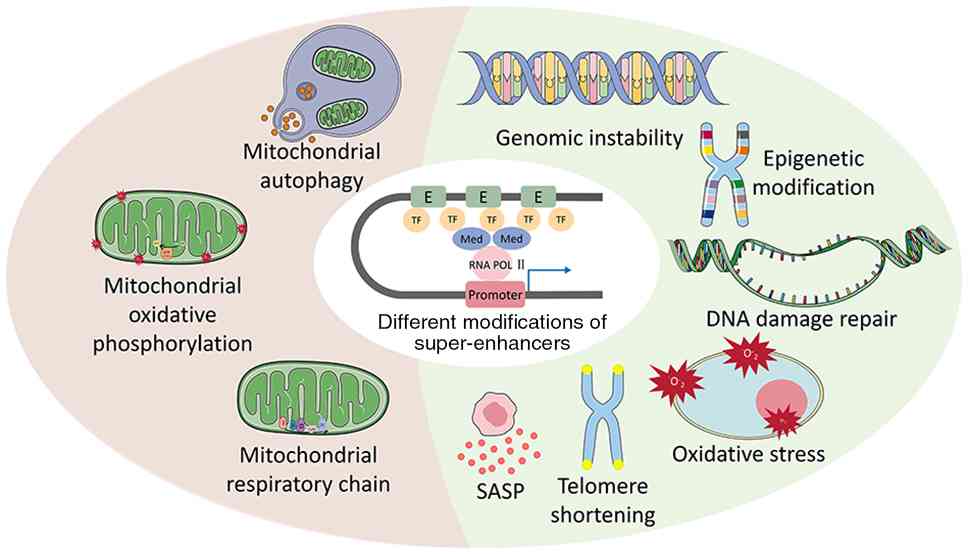
Notably, super-enhancers serve a key role in this process by
interacting with DNA damage repair pathways. Therefore, it is
necessary to elucidate the underlying mechanisms and to identify
potential targets for the protection against DNA damage. As shown
in Fig. 1, the present study
explored the molecular mechanisms by which super-enhancers regulate
cellular senescence through genomic instability, epigenetic
modifications, DNA damage repair, oxidative stress, telomere
shortening and the senescence-associated secretory phenotype (SASP)
in the DNA damage pathway.
Notably, the genome is unstable and any abnormal
changes, such as base substitutions, deletions or insertions and
copy number variations, lead to DNA damage, disrupt cell
proliferation and reproduction, and hinder cell function. Studies
have suggested that super-enhancers can cause DNA damage by
regulating the translocation of chromosomes and the mutation of
genes within the genome. For example, Meng et al (62) reported that super-enhancers can act
on cytidine deaminase-related genes, such as IL4R, IL21R and
NSMCE1, causing them to undergo translocation and leading to DNA
damage. Glodzik et al (63)
revealed that mutations in super-enhancers promote the expression
of genes, such as ESR1 and ZNF217, which in turn induce DNA damage
(63). In addition,
super-enhancers induce activation-induced cytidine deaminase (AID)
through recruitment activation, leading to partial mutation of
antibody genes and chromosomal translocations (64). Abnormal enrichment of AID in the
super-enhancer region may exacerbate DNA damage through antisense
transcription (65).
Epigenetic modifications include DNA methylation,
histone modifications and chromatin remodeling, which are mostly
unstable during the life cycle of somatic cells and are associated
with DNA damage and aging (66–68).
Previous studies have shown that super-enhancers regulate gene
expression through changes in histone modifications, resulting in
DNA damage. For example, the Aire factor in H3K27ac-related
super-enhancers interacts with topoisomerase 1 to promote DNA
damage (69). H3K27ac-related
super-enhancers also alter epigenetic modifications by increasing
the expression of the PDZK1IP1 gene (38). In addition, Zhang et al
(70) demonstrated that deletion
of H3K27ac-related super-enhancers can lead to mutations in BRCA1,
resulting in DNA damage. In addition to histone modifications that
can cause DNA damage, chromatin remodeling can also cause DNA
damage. The SWI/SNF complex is a chromatin remodeling factor
associated with super-enhancer activity, and aging-related
mutations in SWI/SNF subunits, such as SMARCE1, disrupt
super-enhancer-driven gene expression, and exacerbate DNA damage
accumulation and cell senescence (71). By integrating histone modification
and chromatin remodeling, super-enhancers display a dual function
in DNA damage response: On the one hand, they drive the expression
of repair genes to maintain genome stability, and on the other
hand, they may cause genomic instability due to overactive
transcription or abnormal recruitment of injury-related enzymes,
thereby increasing cell senescence.
The interaction with DNA damage repair is also an
important aspect of the regulation of DNA damage by
super-enhancers. Super-enhancers have been confirmed to modulate
genomic stability in senescent cells by the transcriptional
regulation of DNA repair genes or recruitment of repair complexes.
However, aberrant over-activation of these repair sites may
accelerate replication stress, paradoxically accelerating aging
(72). Studies have shown that
super-enhancers drive the expression of the DNA repair gene HNRNPF
by recruiting transcriptional coactivators such as BRD4, MED1 and
P300. HNRNPF further regulates the expression of epigenetic
modification enzymes such as PRMT1, thereby synergistically
regulating the process of ribosome biogenesis and DNA repair
(73). In addition,
super-enhancers maintain the open state of chromatin by enriching
active histone markers such as H3K27ac and H3K4me1, and promote the
rapid recruitment of repair proteins such as 53BP1 and BRCA1 to the
damage site. Notably, RCAN1.4 gene expression is dependent on the
interaction of enhancers and promoters within topologically
associating domains, and disruption of this structure can markedly
impair the efficiency of DNA damage repair (74,75).
Notably, super-enhancers can also promote recruitment of DNA damage
repair factors by binding enhancer RNA (eRNA) produced by
transcription to chromatin remodeling complexes. For example, the
AT-hook domain of BRG1 can bind eRNA and mediate the enrichment of
coactivators such as MLL3/4 and Med1 in the enhancer region,
thereby regulating DNA damage response (76). However, studies have thus far
mainly focused on the regulatory effects of super-enhancers on
repair genes, and it is unclear whether repair pathway-related
proteins regulate the activity of super-enhancers through
epigenetic modification or chromatin remodeling feedback.
In addition to the aforementioned genomic
instability and epigenetic modifications, DNA damage is also
affected by a range of other molecular factors such as oxidative
stress, telomere shortening and the SASP. The essence of oxidative
stress is that free radicals have negative effects on the body and
can directly or indirectly oxidize or damage DNA, proteins and
lipids; induce gene mutations, protein degeneration and lipid
peroxidation; and cause physiological or pathological reactions in
cells and tissues (77). Studies
have shown that genes such as MUTYH, γH2AX and RNaseH2 are involved
in oxidative stress-mediated DNA damage responses, and also promote
physiological and pathological changes such as colorectal
carcinogenesis, disease progression in chronic myeloid leukemia and
the pathogenesis of systemic autoimmune diseases (78–80).
In the absence of telomerase, DNA polymerase cannot fully replicate
linear DNA, resulting in the gradual shortening of telomeres during
karyolysis (81). When telomeres
reach a critical length, DNA damage response proteins mistakenly
recognize the telomeres and activate DNA damage signals (1,56).
Studies have shown that genes such as 53BP1, ATM, γH2AX and TRF2
can cause non-homologous end joining-mediated end-to-end fusion of
short telomeres (82–86), which leads to DNA damage. In
addition, senescent cells are special intervention targets for a
variety of aging-related diseases as they secrete a complex set of
pro-inflammatory cytokines, known as the SASP, which on one hand
maintains and diffuses senescence, and on the other serves a role
in promoting proliferation (87).
Super-enhancers drive formation of the SASP by activating
transcription programs of pro-inflammatory factors and
matrix-remodeling proteins. For example, in DNA damage-induced
aging models, super-enhancers recruit transcriptional regulators,
such as Zscan4 and TAK1, forming a positive feedback loop that
notably enhances the broad-spectrum expression of SASP and promotes
the development of aging (88).
Notably, to the best of our knowledge, little research has been
conducted on whether super-enhancers can mediate DNA damage by
regulating the genes involved in oxidative stress and telomere
shortening, and the specific mechanisms involved have not yet been
elucidated and require further exploration.
Mitochondria are dynamic organelles that produce and
transport ATP for energy, and participate in cell differentiation,
information transmission, fate determination and other processes
(89,90). Mitochondria can malfunction when
stimulated by age-related factors, including mitochondrial
morphological changes, decreased mitochondrial content, impaired
mitochondrial membrane potential (59), reduced electron transport chain
complex activity (91), increased
mitochondrial outer membrane permeability (92), mitochondrial respiratory chain
abnormalities and increased levels of reactive oxygen species (ROS)
(93). Mitochondria therefore
serve an important role in aging and age-related diseases (59). Recently, several studies have
confirmed that super-enhancers are involved in the regulation of
mitochondrial function (94–96).
Therefore, the present review not only aimed to elucidate the
mechanisms by which super-enhancers regulate cellular senescence
through the DNA damage pathway, but also to systematically
summarize the key role of super-enhancers in regulating
mitochondrial functions from the perspectives of mitochondrial
respiratory chain function, oxidative phosphorylation (OXPHOS) and
mitochondrial autophagy (Fig.
1).
The mitochondrial respiratory chain, also known as
the mitochondrial electron transport chain, contains a variety of
enzyme complexes involved in important biological processes, such
as inflammation, thermogenesis, hypoxia tolerance and glycolipid
metabolism (97). When there is an
insufficient oxygen supply to the cell, a chronic lack of nutrients
or mutations in mitochondrial DNA, the mitochondrial respiratory
chain is damaged, which leads to mitochondrial dysfunction, causes
adverse reactions such as cell aging and eventually leads to aging
and age-related diseases of the body. Super-enhancers can affect
the mitochondrial respiratory chain by altering the degree of
mitochondrial oxidative stress and lipid metabolism. For example,
BET) inhibitors can block the action of BRD4-related
super-enhancers, and thus reduce the expression of peroxisome
proliferator-activated receptor γ coactivator 1α (PGC-1α). The
reduced level of PGC-1α hinders mitochondrial respiration and its
antioxidant stress ability, and it eventually damages the
mitochondrial respiratory chain, resulting in the decline or loss
of mitochondrial respiratory function (98). Simultaneously, the inhibition of
BRD4-related super-enhancers reduces the level of mitochondrial
lipid metabolism and destroys complex proteins in the mitochondrial
respiratory chain, resulting in excessive accumulation of lipid
peroxides and ultimately leading to ferroptosis (99). Through epigenetic regulatory
mechanisms, super-enhancers upregulate NADPH oxidase genes, inhibit
the expression of antioxidant oxidase genes and increase the levels
of mitochondrial ROS, resulting in mitochondrial electron transport
chain dysfunction (100).
Cytoplasmic and organelle ion signaling also affects mitochondrial
respiratory chain function by influencing the formation of
respiratory chain complexes (101,102). In addition, it has been shown
that communication between mitochondria and other organelles, or
between themselves, can impact the uptake and release of ion
signals and further affect the mitochondrial respiratory chain
(103). However, the effects of
super-enhancers on the signal transduction of the aforementioned
ion signal transduction and inter-organelle communication in the
mitochondria have not been clearly explained.
The process by which mitochondria supply energy to
cells depends on OXPHOS. ROS are almost entirely produced during
this process and regulate and maintain the normal physiological
functions of the body;. however, when the mitochondria are damaged,
ROS levels markedly increase, preventing them from functioning
normally, thus promoting cell senescence (104,105). It has been demonstrated that
super-enhancers are involved in mitochondrial OXPHOS and ROS
production, thereby affecting mitochondrial function. For example,
histone deactelyase inhibitors can block the H3K27ac super-enhancer
action of the c-Myc gene, resulting in abnormal mitochondrial
OXPHOS, mitochondrial dysfunction and reduced cell viability
(106). By contrast, MLX-related
super-enhancers can regulate the SLC7A11 gene to promote the uptake
of amino acids by cells, thus causing the production of
antioxidants, and maintaining the redox-reduction balance in cells.
However, in the case of MLX knockout, the super-enhancer loses its
regulatory function in maintaining redox balance, leading to the
elevation of intracellular ROS and metabolic disorders, thereby
affecting the OXPHOS of mitochondria (107). Notably, substances such as
glucose, fatty acids and amino acids produced by glycolysis have
key roles in mitochondrial OXPHOS (108). Super-enhancers can change the
energy metabolism balance of mitochondria by driving the high
expression of genes related to glucose metabolism, lipid metabolism
and amino acid metabolism. For example, super-enhancers drive
metabolic reprogramming of cells by activating transcription of key
glycolytic enzyme genes (such as HK2 and LDHA), which manifests as
increased aerobic glycolysis and decreased mitochondrial OXPHOS
(96). Super-enhancers increase
the uptake of amino acids such as glutamine by upregulating the
expression of amino acid transporter alanine-serine-cysteine
transporter 2 and glucose transporter 1, thereby promoting the
mitochondrial tricarboxylic acid cycle to produce ATP. However,
this metabolic reprogramming may exacerbate mitochondrial metabolic
stress (96).
A decline in mitochondrial autophagy is also an
important cause of cellular senescence, in addition to
mitochondrial respiratory chain damage and abnormal OXPHOS.
Mitochondrial autophagy regulates the number of mitochondria
required to meet the energy needs of cells; functionally defective
mitochondria are selectively degraded as much as possible which
prevents the accumulation of functionally defective mitochondria in
the body, which has an important role in maintaining the normal
function and shape of cells (109). Decreased or defective
mitochondrial autophagy can lead to mitochondrial dysfunction and,
if left unchecked, cellular senescence (110). Studies have shown that autophagy
in organelles is associated with the regulation of super-enhancers.
For example, curcumin promotes the expression of H3K4me1- and
H3K27ac-related super-enhancers to promote lysosomal autophagy,
thereby reducing the production of ROS, inhibiting the expression
of BRD4 and effectively improving inflammation in the body
(111,112). In addition, under hypoxia, the
circular RNA calmodulin 4 and PURB proteins promote Beclin1
expression through the regulation of H3K27ac-related
super-enhancers, thereby regulating endoplasmic reticulum autophagy
(113). These findings suggest
that super-enhancers are closely related to autophagy in lysosomes,
the endoplasmic reticulum and other organelles. However, it is
still unclear whether super-enhancers can also regulate
mitochondrial autophagy, and this process requires further
study.
Aging is a key driving factor for the occurrence and
development of various age-related diseases such as
atherosclerosis, Alzheimer's disease, osteoporosis, diabetes,
coronary heart disease, hypertension and cerebral infarction. After
elaborating on the dynamic changes and regulatory roles of
super-enhancers in aging and age-related phenotypes, the present
review will subsequently focus on the specific functions and
molecular mechanisms of super-enhancers involved in
atherosclerosis, Alzheimer's disease, osteoporosis and T2D.
Atherosclerosis is a chronic cardiovascular disease
associated with aging and a common cause of death in older adults.
According to the 2023 World Heart Report released by the World
Heart Federation, 20.5 million individuals died from cardiovascular
diseases worldwide in 2021, accounting for one-third of total
global deaths, whereas in 1990 and 2019, the number of deaths from
cardiovascular diseases was 12.1 million and 18.6 million,
respectively, indicating that the number of deaths from
cardiovascular diseases worldwide is increasing (114,115). Atherosclerosis refers to the
formation of plaques by lipid deposition on the arterial wall of
medium and large arteries, resulting in reduced blood flow or
blocked blood outflow (116),
which is characterized by the migration and infiltration of
macrophages, proliferation of smooth muscle cells, endothelial
damage and accumulation of intracellular and extracellular lipids.
Although various factors contribute to the development of
atherosclerosis, such as chronic inflammation (117), lipid metabolism and accumulation
(118), epigenetics (119), immune disorders (120), hypertension, diabetes, smoking,
mental health and environmental pollution, aging has been confirmed
to be a markedly independent factor when all other factors are
controlled. Thus, the present review aimed to explore the effects
of super-enhancers on atherosclerosis in terms of the effects of
aging and related factors on endothelial cells, smooth muscle cells
and macrophages.
Endothelial cells, which cover the inner surface of
blood vessels and are composed of a layer of flat cells, complete
the metabolic exchange of plasma and interstitial fluid, regulate
blood flow in blood vessels, and synthesize and secrete various
bioactive substances to control blood pressure, vasoconstriction
and relaxation. Endothelial injury is an initial stage of
atherosclerosis. After endothelial injury, the integrity and
permeability of the vascular intima change (121), and the apoptosis and shedding of
endothelial cells promote the adhesion and aggregation of platelets
(122). In addition,
dysfunctional endothelial cells secrete various growth factors,
such as VEGF-A and PDGF (123,124), as well as vasoactive substances,
including endothelin-1 and angiotensin II (125,126), in order to stimulate smooth
muscle cell proliferation and vascular wall contraction,
exacerbating the progression of atherosclerosis. Previous studies
have reported that super-enhancers are involved in endothelial cell
proliferation, vascular development and formation, regulation of
vascular homeostasis and other functions. Mushimiyimana et
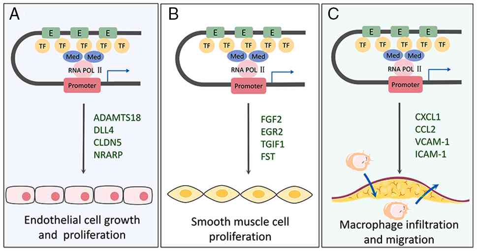
al (127) revealed that the
endothelial cell-specific super-enhancer SE12313 modulates the
ADAMTS18 gene to affect endothelial cell proliferation, leading to
reduced or shortened endothelial sprouts and decreased cell
proliferation. Kalna et al (40) demonstrated that H3K27ac
super-enhancers can regulate the expression of ERG and endothelial
genes, such as DLL4, CLDN5 and NRARP, to affect the development and
generation of blood vessels (Fig.
2A). Endothelial cells not only influence the development of
atherosclerosis through growth and proliferation, but also produce
nitric oxide (NO), which serves an important role in regulating
vascular tone and inhibiting platelet aggregation and leukocyte
adhesion. Lubos et al (128) demonstrated that oxidative stress
caused by endothelium-derived NO and ROS has a key role in vascular
endothelial dysfunction and atherosclerotic thrombosis. Whether
super-enhancers can affect the process of atherosclerosis by
regulating the ability of endothelial cells to produce NO remains
unclear, and further research on the relationship between
super-enhancers and endothelial cells to produce NO is required in
the future.
Vascular smooth muscle cells are the main components
of vascular wall tissue and are essential for the maintenance of
vascular tension; changes in their structure and function affect
the basic function of blood vessels. During atherogenesis, vascular
smooth muscle cells proliferate and migrate abnormally, secreting a
variety of biologically active molecules, including extracellular
matrix components such as collagens, elastin and proteoglycans, as
well as cytokines such as MCP-1 and IL-1β, to form a fibrous cap
that promotes vessel calcification, subsequently leading to plaque
rupture, triggering apoptosis and exacerbating the disease process
(129,130). Currently, there are few studies
on the controlling role of super-enhancers in vascular smooth
muscle cells. Some studies have suggested that super-enhancers can
influence the proliferation function of vascular smooth muscle
cells. For example, Das et al (39) reported that when H3K27ac- and
BRD4-specific super-enhancers were missing, the expression of
angiotensin II-induced FGF2, ERG2, TGIF1 and FST was notably
reduced, which affected the proliferative and the protective
functions of vascular smooth muscle cells (Fig. 2B). However, it is notable that
although super-enhancers have been shown to serve a key role in
vascular smooth muscle cell proliferation for the development of
atherosclerosis, their other functions in the development of this
disease have not been clearly elucidated. This provides a new
research direction: To explore in depth how super-enhancers
contribute to the development of atherosclerosis by influencing
other behaviors and functions of vascular smooth muscle cells.
Future studies should focus on how super-enhancers promote or
inhibit the migration of vascular smooth muscle cells in the vessel
wall, the formation of tough and structurally complex fibrous caps,
and the promotion of vascular calcification.
Macrophages are immune cells with a strong
phagocytic function and antigen-presenting ability that can secrete
a large number of cytokines and chemokines. In the early stages of
atherosclerosis, monocytes are recruited to the subendothelium to
differentiate into macrophages, which further migrate and
infiltrate as the disease progresses, and polarize into different
subtypes according to the altered microenvironment (131). Studies have shown that
super-enhancers recruit monocytes for targeted migration by
directly regulating the expression of CXCL1, CCL2 and other
chemokines (132,133). In addition, super-enhancers
markedly enhance the interaction between macrophages and activated
endothelial cells by upregulating adhesion molecules, such asVCAM-1
and ICAM-1, and promote the retention and differentiation of
inflammatory cells in plaques (134) (Fig.
2C). In plaques, macrophages absorb lipid deposition particles
and transform them into foam cells, thereby inducing an
inflammatory response (135).
Super-enhancers drive the transformation of macrophages into foam
cells by regulating gene networks of glycolysis and lipid
metabolism. Specifically, Wang et al (134) showed that super-enhancers
activate the SREBP1/2 transcription factor, drive the expression of
lipid synthesis genes, and promote the accumulation of
intracellular lipid droplets and the formation of foam cells. In
addition to regulating the migration and transformation of
macrophages, super-enhancers can affect the pathological process of
atherosclerosis by mediating the polarization of macrophages.
Specifically, super-enhancers have been shown to notably increase
the inflammatory response of plaques by driving the expression of
pro-inflammatory genes and regulating the polarization of
macrophages towards M1 type (111,136,137). Macrophages are also found in a
number of other organs and tissues; for example, adipose
tissue-associated macrophages, microglia, liver Kupffer cells,
alveolar macrophages and renal macrophages (138–140). Recent evidence indicates that
adipose tissue-derived exosomes can promote atherosclerosis
progression by delivering bioactive molecules to vascular cells
(141), and that super-enhancers
regulate macrophage polarization and inflammation in
atherosclerosis (136). However,
the specific molecular mechanism of how super-enhancers regulate
adipose tissue macrophages to affect atherosclerosis progression
through exosome-mediated remote signaling still needs to be further
explored.
Alzheimer's disease is a progressive
neurodegenerative disease and is one of the most common causes of
dementia in elderly individuals worldwide. Characteristic
pathological changes include cerebral cortex atrophy, continuous
proliferation of microglia, reduction in the number of memory
neurons, formation of age plaques, β-amyloid deposition and
neurofibrillary tangles (142).
According to the United Nations Department of Economic and Social
Affairs, the global proportion of individuals aged ≥65 years
increased from ~7% in 2000 and is projected to reach 12% by 2030.
Furthermore, according to the Centers for Disease Control and
Prevention, the incidence of Alzheimer's disease approximately
doubles every 5 years after 65 years of age. A total of 50% of
individuals aged ≥ 85 years in the United States have Alzheimer's
disease (143–145). Therefore, aging is considered the
greatest risk factor for Alzheimer's disease (146–148). The present review discusses the
influence of super-enhancers on Alzheimer's disease resulting from
aging and its related factors on the continuous proliferation of
microglia, and the formation of neurofibrillary tangles and amyloid
plaques.
Microglia, which reside in the central nervous
system, are mononuclear macrophages that are widely distributed
throughout the brain and spinal cord, and are important for
maintaining nervous system homeostasis. Microglia, the main
regulators of neuroinflammation, are more sensitive to inflammation
during aging, and their continuous proliferation is considered a
hallmark of Alzheimer's disease (149,150). During aging, persistent
inflammation and overexposure to oxidative environments results in
microglia being continuously activated, leading to
neurodegeneration, neuroinflammation and neurotoxicity (151,152). Previous studies have reported
that microglial super-enhancers serve an important role in the risk
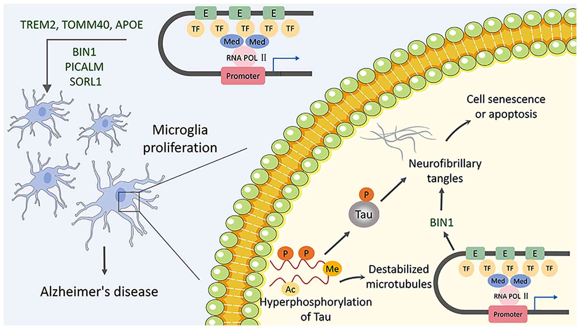
of developing Alzheimer's disease. For example, Nott et al
(153) revealed that H3K27ac
super-enhancers can regulate genes, such as BIN1, PICALM and SORL1,
to promote microglial proliferation, thereby increasing the risk of
Alzheimer's disease. Jansen et al (154) demonstrated that H3K27ac
super-enhancers can regulate TREM2, TOMM40, APOE and other genes to
activate microglia, leading to neurodegeneration (Fig. 3). In Alzheimer's disease, microglia
undergo sustained proliferation, abnormally phagocytose synapses
and mediate synaptic loss. Lipid signaling in neuron-glia
interactions is a key factor in this process, which involves
relevant receptors or lipids such as TREM2, GM1 ganglioside and
ApoE (155). Therefore, whether
super-enhancers can modulate lipid signaling between neurons and
glial cells, and thus the process of phagocytosis of synapses by
the microglia, needs to be investigated in depth in the future.
Two pathological markers of Alzheimer's disease are
the formation of neurofibrillary tangles and amyloid plaques
(148,156). Neurofibrillary tangle formation
is caused by the accumulation of tau proteins via
hyperphosphorylation in cells. Tau protein is a soluble
microcore-related protein that mainly stabilizes microtubules by
phosphorylation. However, it loses its biological activity and
becomes an insoluble protein under hyperphosphorylation,
subsequently aggregating to form neurofibrillary tangles (157). Tau protein phosphorylation can
promote the aging of brain cells, causing memory decline and
neuropathological changes in Alzheimer's disease (158). Studies on neurofibrillary tangles
have mainly focused on the relationship between the tau protein and
BIN1. Chapuis et al (159)
demonstrated that a genetic change caused by a 3-bp insertion in
the 5′ regulatory region of the BIN1 gene (~28 kb upstream of BIN1)
can cause upregulation of BIN1 in vivo, which can increase
the risk of Alzheimer's disease by interfering with tau biology.
Furthermore, Nott et al (153) found that super-enhancers in
microglia can regulate the level of the genetically determined BIN1
gene, increase the expression of the BIN1 gene and thus increase
the risk of Alzheimer's disease, as well as the neuroinflammatory
response and the activity of specific cell signaling pathways that
promote the formation of neurofibrillary tangles (Fig. 3). Super-enhancers may reduce the
neuroinflammatory response by modulating the activity of glial
cells or inhibiting the release of inflammatory factors, thereby
decreasing the extent of neurofibrillary tangles and reducing the
risk of Alzheimer's disease (160–162). Specific cell signaling pathways
also serve key roles in the formation of neurofibrillary tangles.
Super-enhancers may reduce the formation of neurofibrillary tangles
by modulating the activity of signaling pathways, such as the Wnt
or nerve growth factor signaling pathways, in order to affect the
function and structure of neurons (15,163,164). Research into the mechanisms by
which these super-enhancers regulate the formation of
neurofibrillary tangles is required.
The accumulation of amyloid plaques is also a
hallmark of Alzheimer's disease. In a degenerative disease state,
β-secretase and γ-secretase break down amyloid precursor protein
(APP), causing β-amyloid protein (Aβ) to produce Aβ40 and Aβ42
insoluble oligomers, which deposit amyloid plaques outside the cell
and ultimately lead to neuronal death (165). Similar to tau phosphorylation,
increased β-amyloid deposition promotes the aging of brain cells
via mechanisms such as oxidative stress, DNA damage and activation
of the p16/p21 senescence pathways, which in turn serves a key role
in Alzheimer's disease (166,167). Thus far, research on amyloid
plaques has mainly focused on APP gene mutations that lead to the
development of Alzheimer's disease. Tanzi et al (168) reported that mutations in the APP,
PSEN1 and PSEN2 genes alter the sequence of the APP exon encoding
β-amyloid, demonstrating that mutations in these genes can affect
chromosomal genetics. Although studies have determined that
mutations in APP affect amyloid plaque formation, there is no
evidence of a relationship between super-enhancers and APP or
amyloid plaques (169–171). Furthermore, the aggregation and
clearance of β-amyloid and transport of proteins within neurons
also affect amyloid plaque formation (172,173). Notably, super-enhancers may
affect the aggregation and clearance process of β-amyloid; they may
reduce β-amyloid deposition by promoting β-amyloid degradation, for
example by activating autophagy and proteasome pathways within
cells, thereby reducing the formation of amyloid plaques (174,175). Abnormal intra-neuronal transport
may also lead to abnormal aggregation of β-amyloid within neurons,
thereby promoting amyloid plaque formation. Super-enhancers may
regulate protein transport within neurons by affecting
intracellular molecular mechanisms, thereby reducing the
aggregation of β-amyloid within neurons and lowering the formation
of amyloid plaques (15,176,177). An in-depth study of the
aforementioned mechanisms of super-enhancers in regulating the
formation of amyloid plaques will further promote the understanding
of Alzheimer's disease and provide novel options for its
treatment.
Osteoporosis is a bone disease closely related to
aging, characterized by bone mass loss, bone structure damage and
increased bone brittleness, ultimately leading to a notable
increase in the risk of fractures (178). As global population aging
increases, osteoporosis has become one of the important causes of
disability and death among the elderly. Under normal physiological
conditions, the bones maintain skeletal homeostasis through the
dynamic balance between bone formation by osteoblasts and bone
resorption by osteoclasts (179).
However, during the aging process, changes in hormone levels, such
as estrogen and androgens, oxidative stress responses, chronic
inflammation, dysregulation of epigenetic regulation and decreased
differentiation ability of mesenchymal stem cells, all disrupt this
bone homeostasis (180–183). In recent years, more evidence has
shown that super-enhancers serve a key role in determining the fate
of bone formation by osteoblasts and bone resorption by osteoclasts
(22,26,184). Therefore, clarifying the
regulatory network of super-enhancers in osteoporosis is expected
to provide new targets for the treatment of osteoporosis, an
aging-related disease.
In the process of bone formation by osteoblasts,
super-enhancers directly affect the function of osteoblasts by
regulating key transcription factors and signaling pathways
involved in osteoblast differentiation. Yu et al (26) revealed that, during osteoblast
differentiation, osteoblast-related genes such as ZBTB16, RUNX2 and
SP7 are enriched around a large number of H3K27ac super-enhancers.
Among them, the enhanced activity of super-enhancers targeting
ZBTB16 was shown to markedly upregulate the expression of the
ZBTB16 gene, promote osteoblast differentiation and increase the
formation of mineralized nodules (26). Additionally, BRD4, as a key
cofactor of super-enhancers, collaboratively drives the
transcription of genes such as Runx2 and Osterix (also known as
SP7) with H3K27ac super-enhancers during osteoblast bone formation,
and the use of BRD4 inhibitors can notably inhibit osteoblast
differentiation (185,186). However, there is a lack of
dynamic mapping analysis at the single-cell level in aging bone
tissue regarding how super-enhancers integrate upstream signals
from estrogen deficiency or oxidative stress.
In terms of osteoclastic bone resorption,
super-enhancers also affect the occurrence and development of
osteoporosis by regulating osteoclast differentiation and function.
Bae et al (22) discovered
that, during the osteoclast differentiation induced by RANKL,
multiple genes related to osteoclasts, such as NFATC1, C-FOS, ACP5
and CTSK, form new super-enhancers around them. The establishment
of these super-enhancers depends on BRD4 and p300. Notably, RANKL
shows a marked dose-dependent effect on super-enhancers in
osteoclasts: Low concentrations of RANKL only activate ordinary
enhancers, whereas high concentrations of RANKL or the formation of
a chronic inflammatory microenvironment can induce the formation of
super-enhancers, leading to excessive activation of osteoclasts
(22). However, the distribution
of osteoclast super-enhancers in aged bone tissue within the body
and their interaction with osteoblast super-enhancers still require
further investigation. Osteoclast-secreted exosomes are emerging as
critical mediators of intercellular communication in the bone
microenvironment, capable of delivering bioactive cargoes such as
proteins and miRNAs to regulate the function of recipient cells
(187). Additionally, whether
super-enhancers can remotely inhibit osteoblast function by
regulating exosomes secreted by osteoclasts remains unexplored.
Although super-enhancers have shown notable
regulatory potential in osteoporosis, current research still faces
a number of unanswered questions and knowledge gaps. On one hand,
super-enhancers exhibit opposite regulatory characteristics in
different environments. For example, ZBTB16-related super-enhancers
can promote osteoblastic bone formation, while super-enhancers
related to the NF-κB signaling pathway promote osteoclastic bone
resorption and inhibit osteoblastic bone formation (185). On the other hand, the
heterogeneity of super-enhancers in different types of bone cells,
such as osteoblasts, osteocytes and osteoprogenitor cells, has not
yet been systematically mapped. In osteoporosis, bone marrow
mesenchymal stem cells exhibit an imbalanced differentiation
pattern, with increased adipogenic differentiation at the expense
of osteogenic differentiation, leading to bone marrow adiposity
accumulation and reduced bone formation (15,188). Future research on super-enhancers
in osteoporosis should focus on developing in vivo
super-enhancer tracing techniques, clarifying the heterogeneity of
super-enhancers in different bone cell types and assessing the role
of metabolic reprogramming, such as glycolytic reprogramming,
regulated by super-enhancers in the determination of osteogenic and
adipogenic differentiation.
T2D is a chronic metabolic disease characterized by
insulin resistance and impairment of islet β-cell function, which
accounts for 90–95% of all diabetes cases. Insulin resistance is a
key feature of T2D; over time, the pancreas may produce less
insulin in response to the body's needs, leading to hyperglycemia.
The pathogenesis of T2D is complex, involving the interaction
between multiple organs and systems. The main mechanisms include
epigenetic regulation, insulin resistance and functional defects of
islet β cells (189).
As a key element of epigenetic regulation,
super-enhancers notably affect the progression of T2D by regulating
the function of islet β cells, and insulin synthesis and secretion.
Suzuki et al (190) showed
that a T2D risk variant (rs231361) located in the KCNQ1 locus
disrupts the interaction between the super-enhancer and the INS
gene, ultimately triggering insulin secretion dysfunction. Notably,
their single-cell chromatin accessibility analysis further revealed
that the super-enhancers of T2D risk genes are enriched in islet β
cells and can affect their transcriptional regulatory networks
(189,190). Recent studies have suggested that
the understanding of the pathogenesis of T2D needs to break through
the traditional single-organ perspective and turn to multi-organ
interaction network analysis at the level of systems biology.
Super-enhancers may serve a role in the interaction between
intestinal, fat, liver, muscle and islet β cells through dynamic
regulation of metabolic organ core functions and cross-tissue
signal transduction, thus driving the pathological process of T2D
(191–194).
In addition, some clinical studies have revealed
that non-coding single nucleotide polymorphisms (SNPs) serve an
important role in T2D. Sun et al (51) demonstrated through 3D genomics
analysis that T2D SNPs re mostly enriched in space-adjacent
super-enhancers, and participate in chromatin interaction to
regulate and affect transcription factor binding. Such
SNP-super-enhancer interaction modules are markedly enriched in the
Wnt signaling pathway, which may provide novel insights into the
pathogenesis of T2D (51). Future
studies may combine single-cell epigenomics and gene-editing
techniques to further elucidate the dynamic regulatory mechanisms
of super-enhancers in multi-organ interactions.
As a class of cis-regulatory elements with strong
transcriptional activation properties, super-enhancers have
stronger regulatory functions than enhancers and serve key roles in
cell type-specific development, differentiation and the development
of various diseases. Since their discovery, super-enhancers have
received increasing attention from researchers and they are
considered a new target and a new strategy for the treatment of
disease. In addition, in a variety of diseases, such as
atherosclerosis (29), Alzheimer's
disease (149) and autoimmune
diseases (30), super-enhancers
have been shown to regulate the expression of genes and control
cell differentiation, aging, death and other processes. Genetic and
SNP analyses also support the contribution of super-enhancers to
human aging and associated diseases. These findings have prompted
researchers to investigate the role of super-enhancers in
age-related diseases.
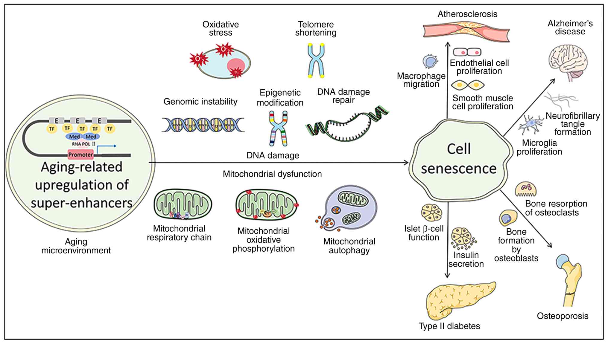
The present review systematically elaborates that
in an aging microenvironment, the expression intensity of
super-enhancers undergoes notable changes, and they drive the
occurrence of cellular senescence through multiple pathways,
thereby promoting the development of various aging-related
diseases. As shown in Fig. 4, in
the aging microenvironment, the expression of aging-related
super-enhancers is upregulated, and induces the occurrence and
development of cellular senescence through DNA damage pathways and
mitochondrial dysfunction pathways. Among them, the DNA damage
pathway includes genomic instability, abnormal epigenetic
modifications, DNA damage repair, oxidative stress and telomere
shortening; and the mitochondrial dysfunction pathway involves
changes in mitochondrial respiratory chain function, OXPHOS and
mitochondrial autophagy. The cellular senescence caused by the
abnormal expression of super-enhancers can further promote the
occurrence and progression of various aging-related diseases, such
as atherosclerosis, Alzheimer's disease, osteoporosis and T2D.
Specifically, cellular senescence affects the proliferation of
endothelial cells and smooth muscle cells, as well as the migration
and infiltration of macrophages, promoting atherosclerosis; it
affects the proliferation of microglia and the formation of
neurofibrillary tangles, promoting Alzheimer's disease; it
regulates the bone formation process of osteoblasts and the bone
resorption process of osteoclasts, promoting osteoporosis; and it
damages the function of pancreatic β cells and insulin secretion,
promoting the occurrence of T2D (Fig.
4). These findings reveal the core role of aging-related
super-enhancers in connecting aging molecular mechanisms and the
development of various pathological processes, providing evidence
for the mechanistic research and exploration of therapeutic
strategies for aging-related diseases.
Cellular senescence is affected by various
endogenous and exogenous factors, partly depending on the action of
super-enhancers, particularly DNA damage, telomere shortening and
mitochondrial dysfunction. With the development of ChIP-seq and its
combination with other omics-related studies, the potential role
and mechanism of super-enhancers in cell aging and aging-related
diseases has gradually been revealed (195–197). Current research on cell
senescence has mainly focused on the effects of super-enhancers on
DNA damage, organelle stress and oncogene activation (198–200). Therefore, the role of
super-enhancers in other factors that influence cell aging, such as
macrophage autophagy, protein homeostasis, nuclear integrity and
nutritional dysregulation, should be further explored in subsequent
studies. Concurrently, current research on age-related diseases has
focused on the influence of super-enhancers on causative factors or
genetics. For example, studies on atherosclerosis have mainly
focused on the regulation of super-enhancers in the accumulation of
endothelial cells, proliferation of smooth muscle cells, and the
migration and infiltration of macrophages. Research on Alzheimer's
disease has focused on the super-enhancer regulation of microglial
proliferation, neurofibrillary tangle formation and amyloid plaque
formation. Therefore, the role of super-enhancers in the
pathogenesis of age-related diseases, such as dysplasia of
phagocyte exocytosis, low-density lipoprotein accumulation,
cholesterol metabolism imbalance in atherosclerosis, synaptic
dysfunction, neurotransmitter imbalance and neuroinflammation in
Alzheimer's disease, should be further explored in subsequent
studies.
Although super-enhancers have received increasing
attention in regulating cellular senescence and related diseases,
current research still faces a series of contradictory findings,
methodological limitations and key knowledge gaps. Firstly, the
functions of super-enhancers in aging show obvious duality. On one
hand, super-enhancers can activate protective transcription
programs to delay aging, for example, the transcription factor
FOXP1 activates SESN3 through the super-enhancer mechanism, thereby
delaying endothelial cellular senescence and inhibiting the
progression of atherosclerosis (201); on the other hand, super-enhancers
can drive pathological aging-related secretory phenotypes and
promote the occurrence of related diseases. Additionally, the
functions of regulatory factors also show contradiction. For
example, the histone acetyltransferase p300 can induce the
generation of new super-enhancers to drive cellular senescence
during aging. However, in pathologically altered cellular states,
such as in certain cancer cells where p300 function is disrupted or
bypassed, or when hijacked by oncogenic transcriptional programs,
p300 can conversely promote proliferation, facilitate senescence
evasion and drive disease progression (16,202,203). Secondly, some methodological
limitations also affect in-depth research on super-enhancers. Most
current evidence comes from immortalized in vitro cell
models, while in vivo studies using aging tissues or cells
face significant hurdles. These include the challenge of detecting
senescent cells with conventional markers that often perform poorly
in native tissues, the extremely low abundance of senescent cells
within tissue and their pronounced biological heterogeneity
(14). The in vitro culture
of senescent cells may not fully reflect the dynamic functions of
senescent cells in the complex microenvironment in vivo. In
terms of detection techniques, the enhancer identification methods
such as ChIP-seq and CUT&Tag are highly dependent on the
quality of antibodies and the setting of signal thresholds, and the
identification results from different laboratories and instruments
may have notable differences, making direct comparison difficult
(133). Finally, it is not yet
clear how super-enhancers integrate upstream signals from DNA
damage response and mitochondrial dysfunction. Whether the
super-enhancers identified from mouse cells have common regulatory
patterns in human cells of different aging-related diseases (such
as atherosclerosis and Alzheimer's disease) has also not been
clarified (204). The research on
the mechanisms of super-enhancers in these aspects is largely
lacking and further exploration is needed in the future.
By considering the differential expression of
super-enhancers in aging-related diseases and their roles in
regulating the occurrence and development of diseases, there is an
urgent need to precisely control the levels of super-enhancers.
Currently, the known super-enhancer modulators include the CDK7
inhibitor THZ1 (205),
CREB-binding protein/p300 inhibitor (206), Minnelide (207), lysine-specific histone
demethylase 1 inhibitor, BET inhibitor combination therapy
(208), BRD4 inhibitor and RET
inhibitor combination therapy (209). The super-enhancer regulators and
their clinical trial stages are all listed in Table II. Although the discovery of these
super-enhancer modulators makes it possible for them to serve as
key targets for the treatment of age-related diseases, several
questions remain unanswered before these modulators can be applied.
First, there are a number of downstream target genes in the region
where the super-enhancer modulator acts, and whether the regulator
acts on these downstream target genes should be considered. Second,
the issue of the dosage of super-enhancer modulators is should be
considered to ensure that the regulator has a chance to work in the
body without causing toxicity. Third, although combination therapy
has an additive effect on super-enhancers, the potential
accumulation of side effects should be considered in a clinical
setting.
Furthermore, with the rapid development of science
and technology, intelligent healthcare devices based on the
Internet of Things have improved the early detection rate of
age-related diseases by tracking the health data of the elderly in
real time, bringing innovative changes (210). At the same time, super-enhancers
are becoming a new hotspot in aging research because of their
central role in gene expression regulation, and their potential to
become biomarkers for disease diagnosis and treatment.
Interventions such as small molecule inhibitors and gene editing
techniques targeting the regulatory mechanism of super-enhancers
may provide innovative strategies for delaying the aging process
(211). Although super-enhancers
as possible candidates for biomarkers show great potential in the
intervention of aging-related diseases, there are still a number of
challenges to achieving this goal. In the future, with the
continuous development of gene sequencing technology, gene editing
technology and epigenetic regulation technology, the dynamic
regulatory mechanism of super-enhancers, as well as the development
of cross-disease common targets and innovative intervention
technologies, can be focused on so as to find more effective and
safe treatment methods.
Not applicable.
The present study was supported by the Natural Science
Foundation of the Guangxi Zhuang Autonomous Region (grant no.
2024GXNSFAA010154), the National Natural Sciences Foundation of
China (grant nos. 82570989 and 82370463), the Guangdong Basic and
Applied Basic Research Foundation (grant no. 2025A1515012522) and
the Youth S&T Talent Support Programme of Guangdong Provincial
Association for Science and Technology (grant no.
SKXRC2026341).
Not applicable.
XW reviewed the literature, synthesized the data,
constructed tables and charts, and drafted and wrote the initial
version of the paper. WD established a literature database,
organizing the references, and made critical revisions to important
academic content. ST was responsible for project management,
created visual content, and participated in the writing and editing
of the paper. HT designed the review framework, formulated the
literature search strategy, verified the data, and made critical
revisions to the initial draft. KY provided institutional
resources, software access rights, and administrative support, and
assisted in the review and editing of the paper. XZ was responsible
for the overall research design, revised the paper, sorted out the
feedback and ultimately approved the final version. Data
authentication is not applicable. All authors read and approved the
final manuscript.
Not applicable.
Not applicable.
The authors declare that they have no competing
interests.
|
1
|
Lin J and Epel E: Stress and telomere
shortening: Insights from cellular mechanisms. Ageing Res Rev.
73:1015072022. View Article : Google Scholar : PubMed/NCBI
|
|
2
|
Guo Y, Guan T, Shafiq K, Yu Q, Jiao X, Na
D, Li M, Zhang G and Kong J: Mitochondrial dysfunction in aging.
Ageing Res Rev. 88:1019552023. View Article : Google Scholar : PubMed/NCBI
|
|
3
|
Wang K, Liu H, Hu Q, Wang L, Liu J, Zheng
Z, Zhang W, Ren J, Zhu F and Liu GH: Epigenetic regulation of
aging: Implications for interventions of aging and diseases. Signal
Transduct Target Ther. 7:3742022. View Article : Google Scholar : PubMed/NCBI
|
|
4
|
López-Otín C, Blasco MA, Partridge L,
Serrano M and Kroemer G: Hallmarks of aging: An expanding universe.
Cell. 186:243–278. 2023. View Article : Google Scholar : PubMed/NCBI
|
|
5
|
Son SM, Park SJ, Breusegem SY, Larrieu D
and Rubinsztein DC: p300 nucleocytoplasmic shuttling underlies
mTORC1 hyperactivation in Hutchinson-Gilford progeria syndrome. Nat
Cell Biol. 26:235–249. 2024. View Article : Google Scholar : PubMed/NCBI
|
|
6
|
Islam MT, Tuday E, Allen S, Kim J, Trott
DW, Holland WL, Donato AJ and Lesniewski LA: Senolytic drugs,
dasatinib and quercetin, attenuate adipose tissue inflammation, and
ameliorate metabolic function in old age. Aging Cell.
22:e137672023. View Article : Google Scholar : PubMed/NCBI
|
|
7
|
Aguado J, Sola-Carvajal A, Cancila V,
Revêchon G, Ong PF, Jones-Weinert CW, Wallén Arzt E, Lattanzi G,
Dreesen O, Tripodo C, et al: Inhibition of DNA damage response at
telomeres improves the detrimental phenotypes of Hutchinson-Gilford
progeria syndrome. Nat Commun. 10:49902019. View Article : Google Scholar : PubMed/NCBI
|
|
8
|
Khan J, Pernicova I, Nisar K and Korbonits
M: Mechanisms of ageing: Growth hormone, dietary restriction, and
metformin. Lancet Diabetes Endocrinol. 11:261–281. 2023. View Article : Google Scholar : PubMed/NCBI
|
|
9
|
Chen S, Gan D, Lin S, Zhong Y, Chen M, Zou
X, Shao Z and Xiao G: Metformin in aging and aging-related
diseases: Clinical applications and relevant mechanisms.
Theranostics. 12:2722–2740. 2022. View Article : Google Scholar : PubMed/NCBI
|
|
10
|
Grosveld F, van Staalduinen J and
Stadhouders R: Transcriptional regulation by (Super)enhancers: From
discovery to mechanisms. Annu Rev Genomics Hum Genet. 22:127–146.
2021. View Article : Google Scholar : PubMed/NCBI
|
|
11
|
Shin HY: Targeting Super-enhancers for
disease treatment and diagnosis. Mol Cells. 41:506–514.
2018.PubMed/NCBI
|
|
12
|
Lavaud M, Tesfaye R, Lassous L, Brounais
B, Baud'huin M, Verrecchia F, Lamoureux F, Georges S and Ory B:
Super-enhancers: Drivers of cells' identities and cells' debacles.
Epigenomics. 16:681–700. 2024. View Article : Google Scholar : PubMed/NCBI
|
|
13
|
Wang M, Chen Q, Wang S, Xie H, Liu J,
Huang R, Xiang Y, Jiang Y, Tian D and Bian E: Super-enhancers
complexes zoom in transcription in cancer. J Exp Clin Cancer Res.
42:1832023. View Article : Google Scholar : PubMed/NCBI
|
|
14
|
Sturmlechner I, Sine CC, Jeganathan KB,
Zhang C, Fierro Velasco RO, Baker DJ, Li H and van Deursen JM:
Senescent cells limit p53 activity via multiple mechanisms to
remain viable. Nat Commun. 13:37222022. View Article : Google Scholar : PubMed/NCBI
|
|
15
|
Wang R, Li A and Pang Z: Current progress
and future perspective of super-enhancers: A viable and effective
bridge between the transcriptional apparatus and disease. Front
Genet. 16:16119052025. View Article : Google Scholar : PubMed/NCBI
|
|
16
|
Di Giorgio E, Paluvai H, Dalla E, Ranzino
L, Renzini A, Moresi V, Minisini M, Picco R and Brancolini C: HDAC4
degradation during senescence unleashes an epigenetic program
driven by AP-1/p300 at selected enhancers and super-enhancers.
Genome Biol. 22:1292021. View Article : Google Scholar : PubMed/NCBI
|
|
17
|
Huang W, Hickson LJ, Eirin A, Kirkland JL
and Lerman LO: Cellular senescence: The good, the bad and the
unknown. Nat Rev Nephrol. 18:611–627. 2022. View Article : Google Scholar : PubMed/NCBI
|
|
18
|
Tasdemir N, Banito A, Roe JS,
Alonso-Curbelo D, Camiolo M, Tschaharganeh DF, Huang CH, Aksoy O,
Bolden JE, Chen CC, et al: BRD4 connects enhancer remodeling to
senescence immune surveillance. Cancer Discov. 6:612–629. 2016.
View Article : Google Scholar : PubMed/NCBI
|
|
19
|
Wang Z, Luo M, Liang Q, Zhao K, Hu Y, Wang
W, Feng X, Hu B, Teng J, You T, et al: Landscape of enhancer
disruption and functional screen in melanoma cells. Genome Biol.
24:2482023. View Article : Google Scholar : PubMed/NCBI
|
|
20
|
Ho CY and Dreesen O: Faces of cellular
senescence in skin aging. Mech Ageing Dev. 198:1115252021.
View Article : Google Scholar : PubMed/NCBI
|
|
21
|
Yang Y, Li X, Meng Z, Liu Y, Qian K, Chu M
and Pan Z: A body map of super-enhancers and their function in pig.
Front Vet Sci. 10:12399652023. View Article : Google Scholar : PubMed/NCBI
|
|
22
|
Bae S, Kim K, Kang K, Kim H, Lee M, Oh B,
Kaneko K, Ma S, Choi JH, Kwak H, et al: RANKL-responsive epigenetic
mechanism reprograms macrophages into bone-resorbing osteoclasts.
Cell Mol Immunol. 20:94–109. 2023. View Article : Google Scholar : PubMed/NCBI
|
|
23
|
Jiang Y, Jiang YY and Lin DC:
Super-enhancer-mediated core regulatory circuitry in human cancer.
Comput Struct Biotechnol J. 19:2790–2795. 2021. View Article : Google Scholar : PubMed/NCBI
|
|
24
|
Lei S, Li C, She Y, Zhou S, Shi H and Chen
R: Roles of super enhancers and enhancer RNAs in skeletal muscle
development and disease. Cell Cycle. 22:495–505. 2023. View Article : Google Scholar : PubMed/NCBI
|
|
25
|
Kai Y, Li BE, Zhu M, Li GY, Chen F, Han Y,
Cha HJ, Orkin SH, Cai W, Huang J and Yuan GC: Mapping the evolving
landscape of super-enhancers during cell differentiation. Genome
Biol. 22:2692021. View Article : Google Scholar : PubMed/NCBI
|
|
26
|
Yu W, Xie Z, Li J, Lin J, Su Z, Che Y, Ye
F, Zhang Z, Xu P, Zeng Y, et al: Super enhancers targeting ZBTB16
in osteogenesis protect against osteoporosis. Bone Res. 11:302023.
View Article : Google Scholar : PubMed/NCBI
|
|
27
|
Bacabac M and Xu W: Oncogenic
super-enhancers in cancer: Mechanisms and therapeutic targets.
Cancer Metastasis Rev. 42:471–480. 2023. View Article : Google Scholar : PubMed/NCBI
|
|
28
|
Liu Q, Guo L, Lou Z, Xiang X and Shao J:
Super-enhancers and novel therapeutic targets in colorectal cancer.
Cell Death Dis. 13:2282022. View Article : Google Scholar : PubMed/NCBI
|
|
29
|
Li J, Zhu J, Gray O, Sobreira DR, Wu D,
Huang RT, Miao B, Sakabe NJ, Krause MD, Kaikkonen MU, et al:
Mechanosensitive super-enhancers regulate genes linked to
atherosclerosis in endothelial cells. J Cell Biol.
223:e2022111252024. View Article : Google Scholar : PubMed/NCBI
|
|
30
|
Ohkura N and Sakaguchi S: Transcriptional
and epigenetic basis of Treg cell development and function: Its
genetic anomalies or variations in autoimmune diseases. Cell Res.
30:465–474. 2020. View Article : Google Scholar : PubMed/NCBI
|
|
31
|
Wibisana JN, Inaba T, Shinohara H, Yumoto
N, Hayashi T, Umeda M, Ebisawa M, Nikaido I, Sako Y and Okada M:
Enhanced transcriptional heterogeneity mediated by NF-κB
super-enhancers. PLoS Genet. 18:e10102352022. View Article : Google Scholar : PubMed/NCBI
|
|
32
|
Jia P, Li X, Wang X, Yao L, Xu Y, Hu Y, Xu
W, He Z, Zhao Q, Deng Y, et al: ZMYND8 mediated liquid condensates
spatiotemporally decommission the latent super-enhancers during
macrophage polarization. Nat Commun. 12:65352021. View Article : Google Scholar : PubMed/NCBI
|
|
33
|
Scholz BA, Sumida N, de Lima CDM, Chachoua
I, Martino M, Tzelepis I, Nikoshkov A, Zhao H, Mehmood R, Sifakis
EG, et al: WNT signaling and AHCTF1 promote oncogenic MYC
expression through super-enhancer-mediated gene gating. Nat Genet.
51:1723–1731. 2019. View Article : Google Scholar : PubMed/NCBI
|
|
34
|
Zhuo R, Zhang Z, Guo X, Cao H, Chen Y, Tao
Y, Yang R, Li X, Pan J and Wang J: Super-enhancer-associated TTC8
alters the nucleocytoplasmic distribution of PHOX2B and activates
MAPK signaling in neuroblastoma. Genes Dis. 10:1210–1213. 2023.
View Article : Google Scholar : PubMed/NCBI
|
|
35
|
Tan M, Pan Q, Gong H, Zhai X, Wan Z, Ge M,
Gu J, Zhang D, Chen X and Xu D: Super-enhancer-associated SNHG15
cooperating with FOSL1 contributes to bladder cancer progression
through the WNT pathway. Pharmacol Res. 197:1069402023. View Article : Google Scholar : PubMed/NCBI
|
|
36
|
Chen S, Yang Z, Wilkinson AW, Deshpande
AJ, Sidoli S, Krajewski K, Strahl BD, Garcia BA, Armstrong SA,
Patel DJ and Gozani O: The PZP Domain of AF10 senses unmodified
H3K27 to regulate DOT1L-mediated methylation of H3K79. Mol Cell.
60:319–327. 2015. View Article : Google Scholar : PubMed/NCBI
|
|
37
|
Ma P, Jin X, Fan Z, Wang Z, Yue S, Wu C,
Chen S, Wu Y, Chen M, Gu D, et al: Super-enhancer receives signals
from the extracellular matrix to induce PD-L1-mediated immune
evasion via integrin/BRAF/TAK1/ERK/ETV4 signaling. Cancer Biol Med.
19:669–684. 2021. View Article : Google Scholar : PubMed/NCBI
|
|
38
|
Zhou RW, Xu J, Martin TC, Zachem AL, He J,
Ozturk S, Demircioglu D, Bansal A, Trotta AP, Giotti B, et al: A
local tumor microenvironment acquired super-enhancer induces an
oncogenic driver in colorectal carcinoma. Nat Commun. 13:60412022.
View Article : Google Scholar : PubMed/NCBI
|
|
39
|
Das S, Senapati P, Chen Z, Reddy MA,
Ganguly R, Lanting L, Mandi V, Bansal A, Leung A, Zhang S, et al:
Regulation of angiotensin II actions by enhancers and
super-enhancers in vascular smooth muscle cells. Nat Commun.
8:14672017. View Article : Google Scholar : PubMed/NCBI
|
|
40
|
Kalna V, Yang Y, Peghaire CR, Frudd K,
Hannah R, Shah AV, Osuna Almagro L, Boyle JJ, Göttgens B, Ferrer J,
et al: The transcription factor ERG regulates Super-enhancers
associated with an Endothelial-specific gene expression program.
Circ Res. 124:1337–1349. 2019. View Article : Google Scholar : PubMed/NCBI
|
|
41
|
Wang J, Lin B, Zhang Y, Ni L, Hu L, Yang
J, Xu L, Shi D and Chen YH: The regulatory role of histone
modification on gene expression in the early stage of myocardial
infarction. Front Cardiovasc Med. 7:5943252020. View Article : Google Scholar : PubMed/NCBI
|
|
42
|
Stratton MS, Bagchi RA, Felisbino MB,
Hirsch RA, Smith HE, Riching AS, Enyart BY, Koch KA, Cavasin MA,
Alexanian M, et al: Dynamic chromatin targeting of BRD4 stimulates
cardiac fibroblast activation. Circ Res. 125:662–677. 2019.
View Article : Google Scholar : PubMed/NCBI
|
|
43
|
Micheletti R, Plaisance I, Abraham BJ,
Sarre A, Ting CC, Alexanian M, Maric D, Maison D, Nemir M, Young
RA, et al: The long noncoding RNA Wisper controls cardiac fibrosis
and remodeling. Sci Transl Med. 9:eaai91182017. View Article : Google Scholar : PubMed/NCBI
|
|
44
|
Man J, Barnett P and Christoffels VM:
Structure and function of the Nppa-Nppb cluster locus during heart
development and disease. Cell Mol Life Sci. 75:1435–1444. 2018.
View Article : Google Scholar : PubMed/NCBI
|
|
45
|
Gong J, Qiu C, Huang D, Zhang Y, Yu S and
Zeng C: Integrative functional analysis of super enhancer SNPs for
coronary artery disease. J Hum Genet. 63:627–638. 2018. View Article : Google Scholar : PubMed/NCBI
|
|
46
|
Marsman J, Gimenez G, Day RC, Horsfield JA
and Jones GT: A non-coding genetic variant associated with
abdominal aortic aneurysm alters ERG gene regulation. Hum Mol
Genet. 29:554–565. 2020. View Article : Google Scholar : PubMed/NCBI
|
|
47
|
Chignon A, Argaud D, Boulanger MC, Mkannez
G, Bon-Baret V, Li Z, Thériault S, Bossé Y and Mathieu P:
Genome-wide chromatin contacts of super-enhancer-associated lncRNA
identify LINC01013 as a regulator of fibrosis in the aortic valve.
PLoS Genet. 18:e10100102022. View Article : Google Scholar : PubMed/NCBI
|
|
48
|
Liu CF, Abnousi A, Bazeley P, Ni Y, Morley
M, Moravec CS, Hu M and Tang WHW: Global analysis of histone
modifications and long-range chromatin interactions revealed the
differential cistrome changes and novel transcriptional players in
human dilated cardiomyopathy. J Mol Cell Cardiol. 145:30–42. 2020.
View Article : Google Scholar : PubMed/NCBI
|
|
49
|
Martinez MF, Medrano S, Brown RI, Tufan T,
Shang S, Bertoncello N, Guessoum O, Adli M, Belyea BC,
Sequeira-Lopez MLS and Gomez RA: Super-enhancers maintain
renin-expressing cell identity and memory to preserve multi-system
homeostasis. J Clin Invest. 128:4787–4803. 2018. View Article : Google Scholar : PubMed/NCBI
|
|
50
|
Gunawardhana KL, Hong L, Rugira T, Uebbing
S, Kucharczak J, Mehta S, Karunamuni DR, Cabera-Mendoza B, Gandotra
N, Scharfe C, et al: A systems biology approach identifies the role
of dysregulated PRDM6 in the development of hypertension. J Clin
Invest. 133:e1600362023. View Article : Google Scholar : PubMed/NCBI
|
|
51
|
Sun W, Yao S, Tang J, Liu S, Chen J, Deng
D and Zeng C: Integrative analysis of super enhancer SNPs for type
2 diabetes. PLoS One. 13:e01921052018. View Article : Google Scholar : PubMed/NCBI
|
|
52
|
Chen MJ, Ramesha S, Weinstock LD, Gao T,
Ping L, Xiao H, Dammer EB, Duong DD, Levey AI, Lah JJ, et al:
Extracellular signal-regulated kinase regulates microglial immune
responses in Alzheimer's disease. J Neurosci Res. 99:1704–1721.
2021. View Article : Google Scholar : PubMed/NCBI
|
|
53
|
Jia Y, Zhou J, Tan TK, Chung TH, Chen Y,
Chooi JY, Sanda T, Fullwood MJ, Xiong S, Toh SHM, et al: Super
enhancer-mediated Upregulation of HJURP promotes growth and
survival of t(4;14)-Positive multiple myeloma. Cancer Res.
82:406–418. 2022. View Article : Google Scholar : PubMed/NCBI
|
|
54
|
Di Micco R, Krizhanovsky V, Baker D and
d'Adda di Fagagna F: Cellular senescence in ageing: From mechanisms
to therapeutic opportunities. Nat Rev Mol Cell Biol. 22:75–95.
2021. View Article : Google Scholar : PubMed/NCBI
|
|
55
|
Kiourtis C, Terradas-Terradas M, Gee LM,
May S, Georgakopoulou A, Collins AL, O'Sullivan ED, Baird DP,
Hassan M, Shaw R, et al: Hepatocellular senescence induces
multi-organ senescence and dysfunction via TGFβ. Nat Cell Biol.
26:2075–2083. 2024. View Article : Google Scholar : PubMed/NCBI
|
|
56
|
Rossiello F, Jurk D, Passos JF and d'Adda
di Fagagna F: Telomere dysfunction in ageing and age-related
diseases. Nat Cell Biol. 24:135–147. 2022. View Article : Google Scholar : PubMed/NCBI
|
|
57
|
Ahmad A, Braden A, Khan S, Xiao J and Khan
MM: Crosstalk between the DNA damage response and cellular
senescence drives aging and age-related diseases. Semin
Immunopathol. 46:102024. View Article : Google Scholar : PubMed/NCBI
|
|
58
|
Wu Z, Qu J and Liu GH: Roles of chromatin
and genome instability in cellular senescence and their relevance
to ageing and related diseases. Nat Rev Mol Cell Biol. 25:979–1000.
2024. View Article : Google Scholar : PubMed/NCBI
|
|
59
|
Miwa S, Kashyap S, Chini E and von
Zglinicki T: Mitochondrial dysfunction in cell senescence and
aging. J Clin Invest. 132:e1584472022. View Article : Google Scholar : PubMed/NCBI
|
|
60
|
Guan Y, Zhang C, Lyu G, Huang X, Zhang X,
Zhuang T, Jia L, Zhang L, Zhang C, Li C and Tao W:
Senescence-activated enhancer landscape orchestrates the
senescence-associated secretory phenotype in murine fibroblasts.
Nucleic Acids Res. 48:10909–10923. 2020. View Article : Google Scholar : PubMed/NCBI
|
|
61
|
Yousefzadeh M, Henpita C, Vyas R,
Soto-Palma C, Robbins P and Niedernhofer L: DNA damage-how and why
we age? Elife. 10:e628522021. View Article : Google Scholar : PubMed/NCBI
|
|
62
|
Meng FL, Du Z, Federation A, Hu J, Wang Q,
Kieffer-Kwon KR, Meyers RM, Amor C, Wasserman CR, Neuberg D, et al:
Convergent transcription at intragenic super-enhancers targets
AID-initiated genomic instability. Cell. 159:1538–1548. 2014.
View Article : Google Scholar : PubMed/NCBI
|
|
63
|
Glodzik D, Morganella S, Davies H, Simpson
PT, Li Y, Zou X, Diez-Perez J, Staaf J, Alexandrov LB, Smid M, et
al: A somatic-mutational process recurrently duplicates germline
susceptibility loci and tissue-specific super-enhancers in breast
cancers. Nat Genet. 49:341–348. 2017. View Article : Google Scholar : PubMed/NCBI
|
|
64
|
Bal E, Kumar R, Hadigol M, Holmes AB,
Hilton LK, Loh JW, Dreval K, Wong JCH, Vlasevska S, Corinaldesi C,
et al: Super-enhancer hypermutation alters oncogene expression in B
cell lymphoma. Nature. 607:808–815. 2022. View Article : Google Scholar : PubMed/NCBI
|
|
65
|
Qian J, Wang Q, Dose M, Pruett N,
Kieffer-Kwon KR, Resch W, Liang G, Tang Z, Mathé E, Benner C, et
al: B cell super-enhancers and regulatory clusters recruit AID
tumorigenic activity. Cell. 159:1524–1537. 2014. View Article : Google Scholar : PubMed/NCBI
|
|
66
|
Soto-Palma C, Niedernhofer LJ, Faulk CD
and Dong X: Epigenetics, DNA damage, and aging. J Clin Invest.
132:e1584462022. View Article : Google Scholar : PubMed/NCBI
|
|
67
|
Song H, Shen R, Liu X, Yang X, Xie K, Guo
Z and Wang D: Histone post-translational modification and the DNA
damage response. Genes Dis. 10:1429–1444. 2022. View Article : Google Scholar : PubMed/NCBI
|
|
68
|
Min S, Ji JH, Heo Y and Cho H:
Transcriptional regulation and chromatin dynamics at DNA
double-strand breaks. Exp Mol Med. 54:1705–1712. 2022. View Article : Google Scholar : PubMed/NCBI
|
|
69
|
Bansal K, Yoshida H, Benoist C and Mathis
D: The transcriptional regulator Aire binds to and activates
super-enhancers. Nat Immunol. 18:263–273. 2017. View Article : Google Scholar : PubMed/NCBI
|
|
70
|
Zhang X, Wang Y, Chiang HC, Hsieh YP, Lu
C, Park BH, Jatoi I, Jin VX, Hu Y and Li R: BRCA1 mutations
attenuate super-enhancer function and chromatin looping in
haploinsufficient human breast epithelial cells. Breast Cancer Res.
21:512019. View Article : Google Scholar : PubMed/NCBI
|
|
71
|
Zhu Z, Chen X, Guo A, Manzano T, Walsh PJ,
Wills KM, Halliburton R, Radko-Juettner S, Carter RD, Partridge JF,
et al: Mitotic bookmarking by SWI/SNF subunits. Nature.
618:180–187. 2023. View Article : Google Scholar : PubMed/NCBI
|
|
72
|
Paukštytė J, López Cabezas RM, Feng Y,
Tong K, Schnyder D, Elomaa E, Gregorova P, Doudin M, Särkkä M,
Sarameri J, et al: Global analysis of aging-related protein
structural changes uncovers enzyme-polymerization-based control of
longevity. Mol Cell. 83:3360–3376.e11. 2023. View Article : Google Scholar : PubMed/NCBI
|
|
73
|
Chang Q, Yan Z, Zhou M, Qu H, He X, Zhang
H, Baskaran L, Al'Aref S, Li H, Zhang S and Metaxas DN: Mining
multi-center heterogeneous medical data with distributed synthetic
learning. Nat Commun. 14:55102023. View Article : Google Scholar : PubMed/NCBI
|
|
74
|
Hartmann HA, Loberg MA, Xu GJ, Schwarzkopf
AC, Chen SC, Phifer CJ, Caroland K, Chen HC, Diaz D, Tigue ML, et
al: Tenascin-C potentiates Wnt signaling in thyroid cancer.
Endocrinology. 166:bqaf0302025. View Article : Google Scholar : PubMed/NCBI
|
|
75
|
Al-Mahrouqi T, Chan MF, Al-Mukhaini M, Al
Shehi M, Al-Huseini S, Jahan F, Al Balushi N, Panchatcharam SM and
Al Alawi M: The differential mediating roles of resilience in the
relationship between meaningful living and stress among college
students during the COVID-19 pandemic. Sci Rep. 13:116712023.
View Article : Google Scholar : PubMed/NCBI
|
|
76
|
Saha D, Animireddy S, Lee J, Thommen A,
Murvin MM, Lu Y, Calabrese JM and Bartholomew B: Enhancer switching
in cell lineage priming is linked to eRNA, Brg1′s AT-hook, and
SWI/SNF recruitment. Mol Cell. 84:1855–1869.e5. 2024. View Article : Google Scholar : PubMed/NCBI
|
|
77
|
Dong C, Zhang NJ and Zhang LJ: Oxidative
stress in leukemia and antioxidant treatment. Chin Med J (Engl).
134:1897–1907. 2021. View Article : Google Scholar : PubMed/NCBI
|
|
78
|
Vodicka P, Urbanova M, Makovicky P,
Tomasova K, Kroupa M, Stetina R, Opattova A, Kostovcikova K,
Siskova A, Schneiderova M, et al: Oxidative damage in sporadic
colorectal cancer: Molecular mapping of base excision repair
glycosylases in colorectal cancer patients. Int J Mol Sci.
21:24732020. View Article : Google Scholar : PubMed/NCBI
|
|
79
|
Popp HD, Kohl V, Naumann N, Flach J,
Brendel S, Kleiner H, Weiss C, Seifarth W, Saussele S, Hofmann WK
and Fabarius A: DNA damage and DNA damage response in chronic
myeloid leukemia. Int J Mol Sci. 21:11772020. View Article : Google Scholar : PubMed/NCBI
|
|
80
|
Souliotis VL, Vlachogiannis NI, Pappa M,
Argyriou A, Ntouros PA and Sfikakis PP: DNA damage response and
oxidative stress in systemic autoimmunity. Int J Mol Sci.
21:552019. View Article : Google Scholar : PubMed/NCBI
|
|
81
|
Guh CY, Shen HJ, Chen LW, Chiu PC, Liao
IH, Lo CC, Chen Y, Hsieh YH, Chang TC, Yen CP, et al: XPF activates
break-induced telomere synthesis. Nat Commun. 13:57812022.
View Article : Google Scholar : PubMed/NCBI
|
|
82
|
Sławińska N and Krupa R: Molecular aspects
of senescence and organismal Ageing-DNA damage response, telomeres,
inflammation and chromatin. Int J Mol Sci. 22:5902021. View Article : Google Scholar : PubMed/NCBI
|
|
83
|
Makins K, Cisneros-Aguirre M,
Lopezcolorado FW and Stark JM: 53BP1-RIF1 and DNA-PKcs show
distinct genetic interactions with diverse chromosomal break repair
outcomes. Nat Commun. 16:103612025. View Article : Google Scholar : PubMed/NCBI
|
|
84
|
Vančevska A, Ahmed W, Pfeiffer V,
Feretzaki M, Boulton SJ and Lingner J: SMCHD1 promotes
ATM-dependent DNA damage signaling and repair of uncapped
telomeres. EMBO J. 39:e1026682020. View Article : Google Scholar : PubMed/NCBI
|
|
85
|
Storchova R, Palek M, Palkova N, Veverka
P, Brom T, Hofr C and Macurek L: Phosphorylation of TRF2 promotes
its interaction with TIN2 and regulates DNA damage response at
telomeres. Nucleic Acids Res. 51:1154–1172. 2023. View Article : Google Scholar : PubMed/NCBI
|
|
86
|
Saxena P, Srivastava J, Rai B, Tripathy
NK, Raza S, Sinha RA, Gupta R, Yadav S, Nityanand S and Chaturvedi
CP: Elevated senescence in the bone marrow mesenchymal stem cells
of acquired aplastic anemia patients: A possible implication of DNA
damage responses and telomere attrition. Biochim Biophys Acta Mol
Basis Dis. 1870:1670252024. View Article : Google Scholar : PubMed/NCBI
|
|
87
|
Calcinotto A, Kohli J, Zagato E,
Pellegrini L, Demaria M and Alimonti A: Cellular senescence: Aging,
cancer, and injury. Physiol Rev. 99:1047–1078. 2019. View Article : Google Scholar : PubMed/NCBI
|
|
88
|
Zhang B, Fu D, Xu Q, Cong X, Wu C, Zhong
X, Ma Y, Lv Z, Chen F, Han L, et al: The senescence-associated
secretory phenotype is potentiated by feedforward regulatory
mechanisms involving Zscan4 and TAK1. Nat Commun. 9:17232018.
View Article : Google Scholar : PubMed/NCBI
|
|
89
|
Bhattacharya D and Scimè A: Mitochondrial
function in muscle stem cell fates. Front Cell Dev Biol. 8:4802020.
View Article : Google Scholar : PubMed/NCBI
|
|
90
|
Rossmann MP, Dubois SM, Agarwal S and Zon
LI: Mitochondrial function in development and disease. Dis Model
Mech. 14:dmm0489122021. View Article : Google Scholar : PubMed/NCBI
|
|
91
|
Yao PJ, Eren E, Goetzl EJ and Kapogiannis
D: Mitochondrial electron transport Chain protein abnormalities
detected in plasma extracellular vesicles in Alzheimer's disease.
Biomedicines. 9:15872021. View Article : Google Scholar : PubMed/NCBI
|
|
92
|
Victorelli S, Salmonowicz H, Chapman J,
Martini H, Vizioli MG, Riley JS, Cloix C, Hall-Younger E, Machado
Espindola-Netto J, Jurk D, et al: Apoptotic stress causes mtDNA
release during senescence and drives the SASP. Nature. 622:627–636.
2023. View Article : Google Scholar : PubMed/NCBI
|
|
93
|
Maldonado E, Morales-Pison S, Urbina F and
Solari A: Aging hallmarks and the role of oxidative stress.
Antioxidants (Basel). 12:6512023. View Article : Google Scholar : PubMed/NCBI
|
|
94
|
Ma C, Wang Z, Zhu X, Pang X, Zhang L, Ou
L, Chen Y, Liu Y, Mei J, Guan X, et al: Super enhancer-driven
LINC01013 mediates hypoxia-induced mitochondrial dysfunction by
HSPA9 to determine pulmonary arterial smooth muscle cell fate. Cell
Mol Life Sci. 83:572026. View Article : Google Scholar : PubMed/NCBI
|
|
95
|
Ma C, Wang X, Zhang L, Zhu X, Bai J, He S,
Mei J, Jiang J, Guan X, Zheng X, et al: Super Enhancer-associated
circular RNA-CircKrt4 regulates hypoxic pulmonary artery
endothelial cell dysfunction in mice. Arterioscler Thromb Vasc
Biol. 43:1179–1198. 2023. View Article : Google Scholar : PubMed/NCBI
|
|
96
|
Zhou Z, Li J, Ousmane D, Peng L, Yuan X
and Wang J: Metabolic reprogramming directed by super-enhancers in
tumors: An emerging landscape. Mol Ther. 32:572–579. 2024.
View Article : Google Scholar : PubMed/NCBI
|
|
97
|
Vercellino I and Sazanov LA: The assembly,
regulation and function of the mitochondrial respiratory chain. Nat
Rev Mol Cell Biol. 23:141–161. 2022. View Article : Google Scholar : PubMed/NCBI
|
|
98
|
Gelato KA, Schöckel L, Klingbeil O,
Rückert T, Lesche R, Toedling J, Kalfon E, Héroult M, Lejeune P,
Mönning U, et al: Super-enhancers define a proliferative
PGC-1α-expressing melanoma subgroup sensitive to BET inhibition.
Oncogene. 37:512–521. 2018. View Article : Google Scholar : PubMed/NCBI
|
|
99
|
Yang M, Liu K, Chen P, Zhu H, Wang J and
Huang J: Bromodomain-containing protein 4 (BRD4) as an epigenetic
regulator of fatty acid metabolism genes and ferroptosis. Cell
Death Dis. 13:9122022. View Article : Google Scholar : PubMed/NCBI
|
|
100
|
Ruprecht JJ, King MS, Zögg T, Aleksandrova
AA, Pardon E, Crichton PG, Steyaert J and Kunji ERS: The molecular
mechanism of transport by the mitochondrial ADP/ATP carrier. Cell.
176:435–447.e15. 2019. View Article : Google Scholar : PubMed/NCBI
|
|
101
|
Wescott AP, Kao JPY, Lederer WJ and Boyman
L: Voltage-energized calcium-sensitive ATP production by
mitochondria. Nat Metab. 1:975–984. 2019. View Article : Google Scholar : PubMed/NCBI
|
|
102
|
Kirkman DL, Robinson AT, Rossman MJ, Seals
DR and Edwards DG: Mitochondrial contributions to vascular
endothelial dysfunction, arterial stiffness, and cardiovascular
diseases. Am J Physiol Heart Circ Physiol. 320:H2080–H2100. 2021.
View Article : Google Scholar : PubMed/NCBI
|
|
103
|
Glancy B, Hartnell LM, Combs CA, Femnou A,
Sun J, Murphy E, Subramaniam S and Balaban RS: Power grid
protection of the muscle mitochondrial reticulum. Cell Rep.
19:487–496. 2017. View Article : Google Scholar : PubMed/NCBI
|
|
104
|
Annesley SJ and Fisher PR: Mitochondria in
health and disease. Cells. 8:6802019. View Article : Google Scholar : PubMed/NCBI
|
|
105
|
Kausar S, Wang F and Cui H: The role of
mitochondria in reactive oxygen species generation and its
implications for neurodegenerative diseases. Cells. 7:2742018.
View Article : Google Scholar : PubMed/NCBI
|
|
106
|
Nguyen TTT, Zhang Y, Shang E, Shu C,
Torrini C, Zhao J, Bianchetti E, Mela A, Humala N, Mahajan A, et
al: HDAC inhibitors elicit metabolic reprogramming by targeting
super-enhancers in glioblastoma models. J Clin Invest.
130:3699–3716. 2020. View Article : Google Scholar : PubMed/NCBI
|
|
107
|
Guo W, Wang X, Lu B, Yu J, Xu M, Huang R,
Cheng M, Yang M, Zhao W and Zou C: Super-enhancer-driven MLX
mediates redox balance maintenance via SLC7A11 in osteosarcoma.
Cell Death Dis. 14:4392023. View Article : Google Scholar : PubMed/NCBI
|
|
108
|
Purandare N, Ghosalkar E, Grossman LI and
Aras S: Mitochondrial oxidative phosphorylation in viral
infections. Viruses. 15:23802023. View Article : Google Scholar : PubMed/NCBI
|
|
109
|
Amorim JA, Coppotelli G, Rolo AP, Palmeira
CM, Ross JM and Sinclair DA: Mitochondrial and metabolic
dysfunction in ageing and age-related diseases. Nat Rev Endocrinol.
18:243–258. 2022. View Article : Google Scholar : PubMed/NCBI
|
|
110
|
Leduc-Gaudet JP, Hussain SNA, Barreiro E
and Gouspillou G: Mitochondrial dynamics and mitophagy in skeletal
muscle health and aging. Int J Mol Sci. 22:81792021. View Article : Google Scholar : PubMed/NCBI
|
|
111
|
Li X, Zhu R, Jiang H, Yin Q, Gu J, Chen J,
Ji X, Wu X, Fu H, Wang H, et al: Autophagy enhanced by curcumin
ameliorates inflammation in atherogenesis via the TFEB-P300-BRD4
axis. Acta Pharm Sin B. 12:2280–2299. 2022. View Article : Google Scholar : PubMed/NCBI
|
|
112
|
Elshazly AM and Gewirtz DA:
Cytoprotective, cytotoxic and cytostatic roles of autophagy in
response to BET inhibitors. Int J Mol Sci. 24:126692023. View Article : Google Scholar : PubMed/NCBI
|
|
113
|
Zhang J, Li Y, Chen Y, Yu X, Wang S, Sun
H, Zheng X, Zhang L, Wang Y and Zhu D: Circ-calm4 regulates
hypoxia-induced pulmonary artery smooth muscle autophagy by binding
Purb. J Mol Cell Cardiol. 176:41–54. 2023. View Article : Google Scholar : PubMed/NCBI
|
|
114
|
Roth GA, Mensah GA, Johnson CO, Addolorato
G, Ammirati E, Baddour LM, Barengo NC, Beaton AZ, Benjamin EJ,
Benziger CP, et al: Global burden of cardiovascular diseases and
risk factors, 1990–2019: Update from the GBD 2019 study. J Am Coll
Cardiol. 76:2982–3021. 2020. View Article : Google Scholar : PubMed/NCBI
|
|
115
|
World Heart, . World Heart Report 2023:
Confronting the World's Number One Killer. Geneva, Switzerland:
World Heart Federation; 2023
|
|
116
|
Zhu Y, Xian X, Wang Z, Bi Y, Chen Q, Han
X, Tang D and Chen R: Research Progress on the Relationship between
Atherosclerosis and Inflammation. Biomolecules. 8:802018.
View Article : Google Scholar : PubMed/NCBI
|
|
117
|
Poznyak A, Grechko AV, Poggio P,
Myasoedova VA, Alfieri V and Orekhov AN: The diabetes
mellitus-atherosclerosis connection: The role of lipid and glucose
metabolism and chronic inflammation. Int J Mol Sci. 21:18352020.
View Article : Google Scholar : PubMed/NCBI
|
|
118
|
Badmus OO, Hillhouse SA, Anderson CD,
Hinds TD and Stec DE: Molecular mechanisms of metabolic associated
fatty liver disease (MAFLD): Functional analysis of lipid
metabolism pathways. Clin Sci (Lond). 136:1347–1366. 2022.
View Article : Google Scholar : PubMed/NCBI
|
|
119
|
Kuznetsova T, Prange KHM, Glass CK and de
Winther MPJ: Transcriptional and epigenetic regulation of
macrophages in atherosclerosis. Nat Rev Cardiol. 17:216–228. 2020.
View Article : Google Scholar : PubMed/NCBI
|
|
120
|
Roy P, Orecchioni M and Ley K: How the
immune system shapes atherosclerosis: Roles of innate and adaptive
immunity. Nat Rev Immunol. 22:251–265. 2022. View Article : Google Scholar : PubMed/NCBI
|
|
121
|
Liu H, Sun M, Wu N, Liu B, Liu Q and Fan
X: TGF-β/Smads signaling pathway, Hippo-YAP/TAZ signaling pathway,
and VEGF: Their mechanisms and roles in vascular remodeling related
diseases. Immun Inflamm Dis. 11:e10602023. View Article : Google Scholar : PubMed/NCBI
|
|
122
|
Gao F, Chen J and Zhu H: A potential
strategy for treating atherosclerosis: Improving endothelial
function via AMP-activated protein kinase. Sci China Life Sci.
61:1024–1029. 2018. View Article : Google Scholar : PubMed/NCBI
|
|
123
|
Dabravolski SA, Khotina VA, Omelchenko AV,
Kalmykov VA and Orekhov AN: The role of the VEGF family in
atherosclerosis development and its potential as treatment targets.
Int J Mol Sci. 23:9312022. View Article : Google Scholar : PubMed/NCBI
|
|
124
|
Zhao Z, Zhang G, Yang J, Lu R and Hu H:
DLEU2 modulates proliferation, migration and invasion of
platelet-derived growth factor-BB (PDGF-BB)-induced vascular smooth
muscle cells (VSMCs) via miR-212-5p/YWHAZ axis. Cell Cycle.
21:2013–2026. 2022. View Article : Google Scholar : PubMed/NCBI
|
|
125
|
Babaahmadi-Rezaei H, Mohamed R, Dayati P,
Mehr RN, Seif F, Sharifat N, Khedri A, Kamato D and Little PJ:
Endothelin-1 dependent expression of GAG genes involves NOX and p38
mediated Smad linker region phosphorylation. Clin Exp Pharmacol
Physiol. 49:710–718. 2022. View Article : Google Scholar : PubMed/NCBI
|
|
126
|
St Paul A, Corbett CB, Okune R and Autieri
MV: Angiotensin II, hypercholesterolemia, and vascular smooth
muscle cells: A perfect trio for vascular pathology. Int J Mol Sci.
21:45252020. View Article : Google Scholar : PubMed/NCBI
|
|
127
|
Mushimiyimana I, Niskanen H, Beter M,
Laakkonen JP, Kaikkonen MU, Ylä-Herttuala S and Laham-Karam N:
Characterization of a functional endothelial super-enhancer that
regulates ADAMTS18 and angiogenesis. Nucleic Acids Res.
49:8078–8096. 2021. View Article : Google Scholar : PubMed/NCBI
|
|
128
|
Lubos E, Handy DE and Loscalzo J: Role of
oxidative stress and nitric oxide in atherothrombosis. Front
Biosci. 13:5323–5344. 2008. View
Article : Google Scholar : PubMed/NCBI
|
|
129
|
Grootaert MOJ and Bennett MR: Vascular
smooth muscle cells in atherosclerosis: Time for a re-assessment.
Cardiovasc Res. 117:2326–2339. 2021. View Article : Google Scholar : PubMed/NCBI
|
|
130
|
Faleeva M, Ahmad S, Theofilatos K, Lynham
S, Watson G, Whitehead M, Marhuenda E, Iskratsch T, Cox S and
Shanahan CM: Sox9 accelerates vascular aging by regulating
extracellular matrix composition and stiffness. Circ Res.
134:307–324. 2024. View Article : Google Scholar : PubMed/NCBI
|
|
131
|
Chen W, Schilperoort M, Cao Y, Shi J,
Tabas I and Tao W: Macrophage-targeted nanomedicine for the
diagnosis and treatment of atherosclerosis. Nat Rev Cardiol.
19:228–249. 2022. View Article : Google Scholar : PubMed/NCBI
|
|
132
|
Liu M, Cao S, He L, Gao J, Arab JP, Cui H,
Xuan W, Gao Y, Sehrawat TS, Hamdan FH, et al: Super enhancer
regulation of cytokine-induced chemokine production in alcoholic
hepatitis. Nat Commun. 12:45602021. View Article : Google Scholar : PubMed/NCBI
|
|
133
|
Li ZR, Wang YY, Zhang C, Shi JS, Yu X,
Ying NT, Xu XK, Li JJ and Guo T: Features and functional mechanisms
of super-enhancers in cardiovascular disorders, cancer, autoimmune
diseases and neurodegenerative disorders. Cell Signal.
138:1122522026. View Article : Google Scholar : PubMed/NCBI
|
|
134
|
Wang X, Zhu L, Liu J, Ma Y, Qiu C, Liu C,
Gong Y, Yuwen Y, Guan G, Zhang Y, et al: Palmitic acid in type 2
diabetes mellitus promotes atherosclerotic plaque vulnerability via
macrophage Dll4 signaling. Nat Commun. 15:12812024. View Article : Google Scholar : PubMed/NCBI
|
|
135
|
Lee-Rueckert M, Lappalainen J, Kovanen PT
and Escola-Gil JC: Lipid-laden macrophages and inflammation in
atherosclerosis and cancer: An integrative view. Front Cardiovasc
Med. 9:7778222022. View Article : Google Scholar : PubMed/NCBI
|
|
136
|
Wang J, Xiao Q, Cai Y, Wang M, Chen C,
Wang L, Ma R, Cao Y, Wang Y, Ding H and Wang DW: ABCA1-super
enhancer RNA promotes cholesterol efflux, reduces
macrophage-mediated inflammation and atherosclerosis. JACC Basic
Transl Sci. 9:1388–1405. 2024. View Article : Google Scholar : PubMed/NCBI
|
|
137
|
Cho M, Kim SM, Lee J, Kwon OC, Woo W, Lee
E, Park HJ, Lee Y, Dho SH, Kim TK, et al: Targeting eRNA-producing
super-enhancers regulates TNFα expression and mitigates chronic
inflammation in mice and patient-derived immune cells. Adv Sci
(Weinh). 12:e052142025. View Article : Google Scholar : PubMed/NCBI
|
|
138
|
Liu Y, Huang X, Sang L, Zhang Y, Cao J and
Kong Q: Modulation and reprogramming of adipose tissue macrophages
in obesity. Biomolecules. 16:3392026. View Article : Google Scholar : PubMed/NCBI
|
|
139
|
Khodabakhsh P and Garaschuk O: Microglial
maturation and functional heterogeneity: Mechanistic links to
neurodevelopmental disorders. Int J Mol Sci. 27:11852026.
View Article : Google Scholar : PubMed/NCBI
|
|
140
|
Aegerter H, Lambrecht BN and Jakubzick CV:
Biology of lung macrophages in health and disease. Immunity.
55:1564–1580. 2022. View Article : Google Scholar : PubMed/NCBI
|
|
141
|
Guo B, Zhuang TT, Li CC, Li F, Shan SK,
Zheng MH, Xu QS, Wang Y, Lei LM, Tang KX, et al: MiRNA-132/212
encapsulated by adipose tissue-derived exosomes Worsen
atherosclerosis progression. Cardiovasc Diabetol. 23:3312024.
View Article : Google Scholar : PubMed/NCBI
|
|
142
|
Jack CR Jr, Andrews JS, Beach TG,
Buracchio T, Dunn B, Graf A, Hansson O, Ho C, Jagust W, McDade E,
et al: Revised criteria for diagnosis and staging of Alzheimer's
disease: Alzheimer's Association Workgroup. Alzheimers Dement.
20:5143–5169. 2024. View Article : Google Scholar : PubMed/NCBI
|
|
143
|
United Nations Department of Economic and
Social Affairs, . World Social Report 2023: Leaving No One Behind
in an Ageing World. 2023.
|
|
144
|
Centers for Disease Control and
Prevention, . At a Glance 2017: Alzheimer's disease-Promoting
Health and Independence for an Aging Population. 2017.
|
|
145
|
Centers for Disease Control and
Prevention, . Announcements: National Alzheimer's disease awareness
month-November 2011. Morb Mortal Wkly Rep. 60:15572011.
|
|
146
|
Butler MJ, Davidson KW and Suls J: The
National institute on aging Edward R. Roybal centers: Past,
present, and future of research to foster healthy aging. J Am
Geriatr Soc. 72:1590–1594. 2024. View Article : Google Scholar : PubMed/NCBI
|
|
147
|
2021 Alzheimer's disease facts and
figures. Alzheimers Dement. 17:327–406. 2021. View Article : Google Scholar : PubMed/NCBI
|
|
148
|
Liu RM: Aging, cellular senescence, and
Alzheimer's disease. Int J Mol Sci. 23:19892022. View Article : Google Scholar : PubMed/NCBI
|
|
149
|
Hu Y, Fryatt GL, Ghorbani M, Obst J,
Menassa DA, Martin-Estebane M, Muntslag TAO, Olmos-Alonso A,
Guerrero-Carrasco M, Thomas D, et al: Replicative senescence
dictates the emergence of disease-associated microglia and
contributes to Abeta pathology. Cell Rep. 35:1092282021. View Article : Google Scholar : PubMed/NCBI
|
|
150
|
Olmos-Alonso A, Schetters ST, Sri S, Askew
K, Mancuso R, Vargas-Caballero M, Holscher C, Perry VH and
Gomez-Nicola D: Pharmacological targeting of CSF1R inhibits
microglial proliferation and prevents the progression of
Alzheimer's-like pathology. Brain. 139:891–907. 2016. View Article : Google Scholar : PubMed/NCBI
|
|
151
|
Merighi S, Nigro M, Travagli A and Gessi
S: Microglia and Alzheimer's disease. Int J Mol Sci. 23:129902022.
View Article : Google Scholar : PubMed/NCBI
|
|
152
|
Fan Z, Brooks DJ, Okello A and Edison P:
An early and late peak in microglial activation in Alzheimer's
disease trajectory. Brain. 140:792–803. 2017.PubMed/NCBI
|
|
153
|
Nott A, Holtman IR, Coufal NG,
Schlachetzki JCM, Yu M, Hu R, Han CZ, Pena M, Xiao J, Wu Y, et al:
Brain cell type-specific enhancer-promoter interactome maps and
disease-risk association. Science. 366:1134–1139. 2019. View Article : Google Scholar : PubMed/NCBI
|
|
154
|
Jansen IE, Savage JE, Watanabe K, Bryois
J, Williams DM, Steinberg S, Sealock J, Karlsson IK, Hägg S,
Athanasiu L, et al: Genome-wide meta-analysis identifies new loci
and functional pathways influencing Alzheimer's disease risk. Nat
Genet. 51:404–413. 2019. View Article : Google Scholar : PubMed/NCBI
|
|
155
|
Bartels T, De Schepper S and Hong S:
Microglia modulate neurodegeneration in Alzheimer's and Parkinson's
diseases. Science. 370:66–69. 2020. View Article : Google Scholar : PubMed/NCBI
|
|
156
|
Hashimoto T, Fujii D, Naka Y,
Kashiwagi-Hakozaki M, Matsuo Y, Matsuura Y, Wakabayashi T and
Iwatsubo T: Collagenous Alzheimer amyloid plaque component impacts
on the compaction of amyloid-beta plaques. Acta Neuropathol Commun.
8:2122020. View Article : Google Scholar : PubMed/NCBI
|
|
157
|
Zhang H, Wei W, Zhao M, Ma L, Jiang X, Pei
H, Cao Y and Li H: Interaction between Aβ and Tau in the
pathogenesis of Alzheimer's disease. Int J Biol Sci. 17:2181–2192.
2021. View Article : Google Scholar : PubMed/NCBI
|
|
158
|
Tiwari S, Atluri V, Kaushik A, Yndart A
and Nair M: Alzheimer's disease: Pathogenesis, diagnostics, and
therapeutics. Int J Nanomedicine. 14:5541–5554. 2019. View Article : Google Scholar : PubMed/NCBI
|
|
159
|
Chapuis J, Hansmannel F, Gistelinck M,
Mounier A, Van Cauwenberghe C, Kolen KV, Geller F, Sottejeau Y,
Harold D, Dourlen P, et al: Increased expression of BIN1 mediates
Alzheimer genetic risk by modulating tau pathology. Mol Psychiatry.
18:1225–1234. 2013. View Article : Google Scholar : PubMed/NCBI
|
|
160
|
Guo SB, Feng XZ, Huang WJ, Zhou ZZ and
Tian XP: Global research hotspots, development trends and prospect
discoveries of phase separation in cancer: A decade-long
informatics investigation. Biomark Res. 12:392024. View Article : Google Scholar : PubMed/NCBI
|
|
161
|
Lai X, Li C, Tang X, Luo X, Wu F, Liang Y,
Huang B and Li H: Super-enhancers in immune system regulation:
Mechanisms, pathological reprogramming, and therapeutic
opportunities. Front Immunol. 16:16523982025. View Article : Google Scholar : PubMed/NCBI
|
|
162
|
Masuda T: Microglia makeover: On-demand
control panel revamp. Immunity. 59:229–231. 2026. View Article : Google Scholar : PubMed/NCBI
|
|
163
|
Kostes WW and Brafman DA: The multifaceted
Role of WNT signaling in Alzheimer's disease onset and age-related
progression. Cells. 12:12042023. View Article : Google Scholar : PubMed/NCBI
|
|
164
|
Bruno F, Abondio P, Montesanto A, Luiselli
D, Bruni AC and Maletta R: The nerve growth factor receptor
(NGFR/p75NTR): A major player in Alzheimer's disease.
Int J Mol Sci. 24:32002023. View Article : Google Scholar : PubMed/NCBI
|
|
165
|
Nicolas G: Recent advances in Alzheimer
disease genetics. Curr Opin Neurol. 37:154–165. 2024. View Article : Google Scholar : PubMed/NCBI
|
|
166
|
Zamani NISM, Hamezah HS, Mediani A,
Ghazali M, Sadikan MZ and Jam FA: Selective elimination of
amyloid-β-induced senescent neuroblastoma cells by Moringa oleifera
leaf extract. Sci Rep. May 20–2026.doi: 10.1038/s41598-026-53311-y
(Epub ahead of print). View Article : Google Scholar
|
|
167
|
Wang J, Zheng B, Yang S, Zhou D and Wang
J: Olmesartan prevents oligomerized amyloid β (Aβ)-induced cellular
senescence in neuronal cells. ACS Chem Neurosci. 12:1162–1169.
2021. View Article : Google Scholar : PubMed/NCBI
|
|
168
|
Tanzi RE and Bertram L: Twenty years of
the Alzheimer's disease amyloid hypothesis: A genetic perspective.
Cell. 120:545–555. 2005. View Article : Google Scholar : PubMed/NCBI
|
|
169
|
Zhang X, Zhang CM, Prokopenko D, Liang Y,
Zhen SY, Weigle IQ, Han W, Aryal M, Tanzi RE and Sisodia SS: An APP
ectodomain mutation outside of the Aβ domain promotes Aβ production
in vitro and deposition in vivo. J Exp Med. 218:e202103132021.
View Article : Google Scholar : PubMed/NCBI
|
|
170
|
Zhou B, Lu JG, Siddu A, Wernig M and
Südhof TC: Synaptogenic effect of APP-Swedish mutation in familial
Alzheimer's disease. Sci Transl Med. 14:eabn93802022. View Article : Google Scholar : PubMed/NCBI
|
|
171
|
Cacace R, Sleegers K and Van Broeckhoven
C: Molecular genetics of early-onset Alzheimer's disease revisited.
Alzheimers Dement. 12:733–748. 2016. View Article : Google Scholar : PubMed/NCBI
|
|
172
|
Takatori S, Kondo M and Tomita T:
Unraveling the complex role of microglia in Alzheimer's disease:
Amyloid beta metabolism and plaque formation. Inflamm Regen.
45:162025. View Article : Google Scholar : PubMed/NCBI
|
|
173
|
Nagano S and Araki T: Axonal transport and
local translation of mRNA in neurodegenerative diseases. Front Mol
Neurosci. 14:6979732021. View Article : Google Scholar : PubMed/NCBI
|
|
174
|
Sun J, Gui Y, Zhou S and Zheng XL:
Unlocking the secrets of aging: Epigenetic reader BRD4 as the
target to combatting aging-related diseases. J Adv Res. 63:207–218.
2024. View Article : Google Scholar : PubMed/NCBI
|
|
175
|
Long Z, Ge C, Zhao Y, Liu Y, Zeng Q, Tang
Q, Dong Z and He G: Enhanced autophagic clearance of amyloid-β via
histone deacetylase 6-mediated V-ATPase assembly and lysosomal
acidification protects against Alzheimer's disease in vitro and in
vivo. Neural Regen Res. 20:2633–2644. 2025. View Article : Google Scholar : PubMed/NCBI
|
|
176
|
Chung KM, Hernández N, Sproul AA and Yu
WH: Alzheimer's disease and the autophagic-lysosomal system.
Neurosci Lett. 697:49–58. 2019. View Article : Google Scholar : PubMed/NCBI
|
|
177
|
Zhao S, Yang Q and Gao C: The mechanism of
super-enhancer regulation of gene expression and research progress
in neurodegenerative diseases. Rev Invest Clin. 77:35–43.
2025.PubMed/NCBI
|
|
178
|
Compston JE, McClung MR and Leslie WD:
Osteoporosis. Lancet. 393:364–376. 2019. View Article : Google Scholar : PubMed/NCBI
|
|
179
|
Kim JM, Lin C, Stavre Z, Greenblatt MB and
Shim JH: Osteoblast-osteoclast communication and bone homeostasis.
Cells. 9:20732020. View Article : Google Scholar : PubMed/NCBI
|
|
180
|
Correnti M, Gammella E, Cairo G and
Recalcati S: The interplay between bone biology and iron
metabolism: Molecular mechanisms and clinical implications.
Biomedicines. 14:3012026. View Article : Google Scholar : PubMed/NCBI
|
|
181
|
Almeida M, Laurent MR, Dubois V, Claessens
F, O'Brien CA, Bouillon R, Vanderschueren D and Manolagas SC:
Estrogens and androgens in skeletal physiology and pathophysiology.
Physiol Rev. 97:135–187. 2017. View Article : Google Scholar : PubMed/NCBI
|
|
182
|
Zhao J, Chen A, Wang R, Qiu D, Chen H, Li
J, Zhang J, Wang T, Wang Y, Lin Y, et al: Bmi-1 epigenetically
orchestrates osteogenic and adipogenic differentiation of bone
marrow mesenchymal stem cells to delay bone aging. Adv Sci (Weinh).
11:e24045182024. View Article : Google Scholar : PubMed/NCBI
|
|
183
|
Jinteng L, Peitao X, Wenhui Y, Guiwen Y,
Feng Y, Xiaojun X, Zepeng S, Jiajie L, Yunshu C, Zhaoqiang Z, et
al: BMAL1-TTK-H2Bub1 loop deficiency contributes to impaired
BM-MSC-mediated bone formation in senile osteoporosis. Mol Ther
Nucleic Acids. 31:568–585. 2023. View Article : Google Scholar : PubMed/NCBI
|
|
184
|
Bai WY, Xia JW, Rong XL, Cong PK,
Khederzadeh S and Zheng HF: Integrative analysis of genomic and
epigenomic data reveal underlying superenhancer-mediated microRNA
regulatory network for human bone mineral density. Hum Mol Genet.
30:2177–2189. 2021. View Article : Google Scholar : PubMed/NCBI
|
|
185
|
Dashti P, Lewallen EA, Gordon JAR,
Montecino MA, Davie JR, Stein GS, van Leeuwen JPTM, van der Eerden
BCJ and van Wijnen AJ: Epigenetic regulators controlling osteogenic
lineage commitment and bone formation. Bone. 181:1170432024.
View Article : Google Scholar : PubMed/NCBI
|
|
186
|
Liu Y, Liu W, Yu Z, Zhang Y, Li Y, Xie D,
Xie G, Fan L and He S: A novel BRD4 inhibitor suppresses
osteoclastogenesis and ovariectomized osteoporosis by blocking
RANKL-mediated MAPK and NF-κB pathways. Cell Death Dis. 12:6542021.
View Article : Google Scholar : PubMed/NCBI
|
|
187
|
Yuan FL, Wu QY, Miao ZN, Xu MH, Xu RS,
Jiang DL, Ye JX, Chen FH, Zhao MD, Wang HJ and Li X:
Osteoclast-derived extracellular vesicles: Novel regulators of
osteoclastogenesis and osteoclast-osteoblasts communication in bone
remodeling. Front Physiol. 9:6282018. View Article : Google Scholar : PubMed/NCBI
|
|
188
|
Wang C, Tian W, Hu SY, Di CX, He CY, Cao
QL, Hao RH, Dong SS, Liu CC, Rong Y, et al: Lineage-selective super
enhancers mediate core regulatory circuitry during adipogenic and
osteogenic differentiation of human mesenchymal stem cells. Cell
Death Dis. 13:8662022. View Article : Google Scholar : PubMed/NCBI
|
|
189
|
Chiou J, Zeng C, Cheng Z, Han JY,
Schlichting M, Miller M, Mendez R, Huang S, Wang J, Sui Y, et al:
Single-cell chromatin accessibility identifies pancreatic islet
cell type- and state-specific regulatory programs of diabetes risk.
Nat Genet. 53:455–466. 2021. View Article : Google Scholar : PubMed/NCBI
|
|
190
|
Suzuki K, Hatzikotoulas K, Southam L,
Taylor HJ, Yin X, Lorenz KM, Mandla R, Huerta-Chagoya A, Melloni
GEM, Kanoni S, et al: Genetic drivers of heterogeneity in type 2
diabetes pathophysiology. Nature. 627:347–357. 2024. View Article : Google Scholar : PubMed/NCBI
|
|
191
|
Zhang P, Liu J, Lee A, Tsaur I, Ohira M,
Duong V, Vo N, Watari K, Su H, Kim JY, et al: IL-22 resolves MASLD
via enterocyte STAT3 restoration of diet-perturbed intestinal
homeostasis. Cell Metab. 36:2341–2354.e6. 2024. View Article : Google Scholar : PubMed/NCBI
|
|
192
|
Zhang LY, Wang CY, Xu Q, Mu ZQ, Lin X, Li
LY, Xiao Y, Wu M and Chen MK: Removal of epigenetic repressive mark
on inflammatory genes in fat liver. J Gastroenterol Hepatol.
38:1426–1437. 2023. View Article : Google Scholar : PubMed/NCBI
|
|
193
|
Shen H, Zhou L, Zhang H, Yang Y, Jiang L,
Wu D, Shu H, Zhang H, Xie L, Zhou K, et al: Dietary fiber
alleviates alcoholic liver injury via Bacteroides acidifaciens and
subsequent ammonia detoxification. Cell Host Microbe.
32:1331–1346.e6. 2024. View Article : Google Scholar : PubMed/NCBI
|
|
194
|
Helsley RN, Park SH, Vekaria HJ, Sullivan
PG, Conroy LR, Sun RC, Romero MDM, Herrero L, Bons J, King CD, et
al: Ketohexokinase-C regulates global protein acetylation to
decrease carnitine palmitoyltransferase 1a-mediated fatty acid
oxidation. J Hepatol. 79:25–42. 2023. View Article : Google Scholar : PubMed/NCBI
|
|
195
|
Zeng A, Liu H, He S, Luo X, Zhang Z, Fu M
and Yu B: Multi-omic integration of single-cell data uncovers
methylation profiles of super-enhancers in skeletal muscle stem
cells. Epigenetics Chromatin. 18:542025. View Article : Google Scholar : PubMed/NCBI
|
|
196
|
Zhang C, Yang T, Luo X, Zhou X, Feng M and
Yuan W: The chromatin accessibility and transcriptomic landscape of
the aging mice cochlea and the identification of potential
functional super-enhancers in age-related hearing loss. Clin
Epigenetics. 16:862024. View Article : Google Scholar : PubMed/NCBI
|
|
197
|
Wang L, Donahue G, Zhang C, Havas A, Lei
X, Xu C, Wang W, Vahedi G, Adams PD and Berger SL: Dynamic enhancer
interactome promotes senescence and aging. bioRxiv. May
22–2023.(Epub ahead of print). doi: 10.1101/2023.05.22.541769.
|
|
198
|
Hidmi O, Shatleh D, Flayshman SO, Monin J
and Aqeilan RI: Superenhancers shape the landscape and repair
dynamics of transcription-associated DNA breaks in cancer. Sci Adv.
12:eaeb63792026. View Article : Google Scholar : PubMed/NCBI
|
|
199
|
Kumar P, Hassan M, Tacke F and Engelmann
C: Mitochondria-endoplasmic reticulum contact sites in hepatocytic
senescence. Cell Mol Biol Lett. 31:132026. View Article : Google Scholar : PubMed/NCBI
|
|
200
|
Wang A, Diao P, Xiao N, Wei Y, Wu Y, Wang
Y, Wang X, Yan E, Jiang H, Li J and Cheng J: A novel
super-enhancer-driven lncRNA LINC00973 governs head and neck
squamous cell carcinoma progression through EN2. Cell Death Dis.
17:1112025. View Article : Google Scholar : PubMed/NCBI
|
|
201
|
Mao L, Liao ZF, Tang D, Qiu Y, Yang M, Li
Y, Xie Y, Feng W, Zheng ZJ, Liu XM, et al: Super-enhancer-driven
core transcription factor FOXP1 delays endothelial cell senescence
via phase separation-mediated SESN3 activation. Theranostics.
16:1386–1409. 2026. View Article : Google Scholar : PubMed/NCBI
|
|
202
|
Sen P, Lan Y, Li CY, Sidoli S, Donahue G,
Dou Z, Frederick B, Chen Q, Luense LJ, Garcia BA, et al: Histone
Acetyltransferase p300 induces de novo super-enhancers to drive
cellular senescence. Mol Cell. 73:684–698.e8. 2019. View Article : Google Scholar : PubMed/NCBI
|
|
203
|
Wei E, Mitanoska A, O'Brien Q, Porter K,
Molina M, Ahsan H, Jung U, Mills L, Kyba M and Bosnakovski D:
Pharmacological targeting of P300/CBP reveals EWS::FLI1-mediated
senescence evasion in Ewing sarcoma. Mol Cancer. 23:2222024.
View Article : Google Scholar : PubMed/NCBI
|
|
204
|
Parish A, Ioannidis JPA, Zhang K, Barardo
D, R Swindell W and de Magalhães JP: Reporting quality, effect
sizes, and biases for aging interventions: A methodological
appraisal of the DrugAge database. NPJ Aging. 11:962025. View Article : Google Scholar : PubMed/NCBI
|
|
205
|
Huang T, Ding X, Xu G, Chen G, Cao Y, Peng
C, Shen S, Lv Y, Wang L and Zou X: CDK7 inhibitor THZ1 inhibits
MCL1 synthesis and drives cholangiocarcinoma apoptosis in
combination with BCL2/BCL-XL inhibitor ABT-263. Cell Death Dis.
10:6022019. View Article : Google Scholar : PubMed/NCBI
|
|
206
|
Chen Q, Yang B, Liu X, Zhang XD, Zhang L
and Liu T: Histone acetyltransferases CBP/p300 in tumorigenesis and
CBP/p300 inhibitors as promising novel anticancer agents.
Theranostics. 12:4935–4948. 2022. View Article : Google Scholar : PubMed/NCBI
|
|
207
|
Skorupan N, Ahmad MI, Steinberg SM, Trepel
JB, Cridebring D, Han H, Von Hoff DD and Alewine C: A phase II
trial of the super-enhancer inhibitor Minnelide in advanced
refractory adenosquamous carcinoma of the pancreas. Future Oncol.
18:2475–2481. 2022. View Article : Google Scholar : PubMed/NCBI
|
|
208
|
Li M, Liu M, Han W, Wang Z, Han D,
Patalano S, Macoska JA, Balk SP, He HH, Corey E, et al: LSD1
Inhibition disrupts super-enhancer-driven oncogenic transcriptional
programs in castration-resistant prostate cancer. Cancer Res.
83:1684–1698. 2023. View Article : Google Scholar : PubMed/NCBI
|
|
209
|
Zheng ZZ, Xia L, Hu GS, Liu JY, Hu YH,
Chen YJ, Peng JY, Zhang WJ and Liu W: Super-enhancer-controlled
positive feedback loop BRD4/ERα-RET-ERα promotes ERα-positive
breast cancer. Nucleic Acids Res. 50:10230–10248. 2022. View Article : Google Scholar : PubMed/NCBI
|
|
210
|
Chakraborty P, Dey S, Patra R, Das NC and
Mukherjee S: IoT-based eHealth solutions for aging with special
emphasis on aging-related inflammatory diseases: Prospects and
challenges. Anter AM, Elhoseny M and Thakare AD: Nature-Inspired
Methods for Smart Healthcare Systems and Medical Data. Springer
Nature; Switzerland, Cham: pp. 133–148. 2024, View Article : Google Scholar
|
|
211
|
Cui S, Wu Q, Liu M, Su M, Liu S, Shao L,
Han X and He H: EphA2 super-enhancer promotes tumor progression by
recruiting FOSL2 and TCF7L2 to activate the target gene EphA2. Cell
Death Dis. 12:2642021. View Article : Google Scholar : PubMed/NCBI
|