Introduction
MicroRNAs (miRNAs or miRs) represent an
evolutionarily conserved group of non-coding RNA sequences with
sizes of 20–22 nucleotides. These small RNA molecules exert their
effects on gene expression by interacting with the 3′ untranslated
region (3′ UTR) of specific genes. Thus, miRNAs affect a broad
spectrum of cellular events such as cell proliferation,
differentiation, development and apoptosis (1–3).
Accumulating research data indicate that aberrant levels of miRNAs
can be causative factors in the pathogenesis of various conditions
(4). For instance, in
triple-negative breast cancer (TNBC), miR-429 tips the scales
towards mischief by suppressing the tumor suppressor gene DLC1,
resulting in enhanced proliferation, motility and invasiveness
(5). Similarly, in lung cancer
(LC), cell migration and invasiveness are reigned in through the
action of miR-193b-3p on the PRNP gene (6). Clearly, these studies have brought to
the fore the essential part that miRNAs play in the etiology of
disease.
Prominent among such miRNAs is miR-200c, which
belongs to the miR-200 family. Specifically, miR-200c exerts
precise repression of the genes encoding for ZEB1 (7,8),
ZEB2 (8,9) and FSTL1 (10). miR-200c plays an important role in
controlling epithelial-mesenchymal transition (EMT) and, in
consequence, in cancer metastasis. However, recent findings from
cell death studies highlight yet another important aspect of
miR-200c, this being the regulation of programmed cell death.
Cell death occurs along three major pathways that
contribute to balanced tissue homeostasis and include apoptosis,
pyroptosis and autophagy (11).
These pathways do not exist as independent processes and
interconnect forming complex interactions that are associated with
disease initiation and progression (12–14).
Despite the number of studies that have already demonstrated
involvement of miR-200c in the regulation of apoptosis, pyroptosis
and autophagy, research mostly focused on one particular death
pathway and a single disease. This, in combination with highly
varying expression of miR-200c among diseases and cells, indicates
that the role of this miRNA in the control of programmed cell death
in various diseases should be investigated.
The current review offers a comprehensive discussion
on the function of miR-200c in the regulation of apoptosis,
autophagy and pyroptosis. The review stresses that it is important
to clarify the molecular mechanism of miR-200c-induced apoptosis in
different diseases. Additionally, the review evaluates the
therapeutic potential of miR-200c from the perspectives of
apoptosis regulation, nano-delivery strategies, and remodeling of
the tumor microenvironment (TME). Collectively, this synthesis aims
to provide a theoretical foundation for translating miR-200c-based
therapeutic approaches into clinical practice.
Overview and physiological functions of
miR-200c
Overview of miR-200c
miR-200c is a member of the miR-200 group, together
with miR-141 residing in the same chromosomal locus of 12p13
(15,16). The transcript of the miR-200c gene
is processed to form two miRNAs; namely miR-200c-3p and
miR-200c-5p. Each of the aforementioned miRNAs is characterized by
their unique seed sequence, where miR-200c-3p has the seed sequence
‘AAUACUG’ and miR-200c-5p the seed sequence ‘GUCUUAC’. This
property allows the identification of various mRNA targets for
these miRNAs. Most of the studies have concentrated only on
miR-200c-3p that targets key transcription factors associated with
EMT like ZEB1 and ZEB2, as well as apoptosis-related genes such as
BCL2 and XIAP. By contrast, miR-200c-5p has received relatively
limited attention.
Apart from their unique target specificity, miR-200c
abundance is regulated by complex epigenetic regulation involving
DNA methylation and histone modifications. A study by Vrba et
al (17) has shown that there
exists an inverse relationship between miR-200c/141 CpG island
methylation and the expression of miR-200c. This is because, in
epithelial cells expressing miR-200c/141, the promoter region is
marked with histone modifications such as H3K27ac and H3K4me3,
which are histone marks associated with permissive environment;
however, those that lack miR-200c/141 are often associated with
non-permissive marks like H3K9me2 (17). Similarly, Wiklund et al
(18) reported that transcription
of the miR-200c/miR-141 cluster is suppressed by DNA methylation
and H3K9me2 modifications.
Furthermore, demethylation is capable of promoting
miR-200c gene expression. For instance, application of the
demethylating drug, 5-aza-2′-deoxycytidine, significantly
upregulates miR-200c gene expression (17). In another study, the use of
CRISPR/dCas9-TET1 method was used by Zahraei et al (19) to design a specific gRNA, which
targets the demethylase, TET1, to the miR-200c CpG island promoter
region, leading to restoration of miR-200c gene expression. These
findings underscore the complexity of miR-200c regulation and
establish a foundation for developing therapeutic approaches that
target epigenetic modifications associated with miR-200c
expression.
Physiological functions of
miR-200c
There exists a body of studies demonstrating that
miR-200c lies at an important juncture during several physiological
ceremonies, ranging from wound healing, to embryo development, and
even mammary gland development (Fig.
1). Indeed, effective skin wound healing relies on a delicate
act called re-epithelialization, which involves the cooperation of
keratinocyte migration, proliferation and differentiation (20,21).
Previous findings suggest that miR-200c acts as an inhibitor of
re-epithelialization by suppressing keratinocyte migration and
promoting cell differentiation, leading to a delay in wound closure
(22). In other words, increased
expression of miR-200c suppresses genes associated with migration,
such as zinc finger E-box-binding homeobox 1 (ZEB1), serum response
factor, chloride channel 4, RAS-related C3 botulinum toxin
substrate 1 and hepatocyte growth factor receptor (Met), thus
hindering keratinocyte migration. Concurrently, it upregulates
differentiation-related genes, including keratin 1, loricrin and
involucrin, resulting in an inhibition of wound healing (22).
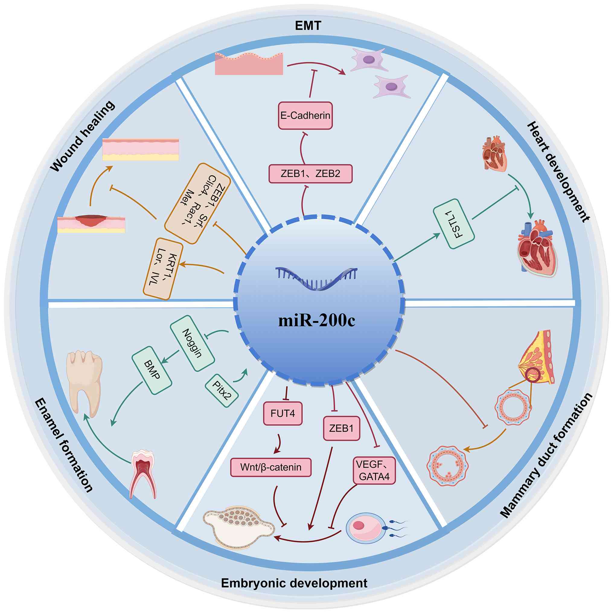 | Figure 1.Overview of the physiological
functions of miR-200c. Arrows indicate promotion (→) or inhibition
(⊣) (figure generated with figdraw). miR, microRNA; EMT,
epithelial-mesenchymal transition; ZEB, Zinc Finger E-box Binding
Homeobox; VEGF, vascular endothelial growth factor; GATA4,
GATA-binding protein 4; BMP, bone morphogenetic protein; Pitx2,
paired-like homeodomain transcription factor 2; Clic4, chloride
channel 4; Lor, loricrin IVL, involucrin; KRT1, keratin 1; FUT4,
fucosyl-transferase 4; PDE7B, phosphodiesterase 7B; Srf, Serum
response factor; Met, MET proto-oncogene, receptor tyrosine kinase;
Rac1, Rac family small GTPase 1; FSTL1, Follistatin-like 1. |
E-cadherin is crucial for maintaining intercellular
adhesion in epithelial tissues, and its expression is negatively
regulated by ZEB1 and ZEB2. Upregulation of E-cadherin inhibits
EMT. miR-200c-3p downregulates ZEB1 and ZEB2, thereby promoting
E-cadherin expression and suppressing EMT. This regulation
highlights the importance of miR-200c-3p in preserving epithelial
phenotype integrity (8,23). Conversely, ZEB1 negatively
regulates miR-200c, indicating a reciprocal negative feedback loop
essential for epithelial stability (24). miR-200c is critical in multiple
developmental processes. During heart development, miR-200c-3p
inhibits the progression of EMT mediated by FSTL1, delaying the
process of tissue regeneration in the heart (10). In tooth development, paired-like
homeodomain transcription factor 2 which is the first molecular
signal for tooth development, induces the expression of
miR-200c/141. This inhibition of noggin which is an antagonist of
bone morphogenetic protein (BMP) antagonist noggin, enhances BMP
signaling, drives differentiation of dental epithelial stem cells,
and facilitates enamel formation (25).
Regulation of miR-200c occurs in different
embryological stages. For instance, regulation of miR-200c promotes
embryonic stem cell differentiation toward endothelial progenitors
and regulates embryonic angiogenesis via direct repression of ZEB1
gene (26). Vascular endothelial
growth factor (VEGF) is an essential molecule for angiogenesis,
whose expression may be induced by hydrogen sulfide
(H2S) (27,28). Importantly, according to Hu et
al (29), H2S
downregulates the expression of miR-200c in human trophoblasts from
the placenta and induces the synthesis of VEGF; thus, promoting the
development of embryos' blood vessels. Conversely, when it comes to
undifferentiated human embryonic stem cells (hESCs), miR-200c
significantly suppresses GATA-binding protein 4 (GATA4), thereby
inhibiting differentiation and embryoid body formation (30).
Additionally, miR-200c participates actively in
embryonic development in early pregnancy and creates the
pre-implantation environment (31). Zheng et al (32) showed that miR-200c downregulates
fucosyl-transferase 4, suppressing Wnt/β-catenin signaling, and
preventing successful embryo implantation. Pinopodes in the
endometrium, which play a key role in receptivity necessary for
embryo implantation, are also inhibited by miR-200c, reducing the
chances of successful attachment (33).
In mammary gland development, miR-200c strongly
inhibits the capability of mammary stem cells to form mammary ducts
in vivo (34). All of these
facts clearly highlight the important role played by miR-200c in
the regulation of various processes in tissues.
Mechanisms of action of miR-200c in
apoptosis, pyroptosis and autophagy
Apoptosis
It is clear that apoptosis plays a central role in
programmed cell death which maintains cell number balance in order
to preserve cellular and tissue homeostasis. Among all cell death
mechanisms, apoptosis is considered essential due to its importance
for the elimination of damaged, senescent and malfunctioning cells
(35,36). An imbalance in the processes of
cell life and death leads to the development of numerous
pathological conditions (37,38).
One of the principal regulators in this process is miR-200c, which
performs a vital role in induction of apoptosis. Mechanisms of
miR-200c-induced apoptosis include controlling intrinsic and
extrinsic pathways of apoptosis induction, AKT-mediated apoptosis,
and other target gene functions. Specific apoptosis-related
molecular mechanisms regulated by miR-200c are represented in
Fig. 2, while target genes are
presented in Table I.
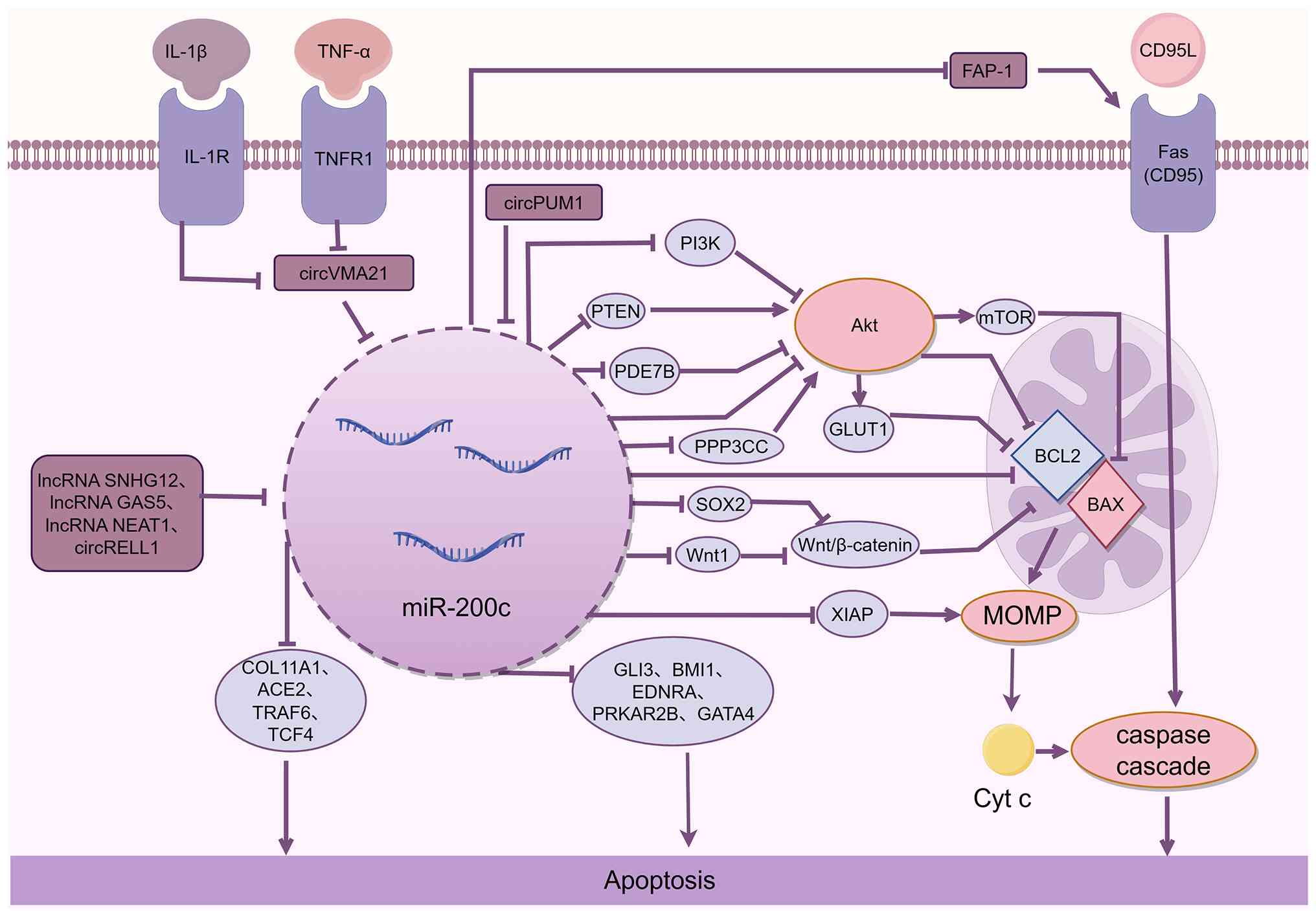 | Figure 2.Molecular mechanisms through which
miR-200c regulates apoptosis via multiple pathways. miR-200c is
regulated by upstream factors, including inflammatory cytokines and
non-coding RNAs, and directly targets a broad range of downstream
genes to modulate apoptosis. Arrows indicate promotion (→) or
inhibition (⊣) (figure generated with figdraw). miR, microRNA;
lncRNA, long non-coding RNA; circ, circular RNA; MOMP,
mitochondrial outer membrane permeabilization; COL11A1, collagen
type XI alpha 1 chain; GATA4, GATA-binding protein 4; ACE2,
angiotensin-converting enzyme 2; IL-1β, interleukin-1β; TNF-α,
tumor necrosis factor-α; PTEN, phosphatase and tensin homolog
deleted on chromosome 10; FAP-1, Fas-associated phosphatase-1;
PPP3CC, protein phosphatase 3 catalytic subunit gamma; SOX2,
SRY-box transcription factor 2; Wnt1, Wnt family member 1; PDE7B,
phosphodiesterase 7B; GLI3, GLI family zinc finger 3; BMI1, BMI1
proto-oncogene, polycomb ring finger; EDNRA, endothelin receptor
type A; PRKAR2B, protein kinase cAMP-dependent type II regulatory
subunit beta; TRAF6, TNF receptor associated factor 6; TCF4,
transcription factor 4; XIAP, X-linked inhibitor of apoptosis; BAX,
BCL2-associated X protein; BCL2, BCL2 apoptosis regulator; GLUT1,
Glucose transporter type 1. |
 | Table I.Target genes regulated by miR-200c
and their mechanisms of action. |
Table I.
Target genes regulated by miR-200c
and their mechanisms of action.
| First author/s,
year | Target gene | Disease | Strand specificity
(3p/5p) | Verification
method | Apoptosis
direction | Regulatory
mechanism | (Refs.) |
|---|
| Zhu et al,
2012 | BCL2 | GC and LC | - | Luciferase reporter
assay and western blotting | Promotion | Direct targeted
inhibition | (47) |
| Zhu et al,
2012; Ren et al, 2014 | XIAP | GC, LC and
TNBC | - | Luciferase reporter
assay, RT-qPCR and western blotting | Promotion | Direct targeted
inhibition | (47,48) |
| Cheng et al,
2018 | XIAP | Intervertebral disc
degeneration | - | Luciferase reporter
assay and western blotting | Promotion | Regulated by
circVMA21 | (49) |
| Li et al,
2019 | SOX2 | RCC | −3p | Bioinformatics
analysis, luciferase reporter assay, RT-qPCR and western
blotting | Promotion | Inactivation of
Wnt/β-catenin signaling pathway | (52) |
| Zhang et al,
2019 | Wnt1 | Preeclampsia | - | Luciferase reporter
assay, RT-qPCR and western blotting | Promotion | Inactivation of
Wnt/β-catenin signaling pathway | (53) |
| Schickel et
al, 2010 | FAP-1 | Renal, ovarian and
colorectal cancers | - | Luciferase reporter
assay, RT-qPCR and western blotting | Promotion | Enhancement of Fas
death receptor signaling | (55) |
| Zhang et al,
2019 | PDE7B | TNBC | - | Luciferase reporter
assay, RT-qPCR and western blotting | Promotion | Elevation of cAMP
and inhibition of Akt activity | (68) |
| Shen et al,
2020 | PTEN | Glaucoma | - | Bioinformatics
analysis, luciferase reporter assay, western blotting and
RT-qPCR | Inhibition | Activation of
PI3K/AKT/mTOR pathway | (62) |
| Liao et al,
2013 | PTEN | Pituitary
adenoma | −3p | Bioinformatics
analysis, luciferase reporter assay and western blotting | Inhibition | Activation of the
PTEN/Akt signaling pathway | (61) |
| Anastasiadou et
al, 2021 | PPP3CC | Epithelial ovarian
cancer | −3p | RT-qPCR and western
blotting | Inhibition | Activation of the
AKT signaling pathway | (70) |
| Yi et al,
2024 | GLI3 | LC | - | Luciferase reporter
assay, RT-qPCR and western blotting | Promotion | Direct targeted
inhibition | (71) |
| Cui et al,
2014 | BMI1 | Thymic
lymphoma | - | Bioinformatics
analysis, luciferase reporter assay, RT-qPCR and western
blotting | Promotion | Direct targeted
inhibition | (72) |
| Wei et al,
2018 | EDNRA | GC | - | Luciferase reporter
assay, RT-qPCR and western blotting | Promotion | Direct targeted
inhibition | (73) |
| Xu et al,
2020 | COL11A1 | RCC | −5p | Luciferase reporter
assay, RT-qPCR and western blotting | Inhibition | Regulated by lncRNA
SNHG12 | (80) |
| Xia et al,
2020 | PRKAR2B | Prostate
cancer | −3p | Bioinformatics
analysis, luciferase reporter assay, RT-qPCR and western
blotting | Inhibition | Direct targeted
inhibition | (85) |
| Zhang et al,
2023 | ACE2 | Acute lung
injury | −3p | Bioinformatics
analysis, luciferase reporter assay, RT-qPCR and western
blotting | Promotion | Inhibition of
ACE2/ANG1-7 axis | (76) |
| Li et al,
2018 | ACE2 | Acute respiratory
distress syndrome | −3p | Western blotting
and RT-qPCR | Promotion | Regulated by lncRNA
GAS5 | (77) |
| Ding et al,
2024 | TCF4 | Osteoarthritis | −3P | Luciferase reporter
assay, RT-qPCR, western blotting and RNA immunoprecipitation | Promotion | Regulated by
circRELL1 | (78) |
| Chen et al,
2017 | GATA4 | Ischemic heart
disease | - | Luciferase reporter
assay, RT-qPCR and western blotting | Promotion | Direct targeted
inhibition | (79) |
| Zhang et al,
2022 | TRAF6 | Chronic
periodontitis | −3P | Luciferase reporter
assay, RT-qPCR and western blotting | Inhibition | Regulated by lncRNA
NEAT1 | (81) |
Regulation of intrinsic and extrinsic
apoptotic pathways
Apoptosis is carried out via two pathways, namely
the intrinsic pathway that arises from cellular dysfunction, and
the extrinsic pathway that involves death signals from outside of
the cell (39). Intrinsic pathway
gets activated upon the disruption of cellular equilibrium caused
by internal stress such as oxidative stress (OS) or damage to DNA,
thus leading to programmed cell death. Mitochondrial outer membrane
permeabilization (MOMP) is the critical event that characterizes
this pathway (40,41). It is mediated by opposing actions
of pro-apoptotic and anti-apoptotic members of BCL2 family
(42). As a consequence of MOMP,
Bax and Bak oligomerize to form pores on the surface of
mitochondria, which results in the release of cytochrome c to the
cytosolic compartment. Subsequently, released cytochrome c
interacts with apoptotic protease-activating factor-1 (Apaf-1),
allowing for the assembly of Apaf-1 into dATP-dependent complexes.
These complexes are known as apoptosomes, which activate caspases
and lead to apoptosis (43).
Conversely, the extrinsic apoptotic pathway is
activated by extracellular death signals, such as Fas ligand
(FasL/CD95L), via ligand-receptor interactions (44). Once this external signal binds to
the corresponding receptor in the cell membrane, a death-inducing
signaling complex that becomes active and sets a chain of reactions
leading to apoptosis (45). While
there may be variations in the beginning stage, both pathways will
ultimately merge and end up activating the caspases, leading the
cell towards programmed death (44,46).
As indicated in the diagram below, miR-200c promotes apoptosis
along both signaling pathways.
Intrinsic apoptotic pathway
The miR-200c facilitates intrinsic apoptosis by
suppressing the amounts of several important anti-apoptotic
proteins such as BCL2 and XIAP. As shown in patients with gastric
cancer (GC) and LC, the microRNA induces apoptosis through the
activation of a chain of programmed cell death by regulating both
BCL2 and XIAP (47). This is also
applicable to TNBC and intervertebral disc degeneration (IVDD) in
that miR-200c initiates apoptosis by downregulating XIAP. In IVDD,
circVMA21 serves as an upstream controller of the miR-200c-induced
apoptosis. The inflammatory stimuli inflammatory cytokines
interleukin-1β (IL-1β) and tumor necrosis factor-α reduce circVMA21
expression levels. Since circVMA21, as a miR-200c inhibitor,
suppresses XIAP through binding to miR-200c, apoptosis occurs when
the protein's amounts decrease due to the reduction of circVMA21,
leading to a higher rate of death of nucleus pulposus cells
(48,49). Collectively, these findings suggest
that miR-200c-induced apoptosis via BCL2/XIAP suppression may
represent a common regulatory mechanism rather than a
disease-specific event, with variability arising from upstream
modulators.
Additionally, the role of miR-200c in the regulation
of intrinsic apoptosis is through modulating the Wnt/β-catenin
pathway, which is intricately linked with mitochondria-driven
apoptosis (50,51). Through suppression of the
Wnt/β-catenin pathway, miR-200c promotes intrinsic apoptosis. This
is evidenced by its ability to indirectly suppress Wnt/β-catenin
pathway via SRY-box transcription factor 2 (SOX2) inhibition in
renal cell carcinoma (RCC) cells, while in trophoblast cells
isolated from rats with preeclampsia, miR-200c acts directly on
Wnt1 (52,53).
Extrinsic apoptotic pathway
The induction of apoptosis mediated by extrinsic
pathways is facilitated by miR-200c through the increased
stimulation of Fas apoptosis pathway mediated by the repression of
Fas-associated phosphatase-1 (FAP-1). CD95 (APO-1/Fas), which acts
as the inducer for the apoptotic signaling, and is inhibited by
FAP-1 through binding of the receptor (54). Schickel et al (55) demonstrated that miR-200c
downregulates FAP-1 expression in clear cell RCC (CCRCC), RCC,
ovarian cancer (OC) and colorectal cancer (CRC) cells, thereby
enhancing Fas-mediated apoptosis (55). It has also been observed in
endothelial cells in a similar fashion. Additionally, Jiang et
al (56) observed that
fluoride exposure induces apoptosis in endothelial cells through
the induction of miR-200c-3p that represses FAP-1 resulting in
Fas-mediated apoptosis. It should be noted that even though studies
(55,56) have shown that miR-200c targets
FAP-1 in cancer cells as well as in endothelial cells, the
conservation of this function in other pathologies has not yet been
determined.
AKT-mediated apoptosis
miR-200c influences the behavior of cells through
regulation of apoptosis caused by AKT signaling, targeting several
molecules, including phosphatase and tensin homolog (PTEN) and
phosphodiesterase 7B (PDE7B). In other words, miR-200c exhibits
dual impacts on apoptosis, depending on the type of disease. For
instance, through PTEN and protein phosphatase 3 catalytic subunit
gamma (PPP3CC) downregulation, miR-200c promotes AKT signaling
activity and inhibition of apoptosis. At the same time, miR-200c
can cause apoptosis either through inhibition of PDE7B or via
inhibition of the PI3K-AKT pathway. The diverse roles of
miR-200c-controlled pathways are discussed in detail below.
PTEN and the PI3K-AKT pathway
However, recent studies indicate that the inhibition
of the PI3K-AKT pathway leads to increased cell death in various
disorders (57). For example,
miR-200c regulates the PI3K-AKT pathway within the trophoblasts in
recurrent spontaneous abortion, resulting in a decrease in the
BCL2/Bax ratio and cell apoptosis (58). The same can be said about
pancreatic cancer where circPUM1 functions as a miR-200c-3p
sponger; in the case of low expression levels of circPUM1, an
increase in miR-200c-3p leads to the slowing of PI3K-AKT pathway
activity and promotes apoptosis (59).
PTEN, an enzyme with dual activity of phosphatase
towards lipids and proteins, acts against AKT through
phosphorolyzing PIP3 into PIP2 (60). Considering PTEN's
apoptosis-inducing function, downregulation of PTEN through
miR-200c leads to AKT phosphorylation and subsequent induction of
AKT pathway and inhibition of apoptosis. As observed by Liao et
al (61), for instance, the
expression of PTEN is decreased by miR-200c in pituitary tumors
causing increased phosphorylated AKT levels with reduced cell
apoptosis in pituitary adenoma (PA) cells. Similarly, miR-200c-3p
induces PTEN suppression to increase phosphorylated AKT and mTOR
levels, along with decreasing cleaved caspase-3 and Bax expression
to inhibit apoptosis in glaucoma (62). Collectively, these findings suggest
that miR-200c's regulation of apoptosis through the PTEN-AKT axis
represents a critical anti-apoptotic mechanism across multiple
disease contexts.
Other mechanisms regulating AKT
activity
miR-200c regulates AKT signaling via processes other
than those related to PTEN/PI3K modulation. In their study, Zhao
et al (63) observed that
miR-200c suppresses phosphorylated AKT levels and subsequent GLUT1
expression in Wilms tumor, thus promoting apoptosis. Besides,
miR-200c regulates AKT signaling by affecting intracellular cAMP
concentrations. Increased intracellular cAMP levels reduce AKT
signaling (64,65), while PDEs, enzymes responsible for
regulating intracellular cAMP degradation, are among the targets of
miR-200c (66,67). Zhang et al (68) revealed that miR-200c suppresses
PDE7B, which led to the increase in intracellular cAMP, inhibition
of AKT signaling, and promotion of apoptosis in TNBC cells.
The next dimension of miR-200c is revealed by its
involvement in inhibiting apoptosis through other targets that
regulate AKT. For instance, the calcineurin-gamma subunit is
involved in mediating the calcium/calmodulin-dependent
serine/threonine phosphatase and is regarded as a tumor suppressor
in apoptosis. The target gene of miR-200c-3p includes PPP3CC
(69). In epithelial OC,
miR-200c-3p--mediated PPP3CC inhibition leads to AKT activation,
increased BCL2 expression and decreased caspase-3, collectively
inhibiting apoptosis (70).
Regulation of apoptosis via other
target genes
Outside the AKT pathway, miR-200c acts to induce
apoptosis via targeting an array of genes, including those related
to development (GLI3 and GATA4), inflammation (TRAF6) and
cell-cycle regulation (BMI1). The aforementioned list indicates the
complexity and interweaving aspects that contribute to the effects
of miR-200c in apoptosis. For instance, miR-200c drives apoptosis
in LC, thymic lymphoma, and GC by inhibiting GLI3, BMI1 and EDNRA,
respectively (71–73). Also, it should be noted that
angiotensin-converting enzyme 2 (ACE2), one of the confirmed
miR-200c-3p targets, serves in various protective functions in
lungs (74,75). Both damage to the lungs due to
seawater aspiration causing acute lung injury (ALI) and the more
extensive condition of acute respiratory distress syndrome (ARDS)
are governed by miR-200c-3p, which facilitates apoptosis by
reducing ACE2 levels. More precisely, in the case of ALI, the
increased apoptosis results from inhibition of the ACE2/ANG1-7
feedback mechanism (76). In ARDS,
inhibition of the long non-coding RNA (lncRNA) GAS5 molecule causes
miR-200c-3p upregulation, reduced ACE2, and apoptosis in A549 cells
(77). Similarly, Ding et
al (78) reported that
circRELL1 knockout elevates miR-200c-3p in osteoarthritis (OA),
inhibiting TCF4 expression and leading to increased apoptosis. In
ischemic heart disease, miR-200c induces cardiomyocyte apoptosis by
directly suppressing GATA4, highlighting potential therapeutic
implications (79).
Conversely, miR-200c exhibits anti-apoptotic effects
in RCC, prostate cancer (PCa), and chronic periodontitis (CP). The
possible mechanism behind these anti-apoptotic functions involves
factors specific to the different cells and competition against
upstream non-coding RNAs. However, the actual pathway needs further
investigation. For example, in RCC, the lncRNA SNHG12 has been
shown to be involved in sequestering miR-200c-5p; upon
downregulation of SNHG12, there is upregulation of miR-200c-5p,
which results in decreased levels of collagen type XI alpha 1 chain
mRNA and apoptosis (80).
Similarly, depletion of NEAT1 in CP leads to an increase in
miR-200c-3p, causing downregulation of TRAF6 and thus preventing
apoptosis (81). Interestingly,
TRAF6 is generally recognized as a key mediator of pro-inflammatory
responses in canonical immune signaling pathways, and its
downregulation can suppress inflammation and promote cell survival
(82,83). Therefore, the observed
downregulation of TRAF6 by miR-200c-3p in CP suggests that
miR-200c-3p may exert anti-apoptotic effects within this
inflammatory microenvironment. Similar anti-apoptotic effects of
miR-200c-3p have also been reported in PCa. Lin et al
(84) reported that miR-200c-3p
directly exerts anti-apoptotic effects in PCa cells (84). Additionally, in PCa, miR-200c-3p
directly exhibits anti-apoptotic properties, with Xia et al
(85) reporting inhibition of
apoptosis via specific targeting of PRKAR2B.
In summary, miR-200c regulates apoptosis via
multiple mechanisms, exhibiting both pro- and anti-apoptotic
effects depending on the cellular context (Fig. 3). Current evidence indicates that
miR-200c predominantly promotes apoptosis in diseases such as GC,
TNBC and Wilms tumor, whereas it inhibits apoptosis in pituitary
tumors, glaucoma, epithelial OC, PCa and CP. Several factors
explain these context-dependent effects, including: i) Cell- and
tissue-specific gene expression and signaling differences; ii) EMT
status, with mesenchymal cells typically less sensitive to
apoptosis (86–88); iii) distinct seed sequences of
miR-200c-3p and miR-200c-5p, targeting different mRNAs; iv)
differential targeting of pro- or anti-apoptotic nodes within
signaling pathways; and v) composition of competing endogenous RNA
regulatory networks, where circular RNAs and lncRNAs competitively
bind miR-200c, altering target gene expression and influencing
apoptosis outcomes.
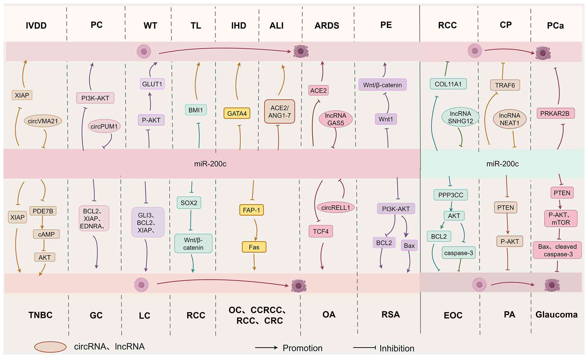 | Figure 3.Molecular mechanisms of
miR-200c-mediated apoptosis in various diseases. Arrows indicate
promotion (→) or inhibition (⊣) (figure generated with figdraw).
IVDD, intervertebral disc degeneration; PC, pancreatic cancer; WT,
Wilms tumor; TL, thymic lymphoma; IHD ischemic heart disease; ALI,
acute lung injury; ARDS, acute respiratory distress syndrome; PE,
preeclampsia; TNBC, triple-negative breast cancer; GC, gastric
cancer; LC, lung cancer; RCC; renal cell carcinoma; OC, ovarian
cancer; CRC, colorectal cancer; CCRCC, clear cell RCC; OA,
osteoarthritis; RSA, recurrent spontaneous abortion; CP, chronic
periodontitis; PCa, prostate cancer; EOC, epithelial OC; PA,
pituitary adenoma; lncRNA, long non-coding RNA; circRNA, circular
RNA; GATA4, GATA-binding protein 4; ACE2, angiotensin-converting
enzyme 2; FAP-1, Fas-associated phosphatase-1; PPP3CC, protein
phosphatase 3 catalytic subunit gamma; PTEN, phosphatase and tensin
homolog deleted on chromosome 10; COL11A1, collagen type XI alpha 1
chain; TRAF6, TNF receptor associated factor 6; PDE7B,
phosphodiesterase 7B; SOX2, SRY-box transcription factor 2; BCL2,
BCL2 apoptosis regulator; XIAP, X-linked inhibitor of apoptosis;
GLUT1, glucose transporter type 1; GLI3, GLI family zinc finger 3;
BMI1, BMI1 proto-oncogene, polycomb ring finger; Fas, Fas cell
surface death receptor; EDNRA, endothelin receptor type A; ANG1-7,
Angiotensin 1–7; TCF4, transcription factor 4; Wnt1, Wnt family
member 1; BAX, BCL2-associated X protein; PRKAR2B, protein kinase
cAMP-dependent type II regulatory subunit beta; XIAP, X-linked
inhibitor of apoptosis; GLUT1, Glucose transporter type 1; GLI3,
GLI family zinc finger 3; TCF4, transcription factor 4. |
Pyroptosis
Pyroptosis is an inflammatory form of programmed
cell death crucially involved in the onset and progression of
various diseases. The activation of the NLRP3 inflammasome and the
cleavage of gasdermin D (GSDMD) constitute the core mechanisms
underlying pyroptosis (89).
Pyroptosis is initiated via two distinct pathways: The classical
and non-classical pathways. The classical pathway involves assembly
of the NLRP3 inflammasome, consisting of NLRP3,
apoptosis-associated speck-like protein, and caspase-1, as well as
caspases −4, −5, and −11. Activated caspase-1 subsequently cleaves
GSDMD, into N-terminal and C-terminal fragments (90). By contrast, the non-classical
pathway involves direct activation of human caspase-4/5 or mouse
caspase-11 by lipopolysaccharides derived from Gram-negative
bacteria. These caspases similarly cleave GSDMD (90,91).
The N-terminal fragment of GSDMD forms pores in the cell membrane,
leading to cell swelling and rupture, and converts inactive
pro-inflammatory cytokines pro-IL-1β and pro-IL-18 into their
mature forms. This process amplifies local inflammatory response,
ultimately driving pyroptotic cell death (92,93).
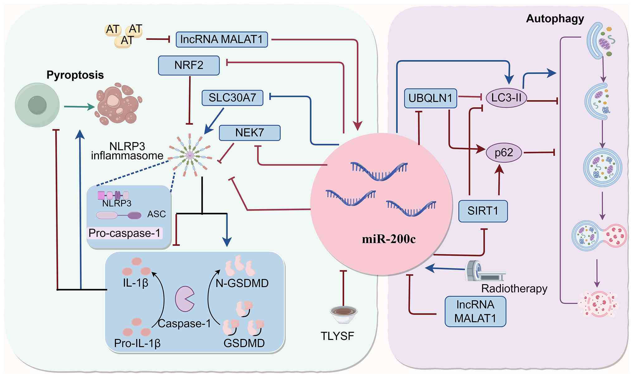
Existing research confirms miR-200c is extensively
regulates pyroptosis. However, current evidence pertains
exclusively to the classical NLRP3/caspase-1 pathway; no studies to
date have examined miR-200c involvement in the non-classical
caspase-4/5/11 pathway. Future research is necessary to address
this knowledge gap.
In diabetes-related complications, such as diabetic
retinopathy (DR) and diabetic nephropathy (DN), miR-200c mediates
pyroptosis predominantly through activation of the NLRP3
inflammasome, though pathways differ diseases. Li et al
(94) demonstrated that
miR-200c-3p promotes pyroptosis in human retinal microvascular
endothelial cells during DR by downregulating SLC30A7, thus
activating the NLRP3 inflammasome. In DN, hyperglycemic conditions
concurrently induce lncRNA MALAT1 and miR-200c, synergistically
inhibiting NRF2 and elevating miR-200c-3p expression in exosomes
derived from glomerular mesangial cells. Both mechanisms can
activate the NLRP3 inflammasome, exacerbating podocyte pyroptosis.
By contrast, the lipid-lowering agent atorvastatin and the
traditional Chinese medicinal formula Tongluo Yishen confer renal
protection by blocking these pathways (95,96).
Notably, in inflammatory bowel disease (IBD),
miR-200c suppresses pyroptosis. Specifically, miR-200c-5p directly
targets and inhibits NEK7, a critical activator of the NLRP3
inflammasome, thus reducing pyroptosis in intestinal epithelial
cells (MODE-K) cells. This inhibition attenuates intestinal
inflammation and mitigates disease progression in IBD (97).
In summary, miR-200c exhibits disease-specific
regulatory roles in pyroptosis, promoting it in diabetic
complications while inhibiting it in inflammatory bowel disease.
This specificity provides a theoretical foundation for developing
miR-200c-targeted therapeutic strategies against
pyroptosis-associated diseases.
Autophagy
Autophagy is a highly conserved process of cellular
self-degradation process in which substantial amounts of
cytoplasmic content and organelles are encapsulated within double-
or multi-membraned autophagosomes and subsequently delivered to
lysosomes for degradation (98,99).
Under pathological conditions, radiation-induced autophagy
typically exerts a cytoprotective effect, thereby reducing the
sensitivity of tumor cell sensitivity (100). For example, Sun et al
(101) demonstrated that miR-200c
inhibits protective autophagy induced by radiation in BC cells by
downregulating UBQLN1, leading to decreased LC-II and increased p62
expression, thus enhancing radiosensitivity. In a cerebral ischemia
model, the lncRNA MALAT1 competitively binds to miR-200c-3p.
Knockdown of MALAT1 increases miR-200c-3p levels, resulting in
downregulated SIRT1, decreased LC3-II expression, increased p62
expression, and ultimately impaired autophagy. This impairment
exacerbates cell death and worsens cerebral ischemic injury
(102).
Conversely, in glioblastoma and non-small cell LC
(NSCLC), combining miR-200c with radiotherapy significantly
elevates LC3-II expression and promotes autophagy-mediated tumor
cell death, enhancing overall cell death rates. This finding
provides a novel mechanistic insight and potential research
direction for combination radiotherapy in cancer treatment
(103). In summary, miR-200c
exerts diverse regulatory effects on pyroptosis and autophagy, with
specific roles dependent on cell type and pathological context
(Fig. 4). These insights broaden
the understanding of miR-200c's complex functions within cell death
pathways and hold promise for developing innovative therapeutic
strategies.
Therapeutic potential of miR-200c
Due to its importance as an apoptosis regulator,
miR-200c has become significant as a tool for reversing drug
resistance and customizing treatment protocols. The current
research involving miR-200c includes four major areas: i)
Regulating miR-200c expression by drugs as a means of inducing
apoptosis; ii) manipulating miR-200c to affect cancer cells'
sensitivity to drugs; iii) designing nano-formulations for delivery
of miR-200c; and iv) utilizing miR-200c as a means of targeting
TME. Fourth, therapeutic strategies based on reshaping the TME via
miR-200c. The following sections describe recent findings and
future directions regarding these areas.
Therapeutic modulation of miR-200c
expression by drugs
Another mechanism that drugs employ for treatment
involves the use of the apoptosis induction effect by adjusting
miR-200c concentrations. However, depending on the particular
disease and tissues involved, miR-200c can be upregulated or
downregulated to achieve desired therapeutic effects (Table II). Therefore, drugs can either
increase or decrease miR-200c expression based on the disease type
and miR-200c involvement. There are instances where increasing
miR-200c leads to better effects. Resveratrol induces apoptosis and
helps inhibit tumorigenesis in CRCs due to increased miR-200c
expression (104). Similarly, the
antitumor activity of metformin in BC is enhanced by increasing
miR-200c expression; as a result, AKT2 and BCL2 proteins get
inhibited, leading to proapoptotic Bax/BCL2 imbalance (105).
| Table II.Mechanisms of drug action mediated by
regulation of miR-200c expression. |
Table II.
Mechanisms of drug action mediated by
regulation of miR-200c expression.
| First author/s,
year | Disease | Therapeutic
drug | miR-200c
change | Strand specificity
(3p/5p) | Verification
method | Target
gene/Signaling pathway | Apoptosis
direction | (Refs.) |
|---|
| Karimi et
al, 2017 | CRC | Resveratrol | ↑ | - | - | - | Promotion | (104) |
| Lozano-Herrera
et al, 2022 | CRC | Naringenin and
FEN | ↓ | - | RT-qPCR and
bioinformatics analysis | Upregulation of
PTEN expression | Promotion | (106) |
| Tang et al,
2020 | CRC | Atractylolide
I | ↓ | - | - | - | Promotion | (107) |
| Zhang et al,
2017 | BC | Metformin | ↑ | - | RT-qPCR and western
blotting of AKT2 and BCL2 | Targeting and
inhibition | Promotion | (105) |
| Wang et al,
2017 | PA | Bromocriptine and
Artesunate | ↓ | - | RT-qPCR, western
blotting and immunocytochemistry | Upregulation of
PTEN expression | Promotion | (108) |
| Chen et al,
2013 | PA | SZ-685C | ↓ | - | - | - | Promotion | (109) |
| Deng et al,
2017; Huang et al, 2022 | MI/RI | Propofol | ↓ | −3p | Luciferase reporter
assay, RT-qPCR and western blotting | Upregulation of
AdipoR2 and activation of STAT3 signaling pathway | Inhibition | (110,111) |
On the contrary, miR-200c may have a role to play in
preventing cell death in specific diseases, therefore any therapy
would require downregulation of miR-200c to restore cell death,
which is necessary for therapeutic effect. According to a study
carried out by Lozano-Herrera et al (106) coadministration of both naringenin
and FEN, along with bisphenol A decreases miR-200c expression, thus
overcoming inhibition of PTEN, expression by miR-200c and restoring
apoptosis of CRC cells (106).
Similarly, atractylenolide I, a phytochemical utilized in
traditional Chinese medicine, exerts an anticancer effect by
downregulating miR-200c in cancer stem cells (107).
A combination of drugs may covertly affect the
regulation of miR-200c, thus enhancing the effect of drug therapy.
For example, in PA, bromocriptine combined with artesunate
downregulates miR-200c expression, resulting in increased PTEN
expression and enhanced antitumor effects (108). Furthermore, the marine-derived
compound SZ-685C effectively reduces miR-200c levels, induces
apoptosis in PA MMQ cells, and increases therapeutic efficacy,
presenting a promising targeted treatment approach for PAs
(109).
Under the conditions of myocardial
ischemia/reperfusion injury (MI/RI), associated with diabetes,
reduction of miR-200c leads to anti-apoptotic effects that enhance
the chances of successful treatment. Propofol, a cardioprotective
agent, exerts its protective effect by suppressing miR-200c-3p,
followed by activation of adiponectin receptor 2 and subsequent
induction of the STAT3 signaling pathway (110). This contributes to protection
against heart muscle cells death and delays progression of MI/RI
(111). In summary, it can be
deduced that the same signaling pathways can be involved in various
types of treatment and lead to diverse effects depending on the
pathophysiological condition. This demonstrates the role of
miR-200c that is disease-dependent. Hence, the targeting of
miR-200c should be adjusted to particular diseases.
miR-200c influences treatment
resistance through regulation of apoptosis
Other than being an effector molecule responsible
for implementing induced by pharmacotherapy, miR-200c is a
regulator of anticancer drug resistance through modulation of
apoptosis. Cisplatin (DDP) is the standard anticancer chemotherapy
drug, and the role played by miR-200c in modulating sensitivity to
DDP has been validated in numerous cancers and proved to be
two-fold. For example, in the study by Hamano et al
(112) on esophageal cancer, the
pro-survival function of miR-200c is observed as it activates
PPP2R1B and the Akt pathway and thus increases DDP resistance.
Conversely, miR-200c enhances DDP sensitivity in GC by specifically
targeting RhoE, and in NSCLC by targeting BCL2 and XIAP, thereby
promoting apoptosis and reversing DDP resistance (47,113). These differences in
miR-200c--mediated effects likely depend on disease-specific
downstream target genes.
Apart from DDP, miR-200c is also responsible for
altering the resistance towards other treatment approaches. As an
instance, miR-200c renders the GC cells sensitive again towards
vincristine through downregulation of the two genes, BCL2 and XIAP
(47). Similarly, in CCRCC,
miR-200c specifically silences the anti-apoptotic protein heme
oxygenase 1. This mechanism increases the expression of the
pro-apoptotic proteins Bid and Bax and decreases the anti-apoptotic
proteins BCL-xL and BCL2, enhancing apoptosis induced by sorafenib
and imatinib, thus overcoming drug resistance (114). miR-200c can overcome resistance
to chemotherapy in LC. The downregulation of miR-200c is
significantly associated with the development of resistance against
methotrexate therapy in NSCLC. Upregulating miR-200c enhances the
efficacy of methotrexate by modulating the p53/p21 signaling
pathway (115). Additionally, Bai
et al (116) demonstrated
that miR-200c upregulates the expression of the pro-apoptotic
markers caspase-3 and caspase-9, enhancing the resveratrol-induced
apoptotic in LC cells. Resistance to paclitaxel continues to be a
significant problem in BC treatment (117). It has been found that regulates
miR-200c-3p regulates SOX2 expression in paclitaxel-resistant
MCF-7/Tax cells, inducing apoptosis and re-establishing
sensitiveness to paclitaxel (118). Additionally, miR-200c mediated
reversal of drug resistance in BC is regulated by lncRNAs. The
lncRNA XIST acts as a competitive endogenous RNA: Its
downregulation reduces ANLN expression, consequently elevating
miR-200c-3p, which promotes apoptosis and reverses doxorubicin
resistance in MDA-MB-231/ADM cells (119).
Of particular importance is the fact that EMT lies
right at the intersection between metastasis and apoptosis
resistance (86–88), and as such acts as one of the
driving forces behind multidrug resistance, steering therapeutic
options in a negative direction (120–122). Numerous cells that go through EMT
become resistant to apoptosis, becoming more robust against any
type of therapy. miR-200c stands out in this respect as an
important inhibitor of EMT by acting as a suppressor of the
transcription factors that drive EMT, specifically ZEB1 and ZEB2,
thus maintaining the epithelial cell phenotype (8,23,123,124). By inhibiting EMT, miR-200c
facilitates apoptosis and restores drug sensitivity in various
cancers.
This process has been confirmed for a number of
cancers. Specifically, the development of epidermal growth factor
receptor (EGFR) tyrosine kinase inhibitors' resistance is very
common during the treatment of patients with EGFR mutations who are
suffering from NSCLC. miR-200c counteracts the development of
resistance to the aforementioned inhibitors as a result of EMT
suppression, thus leading to an increased level of apoptosis in
response to gefitinib and osimertinib (125). Similarly, miR-200c reverses drug
resistance by suppressing the process of EMT through targeting of
the ZEB family in patients with GC and BC. According to Jiang et
al (126), miR-200c
suppresses DDP resistance through the ZEB2 gene, thus contributing
to apoptosis. In BC, miR-200c enhances apoptosis through two
distinct pathways: First, by inhibiting ZEB1 and augmenting
p53-mediated apoptosis to reverse doxorubicin resistance; second,
in doxorubicin-resistant MCF-7 cells, ultrasound-mediated miR-200c
overexpression reduces P-glycoprotein expression via ZEB1
inhibition, restoring drug sensitivity (124,127).
In summary, miR-200c significantly influences
resistance to multiple chemotherapeutic agents (Table III), highlighting its potential
as promising biomarker for predicting chemotherapy resistance.
However, current evidence regarding the association between
miR-200c and drug resistance is primarily derived from cellular
models, with limited clinical data available from in vivo
studies. Further research is required to confirm whether miR-200c
expression levels directly affect therapeutic outcomes in
patients.
| Table III.Regulation of drug resistance by
microRNA-200c. |
Table III.
Regulation of drug resistance by
microRNA-200c.
| First author/s,
year | Disease | Therapeutic
drug | Strand specificity
(3p/5p) | Target
gene/signaling pathway | Verification
method | Effect on
resistance | (Refs.) |
|---|
| Jiang et al,
2017 | Gastric cancer | Cisplatin | - | Targeted inhibition
of ZEB2 | Luciferase reporter
assays, RT-qPCR, western blotting and bioinformatics analysis | ↓ | (126) |
| Ghasabi et
al, 2019 | Gastric cancer | Cisplatin | - | Targeted inhibition
of RhoE | RT-qPCR and western
blotting | ↓ | (113) |
| Zhu et al,
2012 | Gastric cancer | Vincristine | - | Targeted inhibition
of BCL2 and XIAP | Luciferase reporter
assay and western blotting | ↓ | (47) |
| Hamano et
al, 2011 | Esophageal
cancer | Cisplatin | - | Targeted inhibition
of PPP2R1B | Luciferase assay,
western blotting and bioinformatics analysis | ↑ | (112) |
| Bai et al,
2014 | Lung cancer | Resveratrol | - | - | - | ↓ | (116) |
| Zhu et al,
2012 | Lung cancer | Cisplatin | - | Targeted inhibition
of BCL2 and XIAP | Luciferase reporter
assay and western blotting | ↓ | (47) |
| Gao et al,
2014 | Clear cell renal
cell carcinoma | Sorafenib and
imatinib | - | Targeted inhibition
of the anti-apoptotic protein HO-1 | Luciferase reporter
assay, RT-qPCR and western blotting | ↓ | (114) |
| Shan et al,
2016 | Non-small cell lung
cancer | Methotrexate | - | Through the p53/p21
pathway | Western
blotting | ↓ | (115) |
| Wang et al,
2020 | Non-small cell lung
cancer | Gefitinib and
osimertinib | −3p | - | - | ↓ | (125) |
| Chen et al,
2018 | Breast cancer | Paclitaxel | −3p | Targeted inhibition
of SOX2 | Luciferase reporter
assay, RT-qPCR and western blotting | ↓ | (118) |
| Zhang et al,
2020 | Breast cancer | Adriamycin
(doxorubicin) | −3p | Targeted inhibition
of ANLN | Luciferase reporter
assays, RT-qPCR and western blotting | ↓ | (119) |
| Tryndyak et
al, 2010 | Breast cancer | Doxorubicin | - | Targeted inhibition
of ZEB1 | RT-qPCR and western
blotting | ↓ | (124) |
| Huang et al,
2018 | Breast cancer | Doxorubicin | - | Targeted inhibition
of ZEB1 | Luciferase reporter
assay, RT-qPCR and western blotting | ↓ | (127) |
Therapeutic applications in the
TME
miR-200c is a miRNA that acts as an orchestrator
within the TME through various means, allowing specific treatments
for cancers to be developed. It can change the environment of a
tumor through regulation of tumor-associated macrophages (TAMs),
cancer-associated fibroblasts (CAFs), and the immune checkpoint
molecule PD-L1, which contribute to tumor development. The TAMs are
immune cells within the TME. A study has shown that breast cancer
cells in the process of apoptosis are able to transport the miRNA
to macrophages through a CD36 receptor. Inside the macrophages, the
miR-200c blocks the genes related to macrophage movement (such as
PPM1F and MSN). Thus, there will be fewer infiltrating macrophages
within the tumor (128). This
demonstrates that miR-200c can modify the TME through intercellular
transfer, ultimately affecting tumor progression.
Another stromal factor involved in the TME is the
CAFs. Typically, the activation of CAFs results in tumor
progression. However, the role of miR-200c in CAFs is distinct from
its role in macrophages, although the precise mechanism is yet to
be fully elucidated. Typically, the level of miR-200c in CAFs is
downregulated (129). As reported
by Lin et al (130),
OS-mediated miR-200c promoter demethylation leads to increased
expression of this molecule in CAFs, resulting in
mesenchymal-to-epithelial transition reprogramming via
COMMD1-NFκB-HIF suppression. Such reprogramming results in
increased proliferation and reduced apoptosis of cancer cells while
promoting immune suppression and tumor progression (130). PD-L1, an immune checkpoint
protein, mediates tumor immune evasion. miR-200c indirectly
downregulates PD-L1 by inhibiting ZEB1, thereby reducing
immunosuppression of CD8+ T cells, limiting tumor immune
evasion, and preventing distant metastasis (131).
miR-200c-based nanotherapies
With the ability to play the significant role of
conducting apoptosis, pyroptosis and autophagy, miR-200c is an
important element in orchestrating numerous diseases. The more the
roles of this element are known and explored, the more miR-200c
proves itself to be promising as a potential therapeutic element
and has become a worthy drug candidate. However, the way from
research and discovery to treatment faces numerous difficulties.
Firstly, RNA molecules are quite vulnerable because they can easily
degrade when encountering nucleases; besides, their delivery is
unpredictable and non-precise.
The current options for delivering miRNA include
viral and non-viral techniques. Though the viral vectors exhibit
high efficiency in transfection, their safety concerns related to
immunogenicity and tumorigenicity limit their medical applications.
By contrast, the non-viral delivery systems pose little
immunogenicity and are highly biocompatible, thus serving as main
candidates for miR-200c delivery. The list of non-viral techniques
involves the following: Inorganic nanoparticles, polymeric
nanocarriers, lipid vesicles, exosomes and liposomes (132). As for nano-delivery approaches,
they proved particularly encouraging when developing
miR-200c--based drugs.
Qian et al (133) developed miR-200c-loaded
nanoparticles, effectively delivered them into AGS GC cells, and
enhanced apoptosis in combination with radiotherapy, significantly
improving therapeutic efficacy. In BC therapy, Garrido-Cano et
al (134) utilized mesoporous
silica nanoparticles to deliver miR-200c-3p into MDA-MB-231 cells,
effectively downregulating ZEB1 and ZEB2, thereby inhibiting EMT
and achieving potent anticancer effects. Nano-delivery approaches
also exhibit significant potential in OA. Zheng et al
(135) prepared a
lipo-AgPEI-miR-200c-3p complex using a microfluidic device to
encapsulate miR-200c-3p into silver nitrate-polyethyleneimine
nanoparticles. They successfully delivered miR-200c-3p into
chondrocytes, significantly reduced apoptosis by downregulating Bax
and cleaved caspase-3, and increased BCL2 expression, providing a
novel approach for OA treatment.
Even with the potential benefits associated with it,
developing a nanoparticle-based therapy for use in humans presents
challenges that make this pathway a rather congested one. Once they
enter the body through a systemic route, nanoparticles are rapidly
absorbed into the body's immune system, reducing their availability
in the target tissue to an enormous degree. Since miR-200c has
wide-ranging effects on gene expression, it increases the risk of
non-specific effects if it enters other non-target cells after
delivery. Modifications made to miRNA to maintain stability in
blood can reduce its effectiveness within cells. More importantly,
the context-dependent nature of miR-200c's function poses unique
safety challenges: i) miR-200c is involved in normal physiological
processes such as wound healing, cardiac development and embryonic
development; systemic administration may interfere with these
normal physiological functions; ii) exogenous miRNAs and their
delivery vehicles are recognized by the immune system, triggering
an immune response and inflammatory reaction; and iii)
nanoparticles are captured by hepatic macrophages, leading to their
accumulation in the liver; this off-target accumulation may result
in hepatocyte damage, elevated transaminases and abnormal liver
function, and the hepatotoxic consequences of long-term
accumulation remain unclear (136,137).
Nano-delivery approaches can be considered as one of
the feasible options to protect miR-200c from instability and
degradation. While there is a lot of potential in the development
of miR-200c-based nanotherapeutics, various issues need to be
addressed, such as improving efficacy of delivery, limiting adverse
side effects, decreasing the risk of immunological response, and
proving that chemically modified miRNAs are safe for humans.
Further efforts must address all of the aforementioned concerns and
assess safety in animal studies, which would enhance the
effectiveness of therapy.
Conclusion
miR-200c plays an essential role in cell life
because it regulates the processes of apoptosis, autophagy and
pyroptosis through various molecular pathways. When the organism is
healthy, it maintains the harmony of epithelial tissues and
promotes important developmental events (for example, teeth and
cardiac formation), regulating pluripotency vs. differentiation in
ESCs. When the organism suffers from disease, miR-200c controls the
cell's fate by acting on several targets and signaling pathways. It
directly affects apoptosis by silencing genes that regulate the
process or indirectly through AKT pathway. At the same time,
miR-200c also integrates itself in autophagy and pyroptosis,
resulting in a complicated regulation system for cellular decisions
to live or die.
Specifically, miR-200c can be regarded as an
excellent candidate for developing new therapeutic interventions,
considering its sensitivity to medication therapy and capability to
regulate drug resistance in various diseases. However, the
application of drugs that are based on miR-200c requires additional
validation to be used in clinical practice. Although preclinical
trials have demonstrated positive outcomes of the targeted delivery
of miR-200c into a particular location, a lot of research remains
to be conducted regarding delivering the substance efficiently and
accurately. Specifically, three crucial problems should be
addressed by future research, namely: i) Tissue and cellular
targeting to reduce possible negative effects of the off-target
delivery of the substance; ii) ensuring safety and efficiency of
the therapy; and iii) maintaining miR-200c stability and activity
in vivo.
Furthermore, future studies need to elucidate the
mechanism by which miR-200c regulates apoptosis, autophagy and
pyroptosis collectively in one disease context. Currently, the
focus is on exploring these cell death pathways individually using
various diseases as their context, thus making it unknown whether
miR-200c regulates all the three types of cell deaths together in
one disease context. It is only by focusing on one particular model
that it will be possible to determine whether miR-200c acts as a
master regulator.
Complete investigations into how miR-200c acts in
different ways according to the disease stage and type of cells
involved will result in exact windows of therapeutic opportunities
being defined, thus leading to the development of miR-200c-based
therapies for clinical applications. This progress would not only
transform miR-200c a practical therapeutic tool but also deepen our
understanding of programmed cell death regulation, offering new
insights and strategies for disease diagnosis and treatment.
Acknowledgements
Not applicable.
Funding
The present study was funded by the Inner Mongolia Science and
Technology Research Project (grant nos. 2024MS08069 and
2025MS08117), the Science and Technology Program of the Joint Fund
of Scientific Research for the Public Hospitals of Inner Mongolia
Academy of Medical Sciences (grant no. 2024GLLH0323), the Key
Technologies Research and development program of Inner Mongolia
(grant no. 2021GG0170), the general Program of Inner Mongolia
Medical University (grant no. YKD2021MS006), the 14th Five-Year
Plan of Science and Technology Innovation in Inner Mongolia
Autonomous Region (grant no. 2022YFSH0078), the Key project of
Inner Mongolia Medical University (grant no. YKD2021ZD00), the
Zhiyuan Talent Program of Inner Mongolia Medical University (grant
no. ZY0202020 and ZY20242107), the Doctoral Start-up Foundation
Project of Inner Mongolia Medical University (grant no.
YKD2024BSQD026), the Undergraduate Teaching Reform Research and
Practice Project of Inner Mongolia Medical University in 2024
(grant no. NYJXGGSJ20244046), the Inner Mongolia Medical University
2024 Maker Cultivation Project (grant no. 101322024038), the Inner
Mongolia Medical University 2025 Cultivation of Excellence Program
in Science and Technology Innovation for Undergraduates (grant no.
YCPY2025057), the National Natural Science Foundation Of China
(grant no. 82560297), the Joint Project of Inner Mongolia Medical
University (grant no. YKD2022LH049), and the Project of the Inner
Mongolia Autonomous Region Educational Science Research ‘14th
Five-Year Plan’ (grant no. NGJGH2025307).
Availability of data and materials
Not applicable.
Authors' contributions
XW and HD were responsible for manuscript writing,
and conceived and designed the study. YS and XW were responsible
for the collection and assembly of data. YS and HD were responsible
for data analysis and interpretation. All authors read and approved
the final version of the manuscript. Data authentication is not
applicable.
Ethics approval and consent to
participate
Not applicable.
Patient consent for publication
Not applicable.
Competing interests
The authors declare that they have no competing
interests.
References
|
1
|
Saliminejad K, Khorram Khorshid HR,
Soleymani Fard S and Ghaffari SH: An overview of microRNAs:
Biology, functions, therapeutics, and analysis methods. J Cell
Physiol. 234:5451–5465. 2019. View Article : Google Scholar : PubMed/NCBI
|
|
2
|
Bartel DP: MicroRNAs: Genomics,
biogenesis, mechanism, and function. Cell. 116:281–297. 2004.
View Article : Google Scholar : PubMed/NCBI
|
|
3
|
Iorio MV and Croce CM: MicroRNAs in
cancer: Small molecules with a huge impact. J Clin Oncol.
27:5848–5856. 2009. View Article : Google Scholar : PubMed/NCBI
|
|
4
|
Vishnoi A and Rani S: miRNA biogenesis and
regulation of diseases: An updated overview. Methods Mol Biol.
2595:1–12. 2023. View Article : Google Scholar : PubMed/NCBI
|
|
5
|
Li Y, Meng X, Luo Y, Luo S, Li J, Zeng J,
Huang X and Wang J: The oncogenic miR-429 promotes triple-negative
breast cancer progression by degrading DLC1. Aging (Albany NY).
15:9809–9821. 2023. View Article : Google Scholar : PubMed/NCBI
|
|
6
|
Ho HL, Lin SC, Chiang CW, Lin C, Liu CW,
Yeh YC, Chen MY and Chou TY: miR-193b-3p suppresses lung cancer
cell migration and invasion through PRNP targeting. J Biomed Sci.
32:282025. View Article : Google Scholar : PubMed/NCBI
|
|
7
|
Bai WD, Ye XM, Zhang MY, Zhu HY, Xi WJ,
Huang X, Zhao J, Gu B, Zheng GX, Yang AG and Jia LT: MiR-200c
suppresses TGF-β signaling and counteracts trastuzumab resistance
and metastasis by targeting ZNF217 and ZEB1 in breast cancer. Int J
Cancer. 135:1356–1368. 2014. View Article : Google Scholar : PubMed/NCBI
|
|
8
|
Gregory PA, Bert AG, Paterson EL, Barry
SC, Tsykin A, Farshid G, Vadas MA, Khew-Goodall Y and Goodall GJ:
The miR-200 family and miR-205 regulate epithelial to mesenchymal
transition by targeting ZEB1 and SIP1. Nat Cell Biol. 10:593–601.
2008. View Article : Google Scholar : PubMed/NCBI
|
|
9
|
Zhou X, Men X, Zhao R, Han J, Fan Z, Wang
Y, Lv Y, Zuo J, Zhao L, Sang M, et al: miR-200c inhibits
TGF-β-induced-EMT to restore trastuzumab sensitivity by targeting
ZEB1 and ZEB2 in gastric cancer. Cancer Gene Ther. 25:68–76. 2018.
View Article : Google Scholar : PubMed/NCBI
|
|
10
|
Pontemezzo E, Foglio E, Vernucci E,
Magenta A, D'Agostino M, Sileno S, Astanina E, Bussolino F,
Pellegrini L, Germani A, et al: miR-200c-3p regulates
epitelial-to-mesenchymal transition in epicardial mesothelial cells
by targeting epicardial follistatin-related protein 1. Int J Mol
Sci. 22:49712021. View Article : Google Scholar : PubMed/NCBI
|
|
11
|
Tower J: Programmed cell death in aging.
Ageing Res Rev. 23:90–100. 2015. View Article : Google Scholar : PubMed/NCBI
|
|
12
|
Kerr JFR: History of the events leading to
the formulation of the apoptosis concept. Toxicology.
181-182:471–474. 2002. View Article : Google Scholar : PubMed/NCBI
|
|
13
|
Cookson BT and Brennan MA:
Pro-inflammatory programmed cell death. Trends Microbiol.
9:113–114. 2001. View Article : Google Scholar : PubMed/NCBI
|
|
14
|
Tsvetkov P, Coy S, Petrova B, Dreishpoon
M, Verma A, Abdusamad M, Rossen J, Joesch-Cohen L, Humeidi R,
Spangler RD, et al: Copper induces cell death by targeting
lipoylated TCA cycle proteins. Science. 375:1254–1261. 2022.
View Article : Google Scholar : PubMed/NCBI
|
|
15
|
Feng X, Wang Z, Fillmore R and Xi Y:
MiR-200, a new star miRNA in human cancer. Cancer Lett.
344:166–173. 2014. View Article : Google Scholar : PubMed/NCBI
|
|
16
|
Huang GL, Sun J, Lu Y, Liu Y, Cao H, Zhang
H and Calin GA: MiR-200 family and cancer: From a meta-analysis
view. Mol Aspects Med. 70:57–71. 2019. View Article : Google Scholar : PubMed/NCBI
|
|
17
|
Vrba L, Jensen TJ, Garbe JC, Heimark RL,
Cress AE, Dickinson S, Stampfer MR and Futscher BW: Role for DNA
methylation in the regulation of miR-200c and miR-141 expression in
normal and cancer cells. PLoS One. 5:e86972010. View Article : Google Scholar : PubMed/NCBI
|
|
18
|
Wiklund ED, Bramsen JB, Hulf T, Dyrskjøt
L, Ramanathan R, Hansen TB, Villadsen SB, Gao S, Ostenfeld MS,
Borre M, et al: Coordinated epigenetic repression of the miR-200
family and miR-205 in invasive bladder cancer. Int J Cancer.
128:1327–1334. 2011. View Article : Google Scholar : PubMed/NCBI
|
|
19
|
Zahraei M, Azimi Y, Karimipour M,
Rahimi-Jamnani F, Valizadeh V and Azizi M:
CRISPR/dCas9-TET1-mediated epigenetic editing reactivates miR-200c
in breast cancer cells. Sci Rep. 15:317392025. View Article : Google Scholar : PubMed/NCBI
|
|
20
|
Shaw TJ and Martin P: Wound repair at a
glance. J Cell Sci. 122:3209–3213. 2009. View Article : Google Scholar : PubMed/NCBI
|
|
21
|
Pastar I, Stojadinovic O, Yin NC, Ramirez
H, Nusbaum AG, Sawaya A, Patel SB, Khalid L, Isseroff RR and
Tomic-Canic M: Epithelialization in wound healing: A comprehensive
review. Adv Wound Care (New Rochelle). 3:445–464. 2014. View Article : Google Scholar : PubMed/NCBI
|
|
22
|
Aunin E, Broadley D, Ahmed MI, Mardaryev
AN and Botchkareva NV: Exploring a role for regulatory miRNAs in
wound healing during ageing:Involvement of miR-200c in wound
repair. Sci Rep. 7:32572017. View Article : Google Scholar : PubMed/NCBI
|
|
23
|
Park SM, Gaur AB, Lengyel E and Peter ME:
The miR-200 family determines the epithelial phenotype of cancer
cells by targeting the E-cadherin repressors ZEB1 and ZEB2. Genes
Dev. 22:894–907. 2008. View Article : Google Scholar : PubMed/NCBI
|
|
24
|
Burk U, Schubert J, Wellner U, Schmalhofer
O, Vincan E, Spaderna S and Brabletz T: A reciprocal repression
between ZEB1 and members of the miR-200 family promotes EMT and
invasion in cancer cells. EMBO Rep. 9:582–589. 2008. View Article : Google Scholar : PubMed/NCBI
|
|
25
|
Cao H, Jheon A, Li X, Sun Z, Wang J,
Florez S, Zhang Z, McManus MT, Klein OD and Amendt BA: The
Pitx2:miR-200c/141:noggin pathway regulates Bmp signaling and
ameloblast differentiation. Development. 140:3348–3359. 2013.
View Article : Google Scholar : PubMed/NCBI
|
|
26
|
Luo Z, Wen G, Wang G, Pu X, Ye S, Xu Q,
Wang W and Xiao Q: MicroRNA-200C and −150 play an important role in
endothelial cell differentiation and vasculogenesis by targeting
transcription repressor ZEB1. Stem Cells. 31:1749–1762. 2013.
View Article : Google Scholar : PubMed/NCBI
|
|
27
|
Kang Y, Li H, Liu Y and Li Z: Regulation
of VEGF-A expression and VEGF-A-targeted therapy in malignant
tumors. J Cancer Res Clin Oncol. 150:2212024. View Article : Google Scholar : PubMed/NCBI
|
|
28
|
Zhang YX, Jing MR, Cai CB, Zhu SG, Zhang
CJ, Wang QM, Zhai YK, Ji XY and Wu DD: Role of hydrogen sulphide in
physiological and pathological angiogenesis. Cell Prolif.
56:e133742023. View Article : Google Scholar : PubMed/NCBI
|
|
29
|
Hu TX, Wang G, Guo XJ, Sun QQ, He P, Gu H,
Huang Y, Gao L and Ni X: MiR 20a,-20b and −200c are involved in
hydrogen sulfide stimulation of VEGF production in human placental
trophoblasts. Placenta. 39:101–110. 2016. View Article : Google Scholar : PubMed/NCBI
|
|
30
|
Huang HN, Chen SY, Hwang SM, Yu CC, Su MW,
Mai W, Wang HW, Cheng WC, Schuyler SC, Ma N, et al: miR-200c and
GATA binding protein 4 regulate human embryonic stem cell renewal
and differentiation. Stem Cell Res. 12:338–353. 2014. View Article : Google Scholar : PubMed/NCBI
|
|
31
|
Kim J, Lee J and Jun JH: Identification of
differentially expressed microRNAs in outgrowth embryos compared
with blastocysts and non-outgrowth embryos in mice. Reprod Fertil
Dev. 31:645–657. 2019. View Article : Google Scholar : PubMed/NCBI
|
|
32
|
Zheng Q, Zhang D, Yang YU, Cui X, Sun J,
Liang C, Qin H, Yang X, Liu S and Yan Q: MicroRNA-200c impairs
uterine receptivity formation by targeting FUT4 and
α1,3-fucosylation. Cell Death Differ. 24:2161–2172. 2017.
View Article : Google Scholar : PubMed/NCBI
|
|
33
|
Salmasi S, Heidar MS, Khaksary Mahabady M,
Rashidi B and Mirzaei H: MicroRNAs, endometrial receptivity and
molecular pathways. Reprod Biol Endocrinol. 22:1392024. View Article : Google Scholar : PubMed/NCBI
|
|
34
|
Shimono Y, Zabala M, Cho RW, Lobo N,
Dalerba P, Qian D, Diehn M, Liu H, Panula SP, Chiao E, et al:
Downregulation of miRNA-200c links breast cancer stem cells with
normal stem cells. Cell. 138:592–603. 2009. View Article : Google Scholar : PubMed/NCBI
|
|
35
|
Thompson CB: Apoptosis in the pathogenesis
and treatment of disease. Science. 267:1456–1462. 1995. View Article : Google Scholar : PubMed/NCBI
|
|
36
|
Billig H, Chun SY, Eisenhauer K and Hsueh
AJ: Gonadal cell apoptosis: Hormone-regulated cell demise. Hum
Reprod Update. 2:103–117. 1996. View Article : Google Scholar : PubMed/NCBI
|
|
37
|
Morana O, Wood W and Gregory CD: The
apoptosis paradox in cancer. Int J Mol Sci. 23:13282022. View Article : Google Scholar : PubMed/NCBI
|
|
38
|
Rudin CM and Thompson CB: Apoptosis and
disease: Regulation and clinical relevance of programmed cell
death. Annu Rev Med. 48:267–281. 1997. View Article : Google Scholar : PubMed/NCBI
|
|
39
|
Hanahan D and Weinberg RA: Hallmarks of
cancer: The next generation. Cell. 144:646–674. 2011. View Article : Google Scholar : PubMed/NCBI
|
|
40
|
Youle RJ and Strasser A: The BCL-2 protein
family: Opposing activities that mediate cell death. Nat Rev Mol
Cell Biol. 9:47–59. 2008. View Article : Google Scholar : PubMed/NCBI
|
|
41
|
Tait SW and Green DR: Mitochondria and
cell death: Outer membrane permeabilization and beyond. Nat Rev Mol
Cell Biol. 11:621–632. 2010. View Article : Google Scholar : PubMed/NCBI
|
|
42
|
Galluzzi L, Vitale I, Aaronson SA, Abrams
JM, Adam D, Agostinis P, Alnemri ES, Altucci L, Amelio I, Andrews
DW, et al: Molecular mechanisms of cell death: Recommendations of
the nomenclature committee on cell death 2018. Cell Death Differ.
25:486–541. 2018. View Article : Google Scholar : PubMed/NCBI
|
|
43
|
Wu CC and Bratton SB: Regulation of the
intrinsic apoptosis pathway by reactive oxygen species. Antioxid
Redox Signal. 19:546–558. 2013. View Article : Google Scholar : PubMed/NCBI
|
|
44
|
Fuchs Y and Steller H: Live to die another
way: Modes of programmed cell death and the signals emanating from
dying cells. Nat Rev Mol Cell Biol. 16:329–344. 2015. View Article : Google Scholar : PubMed/NCBI
|
|
45
|
Castillo Ferrer C, Berthenet K and Ichim
G: Apoptosis-fueling the oncogenic fire. FEBS J. 288:4445–4463.
2021. View Article : Google Scholar : PubMed/NCBI
|
|
46
|
Thornberry NA and Lazebnik Y: Caspases:
Enemies within. Science. 281:1312–1316. 1998. View Article : Google Scholar : PubMed/NCBI
|
|
47
|
Zhu W, Xu H, Zhu D, Zhi H, Wang T, Wang J,
Jiang B, Shu Y and Liu P: miR-200bc/429 cluster modulates multidrug
resistance of human cancer cell lines by targeting BCL2 and XIAP.
Cancer Chemother Pharmacol. 69:723–731. 2012. View Article : Google Scholar : PubMed/NCBI
|
|
48
|
Ren Y, Han X, Yu K, Sun S, Zhen L, Li Z
and Wang S: microRNA-200c downregulates XIAP expression to suppress
proliferation and promote apoptosis of triple-negative breast
cancer cells. Mol Med Rep. 10:315–321. 2014. View Article : Google Scholar : PubMed/NCBI
|
|
49
|
Cheng X, Zhang L, Zhang K, Zhang G, Hu Y,
Sun X, Zhao C, Li H, Li YM and Zhao J: Circular RNA VMA21 protects
against intervertebral disc degeneration through targeting miR-200c
and X linked inhibitor-of-apoptosis protein. Ann Rheum Dis.
77:770–779. 2018. View Article : Google Scholar : PubMed/NCBI
|
|
50
|
Ming M, Wang S, Wu W, Senyuk V, Le Beau
MM, Nucifora G and Qian Z: Activation of Wnt/β-catenin protein
signaling induces mitochondria-mediated apoptosis in hematopoietic
progenitor cells. J Biol Chem. 287:22683–22690. 2012. View Article : Google Scholar : PubMed/NCBI
|
|
51
|
Anitha P, Priyadarsini RV, Kavitha K,
Thiyagarajan P and Nagini S: Ellagic acid coordinately attenuates
Wnt/β-catenin and NF-κB signaling pathways to induce intrinsic
apoptosis in an animal model of oral oncogenesis. Eur J Nutr.
52:75–84. 2013. View Article : Google Scholar : PubMed/NCBI
|
|
52
|
Li S, Feng Z, Zhang X, Lan D and Wu Y:
Up-regulation of microRNA-200c-3p inhibits invasion and migration
of renal cell carcinoma cells via the SOX2-dependent Wnt/β-catenin
signaling pathway. Cancer Cell Int. 19:2312019. View Article : Google Scholar : PubMed/NCBI
|
|
53
|
Zhang X, Ge YW, Wang ZX, Xu QL, Guo R and
Xu HY: MiR-200c regulates apoptosis of placental trophoblasts in
preeclampsia rats through Wnt/β-catenin signaling pathway. Eur Rev
Med Pharmacol Sci. 23:7209–7216. 2019.PubMed/NCBI
|
|
54
|
Sato T, Irie S, Kitada S and Reed JC:
FAP-1: A protein tyrosine phosphatase that associates with Fas.
Science. 268:411–415. 1995. View Article : Google Scholar : PubMed/NCBI
|
|
55
|
Schickel R, Park SM, Murmann AE and Peter
ME: miR-200c regulates induction of apoptosis through CD95 by
targeting FAP-1. Mol Cell. 38:908–915. 2010. View Article : Google Scholar : PubMed/NCBI
|
|
56
|
Jiang Y, Yang Y, Zhang C, Huang W, Wu L,
Wang J, Su M, Sun D and Gao Y: Upregulation of miR-200c-3p induced
by NaF promotes endothelial apoptosis by activating Fas pathway.
Environ Pollut. 266:1150892020. View Article : Google Scholar : PubMed/NCBI
|
|
57
|
Zhang HP, Jiang RY, Zhu JY, Sun KN, Huang
Y, Zhou HH, Zheng YB and Wang XJ: PI3K/AKT/mTOR signaling pathway:
An important driver and therapeutic target in triple-negative
breast cancer. Breast Cancer. 31:539–551. 2024. View Article : Google Scholar : PubMed/NCBI
|
|
58
|
Yue L and Xu H: MicroRNA-200c promotes
trophoblast cell dysfunction via inhibition of PI3K/Akt signaling
in unexplained recurrent spontaneous abortion. Reprod Biol.
24:1009512024. View Article : Google Scholar : PubMed/NCBI
|
|
59
|
Chen Y, Liu Y, Tao Q, Fan Y, Ma C, Li D,
Huang F and Tang D: circPUM1 activates the PI3K/AKT signaling
pathway by sponging to promote the proliferation, invasion and
glycolysis of pancreatic cancer. Curr Pharm Biotechnol.
23:1405–1414. 2022. View Article : Google Scholar : PubMed/NCBI
|
|
60
|
Álvarez-Garcia V, Tawil Y, Wise HM and
Leslie NR: Mechanisms of PTEN loss in cancer: It's all about
diversity. Semin Cancer Biol. 59:66–79. 2019. View Article : Google Scholar : PubMed/NCBI
|
|
61
|
Liao C, Chen W, Fan X, Jiang X, Qiu L,
Chen C, Zhu Y and Wang H: MicroRNA-200c inhibits apoptosis in
pituitary adenoma cells by targeting the PTEN/Akt signaling
pathway. Oncol Res. 21:129–136. 2013. View Article : Google Scholar : PubMed/NCBI
|
|
62
|
Shen Y, Zhu Y and Rong F: miR-200c-3p
regulates the proliferation and apoptosis of human trabecular
meshwork cells by targeting PTEN. Mol Med Rep. 22:1605–1612. 2020.
View Article : Google Scholar : PubMed/NCBI
|
|
63
|
Zhao GZ, Niu YQ, Li ZM, Kou D and Zhang L:
MiR-200c inhibits proliferation and promotes apoptosis of Wilms
tumor cells by regulating akt signaling pathway. Eur Rev Med
Pharmacol Sci. 24:6623–6631. 2020.PubMed/NCBI
|
|
64
|
Wang W, Li Y, Zhu JY, Fang D, Ding HF,
Dong Z, Jing Q, Su SB and Huang S: Triple negative breast cancer
development can be selectively suppressed by sustaining an elevated
level of cellular cyclic AMP through simultaneously blocking its
efflux and decomposition. Oncotarget. 7:87232–87245. 2016.
View Article : Google Scholar : PubMed/NCBI
|
|
65
|
Brennesvik EO, Ktori C, Ruzzin J, Jebens
E, Shepherd PR and Jensen J: Adrenaline potentiates
insulin-stimulated PKB activation via cAMP and Epac: Implications
for cross talk between insulin and adrenaline. Cell Signal.
17:1551–1559. 2005. View Article : Google Scholar : PubMed/NCBI
|
|
66
|
Bender AT and Beavo JA: Cyclic nucleotide
phosphodiesterases: Molecular regulation to clinical use. Pharmacol
Rev. 58:488–520. 2006. View Article : Google Scholar : PubMed/NCBI
|
|
67
|
Maurice DH, Ke H, Ahmad F, Wang Y, Chung J
and Manganiello VC: Advances in targeting cyclic nucleotide
phosphodiesterases. Nat Rev Drug Discov. 13:290–314. 2014.
View Article : Google Scholar : PubMed/NCBI
|
|
68
|
Zhang DD, Li Y, Xu Y, Kim J and Huang S:
Phosphodiesterase 7B/microRNA-200c relationship regulates
triple-negative breast cancer cell growth. Oncogene. 38:1106–1120.
2019. View Article : Google Scholar : PubMed/NCBI
|
|
69
|
Zhang Q, Wang W, Xu W, Du' M, Ma G, Liu H,
Chu H, Tong NA, Wang M, Shao J, et al: Genetic variations in the
3′-untranslated regions of genes involved in the cell cycle and
apoptosis pathways affect bladder cancer risk. Cancer Genomics
Proteomics. 15:67–72. 2018.PubMed/NCBI
|
|
70
|
Anastasiadou E, Messina E, Sanavia T,
Labruna V, Ceccarelli S, Megiorni F, Gerini G, Pontecorvi P, Camero
S, Perniola G, et al: Calcineurin gamma catalytic subunit PPP3CC
inhibition by miR-200c-3p affects apoptosis in epithelial ovarian
cancer. Genes (Basel). 12:14002021. View Article : Google Scholar : PubMed/NCBI
|
|
71
|
Yi X, Chen X and Li Z: miR-200c targeting
GLI3 inhibits cell proliferation and promotes apoptosis in
non-small cell lung cancer cells. Medicine (Baltimore).
103:e396582024. View Article : Google Scholar : PubMed/NCBI
|
|
72
|
Cui J, Cheng Y, Zhang P, Sun M, Gao F, Liu
C and Cai J: Down regulation of miR200c promotes radiation-induced
thymic lymphoma by targeting BMI1. J Cell Biochem. 115:1033–1042.
2014. View Article : Google Scholar : PubMed/NCBI
|
|
73
|
Wei W, Shi L, Chen W, Hu L, Chen D, Shi X,
Xiang H, Guo C and Wu Z: miR-200c regulates the proliferation,
apoptosis and invasion of gastric carcinoma cells through the
downregulation of EDNRA expression. Int J Mol Med. 41:1619–1626.
2018.PubMed/NCBI
|
|
74
|
Soltani S and Zandi M: miR-200c-3p
upregulation and ACE2 downregulation via bacterial LPS and LTA as
interesting aspects for COVID-19 treatment and immunity. Mol Biol
Rep. 48:5809–5810. 2021. View Article : Google Scholar : PubMed/NCBI
|
|
75
|
Ji Y, Gao F, Sun B, Hao J and Liu Z:
Angiotensin-converting enzyme 2 inhibits apoptosis of pulmonary
endothelial cells during acute lung injury through suppressing
SMAD2 phosphorylation. Cell Physiol Biochem. 35:2203–2212. 2015.
View Article : Google Scholar : PubMed/NCBI
|
|
76
|
Zhang M and Xie L: MicroRNA-200c-3p
regulates seawater-induced acute lung injury via ANGII and
ACE2/ANG1-7 pathways. Exp Ther Med. 26:5822023. View Article : Google Scholar : PubMed/NCBI
|
|
77
|
Li HB, Zi PP, Shi HJ, Gao M and Sun RQ:
Role of signaling pathway of long non-coding RNA growth
arrest-specific transcript 5/microRNA-200c-3p/angiotensin
converting enzyme 2 in the apoptosis of human lung epithelial cell
A549 in acute respiratory distress syndrome. Zhonghua Yi Xue Za
Zhi. 98:3354–3359. 2018.(In Chinese). PubMed/NCBI
|
|
78
|
Ding H, Chen H, Dou L and Li Y: CircRELL1
promotes osteoarthritis progression by regulating miR-200c-3p.
Heliyon. 10:e342512024. View Article : Google Scholar : PubMed/NCBI
|
|
79
|
Chen Z, Zhang S, Guo C, Li J and Sang W:
Downregulation of miR-200c protects cardiomyocytes from
hypoxia-induced apoptosis by targeting GATA-4. Int J Mol Med.
39:1589–1596. 2017. View Article : Google Scholar : PubMed/NCBI
|
|
80
|
Xu C, Liang H, Zhou J, Wang Y, Liu S, Wang
X, Su L and Kang X: lncRNA small nucleolar RNA host gene 12
promotes renal cell carcinoma progression by modulating the
miR-200c-5p/collagen type XI α1 chain pathway. Mol Med Rep.
22:3677–3686. 2020.PubMed/NCBI
|
|
81
|
Zhang L, Lv H, Cui Y and Shi R: The role
of long non-coding RNA (lncRNA) nuclear paraspeckle assembly
transcript 1 (NEAT1) in chronic periodontitis progression.
Bioengineered. 13:2336–2345. 2022. View Article : Google Scholar : PubMed/NCBI
|
|
82
|
Inoue J, Ishida T, Tsukamoto N, Kobayashi
N, Naito A, Azuma S and Yamamoto T: Tumor necrosis factor
receptor-associated factor (TRAF) family: Adapter proteins that
mediate cytokine signaling. Exp Cell Res. 254:14–24. 2000.
View Article : Google Scholar : PubMed/NCBI
|
|
83
|
Xing PC, An P, Hu GY, Wang DL and Zhou MJ:
LncRNA MIAT promotes inflammation and oxidative stress in
sepsis-induced cardiac injury by targeting miR-330-5p/TRAF6/NF-κB
axis. Biochem Genet. 58:783–800. 2020. View Article : Google Scholar : PubMed/NCBI
|
|
84
|
Lin J, Lu Y, Zhang X, Mo Q and Yu L:
Effect of miR-200c on proliferation, invasion and apoptosis of
prostate cancer LNCaP cells. Oncol Lett. 17:4299–4304.
2019.PubMed/NCBI
|
|
85
|
Xia L, Han Q, Chi C, Zhu Y, Pan J, Dong B,
Huang Y, Xia W, Xue W and Sha J: Transcriptional regulation of
PRKAR2B by miR-200b-3p/200c-3p and XBP1 in human prostate cancer.
Biomed Pharmacother. 124:1098632020. View Article : Google Scholar : PubMed/NCBI
|
|
86
|
Brabletz S and Brabletz T: The ZEB/miR-200
feedback loop-a motor of cellular plasticity in development and
cancer? EMBO Rep. 11:670–677. 2010. View Article : Google Scholar : PubMed/NCBI
|
|
87
|
Li Y, VandenBoom TG II, Kong D, Wang Z,
Ali S, Philip PA and Sarkar FH: Up-regulation of miR-200 and let-7
by natural agents leads to the reversal of
epithelial-to-mesenchymal transition in gemcitabine-resistant
pancreatic cancer cells. Cancer Res. 69:6704–6712. 2009. View Article : Google Scholar : PubMed/NCBI
|
|
88
|
Li QQ, Xu JD, Wang WJ, Cao XX, Chen Q,
Tang F, Chen ZQ, Liu XP and Xu ZD: Twist1-mediated
adriamycin-induced epithelial-mesenchymal transition relates to
multidrug resistance and invasive potential in breast cancer cells.
Clin Cancer Res. 15:2657–2665. 2009. View Article : Google Scholar : PubMed/NCBI
|
|
89
|
Chen X, Liu G, Yuan Y, Wu G, Wang S and
Yuan L: NEK7 interacts with NLRP3 to modulate the pyroptosis in
inflammatory bowel disease via NF-κB signaling. Cell Death Dis.
10:9062019. View Article : Google Scholar : PubMed/NCBI
|
|
90
|
Gaidt MM, Ebert TS, Chauhan D, Schmidt T,
Schmid-Burgk JL, Rapino F, Robertson AA, Cooper MA, Graf T and
Hornung V: Human monocytes engage an alternative inflammasome
pathway. Immunity. 44:833–846. 2016. View Article : Google Scholar : PubMed/NCBI
|
|
91
|
Kayagaki N, Stowe IB, Lee BL, O'Rourke K,
Anderson K, Warming S, Cuellar T, Haley B, Roose-Girma M, Phung QT,
et al: Caspase-11 cleaves gasdermin D for non-canonical
inflammasome signalling. Nature. 526:666–671. 2015. View Article : Google Scholar : PubMed/NCBI
|
|
92
|
Compan V, Martín-Sánchez F, Baroja-Mazo A,
López--Castejón G, Gomez AI, Verkhratsky A, Brough D and Pelegrín
P: Apoptosis-associated speck-like protein containing a CARD forms
specks but does not activate caspase-1 in the absence of NLRP3
during macrophage swelling. J Immunol. 194:1261–1273. 2015.
View Article : Google Scholar : PubMed/NCBI
|
|
93
|
Ding J, Wang K, Liu W, She Y, Sun Q, Shi
J, Sun H, Wang DC and Shao F: Pore-forming activity and structural
autoinhibition of the gasdermin family. Nature. 535:111–116. 2016.
View Article : Google Scholar : PubMed/NCBI
|
|
94
|
Li W, Yang S, Chen G and He S: MiR-200c-3p
regulates pyroptosis by targeting SLC30A7 in diabetic retinopathy.
Hum Exp Toxicol. 41:96032712210995892022. View Article : Google Scholar : PubMed/NCBI
|
|
95
|
Zuo Y, Chen L, He X, Ye Z, Li L, Liu Z and
Zhou S: Atorvastatin regulates MALAT1/miR-200c/NRF2 activity to
protect against podocyte pyroptosis induced by high glucose.
Diabetes Metab Syndr Obes. 14:1631–1645. 2021. View Article : Google Scholar : PubMed/NCBI
|
|
96
|
Yan W, Han B, Lin L, Chen L, Qu J and Li
X: Study on the promotion of podocyte pyroptosis by high
glucose-stimulated GMC-derived exosomes and the intervention
effects of Tongluo Yishen formula. Xi Bao Yu Fen Zi Mian Yi Xue Za
Zhi. 41:495–504. 2025.(In Chinese). PubMed/NCBI
|
|
97
|
Wu G, Zhang D, Yang L, Wu Q and Yuan L:
MicroRNA-200c-5p targets NIMA Related Kinase 7 (NEK7) to inhibit
NOD-like receptor 3 (NLRP3) inflammasome activation, MODE-K cell
pyroptosis, and inflammatory bowel disease in mice. Mol Immunol.
146:57–68. 2022. View Article : Google Scholar : PubMed/NCBI
|
|
98
|
Gozuacik D and Kimchi A: Autophagy as a
cell death and tumor suppressor mechanism. Oncogene. 23:2891–2906.
2004. View Article : Google Scholar : PubMed/NCBI
|
|
99
|
Mizushima N, Levine B, Cuervo AM and
Klionsky DJ: Autophagy fights disease through cellular
self-digestion. Nature. 451:1069–1075. 2008. View Article : Google Scholar : PubMed/NCBI
|
|
100
|
Gewirtz DA: The switch between protective
and nonprotective autophagy; implications for autophagy inhibition
as a therapeutic strategy in cancer. Biology (Basel).
9:122020.PubMed/NCBI
|
|
101
|
Sun Q, Liu T, Yuan Y, Guo Z, Xie G, Du S,
Lin X, Xu Z, Liu M, Wang W, et al: MiR-200c inhibits autophagy and
enhances radiosensitivity in breast cancer cells by targeting
UBQLN1. Int J Cancer. 136:1003–1012. 2015. View Article : Google Scholar : PubMed/NCBI
|
|
102
|
Wang S, Han X, Mao Z, Xin Y, Maharjan S
and Zhang B: MALAT1 lncRNA induces autophagy and protects brain
microvascular endothelial cells against oxygen-glucose deprivation
by binding to miR-200c-3p and upregulating SIRT1 expression.
Neuroscience. 397:116–126. 2019. View Article : Google Scholar : PubMed/NCBI
|
|
103
|
Koo T, Cho BJ, Kim DH, Park JM, Choi EJ,
Kim HH, Lee DJ and Kim IA: MicroRNA-200c increases radiosensitivity
of human cancer cells with activated EGFR-associated signaling.
Oncotarget. 8:65457–65468. 2017. View Article : Google Scholar : PubMed/NCBI
|
|
104
|
Karimi Dermani F, Saidijam M, Amini R,
Mahdavinezhad A, Heydari K and Najafi R: Resveratrol inhibits
proliferation, invasion, and epithelial-mesenchymal transition by
increasing miR-200c expression in HCT-116 colorectal cancer cells.
J Cell Biochem. 118:1547–1555. 2017. View Article : Google Scholar : PubMed/NCBI
|
|
105
|
Zhang J, Li G, Chen Y, Fang L, Guan C, Bai
F, Ma M, Lyu J and Meng QH: Metformin inhibits tumorigenesis and
tumor growth of breast cancer cells by upregulating miR-200c but
downregulating AKT2 expression. J Cancer. 8:1849–1864. 2017.
View Article : Google Scholar : PubMed/NCBI
|
|
106
|
Lozano-Herrera SJ, Luna-Bárcenas G,
Guevara-González RG, Campos-Vega R, Solís-Sáinz JC, Hernández-Puga
AG and Vergara-Castañeda HA: Fermentation extract of naringenin
increases the expression of estrogenic receptor β and modulates
genes related to the p53 signalling pathway, miR-200c and miR-141
in human colon cancer cells exposed to BPA. Molecules. 27:65882022.
View Article : Google Scholar : PubMed/NCBI
|
|
107
|
Tang D, Xu X, Ying J, Xie T and Cao G:
Transfer of metastatic traits via miR-200c in extracellular
vesicles derived from colorectal cancer stem cells is inhibited by
atractylenolide I. Clin Transl Med. 10:e1392020. View Article : Google Scholar : PubMed/NCBI
|
|
108
|
Wang X, Du Q, Mao Z, Fan X, Hu B, Wang Z,
Chen Z, Jiang X, Wang Z, Lei N, et al: Combined treatment with
artesunate and bromocriptine has synergistic anticancer effects in
pituitary adenoma cell lines. Oncotarget. 8:45874–45887. 2017.
View Article : Google Scholar : PubMed/NCBI
|
|
109
|
Chen CH, Xiao WW, Jiang XB, Wang JW, Mao
ZG, Lei N, Fan X, Song BB, Liao CX, Wang HJ, et al: A novel marine
drug, SZ-685C, induces apoptosis of MMQ pituitary tumor cells by
downregulating miR-200c. Curr Med Chem. 20:2145–2154. 2013.
View Article : Google Scholar : PubMed/NCBI
|
|
110
|
Deng F, Wang S, Zhang L, Xie X, Cai S, Li
H, Xie GL, Miao HL, Yang C, Liu X and Xia Z: Propofol through
upregulating caveolin-3 attenuates post-hypoxic mitochondrial
damage and cell death in H9C2 cardiomyocytes during hyperglycemia.
Cell Physiol Biochem. 44:279–292. 2017. View Article : Google Scholar : PubMed/NCBI
|
|
111
|
Huang L, Ding L, Yu S, Huang X and Ren Q:
Propofol postconditioning alleviates diabetic myocardial
ischemia-reperfusion injury via the miR-200c-3p/AdipoR2/STAT3
signaling pathway. Mol Med Rep. 25:1372022. View Article : Google Scholar : PubMed/NCBI
|
|
112
|
Hamano R, Miyata H, Yamasaki M, Kurokawa
Y, Hara J, Moon JH, Nakajima K, Takiguchi S, Fujiwara Y, Mori M and
Doki Y: Overexpression of miR-200c induces chemoresistance in
esophageal cancers mediated through activation of the Akt signaling
pathway. Clin Cancer Res. 17:3029–3038. 2011. View Article : Google Scholar : PubMed/NCBI
|
|
113
|
Ghasabi M, Majidi J, Mansoori B, Mohammadi
A, Shomali N, Shirafkan N, Baghbani E, Kazemi T and Baradaran B:
The effect of combined miR-200c replacement and cisplatin on
apoptosis induction and inhibition of gastric cancer cell line
migration. J Cell Physiol. 234:22581–22592. 2019. View Article : Google Scholar : PubMed/NCBI
|
|
114
|
Gao C, Peng FH and Peng LK: MiR-200c
sensitizes clear-cell renal cell carcinoma cells to sorafenib and
imatinib by targeting heme oxygenase-1. Neoplasma. 61:680–689.
2014. View Article : Google Scholar : PubMed/NCBI
|
|
115
|
Shan W, Zhang X, Li M, Deng F and Zhang J:
Over expression of miR-200c suppresses invasion and restores
methotrexate sensitivity in lung cancer A549 cells. Gene.
593:265–271. 2016. View Article : Google Scholar : PubMed/NCBI
|
|
116
|
Bai T, Dong DS and Pei L: Synergistic
antitumor activity of resveratrol and miR-200c in human lung
cancer. Oncol Rep. 31:2293–2297. 2014. View Article : Google Scholar : PubMed/NCBI
|
|
117
|
Abu Samaan TM, Samec M, Liskova A, Kubatka
P and Büsselberg D: Paclitaxel's mechanistic and clinical effects
on breast cancer. Biomolecules. 9:7892019. View Article : Google Scholar : PubMed/NCBI
|
|
118
|
Chen J, Tian W, He H, Chen F, Huang J,
Wang X and Chen Z: Downregulation of miR-200c-3p contributes to the
resistance of breast cancer cells to paclitaxel by targeting SOX2.
Oncol Rep. 40:3821–3829. 2018.PubMed/NCBI
|
|
119
|
Zhang M, Wang F, Xiang Z, Huang T and Zhou
WB: LncRNA XIST promotes chemoresistance of breast cancer cells to
doxorubicin by sponging miR-200c-3p to upregulate ANLN. Clin Exp
Pharmacol Physiol. 47:1464–1472. 2020. View Article : Google Scholar : PubMed/NCBI
|
|
120
|
Aderetti DA, Hira VVV, Molenaar RJ and van
Noorden CJF: The hypoxic peri-arteriolar glioma stem cell niche, an
integrated concept of five types of niches in human glioblastoma.
Biochim Biophys Acta Rev Cancer. 1869:346–354. 2018. View Article : Google Scholar : PubMed/NCBI
|
|
121
|
Safa AR: Epithelial-mesenchymal
transition: A hallmark in pancreatic cancer stem cell migration,
metastasis formation, and drug resistance. J Cancer Metastasis
Treat. 6:362020.PubMed/NCBI
|
|
122
|
Tong X, Tang R, Xiao M, Xu J, Wang W,
Zhang B, Liu J, Yu X and Shi S: Targeting cell death pathways for
cancer therapy: Recent developments in necroptosis, pyroptosis,
ferroptosis, and cuproptosis research. J Hematol Oncol. 15:1742022.
View Article : Google Scholar : PubMed/NCBI
|
|
123
|
Korpal M, Lee ES, Hu G and Kang Y: The
miR-200 family inhibits epithelial-mesenchymal transition and
cancer cell migration by direct targeting of E-cadherin
transcriptional repressors ZEB1 and ZEB2. J Biol Chem.
283:14910–14914. 2008. View Article : Google Scholar : PubMed/NCBI
|
|
124
|
Tryndyak VP, Beland FA and Pogribny IP:
E-cadherin transcriptional down-regulation by epigenetic and
microRNA-200 family alterations is related to mesenchymal and
drug-resistant phenotypes in human breast cancer cells. Int J
Cancer. 126:2575–2583. 2010. View Article : Google Scholar : PubMed/NCBI
|
|
125
|
Wang HY, Liu YN, Wu SG, Hsu CL, Chang TH,
Tsai MF, Lin YT and Shih JY: MiR-200c-3p suppression is associated
with development of acquired resistance to epidermal growth factor
receptor (EGFR) tyrosine kinase inhibitors in EGFR mutant non-small
cell lung cancer via a mediating epithelial-to-mesenchymal
transition (EMT) process. Cancer Biomark. 28:351–363. 2020.
View Article : Google Scholar : PubMed/NCBI
|
|
126
|
Jiang T, Dong P, Li L, Ma X, Xu P, Zhu H,
Wang Y, Yang B, Liu K, Liu J, et al: MicroRNA-200c regulates
cisplatin resistance by targeting ZEB2 in human gastric cancer
cells. Oncol Rep. 38:151–158. 2017. View Article : Google Scholar : PubMed/NCBI
|
|
127
|
Huang C, Huang S, Li H, Li X, Li B, Zhong
L, Wang J, Zou M, He X, Zheng H, et al: The effects of ultrasound
exposure on P-glycoprotein-mediated multidrug resistance in vitro
and in vivo. J Exp Clin Cancer Res. 37:2322018. View Article : Google Scholar : PubMed/NCBI
|
|
128
|
Raue R, Frank AC, Fuhrmann DC, de la
Cruz-Ojeda P, Rösser S, Bauer R, Cardamone G, Weigert A, Syed SN,
Schmid T and Brüne B: MicroRNA-200c attenuates the
tumor-infiltrating capacity of macrophages. Biology (Basel).
11:3492022.PubMed/NCBI
|
|
129
|
Sun Y, Yang D, Xi L, Chen Y, Fu L, Sun K,
Yin J, Li X, Liu S, Qin Y, et al: Primed atypical ductal
hyperplasia-associated fibroblasts promote cell growth and polarity
changes of transformed epithelium-like breast cancer MCF-7 cells
via miR-200b/c-IKKβ signaling. Cell Death Dis. 9:1222018.
View Article : Google Scholar : PubMed/NCBI
|
|
130
|
Lin Z, Roche ME, Díaz-Barros V,
Domingo-Vidal M, Whitaker-Menezes D, Tuluc M, Uppal G, Caro J,
Curry JM and Martinez-Outschoorn U: MiR-200c reprograms fibroblasts
to recapitulate the phenotype of CAFs in breast cancer progression.
Cell Stress. 8:1–20. 2024. View Article : Google Scholar : PubMed/NCBI
|
|
131
|
Chen L, Gibbons DL, Goswami S, Cortez MA,
Ahn YH, Byers LA, Zhang X, Yi X, Dwyer D, Lin W, et al: Metastasis
is regulated via microRNA-200/ZEB1 axis control of tumour cell
PD-L1 expression and intratumoral immunosuppression. Nat Commun.
5:52412014. View Article : Google Scholar : PubMed/NCBI
|
|
132
|
Roy B, Ghose S and Biswas S: Therapeutic
strategies for miRNA delivery to reduce hepatocellular carcinoma.
Semin Cell Dev Biol. 124:134–144. 2022. View Article : Google Scholar : PubMed/NCBI
|
|
133
|
Qian L, Liu F, Chu Y, Zhai Q, Wei X, Shao
J, Li R, Xu Q, Yu L, Liu B and Liu Q: MicroRNA-200c nanoparticles
sensitized gastric cancer cells to radiotherapy by regulating PD-L1
expression and EMT. Cancer Manag Res. 12:12215–12223. 2020.
View Article : Google Scholar : PubMed/NCBI
|
|
134
|
Garrido-Cano I, Adam-Artigues A,
Lameirinhas A, Blandez JF, Candela-Noguera V, Lluch A, Bermejo B,
Sancenón F, Cejalvo JM, Martínez-Máñez R and Eroles P: Delivery of
miR-200c-3p using tumor-targeted mesoporous silica nanoparticles
for breast cancer therapy. ACS Appl Mater Interfaces.
15:38323–38334. 2023. View Article : Google Scholar : PubMed/NCBI
|
|
135
|
Zheng D, Chen T, Yang K, Yin G, Chen Y,
Gui J, Xu C and Lv S: Microfluidic synthesis of miR-200c-3p lipid
nanoparticles: Targeting ZEB2 to alleviate chondrocyte damage in
osteoarthritis. Int J Nanomedicine. 20:505–521. 2025. View Article : Google Scholar : PubMed/NCBI
|
|
136
|
He Y, Wang Y, Wang L, Jiang W and Wilhelm
S: Understanding nanoparticle-liver interactions in nanomedicine.
Expert Opin Drug Deliv. 21:829–843. 2024. View Article : Google Scholar : PubMed/NCBI
|
|
137
|
Skorzynski M, Krol M and Braniewska A:
Hepatotoxicity of nanoparticle-based anti-cancer drugs: Insights
into toxicity and mitigation strategies. Int J Nanomedicine.
20:11697–11715. 2025. View Article : Google Scholar : PubMed/NCBI
|