Introduction
The chemokine-like factor (CKLF) MARVEL
transmembrane domain-containing 6 (CMTM6) is a key member of the
human CKLF gene superfamily, first identified by the Human Genetics
Research Center at Peking University (Beijing, China) in 2003
(1–3). The CMTM6 gene is located on
chromosome 3p22.3 and encodes a protein comprising 183 amino acids,
characterized by a typical MARVEL domain (2). CMTM6 is expressed in several tissue
types, with particularly high levels observed in immune-related
organs, such as the lung, liver, spleen, lymph nodes and tonsils,
as well as in reproductive tissues (4,5).
Moreover, CMTM6 expression is upregulated during myelination,
serving a critical role in the development of functional myelin and
the enhancement of neural function (6,7). In
neural cells, CMTM6 is localized on the cell membrane of axonal
Schwann cells, where it participates in regulating the
transmembrane transport of secretory proteins. Notably, it is the
first myelin-associated protein identified to limit axon diameter
(8).
CMTM6 is a tetrameric protein characterized by six
transmembrane domains, two extracellular loop domains, and short N-
and C-terminal regions containing MARVEL domains that extend into
the cytoplasm (8). The MARVEL
domain of CMTM6 serves a crucial role in numerous biological
processes, including vesicular trafficking and tight junction
formation, and is implicated in the pathogenesis of multiple
diseases (9). Additionally, the
extracellular loops within the MARVEL domain are essential for
binding programmed death ligand 1 (PD-L1) and CD58 (10–12).
In recent years, emerging evidence has highlighted the involvement
of CMTM6 in the pathogenesis and progression of tumors and
immune-related diseases (13–15).
Consequently, elucidating the signaling pathways, mechanisms of
action and expression patterns of CMTM6 in these conditions offers
promising novel opportunities for understanding disease mechanisms
and developing targeted therapeutic strategies.
Mechanisms of action of CMTM6
Programmed cell death protein 1
(PD-1)/PD-L1 signaling pathway
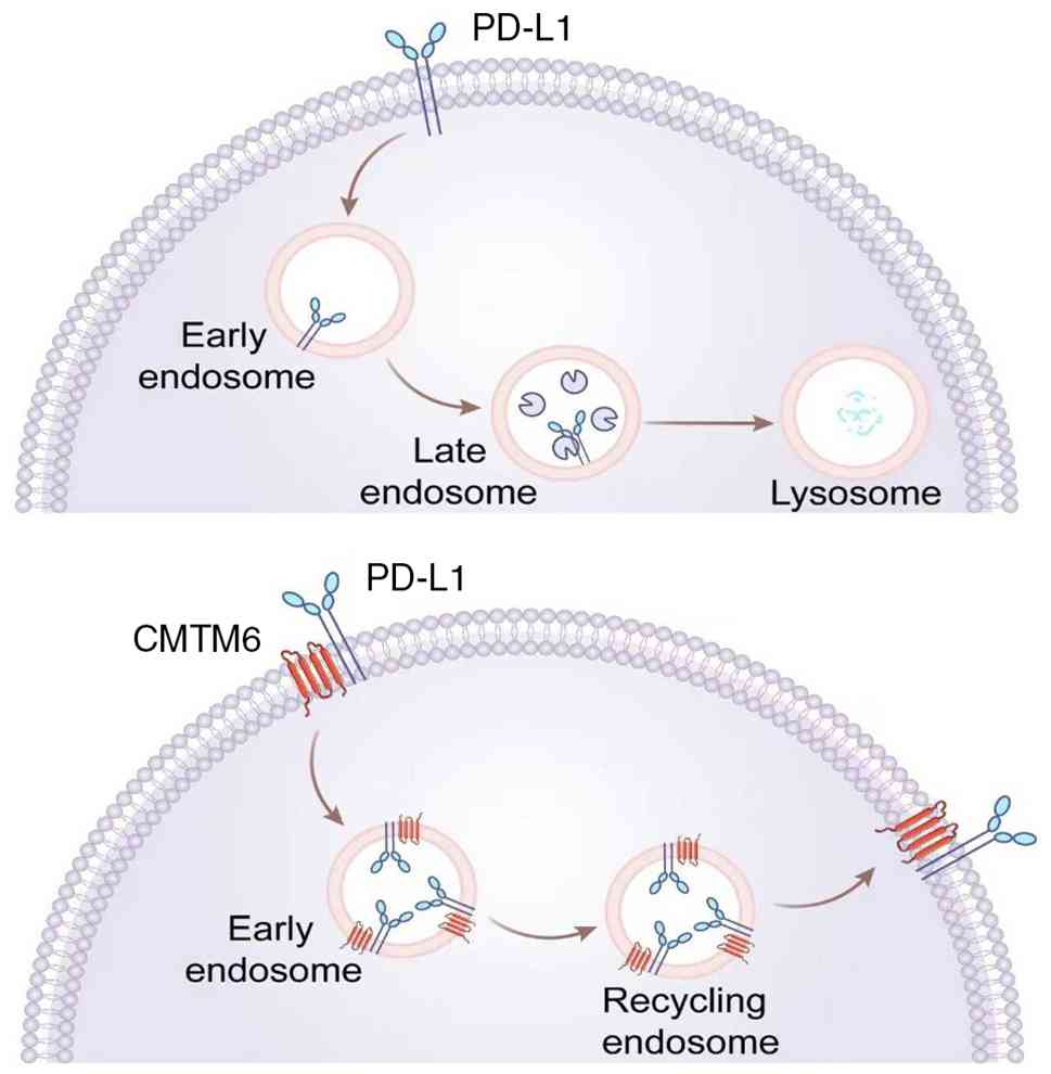
CMTM6 is recognized as a key regulator of PD-L1
protein stability. By binding to PD-L1, it prevents lysosomal
degradation, thereby increasing PD-L1 expression on the surface of
tumor cells and facilitating tumor immune evasion (Fig. 1). This function, validated across
multiple cancer types, represents one of the most well-established
and critical biological roles of CMTM6 (4,16–18).
Through positive regulation of PD-L1, CMTM6 markedly contributes to
tumor progression and PD-L1 expression itself holds considerable
clinical relevance in several malignancies (19–21).
In vitro and in vivo studies have both reported that
loss of CMTM6 markedly reduces PD-L1 levels, thereby alleviating
suppression of tumor-specific T-cell activity (4,16).
CMTM6 exhibits high specificity for PD-L1; its depletion decreases
PD-L1 expression without affecting the surface levels or
transcription of major histocompatibility class I molecules
(22,23). Moreover, CMTM6 expression levels
are notably positively associated with CD58. Under CMTM6-deficient
conditions, the expression of T-cell activation-related markers,
such as CD137 and IL-2, is markedly downregulated. Mechanistically,
CMTM6 serves as a critical regulator of CD58 protein stability,
physically interacting with CD58 on recycling endosomes and
protecting it from lysosomal degradation, thereby maintaining its
cell surface expression (24,25).
However, PD-L1 and CD58 compete for binding to the MARVEL domain of
CMTM6. When PD-L1 expression is upregulated, its competitive
advantage displaces CD58 from CMTM6 binding, leaving CD58
unprotected and shunting it toward the lysosomal degradation
pathway (10,24,25).
Additionally, a study reported that Hsc70 competes with CMTM6 for
binding to PD-L1, with both proteins sharing the same PD-L1 binding
region (amino acids 161–241). Knockdown of CMTM6 enhances the
interaction between Hsc70 and PD-L1, promoting PD-L1 degradation
via the endosomal microautophagy pathway (26).
Other signaling pathways associated
with CMTM6
Within the Wnt/β-catenin signaling pathway, CMTM6
interacts with membrane-bound enolase-1 (ENO-1), activating the
AKT/glycogen synthase kinase-3β (GSK3β) axis and thereby modulating
Wnt signaling. This mechanism is closely associated with the
maintenance of cancer stem cells, epithelial-mesenchymal transition
(EMT) and resistance to cisplatin (27). Furthermore, CMTM6 regulates the Wnt
pathway via β-catenin and is positively associated with
infiltration of CD66b+ neutrophils (28). In the ERK1/2 signaling pathway,
exosomal CMTM6 promotes ERK1/2 phosphorylation in macrophages,
enhancing the expression of M2 macrophage markers such as IL-10,
arginase-1 and CD163, thereby inducing M2 polarization (29). Concurrently, CMTM6 suppresses
activation of the pRB/E2F pathway, leading to inhibition of
hepatocellular carcinoma (HCC) cell proliferation, reduced colony
formation and G0/G1 phase cell cycle arrest
(30). Additionally, CMTM6
activates the MAPK signaling pathway while inhibiting MAPK/ERK
phosphorylation, which collectively promotes cell proliferation,
migration, invasion and EMT, and simultaneously suppresses
sodium-iodide symporter expression and apoptosis (31).
The expression of CMTM6 is also closely associated
with activation of the mTOR signaling pathway. Knockdown of CMTM6
results in the downregulation of key mTOR pathway proteins,
including phosphorylated mTOR and phosphorylated p70S6 kinase
(32). Conversely, overexpression
of CMTM6 upregulates critical components of the PI3K/AKT/mTOR and
MAPK pathways, such as PI3K, AKT, MEK and ERK, thereby stabilizing
the HER2 protein and inhibiting its ubiquitination (33). In cervical cancer (CC), CMTM6
promotes EMT and metastasis through the MAPK JNK/p38 pathway,
characterized by decreased E-cadherin and increased vimentin
expression. Additionally, CMTM6 suppresses the proliferation of
endocervical adenocarcinoma (ECA) through the p53 pathway (34,35).
Previous studies that performed RNA sequencing on CMTM6-knockdown
ECA cell lines reported that differentially expressed genes were
notably enriched in the p53 signaling pathway. Subsequent in
vitro functional assays demonstrated that small interfering
RNA-mediated CMTM6 knockdown markedly promoted the proliferative
capacity of ECA cells, and this effect was mediated through
activation of the p53 pathway (34,35).
Gene set enrichment analysis further revealed a positive
association between high CMTM6 expression and activation of the
IL-6-JAK-STAT3 signaling pathway, suggesting a role in
inflammation-related signaling and indirect promotion of
tumorigenesis (36). Moreover,
CMTM6 has been reported to regulate C-C motif chemokine ligand
(CCL)4 expression through the AKT/GSK3β/β-catenin pathway in a
PD-L1-independent manner, indicating a distinct mechanism involved
in tumor progression (Fig. 2)
(37).
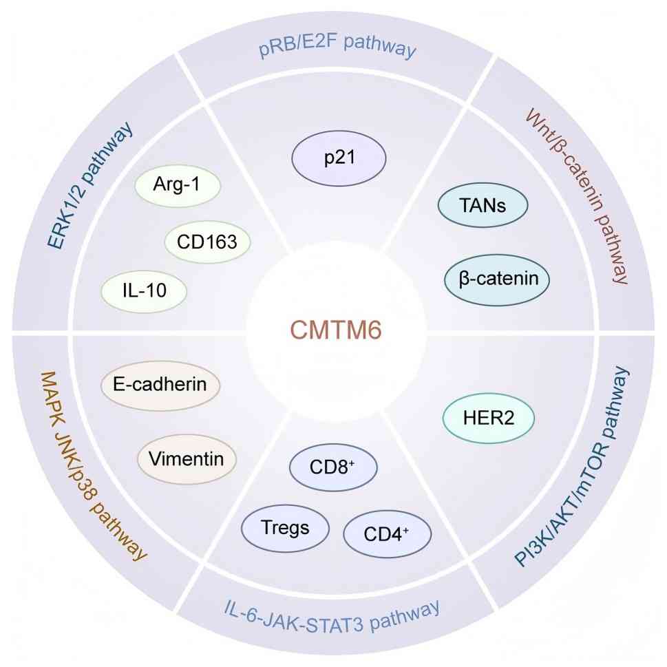 | Figure 2.CMTM6 is involved in multiple
signaling pathways and serves a multifaceted role in tumor
development and prognosis. CMTM6 is implicated in the pRB/E2F
pathway, p21-mediated cell cycle arrest epithelial-mesenchymal
transition, and the regulation of various immunosuppressive
factors, including Arg-1, CD163, IL-10, TANs and Tregs.
Additionally, it influences CD4+ and CD8+ T-cell dynamics. Arg-1,
arginase-1; CMTM6, chemokine-like factor-like MARVEL transmembrane
domain-containing protein 6; TANs, tumor-associated neutrophils;
Tregs, regulatory T cells. |
Mechanisms regulating CMTM6
expression
Research has reported that human antigen R (HuR)
binds to AU-rich elements within the 3′ untranslated region of
CMTM6 mRNA, stabilizing the transcript and upregulating its
expression (38). Further research
has revealed that the cerebellar degeneration-related protein 1
transcript upregulates CMTM6, potentially by modulating the
expression and/or activity of transcription factors essential for
CMTM6 expression (39).
Experimental evidence also suggests that WEE1 and ATM inhibitors
reduce PD-L1 expression by downregulating CMTM6 (40). Concurrently, EMT transcription
factors, particularly SNAIL, have been implicated in upregulating
CMTM6 expression (41). Yamamoto
et al (42) reported that
the hepatitis B virus drug entecavir increased PD-L1 expression
through upregulation of CMTM6. Additionally, tumor cells may
transfer CMTM6 to surrounding cells via exosomes, thereby enhancing
PD-L1 expression on tumor stromal cells, including immune cells
(29). Furthermore, five microRNAs
(miRs), including hsa-miR-545-5p and hsa-miR-514a-3p, have been
reported to exhibit a notable negative association with CMTM6
expression (34). Transcription
factors such as BCL3, CREB1 and FOXA1 are also predicted to
suppress CMTM6 transcription, while E2F4 and ELK1 are potential
activators of its expression (34).
In summary, CMTM6 is a widely expressed protein that
serves crucial roles in tumor occurrence, development and treatment
(24,33,34).
It stabilizes PD-L1 and CD58 by protecting them from lysosomal
degradation and extending their protein half-lives, thereby
modulating the activation threshold of T cells. CMTM6 is also
involved in multiple signaling pathways and is regulated at several
levels by factors such as HuR, miRs, EMT transcription factors and
antiviral therapeutics. Additionally, CMTM6 can be horizontally
transferred via exosomes. Together, these mechanisms enable CMTM6
to promote tumor immune evasion and cancer progression,
highlighting its potential as a promising target for cancer
immunotherapy.
Expression of CMTM6 in diseases
CMTM6 and cancer
A comprehensive study integrating data from
databases such as The Cancer Genome Atlas, Human Protein Atlas and
The Cancer Proteome Atlas analyzed the expression patterns and
genetic alteration profiles of CMTM6 across 33 cancer types,
alongside its relationship with the tumor immune microenvironment
(TIME) (Fig. 3) (43). The findings revealed that CMTM6
expression exhibits notable cancer type specificity and is closely
associated with the TIME, particularly PD-L1 expression and immune
cell infiltration. Moreover, CMTM6 demonstrated independent
prognostic value in multiple types of cancer. These results
highlight its potential as both a target for immunotherapy and a
prognostic biomarker (Table I)
(43).
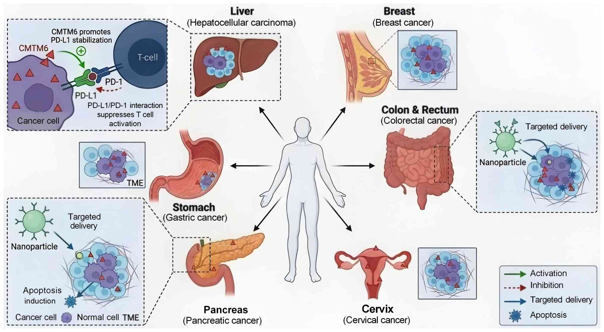 | Figure 3.CMTM6-driven PD-L1 stabilization in
multiple cancer types and nanoparticle-based targeted delivery
strategy. CMTM6 enhances the stabilization of PD-L1 across various
tumor types, including liver, breast, colorectal, gastric,
pancreatic and cervical cancer. The increased levels of PD-L1
interact with PD-1 on T cells, leading to the suppression of T-cell
activation. Additionally, the figure illustrates a nanoparticle
system designed for targeted delivery to cancer cells within the
TME, promoting apoptosis while sparing normal cells. CMTM6,
chemokine-like factor-like MARVEL transmembrane domain-containing
protein 6; PD-1, programmed death 1; PD-L1, programmed death ligand
1; TME, tumor microenvironment. |
 | Table I.Expression profile of CMTM6 in
various types of cancer. |
Table I.
Expression profile of CMTM6 in
various types of cancer.
| First author,
year | Type of cancer | Proposed
mechanism | Clinical sample
validation | Experimental
models | (Refs.) |
|---|
| Jia et al,
2023 | Lung cancer | High CMTM6
expression activates the following: Angiogenesis, IL-6-JAK-STAT3,
KRAS, PI3K-AKT-mTOR and TGF-β signaling pathways | Yes, n=110 | Cell lines: A549,
H358, Beas-2B | (36) |
| Gao et al,
2019 | Lung cancer | CMTM6 promotes
immune escape by stabilizing PD-L1 | Yes, n=141 | None (clinical
trial) | (49) |
| Zugazagoitia et
al, 2019 | Lung cancer | CMTM6 expression is
strongly positively associated with PD-L1 expression | Yes, n=180 | None (clinical
trial) | (50) |
| Koh et al,
2019 | Lung cancer | PD-L1 expression is
strictly dependent on the presence of CMTM6 | Yes, n=123 | None (clinical
trial) | (48) |
| Kong et al,
2024 | HCC | CMTM6 modulates the
Wnt pathway by targeting β-catenin | Yes, n=60 | Cell lines: HepG2,
HuH-7, MHCC97H, LO2 Animal model: Nude mouse xenograft | (28) |
| Yugawa et
al, 2021 | HCC | CMTM6 extends the
half-life of PD-L1, and thereby promotes EMT and the stemness
phenotype | Yes, n=259 | Cell lines:
HuH-7/Hep3B | (58) |
| Huang et al,
2022 | HCC | CMTM6 binds to p21
and inhibits its ubiquitination, thereby suppressing activation of
the pRB/E2F pathway | Yes, n=167 | Cell lines: HepG2,
Hep3B, HuH-7 Animal model: Nude xenograft | (30) |
| Huang et al,
2021 | HCC | CMTM6 stabilizes
vimentin to induce EMT, thereby promoting HCC metastasis | Yes, n=130 | Cell lines:
MHCC-97H, BEL-7402 HCCLM3 Animal model: Nude mouse xenograft. | (56) |
| Muranushi et
al, 2021 | HCC | CMTM6 promotes HCC
recurrence by regulating the expression of B7 family ligands | Yes, n=84 | Cell lines:
PLC/PRF/5, Hep3B | (59) |
| Miao et al,
2023 | Colorectal
cancer | CMTM6 stabilizes
CD58 protein by inhibiting its lysosomal degradation pathway | Yes, n=102 | Cell lines: 8505C,
A375, RKO, HAP1 | (24) |
| Shaha et al,
2024 | Colorectal
cancer | CMTM6 forms a
complex with Glut1 and Rab11, maintaining Rab11 mRNA level and
activity to promote Glut1 membrane localization | No | Cell lines: HCT116,
KM12L4, MC38 Animal model: Nude mouse xenograft. C57BL/6 syngeneic
mouse model | (71) |
| Peng et al,
2021 | Colorectal
cancer | CMTM6 expression is
positively associated with CD4+ and CD8+
T-cell infiltration | Yes, n=286 | None (clinical
trial) | (67) |
| Wu et al,
2021 | Colorectal
cancer | CMTM6 expression is
positively associated with CD163+ M2 macrophage
density | Yes, n=32 | None (clinical
trial) | (68) |
| Li et al,
2022 | Colorectal
cancer | CMTM6 may modulate
the immune microenvironment via the Wnt/β-catenin pathway | Yes, n=704 | None (clinical
trial) | (69) |
| Zhang et al,
2021 | Gastric cancer | Knockdown of CMTM6
leads to decreased PD-L1 expression | Yes, n=185 | None (clinical
trial) | (76) |
| Li et al,
2020 | Gastric cancer | CMTM6 expression is
strongly positively associated with PD-L1 expression | Yes, n=122 | None (clinical
trial) | (74) |
| Nishi et al,
2021 | Gastric cancer | CMTM6 promotes
immune escape by stabilizing PD-L1 | Yes, n=105 | None (clinical
trial) | (75) |
| Gao et al,
2023 | Pancreatic
cancer | CMTM6 enhances cell
proliferation, migration and invasion by stabilizing PD-L1 | Yes, n=179 | Cell lines: AsPC-1,
PANC-1 | (80) |
| Huang et al,
2022 | CC | CMTM6 promotes EMT
and metastasis through the MAPK (JNK/p38) pathway | Yes, n=306 | Cell line: CC
epithelial cells Animal models: Subcutaneous trans-planted tumor
model, experimental lung metastasis model | (34) |
| Liang et al,
2023 | CC | CMTM6 suppresses
ECA proliferation through the p53 pathway | Yes, n=241 | Cell line: HeLa,
SiHa Animal model: Female BALB/c nude mice | (35) |
| Ma et al,
2023 | CC | CMTM6 promotes
immune escape by stabilizing PD-L1 | Yes, n=102 | None (clinical
trial) | (83) |
| Yin et al,
2025 | CC | Exosomal transfer
of CMTM6 to macrophages induces M2a polarization via mTOR
activation, leading to C-C motif chemokine ligand 2-mediated
promotion of colorectal cancer progression | Yes, n=30 | Cell lines: TC-1,
HeLa, SiHa Animal models: Subcutaneous trans-planted tumor model,
experimental lung metastasis model | (13) |
| Xing et al,
2023 | Breast cancer | CMTM6 stabilizes
HER2 protein by inhibiting its ubiquitination | Yes, n=76 | Cell lines: SKBR3,
JIMT-1, BT-474 Animal model: Nude mouse xenograft | (33) |
| Xiao et al,
2021 | Breast cancer | EMT regulates PD-L1
expression by inducing CMTM6 and CMTM7 | Yes, n=299 | Cell lines: MCF-7,
MDA-MB-231 | (41) |
| Xiao et al,
2022 | Breast cancer | SNAI1-induced EMT
upregulates PD-L1 and CMTM6/7 synchronously in MCF-7 mesenchymal
cells | No | Cell lines: MCF-7,
MDA-MB-231 | (88) |
| Zheng et al,
2020 | OSCC | CMTM6 promotes
tumorigenesis by inducing NRP1 expression | Yes, n=244 | Cell lines: HN4,
HN6, HOK | (93) |
|
|
|
|
| Animal model: Nude
mouse xenograft model |
|
| Pang et al,
2021 | OSCC | Exosomal CMTM6 from
OSCC activates the ERK1/2 signaling pathway, inducing M2 macrophage
polarization | Yes, n=45 | Cell lines: Cal-27,
SCC25, THP-1 Animal model: 4NQO-induced OSCC mouse model | (29) |
| Martinez-Morilla
et al, 2020 | Melanoma | CMTM6 expression is
strongly positively associated with PD-L1 expression | Yes, n=60 | Cell lines: CD68
macrophages, CD3 T cells, CD20 B cells | (98) |
| Miao et al,
2023 | Melanoma | CMTM6 positively
regulates CD58 expression, while CD58 and PD-L1 exhibit competitive
binding to the CMTM6 MARVEL domain | Yes, n=88 | Cell lines: 8505C,
A375, RKO, HAP1 Animal models: in vivo xenograft models | (24) |
| Xue et al,
2022 | Glioma | CMTM6 expression is
strongly positively associated with PD-L1 expression | Yes, n=177 | None (clinical
trial) | (101) |
| Wei et al,
2022 | Glioma | Knockdown of CMTM6
down-regulates key proteins in the mTOR pathway, leading to cell
cycle arrest at the G1/G0 phase | Yes, n=44 | Cell lines: U87,
U251, T98G, U373, HEB Animal model: Nude mouse xenograft model | (32) |
| Li et al,
2025 | Glioma | CMTM6 inhibits
antigen presentation by stabilizing PD-L1 and promoting M2
polarization | No | Cell line: HMC3
human microglial cells Animal model: GL261 syngeneic mouse model of
glioblastoma | (103) |
| Yin et al,
2022 | Ovarian cancer | CMTM6 expression is
positively associated with CD4+ T-cell and neutrophil
infiltration | No | Cell lines: IOSE80,
A2780, ES2, Hey, SKOV3 | (104) |
| Wang et al,
2022 | Clear cell renal
cell carcinoma | CMTM6 deficiency
induces DNA damage and consequently causes cell cycle arrest at the
G2/M phase | Yes, n=144 | Cell lines: HKC,
A498, Caki-1, SN12-PM6 Animal models: Mouse orthotopic syngeneic
graft and xenograft renal cancer model | (107) |
| Ishihara et
al, 2021 | Undiffe rentiated
pleomorphic sarcoma | CMTM6 expression is
strongly positively associated with PD-L1 expression | Yes, n=51 | Cell line: CD8 T
cells | (108) |
| Chen et al,
2024 | Thyroid cancer | CMTM6 promotes cell
proliferation, migration, invasion and EMT via MAPK pathway
activation, while suppressing NIS expression and apoptosis | No | Cell lines: TEC,
FTC133, BCPAP, TPC-1, SW1736 | (31) |
| Zwick et al,
2025 | Acute leukemia | CMTM6 is
stabilizied by the FLT3-ITD mutation via protein interaction,
reducing its lysosomal and proteasomal degradation | Yes, n=15 | Cell lines: MV4-11,
Ba/F3, 32D, WEHI-3B Animal models: Allogeneic hematopoietic cell
transplantation model, MV4-11 ×enograft model | (14) |
CMTM6 in lung cancer
Lung cancer is one of the most prevalent and deadly
malignancies worldwide, encompassing two primary categories: Small
cell lung cancer (SCLC) and non-SCLC (NSCLC) (44,45).
The development and progression of lung cancer involve complex
signaling pathways that regulate key cellular processes, such as
proliferation, survival, metastasis and therapeutic resistance
(46). In NSCLC, CMTM6 expression
is markedly elevated compared with that in normal tissues (47). Immunodetection studies have
reported the presence of CMTM6 not only in cancer cells but also in
tumor-associated macrophages, lymphocytes and adjacent bronchial
epithelial cells (48,49). A retrospective analysis of patients
with NSCLC revealed that CMTM6 expression exhibits histological
subtype specificity in lung cancer, with notably higher levels in
squamous cell carcinoma than in adenocarcinoma. Additionally,
patients exhibiting high CMTM6 expression demonstrated a shorter
overall survival (OS) time than those with low expression levels.
Furthermore, CMTM6 expression was found to be elevated in cases
with tumor-infiltrating lymphocytes (TILs) when compared to cases
without such infiltration (47,50).
In lung adenocarcinoma, CMTM6 is markedly upregulated and notably
associated with advanced T stage and lymph node metastasis
(36). In patients with lung
squamous cell carcinoma (LUSC), CMTM6 expression has been reported
to be positively associated with PD-L1 at both mRNA and protein
levels. Furthermore, CMTM6 expression is positively associated with
infiltration of CD8+ T cells, macrophages, neutrophils and
dendritic cells, while it is negatively associated with CD4+ T-cell
infiltration, underscoring its notable role in modulating immune
cell dynamics within the LUSC tumor microenvironment (TME)
(51). Additionally, in SCLC,
elevated CMTM6 expression has been reported in high-risk patient
subgroups, suggesting its association with specific immune
phenotypes and supporting its role as a novel independent
prognostic factor (52).
CMTM6 in HCC
HCC, the most common form of primary liver cancer,
poses a notable global health challenge due to its prolonged
latency period and lack of distinct early symptoms, often resulting
in late-stage diagnosis and high mortality rates (53–55).
Numerous studies have reported that elevated CMTM6 expression in
HCC is closely associated with tumor progression and poor
prognosis. Increased CMTM6 levels are negatively associated with
patient survival and positively associated with α-fetoprotein
levels (15,56). Mechanistically, CMTM6 promotes
tumor cell proliferation by activating the Wnt/β-catenin signaling
pathway through stabilization of β-catenin (15). Moreover, CMTM6 facilitates the
infiltration of tumor-associated neutrophils and synergizes with
PD-L1 to regulate T cell-mediated immune responses (28). It also modulates the infiltration
patterns of both CD4+ and CD8+ T cells within
the TME (57). Clinically, high
co-expression of CMTM6 and PD-L1 is associated with poor tumor
differentiation, micro-intrahepatic metastasis, frequent
recurrence, and consequently, shorter overall and recurrence-free
survival in patients with HCC (58).
Moreover, CMTM6 promotes the proliferation and
invasion of HCC cells by stabilizing vimentin, thereby inducing EMT
(56). Elevated CMTM6 expression
has been associated with increased Ki-67 levels and shorter
recurrence-free survival (59).
Additionally, CMTM6 promotes HCC recurrence by regulating the
expression of B7 family ligands, suggesting its potential as both a
biomarker for HCC recurrence risk and a therapeutic target
(59). Furthermore, in the
macrotrabecular-massive (MTM) subtype of HCC, co-expression of
CMTM6 and PD-L1 is associated with higher inflammatory cell density
and elevated PD-L1 expression, which corresponds with increased
risks of HCC progression and mortality. An immune classification
system based on CMTM6/PD-L1 co-expression effectively stratifies
patients with MTM HCC by prognosis, highlighting the potential of
CMTM6 as a biomarker and therapeutic target in HCC management
(60). By contrast, Huang et
al (30) and Zhu et al
(61) reported low CMTM6
expression in HCC tissues, with higher CMTM6 levels associated with
improved patient prognosis (30,61,62).
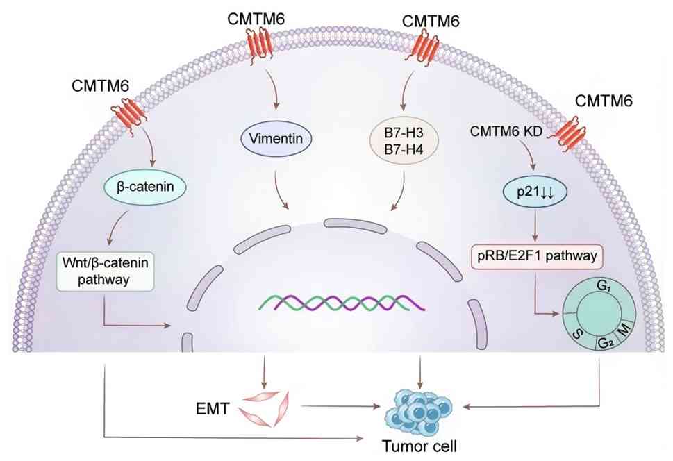
Huang et al (30) reported
that downregulation of CMTM6 in HCC cells reduced p21 levels,
leading to activation of the pRB/E2F1 pathway and promoting
G1/S phase transition (Fig.
4).
Based on the relevant literature, three principal
factors account for the discrepancies regarding CMTM6 in HCC.
First, the etiological heterogeneity across cohorts, including
variations in hepatitis virus type, the presence of cirrhosis,
histological subtype and metabolic dysfunction-associated fatty
liver disease, markedly influences the TIME. Second, CMTM6 exhibits
bidirectional, context-dependent functions; it stabilizes PD-L1 to
suppress antitumor immunity, binds to p21 to inhibit
G1/S transition, or interacts with vimentin to promote
EMT, with the net effects varying according to the molecular
partner involved. Third, the complexity of TIME further diversifies
the observed outcomes. Wei et al (32). reported that the co-expression of
CMTM6 and PD-L1, along with CD4+ T-cell infiltration,
was associated with a favorable prognosis, whereas Kong et
al (28) revealed that CMTM6
can promote tumor-associated neutrophil infiltration through the
Wnt/β-catenin pathway, which is associated with a poor prognosis.
Furthermore, differences among studies regarding sample size,
clinical stage distribution, detection methodologies and assessment
criteria may contribute to the inconsistent conclusions.
CMTM6 in colorectal cancer (CRC)
CRC ranks as the third most commonly diagnosed
cancer and the second leading cause of cancer-related mortality
worldwide (63,64). Its complexity is driven by diverse
signal transduction networks that regulate cellular proliferation,
survival, differentiation and apoptosis (65). CMTM6 expression has been reported
to be elevated in CRC tissues compared with that in matched normal
tissues, with high CMTM6 levels associated with increased PD-L1
expression and greater immune cell infiltration (66). Notably, CMTM6 levels tend to be
lower in advanced-stage CRC tumors compared with those in
early-stage tumors. High CMTM6 expression is associated with lower
pathological T stage and increased density of TILs, suggesting a
notable role for CMTM6 in modulating the TIME (67). Moreover, Wu et al (68) reported that in patients with rectal
cancer undergoing PD-1/PD-L1 inhibitor therapy, CMTM6 expression in
M2 macrophages can serve as a promising biomarker for predicting
treatment response. Furthermore, an immunohistochemical analysis of
a tissue microarray from 704 patients with CRC assessing β-catenin,
CMTM6, PD-L1 and mismatch repair protein expression revealed a
positive association between CMTM6/PD-L1 co-expression and
β-catenin levels, suggesting that CMTM6/PD-L1 may influence the
immune microenvironment via the Wnt/β-catenin signaling pathway
(69).
Liver metastasis of CRC is a major contributor to
CRC-related mortality (70). A
study reported that CMTM6 promotes CRC liver metastasis by
mediating the Warburg effect. Mechanistically, CMTM6 forms a
complex with glucose transporter 1 (Glut1) and Rab11 in the
endoplasmic reticulum, facilitating Rab11-dependent trafficking of
Glut1 to the plasma membrane, which enhances glucose uptake and
glycolysis (71). CMTM6 serves a
critical role in sustaining CRC cell cycle progression and liver
metastasis, contrasting with its function in HCC, where it
suppresses G1 to S phase transition by stabilizing p21
and inactivating the pRB/E2F pathway (30).
CMTM6 in gastric cancer (GC)
GC is a complex and heterogeneous disease
characterized by marked phenotypic and genetic variability
(72). Studies have reported that
CMTM6 expression is markedly elevated in GC tissues compared with
that in normal gastric tissues, with even higher levels observed in
advanced-stage disease (73).
Immunohistochemical analysis of 122 postoperative GC tissue
specimens revealed a notable association between CMTM6 and PD-L1
protein expression (74). A
retrospective study involving 105 patients with stage II/III GC
performed by Nishi et al (75) reported markedly lower 5-year OS and
disease-free survival rates in the CMTM6 high-expression group
compared with that in the low-expression group, supporting the role
of CMTM6-mediated immune escape via PD-L1 (75). Furthermore, immunohistochemical
staining of 185 GC specimens from radical gastrectomies indicated
that high co-expression of CMTM6 and PD-L1 was notably associated
with Borrmann type, lymph node metastasis, peritoneal metastasis
and Tumor-Node-Metastasis stage, but showed no association with
patient age, sex, tumor size, differentiation grade, tumor location
or T stage. Survival analysis further demonstrated that high CMTM6
expression was associated with a shorter OS and worse prognosis,
with the worst outcomes observed in cases with concomitant high
PD-L1 expression (76).
Additionally, in gastric adenocarcinoma, CMTM6 expression has been
shown to be higher in tumor tissues than in adjacent normal
tissues, and to be positively associated with PD-L1 expression in
both epithelial and stromal compartments. CMTM6 expression also
showed positive associations with tumor burden and the Ki-67
proliferation index, indicating that membrane co-expression of
CMTM6 and PD-L1 on tumor epithelial cells serves as a marker of
poor prognosis in gastric adenocarcinoma (77).
CMTM6 in pancreatic cancer (PC)
PC remains one of the most lethal malignancies
worldwide, with limited advances in effective treatments over
recent decades (78,79). Notably, studies have identified
upregulated CMTM6 expression in pancreatic adenocarcinoma tissues
(40,80). Functional analyses have
demonstrated that CMTM6 promotes the proliferation, migration and
invasion of PC cells (80). CMTM6
expression has also been reported to be positively associated with
PD-L1 levels, and CMTM6 has been shown to co-immunoprecipitate with
PD-L1 protein in PC cell lines. Upregulation of CMTM6 contributes
to shaping an inflammatory TME characterized by a heightened immune
response (80). Furthermore,
research has indicated that inhibition of WEE1, alone or in
combination with ATM inhibition, downregulates PD-L1 expression by
blocking phosphorylation of GSK3β at serine 9 and reducing CMTM6
levels. In Capan-1 mouse xenograft models, treatment with AZD1775
(a WEE1 inhibitor) combined with AZD0156 (an ATM inhibitor) has
been shown to reduce tumor growth and downregulate the expression
of PD-L1, CMTM6, CD163 and CXCR2 in tumors, factors known to
contribute to tumor immune evasion (40).
CMTM6 in CC
CC, primarily driven by human papillomavirus
infection, is considered preventable and manageable due to its
well-established etiology (81,82).
CMTM6 serves a critical role in CC, with its expression closely
associated with the biological behavior of the tumor. Studies have
reported that CMTM6 expression is markedly upregulated in CC
tissues compared with that in adjacent non-cancerous tissues, and
positively associated with PD-L1 expression. This co-expression is
strongly associated with poor patient prognosis (13,83).
In vitro experiments have demonstrated that CMTM6 promotes
invasion, migration, proliferation and EMT of CC cells by
activating the MAPK JNK/p38 signaling pathway (34). Conversely, CMTM6 inhibits the
proliferation of ECA cells through the p53 pathway and recruits T
cells, thereby enhancing the antitumor immune response (35). The multiple molecular functions of
CMTM6 underpin its dual role; it stabilizes PD-L1 to suppress
antitumor immunity, binds to p21 to inhibit G1/S
transition, or interacts with vimentin to promote EMT, with the net
effects varying according to the molecular partner involved.
Within the TIME, CMTM6 expression has been reported
to be positively associated with immune checkpoint components.
Silencing CMTM6 increases infiltration of CD8+ and
CD4+ T cells while reducing the proportion of exhausted
T cells (84). Moreover, soluble
CMTM6 levels are markedly elevated in the plasma of patients with
CC and are positively associated with exosomal PD-L1, suggesting a
role for CMTM6 in tumor immune evasion via the exosomal pathway
(85). A recent study further
reported that exosomes secreted by CC cells carry CMTM6, which
activates the mTOR signaling pathway in macrophages, inducing M2a
polarization. These M2a macrophages subsequently secrete CCL2,
enhancing the migration and invasion of CC cells, and promoting
disease progression (13).
Collectively, these findings highlight CMTM6 not only as a
potential prognostic biomarker for CC but also as a promising
target for immunotherapy.
CMTM6 in breast cancer
Breast cancer is the most common malignancy among
women worldwide. Triple-negative breast cancer (TNBC), which
accounts for 10–20% of all breast cancer cases, is an aggressive
subtype associated with a poor prognosis (86). A study reported that CMTM6
expression is markedly higher in patients with TNBC compared with
that in those with HER2-positive breast cancer (87). Moreover, elevated CMTM6 levels are
associated with shorter progression-free survival (PFS) in patients
with TNBC, and multivariate Cox regression analysis has identified
CMTM6 as an independent risk factor for PFS (87). Furthermore, CMTM6 expression has
been reported to be positively associated with both the EMT score
in breast cancer cell lines and the key EMT marker vimentin in TNBC
(41). In SNAI1-induced MCF-7
cells (MCF-7-Mes), acquisition of a mesenchymal phenotype leads to
increased PD-L1 and CMTM6 expression. Silencing CMTM6 in MCF-7-Mes
cells partially reduces PD-L1 surface expression, indicating that
PD-L1 expression is partially dependent on CMTM6. Furthermore, dual
knockdown of CMTM6 and CMTM7 markedly decreases PD-L1 surface
expression, suggesting that these proteins cooperatively regulate
PD-L1 levels (41,88). Additionally, research has reported
that hsa_circ_0067842 upregulates CMTM6 expression via HuR, while
knockdown of HuR reduces CMTM6 mRNA stability. These findings
establish the hsa_circ_0067842/HuR/CMTM6/PD-L1 axis as a potential
prognostic biomarker and therapeutic target for breast cancer
immunotherapy (89).
CMTM6 in oral squamous cell carcinoma
(OSCC)
OSCC, the predominant subtype of head and neck
squamous cell carcinoma, has a 5-year survival rate of <50%,
with its precise etiology still unclear (90–92).
CMTM6 holds notable clinicopathological relevance in OSCC, as its
high expression is closely associated with tumor progression and
poor prognosis. A study reported that both CMTM6 and neuropilin-1
(NRP1) are upregulated in OSCC tissues and physically interact.
NRP1 contributes to the degradation of CMTM6, while CMTM6 promotes
tumorigenesis by inducing NRP1 expression (93). Furthermore, CMTM6 expression is
positively associated with PD-1 and PD-L1 levels, suggesting a
potential synergistic role in immune pathways that facilitate tumor
immune escape in OSCC (94). CMTM6
can also be transferred to macrophages via exosomes, where it
activates the ERK1/2 signaling pathway and induces M2 macrophage
polarization, thereby promoting malignant progression of OSCC
(95). Additionally, research has
shown that CMTM6 interacts with membrane-bound ENO-1 to activate
the AKT/GSK3β axis, which modulates the Wnt signaling pathway,
driving EMT and cisplatin resistance (27). Thus, CMTM6 not only regulates tumor
cell behavior in OSCC but also markedly influences the immune
microenvironment, underscoring its potential as a therapeutic
target (Fig. 5).
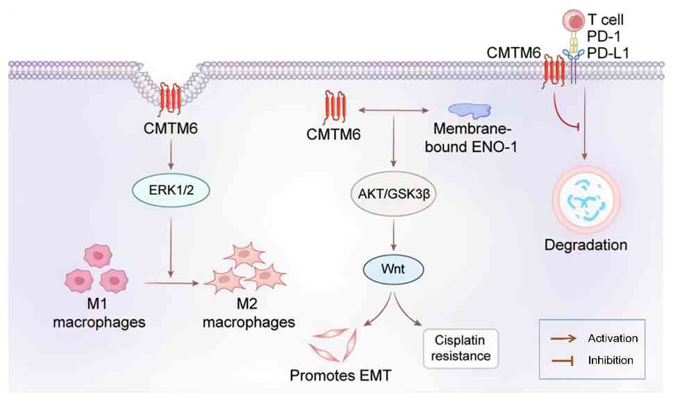 | Figure 5.Mechanisms of CMTM6 action in oral
squamous cell carcinoma. CMTM6 can be transferred to macrophages
via exosomes, where it activates the ERK1/2 signaling pathway and
induces M2 macrophage polarization. Additionally, CMTM6 interacts
with membrane-bound ENO1 to activate the AKT/GSK3β axis, which
modulates the Wnt signaling pathway, thereby driving EMT and
contributing to cisplatin resistance. Furthermore, CMTM6 maintains
PD-L1 expression on the surface of tumor cells by inhibiting its
lysosomal degradation. By stabilizing PD-L1, CMTM6 facilitates the
binding of PD-L1 to PD-1 on T cells, thus suppressing T-cell
activation. CMTM6, chemokine-like factor-like MARVEL transmembrane
domain-containing protein 6; EMT, epithelial-mesenchymal
transition; ENO-1, enolase-1; GSK3β, glycogen synthase kinase-3β;
PD-1, programmed cell death protein 1; PD-L1, programmed death
ligand 1. |
CMTM6 in melanoma
Melanoma, which arises from melanocytes, is the most
lethal form of skin cancer (96,97).
In melanoma, CMTM6 expression has been reported to be markedly
associated with several immune markers, including PD-L1, CD3, CD20
and CD68. Furthermore, high CMTM6 expression within stromal and
immune compartments has been shown to be significantly associated
with improved survival following immunotherapy (P=0.007) and
demonstrates notable prognostic value in multivariate analyses
(98). Furthermore, high
co-expression of CMTM6 and PD-L1 in the stromal compartment has
been significantly associated with prolonged survival (P=0.028),
highlighting CMTM6 as a potential predictive biomarker for immune
checkpoint inhibitor therapy (98). Further studies have reported that
CMTM6 is essential for maintaining CD58 stability in melanoma,
where intact CD58 expression and its interaction with CD2 are
critical for effective antitumor immunity. When CD58 is lost, CMTM6
facilitates PD-L1 upregulation, thereby promoting immune evasion.
The competitive interactions among CMTM6, CD58 and PD-L1 regulate
their endosomal recycling and lysosomal degradation rates,
establishing a molecular mechanism by which cancer cells balance
immunosuppressive and immunostimulatory signals (10). Collectively, these findings
underscore the marked role of CMTM6 in melanoma immunotherapy,
positioning it as both a promising prognostic biomarker and
therapeutic target.
CMTM6 in glioma
Glioma is the most prevalent and aggressive
malignant brain tumor in adults, posing notable challenges in
oncology due to its rapid progression, therapeutic resistance and
poor prognosis (99,100). In glioma, CMTM6 expression is
closely associated with several biological processes and
therapeutic responses. A study reported that both CMTM6 and CMTM4
are markedly associated with PD-L1 expression in glioma, with
macrophages identified as the primary cell type expressing these
proteins. High expression levels of CMTM6 and CMTM4 are associated
with reduced OS, suggesting their potential as biomarkers for
immune-targeted interventions in glioma (101). Moreover, elevated CMTM6
expression in glioma has been associated with poor prognosis.
Knockdown of CMTM6 inhibits the proliferation, invasion and
migration of U87 and U251 glioma cells, increases apoptosis rates,
inactivates the mTOR signaling pathway, and reduces tumor volume
and Ki-67 expression (32). CMTM6
is highly expressed in glioma and is strongly associated with the
TIME and inflammatory responses (102). A recent study further
demonstrated that CMTM6 is markedly upregulated in high-grade
glioma and associated with poor patient outcomes. CMTM6 knockout
resulted in downregulation of PD-L1 and TGFβ1/2/3, alongside
upregulation of pro-inflammatory factors IL-6 and CCL3, promoting
microglial polarization toward the M1 phenotype and thereby
modulating the immune microenvironment (103). Collectively, these findings
underscore the crucial role of CMTM6 in glioma pathogenesis,
progression and immune regulation, highlighting its potential as
both a diagnostic biomarker and therapeutic target.
CMTM6 in other types of cancer
Beyond the aforementioned malignancies, CMTM6 serves
pivotal roles in several other tumors, with its functional impact
and mechanisms differing across cancer types. In ovarian cancer,
the most lethal gynecological malignancy, CMTM6 upregulation is
paradoxically associated with a more favorable prognosis. This may
be attributed to its modulation of immune cell infiltration and the
expression of immune cell markers, with copy number variations and
DNA methylation implicated in its aberrant expression (104–106). In clear cell renal cell
carcinoma, CMTM6 depletion has been reported to notably inhibit
cell proliferation, migration and invasion, while enhancing
antitumor immunity, evidenced by increased infiltration of CD4+ and
CD8+ T cells. Loss of CMTM6 also induces a DNA damage response,
leading to cellular senescence and secretion of inflammatory
factors (107). In
undifferentiated pleomorphic sarcoma, CMTM6 expression has been
reported to be strongly associated with high PD-L1 levels, and
elevated expression is markedly associated with poor prognosis
(108). In thyroid carcinoma,
CMTM6 promotes the development of thyroid cancer by inhibiting
sodium/iodide symporter activity through activating the MAPK
signaling pathway, with its expression closely associated with
tumor metastasis (31). In acute
myeloid leukemia, FLT3-ITD mutations enhance CMTM6 protein
stability through protein-protein interactions, thereby promoting
PD-L1 expression and facilitating immune evasion (14). Collectively, these studies
underscore the complex and diverse mechanisms through which CMTM6
influences tumor biology, emphasizing its potential as a
therapeutic target across multiple cancer types.
CMTM6 in non-neoplastic diseases
Beyond its roles in oncology, CMTM6 has emerged as a
key regulator in numerous non-neoplastic pathological conditions.
In dry eye disease (DED), reduced CMTM6 expression is associated
with increased disease severity. Its downregulation contributes to
decreased tear secretion, corneal epithelial defects and heightened
inflammatory responses, implicating the CMTM6-NF-κB p65 signaling
axis as a potential therapeutic target for DED (109). In primary Sjögren's syndrome,
CMTM6 levels are notably elevated in both the serum and labial
salivary gland tissues of patients, and are positively associated
with PD-1/PD-L1 levels, disease activity (as assessed by EULAR
Sjögren's Syndrome Disease Activity Index) and immunological
indices, including IgG and erythrocyte sedimentation rate. By
contrast, serum CMTM6 levels are negatively correlated with
lymphocyte counts and CD3+ T cells (r=−0.397). These findings
suggest that CMTM6 may participate in the PD-1/PD-L1 pathway by
regulating PD-L1 stability, thereby influencing lymphocyte
activation and peripheral immune tolerance (110). Additionally, in Helicobacter
pylori-infected gastric mucosal cells, CMTM6 stabilizes PD-L1
protein by preventing its degradation. CRISPR/Cas9-mediated
knockout of CMTM6 alters protein ubiquitination and affects the
expression of cell surface receptors, highlighting its regulatory
role in infection-associated immune modulation (111).
Furthermore, CMTM6 contributes to the pathogenesis
of autoimmune diseases. In antineutrophil cytoplasmic
antibody-associated vasculitis (AAV), reduced CMTM6 expression
leads to increased lysosomal degradation of PD-L1, thereby
impairing negative regulatory signaling in T cells. This suggests
that targeting lysosomal function could offer a novel therapeutic
approach for AAV (112). CMTM6
expression is also upregulated in neutrophils from patients with
antiphospholipid syndrome (APS), and neutrophil extracellular traps
(NETs) serve a critical pathogenic role in thrombosis associated
with APS. It is therefore hypothesized that CMTM6 may contribute to
the pro-thrombotic process in APS by promoting or sustaining
excessive neutrophil activation and enhancing NET release; however,
the underlying molecular mechanisms, including the potential
involvement of PD-L1 stabilization or other signaling pathways,
remain to be elucidated (113,114). A recent study identified CMTM6 as
a key regulator in the pathogenesis of rheumatoid arthritis (RA),
acting through the CMTM6-TAK1-NF-κB/MAPK axis to modulate
fibroblast-like synoviocyte function and macrophage polarization,
thereby promoting RA progression. Targeting CMTM6 thus represents a
potential therapeutic direction for RA, although the specific
binding sites between CMTM6 and TAK1, as well as the particular
ubiquitin ligases and deubiquitinases involved in regulating this
axis, require further analysis (115). In summary, CMTM6 has emerged as a
promising diagnostic and therapeutic target for several
non-neoplastic diseases; however, the precise mechanisms by which
CMTM6 influences the onset and progression of autoimmune disorders
require further investigation.
CMTM6 and clinical translation
Cancer is a major global public health challenge. A
critical mechanism of cancer immune evasion involves immune
checkpoint molecules, and leveraging the immune system to combat
malignant tumors has become a central focus in cancer therapy.
CMTM6 serves a pivotal role in the treatment of several types of
cancer, primarily by stabilizing key proteins, regulating signaling
pathways and modulating therapy resistance (Table II). In lung cancer, CMTM6
influences DNA double-strand break repair by regulating the nuclear
translocation of PD-L1, thereby contributing to radiotherapy
resistance (74). In NSCLC, CMTM6
co-localizes with EGFR in recycling endosomes, preventing its
lysosomal degradation. Elevated CMTM6 expression is associated with
resistance against tyrosine kinase inhibitors, and targeting CMTM6
effectively suppresses the proliferation of these resistant cancer
cells (116).
| Table II.Role of CMTM6 in cancer therapy. |
Table II.
Role of CMTM6 in cancer therapy.
| First author/s,
year | Cancer type | Mechanism | Methods | Drugs | Clinical
trials | (Refs.) |
|---|
| Huang et al,
2022 | Hepatocellular
carcinoma | CMTM6 binds to p21
and inhibits its ubiquitination, thereby inducing
G0/G1 phase arrest | Co-IP,
ubiquitination assay, flow cytometry | Doxorubicin,
cisplatin | No | (30) |
| Xia et al,
2025 | Lung cancer | CMTM6 binds to EGFR
and thereby inhibits its lysosomal degradation | Proteomic screening
and in vitro/in vivo functional validation | Anti-CMTM6 nanobody
(1A5) | Yes, n=92 | (116) |
| Xing et al,
2023 | Breast cancer | CMTM6 stabilizes
the HER2 protein by inhibiting its ubiquitination-mediated
degradation and activates downstream signaling | Ubiquitination
assay and in vitro/in vivo functional validation | Trastuzumab | Yes, n=72 | (33) |
| Mohapatra et
al, 2021 | Oral squamous cell
carcinoma | CMTM6 activates Wnt
signaling through the ENO-1/AKT/GSK3β axis, promoting cancer stem
cell properties and drug resistance | Co-IP, IHC,
immunofluore-scence, RT-qPCR, RNA sequencing, in vitro/in
vivo functional validation | Cisplatin | Yes, n=34 | (27) |
| Zhu et al,
2024 | Pancreatic ductal
adenocarcinoma | EP300 mediates
H3K27ac to activate CMTM6 transcription; CMTM6 subsequently
stabilizes IGF2BP1 by inhibiting its ubiquitination | Co-IP, liquid
chromatography with tandem mass spectrometry, chromatin
immunoprecipitation sequencing RNA immunopreci-pitation
sequencing, in vitro/in vivo functional validation | Gemcitabine | Yes, n=45 | (118) |
| Chabu et al,
2026 | Pancreatic ductal
adenocarcinoma | iSTORM delivers
CMTM6-targeting shRNA, disrupting PD-L1 stability and inhibiting
myeloid-mediated immunosuppression, thereby enhancing CD8+ T-cell
activation and antitumor function | In vitro
experiments and multiple mouse models | Tumor-targeting
microbial immunotherapy | No | (119) |
| Meng et al,
2022 | GBM | CMTM6 enhances GBM
cell migration through EMT regulation; PL markedly inhibits GBM
cell proliferation and induces CMTM6 | In
vitro/in vivo functional validation, RT-qPCR, western
blotting, flow cytometry | PL | No | (120) |
| Tulchiner et
al, 2021 | Renal cell
carcinoma | Higher CMTM6
expression in immune cells vs. tumor cells, and it is positively
associated with PD-L1 | IHC | Nivolumab | Yes, n=16 | (121) |
| Wang et al,
2025 | Breast cancer | Knockout of
circUBR5 induces an 8-fold increase in miR-340-5p expression,
whereas miR-340-5p directly targets and regulates CMTM6 | Western blotting,
flow cytometry, RNA-FISH, in vitro/in vivo functional
validation | Docetaxel | No | (122) |
In cisplatin-resistant OSCC, CMTM6 promotes
chemoresistance by regulating ribosome biogenesis and the Wnt
signaling pathway, while its inhibition restores cisplatin
sensitivity (27,117). In pancreatic ductal
adenocarcinoma (PDAC), CMTM6 forms a positive feedback loop with
insulin-like growth factor 2 mRNA-binding protein 1, enhancing
tumor stemness and contributing to gemcitabine resistance.
Combining an EP300 inhibitor with gemcitabine effectively overcomes
this resistance (118). A recent
study on novel microbial immunotherapy for PDAC has characterized
the tumor-targeting capability, immune-remodeling properties and
safety profile of an attenuated Salmonella typhimurium
strain, CRC2631, and its next-generation derivative, iSTORM.
Notably, iSTORM exploits its bacterial carrier CRC2631 to
selectively colonize PDAC tumors and locally deliver
CMTM6-silencing short hairpin RNA. This then disrupts PD-L1
stability and inhibits myeloid-mediated immunosuppression,
ultimately enhancing CD8+ T-cell activation and antitumor function
(119). In glioblastoma
multiforme, elevated CMTM6 expression is closely associated with
the TIME, promoting cell migration and EMT. Notably, the anticancer
compound piperlongumine (PL) induces CMTM6 expression, but the deep
mechanism of the relationship between PL and CMTM6 needs further
study (120). Moreover, in renal
cell carcinoma, CMTM6 expression is higher in immune cells than in
tumor cells. It is also positively associated with PD-L1, and its
elevated levels are associated with a shorter PFS in patients
receiving nivolumab treatment (121).
In HER2-positive breast cancer, CMTM6 stabilizes the
HER2 protein by inhibiting its ubiquitination, resulting in
trastuzumab resistance. Knockdown of CMTM6 restores drug
sensitivity and evidence suggests that combining trastuzumab-based
therapies with CMTM6 inhibitors, such as rapamycin, lapatinib and
pyrotinib, may enhance treatment efficacy (33). In TNBC, CMTM6 modulates docetaxel
resistance through c-MYC-mediated ribosome biogenesis, while
knockdown of circUBR5 enhances the activity of miR-340-5p and
reduces the expression of CMTM6 and c-MYC, thereby improving
chemosensitivity (122).
Additionally, research has indicated that REIC/Dkk-3 competitively
binds CMTM6, releasing PD-L1 and promoting its degradation, thereby
suppressing tumor progression (123). A recent study reported that a
novel anti-PD-L1 antibody (H1A) can disrupt the PD-L1-CMTM6
interaction, induce PD-L1 degradation, and enhance myeloid cell
activation and cytotoxic T-cell expansion, improving therapeutic
outcomes in tumors with limited responses to immune checkpoint
inhibitors (124). Another study
constructed a stable full-length membrane protein complex of
CMTM6-PD-L1 using membrane simulation strategies. Based on this
complex, an anti-CMTM6 nanobody, 1A5, was screened, which can
reduce T cell immunosuppression in vitro and exerts its
antitumor effect primarily through CD8+ T cells in mouse
models. This confirms that CMTM6 can serve as a novel target for
antibody-mediated tumor immunotherapy (125). Notably, a study on the treatment
of skin malignant melanoma found that by designing intermediate
chimeric peptide (PEP)-PDL1, it could specifically bind to the PD-1
binding domain of PD-L1, rather than the Hsc70/CMTM6 binding
domain, thereby avoiding competitive inhibition by CMTM6.
Simultaneously, it binds to Hsc70, mediating the degradation of
PD-L1 into lysosomes, thus restoring antitumor T-cell immunity in a
B16F10-derived malignant melanoma mouse model (126).
In summary, targeting CMTM6 and its associated
pathways may represent a potential therapeutic strategy for
multiple cancer types. However, the translation of CMTM6-targeted
therapy from basic research to clinical application faces several
challenges. The first is off-target effects and safety concerns:
CMTM6 is broadly expressed in normal tissues with a substrate
spectrum extending beyond PD-L1, simultaneously stabilizing the
inhibitory ligand PD-L1 and the co-stimulatory molecule CD58;
non-selective inhibition may therefore compromise antitumor
immunity. Moreover, strong and specific binding of CMTM6 to FAS has
been observed in mouse models, enhancing the sensitivity of mouse
cells to FAS ligand-induced cytotoxicity. However, in human
cell-based studies, this interaction is absent for human FAS, which
can be attributed to a three-amino-acid divergence at the boundary
between the extracellular and transmembrane domains. This
divergence disrupts the binding capacity, creating a
species-specific translational pitfall (127). The second challenge is tumor
heterogeneity: CMTM6 exhibits highly spatiotemporally dynamic
expression across different cancer types, diverse
microenvironmental cell populations, and under therapeutic
pressure, rendering uniform strategies ineffective. The third is
functional complexity: CMTM6 can drive chemotherapy resistance,
stabilize EGFR signaling and reshape the immunosuppressive
microenvironment via exosomes, all independently of PD-L1. In
response to these challenges, current strategies are transitioning
from global suppression to precision modulation. This includes the
development of novel antibodies, mesoscopic chimeric peptides and
nanobodies that disrupt the PD-L1-CMTM6 interaction to achieve
substrate-selective intervention. Additionally, combining these
strategies with immune checkpoint blockade addresses CD58
downregulation (10,125,126). The utilization of exosomal CMTM6
also facilitates non-invasive patient stratification. Collectively,
these efforts aim to advance CMTM6-targeted therapy towards
precision and personalized medicine.
Conclusions and future perspectives
The present comprehensive review systematically
assessed the biological characteristics of CMTM6, its involvement
in key signaling pathways, and its expression patterns and
functional roles across diverse diseases. As a crucial
transmembrane protein, CMTM6 serves a central role in fundamental
biological processes such as cell proliferation, immune regulation
and signal transduction. Aberrant CMTM6 expression has been
identified in numerous types of cancer, including lung, colorectal
and breast cancer, where it is associated with tumor
aggressiveness, patient prognosis and therapeutic response. Beyond
oncology, altered CMTM6 expression is increasingly recognized in
non-neoplastic conditions, including autoimmune and chronic
inflammatory diseases, suggesting its involvement in their
pathogenesis and progression.
In the field of oncological therapeutics, CMTM6 has
emerged as a potential therapeutic target, with its roles in
immunotherapy, targeted therapy and combination treatment
strategies being progressively elucidated. However, its clinical
translation has numerous challenges: The broad physiological
expression of CMTM6, its multi-substrate regulatory spectrum and
species-specific differences constitute core safety concerns and
off-target effect risks; the expression heterogeneity across tumor
types, cell populations and during the course of treatment
necessitates precisely stratified therapeutic strategies; and its
multidimensional tumor-promoting functions independent of PD-L1,
including driving chemotherapy resistance, stabilizing EGFR and
remodeling the immunosuppressive microenvironment, not only add to
the complexity of intervention but also provide a rationale for
combination therapy.
The current research focus is transitioning from the
question of whether to target CMTM6 to how to effectively target
it. Strategies such as the novel antibody H1A, the mesoscopic
chimeric peptide PEP-PDL1 and anti-CMTM6 nanobodies exemplify a
paradigm shift from broad suppression to precision modulation.
Future research should prioritize the following directions: i)
Employing structural biology approaches to resolve the key
structural domains mediating CMTM6 interactions with its numerous
substrates, thereby providing a structural basis for the
development of highly specific inhibitors; ii) establishing
preclinical evaluation systems based on humanized models,
particularly utilizing humanized PD-1/PD-L1 mouse models to bridge
the gap arising from species-specific differences; iii) developing
liquid biopsy biomarkers such as exosomal CMTM6 for patient
stratification and therapeutic efficacy monitoring; and iv)
exploring optimal combination regimens integrating CMTM6-targeted
therapy with existing immune checkpoint inhibitors, chemotherapy
and targeted therapy, and validating their safety and efficacy in
rigorously designed clinical trials. Through these systematic
endeavors, CMTM6-targeted therapy is poised to advance from proof
of concept to clinical benefit.
Acknowledgements
Not applicable.
Funding
Funding: No funding was received.
Availability of data and materials
Not applicable.
Authors' contributions
RH was responsible for conceptualization, writing,
reviewing and editing, and project administration. DM, HH, MZ and
WY conducted the literature search and contributed to the writing
and editing of the manuscript. BC proposed the research design,
supervised the study and critically revised the manuscript for
important intellectual content. Data authentication is not
applicable. All authors read and approved the final manuscript.
Ethics approval and consent to
participate
Not applicable.
Patient consent for publication
Not applicable.
Competing interests
The authors declare that they have no competing
interests.
Glossary
Abbreviations
Abbreviations:
|
CMTM6
|
chemokine-like factor-like MARVEL
transmembrane domain-containing 6
|
|
PD-L1
|
programmed death ligand 1
|
|
EMT
|
epithelial-mesenchymal transition
|
|
PFS
|
progression-free survival
|
|
TNBC
|
triple-negative breast cancer
|
|
HCC
|
hepatocellular carcinoma
|
|
CC
|
cervical cancer
|
|
GC
|
gastric cancer
|
|
PC
|
pancreatic cancer
|
|
NSCLC
|
non-small cell lung cancer
|
|
SCLC
|
small cell lung cancer
|
|
LUSC
|
lung squamous carcinoma
|
|
OSCC
|
oral squamous cell carcinoma
|
|
GBM
|
glioblastoma
|
|
OS
|
overall survival
|
|
DED
|
dry eye disease
|
|
ENO-1
|
enolase-1
|
|
GSK3β
|
glycogen synthase kinase-3β
|
|
ECA
|
endocervical adenocarcinoma
|
|
MTM
|
macrotrabecular-massive
|
|
TME
|
tumor microenvironment
|
|
AAV
|
antibody-associated vasculitis
|
References
|
1
|
Han W, Lou Y, Tang J, Zhang Y, Chen Y, Li
Y, Gu W, Huang J, Gui L, Tang Y, et al: Molecular cloning and
characterization of chemokine-like factor 1 (CKLF1), a novel human
cytokine with unique structure and potential chemotactic activity.
Biochem J. 357:127–135. 2001. View Article : Google Scholar : PubMed/NCBI
|
|
2
|
Han W, Ding P, Xu M, Wang L, Rui M, Shi S,
Liu Y, Zheng Y, Chen Y, Yang T and Ma D: Identification of eight
genes encoding chemokine-like factor superfamily members 1–8
(CKLFSF1-8) by in silico cloning and experimental validation.
Genomics. 81:609–617. 2003. View Article : Google Scholar : PubMed/NCBI
|
|
3
|
Wu J, Li L, Wu S and Xu B: CMTM family
proteins 1–8: Roles in cancer biological processes and potential
clinical value. Cancer Biol Med. 17:528–542. 2020. View Article : Google Scholar : PubMed/NCBI
|
|
4
|
Burr ML, Sparbier CE, Chan YC, Williamson
JC, Woods K, Beavis PA, Lam EYN, Henderson MA, Bell CC, Stolzenburg
S, et al: CMTM6 maintains the expression of PD-L1 and regulates
anti-tumour immunity. Nature. 549:101–105. 2017. View Article : Google Scholar : PubMed/NCBI
|
|
5
|
Lu J, Wu QQ, Zhou YB, Zhang KH, Pang BX,
Li L, Sun N, Wang HS, Zhang S, Li WJ, et al: Cancer research
advance in CKLF-like MARVEL transmembrane domain containing member
family (review). Asian Pac J Cancer Prev. 17:2741–2744.
2016.PubMed/NCBI
|
|
6
|
Elazar N and Peles E: Schwann-cell-derived
CMTM6 restricts radial axonal growth. Nat Commun. 11:50442020.
View Article : Google Scholar : PubMed/NCBI
|
|
7
|
Eichel MA, Gargareta V, D'Este E, Fledrich
R, Kungl T, Buscham TJ, Lüders KA, Miracle C, Jung RB, Distler U,
et al: CMTM6 expressed on the adaxonal Schwann cell surface
restricts axonal diameters in peripheral nerves. Nat Commun.
11:45142020. View Article : Google Scholar : PubMed/NCBI
|
|
8
|
Liang J, Li S, Li W, Rao W, Xu S, Meng H,
Zhu F, Zhai D, Cui M, Xu D, et al: CMTM6, a potential immunotherapy
target. J Cancer Res Clin Oncol. 148:47–56. 2022. View Article : Google Scholar : PubMed/NCBI
|
|
9
|
Sánchez-Pulido L, Martín-Belmonte F,
Valencia A and Alonso MA: MARVEL: A conserved domain involved in
membrane apposition events. Trends Biochem Sci. 27:599–601. 2002.
View Article : Google Scholar : PubMed/NCBI
|
|
10
|
Ho P, Melms JC, Rogava M, Frangieh CJ,
Poźniak J, Shah SB, Walsh Z, Kyrysyuk O, Amin AD, Caprio L, et al:
The CD58-CD2 axis is co-regulated with PD-L1 via CMTM6 and shapes
anti-tumor immunity. Cancer Cell. 41:1207–1221.e12. 2023.
View Article : Google Scholar : PubMed/NCBI
|
|
11
|
Wu K, Li X, Gu H, Yang Q, Liu Y and Wang
L: Research advances in CKLF-like MARVEL transmembrane
domain-containing family in non-small cell lung cancer. Int J Biol
Sci. 15:2576–2583. 2019. View Article : Google Scholar : PubMed/NCBI
|
|
12
|
Duan HJ, Li XY, Liu C and Deng XL:
Chemokine-like factor-like MARVEL transmembrane domain-containing
family in autoimmune diseases. Chin Med J (Engl). 133:951–958.
2020. View Article : Google Scholar : PubMed/NCBI
|
|
13
|
Yin B, Chen C, Huang B, Ding J, Hu H, Zhou
H, Zhu Y, Huang T, He X, Lu Y and Han L: Oncogenic CMTM6 drives M2a
macrophages formation and fuels cervical cancer progression. Front
Immunol. 16:16218162025. View Article : Google Scholar : PubMed/NCBI
|
|
14
|
Zwick M, Zinkel B, Spohr C, Rückert T,
Halbach S, Shoumariyeh K, Scheid J, Baur AS, Braun LM, Angel M, et
al: FLT3-ITD induces CMTM6 and enhances immune escape in acute
myeloid leukemia. Cancer Res. 86:367–386. 2026. View Article : Google Scholar : PubMed/NCBI
|
|
15
|
Liu Q, Wang J, Guo Z, Zhang H, Zhou Y,
Wang P, Li T, Lu W, Liu F and Han W: CMTM6 promotes hepatocellular
carcinoma progression through stabilizing β-catenin. Cancer Lett.
583:2165852024. View Article : Google Scholar : PubMed/NCBI
|
|
16
|
Mezzadra R, Sun C, Jae LT, Gomez-Eerland
R, de Vries E, Wu W, Logtenberg MEW, Slagter M, Rozeman EA, Hofland
I, et al: Identification of CMTM6 and CMTM4 as PD-L1 protein
regulators. Nature. 549:106–110. 2017. View Article : Google Scholar : PubMed/NCBI
|
|
17
|
Yaseen MM, Abuharfeil NM and Darmani H:
CMTM6 as a master regulator of PD-L1. Cancer Immunol Immunother.
71:2325–2340. 2022. View Article : Google Scholar : PubMed/NCBI
|
|
18
|
Mamessier E, Birnbaum DJ, Finetti P,
Birnbaum D and Bertucci F: CMTM6 stabilizes PD-L1 expression and
refines its prognostic value in tumors. Ann Transl Med. 6:542018.
View Article : Google Scholar : PubMed/NCBI
|
|
19
|
Chen L, Lucas E, Zhang X, Liu Q, Zhuang Y,
Lin W, Chen H and Zhou F: Programmed death-ligand 1 expression in
human papillomavirus-independent cervical adenocarcinoma and its
prognostic significance. Histopathology. 80:338–347. 2022.
View Article : Google Scholar : PubMed/NCBI
|
|
20
|
Jiang X, Wang J, Deng X, Xiong F, Ge J,
Xiang B, Wu X, Ma J, Zhou M, Li X, et al: Role of the tumor
microenvironment in PD-L1/PD-1-mediated tumor immune escape. Mol
Cancer. 18:102019. View Article : Google Scholar : PubMed/NCBI
|
|
21
|
Karpathiou G, Chauleur C, Mobarki M and
Peoc'h M: The immune checkpoints CTLA-4 and PD-L1 in carcinomas of
the uterine cervix. Pathol Res Pract. 216:1527822020. View Article : Google Scholar : PubMed/NCBI
|
|
22
|
Lei Q, Wang D, Sun K, Wang L and Zhang Y:
Resistance mechanisms of anti-PD1/PDL1 therapy in solid tumors.
Front Cell Dev Biol. 8:6722020. View Article : Google Scholar : PubMed/NCBI
|
|
23
|
Yoo SH, Keam B, Ock CY, Kim S, Han B, Kim
JW, Lee KW, Jeon YK, Jung KC, Chung EJ, et al: Prognostic value of
the association between MHC class I downregulation and PD-L1
upregulation in head and neck squamous cell carcinoma patients. Sci
Rep. 9:76802019. View Article : Google Scholar : PubMed/NCBI
|
|
24
|
Miao B, Hu Z, Mezzadra R, Hoeijmakers L,
Fauster A, Du S, Yang Z, Sator-Schmitt M, Engel H, Li X, et al:
CMTM6 shapes antitumor T cell response through modulating protein
expression of CD58 and PD-L1. Cancer Cell. 41:1817–1828.e9. 2023.
View Article : Google Scholar : PubMed/NCBI
|
|
25
|
Yamaguchi H and Hung MC: PD-L1 and CD58
co-regulated by CMTM6 play yin and yang to shape anti-tumor
immunity. Cancer Cell. 41:1701–1704. 2023. View Article : Google Scholar : PubMed/NCBI
|
|
26
|
Xu X, Xie T, Zhou M, Sun Y, Wang F, Tian
Y, Chen Z, Xie Y, Wu R, Cen X, et al: Hsc70 promotes anti-tumor
immunity by targeting PD-L1 for lysosomal degradation. Nat Commun.
15:42372024. View Article : Google Scholar : PubMed/NCBI
|
|
27
|
Mohapatra P, Shriwas O, Mohanty S, Ghosh
A, Smita S, Kaushik SR, Arya R, Rath R, Das Majumdar SK, Muduly DK,
et al: CMTM6 drives cisplatin resistance by regulating Wnt
signaling through the ENO-1/AKT/GSK3β axis. JCI Insight.
6:e1436432021. View Article : Google Scholar : PubMed/NCBI
|
|
28
|
Kong P, Yang H, Liu H, Tong Q, Yi MA, Zhao
Y and Yan D: CMTM6 promotes hepatocellular carcinoma invasion and
metastasis and tumor-associated neutrophil immunoinfiltration
through the Wnt/β-catenin pathway. Eur J Med Res. 29:5952024.
View Article : Google Scholar : PubMed/NCBI
|
|
29
|
Pang X, Wang SS, Zhang M, Jiang J, Fan HY,
Wu JS, Wang HF, Liang XH and Tang YL: OSCC cell-secreted exosomal
CMTM6 induced M2-like macrophages polarization via ERK1/2 signaling
pathway. Cancer Immunol Immunother. 70:1015–1029. 2021. View Article : Google Scholar : PubMed/NCBI
|
|
30
|
Huang Y, Zhu Y, Yang J, Pan Q, Zhao J,
Song M, Yang C, Han Y, Tang Y, Wang Q, et al: CMTM6 inhibits tumor
growth and reverses chemoresistance by preventing ubiquitination of
p21 in hepatocellular carcinoma. Cell Death Dis. 13:2512022.
View Article : Google Scholar : PubMed/NCBI
|
|
31
|
Chen X, Shen H, Liu H, Tan L and Zhang N:
CMTM 6 promotes the development of thyroid cancer by inhibiting NIS
activity through activating the MAPK signaling pathway. Funct
Integr Genomics. 24:102024. View Article : Google Scholar : PubMed/NCBI
|
|
32
|
Wei L, Wei Q, Yang X and Zhou P: CMTM6
knockdown prevents glioma progression by inactivating the mTOR
pathway. Ann Transl Med. 10:1812022. View Article : Google Scholar : PubMed/NCBI
|
|
33
|
Xing F, Gao H, Chen G, Sun L, Sun J, Qiao
X, Xue J and Liu C: CMTM6 overexpression confers trastuzumab
resistance in HER2-positive breast cancer. Mol Cancer. 22:62023.
View Article : Google Scholar : PubMed/NCBI
|
|
34
|
Huang X, Liu W, Liu C, Hu J, Wang B, Ren
A, Huang X, Yuan Y, Liu J and Li M: CMTM6 as a candidate risk gene
for cervical cancer: Comprehensive bioinformatics study. Front Mol
Biosci. 9:9834102022. View Article : Google Scholar : PubMed/NCBI
|
|
35
|
Liang HY, Chen SL, Cai SH, Zhang SW, Yang
X, Wei LJ, Luo RZ and Liu LL: CMTM6 recruits T cells within the
endocervical adenocarcinoma microenvironment and suppresses cell
proliferation via the p53 pathway. J Med Virol. 95:e286052023.
View Article : Google Scholar : PubMed/NCBI
|
|
36
|
Jia D, Xiong L, Xue H and Li J: CMTM6 is
highly expressed in lung adenocarcinoma and can be used as a
biomarker of a poor diagnosis. PeerJ. 11:e146682023. View Article : Google Scholar : PubMed/NCBI
|
|
37
|
Long Y, Chen R, Yu X, Tong Y, Peng X, Li
F, Hu C, Sun J and Gong L: Suppression of tumor or host intrinsic
CMTM6 drives antitumor cytotoxicity in a PD-L1-independent manner.
Cancer Immunol Res. 11:241–260. 2023. View Article : Google Scholar : PubMed/NCBI
|
|
38
|
Liu Y, Li X, Zhang H, Zhang M and Wei Y:
HuR up-regulates cell surface PD-L1 via stabilizing CMTM6
transcript in cancer. Oncogene. 40:2230–2242. 2021. View Article : Google Scholar : PubMed/NCBI
|
|
39
|
Tanaka E, Miyakawa Y, Kishikawa T, Seimiya
T, Iwata T, Funato K, Odawara N, Sekiba K, Yamagami M, Suzuki T, et
al: Expression of circular RNA CDR1-AS in colon cancer cells
increases cell surface PD-L1 protein levels. Oncol Rep.
42:1459–1466. 2019.PubMed/NCBI
|
|
40
|
Jin MH, Nam A, Park JE, Bang J, Bang Y and
Oh D: Therapeutic Co-targeting of WEE1 and ATM downregulates PD-L1
expression in pancreatic cancer. Cancer Res Treat. 52:149–166.
2020. View Article : Google Scholar : PubMed/NCBI
|
|
41
|
Xiao M, Hasmim M, Lequeux A, Moer KV, Tan
TZ, Gilles C, Hollier BG, Thiery JP, Berchem G, Janji B and Noman
MZ: Epithelial to mesenchymal transition regulates surface PD-L1
via CMTM6 and CMTM7 induction in breast cancer. Cancers (Basel).
13:11652021. View Article : Google Scholar : PubMed/NCBI
|
|
42
|
Yamamoto Y, Kakizaki M, Shimizu T,
Carreras J, Chiba T, Chamoto K, Kagawa T, Aoki T, Nakamura N, Ando
K and Kotani A: PD-L1 is induced on the hepatocyte surface via
CKLF-like MARVEL transmembrane domain-containing protein 6
up-regulation by the anti-HBV drug entecavir. Int Immunol.
32:519–531. 2020. View Article : Google Scholar : PubMed/NCBI
|
|
43
|
Zhao Y, Zhang M, Pu H, Guo S, Zhang S and
Wang Y: Prognostic implications of pan-cancer CMTM6 expression and
its relationship with the immune microenvironment. Front Oncol.
10:5859612021. View Article : Google Scholar : PubMed/NCBI
|
|
44
|
Wu Y, He S, Cao M, Teng Y, Li Q, Tan N,
Wang J, Zuo T, Li T, Zheng Y, et al: Corrigendum: Comparative
analysis of cancer statistics in China and the United States in
2024. Chin Med J (Engl). 138:12602025. View Article : Google Scholar : PubMed/NCBI
|
|
45
|
Xia C, Dong X, Li H, Cao M, Sun D, He S,
Yang F, Yan X, Zhang S, Li N and Chen W: Cancer statistics in China
and United States, 2022: Profiles, trends, and determinants. Chin
Med J (Engl). 135:584–590. 2022. View Article : Google Scholar : PubMed/NCBI
|
|
46
|
Tao Z, Shao W, Zhou H, Xia S, Zeng J, Ren
J, Wang X and Zhu H: Role of signaling pathways in lung cancer
development and advances in targeted therapies (review). Oncol
Lett. 30:5892025. View Article : Google Scholar : PubMed/NCBI
|
|
47
|
Dai F, Duan Y, Feng Q, Song S, Yang J and
Lv T: CMTM6: A critical prognostic indicator in non-small cell lung
cancer. J Cancer. 15:2373–2379. 2024. View Article : Google Scholar : PubMed/NCBI
|
|
48
|
Koh YW, Han J, Haam S, Jung J and Lee HW:
Increased CMTM6 can predict the clinical response to PD-1
inhibitors in non-small cell lung cancer patients. Oncoimmunology.
8:e16292612019. View Article : Google Scholar : PubMed/NCBI
|
|
49
|
Gao F, Chen J, Wang J, Li P, Wu S, Wang J
and Ji Y: CMTM6, the newly identified PD-L1 regulator, correlates
with PD-L1 expression in lung cancers. Biochem Biophys Rep.
20:1006902019.PubMed/NCBI
|
|
50
|
Zugazagoitia J, Liu Y, Toki M, McGuire J,
Ahmed FS, Henick BS, Gupta R, Gettinger SN, Herbst RS, Schalper KA
and Rimm DL: Quantitative assessment of CMTM6 in the tumor
microenvironment and association with response to PD-1 pathway
blockade in advanced-stage non-small cell lung cancer. J Thorac
Oncol. 14:2084–2096. 2019. View Article : Google Scholar : PubMed/NCBI
|
|
51
|
Shang X, Li J and Wang H, Li Z, Lin J,
Chen D and Wang H: CMTM6 is positively correlated with PD-L1
expression and immune cells infiltration in lung squamous
carcinoma. Int Immunopharmacol. 88:1068642020. View Article : Google Scholar : PubMed/NCBI
|
|
52
|
Zhang Z, Wu P, Zhang C, Luo Y, Zhang G,
Zeng Q, Wang L, Yang Z, Sun N and He J: Tumor necrosis factor
family member profile predicts prognosis and adjuvant chemotherapy
benefit for patients with small-cell lung cancer. Front Immunol.
12:7457692021. View Article : Google Scholar : PubMed/NCBI
|
|
53
|
Rumgay H, Arnold M, Ferlay J, Lesi O,
Cabasag CJ, Vignat J, Laversanne M, McGlynn KA and Soerjomataram I:
Global burden of primary liver cancer in 2020 and predictions to
2040. J Hepatol. 77:1598–1606. 2022. View Article : Google Scholar : PubMed/NCBI
|
|
54
|
Sankar K, Gong J, Osipov A, Miles SA,
Kosari K, Nissen NN, Hendifar AE, Koltsova EK and Yang JD: Recent
advances in the management of hepatocellular carcinoma. Clin Mol
Hepatol. 30:1–15. 2024. View Article : Google Scholar : PubMed/NCBI
|
|
55
|
Yen C, Yen C, Shan Y, Lin YJ, Liu IT,
Huang HY, Yeh MM, Chan SH and Tsai HW: Comparing the
clinicopathological characteristics of combined
hepatocellular-cholangiocarcinoma with those of other primary liver
cancers by use of the updated World Health Organization
classification. Histopathology. 79:556–572. 2021. View Article : Google Scholar : PubMed/NCBI
|
|
56
|
Huang X, Xiang L, Wang B, Hu J, Liu C, Ren
A, Du K, Ye G, Liang Y, Tang Y, et al: CMTM6 promotes migration,
invasion, and EMT by interacting with and stabilizing vimentin in
hepatocellular carcinoma cells. J Transl Med. 19:1202021.
View Article : Google Scholar : PubMed/NCBI
|
|
57
|
Wei Z, Guo X, Li D, Wang J, Lin C, Tan C,
Wang Y, Zhu X and Tan S: Prognostic value of CMTM6 protein in
hepatocellular carcinoma involving the regulation of the immune
microenvironment. Int J Biol Macromol. 275:1336182024. View Article : Google Scholar : PubMed/NCBI
|
|
58
|
Yugawa K, Itoh S, Yoshizumi T, Iseda N,
Tomiyama T, Morinaga A, Toshima T, Harada N, Kohashi K, Oda Y and
Mori M: CMTM6 stabilizes PD-L1 expression and is a new prognostic
impact factor in hepatocellular carcinoma. Hepatol Commun.
5:334–348. 2020. View Article : Google Scholar : PubMed/NCBI
|
|
59
|
Muranushi R, Araki K, Yokobori T,
Chingunjav B, Hoshino K, Dolgormaa G, Hagiwara K, Yamanaka T, Ishii
N, Tsukagoshi M, et al: High membrane expression of CMTM6 in
hepatocellular carcinoma is associated with tumor recurrence.
Cancer Sci. 112:3314–3323. 2021. View Article : Google Scholar : PubMed/NCBI
|
|
60
|
Liu L, Zhang S, Chao X, Wang CH, Yang X,
Zhang XK, Wen YL, Yun JP and Luo RZ: Coexpression of CMTM6 and
PD-L1 as a predictor of poor prognosis in macrotrabecular-massive
hepatocellular carcinoma. Cancer Immunol Immunother. 70:417–429.
2021. View Article : Google Scholar : PubMed/NCBI
|
|
61
|
Zhu X, Qi G, Li C, Bei C, Tan C, Zhang Y,
Shi W, Zeng W, Kong J, Fu Y and Tan S: Expression and clinical
significance of CMTM6 in hepatocellular carcinoma. DNA Cell Biol.
38:193–197. 2019. View Article : Google Scholar : PubMed/NCBI
|
|
62
|
Li D, Huang S, Luo C, Xu Y, Fu S, Liu K
and Wu J: CKLF as a prognostic biomarker and its association with
immune infiltration in hepatocellular carcinoma. Curr Oncol.
30:2653–2672. 2023. View Article : Google Scholar : PubMed/NCBI
|
|
63
|
Patel SG, Karlitz JJ, Yen T, Lieu CH and
Boland CR: The rising tide of early-onset colorectal cancer: A
comprehensive review of epidemiology, clinical features, biology,
risk factors, prevention, and early detection. Lancet Gastroenterol
Hepatol. 7:262–274. 2022. View Article : Google Scholar : PubMed/NCBI
|
|
64
|
Dekker E, Tanis PJ, Vleugels JLA, Kasi PM
and Wallace MB: Colorectal cancer. Lancet. 394:1467–1480. 2019.
View Article : Google Scholar : PubMed/NCBI
|
|
65
|
Li Q, Geng S, Luo H, Wang W, Mo YQ, Luo Q,
Wang L, Song GB, Sheng JP and Xu B: Signaling pathways involved in
colorectal cancer: Pathogenesis and targeted therapy. Signal
Transduct Target Ther. 9:2662024. View Article : Google Scholar : PubMed/NCBI
|
|
66
|
Lu ZM, Pan SL, Yuan WL, Feng JL, Tian D
and Shang XQ: Molecular and immunological characteristics of
patients with CMTM6 low expression colorectal cancer. Medicine
(Baltimore). 102:e364802023. View Article : Google Scholar : PubMed/NCBI
|
|
67
|
Peng QH, Wang CH, Chen HM, Zhang RX, Pan
ZZ, Lu ZH, Wang GY, Yue X, Huang W and Liu RY: CMTM6 and PD-L1
coexpression is associated with an active immune microenvironment
and a favorable prognosis in colorectal cancer. J Immunother
Cancer. 9:e0016382021. View Article : Google Scholar : PubMed/NCBI
|
|
68
|
Wu X, Lan X, Hu W, Zhang W, Lai X, Xu S,
Li J, Qiu W, Wang W, Xiao J, et al: CMTM6 expression in M2
macrophages is a potential predictor of PD-1/PD-L1 inhibitor
response in colorectal cancer. Cancer Immunol Immunother.
70:3235–3248. 2021. View Article : Google Scholar : PubMed/NCBI
|
|
69
|
Li B, Wu S, Qiu Z, Zhong X, Chen Z, Luo F,
Li J and Liu K: The predictive value and correlation of β-catenin,
CMTM6, and PD-L1 expression in colorectal cancer. Neoplasma.
69:691–699. 2022. View Article : Google Scholar : PubMed/NCBI
|
|
70
|
Wang Y, Zhong X, He X, Hu Z, Huang H, Chen
J, Chen K, Zhao S, Wei P and Li D: Liver metastasis from colorectal
cancer: Pathogenetic development, immune landscape of the tumour
microenvironment and therapeutic approaches. J Exp Clin Cancer Res.
42:1772023. View Article : Google Scholar : PubMed/NCBI
|
|
71
|
Shaha A, Wang Y, Wang X, Wang D, Guinovart
D, Liu B and Kang N: CMTM6 mediates the Warburg effect and promotes
the liver metastasis of colorectal cancer. Exp Mol Med.
56:2002–2015. 2024. View Article : Google Scholar : PubMed/NCBI
|
|
72
|
Ma Y, Jiang Z, Pan L, Zhou Y, Xia R, Liu Z
and Yuan L: Current development of molecular classifications of
gastric cancer based on omics (review). Int J Oncol. 65:892024.
View Article : Google Scholar : PubMed/NCBI
|
|
73
|
Dai M, Lan T, Li X and Xiao B: High
expression of CMTM6 is a risk factor for poor prognosis of
gastrointestinal tumors: A meta-analysis. Asian J Surg. 46:66–72.
2023. View Article : Google Scholar : PubMed/NCBI
|
|
74
|
Li X, Chen L, Gu C, Sun Q and Li J: CMTM6
significantly relates to PD-L1 and predicts the prognosis of
gastric cancer patients. PeerJ. 8:e95362020. View Article : Google Scholar : PubMed/NCBI
|
|
75
|
Nishi M, Shimada M, Yoshikawa K,
Higashijima J, Tokunaga T, Kashihara H, Takasu C, Eto S and
Yoshimoto T: Impact of CKLF-like MARVEL transmembrane domain
containing 6 (CMTM6) expression in gastric cancer. J Med Invest.
68:362–367. 2021. View Article : Google Scholar : PubMed/NCBI
|
|
76
|
Zhang C, Zhao S and Wang X: Co-expression
of CMTM6 and PD-L1: A novel prognostic indicator of gastric cancer.
Cancer Cell Int. 21:782021. View Article : Google Scholar : PubMed/NCBI
|
|
77
|
Wang Z, Peng Z, Liu Q, Guo Z, Menatola M,
Su J, Li T, Ge Q, Wang P, Shen L and Jin R: Co-Expression with
membrane CMTM6/4 on tumor epithelium enhances the prediction value
of PD-L1 on anti-PD-1/L1 therapeutic efficacy in gastric
adenocarcinoma. Cancers (Basel). 13:51752021. View Article : Google Scholar : PubMed/NCBI
|
|
78
|
Atay S: Integrated transcriptome
meta-analysis of pancreatic ductal adenocarcinoma and matched
adjacent pancreatic tissues. PeerJ. 8:e101412020. View Article : Google Scholar : PubMed/NCBI
|
|
79
|
Siegel RL, Kratzer TB, Giaquinto AN, Sung
H and Jemal A: Cancer statistics, 2025. CA Cancer J Clin. 75:10–45.
2025.PubMed/NCBI
|
|
80
|
Gao H, Yin J, Guan X, Zhang S, Peng S, Liu
X and Xing F: CMTM6 as a potential therapy target is associated
with immunological tumor microenvironment and can promote migration
and invasion in pancreatic adenocarcinoma. Funct Integr Genomics.
23:3062023. View Article : Google Scholar : PubMed/NCBI
|
|
81
|
Xu M, Cao C, Wu P, Huang X and Ma D:
Advances in cervical cancer: Current insights and future
directions. Cancer Commun (Lond). 45:77–109. 2025. View Article : Google Scholar : PubMed/NCBI
|
|
82
|
Khan A, Das BC, Abiha U, Sisodiya S,
Chikara A, Nazir SU, Das AM, Rodrigues AG, Passari AK, Tanwar P, et
al: Insights into the role of complement regulatory proteins in HPV
mediated cervical carcinogenesis. Semin Cancer Biol. 86:583–589.
2022. View Article : Google Scholar : PubMed/NCBI
|
|
83
|
Ma H, Shi S, Ma Z, Sun J, Liu X, Niu S,
Liu H and Zhang Z: Clinicopathologic and prognostic significance of
CMTM6 and PD-L1 expression in cervical squamous cell carcinoma. Int
J Clin Exp Pathol. 16:368–377. 2023.PubMed/NCBI
|
|
84
|
Chen L, Yang QC, Li YC, Yang LL, Liu JF,
Li H, Xiao Y, Bu LL, Zhang WF and Sun ZJ: Targeting CMTM6
suppresses stem cell-like properties and enhances antitumor
immunity in head and neck squamous cell carcinoma. Cancer Immunol
Res. 8:179–191. 2020. View Article : Google Scholar : PubMed/NCBI
|
|
85
|
Urciaga-Gutierrez PI, Franco-Topete RA,
Bastidas-Ramirez BE, Solorzano-Ibarra F, Rojas-Diaz JM,
Garcia-Barrientos NT, Klimov-Kravtchenko K, Tellez-Bañuelos MC,
Ortiz-Lazareno PC, Peralta-Zaragoza O, et al: New advances in the
study of CMTM6, a focus on its novel non-canonical cellular
locations, and functions beyond its role as a PD-L1 stabilizer.
Cancers (Basel). 16:31262024. View Article : Google Scholar : PubMed/NCBI
|
|
86
|
Derakhshan F and Reis-Filho JS:
Pathogenesis of triple-negative breast cancer. Annu Rev Pathol.
17:181–204. 2022. View Article : Google Scholar : PubMed/NCBI
|
|
87
|
Tian Y, Sun X, Cheng G, Ji E, Yang S, Feng
J and Zheng L: The association of CMTM6 expression with prognosis
and PD-L1 expression in triple-negative breast cancer. Ann Transl
Med. 9:1312021. View Article : Google Scholar : PubMed/NCBI
|
|
88
|
Xiao M, Duhem C, Chammout A, Berchem G and
Janji B: CMTM6 and CMTM7: New leads for PD-L1 regulation in breast
cancer cells undergoing EMT. J Cell Biochem. 123:1025–1031. 2022.
View Article : Google Scholar : PubMed/NCBI
|
|
89
|
Li J, Dong X, Kong X, Wang Y, Li Y, Tong
Y, Zhao W, Duan W, Li P, Wang Y and Wang C: Circular RNA
hsa_circ_0067842 facilitates tumor metastasis and immune escape in
breast cancer through HuR/CMTM6/PD-L1 axis. Biol Direct. 18:482023.
View Article : Google Scholar : PubMed/NCBI
|
|
90
|
Li CC, Shen Z, Bavarian R, Yang F and
Bhattacharya A: Oral cancer: Genetics and the role of precision
medicine. Surg Oncol Clin N Am. 29:127–144. 2020. View Article : Google Scholar : PubMed/NCBI
|
|
91
|
Chow LQM: Head and neck cancer. N Engl J
Med. 382:60–72. 2020. View Article : Google Scholar : PubMed/NCBI
|
|
92
|
Gau M, Karabajakian A, Reverdy T,
Neidhardt EM and Fayette J: Induction chemotherapy in head and neck
cancers: Results and controversies. Oral Oncol. 95:164–169. 2019.
View Article : Google Scholar : PubMed/NCBI
|
|
93
|
Zheng Y, Wang C, Song A, Jiang F, Zhou J,
Li G, Zhang W, Ye J, Ding X, Zhang W, et al: CMTM6 promotes cell
proliferation and invasion in oral squamous cell carcinoma by
interacting with NRP1. Am J Cancer Res. 10:1691–1709.
2020.PubMed/NCBI
|
|
94
|
Zhang S, Yan Q, Wei S, Feng X, Xue M, Liu
L, Cui J and Zhang Y: CMTM6 and PD-1/PD-L1 overexpression is
associated with the clinical characteristics of malignancy in oral
squamous cell carcinoma. Oral Surg Oral Med Oral Pathol Oral
Radiol. 132:202–209. 2021. View Article : Google Scholar : PubMed/NCBI
|
|
95
|
Pang X, Wang SS, Zhang M, Jiang J, Fan HY,
Wu JS, Wang HF, Liang XH and Tang YL: Correction to: OSCC
cell-secreted exosomal CMTM6 induced M2-like macrophages
polarization via ERK1/2 signaling pathway. Cancer Immunol
Immunother. 71:505–506. 2022. View Article : Google Scholar : PubMed/NCBI
|
|
96
|
Lauters R, Brown AD and Harrington KCA:
Melanoma: Diagnosis and treatment. Am Fam Physician. 110:367–377.
2024.PubMed/NCBI
|
|
97
|
Tasdogan A, Sullivan RJ, Katalinic A,
Lebbe C, Whitaker D, Puig S, van de Poll-Franse LV, Massi D and
Schadendorf D: Cutaneous melanoma. Nat Rev Dis Primers. 11:232025.
View Article : Google Scholar : PubMed/NCBI
|
|
98
|
Martinez-Morilla S, Zugazagoitia J, Wong
PF, Kluger HM and Rimm DL: Quantitative analysis of CMTM6
expression in tumor microenvironment in metastatic melanoma and
association with outcome on immunotherapy. Oncoimmunology.
10:18649092020. View Article : Google Scholar : PubMed/NCBI
|
|
99
|
Pouyan A, Ghorbanlo M, Eslami M,
Jahanshahi M, Ziaei E, Salami A, Mokhtari K, Shahpasand K, Farahani
N, Meybodi TE, et al: Glioblastoma multiforme: Insights into
pathogenesis, key signaling pathways, and therapeutic strategies.
Mol Cancer. 24:582025. View Article : Google Scholar : PubMed/NCBI
|
|
100
|
Darlix A, Zouaoui S, Rigau V, Bessaoud F,
Figarella-Branger D, Mathieu-Daudé H, Trétarre B, Bauchet F, Duffau
H, Taillandier L and Bauchet L: Epidemiology for primary brain
tumors: A nationwide population-based study. J Neurooncol.
131:525–546. 2017. View Article : Google Scholar : PubMed/NCBI
|
|
101
|
Xue H, Qiu B, Wang H, Jiang P, Zhang W,
Xue L and Wang J: Analysis of CMTM6 and CMTM4 expression as
potential regulators of the PD-L1 protein and its association with
prognosis in glioma cancer. Acta Biochim Biophys Sin (Shanghai).
54:1193–1196. 2022. View Article : Google Scholar : PubMed/NCBI
|
|
102
|
Wang J, Liu B, Yao J, Liu Z, Wang H, Zhang
B, Lian X, Ren Z, Liu L and Gao Y: Interleukin-1
receptor-associated kinase 4 as a potential biomarker:
Overexpression predicts poor prognosis in patients with glioma.
Oncol Lett. 21:2542021. View Article : Google Scholar : PubMed/NCBI
|
|
103
|
Li X, Han W, Cai W, Yang R, Bai G, Sun C,
Ge S, Zhang C, Han X, Yang H, et al: CMTM6 drives glioblastoma
progression by promoting M2 polarization and suppressing antigen
presentation in microglia/macrophages. Eur J Med Res. 30:10182025.
View Article : Google Scholar : PubMed/NCBI
|
|
104
|
Yin B, Ding J, Hu H, Yang M, Huang B, Dong
W, Li F and Han L: Overexpressed CMTM6 improves prognosis and
associated with immune infiltrates of ovarian cancer. Front Mol
Biosci. 9:7690322022. View Article : Google Scholar : PubMed/NCBI
|
|
105
|
Ferlay J, Soerjomataram I, Dikshit R, Eser
S, Mathers C, Rebelo M, Parkin DM, Forman D and Bray F: Cancer
incidence and mortality worldwide: Sources, methods and major
patterns in GLOBOCAN 2012. Int J Cancer. 136:E359–E386. 2015.
View Article : Google Scholar : PubMed/NCBI
|
|
106
|
Lisio MA, Fu L, Goyeneche AG, Gao ZH and
Telleria C: High-grade serous ovarian cancer: Basic sciences,
clinical and therapeutic standpoints. Int J Mol Sci. 20:9522019.
View Article : Google Scholar : PubMed/NCBI
|
|
107
|
Wang H, Fan Y, Chen W, Lv Z, Wu S, Xuan Y,
Wang C, Lu Y, Guo T, Shen D, et al: Loss of CMTM6 promotes DNA
damage-induced cellular senescence and antitumor immunity.
Oncoimmunology. 11:20116732022. View Article : Google Scholar : PubMed/NCBI
|
|
108
|
Ishihara S, Iwasaki T, Kohashi K, Yamada
Y, Toda Y, Ito Y, Susuki Y, Kawaguchi K, Takamatsu D, Kawatoko S,
et al: The association between the expression of PD-L1 and CMTM6 in
undifferentiated pleomorphic sarcoma. J Cancer Res Clin Oncol.
147:2003–2011. 2021. View Article : Google Scholar : PubMed/NCBI
|
|
109
|
Zhou Y, Ma B, Liu Q, Duan H, Huo Y, Zhao
L, Chen J, Han W and Qi H: Transmembrane protein CMTM6 alleviates
ocular inflammatory response and improves corneal epithelial
barrier function in experimental dry eye. Invest Ophthalmol Vis
Sci. 65:42024. View Article : Google Scholar
|
|
110
|
Qian S, Xu J, Zhao S, Yang P and Yang C:
CMTM6: Increased circulating level and up-regulated expression in
labial salivary glands in patients with primary Sjogren's syndrome.
Clin Exp Immunol. 207:65–71. 2022. View Article : Google Scholar : PubMed/NCBI
|
|
111
|
Fu W, Ning J, Fu W, Zhang J and Ding S:
Effect of CMTM6 on PD-L1 in Helicobacter pylori infected
gastric epithelial cells. Beijing Da Xue Xue Bao Yi Xue Ban.
57:245–252. 2025.(In Chinese). PubMed/NCBI
|
|
112
|
Zeisbrich M, Chevalier N, Sehnert B, Rizzi
M, Venhoff N, Thiel J and Voll RE: CMTM6-deficient monocytes in
ANCA-associated vasculitis fail to present the immune checkpoint
PD-L1. Front Immunol. 12:6739122021. View Article : Google Scholar : PubMed/NCBI
|
|
113
|
Knight JS, Meng H, Coit P, Yalavarthi S,
Sule G, Gandhi AA, Grenn RC, Mazza LF, Ali RA, Renauer P, et al:
Activated signature of antiphospholipid syndrome neutrophils
reveals potential therapeutic target. JCI Insight. 2:e938972017.
View Article : Google Scholar : PubMed/NCBI
|
|
114
|
Ge YY, Duan HJ and Deng XL: Possible
effects of chemokine-like factor-like MARVEL transmembrane
domain-containing family on antiphospholipid syndrome. Chin Med J
(Engl). 134:1661–1668. 2021. View Article : Google Scholar : PubMed/NCBI
|
|
115
|
Luo X, Xu L, Cai J, Ma S, Song H, Yang P
and Yang C: CMTM6 promotes synovial proliferation and macrophage
polarization by preventing ubiquitination of TAK1 in rheumatoid
arthritis. J Orthop Translat. 56:1010052026. View Article : Google Scholar : PubMed/NCBI
|
|
116
|
Xia L, Wang J, Xue H, Li H, Li Q, Qin S,
Yu C, Liu Y, Gao Y, Li L, et al: A CMTM6 nanobody overcomes
EGFR-TKI resistance in non-small cell lung cancer. Adv Sci (Weinh).
12:e24109452025. View Article : Google Scholar : PubMed/NCBI
|
|
117
|
Mohapatra P, Mohanty S, Ansari SA, Shriwas
O, Ghosh A, Rath R, Majumdar SKD, Swain RK, Raghav SK and Dash R:
CMTM6 attenuates cisplatin-induced cell death in OSCC by regulating
AKT/c-Myc-driven ribosome biogenesis. FASEB J. 36:e225662022.
View Article : Google Scholar : PubMed/NCBI
|
|
118
|
Zhu YQ, Huang Y, Shi YH, Huang CS, Zhao
GY, Liu ZD, Ma MJ, Ye JY, Xu X, Liu Q, et al: Epigenetic activation
of the CMTM6-IGF2BP1-EP300 positive feedback loop drives
gemcitabine resistance in pancreatic ductal adenocarcinoma. Adv Sci
(Weinh). 11:e24067142024. View Article : Google Scholar : PubMed/NCBI
|
|
119
|
Chabu CY, Kazmierczak R, Hasani M,
Patterson N, Wang Q, Canti L, Tesfay MZ, Cios A, Dhagat B, Pastor
MQ, et al: CMTM6-silencing microbial immunotherapy reprograms PDAC
tumors and restores T-cell function. bioRxiv [Preprint].
2026.01.26.701790. 2026.PubMed/NCBI
|
|
120
|
Meng H, Li S, Li Q, Wang Y, Wang G and Qu
Y: Chemokine-like factor-like MARVEL transmembrane domain
containing 6: Bioinformatics and experiments in vitro analyze in
glioblastoma multiforme. Front Mol Neurosci. 15:10269272023.
View Article : Google Scholar : PubMed/NCBI
|
|
121
|
Tulchiner G, Brunner A, Schmidinger M,
Staudacher N, Orme JJ, Thurnher M, Horninger W, Culig Z and Pichler
R: CMTM6 expression as a potential biomarker for immunotherapy in
metastatic renal cell carcinoma. BJU Int. 128:29–32. 2021.
View Article : Google Scholar : PubMed/NCBI
|
|
122
|
Wang X, Wang X, Gu J, Wei Y and Wang Y:
circUBR5 promotes ribosome biogenesis and induces docetaxel
resistance in triple-negative breast cancer cell lines via the
miR-340-5p/CMTM6/c-MYC axis. Neoplasia. 59:1010622025. View Article : Google Scholar : PubMed/NCBI
|
|
123
|
Gohara Y, Tomonobu N, Kinoshita R, Futami
J, Audebert L, Chen Y, Komalasari NLGY, Jiang F, Yoshizawa C,
Murata H, et al: Novel extracellular role of REIC/Dkk-3 protein in
PD-L1 regulation in cancer cells. J Mol Med (Berl). 101:431–447.
2023. View Article : Google Scholar : PubMed/NCBI
|
|
124
|
Hsu MA, Liu X, Barham W, Pavelko KD,
Harrington SM, Lin C, Hirdler JB, Mao Z, Zhang W, Dellacecca ER, et
al: Targeting PD-L1-CMTM6 interactions in myeloid cells triggers
PD-L1 degradation and enhances cytotoxic T-cell expansion. J
Immunother Cancer. 13:e0121642025. View Article : Google Scholar : PubMed/NCBI
|
|
125
|
Jia XM, Long YR, Yu XL, Chen RQ, Gong LK
and Geng Y: Construction of stable membranal CMTM6-PD-L1
full-length complex to evaluate the PD-1/PD-L1-CMTM6 interaction
and develop anti-tumor anti-CMTM6 nanobody. Acta Pharmacol Sin.
44:1095–1104. 2023. View Article : Google Scholar : PubMed/NCBI
|
|
126
|
Zhao Y, Liu D, Yang W, He W, Yan J and Yao
L: Resetting the Hsc70-mediated lysosomal degradation of PD-L1 via
a supramolecular meso peptide for the restoration of acquired
anti-tumor T cell immunity. J Nanobiotechnology. 23:792025.
View Article : Google Scholar : PubMed/NCBI
|
|
127
|
Semberova T, Pribikova M, Cimermanova V,
Trivic T, Haderbache R, Paprckova D, Christen L, Kissiova H,
Stepanek O and Draber P: CMTM6 suppresses cell-surface expression
of death receptor FAS in mice but not in humans. EMBO Rep.
27:1301–1324. 2026. View Article : Google Scholar : PubMed/NCBI
|