Introduction
Mesenchymal stromal cells (MSCs) are multipotent
cells that play notable roles in tissue homeostasis and
regeneration (1,2); however, MSCs undergo cellular
senescence during aging. In addition to a decline in cell
population, aged MSCs are associated with impaired osteogenic
differentiation and enhanced adipogenic differentiation (3–5).
This imbalance between osteogenesis and adipogenesis is associated
with the development of age-related bone loss and metabolic
disorders. Aged MSCs also secrete pro-inflammatory cytokines,
growth factors and extracellular matrix-remodeling proteins,
collectively known as the senescence-associated secretory phenotype
(SASP), which creates a hostile microenvironment that inhibits
osteogenesis and promotes adipogenesis (6,7).
Cellular senescence is driven by a variety of
intrinsic and extrinsic factors. Oxidative stress, particularly
reactive oxygen species (ROS), is a key driver of senescence. As
cells age, ROS levels increase, leading to DNA damage, protein
modifications and lipid peroxidation (8,9).
This damage activates DNA repair mechanisms, ultimately leading to
cell cycle arrest and senescence (10). In addition, aging is often
associated with a persistent inflammatory state (11,12).
This chronic low-grade inflammation may be attributed to genomic
instability, including expression of retrotransposable elements,
telomere attrition and epigenetic alterations (13–15).
Ultimately, inflammatory factors, such as SASP, further promote
cellular senescence (7).
Stanniocalcin-1 (STC1) was originally identified as
a glycoprotein hormone secreted by the corpuscles of Stannius in
teleost fish, where it regulates phosphate and calcium homeostasis
(16). More recently, STC1 has
been identified as a core component of the SASP factors (17); however, its effects on inflammation
appear to be tissue-specific and cell-type dependent. For example,
STC1 exhibits a protective effect by suppressing the inflammatory
cascade in lipopolysaccharide-induced lung injury. However, STC1
also mediates oxidative stress-induced parthanatos, a form of
programmed cell death, and amplifies inflammation in dextran
sulfate sodium-induced colitis (18).
Furthermore, STC1 is expressed in bone tissue,
including osteoblasts and chondrocytes (19). Transgenic mice overexpressing
STC1 show a marked decrease in birth weight and reduced
adult body size, possibly owing to the inhibitory effects of
additional STC1 on longitudinal bone growth at the growth plate
(20–22). However, the role of STC1 in the
regulation of osteogenesis remains under debate. Yoshiko et
al (23) reported that STC1
stimulated the maturation of osteoblasts derived from rat calvaria
bone, whereas Kim et al (24) observed that STC1 inhibited
BMP2-induced osteoblast differentiation. Notably, these
discrepancies may reflect the cell type- and differentiation
stage-dependent functions of STC1 in skeletal lineage cells. In
addition to its roles in differentiated skeletal cells, emerging
evidence suggests that STC1 also participates in the regulation of
MSC function. Previous studies have shown that MSC-derived STC1
exerts anti-inflammatory, cytoprotective and ROS-regulatory effects
in multiple disease models, including in vivo mouse
pulmonary fibrosis and zymosan-induced peritonitis models, in
vitro oxidative damage models of lung epithelial cells,
UV-irradiated fibroblasts and NLRP3 inflammasome-activated
macrophages (25–28). STC1 secretion by MSCs is responsive
to cellular stress and mechanotransduction signals and has been
implicated in immunomodulatory adaptation and maintenance of
cellular homeostasis (25,26). Moreover, inhibition of STC1 in
tonsil-derived MSCs was reported to increase intracellular ROS
levels and impair MSC proliferative capacity, supporting a role for
STC1 in MSC stress regulation (27). Nevertheless, the role of STC1 in
MSC senescence and early differentiation remains unclear. In the
present study, it was demonstrated that STC1 knockdown
inhibited MSC senescence and its effect on differentiation and
inflammatory response in MSCs were further investigated. The
present study aimed to determine whether STC1 regulates MSC
senescence and to define its functional roles in osteogenic or
adipogenic differentiation and inflammatory responses. STC1
knockdown was used to assess its effects on these processes in
MSCs.
Materials and methods
Mice
All animal procedures were approved by the
Institutional Animal Care and Use Committee of Zhejiang University
(approval nos. ZJU20230513 and ZJU20240033) and were conducted in
accordance with institutional guidelines and the ARRIVE
recommendations. A total of 40 mice were used in the present study.
Animals were purchased from Laboratory Animal Center of Zhejiang
University and housed under specific pathogen-free (SPF) conditions
at 22±2°C with 50±10% relative humidity under a 12-h light/dark
cycle, with free access to standard chow and water. Animals were
monitored daily for general health status, including body
condition, activity, grooming behavior, food and water intake and
signs of pain or distress. Humane endpoints were predefined as
follows: Body weight loss of 20–25% or evidence of cachexia and
wasting syndrome; complete anorexia for 24 h; partial anorexia
(<50% of normal caloric intake) for 3 consecutive days; and
inability or extreme reluctance to stand or inability to obtain
food or water, persisting for 24 h in the absence of anesthesia or
analgesia. No animals were found dead prior to scheduled euthanasia
or reached the predefined humane endpoints during the study. No
longitudinal in vivo intervention was performed in the
present study. Animals were euthanized immediately prior to tissue
collection for MSC isolation, and all tissue collection procedures
were completed within the same day. Animals were euthanized by
gradual-fill CO2 inhalation at a flow rate of 30% of the
chamber volume per minute, followed by cervical dislocation. Death
was confirmed by the absence of heartbeat and respiration together
with loss of corneal and pedal reflexes before tissue collection.
The mice used in the present study were male and female in equal
proportions, including both young (8 weeks old, weighing 20–25 g)
and aged (20 months old, weighing 35–45 g) at the time of
experiments.
Cell culture
MSCs were isolated from the femora and tibiae of
young (2-month-old) and aged (20-month-old) C57BL/6 mice via
flushing and were cultured in Dulbecco's Modified Eagle Medium high
glucose (4.5 g/l) (DMEM high glucose (4.5 g/l); Gibco; Thermo
Fisher Scientific, Inc.) supplemented with 10% FBS (Gibco; Thermo
Fisher Scientific, Inc.) and 1% penicillin/streptomycin (Gibco;
Thermo Fisher Scientific, Inc.) at 37°C within a 5% CO2
incubator. After 7–10 days of culture, the cells were passaged
using 0.25% trypsin (Gibco; Thermo Fisher Scientific, Inc.). BAY
11–7082 (1 and 2 µM), JSH-23 (20 and 50 µM) and vehicle controls
were added to media as indicated. After 24 h of treatment, the
culture medium was refreshed.
RNA interference
To knock down STC1 in MSCs, Stc1 small
interfering RNAs (siRNAs; si-Stc1) or scrambled siRNAs
(si-Scr) were transfected into MSCs using
Lipofectamine® RNAiMax reagent (Thermo Fisher
Scientific, Inc.). Specifically, siRNAs (final concentration; 50
nM) and Lipofectamine® RNAiMax were diluted separately
in Opti-MEM medium (Gibco; Thermo Fisher Scientific, Inc.),
combined and then incubated at room temperature for 10 min to
induce complex formation. The transfection mixture was added
directly to the cell culture medium. The transfection was performed
at 37°C in a 5% CO2 incubator for 6 h, after which the
medium was replaced with fresh complete medium, and subsequent
experiments were conducted 48 h after transfection. Table I lists the siRNA sequences.
 | Table I.Related to methods, the sequences of
primers used in the present study. |
Table I.
Related to methods, the sequences of
primers used in the present study.
| Gene | Application | Forward primer
sequences | Reverse primer
sequences |
|---|
| si-Scr | Transfection |
UUCUCCGAACGUGUCACGUTT |
ACGUGACACGUUCGGAGAATT |
|
si-Stc1−1 | Transfection |
CGGUCAGUACAAUCAGAGA |
UCUCUGAUUGUACUGACCG |
|
si-Stc1−2 | Transfection |
CUUGUACAGUGCUGCUAAA |
UUUAGCAGCACUGUACAAG |
| Stc1 | RT-qPCR |
AGGAGGACTGCTACAGCAAGCT |
TCCAGAAGGCTTCGGACAAGTC |
| Alp | RT-qPCR |
CCAGAAAGACACCTTGACTGTGG |
TCTTGTCCGTGTCGCTCACCAT |
| Ibsp | RT-qPCR |
ACAATCCGTGCCACTCACTC |
CCGGTACTTAAAGACCCCGTT |
| Ocn | RT-qPCR |
GGACCATCTTTCTGCTCACTCTGC |
TCCTGCTTGGACATGAAGGCTTTG |
| Adipoq | RT-qPCR |
GTTGCAAGCTCTCCTGTTCC |
ATCCAACCTGCACAAGTTCC |
| Cebpα | RT-qPCR |
ACTCCTCCTTTTCCTACCG |
AGGAAGCAGGAATCCTCC |
| Lpl | RT-qPCR |
GGGAGTTTGGCTCCAGAGTTT |
TGTGTCTTCAGGGGTCCTTAG |
| Sox9 | RT-qPCR |
CGGAACAGACTCACATCTCTCC |
GCTTGCACGTCGGTTTTGG |
| Col2a1 | RT-qPCR |
CCTCAAGGCAAAGTTGGTCCT |
CTCCCGTCTCACCGTCTTTT |
| Il-1α | RT-qPCR |
GAGAGCCGGGTGACAGTATC |
TGACAAACTTCTGCCTGACG |
| Tnf-α | RT-qPCR |
CCTGTAGCCCACGTCGTAG |
GGGAGTAGACAAGGTACAACCC |
| β-actin | RT-qPCR |
CATTGCTGACAGGATGCAGAAGG |
TGCTGGAAGGTGGACAGTGAGG |
Alkaline phosphatase (ALP), alizarin
red S (ARS), Oil red O and alcian blue staining
Osteogenic and adipogenic differentiation of MSCs
was conducted as previously described (29,30).
Briefly, osteogenic differentiation was induced by culturing MSCs
in osteogenic induction medium consisting of 100 µM ascorbic acid
(MilliporeSigma), 2 mM β-glycerophosphate (MilliporeSigma), and
10−7 M dexamethasone (MilliporeSigma). Adipogenic
differentiation was induced using adipogenic induction medium
containing 1 µM dexamethasone (MilliporeSigma), 10 µg/ml insulin
(MilliporeSigma), 0.5 mM 3-isobutyl-1-methylxanthine
(MilliporeSigma), and 0.2 mM indomethacin (MilliporeSigma). For
chondrogenic induction, the inducing media were prepared using DMEM
high glucose (4.5 g/l) supplemented with 10−7 M
dexamethasone (MilliporeSigma), 1 µM ascorbate-2-phosphate
(MilliporeSigma) and 10 ng/ml transforming growth factor-b1
(ABclonal Biotech Co., Ltd.), along with 10% FBS and 1%
penicillin/streptomycin.
For ALP staining, osteogenic induction medium was
added for 1 week (with the medium refreshed every 3 days), until
>70% confluency was confirmed. Cells were fixed using 70%
ethanol at room temperature for 15 min and incubated with ALP
staining solution (Beyotime Biotechnology) at 37°C in the dark for
5 min. Images were acquired using a CanoScan LiDE 110 scanner
(Canon, Inc.). For quantification of ALP activity, cell lysates
were prepared in RIPA buffer (without protease/phosphatase
inhibitors; Beyotime Biotechnology) and centrifuged at 10,000 × g
at 4°C for 5 min. The resulting supernatants were analyzed using an
ALP assay kit (Beyotime Biotechnology) according to the
manufacturer's instructions. ALP activity was normalized to the
total protein concentration of each lysate, as determined using a
bicinchoninic acid (BCA) protein assay kit (Beyotime
Biotechnology).
For ARS staining, osteogenic induction medium was
added for 3 weeks (with the medium refreshed every 3 days), until
>70% confluency was confirmed. Cells were fixed using 70%
ethanol at room temperature for 1 h and stained using 1% ARS
solution (MilliporeSigma) at room temperature for 30 min. For
quantification, the plate was destained using 10% cetylpyridinium
chloride solution (MilliporeSigma), and optical density was
measured at 562 nm.
For Oil Red O staining, adipogenic induction medium
was added for 1 week (with the medium refreshed every 3 days),
until >90% confluency was confirmed. Cells were fixed using 70%
at room temperature ethanol for 10 min and then stained using Oil
Red O working solution at room temperature for 5 min. After removal
of the staining solution, the cells were rinsed twice using double
distilled H2O. The plate was air-dried, and the stained
lipid droplets were eluted in isopropanol. Absorbance was measured
at 450 nm using a microplate reader (Omega Bio-Tek, Inc.) to
quantify lipid accumulation.
For Alcian blue staining, chondrogenic induction
medium (prepared as aforementioned in this section) was added for
3–4 weeks (with the medium refreshed every 3 days), until >90%
confluency was confirmed. Cells were fixed using 4%
paraformaldehyde at room temperature for 15 min and then stained
with Alcian blue solution (MilliporeSigma) for at room temperature
30 min. After image acquisition using the aforementioned scanner,
stained Alcian blue was extracted using 6 M guanidine hydrochloride
for 6 h at room temperature. Absorbance was measured at 630 nm.
Senescence associate β-galactosidase
(SA-β-Gal) staining
SA-β-Gal staining was performed following previously
published procedures (29,31). Cells were fixed using a freshly
prepared solution containing 2% formaldehyde and 0.2%
glutaraldehyde for 5 min at room temperature. After fixation, the
cells were rinsed twice using PBS and incubated with X-gal staining
solution (containing 1 mg/ml X-gal (Shanghai Macklin Biochemical
Co., Ltd.), 5 mM potassium ferricyanide (Shanghai Macklin
Biochemical Co., Ltd.), 5 mM potassium ferrocyanide (Shanghai
Macklin Biochemical Co., Ltd.), 150 mM NaCl (Shanghai Macklin
Biochemical Co., Ltd.) and 2 mM MgCl2 (Shanghai Macklin
Biochemical Co., Ltd.) in a pH 6.0 buffer system) at 37°C in the
dark for 6–8 h. Images were captured under a bright-field
microscope (Leica Microsystems GmbH) from five randomly selected
fields per sample. SA-β-Gal-positive cells were quantified as the
percentage of blue stained cells among the total number of cells
and statistically analyzed.
Cell counting kit (CCK)-8 assay
Cells were seeded in 96-well plates at a density of
1×10⁴ cells per well. On days 1, 3 and 5 after seeding,
CCK-8 reagent (Beyotime Biotechnology) was added to each well
according to the manufacturer's instructions. Afterward, the plates
were incubated to allow the reagent to react with the cells. After
2 h of incubation at 37°C, absorbance was measured at 450 nm.
Reverse transcription-quantitative
(q)PCR analysis
Total RNA was extracted using TRIzol™ reagent
(Invitrogen; Thermo Fisher Scientific, Inc.), and 1 µg aliquots of
RNA were transcribed to cDNA using reverse transcriptase (Vazyme
Biotech Co., Ltd.) at 50°C for 10 min followed by 85°C for 10 sec.
Afterward, qPCR was performed using an SYBR Green PCR kit (Vazyme
Biotech Co., Ltd.) on a QuantStudio™ 7 Flex System (Applied
Biosystems; Thermo Fisher Scientific, Inc.). PCR cycling involved
initial denaturation at 95°C for 5 min, followed by 40 cycles of
denaturation at 95°C for 30 sec, annealing at 60°C for 30 sec and
extension at 72°C for 30 sec. Relative gene expression was
normalized to β-actin and analyzed via the 2−∆∆Cq
method (32). Table I contains the primer sequences.
Western blot analysis
Total cellular protein was extracted using RIPA
lysis buffer (Beyotime Biotechnology) on ice. Protein concentration
in the lysates was determined using a BCA assay kit according to
the manufacturer's instructions. Aliquots of the lysates were
separated via 10% SDS-PAGE and subsequently transferred onto a PVDF
membrane. A total of 20–30 µg of protein per lane was loaded. The
membrane was blocked using 5% BSA (Beyotime Biotechnology) in TBST
(Tris-buffered saline containing 0.1% Tween-20) for 1 h at room
temperature, followed by incubation with specific primary
antibodies overnight at 4°C. After washing with TBST, the membrane
was incubated with HRP-conjugated secondary antibodies for 1 h at
room temperature. Protein bands were visualized using an enhanced
chemiluminescence substrate (Beyotime Biotechnology) and recorded
using a chemiluminescence imaging system (Bio-Rad Laboratories,
Inc.). The following antibodies were used: Rabbit anti-NF-κB p65
(1:1,000; cat. no. A19653; ABclonal Biotech Co., Ltd.), rabbit
anti-phospho-NF-κB p65 (1:1,000; cat. no. AP0124; ABclonal Biotech
Co., Ltd.), rabbit anti-IκBα (1:1,000; cat. no. A19714; ABclonal
Biotech Co., Ltd.), rabbit anti-phospho-IκBα (1:1,000; cat. no.
AP0707; ABclonal Biotech Co., Ltd.), rabbit anti-β-Actin (1:50,000;
cat. no. AC050; ABclonal Biotech Co., Ltd.) and HRP-conjugated goat
anti-rabbit IgG (1:70,000; cat. no. HA1001; HUABIO).
RNA-sequencing library construction
and analysis
Total RNA was isolated using TRIzol reagent. Library
construction and RNA sequencing were performed at Shanghai OE
Biomedical Technology Co., Ltd. RNA libraries were prepared using
VAHTS Universal V10 RNA-seq Library Prep Kit (cat. no. NR616-01;
Vazyme Biotech Co., Ltd.) according to the manufacturer's
instructions. Subsequently, the FASTQ files of raw paired-end
RNA-seq reads were mapped to the mouse reference genome NCBI38/mm10
using STAR (version 2.7.11a; http://github.com/alexdobin/STAR). The mapped reads
were then quantified to generate raw counts using featureCounts
(version 2.0.3) (33,34). Principal component analysis (PCA)
was performed, and significantly differentially expressed genes
(DEGs) (si-Stc1 versus si-Scr) were identified using
the DESeq2 package (version 1.48.2; http://github.com/thelovelab/DESeq2) in R (4.5.1;
RStudio, Inc.), with a threshold P<0.05 and fold changes of
>1.5. Heatmaps of the relative expression of representative
genes were further generated using the Complexheatmap package
(version 2.24.1; http://github.com/jokergoo/ComplexHeatmap) in R.
The gene expression omnibus (GEO) dataset (accession
GSE113253, https://www.ncbi.nlm.nih.gov/geo/query/acc.cgi?acc=GSE113253,
http://pubmed.ncbi.nlm.nih.gov/30833796/), originally
reported by Rauch et al (35), comprises transcriptional profiles
of primary MSCs at multiple time points (0 days, 4 h, 1 day, 3 days
and 7 days) during osteoblast and adipocyte differentiation
(36). Raw expression matrices
were downloaded from GEO and processed in R (version 4.3.1). DEGs
were identified using DESeq2 and subsequently subjected to K-means
clustering analysis. Read counts were normalized to transcripts per
million to facilitate downstream downstream analyses. Gene set
enrichment analysis (GSEA) was performed using the R package
clusterProfiler (version 4.10.0, http://bioconductor.org/packages/clusterProfiler/)
based on Hallmark and KEGG gene sets obtained from the Molecular
Signatures Database (https://www.gsea-msigdb.org/gsea/msigdb).
Assay for transposase-accessible
chromatin (ATAC)-sequencing library construction and analysis
The ATAC-sequencing library was constructed using a
Hyperactive ATAC-seq Library Prep Kit (Vazyme Biotech Co., Ltd.)
according to the manufacturer's instructions. Specifically, nuclei
were isolated from 50,000 cells via incubation on ice for 5 min,
followed by centrifugation at 500 × g at 4°C. Tagmentation was
performed at 37°C for 30 min. After purification, DNA fragments
were amplified using PCR enzymes and indexed primers (Vazyme
Biotech Co., Ltd.). Size selection was performed using ATAC DNA
Clean Beads (Vazyme Biotech Co., Ltd.), and libraries were
sequenced on the Illumina Xplus platform (Illumina, Inc.) using
PE150. Raw ATAC-seq read FASTQ files were first processed using
Trim Galore (version 0.6.10; http://github.com/FelixKrueger/TrimGalore) to remove
the adaptors and were then aligned to NCBI38/mm10 mouse reference
genome using Bowtie2 (version 2.4.4–1) (37). Uniquely mapped reads were retained
for peak calling using MACS2 with a significance threshold of
P<0.05 (38). To generate a
consensus set of unique peaks across all samples, read counts
within these peak regions were quantified using featureCounts.
Differentially accessible regions (DARs) were identified using the
DESeq2 package in R, with a P<0.05 and fold change of >1.5.
Subsequently, the ChIPseeker package in R (version 1.44.0;
http://github.com/YuLab-SMU/ChIPseeker) in R was used
to identify the nearest genes, with the transcription start site
region defined as −3 kb to +3 kb.
Statistical analysis
Data are presented as the mean ± standard deviation.
Data analysis was conducted using GraphPad Prism 10.0 software
(Dotmatics). Differences between groups were assessed using
unpaired t-tests, one-way ANOVA (with Dunnett's post hoc test) or
two-way ANOVA (with Tukey's post hoc test). P<0.05 was
considered to indicate a statistically significantly
difference.
Results
Stc1 upregulation during cellular
senescence and osteogenic and adipogenic differentiation of
MSCs
Considering that STC1 has been reported to be
upregulated in multiple aged tissues, its expression levels during
MSC senescence were first examined (17), and MSCs were isolated from 2- and
20-month-old mice. As hypothesized, MSCs from 20-month-old mice
exhibited significantly higher Stc1 expression than those
from 2-month-old mice (Fig. S1A).
Next, publicly available sequencing data (GEO: GSE113253) on
osteogenic and adipogenic differentiation of MSCs was analyzed.
Gene clustering analysis revealed multiple modules with distinct
temporal expression patterns during MSC differentiation into
osteoblasts and adipocytes (Fig.
1A). Specifically, the C1 and C2 modules captured gene
expression changes associated with osteogenic differentiation.
Functional enrichment analysis revealed that these modules were
significantly associated with osteogenesis-related biological
processes, including ‘ossification’, the ‘canonical Wnt signaling
pathway’ and ‘bone development’. By contrast, the C3 and C4 modules
represented gene expression programs associated with adipogenic
differentiation and were enriched in ‘fatty acid metabolic
process’, reflecting commitment to the adipocyte lineage.
Consistent with these findings, GSEA demonstrated that
osteoblast-related signaling pathways were significantly activated
during osteogenic differentiation (Fig. 1B), whereas adipogenesis-related
pathways were robustly enriched during adipogenic differentiation
(Fig. 1C). Collectively, these
results highlighted the stage-specific transcriptional programs
underlying MSC lineage specification into osteoblasts and
adipocytes. Furthermore, the temporal expression dynamics of
Stc1 during MSC differentiation were examined. During
osteogenic induction, Stc1 expression exhibited marked
temporal variation, with notably higher levels observed at days 3
and 14 (Fig. 1D). By contrast,
Stc1 expression exhibited a continuous upward trend
throughout adipogenic differentiation (Fig. 1E). These observations indicated
that Stc1 was dynamically regulated during MSC lineage
commitment and might participate in both osteogenic and adipogenic
differentiation processes. To further validate the upregulation of
Stc1, mRNA was collected from mouse MSCs treated with
osteogenic and adipogenic stimuli for 3 and 7 days, respectively.
Consistently, Stc1 mRNA levels increased following
osteogenic and adipogenic stimulation, with substantially higher
expression observed under adipogenic conditions at day 3 (Fig. 1F and G). In addition, Stc1
expression was examined during chondrogenic differentiation of
MSCs, and it was observed that Stc1 expression remained
largely unchanged upon chondrogenic stimulation (Fig. S2A).
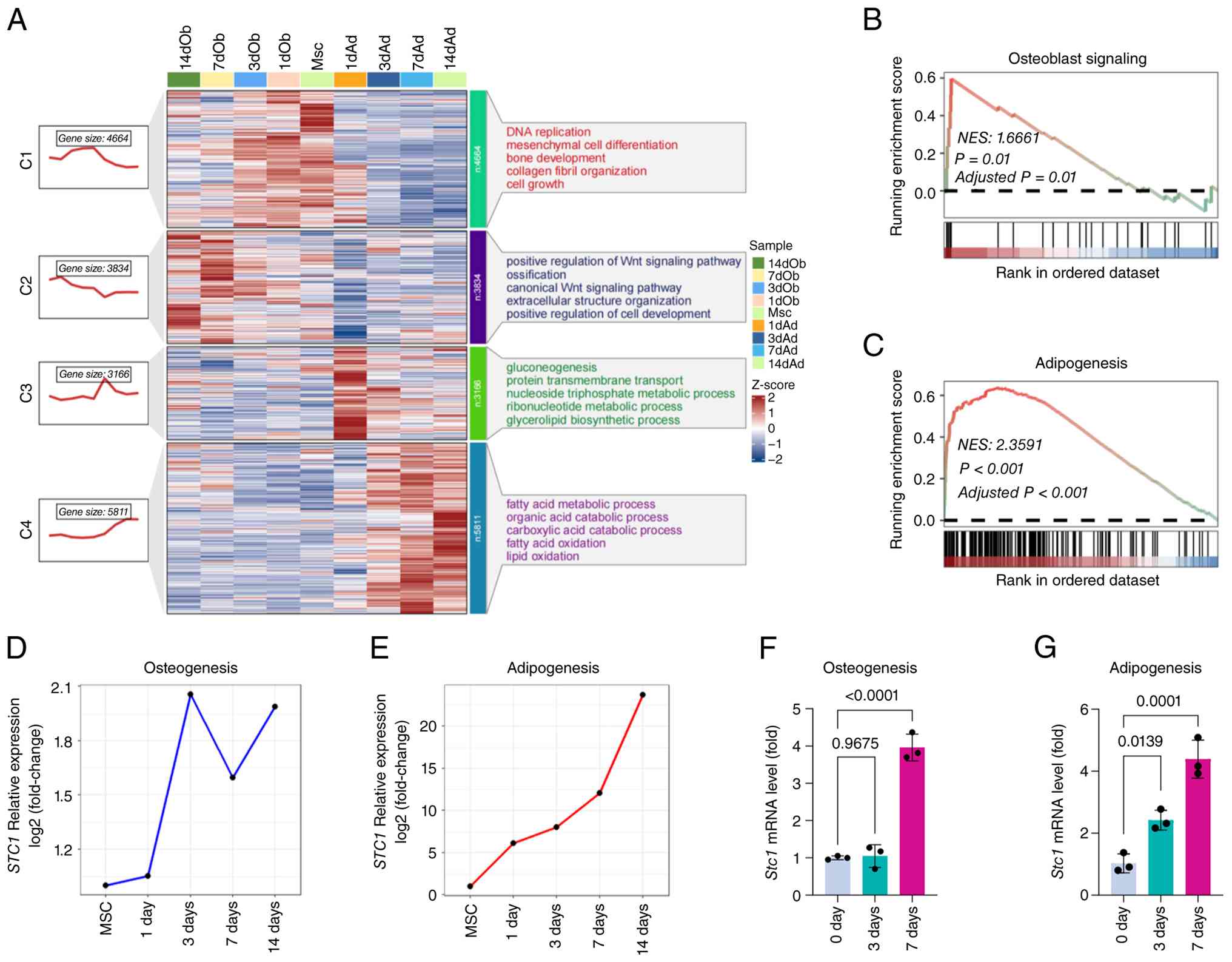 | Figure 1.Dynamic transcriptional programs
underlying osteogenic and adipogenic differentiation of MSCs. (A)
Heatmap showing the expression patterns of differentially expressed
genes across MSCs and multiple time points during osteogenic (left)
and adipogenic (right) differentiation (1, 3, 7 and 14 d).
Differentially expressed genes were grouped into distinct clusters
based on their temporal expression trajectories. Functional
annotations and representative expression trends for each gene
cluster, along with the corresponding gene numbers, are shown on
the right. (B) GSEA demonstrating significant enrichment of
osteoblast-related signaling pathways during osteogenic
differentiation, comparing MSCs with 7-day osteogenic induction.
(C) GSEA showing significant enrichment of adipogenesis-related
pathways during adipogenic differentiation, comparing MSCs with
7-day adipogenic induction. (D and E) Line plot illustrating the
dynamic changes of STC1 from MSCs upon (D) osteogenic and
(E) adipogenic differentiation, respectively. (F and G) STC1
mRNA levels after 3 and 7 days of (F) osteogenic and (G) adipogenic
induction, compared with undifferentiated MSCs. n=3. MSCs,
mesenchymal stromal cells; GSEA, gene set enrichment analysis; d,
days; STC1, stanniocalcin-1. |
Stc1 depletion inhibits senescence and
osteogenic differentiation potential of MSCs
Furthermore, it was examined whether Stc1
depletion affects MSC senescence and lineage specification.
Specifically, two siRNAs targeting Stc1 (si-Stc1−1
and si-Stc1−2) were used to knock down its expression
(Fig. 2A). To minimize off-target
effects, the two siRNAs were mixed at a ratio of 1:1 and used for
transfection (si-Stc1-mix, hereafter referred to as
si-Stc1). Stc1 depletion significantly reduced
cellular senescence in MSCs, as evidenced by decreased SA-β-Gal
staining and downregulated p21 expression, while p16 expression
slightly decreased (Fig. S1B-D).
Consistently, MSCs exhibited an increased proliferation rate
following Stc1 depletion (Fig.
S1E).
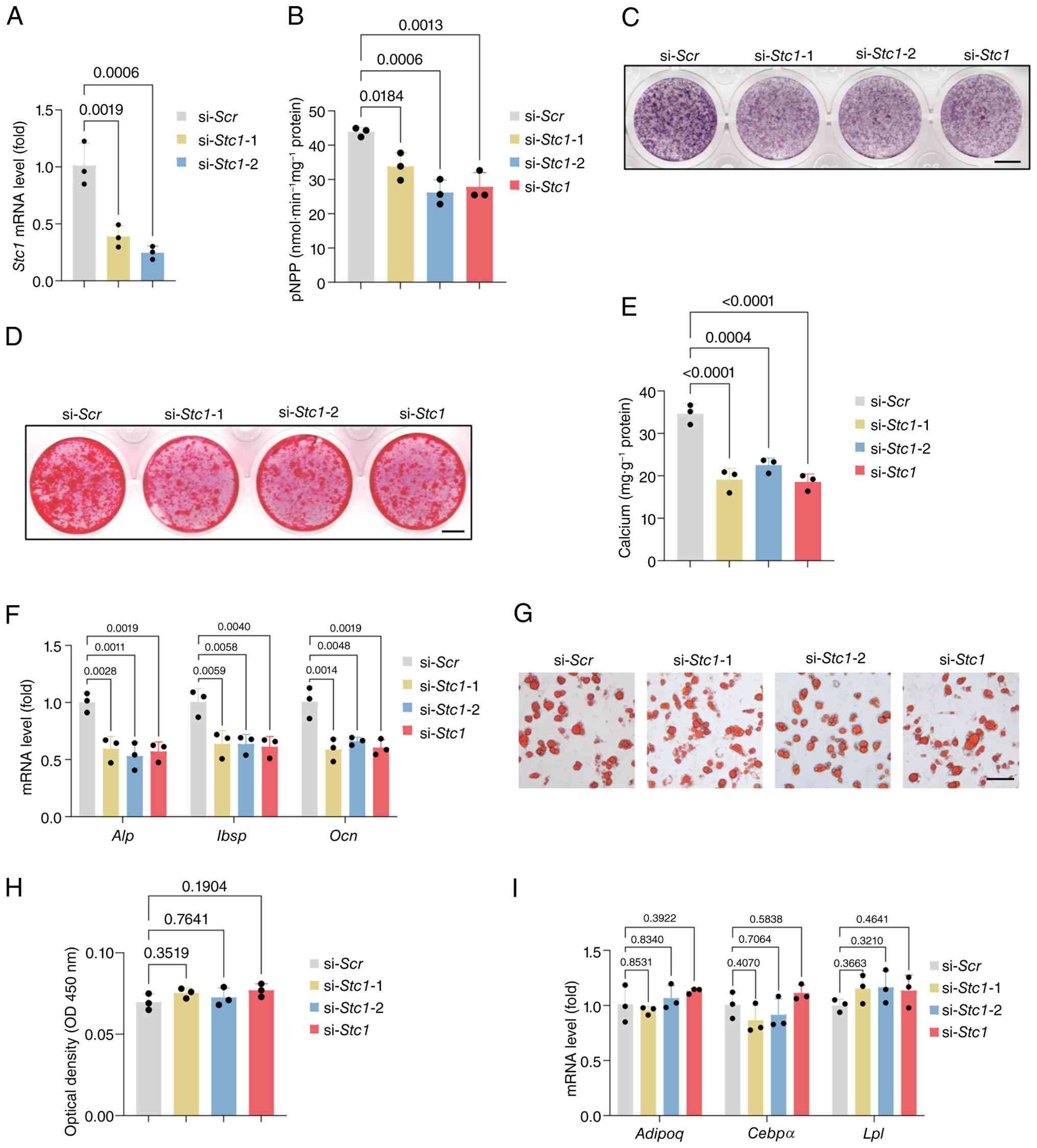 | Figure 2.Knockdown of STC1 impairs osteogenic
differentiation of MSCs. (A) Relative mRNA level of Stc1 in
MSCs after knockdown of STC1 (n=3). (B and C) ALP staining and
quantitative ALP activity assay of MSCs underwent osteogenic
induction after knockdown of STC1. Scale bar, 5 mm (n=3). (D and E)
ARS staining and quantification of calcium deposit in MSCs
underwent osteogenic induction after knockdown of STC1. Scale bar,
5 mm (n=3). (F) mRNA expression of osteogenic marker genes Alp,
Ibsp and Ocn in MSCs underwent osteogenic induction
after knockdown of STC1 (n=3). (G and H) Oil Red O staining of
lipid droplets in MSCs underwent adipogenic induction after
knockdown of STC1. Scale bar, 100 µm (n=3). (I) mRNA expression of
adipogenic marker genes Adipoq, Cebpα and Lpl in MSCs
underwent adipogenic induction after knockdown of STC1 (n=3). MSCs,
mesenchymal stromal cells; ALP, alkaline phosphatase; ARS, alizarin
red S; STC1, stanniocalcin-1; Scr, scrambled. |
ALP staining was performed to assess the effects of
Stc1 knockdown on the osteogenic commitment of MSCs.
Notably, ALP activity, an indicator of early osteogenesis, was
inhibited by Stc1 knockdown (Fig. 2B and C). Extracellular matrix
mineralization also decreased following Stc1 knockdown, as
determined via ARS staining (Fig. 2D
and E). Consistently, the expression levels of
osteogenic-related genes were downregulated by Stc1
knockdown (Fig. 2F). Notably,
although Stc1 was notably upregulated during adipogenesis
(39), significant differences
were not observed between the Stc1 knockdown groups and
controls in lipid droplet formation or the expression of
adipogenic-related genes in MSCs (Fig.
2G-I). In addition, to assess whether STC1 is involved in
chondrogenic differentiation, MSCs with STC1 knockdown were
subjected to chondrogenic induction and evaluated by multiple
readouts, including Alcian blue staining, quantitative
glycosaminoglycan (GAG) measurement and expression of chondrogenic
marker genes (Sox9 and Col2a1). As shown in Fig. S2B-E, STC1 depletion did not
significantly alter matrix deposition, GAG content, or chondrogenic
gene expression, indicating that STC1 is dispensable for MSC
chondrogenesis under the tested conditions.
Stc1 depletion suppresses osteogenic
programs while activating inflammatory signaling
To investigate the effect of Stc1 knockdown
on the transcriptional profile of MSCs, RNA sequencing was
performed 3 days after transfection with si-Stc1 (n=3). PCA
revealed a notable separation between control and
Stc1-depleted MSCs, indicating that Stc1 knockdown
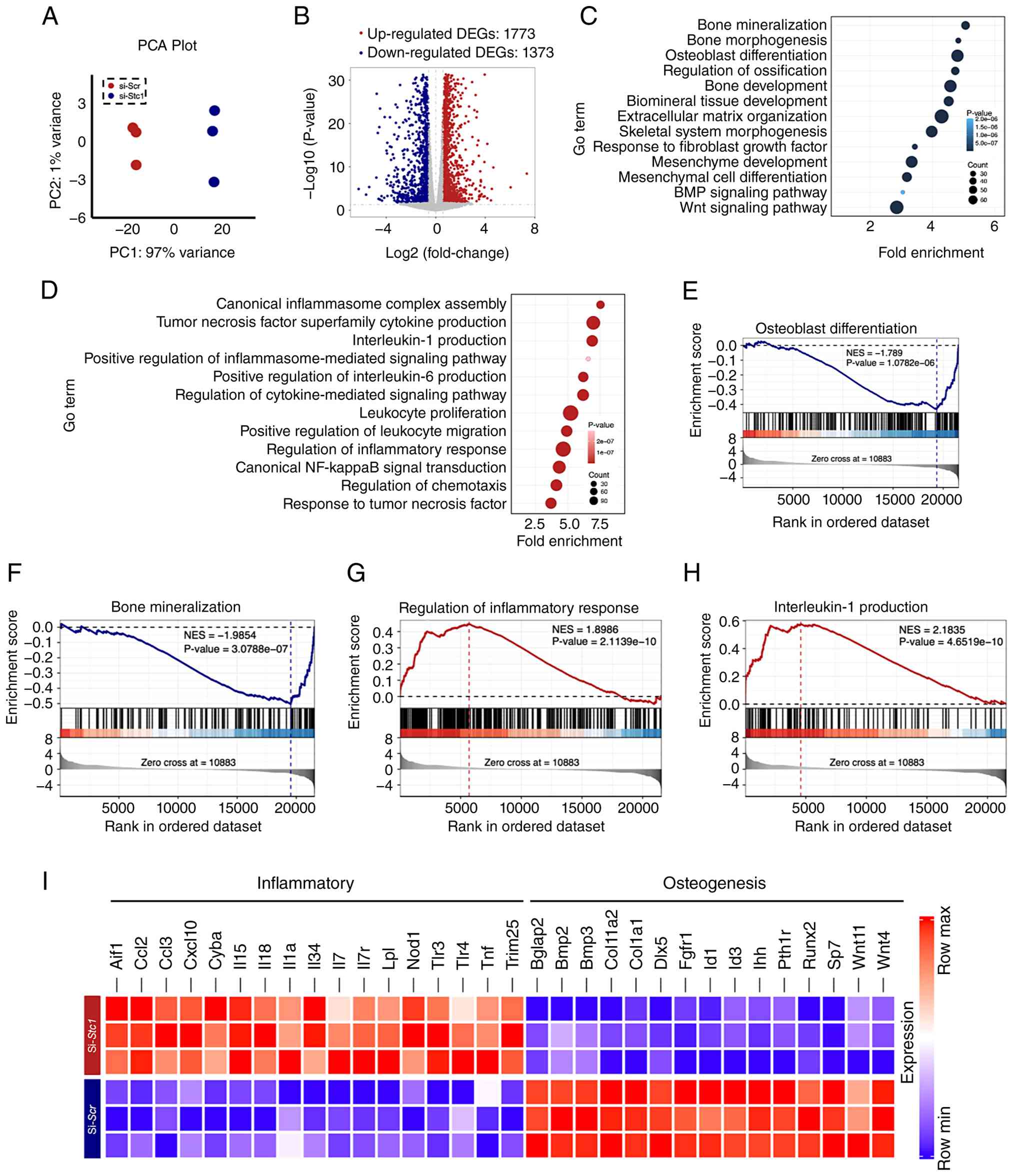
induced a marked global change in gene expression (Fig. 3A). Differential expression analysis
identified a total of 3,146 DEGs based on a cutoff of P<0.05 and
fold change >1.5, including 1,373 downregulated and 1,773
upregulated genes following Stc1 knockdown (Fig. 3B). Gene Ontology (GO) enrichment
analysis revealed that the downregulated genes were significantly
enriched in biological processes related to ‘osteogenic
differentiation’ and ‘bone mineralization’ (Fig. 3C). By contrast, the upregulated
genes were predominantly associated with inflammatory responses,
including pathways related to ‘interleukin-1 production’, ‘positive
regulation of interleukin-6 production’, as well as ‘canonical
NF-κB signal transduction’ (Fig.
3D). GSEA further demonstrated that gene sets associated with
osteogenesis and bone mineralization were significantly negatively
enriched in Stc1-depleted MSCs, whereas gene sets related to
inflammatory and immune responses were positively enriched
following Stc1 knockdown (Fig.
3E-H). A heatmap of representative genes further confirmed
enhanced inflammatory responses and impaired osteogenic capacity
following Stc1 knockdown (Fig.
3I).
Furthermore, an ATAC-sequencing was performed to
examine the impact of Stc1 depletion on chromatin
accessibility in MSCs. Overall, Stc1 knockdown resulted in
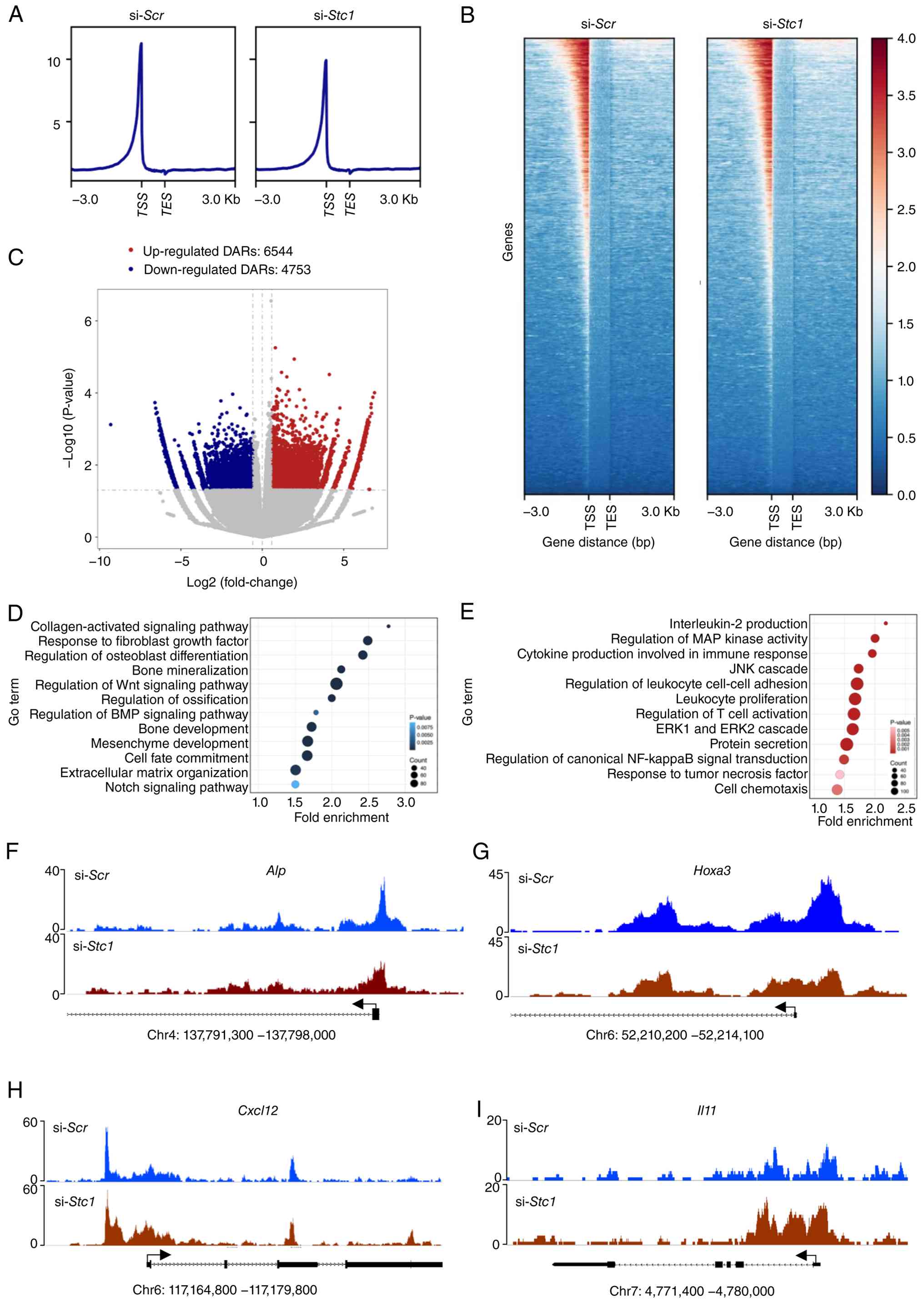
only modest changes in global chromatin accessibility (Fig. 4A and B). Differential accessibility
analysis identified 4,753 DARs with decreased accessibility and
6,544 DARs with increased accessibility following Stc1
knockdown, based on a cutoff of P<0.05 (Fig. 4C). Functional annotation of DARs
revealed a notable functional divergence between regions gaining or
losing accessibility following Stc1 knockdown. Regions with
reduced accessibility were preferentially associated with genes
involved in ‘collagen-activated signaling pathway’, ‘regulation of
osteoblast differentiation’ and ‘bone mineralization’, indicating
selective chromatin closure at osteogenesis-related loci (Fig. 4D). By contrast, regions exhibiting
increased accessibility were associated with genes involved in
inflammatory processes, including ‘interleukin-2 production’,
‘regulation of canonical NF-κB signal transduction’ and ‘cell
chemotaxis’ (Fig. 4E). Genome
track visualization further illustrated these chromatin
accessibility changes at representative loci. The promoter regions
of osteogenic genes, including Alp and Hoxa3,
exhibited notably reduced accessibility following Stc1
knockdown (Fig. 4F and G). By
contrast, increased chromatin accessibility was observed at the
promoters of inflammation-related genes, such as Cxcl12 and
Il11 (Fig. 4H and I).
Collectively, transcriptomic and chromatin accessibility analysis
indicated that STC1 knockdown selectively repressed osteogenic
programs while concomitantly activating inflammatory pathways.
Stc1 depletion enhancing NF-κB
signaling in MSCs
To further elucidate the coordinated transcriptional
and chromatin accessibility changes induced by Stc1
knockdown, an integrative analysis of the RNA-sequencing and
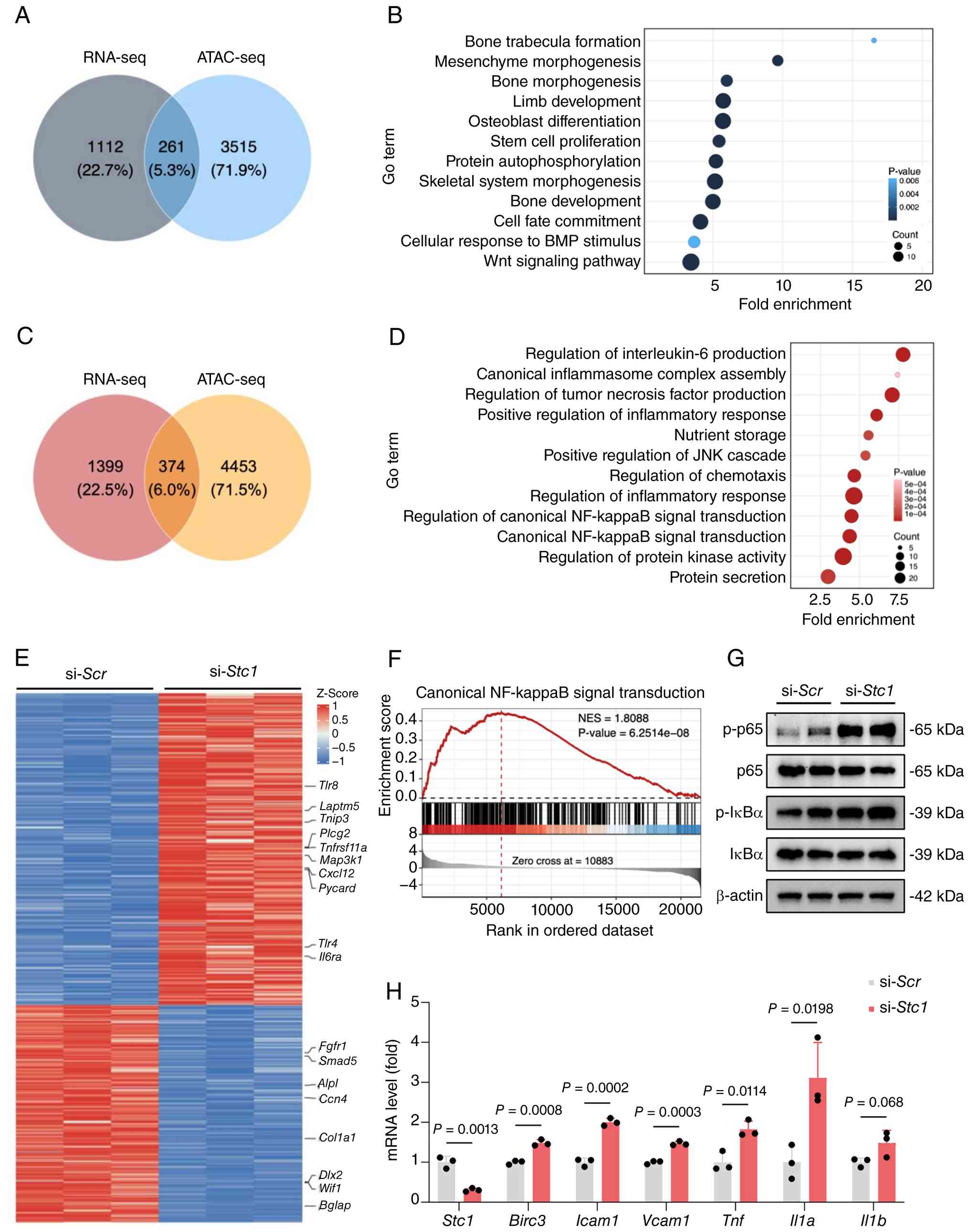
ATAC-sequencing datasets was performed. A total of 261 overlapping
genes exhibited both decreased transcript abundance in
RNA-sequencing and reduced chromatin accessibility in
ATAC-sequencing (Fig. 5A).
Functional enrichment analysis of this gene set revealed an
association with osteogenesis-related processes, including ‘bone
trabecular formation’, ‘osteoblast differentiation’ and ‘skeletal
system morphogenesis’ (Fig. 5B).
Conversely, 374 overlapping genes were identified that exhibited
upregulation at the transcript level and increased chromatin
accessibility (Fig. 5C).
Enrichment analysis of these genes indicated predominant
involvement in inflammatory and immune-related pathways, such as
‘regulation of interleukin-6 production’, ‘canonical inflammasome
complex assembly’ and ‘regulation of canonical NF-κB signal
transduction’ (Fig. 5D).
Consistent with the Venn diagram analysis, the integrative heatmap
revealed that inflammatory and NF-κB-related genes (including
Tlr4, Il6ra, Pycard and Cxcl12) were upregulated and
exhibited increased chromatin accessibility, whereas
osteogenesis-related genes (including Col1a1, Bglap, Alpl,
Smad5 and Dlx2) were downregulated and exhibited reduced
accessibility (Fig. 5E). These
findings indicated that Stc1 knockdown was associated with
coordinated transcriptional and chromatin accessibility changes
related to inflammatory activation and impaired osteogenic
programs. NF-κB signaling has been well established as a central
regulator of MSC osteogenic differentiation and inflammatory
responses (40,41). Given the consistent enrichment of
inflammatory and NF-κB signaling pathways, it was examined whether
Stc1 knockdown activates NF-κB signaling in MSCs. To further
confirm this pathway activation, GSEA was performed on the
RNA-sequencing dataset, which revealed significant enrichment of
the canonical NF-κB signaling gene set in the Stc1 knockdown
group (Fig. 5F). Western blot
analysis revealed a notable increase in p65 phosphorylation,
accompanied by a slight increase in phosphorylated IκBα, indicating
early activation of canonical NF-κB signaling (Fig. 5G). Consistently, the expression
levels of several well-established NF-κB downstream target genes,
including Birc3, Icam1, Vcam1, Tnf and Il1α (42), were significantly upregulated
following Stc1 knockdown (Fig.
5H). Collectively, these results provided molecular evidence
that STC1 depletion activates the NF-κB signaling pathway in
MSCs.
NF-κB signaling inhibition mitigating
impaired osteogenic differentiation of MSCs
To determine whether inhibition of NF-κB signaling
could mitigate the impaired osteogenic differentiation induced by
Stc1 knockdown, MSCs were treated with the NF-κB inhibitor
BAY 11-7082 (43,44). To exclude potential cytotoxic
effects, cell viability was first assessed via a CCK-8 assay.
Treatment with BAY 11-7082 at the concentrations used in the
present study did not significantly affect MSC viability (Fig. 6A). Consequently, the inhibitory
efficiency of BAY 11-7082 was verified via western blot analysis
(Fig. 6B). As shown, treatment
with BAY 11-7082 markedly reduced the phosphorylation levels of p65
and IκBα. Densitometric analysis revealed that p-p65 and p-IκBα
levels were decreased by approximately 60 and 36%, respectively,
compared with the control group. By contrast, total p65 and IκBα
protein levels remained largely unchanged, indicating effective
suppression of NF-κB pathway activation rather than alterations in
protein expression. ALP activity assays revealed that treatment
with BAY 11-7082 at concentrations of 1 and 2 µM increased ALP
activity during osteogenic differentiation of MSCs (Fig. 6C and D). Consistent with this
observation, ARS staining revealed that BAY 11-7082 treatment
effectively mitigated the reduction in extracellular matrix
mineralization induced by Stc1 depletion (Fig. 6E and F). Moreover, the expression
levels of osteogenic marker genes, Alp and Ocn,
exhibited a similar increasing trend following BAY 11-7082
treatment (Fig. 6G and H).
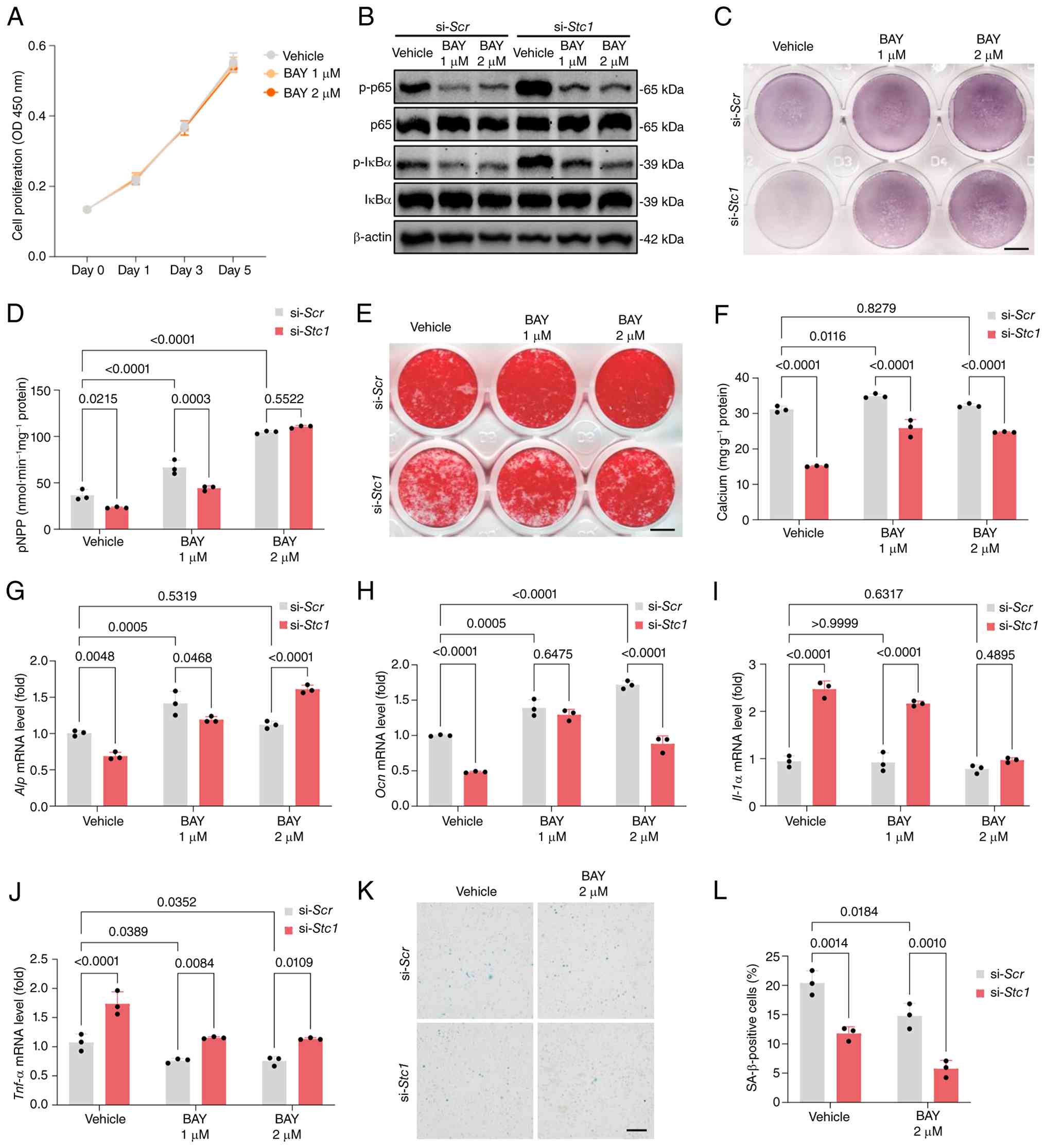 | Figure 6.STC1 knockdown and NF-κB inhibition
by BAY11-7082 promotes functional rejuvenation of MSCs. (A) Cell
counting kit 8 assay of MSCs treated with vehicle or BAY 11-7082 (1
and 2 µM; n=3. (B) Western blot assay for p-p65, p65 p-IκBα and
IκBα protein expression in STC1-depleted MSCs treated with vehicle
or BAY 11-7082 (1 and 2 µM). (C and D) ALP staining and
quantitative ALP activity assay of STC1-knockdown MSCs treated with
vehicle or BAY 11-7082 (1 and 2 µM) upon osteogenic induction.
Scale bar, 5 mm (n=3). (E and F) ARS staining and quantification of
extracellular matrix mineralization in STC1-depleted MSCs treated
with vehicle or BAY 11-7082 (1 and 2 µM) upon osteogenic induction.
Scale bar, 5 mm (n=3). (G and H) mRNA expression levels of
osteogenic marker genes (G) Alp and (H) Ocn in
STC1-depleted MSCs treated with vehicle or BAY 11-7082 (1 and 2 µM)
upon osteogenic induction. n=3. (J and K) mRNA expression levels of
NF-κB downstream target genes (I) Il-1α and (J) Tnf-α
in STC1-depleted MSCs treated with vehicle or BAY 11-7082 (1 and 2
µM). n=3. (K and L) SA-β-Gal staining and the quantitation of
positive cells in STC1-depleted MSCs, after treatment with vehicle,
BAY 11-7082 (1 or 2 µM). Scale bar, 200 µm. n=3. BAY, BAY11-7082;
MSCs, mesenchymal stromal cells; STC1, stanniocalcin-1; ALP,
alkaline phosphatase; ARS, alizarin red S; Scr, scrambled;
SA-β-Gal, senescence associate β-galactosidase. |
To further validate these findings, MSCs were
treated with JSH-23, an alternative NF-κB inhibitor, at
concentrations of 20 and 50 µM during osteogenic induction
(44). To exclude potential
cytotoxic effects, cell viability was first assessed via a CCK-8
assay, which demonstrated that JSH-23 at 20 and 50 µM did not
significantly affect MSC viability (Fig. S3A). Similar to BAY 11-7082, JSH-23
treatment enhanced osteogenic differentiation and restored
osteogenic capacity in Stc1-knockdown MSCs (Fig. S3B-G). Overall, following treatment
with either BAY 11-7082 or JSH-23, osteogenic differentiation in
Stc1-depleted MSCs reached or even exceeded that observed in
scrambled siRNA control cells treated with vehicle, indicating a
robust mitigating effect.
Given the essential role of NF-κB in the regulation
of inflammation, BAY 11-7082 treatment was expected to inhibit the
expression of inflammatory factors, Il1a and Tnf,
induced by Stc1 depletion. Notably, treatment with 2 µM BAY
11-7082 reduced their expression (Fig.
6I and J). Inhibition of NF-κB signaling by BAY 11-7082 further
enhanced the effect of Stc1 depletion on cellular
senescence, as evidenced by the SA-β-gal assay (Fig. 6K and L). Similarly, treatment with
JSH-23 also inhibited the expression of Il1a and Tnfa
and further mitigated senescence in STC1-depleted MSCs (Fig. S3H-K).
Collectively, these results identify STC1 as a key
regulator of osteogenic differentiation and inflammation in MSCs.
These findings provide mechanistic insight into the role of STC1 in
MSC biology and highlight that modulation of STC1 and NF-κB
signaling requires further investigation in more physiologically
relevant and in vivo models of age-related bone loss.
Discussion
Age-related bone loss represents a major global
health challenge in aging societies, leading to increased fracture
risk, disability and mortality. Accumulating evidence indicates
that the functional decline of MSCs, encompassing reduced cell
number, increased cellular senescence, and impaired osteogenic
differentiation, is a key contributor to age-related bone loss
(3–5). In the present study, it was
demonstrated that Stc1 expression is high in aged MSCs and
that Stc1 depletion attenuates MSC senescence. Although
Stc1 upregulation in aged MSCs may reflect a stress-adaptive
response, the present results indicate that targeting STC1
represents a potential strategy for modulating MSC senescence and
mitigating age-related bone loss.
Previous studies have reported both activating and
suppressing effects of STC1 on NF-κB signaling, depending on tissue
context and disease state. For example, STC1 has been shown to
regulate ROS homeostasis and inflammatory responses in mesenchymal
stromal cells, macrophages and lung cancer models (27,28).
In MSCs, it was observed in the present study that STC1 depletion
results in robust activation of NF-κB signaling, accompanied by
increased expression of downstream inflammatory target genes. These
results indicate that the regulatory relationship between STC1 and
NF-κB is cell- and context-dependent. Specifically, STC1 influences
NF-κB activity indirectly through regulation of intracellular redox
homeostasis, as STC1 has been shown to modulate ROS production in
multiple cell types (27,28). Notably, pharmacological inhibition
of NF-κB signaling mitigated the osteogenic defects induced by STC1
knockdown, indicating that NF-κB signaling is possibly involved in
mediating the effects of STC1 depletion on MSC osteogenic
potential.
Chronic low-grade inflammation is increasingly
recognized as a hallmark and driving force of aging across multiple
tissues (11,12). In the skeletal system, inflammatory
signaling accelerates bone resorption and suppresses MSC osteogenic
differentiation. Senescent MSCs exhibit a pro-inflammatory
secretory phenotype enriched in cytokines and chemokines such as
IL-1α, IL-6 and CXCL family members, which further exacerbate
tissue dysfunction (6,7). STC1 has been reported to modulate
inflammatory responses in diverse biological contexts (25–28).
Notably, it was observed in the present study that STC1 depletion
in MSCs leads to activation of inflammatory genes, indicating a
previously unrecognized anti-inflammatory role of STC1 in this cell
type. The ability of NF-κB inhibition to rescue osteogenic
differentiation in STC1-deficient MSCs may therefore be attributed,
at least in part, to suppression of inflammation-associated
inhibitory signals. Notably, while STC1 knockdown attenuated
certain senescence-associated phenotypes in MSCs, it concurrently
suppressed osteogenic programs and activated inflammatory/NF-κB
signaling. In this context, the inhibitory effects on osteogenic
differentiation outweighed the potential benefits of reduced
senescence. These results suggest that in MSCs, regulation of
senescence and osteogenesis are not necessarily coupled processes,
highlighting the complex role of STC1 in coordinating cellular
fate.
Furthermore, the role of STC1 appears to be notably
cell type- and context-dependent. Previous studies investigating
STC1 in bone biology largely focused on mature osteoblasts,
osteocytes or chondrocytes, where STC1 has been reported to exert
either promotive or inhibitory effects on osteogenic activity
(20–24). For example, STC1 has been reported
to inhibit longitudinal bone growth in growth plate chondrocytes
(20) and to negatively regulate
osteoblast differentiation and bone formation in transgenic mouse
bone marrow-derived MSCs (24). By
contrast, STC1 has been shown to promote osteogenic differentiation
in human MSCs (45) and dental
follicle cells (46) and to
contribute to bone-protective effects in pathological settings such
as CCl4-induced bone loss (47).
MSCs are notably responsive to inflammatory and
senescence-associated cues, and their lineage commitment is
markedly regulated by the balance between osteogenic and
inflammatory signaling pathways (40,48).
Emerging evidence also suggests that MSC-derived STC1 participates
in stress adaptation and immunomodulatory signaling in several
disease models (25–27). The present findings reveal that, in
MSCs, STC1 mainly functions as a suppressor of NF-κB-associated
inflammatory signaling and a maintainer of osteogenic potential.
Therefore, the effects of STC1 observed in differentiated bone
cells may not fully reflect its regulatory role in MSC fate
determination during aging. These observations further highlight
the functional heterogeneity of STC1 across different cell types
and indicate that the biological effects of STC1 should be
interpreted within a specific cellular and developmental
context.
From a therapeutic perspective, modulation of STC1
in MSCs may represent a potential strategy for improving osteogenic
capacity under inflammatory conditions. This improvement can
theoretically be achieved through localized delivery approaches,
such as bone-targeted gene or mRNA delivery systems. However, STC1
is widely expressed in multiple tissues as a secreted protein,
which raises concerns regarding target specificity and potential
off-target effects. Therefore, any future therapeutic application
would require precise spatial control of STC1 activity within the
bone marrow niche.
Finally, several limitations of the present study
should be acknowledged. First, all experiments were performed using
cultured MSCs in vitro. Therefore, the physiological
relevance of these findings in vivo is yet to be
established. In addition, although the present data support the
involvement of NF-κB signaling following STC1 depletion, the
precise molecular mechanisms associating STC1 with NF-κB activation
require further investigation. Furthermore, future studies using
in vivo or ex vivo osteoporotic models may further
clarify the role of STC1 in the bone microenvironment and assess
its potential relevance in age-related bone loss.
In summary, the present study revealed a novel role
of STC1 in coordinating cellular senescence, inflammation, and
osteogenic differentiation in MSCs. By inhibiting NF-κB activation,
STC1 preserves osteogenic potential and limits inflammation-driven
lineage imbalance.
Supplementary Material
Supporting Data
Acknowledgements
Not applicable.
Funding
The present study was supported by the National Natural Science
Foundation of China (grant nos. 82370918, U25A6003, 82571774 and
82301074), Department of Science and Technology of Zhejiang
Province (grant nos. 2024C03193 and 2025C02100) and the Sichuan
Science and Technology Program (grant no. 2026YFHZ0149).
Availability of data and materials
All sequencing data in the present study are
available at National Genomics Data Center, China National Center
for Bioinformation under the accession number CRA038035 (https://ngdc.cncb.ac.cn/gsa/browse/CRA038035). All
other data generated in the present study may be requested from the
corresponding author.
Author's contributions
LF, PD and QMC conceived the present study. LF, PD,
QMC, XC and XX designed the experiments. CY, XL, PW, YC, EW, QC,
JH, FZ, LG, JL, MZ, JZ, BH, AAB, YM, JKK, IC, JC, PHK and LH
performed the experiments. CY, XL, PW and EW performed the data
analysis. CY, XL, XC, PD and QMC wrote the manuscript. All authors
confirm the authenticity of all the raw data, and read and approved
the final version of the manuscript.
Ethics approval and consent to
participate
All procedures were performed according to
established ethical guidelines and approved by the Institutional
Animal Care and Use Committee of Zhejiang University (approval nos.
ZJU20230513 and ZJU20240033).
Patient consent for publication
Not applicable.
Competing interests
The authors declare that they have no competing
interests.
Glossary
Abbreviations
Abbreviations:
|
MSCs
|
mesenchymal stromal cells
|
|
STC1
|
stanniocalcin-1
|
|
SASP
|
senescence-associated secretory
phenotype
|
|
DARs
|
differentially accessible regions
|
|
ROS
|
reactive oxygen species
|
References
|
1
|
Zhou J and Shi Y: Mesenchymal stem/stromal
cells (MSCs): Origin, immune regulation, and clinical applications.
Cell Mol Immunol. 20:555–557. 2023. View Article : Google Scholar : PubMed/NCBI
|
|
2
|
Bianco P, Cao X, Frenette PS, Mao JJ,
Robey PG, Simmons PJ and Wang CY: The meaning, the sense and the
significance: Translating the science of mesenchymal stem cells
into medicine. Nat Med. 19:35–42. 2013. View Article : Google Scholar : PubMed/NCBI
|
|
3
|
Zhang L, Wang Z, Zhang Y, Ji R, Li Z, Zou
J and Gao B: Regulatory cellular and molecular networks in the bone
microenvironment during aging. Life Med. 3:nae0192024. View Article : Google Scholar
|
|
4
|
Deng P, Yuan Q, Cheng Y, Li J, Liu Z, Liu
Y, Li Y, Su T, Wang J, Salvo ME, et al: Loss of KDM4B exacerbates
bone-fat imbalance and mesenchymal stromal cell exhaustion in
skeletal aging. Cell Stem Cell. 28:1057–1073.e7. 2021. View Article : Google Scholar : PubMed/NCBI
|
|
5
|
Cheng M, Yuan W, Moshaverinia A and Yu B:
Rejuvenation of mesenchymal stem cells to ameliorate skeletal
aging. Cells. 12:9982023. View Article : Google Scholar : PubMed/NCBI
|
|
6
|
Gnani D, Crippa S, Della Volpe L, Rossella
V, Conti A, Lettera E, Rivis S, Ometti M, Fraschini G, Bernardo ME
and Di Micco R: An early-senescence state in aged mesenchymal
stromal cells contributes to hematopoietic stem and progenitor cell
clonogenic impairment through the activation of a pro-inflammatory
program. Aging Cell. 18:e129332019. View Article : Google Scholar : PubMed/NCBI
|
|
7
|
Wang B, Han J, Elisseeff JH and Demaria M:
The senescence-associated secretory phenotype and its physiological
and pathological implications. Nat Rev Mol Cell Biol. 25:958–978.
2024. View Article : Google Scholar : PubMed/NCBI
|
|
8
|
Vono R, Jover Garcia E, Spinetti G and
Madeddu P: Oxidative stress in mesenchymal stem cell senescence:
Regulation by coding and noncoding RNAs. Antioxid Redox Signal.
29:864–879. 2018. View Article : Google Scholar : PubMed/NCBI
|
|
9
|
Choo KB, Tai L, Hymavathee KS, Wong CY,
Nguyen PN, Huang CJ, Cheong SK and Kamarul T: Oxidative
Stress-induced premature senescence in Wharton's Jelly-derived
mesenchymal stem cells. Int J Med Sci. 11:1201–1207. 2014.
View Article : Google Scholar : PubMed/NCBI
|
|
10
|
Finkel T and Holbrook NJ: Oxidants,
oxidative stress and the biology of ageing. Nature. 408:239–247.
2000. View Article : Google Scholar : PubMed/NCBI
|
|
11
|
Baechle JJ, Chen N, Makhijani P, Winer S,
Furman D and Winer DA: Chronic inflammation and the hallmarks of
aging. Mol Metab. 74:1017552023. View Article : Google Scholar : PubMed/NCBI
|
|
12
|
Andonian BJ, Hippensteel JA, Abuabara K,
Boyle EM, Colbert JF, Devinney MJ, Faye AS, Kochar B, Lee J, Litke
R, et al: Inflammation and aging-related disease: A
transdisciplinary inflammaging framework. Geroscience. 47:515–542.
2025. View Article : Google Scholar : PubMed/NCBI
|
|
13
|
De Cecco M, Ito T, Petrashen AP, Elias AE,
Skvir NJ, Criscione SW, Caligiana A, Brocculi G, Adney EM, Boeke
JD, et al: L1 drives IFN in senescent cells and promotes
age-associated inflammation. Nature. 566:73–78. 2019. View Article : Google Scholar : PubMed/NCBI
|
|
14
|
Liu X, Liu Z, Wu Z, Ren J, Fan Y, Sun L,
Cao G, Niu Y, Zhang B, Ji Q, et al: Resurrection of endogenous
retroviruses during aging reinforces senescence. Cell.
186:287–304.e26. 2023. View Article : Google Scholar : PubMed/NCBI
|
|
15
|
Yang JH, Hayano M, Griffin PT, Amorim JA,
Bonkowski MS, Apostolides JK, Salfati EL, Blanchette M, Munding EM,
Bhakta M, et al: Loss of epigenetic information as a cause of
mammalian aging. Cell. 186:305–326.e27. 2023. View Article : Google Scholar : PubMed/NCBI
|
|
16
|
Kløverpris S, Mikkelsen JH, Pedersen JH,
Jepsen MR, Laursen LS, Petersen SV and Oxvig C: Stanniocalcin-1
Potently inhibits the proteolytic activity of the metalloproteinase
Pregnancy-associated plasma Protein-A. J Biol Chem.
290:21915–21924. 2015. View Article : Google Scholar : PubMed/NCBI
|
|
17
|
Basisty N, Kale A, Jeon OH, Kuehnemann C,
Payne T, Rao C, Holtz A, Shah S, Sharma V, Ferrucci L, et al: A
proteomic atlas of senescence-associated secretomes for aging
biomarker development. PLoS Biol. 18:e30005992020. View Article : Google Scholar : PubMed/NCBI
|
|
18
|
Zhu L, Xie Z, Yang G, Zhou G, Li L and
Zhang S: Stanniocalcin-1 Promotes PARP1-Dependent cell death via
JNK activation in colitis. Adv Sci (Weinh). 11:e23041232024.
View Article : Google Scholar : PubMed/NCBI
|
|
19
|
Yoshiko Y, Son A, Maeda S, Igarashi A,
Takano S, Hu J and Maeda N: Evidence for stanniocalcin gene
expression in mammalian bone. Endocrinology. 140:1869–1874. 1999.
View Article : Google Scholar : PubMed/NCBI
|
|
20
|
Wu S, Yoshiko Y and De Luca F:
Stanniocalcin 1 acts as a paracrine regulator of growth plate
chondrogenesis. J Biol Chem. 281:5120–5127. 2006. View Article : Google Scholar : PubMed/NCBI
|
|
21
|
Filvaroff EH, Guillet S, Zlot C, Bao M,
Ingle G, Steinmetz H, Hoeffel J, Bunting S, Ross J, Carano RA, et
al: Stanniocalcin 1 alters muscle and bone structure and function
in transgenic mice. Endocrinology. 143:3681–3690. 2002. View Article : Google Scholar : PubMed/NCBI
|
|
22
|
Varghese R, Gagliardi AD, Bialek PE, Yee
SP, Wagner GF and Dimattia GE: Overexpression of human
stanniocalcin affects growth and reproduction in transgenic mice.
Endocrinology. 143:868–876. 2002. View Article : Google Scholar : PubMed/NCBI
|
|
23
|
Yoshiko Y, Maeda N and Aubin JE:
Stanniocalcin 1 stimulates osteoblast differentiation in rat
calvaria cell cultures. Endocrinology. 144:4134–4143. 2003.
View Article : Google Scholar : PubMed/NCBI
|
|
24
|
Kim JH, Kim K, Kim I, Seong S, Koh JT and
Kim N: Stanniocalcin 1 and 1,25-dihydroxyvitamin D3 cooperatively
regulate bone mineralization by osteoblasts. Exp Mol Med.
56:1991–2001. 2024. View Article : Google Scholar : PubMed/NCBI
|
|
25
|
Zonderland J, Gomes DB, Pallada Y, Moldero
IL, Camarero-Espinosa S and Moroni L: Mechanosensitive regulation
of stanniocalcin-1 by zyxin and actin-myosin in human mesenchymal
stromal cells. Stem Cells. 38:948–959. 2020. View Article : Google Scholar : PubMed/NCBI
|
|
26
|
Bartosh TJ, Ylöstalo JH, Bazhanov N,
Kuhlman J and Prockop DJ: Dynamic compaction of human mesenchymal
stem/precursor cells into spheres self-activates caspase-dependent
IL1 signaling to enhance secretion of modulators of inflammation
and immunity (PGE2, TSG6, and STC1). Stem Cells. 31:2443–2456.
2013. View Article : Google Scholar : PubMed/NCBI
|
|
27
|
Seo Y, Shin TH, Ahn JS, Oh SJ, Shin YY,
Yang JW, Park HY, Shin SC, Kwon HK, Kim JM, et al: Human
Tonsil-derived mesenchymal stromal cells maintain proliferating and
ROS-Regulatory properties via Stanniocalcin-1. Cells. 9:6362020.
View Article : Google Scholar : PubMed/NCBI
|
|
28
|
Ohkouchi S, Block GJ, Katsha AM, Kanehira
M, Ebina M, Kikuchi T, Saijo Y, Nukiwa T and Prockop DJ:
Mesenchymal stromal cells protect cancer cells from ROS-induced
apoptosis and enhance the Warburg effect by secreting STC1. Mol
Ther. 20:417–423. 2012. View Article : Google Scholar : PubMed/NCBI
|
|
29
|
Deng P, Chang I, Wang J, Badreldin AA, Li
X, Yu B and Wang CY: Loss of KDM4B impairs osteogenic
differentiation of OMSCs and promotes oral bone aging. Int J Oral
Sci. 14:242022. View Article : Google Scholar : PubMed/NCBI
|
|
30
|
Liu Z, Lee HL, Suh JS, Deng P, Lee CR,
Bezouglaia O, Mirnia M, Chen V, Zhou M, Cui ZK, et al: The
ERα/KDM6B regulatory axis modulates osteogenic differentiation in
human mesenchymal stem cells. Bone Res. 10:32022. View Article : Google Scholar : PubMed/NCBI
|
|
31
|
Zhu M, Min S, Mao X, Zhou Y, Zhang Y, Li
W, Li L, Wu L, Cong X and Yu G: Interleukin-13 promotes cellular
senescence through inducing mitochondrial dysfunction in
IgG4-related sialadenitis. Int J Oral Sci. 14:292022. View Article : Google Scholar : PubMed/NCBI
|
|
32
|
Livak KJ and Schmittgen TD: Analysis of
relative gene expression data using real-time quantitative PCR and
the 2(−Delta Delta C(T)) method. Methods. 25:402–408. 2001.
View Article : Google Scholar : PubMed/NCBI
|
|
33
|
Dobin A, Davis CA, Schlesinger F, Drenkow
J, Zaleski C, Jha S, Batut P, Chaisson M and Gingeras TR: STAR:
Ultrafast universal RNA-seq aligner. Bioinformatics. 29:15–21.
2012. View Article : Google Scholar : PubMed/NCBI
|
|
34
|
Liao Y, Smyth GK and Shi W: featureCounts:
An efficient general purpose program for assigning sequence reads
to genomic features. Bioinformatics. 30:923–930. 2013. View Article : Google Scholar : PubMed/NCBI
|
|
35
|
Rauch A, Haakonsson AK, Madsen JGS, Larsen
M, Forss I, Madsen MR, Van Hauwaert EL, Wiwie C, Jespersen NZ,
Tencerova M, et al: Osteogenesis depends on commissioning of a
network of stem cell transcription factors that act as repressors
of adipogenesis. Nat Genet. 51:716–727. 2019. View Article : Google Scholar : PubMed/NCBI
|
|
36
|
Wang C, Tian W, Hu SY, Di CX, He CY, Cao
QL, Hao RH, Dong SS, Liu CC, Rong Y, et al: Lineage-selective super
enhancers mediate core regulatory circuitry during adipogenic and
osteogenic differentiation of human mesenchymal stem cells. Cell
Death Dis. 13:8662022. View Article : Google Scholar : PubMed/NCBI
|
|
37
|
Langmead B and Salzberg SL: Fast
gapped-read alignment with Bowtie 2. Nat Methods. 9:357–359. 2012.
View Article : Google Scholar : PubMed/NCBI
|
|
38
|
Zhang Y, Liu T, Meyer CA, Eeckhoute J,
Johnson DS, Bernstein BE, Nusbaum C, Myers RM, Brown M, Li W and
Liu XS: Model-based analysis of ChIP-Seq (MACS). Genome Biol.
9:R1372008. View Article : Google Scholar : PubMed/NCBI
|
|
39
|
Serlachius M and Andersson LC: Upregulated
expression of stanniocalcin-1 during adipogenesis. Exp Cell Res.
296:256–264. 2004. View Article : Google Scholar : PubMed/NCBI
|
|
40
|
Chang J, Liu F, Lee M, Wu B, Ting K, Zara
JN, Soo C, Al Hezaimi K, Zou W, Chen X, et al: NF-κB inhibits
osteogenic differentiation of mesenchymal stem cells by promoting
β-catenin degradation. Proc Natl Acad Sci USA. 110:9469–9474. 2013.
View Article : Google Scholar : PubMed/NCBI
|
|
41
|
Wang L, Zhao Y, Liu Y, Akiyama K, Chen C,
Qu C, Jin Y and Shi S: IFN-γ and TNF-α synergistically induce
mesenchymal stem cell impairment and tumorigenesis via NFκB
signaling. Stem Cells. 31:1383–1395. 2013. View Article : Google Scholar : PubMed/NCBI
|
|
42
|
Guo Q, Jin Y, Chen X, Ye X, Shen X, Lin M,
Zeng C, Zhou T and Zhang J: NF-κB in biology and targeted therapy:
New insights and translational implications. Signal Transduct
Target Ther. 9:532024. View Article : Google Scholar : PubMed/NCBI
|
|
43
|
Hou Y, Liang H, Rao E, Zheng W, Huang X,
Deng L, Zhang Y, Yu X, Xu M, Mauceri H, et al: Non-canonical NF-κB
Antagonizes STING Sensor-Mediated DNA sensing in radiotherapy.
Immunity. 49:490–503.e4. 2018. View Article : Google Scholar : PubMed/NCBI
|
|
44
|
Mori N, Yamada Y, Ikeda S, Yamasaki Y,
Tsukasaki K, Tanaka Y, Tomonaga M, Yamamoto N and Fujii M: Bay
11-7082 inhibits transcription factor NF-kappaB and induces
apoptosis of HTLV–I-infected T-cell lines and primary adult T-cell
leukemia cells. Blood. 100:1828–1834. 2002. View Article : Google Scholar : PubMed/NCBI
|
|
45
|
Brum AM, van de Peppel J, Nguyen L, Aliev
A, Schreuders-Koedam M, Gajadien T, van der Leije CS, van Kerkwijk
A, Eijken M, van Leeuwen JPTM and van der Eerden BCJ: Using the
Connectivity Map to discover compounds influencing human osteoblast
differentiation. J Cell Physiol. 233:4895–4906. 2018. View Article : Google Scholar : PubMed/NCBI
|
|
46
|
Felthaus O, Gosau M and Morsczeck C:
ZBTB16 induces osteogenic differentiation marker genes in dental
follicle cells independent from RUNX2. J Periodontol. 85:e144–e151.
2014. View Article : Google Scholar : PubMed/NCBI
|
|
47
|
Sonoda S, Murata S, Yamaza H, Yuniartha R,
Fujiyoshi J, Yoshimaru K, Matsuura T, Oda Y, Ohga S, Tajiri T, et
al: Targeting hepatic oxidative stress rescues bone loss in liver
fibrosis. Mol Metab. 66:1015992022. View Article : Google Scholar : PubMed/NCBI
|
|
48
|
Deshpande S, James AW, Blough J, Donneys
A, Wang SC, Cederna PS, Buchman SR and Levi B: Reconciling the
effects of inflammatory cytokines on mesenchymal cell osteogenic
differentiation. J Surg Res. 185:278–285. 2013. View Article : Google Scholar : PubMed/NCBI
|