Introduction
Chronic kidney disease (CKD) is a growing public
health problem due to its high prevalence and mortality. Globally,
the number of adults with CKD was 788 million in 2023, with an
age-standardised prevalence of 14.2%. CKD is currently the ninth
leading cause of death worldwide, accounting for 1.48 million
deaths annually (1). Renal
fibrosis is a representative pathological trait of renal aging and
CKD and is characterized by myofibroblast transdifferentiation
(MTD) and excessive deposition of extracellular matrix (ECM)
proteins in the renal interstitium (2). Kidney fibrosis is regulated by the
interaction between profibrotic and antifibrotic signals and
regulatory factors. Profibrotic pathways such as the TGFβ/Smad and
Wnt/β-catenin pathways facilitate MTD and ECM (3), while fibrosis inhibitors such as
Klotho, BMP-7 or Smad6/7 can clog or disturb profibrotic pathways.
Moreover, patients with CKD with the same basic disease have
different susceptibilities and severities of renal fibrosis
(4). To date, the underlying
mechanisms of renal fibrosis remain unclear. Therefore, probing the
molecular mechanisms of renal fibrosis is key.
The Klotho protein family comprises three isoforms
(α, β and γ). The present study focuses on αKlotho, which is
predominantly expressed in the kidney and has well-established
anti-fibrotic and anti-aging properties in the renal tubular
epithelium. In the context of renal fibrosis and CKD research, the
term ‘Klotho’ without a prefix has been widely accepted to refer
specifically to αKlotho. Therefore, the present study and all
subsequent references to ‘Klotho’ refer to αKlotho.
Klotho-deficient mice exhibit multifarious aging phenotypes in the
majority of organs and naturally develop kidney fibrosis (5). Klotho is a pivotal negative modulator
of the Wnt pathway. It restrains kidney fibrosis by binding to the
Wnt receptor, thereby inhibiting the profibrotic Wnt/β-catenin
pathway (6). Second, some studies
have shown that aging-associated inflammation and the progression
of early CKD may be interrelated with the downregulation of Klotho
and the induction of the RIG-I/NF-κB pathway (7). In diabetic db/db mice, Klotho
expression was decreased in the kidney and corresponded to
increased NF-κB activation (8).
The mechanism of action and downstream targets of Klotho in renal
fibrosis have been extensively explored. However, the upstream
regulatory mechanisms of Klotho have rarely been reported.
The majority of studies have shown that histone
modifications may also mediate pathways involved in CKD and
fibrosis. Sun et al (9)
reported that TGF-β increased histone H3 lysine methylation
(H3K4me1, H3K4me2 and H3K4me3) and augmented the revitalization of
ECM genes associated with tissue growth factor, collagen-1α1 and
plasminogen in mesangial cells. Moreover, blockade of class I
histone deacetylation by the alternative HDAC suppressor MS-275
restrained TGF-β signal transduction and blocked the revitalization
of kidney fibroblasts (10). HDACs
are an enzyme family that removes acetyl groups from histone and
non-histone proteins and they play important roles in the
modulation of gene transcription and protein function (11). HDAC3 is a class I HDAC. This type
of HDAC is deemed the ‘classical’ HDAC and the main target of
current pen-HDAC inhibitors in cancer treatment (12) and in the kidney (13). Abnormal expression of HDAC3 is an
essential part of CKD renal damage (14–16),
indicating that HDAC3 has potential as a target for the treatment
of CKD.
Activating transcription factor 3 (ATF3) belongs to
the ATF/CREB transcription factor family, which is diverse in size,
protein sequence and biological function, and stress-induced ATF3
can act as a transcriptional activator or inhibitor (17). ATF3 is associated with HDAC3. For
example, HDAC3 mediates deacetylation of ATF3, enhancing its
transcriptional inhibitory activity (18–20).
There is also the possibility that ATF3 may negatively affect the
binding of HDAC3 at DA-independent sites (19). In addition, ATF3 facilitates
ferroptosis and renal tubular cell injury in CKD through nuclear
translocation and transcriptional suppression (21–23).
The transcriptional inhibition of HDAC3 is also achieved by binding
to ATF3 (24). The ATF3 protein
directly binds to HDAC6 and HDAC3 and does not require
supernumerary proteins for binding (24). The function of ATF3 in renal
fibrosis has not been extensively explored. Therefore, we
hypothesize that ATF3 may influence Klotho transcription by
combining with HDAC3.
The present study probed the upstream regulatory
mechanism of Klotho. The results showed that Klotho produced
abnormal H3K9ac modifications in renal fibrosis samples and in a
TGF-β-induced HK2 cell model. This modification is mediated by
HDAC3. HDAC3 combines with ATF3 to repress Klotho transcription,
thereby inducing the development of renal fibrosis.
Materials and methods
Clinical samples
Renal tissues from 35 patients with CKD (kidney
puncture sample) and 20 patients without CDK (paracancerous tissue
of renal cancer patients) were retrieved from Yan'an Hospital of
Kunming City. Among the patients with CKD who underwent renal
puncture biopsy in our hospital from 2013 to 2020, the distribution
of renal interstitial fibrosis in the sections obtained accounted
for >75% of the total area, and the glomerular filtration rate
was less than 15 ml/min/1.73 m2 calculated by EPI formula, that is,
patients with stage 5 chronic kidney disease. The kidney tissue was
harvested and stored at liquid nitrogen.
Cell culture
Human renal tubular HK2 cells were purchased from
Otwo Biotechnology Co., Ltd. and cultured in DMEM/F12 (Gibco;
Thermo Fisher Scientific, Inc.) supplemented with 10% FBS (Gibco;
Thermo Fisher Scientific, Inc.) and 1% penicillin/streptomycin
(MilliporeSigma) at 5% CO2 and 37°C. HK2 cells treated
with 10 ng/ml TGF-β (cat. no. 240-B-002; R&D Systems, Inc.) for
48 h were used to construct a renal fibrosis cell model. To explore
the downstream mechanisms, HK2 cells were treated with 5 µM RGFP966
(Selleck Chemicals, cat. No. S7229), a selective HDAC3 inhibitor,
or an equivalent volume of vehicle (0.1% DMSO) as a control, at
37°C for 4 h.
Cell transfection
The cells were cultured in 24-well plates overnight
to reach 70–80% confluence prior to transfection. The fulllength
coding sequences of Klotho and ATF3 were amplified by PCR, digested
with restriction enzymes, and ligated into the pcDNA3.1(+) vector
to generate the overexpression plasmids oe-Klotho and oe-ATF3.
Small interfering RNA targeting ATF3 (si-ATF3), Klotho (si-Klotho),
and the corresponding negative control siRNA (si-NC) were purchased
from Shanghai GenePharma Co., Ltd. The siRNA sequences are listed
in Table SI. For transfection,
oe-Klotho (0.8 µg/well), oe-ATF3 (0.8 µg/well), si-ATF3 (50
pmol/well), si-Klotho (50 pmol/well), or si-NC (50 pmol/well) were
diluted in Opti-MEM reduced serum and mixed with
Lipofectamine® 3000 reagent (Invitrogen; Thermo Fisher
Scientific, Inc.) according to the manufacturer's instructions. The
transfection mixtures were added to at 37°C in a humidified
atmosphere containing 5% CO2 for 6 h, after which the
medium was replaced with fresh complete culture medium. The cells
were cultured under the same conditions for an additional 48 h
prior to subsequent experimentation. Transfection efficiency was
verified by Western blot analysis.
Animals and experiments
Specific pathogen-free-grade 8-week-old male C57BL/6
mice (20±0.5 g) were obtained from the Animal Experiment Center of
Kunming Medical University (Kunming, China). All animals were
housed under controlled conditions at 22±2°C with a relative
humidity of 50–60%, on a 12-h light/dark cycle, and had ad libitum
access to food and water. A total of 50 mice were used in the
present study. The animal study was authorized by the Animal Care
Committee of Yan'an Hospital Affiliated with Kunming Medical
University in conformance with the institutional guidelines
(approval no. 2021044). The mouse kidney fibrosis model was
established using the unilateral ureteric obstruction (UUO)
procedure described previously (25). In the sham-operated group, the
ureter was exposed but not ligated. The mice were randomly divided
into 5 groups: i) Sham-operated (Sham); ii) renal fibrosis group
(UUO); iii) mice injected with si-ATF3 via the tail vein (UUO +
si-ATF3, 5 nmol in 100 µl PBS, twice per week for 2 weeks) prior to
UUO modeling; iv) mice were renally injected with oe-Klotho (UUO +
oeKlotho, genome copy number containing 5×10⁹ virus particles) 1
week prior to UUO modeling; and v) the group in which the mice were
renally injected with oeKlotho and oe-ATF3 (UUO + OE-Klotho +
oe-ATF3, genome copy number containing 5×10⁹ virus particles each)
1 week prior to UUO modeling. Throughout the experiment, animal
health and behaviour were monitored daily. Animals were euthanized
immediately upon reaching the predefined humane endpoint: i)
>15% weight loss within 1–2 days, or >20% overall weight
loss; ii) signs of severe distress, including lethargy,
piloerection, hunched posture, dehydration, or reduced
responsiveness to external stimuli. No animals reached the humane
endpoint during the experiment. The experiment lasted for 14 days
after UUO surgery. During this period, a total of 9 mice died
during the experiment: 4 due to anesthesia-related complications, 3
due to surgical failure, and 2 due to postoperative infection;
these animals were excluded from the final analysis. After UUO
surgery for 14 days, the mice were all anesthetized by
intraperitoneal injection of sodium pentobarbital (50 mg/kg body
weight). Following the loss of consciousness and lack of response
to toe pinch, 0.5–1 ml of whole blood was collected from each mouse
via cardiac puncture for serum cytokine analysis. The blood samples
were stored at room temperature for 30 min to 2 h to allow natural
clotting, and serum was separated by centrifugation at 1,000 × g
for 10 min at 4°C and stored at −80°C until further use. After
blood collection, the kidneys were harvested and stored at −80°C.
All procedures were performed in accordance with institutional
guidelines.
Cytokine assay
The whole blood of the mice was stored at room
temperature for 30 min to 2 h. After the whole blood had naturally
clotted, the serum was aspirated. The expression levels of TNF-α,
IL-1β and IL-6 in mouse serum were detected using commercial ELISA
kits according to the manufacturer's instructions (cat. no. PT512,
PI301 and PI326; all Beyotime Biotechnology). Flow cytometric
analysis was performed using a BD FACSCanto II flow cytometer (BD
Biosciences).
Reverse transcription-quantitative PCR
(RT-qPCR)
Total RNA was extracted from tissue or cell samples
via TRIzol® reagent (Invitrogen; Thermo Fisher
Scientific, Inc.), and the RNA was reverse transcribed into cDNA
using a First Strand cDNA synthesis kit (Beijing Genenode Biotech
Co., Ltd.). SYBR Green real-time fluorescence qPCR was performed
using a SYBR Green reaction mix (Beijing Solarbio Science &
Technology Co., Ltd.) on a real-time PCR system (Roche
LightCycler® 480 II) according to the manufacturer's
protocol. The thermocycling conditions were as follows: initial
denaturation at 95°C for 2 min, followed by 40 cycles of 95°C for
15 sec and 60°C for 60 sec, with a final melt curve stage. GAPDH
was used as the internal reference gene. The relative expression
levels of target genes were calculated using the 2-ΔΔCq method
(26). The primer sequences used
are listed in Table I.
 | Table I.PCR primer sequences. |
Table I.
PCR primer sequences.
| Genes | Primer | Sequence |
|---|
| Klotho | Forward |
5′-TCCCAACCTGAGGCAACT-3′ |
|
| Reverse |
5′-TGGTAGAACAAGGCTGAAGA-3′ |
| HDAC3 | Forward |
5′-CGTCTTCAAGCCATACCA-3′ |
|
| Reverse |
5′-GCTCCAGGATGCCAATCA-3′ |
| GAPDH | Forward |
5′-TGACCACAGTCCATGCCATCAC-3′ |
|
| Reverse |
5′-CGCCTGCTTCACCACCTTCTT-3′ |
Western blotting
Total protein was extracted from cells or tissues
with RIPA buffer (MilliporeSigma) containing 1% protease inhibitor
and phosphatase inhibitor. Protein concentration was determined
using a BCA protein assay kit. Equal amounts of protein (30
µg/lane) were separated by 10% SDS-PAGE and transferred to PVDF
membranes. After the membranes were blocked with 5% skim milk for 2
h at room temperature, they were incubated with the primary
antibody at 4°C overnight, after which they were incubated with an
HRP-conjugated secondary antibody (1:2,000; cat. no. ab205718) for
1 h at room temperature. The proteins were visualized via
chemiluminescence. All western blotting experiments were repeated
at least three times with independent biological samples, and
representative images are shown. The protein bands were analyzed
using ImageJ v1.8.0 software (National Institutes of Health). The
following antibodies were used: Anti-Klotho (1:1,000; cat. no.
ab181373), anti-HDAC3 (1:5,000; cat. no. ab32369; Abcam),
anti-collagen I (1:1,000; cat. no. ab260043; Abcam), anti-collagen
IV (1:1,000; cat. no. ab6586; all Abcam), anti-fibronectin
(1:1,000; cat. no. ab2413; Abcam), anti-E-cadherin (1:1,000; cat.
no. ab231303; Abcam), anti-α-SMA (1:1,000; cat. no. ab5831; Abcam),
anti-ATF3 (1:1,000; cat. no. ab254268; Abcam), anti-Wnt1 (1:1,000;
cat. no. ab15251; Abcam), anti-β-catenin (1:5,000; cat. no.
ab32572; Abcam), anti-c-MYC (1:2,000; cat. no. ab185656; Abcam),
anti-cyclin D1 (1:2,000; cat. no. ab16663; Abcam) and anti-β-actin
(1:1,000; cat. no. ab8226; Abcam). The membranes were then
incubated with HRP-conjugated goat anti-rabbit IgG (1:2,000; cat.
no. ab205718; Abcam) or HRP-conjugated goat anti-mouse IgG
(1:2,000; cat. no. ab205719; Abcam).
Histone H3 acetylation level
After the histones in each group were extracted, the
acetylation level of total histone H3 was detected by a total
histone H3 acetylation detection kit (cat. no. P-4030; EpigenTek
Group Inc.) according to the manufacturer's instructions.
Detection of reactive oxygen species
(ROS) by flow cytometry
After trypsin digestion, the concentrations of the
HK2 cells in each group were adjusted using cell culture medium and
20 µl of DCFH-DA solution was added to the samples. The cells were
incubated in a cell incubator in the dark for 30 min and shaken
every 5 min. The cells were then centrifuged at 1,000 × g for 5 min
at 4°C and washed twice with PBS. Serum-free culture medium was
added again to resuspend the cells, and the levels of ROS in the
cells were measured using a flow cytometer (BD Biosciences).
Malondialdehyde (MDA) and superoxide
dismutase (SOD) content assays
MDA and SOD levels in HK2 cells were measured using
commercial assay kits according to the manufacturers' instructions
(cat. no. BC0025; SOD kit; cat. no. BC5165; both, Beijing Solarbio
Science & Technology Co., Ltd.). Briefly, the cells were lysed
with RIPA lysis buffer (Beijing Solarbio Science & Technology
Co., Ltd.) on ice for 30 min with vortexing every 10 min, and then
centrifuged at 10,000 × g for 4 min at 4°C. The supernatants were
collected, and the MDA and SOD levels were measured according to
the manufacturer's instructions.
Chromatin immunoprecipitation-qPCR
(ChIP-qPCR)
In brief, the cells were crosslinked with 1%
polyformaldehyde at room temperature for 10 min, and the reaction
was quenched with 0.125 M glycine. Then, the cells were spun down,
washed, resuspended, lysed and ultrasonicated to obtain chromatin
extracts. The fragmented chromatin extract was precultured with
protein A/G beads (Thermo Fisher Scientific, Inc.) and incubated
overnight with anti-H3K9ac antibody at 4°C (1:50; cat. no. ab4441;
Abcam). After thorough elution and reverse cross-linking, DNA was
extracted for qPCR analysis.
Co-IP
The cells or tissues were harvested and lysed using
IP lysis buffer (Beyotime Biotechnology; cat. no. P0013) containing
protease and phosphatase inhibitors on ice for 30 min, followed by
centrifugation at 12,000 × g for 15 min at 4°C to collect the
supernatants. A total of 500 µg of protein lysate was used per
immunoprecipitation reaction. Protein A/G Sepharose beads (Santa
Cruz Biotechnology, Inc.; cat. no. sc-2003) were pre-incubated with
an anti-HDAC3 antibody (1:20; cat. no. ab32369; Abcam) at 4°C with
slow shaking for 60 min and then washed twice with lysis buffer.
The antibody-bead complexes were then incubated with the lysates
overnight at 4°C with slow shaking. After immunoprecipitation, the
beads were collected by centrifugation at 1,000 × g for 5 min at
4°C and washed three times with lysis buffer. The
immunoprecipitates were eluted by boiling the beads in 2× SDS-PAGE
loading buffer at 95°C for 10 min and then analyzed by Western
blotting.
Masson's trichrome staining
For histological analysis, kidney tissues were fixed
in 4% paraformaldehyde at room temperature for 24 h, embedded in
paraffin and sectioned at a thickness of 4 µm. Masson's trichrome
staining was performed to evaluate collagen deposition and renal
fibrosis. Briefly, the sections were deparaffinized, rehydrated and
stained using a Masson's trichrome staining kit (Beijing Solarbio
Science & Technology Co., Ltd., cat. no. G1340) according to
the manufacturer's instructions. The stained sections were imaged
under a light microscope (Nikon Corporation). The extent of
collagen deposition was assessed based on the blue-stained areas,
and the images were analyzed using ImageJ v1.8.0 software (National
Institutes of Health, Bethesda, MD, USA).
Immunofluorescence staining
The cells were seeded in a 24-well plate
(2×10⁴ cells/well). After 24 h, the cells were washed
twice with PBS (pH 7.4), fixed with 4% paraformaldehyde for 30 min
at room temperature, permeabilized with 0.5% Triton X-100 for 10
min and blocked with bovine serum albumin for 1 h at room
temperature. Subsequently, the cells were incubated overnight with
primary antibodies against p65 (1:100; cat. no. ab32536; Abcam) at
4°C. The next day, the cells were incubated with Alexa Fluor
488-conjugated goat anti-rabbit IgG (1:500; cat. no. A-11008;
Thermo Fisher Scientific, Inc.) for 1 h at room temperature in the
dark, followed by DAPI staining (1:1,000; room temperature; 5 min).
Finally, the stained cells were observed and imaged under a
fluorescence microscope (Nikon Corporation).
Statistical analysis
GraphPad Prism version 8 software (Dotmatics) was
used to analyze the data, and the data are presented as the mean ±
standard deviation. Comparisons between two groups were performed
using unpaired two-tailed Student's t-test. For comparisons
involving >2 groups, one- or two-way ANOVA was used, followed by
Tukey's post hoc test for multiple comparisons. P<0.05 was
considered to indicate a statistically significant difference.
Results
Low expression of Klotho in renal
fibrosis may be associated with HDAC3-mediated histone
deacetylation
Studies have shown that impaired Klotho accelerates
the transition of the profibrotic phenotype in renal tubular
epithelial cells and strengthens the proliferation of fibroblasts,
thereby promoting renal fibrosis (27–29).
Accordingly, the present study used kidney tissue samples from 20
non-CKD controls and 35 patients with CKD for verification and
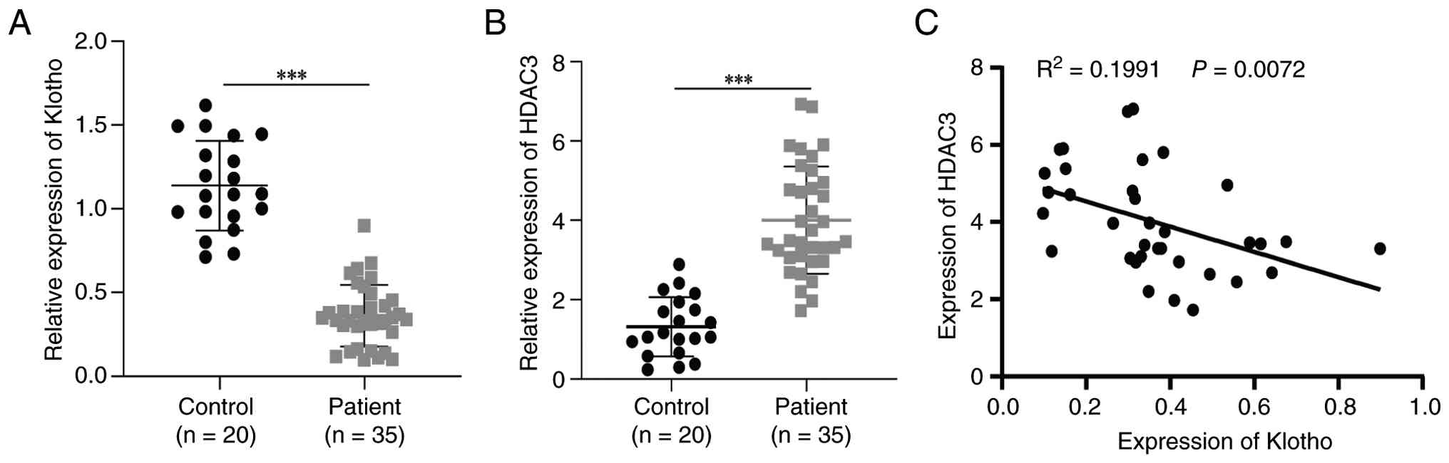
found low expression of Klotho and high expression of HDAC3 in CKD
samples (Fig. 1A and B). In
addition, correlation analysis at the clinical level indicated a
negative correlation between HDAC3 and Klotho (Fig. 1C). UUO is a typical model of renal
fibrosis, and it has been shown that urine flow obstruction leads
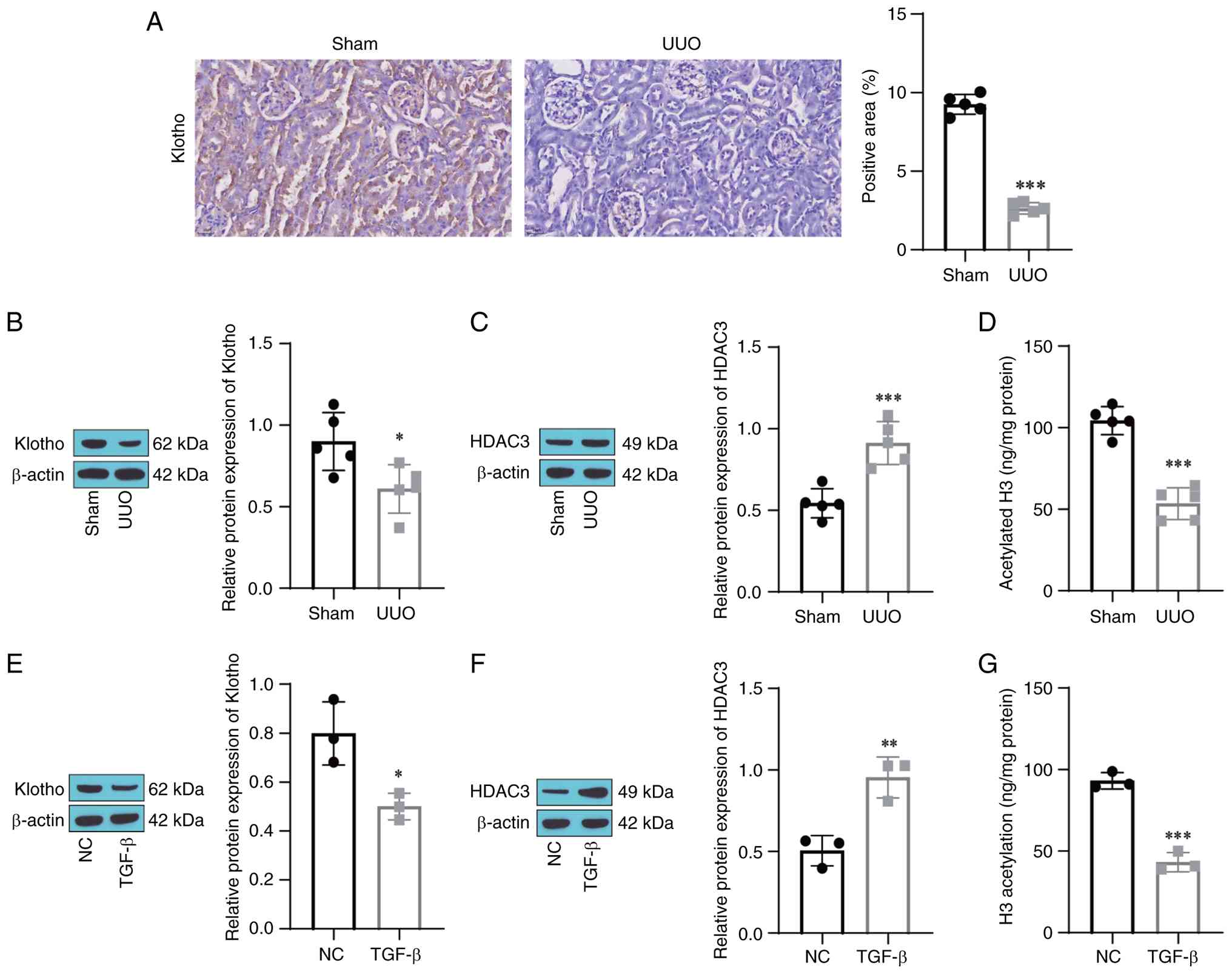
to extensive renal tubular lesions and fibrosis (30). After UUO-induced kidney fibrosis
was established, low expression of Klotho was observed in the renal
tissue of UUO mice (Fig. 2A).
Western blot analysis showed decreased Klotho and increased HDAC3
in the renal tissue of UUO mice (Fig.
2B and C). Histone acetylation plays a pivotal role in the
control of gene expression and is interrelated with the modulation
of several diseases, including tissue fibrosis (31). This modification of histones and
several non-histone proteins are under the control of histone
acetyltransferase and histone deacetylases (HDACs) (32), including HDAC3. Therefore, we
hypothesized that the low expression of Klotho in renal fibrosis
was interrelated with HDAC3-mediated histone deacetylation.
Compared with those in the sham group, histone H3 acetylation
levels were reduced in the UUO group (Fig. 2D). Similarly, decreased Klotho and
increased HDAC3 were observed in HK2 cells treated with TGF-β
(Fig. 2E and F). Decreased histone
H3 acetylation was detected in TGF-β-treated HK2 cells (Fig. 2G). These results indicate that
HDAC3-mediated histone deacetylation may be observably interrelated
with the abnormal downregulation of Klotho in renal fibrosis.
HDAC3 inhibits Klotho transcription
and promotes HK2 cell fibrosis through histone deacetylation
K9 in histone H3 is a unique target of HDAC3, and
HDAC3-mediated deacetylation of H3K9ac is important for H3K9
methylation (33), while increased
H3K9ac modification acts as a transcriptional activator and can
increase the levels of multiple genes. To further verify the effect
of HDAC3 on renal fibrosis via the inhibition of Klotho expression
through histone deacetylation, HK2 cells were treated with an HDAC3
selective inhibitor (RGFP966). ChIP-qPCR experiments showed that
H3K9ac and Klotho binding was markedly decreased after TGF-β
treatment while H3K9ac was increased in the Klotho group after the
addition of RGFP966 (Fig. 3A),
suggesting that a change in the H3K9ac level of Klotho occurs
during renal fibrosis, which is associated with HDAC3. RT-qPCR and
western blot analyses demonstrated that TGF-β inhibited the
expression of Klotho and that RGFP966 clearly restrained the
influence of TGF-β on Klotho (Fig. 3B
and C). To examine the function of HDAC3 and Klotho in renal
fibrosis, two samples of si-Klotho were designed and transfected
into HK2 cells. The knockdown efficiency was detected by western
blotting. The results showed that compared with the si-NC group,
the expression of Klotho in the si-Klotho-1 and si-Klotho-2 groups
decreased, where the si-Klotho-2 group showed significant
differences (Fig. 3D). Subsequent
experiments were conducted with si-Klotho-2. Further analysis
suggested that TGF-β increased the levels of ECM-related proteins,
including collagen I, Collagen IV and Fibronectin, and the
fibrosis-related protein α-SMA in HK2 cells. The addition of
RGFP966 prominently decreased the levels of Collagen I, Collagen
IV, Fibronectin and α-SMA, and these effects were reversed by
si-Klotho (Fig. 3E). These results
imply suppression of HDAC3 can upregulate Klotho expression through
histone acetylation, thereby suppressing TGF-β-induced fibrosis in
HK2 cells.
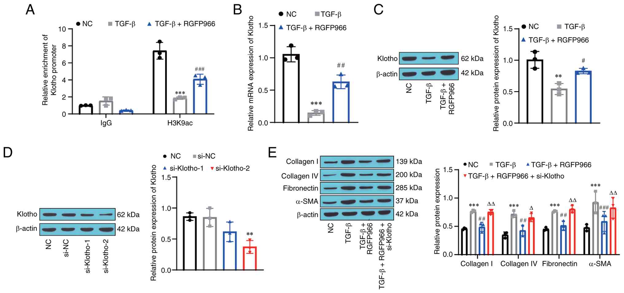 | Figure 3.HDAC3 inhibits Klotho transcription
and promotes HK2 cell fibrosis through histone deacetylation. (A)
H3K9ac levels in the Klotho promoter region were detected by
ChIP-qPCR. (B) Klotho mRNA expression levels in HK2 cells were
detected by RT-qPCR. (C) Klotho protein levels in HK2 cells were
detected by western blotting. (D) Validation of Klotho knockdown
efficiency by siRNA in HK2 cells. (E) Collagen I, Collagen IV,
Fibronectin and α-SMA protein expression levels in HK2 cells were
detected by western blotting. Data were analyzed using one-way
ANOVA followed by Tukey's post hoc test. **P<0.01, ***P<0.001
vs. NC group; #P<0.05, ##P<0.01,
###P<0.001 vs. the TGF-β group;
△P<0.05, △△P<0.01 vs. the TGF-β+RGFP966
group. ChIP, chromatin immunoprecipitation; RT-qPCR, reverse
transcription-quantitative PCR; siRNA, small interfering RNA;
α-SMA, α-smooth muscle actin; NC, negative control. |
HDAC3 binding to ATF3
transcriptionally downregulates Klotho
ATF3 can bind to HDAC3 without the need for
additional proteins (24). The
downregulation of Klotho was accompanied by the upregulation and
nuclear translocation of ATF3, and ATF3 overexpression restrained
the transcriptional activity of Klotho. Therefore, the present
study speculated that HDAC3 binds to ATF3 to mediate the
transcriptional repression of Klotho in renal fibrosis. Western
blot analysis showed that the ATF3 protein level was markedly
increased in UUO mice and in TGF-β-treated HK2 cells (Fig. 4A and B). Co-IP indicated that HDAC3
directly interacted with ATF3 both in vitro and in
vivo (Fig. 4C and D).
ChIP-qPCR analysis revealed that ATF3 was enriched in the Klotho
promoter region after TGF-β treatment, while the binding of ATF3 to
the Klotho promoter region decreased after the addition of RGFP966
(Fig. 4E). In order to explore the
regulatory effect of ATF3 and HDAC3 on Klotho, the present study
designed two si-ATF3 and transfected si-ATF3 into HK2 cells.
Western blot analysis showed that, compared with the si-NC group,
the expression of ATF3 was reduced in the si-ATF3-1 group and the
si-ATF3-2 group, and the reduction of ATF3 in the si-ATF3-2 group
was significantly different (Fig.
4F). si-ATF3-2 showed improved knockdown efficiency and was
used for subsequent experiments. Furthermore, TGF-β inhibited the
expression of Klotho in HK2 cells, and silencing ATF3 promoted the
expression of Klotho (Fig. 4G and
H). Consistently, to determine the ATF3-mediated effects in
vivo, ATF3 was knocked down in mouse kidneys, as shown in
Fig. S1. Similarly, lower
expression of Klotho was detected in the renal tissue of UUO mice,
and si-ATF3 increased the expression of Klotho (Fig. 4I). In addition, compared with those
in the UOO group, si-ATF3 decreased the levels of Collagen I,
Collagen IV, Fibronectin and α-SMA in the renal tissue of the mice,
suggesting that ATF3 knockdown can effectively inhibit those
proteins (Fig. 4J). Masson's
trichrome staining showed glomerular collagen fibril hyperplasia in
UUO mice, and collagen fibrils were extensively deposited in the
interstitium, ATF3 knockdown decreased collagen deposition
(Fig. 4K). These results point
toward HDAC3 binding to ATF3 to repress Klotho transcription and
promote renal fibrosis.
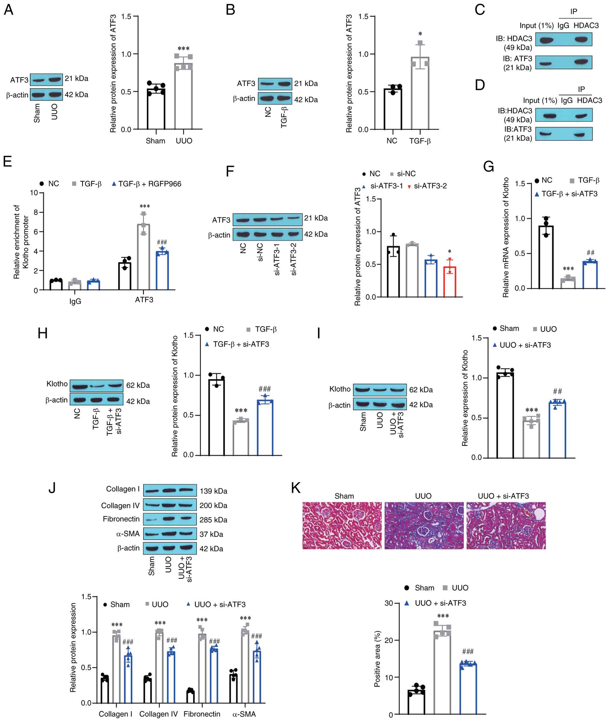 | Figure 4.HDAC3 binding to ATF3
transcriptionally downregulates Klotho. (A) ATF3 protein expression
in renal tissue of mice was detected by western blotting. (B) ATF3
protein expression in HK2 cells was confirmed by western blotting.
The binding of (C) HDAC3 and (D) ATF3 was corroborated by
coimmunoprecipitation in HK2 cells and renal tissue of mice. (E)
The binding of ATF3 to the Klotho promoter region was validated by
ChIP-qPCR. (F) Validation of ATF3 knockdown efficiency by siRNA in
HK2 cells. (G) ATF3 mRNA expression in HK2 cells was distinguished
by RT-qPCR. (H) Klotho protein expression in HK2 cells was detected
by western blotting. (I) Klotho protein expression in renal tissue
of mice was identified by western blotting. (J) Collagen I and IV,
Fibronectin and α-SMA protein expression in renal tissue of mice
were confirmed by western blotting. (K) Mouse pathological sections
were observed by Masson's Trichrome staining. Data were analyzed
using one-way ANOVA followed by Tukey's post hoc test. *P<0.05,
***P<0.001 vs. the NC/sham group; ##P<0.01,
###P<0.001 vs. the TGF-β group. ATF3, activates
transcription factor 3; UUO, unilateral ureteric obstruction; ChIP,
chromatin immunoprecipitation; RT-qPCR, reverse
transcription-quantitative PCR; siRNA, small interfering RNA;
α-SMA, α-smooth muscle actin; NC, negative control. |
ATF3 mediates an inhibitory effect of
Klotho on the Wnt/β-catenin signaling pathway
Blocking Wnt/β-catenin signaling may be beneficial
for alleviating renal fibrosis (34–36).
To explore the downstream mechanism of Klotho in kidney fibrosis, a
Klotho overexpression vector was constructed (Fig. S2A) and used to treat HK2 cells and
mice. Western blot analysis showed that the levels of Wnt1,
β-catenin, c-MYC and Cyclin D1 were markedly increased in the TGF-β
group, while the levels of Wnt1, β-catenin, c-MYC and Cyclin D1
were decreased in the TGF-β+oe-Klotho group; moreover, the
overexpression of ATF3 prevented the aforementioned results
(Fig. 5A). Compared with that in
the TGF-β group, Klotho overexpression inhibited the expression of
ECM-related proteins and fibrosis-related proteins, and the ATF3
overexpression vector that was constructed (Fig. S2B) increased the expression of
these proteins (Fig. 5B). In
addition, increased HDAC3 was detected in TGF-β-induced cells,
oe-Klotho had no effect on HDAC3, while overexpression of ATF3
promoted the expression of HDAC3 (Fig.
5C). Similarly, activated Wnt/β-catenin signaling and increased
renal fibrosis were observed in UUO mice. To confirm the in
vivo effects of Klotho and ATF3, Klotho and ATF3 were
overexpressed in kidney tissue (Fig.
S1). Overexpression of Klotho inhibited Wnt/β-catenin signaling
and the expression of ECM-related proteins and fibrosis-related
proteins. Further treatment of overexpression of ATF3 prevented the
effect of overexpression of Klotho to some extent (Fig. 5D and E). In addition, increased
HDAC3 was detected in the renal tissue of UUO mice. oe-Klotho had
no effect on HDAC3, while the overexpression of ATF3 promoted the
expression of HDAC3 (Fig. 5F).
Masson's trichrome staining showed that glomerular collagen fibrils
proliferated and were extensively deposited in the interstitium of
UUO mice, and collagen deposition was significantly reduced after
Klotho treatment, while overexpression of ATF3 could weaken the
effect of Klotho on renal fibrosis (Fig. 5G). The results suggest that the
alleviating effect of Klotho on TGF-β-induced fibrosis in HK2 cells
and renal fibrosis in UUO mice may be related to the Wnt/β-catenin
signaling pathway, and ATF3 prevents the inhibitory effect of
Klotho on Wnt/β-catenin signaling pathway.
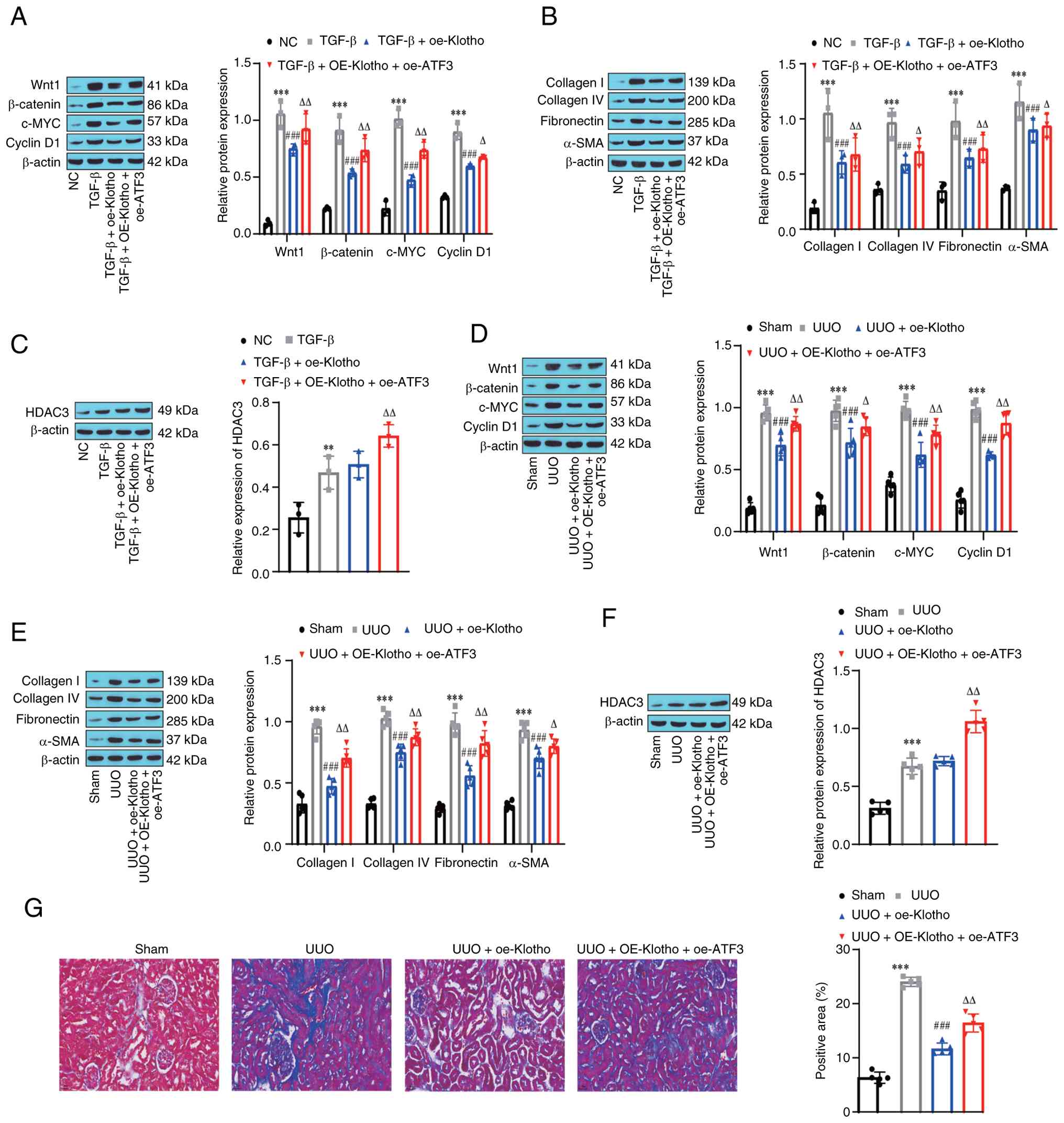 | Figure 5.ATF3 mediates the inhibitory effect
of Klotho on Wnt/β-catenin signaling pathway. (A) Wnt1, β-catenin,
c-MYC and Cyclin D1 protein expression in HK2 cells was detected by
western blotting. (B) Collagen I, Collagen IV, Fibronectin and
α-SMA protein expression in HK2 cells was identified by western
blotting. (C) HDAC3 protein expression in HK2 cells was detected by
western blotting. (D) Wnt1, β-catenin, c-MYC and Cyclin D1 proteins
in renal tissue of mice were detected by western blotting. (E)
Collagen I, Collagen IV, Fibronectin and α-SMA protein expression
in renal tissue of mice was detected by western blotting. (F) HDAC3
protein expression in renal tissue of mice was identified by
western blotting. (G) Mouse pathological section was observed by
Masson's Trichrome staining. Data were analyzed using one-way ANOVA
followed by Tukey's post hoc test. **P<0.01, ***P<0.001 vs.
the NC/sham group; ###P<0.001 vs. the TGF-β/UUO
group; △P<0.05, △△P<0.01 vs. the TGF-β
+ oe-Klotho/UUO + oe-Klotho group. NC, negative control; c-MYC,
cellular myelocytomatosis viral oncogene homolog; ATF3, activates
transcription factor 3; UUO, unilateral ureteric obstruction;
α-SMA, α-smooth muscle actin; HDAC3, histone deacetylase 3; oe,
overexpression. |
ATF3 mediates the inhibitory effect of
Klotho on the NF-κB signaling pathway
A previous study showed that Klotho itself has
antioxidant and anti-inflammatory effects and that the canonical
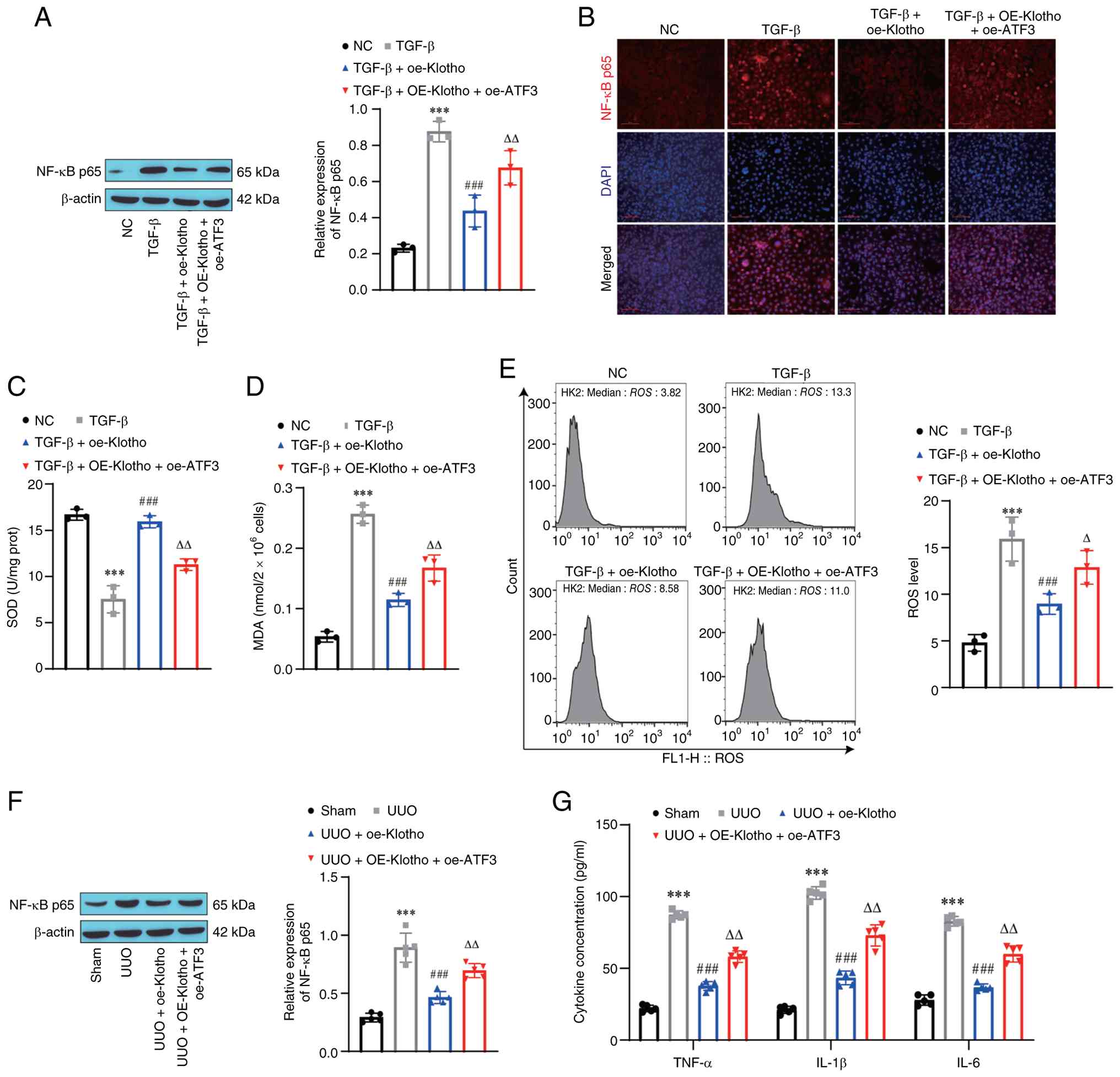
NF-κB component RelA is one of its targets (37). Therefore, the present study
subsequently detected the protein level of NF-κB. Western blot
analysis indicated that the level of NF-κB p65 was increased in the
TGF-β group, while NF-κB p65 was decreased after the addition of
oe-Klotho, and ATF3 promoted the level of NF-κB p65 (Fig. 6A). The immunofluorescence results
were consistent with the western blotting results (Fig. 6B). Moreover, TGF-β inhibited SOD
levels and increased MDA and ROS levels. Compared with those in the
TGF-β group, increased SOD levels and decreased MDA and ROS levels
were observed after the addition of oe-Klotho. Furthermore, oe-ATF3
promoted oxidative stress and inflammation (Fig. 6C-E). Similarly, high NF-κB p65
expression was observed in the renal tissue of UUO mice, Klotho
inhibited NF-κB p65 expression, and ATF3 prevented the influence of
Klotho (Fig. 6F). In addition,
inflammatory factor levels in murine serum were measured. UUO mice
had increased levels of TNF-α, IL-1β and IL-6 and Klotho decreased
the levels of these cytokines. ATF3 overexpression increased the
levels of TNF-α, IL-1β and IL-6 (Fig.
6G). The results suggested that the alleviating effect of
Klotho on TGF-β-induced fibrosis in HK2 cells and ATF3 prevents the
inhibitory effect of Klotho on the NF-κB signaling pathway.
Discussion
Renal fibrosis is a common kidney disease
characterized by depauperation and fibrosis of the renal tubules
and interstitium, resulting in the gradual deterioration of renal
function (38). Renal fibrosis is
currently considered the typical pathological change in renal aging
and CKD and is characterized by the transdifferentiation of
myofibroblasts and exorbitant sedimentation of renal interstitial
ECM protein (2). Currently,
clinical treatments for renal fibrosis are lacking, and new methods
aimed at probing the molecular biological mechanisms of renal
fibrosis have shown good efficacy (39). For example, exendin-4 imparts a
protective effect on renal fibrosis by binding to the GLP-1
receptor (40). Klotho, a gene
involved in mammalian aging, affects various disease processes by
regulating phosphate homeostasis and the activity of fibroblast
growth factor family members (5).
For example, the absence of Klotho can exacerbate the fibrosis
process of retinal cells and induce cardiac aging and heart failure
(41,42). A previous study suggests that
Klotho can restrain the progression of renal fibrosis (43). Klotho defects can accelerate the
transition of the profibrotic phenotype in renal tubular epithelial
cells and enhance the proliferation of fibroblasts, thereby
facilitating renal fibrosis (27).
Use of Klotho for the treatment of renal fibrosis has been widely
proposed (29,44,45).
Therefore, elucidation of the upstream and downstream molecular
mechanisms of Klotho could be helpful for targeting Klotho to
remedy renal fibrosis.
H3K9ac modification is a major class of histone
posttranslational modification that represses the transcriptional
activation of downstream genes by affecting nucleosomes (46). In the present study, abnormal
expression of Klotho in a renal fibrosis animal model or a
TGF-β-induced cell model was significantly associated with the
H3K9ac modification of Klotho. Previous research also found that in
renal fibrosis, Klotho has abnormal histone acetylation (47). HDACs are a family of histone
deacetylases that play important roles in mediating histone
modifications. To the best of our knowledge, the abnormal
expression of HDAC isoforms and their role in renal fibrosis have
not been consistently reported. Several class IIa HDAC inhibitors
exert antifibrotic effects by reducing HDAC4 and HDAC5 levels
(48). HDAC3 is a major member of
the HDAC family and plays a key role in various kidney diseases
(49). The clinical observation
found that renal HDAC3 expression was elevated while Klotho
expression was markedly reduced in patients with CKD compared with
non-CKD controls, with a negative association between the two
(Fig. 7). These clinical data
prompted the investigation of the mechanistic link between HDAC3
and Klotho in the pathogenesis of renal fibrosis. The present study
showed that HDAC3 was abnormally high in renal fibrosis and
suppressed Klotho expression by inhibiting the H3K9ac modification
level of Klotho and promoting the activation of fibrosis-associated
proteins (Collagen-I, Collagen-IV, Fibronectin and α-SMA). Chen
et al (50) found that
selective suppression of HDAC3 and genetic knockout of HDAC3
attenuated the pathology of renal fibrosis, which was consistent
with the findings of the present study and the present study
further confirmed that HDAC3 repressed Klotho transcriptional
activation via H3K9ac.
The function of the HDAC family is usually to act
together with transcription factors with transcriptional repression
activity. For example, HDAC3 binds to the transcriptional repressor
ZNF22 to mediate the transcriptional repression of ZO-1, Occludin
and Claudin-5, promoting blood-tumor barrier permeability in the
blood-tumor barrier of tumors (51). ATF3 is one of the most widespread
transcription repressors. ATF3 mediates the activation effect of
LPS on inflammation by binding to HDAC3 (19,24,52).
The present study showed that ATF3 was highly expressed in renal
fibrosis and Sung et al (53) reported that high expression of ATF3
facilitates the progression of renal fibrosis. Further studies
found that inhibition of HDAC3 alters the nuclear localization of
ATF3 and inhibit its expression in the Klotho promoter region
(18,19,24).
HDAC3 inhibits H3K9ac modification in the Klotho promoter region,
and conversely, ATF3 directly inhibits Klotho transcription. Klotho
is commonly shown to play an important role in fibrosis-related
diseases by inhibiting the activation of the Wnt/β-catenin
signaling pathway to inhibit the expression of fibrotic proteins
(54), and Klotho can regulate the
expression of various cytokines and chemokines (55,56).
In the present study, overexpression of Klotho inhibited oxidative
stress and inflammation by repressing the Wnt/β-catenin and NF-κB
pathways, thereby alleviating fibrosis. The overexpression of ATF3
prevented the aforementioned effects.
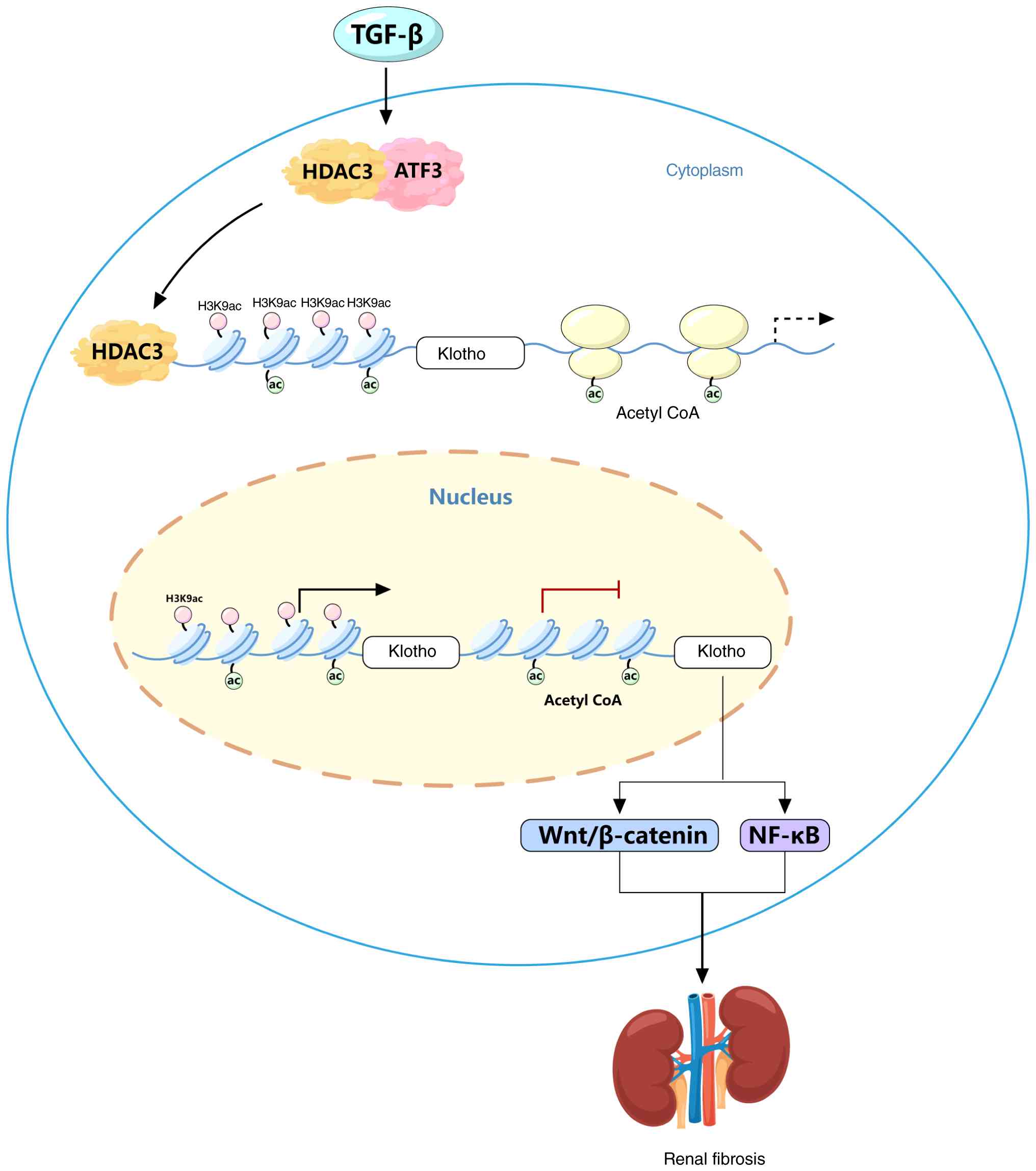
In brief, the present findings establish an
HDAC3-ATF3-Klotho regulatory axis in which HDAC3 recruits ATF3 to
the Klotho promoter, leading to Klotho transcriptional repression.
Downregulation of Klotho may activate the Wnt/β-catenin and NF-κB
pathways to promote oxidative stress and inflammation, thereby
exacerbating the fibrotic process (Fig. 6). This result demonstrated the
therapeutic potential of HDAC3-selective inhibitors for
upregulating Klotho expression in the treatment of renal fibrosis
disease. However, the present study did not explore other
chromosomal-level modifications of Klotho, nor did it conduct
relevant tests on DNA-level modifications of Klotho, which is the
aim of future research. The present study only used HK2 cells to
verify the phenotypes of renal fibrosis or its regulatory
mechanisms. In future studies, other kidney cell lines (such as
mesangial cells and renal interstitial cells) will be used to
further validate the findings. One limitation of the in vivo
experiments should be acknowledged. To confirm ATF3 knockdown in
the kidney, si-ATF3 was administered in combination with other
constructs (oe-Klotho and/or oe-ATF3) rather than as a single
agent. While the reduced ATF3 expression observed in the
UUO+si-ATF3 group compared with the UUO group supports the efficacy
of si-ATF3, the present study did not include a group treated with
si-ATF3 alone. Consequently, the possibility that co-administration
of other constructs may have influenced ATF3 expression or the
overall phenotype cannot be entirely ruled out. Future studies
using si-ATF3 alone in vivo would be valuable to confirm the
specific contribution of ATF3 knockdown in this model. Notably,
HDAC inhibitors have been approved by the FDA for the treatment of
skin and peripheral T-cell lymphoma and multiple myeloma (57–59).
However, HDAC-based methods or drugs have not yet been used to
treat kidney disease (60,61). The present study provides evidence
for the application of HDAC suppressors in renal fibrosis-related
diseases.
Supplementary Material
Supporting Data
Supporting Data
Acknowledgements
Not applicable.
Funding
This work was supported by the National Natural Science
Foundation of China (grant no. 82370720) and the Science and
Technology Plan Project of Yunnan Provincial Science and Technology
Department (grant no. 202201AY070001-193).
Availability of data and materials
The data generated in the present study may be
requested from the corresponding author.
Authors' contributions
ZY, WL and GL designed the study. ZY and XW
performed the experiments. ZY, XW and HC contributed to data
analysis and drafted the manuscript. ZY wrote the manuscript. ZY,
XW and GL revised the manuscript. All authors read and approved the
final manuscript. ZY and GL confirm the authenticity of all the raw
data.
Ethics approval and consent to
participate
The present study was reviewed and approved by the
Animal Care Committee of Yan'an Hospital Affiliated with Kunming
Medical University (Kunming, China) in conformance with the
institutional guidelines (approval no. 2021044) and was carried out
under the ARRIVE Guidelines 2.0.
Patient consent for publication
Not applicable.
Competing interests
The authors declare that they have no competing
interests.
References
|
1
|
Mark PB, Stafford LK, Grams ME, Aalruz H,
ElHafeez SA, Abdelgalil AA, Abdulkader RS, Abeywickrama HM, Abiodun
OO, Abramov D, et al: Global, regional, and national burden of
chronic kidney disease in adults, 1990–2023, and its attributable
risk factors: A systematic analysis for the global burden of
disease study 2023. Lancet. 406:2461–2482. 2025. View Article : Google Scholar : PubMed/NCBI
|
|
2
|
Zeisberg M and Neilson EG: Mechanisms of
tubulointerstitial fibrosis. J Am Soc Nephrol. 21:1819–1834. 2010.
View Article : Google Scholar : PubMed/NCBI
|
|
3
|
Woroniecki R, Gaikwad AB and Susztak K:
Fetal environment, epigenetics, and pediatric renal disease.
Pediatr Nephrol. 26:705–711. 2011. View Article : Google Scholar : PubMed/NCBI
|
|
4
|
Wing MR, Ramezani A, Gill HS, Devaney JM
and Raj DS: Epigenetics of progression of chronic kidney disease:
Fact or fantasy? Semin Nephrol. 33:363–374. 2013. View Article : Google Scholar : PubMed/NCBI
|
|
5
|
Buchanan S, Combet E, Stenvinkel P and
Shiels PG: Klotho, aging, and the failing kidney. Front Endocrinol
(Lausanne). 11:5602020. View Article : Google Scholar : PubMed/NCBI
|
|
6
|
Satoh M, Nagasu H, Morita Y, Yamaguchi TP,
Kanwar YS and Kashihara N: Klotho protects against mouse renal
fibrosis by inhibiting Wnt signaling. Am J Physiol Renal Physiol.
303:F1641–F1651. 2012. View Article : Google Scholar : PubMed/NCBI
|
|
7
|
Zeng Y, Wang PH, Zhang M and Du JR:
Aging-related renal injury and inflammation are associated with
downregulation of Klotho and induction of RIG-I/NF-κB signaling
pathway in senescence-accelerated mice. Aging Clin Exp Res.
28:69–76. 2016. View Article : Google Scholar : PubMed/NCBI
|
|
8
|
Zhao Y, Banerjee S, Dey N, LeJeune WS,
Sarkar PS, Brobey R, Rosenblatt KP, Tilton RG and Choudhary S:
Klotho depletion contributes to increased inflammation in kidney of
the db/db mouse model of diabetes via RelA (serine)536
phosphorylation. Diabetes. 60:1907–1916. 2011. View Article : Google Scholar : PubMed/NCBI
|
|
9
|
Sun G, Reddy MA, Yuan H, Lanting L, Kato M
and Natarajan R: Epigenetic histone methylation modulates fibrotic
gene expression. J Am Soc Nephrol. 21:2069–2080. 2010. View Article : Google Scholar : PubMed/NCBI
|
|
10
|
Liu N, He S, Ma L, Ponnusamy M, Tang J,
Tolbert E, Bayliss G, Zhao TC, Yan H and Zhuang S: Blocking the
class I histone deacetylase ameliorates renal fibrosis and inhibits
renal fibroblast activation via modulating TGF-beta and EGFR
signaling. PLoS One. 8:e540012013. View Article : Google Scholar : PubMed/NCBI
|
|
11
|
Singh BN, Zhang G, Hwa YL, Li J, Dowdy SC
and Jiang SW: Nonhistone protein acetylation as cancer therapy
targets. Expert Rev Anticancer Ther. 10:935–954. 2010. View Article : Google Scholar : PubMed/NCBI
|
|
12
|
Pang M and Zhuang S: Histone deacetylase:
A potential therapeutic target for fibrotic disorders. J Pharmacol
Exp Ther. 335:266–272. 2010. View Article : Google Scholar : PubMed/NCBI
|
|
13
|
Gilbert RE, Huang Q, Thai K, Advani SL,
Lee K, Yuen DA, Connelly KA and Advani A: Histone deacetylase
inhibition attenuates diabetes-associated kidney growth: Potential
role for epigenetic modification of the epidermal growth factor
receptor. Kidney Int. 79:1312–1321. 2011. View Article : Google Scholar : PubMed/NCBI
|
|
14
|
Lin W, Zhang Q, Liu L, Yin S, Liu Z and
Cao W: Klotho restoration via acetylation of peroxisome
proliferation-activated receptor γ reduces the progression of
chronic kidney disease. Kidney Int. 92:669–679. 2017. View Article : Google Scholar : PubMed/NCBI
|
|
15
|
Wang Y, Jiao B, Hu Z and Wang Y: Critical
role of histone deacetylase 3 in the regulation of kidney
inflammation and fibrosis. Kidney Int. 105:775–790. 2024.
View Article : Google Scholar : PubMed/NCBI
|
|
16
|
Chen F, Zhang L, Liu W, Zhang B, Wang S,
Zhou Z, Wang W, Shen J, Deng Y and Cao W: Epigenetic suppression of
RASAL1 by HDAC3 and cofactor YY1 promotes fibroblast-myofibroblast
transition and renal fibrosis. Research (Wash D C).
9:10732026.PubMed/NCBI
|
|
17
|
Cui H, Guo M, Xu D, Ding ZC, Zhou G, Ding
HF, Zhang J, Tang Y and Yan C: The stress-responsive gene ATF3
regulates the histone acetyltransferase Tip60. Nat Commun.
6:67522015. View Article : Google Scholar : PubMed/NCBI
|
|
18
|
Zhu J, Wang F, Wang L, Dai B, Xu G, Zhao
L, Jiang H, Gao W, Zhang T, Zhao C, et al: HDAC inhibition
increases CXCL12 secretion to recruit natural killer cells in
peripheral T-cell lymphoma. Cancer Res. 84:2450–2467. 2024.
View Article : Google Scholar : PubMed/NCBI
|
|
19
|
Nguyen HCB, Adlanmerini M, Hauck AK and
Lazar MA: Dichotomous engagement of HDAC3 activity governs
inflammatory responses. Nature. 584:286–290. 2020. View Article : Google Scholar : PubMed/NCBI
|
|
20
|
Li HF, Cheng CF, Liao WJ, Lin H and Yang
RB: ATF3-mediated epigenetic regulation protects against acute
kidney injury. J Am Soc Nephrol. 21:1003–1013. 2010. View Article : Google Scholar : PubMed/NCBI
|
|
21
|
Zhu B, Ni Y, Gong Y, Kang X, Guo H, Liu X,
Li J and Wang L: Formononetin ameliorates ferroptosis-associated
fibrosis in renal tubular epithelial cells and in mice with chronic
kidney disease by suppressing the Smad3/ATF3/SLC7A11 signaling.
Life Sci. 315:1213312023. View Article : Google Scholar : PubMed/NCBI
|
|
22
|
Wang L, Liu Y, Du T, Yang H, Lei L, Guo M,
Ding HF, Zhang J, Wang H, Chen X and Yan C: ATF3 promotes
erastin-induced ferroptosis by suppressing system Xc. Cell Death
Differ. 27:662–675. 2020. View Article : Google Scholar : PubMed/NCBI
|
|
23
|
Tang Q, Xie J, Wang Y, Dong C and Sun Q:
Exosomes secreted by ATF3/Nrf2-mediated ferroptotic renal tubular
epithelial cells promote M1/M2 ratio imbalance inducing renal
interstitial fibrosis following ischemia and reperfusion injury.
Front Immunol. 16:15105002025. View Article : Google Scholar : PubMed/NCBI
|
|
24
|
Darlyuk-Saadon I, Weidenfeld-Baranboim K,
Yokoyama KK, Hai T and Aronheim A: The bZIP repressor proteins,
c-Jun dimerization protein 2 and activating transcription factor 3,
recruit multiple HDAC members to the ATF3 promoter. Biochim Biophys
Acta. 1819:1142–1153. 2012. View Article : Google Scholar : PubMed/NCBI
|
|
25
|
Li J, Lin Q, Shao X, Li S, Zhu X, Wu J,
Mou S, Gu L, Wang Q, Zhang M, et al: HIF1α-BNIP3-mediated mitophagy
protects against renal fibrosis by decreasing ROS and inhibiting
activation of the NLRP3 inflammasome. Cell Death Dis. 14:2002023.
View Article : Google Scholar : PubMed/NCBI
|
|
26
|
Livak KJ and Schmittgen TD: Analysis of
relative gene expression data using real-time quantitative PCR and
the 2(−Delta Delta C(T)) method. Methods. 25:402–408. 2001.
View Article : Google Scholar : PubMed/NCBI
|
|
27
|
Guan X, Nie L, He T, Yang K, Xiao T, Wang
S, Huang Y, Zhang J, Wang J, Sharma K, et al: Klotho suppresses
renal tubulo-interstitial fibrosis by controlling basic fibroblast
growth factor-2 signalling. J Pathol. 234:560–572. 2014. View Article : Google Scholar : PubMed/NCBI
|
|
28
|
Zhang D, Li Z, Gao Y and Sun H: MiR-556-3p
mediated repression of klotho under oxidative stress promotes
fibrosis of renal tubular epithelial cells. Sci Rep. 15:121822025.
View Article : Google Scholar : PubMed/NCBI
|
|
29
|
Wang Y and Zhao J: The protective function
of αKlotho in chronic kidney disease: Evidence and therapeutic
implications. Integr Med Nephrol Androl. 11:1–11. 2024. View Article : Google Scholar
|
|
30
|
Nan QY, Piao SG, Jin JZ, Chung BH, Yang CW
and Li C: Pathogenesis and management of renal fibrosis induced by
unilateral ureteral obstruction. Kidney Res Clin Pract. 43:586–599.
2024. View Article : Google Scholar : PubMed/NCBI
|
|
31
|
Tang J and Zhuang S: Epigenetics in acute
kidney injury. Curr Opin Nephrol Hypertens. 24:351–358.
2015.PubMed/NCBI
|
|
32
|
Zhuang S: Epigenetic targeting for acute
kidney injury. Nephrology (Carlton). 23 (Suppl 4):S21–S25. 2018.
View Article : Google Scholar
|
|
33
|
Ji H, Zhou Y, Zhuang X, Zhu Y, Wu Z, Lu Y,
Li S, Zeng Y, Lu QR, Huo Y, et al: HDAC3 deficiency promotes liver
cancer through a defect in H3K9ac/H3K9me3 transition. Cancer Res.
79:3676–3688. 2019. View Article : Google Scholar : PubMed/NCBI
|
|
34
|
Xie H, Miao N, Xu D, Zhou Z, Ni J, Yin F,
Wang Y, Cheng Q, Chen P, Li J, et al: FoxM1 promotes Wnt/β-catenin
pathway activation and renal fibrosis via transcriptionally
regulating multi-Wnts expressions. J Cell Mol Med. 25:1958–1971.
2021. View Article : Google Scholar : PubMed/NCBI
|
|
35
|
Li SS, Sun Q, Hua MR, Suo P, Chen JR, Yu
XY and Zhao YY: Targeting the Wnt/β-catenin signaling pathway as a
potential therapeutic strategy in renal tubulointerstitial
fibrosis. Front Pharmacol. 12:7198802021. View Article : Google Scholar : PubMed/NCBI
|
|
36
|
Ma J, Jiao N, Liu X and Lu X: Saracatinib
delays the progression of renal interstitial fibrosis by inhibiting
the Wnt/β-catenin pathway. Kidney360. 7:25–34. 2026. View Article : Google Scholar : PubMed/NCBI
|
|
37
|
Izquierdo MC, Perez-Gomez MV, Sanchez-Niño
MD, Sanz AB, Ruiz-Andres O, Poveda J, Moreno JA, Egido J and Ortiz
A: Klotho, phosphate and inflammation/ageing in chronic kidney
disease. Nephrol Dial Transplant. 27 (Suppl 4):iv6–iv10. 2012.
View Article : Google Scholar : PubMed/NCBI
|
|
38
|
Cheikh Hassan HI, Tang M, Djurdjev O,
Langsford D, Sood MM and Levin A: Infection in advanced chronic
kidney disease leads to increased risk of cardiovascular events,
end-stage kidney disease and mortality. Kidney Int. 90:897–904.
2016. View Article : Google Scholar : PubMed/NCBI
|
|
39
|
Chen C, Lin XH, Xie YM, Xiong SL, Hou SZ,
Huang S, Jian HL, Wen YF, Jiang XY and Liang J: Shengjiang Xiexin
Decoction ameliorates DSS-induced ulcerative colitis via activating
Wnt/β-Catenin signaling to enhance epithelium renovation and
modulating intestinal flora. Phytomedicine. 139:1564562025.
View Article : Google Scholar : PubMed/NCBI
|
|
40
|
Jia Y, Zheng Z, Guan M, Zhang Q, Li Y,
Wang L and Xue Y: Exendin-4 ameliorates high glucose-induced
fibrosis by inhibiting the secretion of miR-192 from injured renal
tubular epithelial cells. Exp Mol Med. 50:1–13. 2018. View Article : Google Scholar : PubMed/NCBI
|
|
41
|
Xie L, Wang Y, Li Q, Ji X, Tu Y, Du S, Lou
H, Zeng X, Zhu L, Zhang J and Zhu M: The
HIF-1α/p53/miRNA-34a/Klotho axis in retinal pigment epithelial
cells promotes subretinal fibrosis and exacerbates choroidal
neovascularization. J Cell Mol Med. 25:1700–1711. 2021. View Article : Google Scholar : PubMed/NCBI
|
|
42
|
Chen K, Wang S, Sun QW, Zhang B, Ullah M
and Sun Z: Klotho deficiency causes heart aging via impairing the
Nrf2-GR pathway. Circ Res. 128:492–507. 2021. View Article : Google Scholar : PubMed/NCBI
|
|
43
|
Yuan Q, Ren Q, Li L, Tan H, Lu M, Tian Y,
Huang L, Zhao B, Fu H, Hou FF, et al: A Klotho-derived peptide
protects against kidney fibrosis by targeting TGF-β signaling. Nat
Commun. 13:4382022. View Article : Google Scholar : PubMed/NCBI
|
|
44
|
Hajare AD, Dagar N and Gaikwad AB: Klotho
antiaging protein: Molecular mechanisms and therapeutic potential
in diseases. Mol Biomed. 6:192025. View Article : Google Scholar : PubMed/NCBI
|
|
45
|
Nijhawan P: Soluble alpha klotho for
reversal of renal interstitial fibrosis and renal regeneration: A
hypothesis based on developmental biology and preclinical evidence:
TH-PO0573. J Am Soc Nephrol. 36((10S)): TH–PO0573. 2025.
|
|
46
|
Zhang Y, Zhang Q, Zhang Y and Han J: The
role of histone modification in DNA replication-coupled nucleosome
assembly and cancer. Int J Mol Sci. 24:49392023. View Article : Google Scholar : PubMed/NCBI
|
|
47
|
Xia J and Cao W: Epigenetic modifications
of Klotho expression in kidney diseases. J Mol Med (Berl).
99:581–592. 2021. View Article : Google Scholar : PubMed/NCBI
|
|
48
|
Xiong C, Guan Y, Zhou X, Liu L, Zhuang MA,
Zhang W, Zhang Y, Masucci MV, Bayliss G, Zhao TC and Zhuang S:
Selective inhibition of class IIa histone deacetylases alleviates
renal fibrosis. FASEB J. 33:8249–8262. 2019. View Article : Google Scholar : PubMed/NCBI
|
|
49
|
Zhang L and Cao W: Histone deacetylase 3
(HDAC3) as an important epigenetic regulator of kidney diseases. J
Mol Med (Berl). 100:43–51. 2022. View Article : Google Scholar : PubMed/NCBI
|
|
50
|
Chen F, Gao Q, Wei A, Chen X, Shi Y, Wang
H and Cao W: Histone deacetylase 3 aberration inhibits Klotho
transcription and promotes renal fibrosis. Cell Death Differ.
28:1001–1012. 2021. View Article : Google Scholar : PubMed/NCBI
|
|
51
|
Zhu B, Zhang L, Zhou X, Ning H and Ma T:
Transcription factor ZNF22 regulates blood-tumor barrier
permeability by interacting with HDAC3 protein. Front Mol Neurosci.
15:10279422022. View Article : Google Scholar : PubMed/NCBI
|
|
52
|
Jeon HB and Jang Y: Reversible
inactivation of bovine plasma amine oxidase by cysteamine and
related analogs. Biochem Biophys Res Commun. 403:442–446. 2010.
View Article : Google Scholar : PubMed/NCBI
|
|
53
|
Sung CC, Poll BG, Lin SH,
Murillo-de-Ozores AR, Chou CL, Chen L, Yang CR, Chen MH, Hsu YJ and
Knepper MA: Early molecular events mediating loss of aquaporin-2
during ureteral obstruction in rats. J Am Soc Nephrol.
33:2040–2058. 2022. View Article : Google Scholar : PubMed/NCBI
|
|
54
|
Miao J, Liu J, Niu J, Zhang Y, Shen W, Luo
C, Liu Y, Li C, Li H, Yang P, et al: Wnt/β-catenin/RAS signaling
mediates age-related renal fibrosis and is associated with
mitochondrial dysfunction. Aging Cell. 18:e130042019. View Article : Google Scholar : PubMed/NCBI
|
|
55
|
Guo Y, Zhuang X, Huang Z, Zou J, Yang D,
Hu X, Du Z, Wang L and Liao X: Klotho protects the heart from
hyperglycemia-induced injury by inactivating ROS and NF-κB-mediated
inflammation both in vitro and in vivo. Biochim Biophys Acta Mol
Basis Dis. 1864:238–251. 2018. View Article : Google Scholar : PubMed/NCBI
|
|
56
|
Lai L, Li Y, Liu J, Luo L, Tang J, Xue J
and Liu T: Bovine serum albumin aggravates macrophage M1 activation
and kidney injury in heterozygous Klotho-deficient mice via the gut
microbiota-immune axis. Int J Biol Sci. 17:742–755. 2021.
View Article : Google Scholar : PubMed/NCBI
|
|
57
|
Chun P: Histone deacetylase inhibitors in
medical therapeutics. Medical Epigenetics. Elsevier; pp. 633–655.
2016, View Article : Google Scholar
|
|
58
|
Li Y, Wang F, Chen X, Wang J, Zhao Y, Li Y
and He B: Zinc-dependent deacetylase (HDAC) inhibitors with
different zinc binding groups. Curr Top Med Chem. 19:223–241. 2019.
View Article : Google Scholar : PubMed/NCBI
|
|
59
|
El Omari N, Bakrim S, Elhrech H, Aanniz T,
Balahbib A, Lee LH, Al Abdulmonem W and Bouyahya A: Clinical
efficacy and mechanistic insights of FDA-approved HDAC inhibitors
in the treatment of lymphoma. Eur J Pharm Sci. 208:1070572025.
View Article : Google Scholar : PubMed/NCBI
|
|
60
|
Cappellacci L, Perinelli DR, Maggi F,
Grifantini M and Petrelli R: Recent progress in histone deacetylase
inhibitors as anticancer agents. Curr Med Chem. 27:2449–2493. 2020.
View Article : Google Scholar : PubMed/NCBI
|
|
61
|
Li J, Yu C, Shen F, Cui B, Liu N and
Zhuang S: Class IIa histone deacetylase inhibition ameliorates
acute kidney injury by suppressing renal tubular cell apoptosis and
enhancing autophagy and proliferation. Front Pharmacol.
13:9461922022. View Article : Google Scholar : PubMed/NCBI
|