Introduction
Hypertension is a notable global public health
challenge, which affects >1 billion individuals worldwide, and
remains a predominant contributor to cardiovascular morbidity and
mortality (1). The chronic
pressure overload induced by hypertension initiates a pathological
cascade that drives cardiac fibrosis, ultimately culminating in
serious adverse outcomes such as heart failure. This pathological
process is characterized by the differentiation of cardiac
fibroblasts (CFs) into myofibroblasts, marked by the emergence of
α-smooth muscle actin (α-SMA) expression (2) and the excessive deposition of
extracellular matrix (ECM) proteins, including collagen I and
fibronectin (3). Notably,
targeting this fibrogenic cascade has considerable therapeutic
potential for preserving cardiac function and improving survival in
patients with cardiovascular disease (4).
Mitochondrial homeostasis, which is defined as the
dynamic equilibrium of mitochondrial function, morphology and mass,
has increasingly been recognized as a key determinant underlying
fibrotic progression (5–7). Mitophagy serves as a pivotal
mechanism in orchestrating mitochondrial homeostasis. Mitophagy is
a selective form of autophagy responsible for clearing damaged
mitochondria, fine-tuning mitochondrial quality control largely
through the canonical PTEN-induced kinase 1 (PINK1)/Parkin-mediated
pathway (8–10). This pathway has been extensively
documented to exert cardioprotective effects in pathological
contexts, such as myocardial ischemia-reperfusion injury and
cardiac hypertrophy (11–13). However, under sustained
pathological stimuli, such as hypertension-induced pressure
overload, mitophagy overactivation disrupts cardiac mitochondrial
homeostasis by depleting functional mitochondria, triggering a
bioenergetic crisis and thus exacerbating the progression of
cardiovascular diseases (14–17).
The precise regulatory mechanism of excessive mitophagy has not yet
been fully elucidated.
Transcriptional regulation represents a central
control layer in numerous biological processes, which notably
contributes to the pathogenesis of cardiovascular diseases. POU
domain class 2 transcription factor 1 (POU2F1) has been implicated
in both oncogenesis and cardiovascular pathologies (18,19),
where it facilitates disease progression through modulation of
angiotensin II (Ang II) signaling, AMPK activation and
stress-responsive genes, including sestrin 2 (20,21).
A recent study revealed that exercise training alleviates Ang
II-induced activation of CFs by reducing POU2F1 expression
(22). However, whether POU2F1
regulates mitophagy, particularly through the PINK1/Parkin pathway,
in hypertensive cardiac fibrosis remains to be elucidated.
In the present study, RNA sequencing was performed
to identify candidate transcription factors associated with
hypertensive cardiac fibrosis. The present study aimed to
investigate the functional role of POU2F1 in cardiac fibrotic
remodeling and elucidate the molecular mechanisms underlying its
regulation of mitochondrial homeostasis, with emphasis on the
PINK1/Parkin-dependent mitophagy pathway.
Materials and methods
Animals
Male SHRs and male Wistar-Kyoto (WKY) rats (age, 5
weeks; weight, ~120 g) were purchased from Beijing Vital River
Laboratory Animal Technology Co., Ltd. A total of 18 SHRs and 9 WKY
rats were used in the current study. The rats were randomly divided
into the following groups: WKY rats served as the normotensive
control group (n=9). SHRs were randomly assigned to three groups:
Untreated SHR group (n=10), SHR + AAV9-shNC group (n=4), and SHR +
AAV9-shPOU2F1 (n=4). SHRs in the latter two groups received tail
vein injections of AAV9-shNC or AAV9-shPOU2F1, respectively, at 14
weeks of age and were maintained for an additional 4 weeks before
subsequent analyses. All rats were acclimated and maintained in the
laboratory until 14 weeks of age, at which time SHRs had developed
established hypertension and myocardial fibrosis and were
subsequently subjected to further analyses. Hypertension was
confirmed by measuring systolic blood pressure (SBP) using the
tail-cuff method, and rats with SBP consistently >150 mmHg were
considered hypertensive. Myocardial fibrosis was verified by
Masson's trichrome staining and fibrosis-related protein expression
analyses (23,24). The rats were housed under
controlled temperature (22±2°C) and relative humidity (50–60%) with
a 12-h light/dark cycle. Animals were monitored daily for health
and behavior throughout the study, with free access to food and
water. Humane endpoints included severe weight loss (>20%),
inability to access food or water, persistent lethargy or signs of
severe distress; no animals reached these humane endpoints and no
unexpected deaths occurred. All procedures were approved by the
Animal Care and Use Committee of Yueyang Hospital of Integrated
Traditional Chinese and Western Medicine (approval no.
YYLAC-2023-182-1; Shanghai, China) and were conducted following the
National Institutes of Health Guide for the Care and Use of
Laboratory Animals (25). At 18
weeks of age, rats were anesthetized with inhaled isoflurane (3%
for induction and 1.5–2% for maintenance in oxygen), administered
via a nose cone, and subjected to echocardiographic examination.
Euthanasia was performed using an overdose of inhaled isoflurane
(5% in oxygen) for 10 min until complete respiratory arrest
occurred. Death was confirmed by the cessation of spontaneous
respiration, absence of heartbeat and loss of corneal reflex.
Cell culture
CFs were prepared from neonatal rats and cultured as
described previously (26).
Primary cardiac fibroblasts were isolated from the ventricles of 36
neonatal Sprague-Dawley rats (age, 1 day) obtained from Beijing
Vital River Laboratory Animal Technology Co., Ltd. The neonatal
rats were euthanized by decapitation prior to heart collection.
Subsequently, the ventricles were isolated from the hearts, minced
into small pieces in ice-cold Hank's balanced salt solution, and
subjected to repeated enzymatic digestion with collagenase type II
(cat. no. 17101-015; both Gibco, Thermo Fisher Scientific, Inc.) at
37°C. The cells were then resuspended, plated in 10-cm dishes with
Dulbecco's Modified Eagle Medium (cat. no. SH30243.01B; HyClone;
Cytiva) supplemented with 10% fetal bovine serum (cat. no.
16000-044; Gibco; Thermo Fisher Scientific, Inc.) and cultured at
37°C in 5% CO2. After a 24-h incubation, the cells were
washed with phosphate-buffered saline (PBS) to remove cellular
debris and non-adherent cells. Cell cultures yielded by this
procedure were used as CFs based on their typical spindle-shaped
morphology and adherence characteristics. Ang II (cat. no.
HY-13948; MedChemExpress) was dissolved in sterile ultrapure water
(cat. no. ST872; Beyotime Biotechnology) to prepare the stock
solution. Cells treated with the corresponding volume of sterile
ultrapure water were used as the vehicle control. Primary CFs were
treated with Ang II at a concentration of 1 µmol/l for 48 h at
37°C, whereas control cells were maintained in serum-supplemented
medium.
Echocardiography
Cardiac function was evaluated using
echocardiography (27,28). Rats were anesthetized with inhaled
isoflurane (3% for induction and 1.5–2% for maintenance in oxygen),
which was administered via a nose cone, and were then positioned in
a supine position on a heated platform. Echocardiographic M-mode
images were obtained from a parasternal short axis view at the
level of the papillary muscles. A Vevo 2100 instrument
(VisualSonics, Inc.) equipped with an MS-400 imaging transducer was
used to measure interventricular septal diastolic thickness (IVSD),
interventricular septal systolic thickness (IVSS), left ventricular
end-diastolic diameter (LVIDD), LV end-systolic diameter (LVIDS),
left ventricular posterior wall diastolic thickness (LVPWD) and
left ventricular posterior wall systolic thickness (LVPWS).
Histopathological examination
Cardiac tissues were removed, rinsed with PBS, fixed
in 4% paraformaldehyde at 4°C for 24 h, dehydrated with graded
ethanol, embedded in paraffin and cut into 5-µm sections. The heart
sections were subsequently processed for Masson trichrome staining
to evaluate myocardial fibrosis as previously described (29,30).
For immunohistochemical staining, the sections were deparaffinized,
rehydrated and subjected to microwave-assisted antigen retrieval in
citrate buffer (pH 6.0) at 100°C for 20 min, followed by three
washes with PBS. After blocking with 5% BSA (cat. no. ST023;
Beyotime Biotechnology) at room temperature for 1 h, the sections
were incubated with primary antibodies against POU2F1 (cat. no.
10387-1-AP; 1:1,000; Proteintech Group, Inc.), collagen I (cat. no.
14695-1-AP; 1:500; Proteintech Group, Inc.) and α-SMA (cat. no.
ab7817; 1:1,000; Abcam) overnight at 4°C, followed by incubation
with horseradish peroxidase-conjugated goat anti-rabbit (cat. no.
A0208; 1:1,000; Beyotime Biotechnology) and anti-mouse IgG (cat.
no. A0216; 1:1,000; Beyotime Biotechnology) at room temperature for
1 h. Immunoreactive signals were visualized using 3DAB), followed
by hematoxylin counterstaining. Images were captured using a light
microscope, and fibrosis was quantified using ImageJ software
(version 1.8.0; National Institutes of Health).
Transmission electron microscopy
(TEM)
Cardiac tissues (1 mm3) were fixed in
2.5% glutaraldehyde at 4°C for 24 h, washed with PBS and post-fixed
with 1% osmium tetroxide at 4°C for 2 h. Dehydration was carried
out through sequential immersion in 30, 50, 70 and 80% ethanol
solutions, followed by a 1:1 mixture of 90% ethanol and 90%
acetone. The samples were embedded in epoxy resin at 60°C for 48 h.
Ultrathin sections (50 nm) were stained with 2% uranyl acetate for
15 min at room temperature and 2% lead citrate for 10 min at room
temperature, then examined under a transmission electron microscope
(Tecnai G2 Spirit BioTWIN; Thermo Fisher Scientific, Inc.)
(31,32).
RNA-seq
Total RNA was extracted from cardiac tissues using
TRIzol® reagent (Invitrogen; Thermo Fisher Scientific,
Inc.). RNA concentration was measured with a NanoDrop ND-2000
spectrophotometer (NanoDrop; Thermo Fisher Scientific, Inc.) and
RNA integrity (RNA integrity number ≥7) was assessed using an
Agilent 2100 Bioanalyzer (Agilent Technologies, Inc.). Library
preparation and 150-bp paired-end sequencing were performed on an
Illumina NovaSeq 6000 platform (Illumina, Inc.) by Majorbio
BioPharm Technology Co., Ltd. (33). Raw reads were quality-controlled
using fastp (v0.23.2, github.com/OpenGene/fastp), and
differentially expressed genes (DEGs) between the SHR and WKY
groups were identified using DESeq2 (v1.38.3, bioconductor.org)
with thresholds of |log2(fold change)|>1 and adjusted
P≤0.05. Hierarchical cluster analysis of DEGs was performed using
the OECloud tools (v1.26, cloud.oebiotech.com) to demonstrate the
expression pattern of genes in different groups and samples. The
transcription factor data of rats was obtained from AnimalTFDB v4.0
(https://guolab.wchscu.cn/AnimalTFDB4//#/).
Adeno-associated virus serotype 9
(AAV9) injection
To determine the role of POU2F1 in PINK1-mediated
mitophagy and myocardial fibrosis in vivo, short hairpin RNA
(shRNA) targeting POU2F1 (5′-TGCACAGGATCTTCAACAATT-3′) or a
negative control (NC) sequence (shNC, 5′-TTCTCCGAACGTGTCACGT-3′)
were cloned into AAV9 vectors and packaged into recombinant AAV9
particles, generating AAV9-shPOU2F1 and AAV9-shNC, respectively.
The shRNA and the recombinant AAV9 vectors were constructed and
packaged by Shanghai GenePharma Co., Ltd. SHRs were injected via
the tail vein at 14 weeks of age with 2×10¹¹ viral
genomes/rat AAV9-shPOU2F1 or AAV9-shNC (in 200 µl PBS). Both groups
were fed a standard diet and echocardiographic assessments were
performed at 18 weeks of age. Subsequently, the rats were
euthanized using an overdose of inhaled isoflurane as
aforementioned.
Immunofluorescence staining
The CFs were fixed with 4% paraformaldehyde for 15
min, permeabilized with Triton X-100 for 10 min and blocked with 1%
BSA (cat. no. ST023; Beyotime Biotechnology) for 1 h, all at room
temperature. Subsequently, the cells were incubated overnight at
4°C with a primary antibody against α-SMA (cat. no. ab7817; 1:100;
Abcam). Alexa Fluor® 488-conjugated goat anti-mouse IgG
(cat. no. A0428; Beyotime Biotechnology) was then applied at room
temperature for 1 h, followed by DAPI (cat. no. C1002; Beyotime
Biotechnology) counterstaining at room temperature for 5 min.
Fluorescent images were captured using a fluorescence microscope
(ECLIPSE Ni; Nikon Corporation).
MitoSOX and EdU staining
To measure mitochondrial superoxide levels, the CFs
were incubated with MitoSOX Red (cat. no. M36008; Thermo Fisher
Scientific, Inc.) for 10 min at 37°C in the dark, according to the
manufacturer's instructions. DAPI was used to stain the nuclei at
room temperature for 10 min. Red fluorescence reflecting
mitochondrial superoxide levels was captured using a confocal laser
scanning microscope. For proliferation analysis, EdU incorporation
was performed using the EdU kit (cat. no. C0071L; Beyotime
Biotechnology) according to the manufacturer's protocol. Hoechst
33342 was used to stain the nuclei at room temperature for 10 min.
ImageJ software (version 1.8.0) was employed to quantify MitoSOX
fluorescence intensity and count the number of EdU-positive
cells.
Gene overexpression and knockdown
For gene overexpression, the coding sequence of rat
POU2F1 was cloned into the pcDNA3.1(+) vector (General Biol,). The
CFs were then transfected with the POU2F1 plasmid or an empty
vector control. For gene knockdown, shRNA constructs targeting
POU2F1 or PINK1, and a NC, were purchased from Shanghai GenePharma
Co., Ltd.; the selected sequences are listed in Table SI. CFs were seeded at a density of
2×10⁵ cells/well in 6-well plates and transfected 24 h
after seeding. Transfection was performed using
Lipofectamine® 2000 (Invitrogen; Thermo Fisher
Scientific, Inc.) according to the manufacturer's instructions
(34). Specifically, 2.5 µg
plasmid DNA and 6 µl Lipofectamine 2000 reagent were incubated at
37°C. The culture medium was replaced with fresh medium at 6 h, and
the cells were cultured for 48 h after transfection before being
harvested for subsequent experiments. CFs were co-transfected with
oe-POU2F1 and sh-PINK1 plasmids simultaneously.
Dual-luciferase reporter gene
assay
First, the rat PINK1 promoter sequence (−2,000 bp)
was obtained from the National Center for Biotechnology Information
(NCBI; ncbi.nlm.nih.gov/gene/), and the POU2F1 binding motif was
predicted using Jaspar (jaspar.genereg.net/). The predicted
POU2F1-binding sequence was identified as 5′-TATTTAAAT-3′
(wild-type). A mutant sequence (5′-ATAAAGGGA-3′) was generated by
replacing the predicted POU2F1-binding motif. Both sequences were
synthesized and cloned into the pGL3-Basic luciferase reporter
vector (Promega Corporation). Subsequently, the synthesized
plasmids were subjected to Sanger sequencing to confirm the
accuracy of the inserted sequences. The plasmids were then
extracted using a plasmid extraction kit (Beijing Solarbio Science
& Technology Co., Ltd.). 293T cells were co-transfected with WT
or MUT PINK1 promoter reporter plasmids together with either the
POU2F1 overexpression plasmid (pcDNA3.1-POU2F1, General Biol,
Anhui, China) or the corresponding empty vector control (pcDNA3.1,
both General Biol, Anhui, China). A total of 4 µg of plasmids was
transfected into cells in each group using Lipofectamine 3000 (cat.
no. L3000001; Invitrogen; Thermo Fisher Scientific, Inc.). After 6
h of incubation, the transfection medium was replaced with complete
medium. At 48 h after transfection, cells were lysed, and firefly
and Renilla luciferase activities were determined using the
Dual-Luciferase Reporter Assay System (Promega Corporation).
Firefly luciferase activity was normalized to Renilla luciferase
activity, and relative promoter activity was calculated as the
firefly/Renilla luciferase ratio (35).
Electrophoretic mobility shift assay
(EMSA)
EMSA was performed using the Chemiluminescent EMSA
Kit (cat. no. GS009; Beyotime Biotechnology) according to the
manufacturer's instructions (36).
The WT and MUT oligonucleotide probes were designed according to
the predicted POU2F1-binding site in the PINK1 promoter and
synthesized by Beyotime Biotechnology. For the EMSA binding
reaction, 4 µg purified protein (Cat. no. P04804; Solarbio
Biotechnology) was incubated with 1 pmol biotin-labeled probe in
binding buffer for 20 min at room temperature. For competitive
EMSA, 100 pmol unlabeled competitor probes were added to the
reaction mixture 20 min prior to the addition of the biotin-labeled
probe. In the mutation assay, mutated biotin-labeled probes were
used in place of WT probe. The reaction mixtures were resolved on a
6% polyacrylamide gel and transferred to a nylon membrane. The DNA
oligomers were then crosslinked to the membrane via UV irradiation,
and the biotin-labeled probes were detected using a
chemiluminescent imaging system.
Seahorse metabolic assays
According to the manufacturer's instructions,
mitochondrial stress was assessed using the Agilent Seahorse XF
Cell Mito Stress Test Kit (cat. no. 103015-100; (Agilent
Technologies, Inc.) to measure the oxygen consumption rate (OCR) of
the CFs. For ex vivo experiments, CFs isolated from SHR and
WKY rat hearts, as well as CFs isolated from AAV9-shNC- or
AAV9-shPOU2F1-treated SHR rats, were subjected to Seahorse
analysis. For in vitro experiments, Ang II-induced CFs
transfected with shPOU2F1 or shNC were used. Briefly, 16,000
cells/well were seeded in a 96-well XF Cell Culture Microplate
(Agilent Technologies, Inc.). OCR was measured at three timepoints,
in basal conditions and following the addition of oligomycin (1.0
µg/ml), carbonyl cyanide-4-trifluoromethoxy phenylhydrazone (1.0
µmol/l) and rotenone + antimycin A (both at 0.5 µmol/l). Data were
analyzed using Seahorse XF software (version 2.6; Agilent
Technologies, Inc.).
Western blotting
The protein expression was analyzed by western
blotting as described previously (37), with GAPDH used as an endogenous
control. After total protein was extracted from rat ventricular
tissue and CFs using RIPA buffer (Beyotime Biotechnology)
supplemented with protease inhibitors and quantified by BCA assay,
protein samples (25 µg/lane) were separated by 10% SDS-PAGE and
transferred onto polyvinylidene difluoride membranes. The membranes
were blocked with 5% skimmed milk at room temperature for 1 h and
incubated with primary antibodies at 4°C overnight. The next day,
the membranes were washed three times with TBS-0.1% Tween-20 wash
buffer for 10 min each. The membranes were incubated with secondary
antibodies for 1 h at room temperature. Membranes were washed with
TBS-0.1% Tween-20 wash buffer. The following antibodies were used
in western blotting: Collagen I (cat. no. 14695-1-AP; 1:1,000;
Proteintech Group, Inc.), α-SMA (cat. no. ab7817; 1:1,000; Abcam),
POU2F1 (cat. no. 10387-1-AP; 1:2,000; Proteintech Group, Inc.),
PINK1 (cat. no. 23274-1-AP; 1:1,000; Proteintech Group, Inc.),
Parkin (cat. no. 14060-1-AP; 1:1,000; Proteintech Group, Inc.),
LC3B (cat. no. 14600-1-AP; 1:1,000; Proteintech Group, Inc.), P62
(cat. no. 18420-1-AP; 1:5,000; Proteintech Group, Inc.) and GAPDH
(cat. no. 10494-1-AP; 1:5,000; Proteintech Group, Inc.) primary
antibodies, horseradish peroxidase-conjugated goat anti-mouse IgG
(cat. no. A0216; 1:1,000; Beyotime Biotechnology), and horseradish
peroxidase-conjugated goat anti-rabbit IgG secondary antibody (cat.
no. A0208; 1:1,000; Beyotime Biotechnology). Chemiluminescent
detection was performed and images were captured using the Tanon
5200 system (Tanon Science and Technology Co., Ltd.). The gray
values were measured and analyzed using ImageJ software (version
1.8.0; National Institutes of Health).
Reverse transcription-quantitative PCR
(RT-qPCR)
Total RNA was extracted from cardiac tissue and CFs
using a Total RNA Extraction Reagent (Shanghai Yeasen Biotechnology
Co., Ltd.). After DNase treatment, the RNA was reverse-transcribed
into cDNA using a cDNA Synthesis Kit (Shanghai Yeasen Biotechnology
Co., Ltd.) according to the manufacturer's instructions. qPCR was
performed using SYBR Green Master Mix (Shanghai Yeasen
Biotechnology Co., Ltd.). The thermocycling conditions were as
follows: initial denaturation at 95°C for 5 min, followed by 40
cycles of denaturation at 95°C for 10 sec and annealing/extension
at 60°C for 30 sec. A melting curve analysis was performed to
verify amplification specificity. GAPDH was used as an internal
control for normalization, and relative mRNA expression levels were
calculated using the 2−ΔΔCq method (38). Primer sequences are listed in
Table SII.
Chromatin immunoprecipitation
(ChIP)-qPCR assay
CFs were cultured to 80–90% confluence. CFs were
cross-linked with 1% paraformaldehyde at room temperature for 10
min, and the crosslinking reaction was quenched with glycine at
room temperature for 5 min. The cells were then resuspended in ChIP
Sonication Cell Lysis Buffer (cat. no. 81804; Cell Signaling
Technology, Inc.) supplemented with Protease Inhibitor Cocktail
(PIC) and incubated on ice. Subsequently, chromatin was fragmented
by sonication on ice using a sonicator (power, 20% amplitude; pulse
duration, 5 sec on/5 sec off; total duration, 10 min) to obtain DNA
fragments of appropriate size. The fragmented chromatin was diluted
in ChIP Buffer (cat. no. 14231; Cell Signaling Technology, Inc.)
containing PIC to prepare a 2% input sample. The remaining
chromatin was diluted in ChIP Dilution Buffer containing PIC and
incubated overnight at 4°C with an anti-POU2F1 antibody (cat. no.
ab178869) or normal rabbit IgG (both 5 µg; cat. no. ab313801) (both
from Abcam) as a NC. qPCR was performed using
SimpleChIP® Universal qPCR Master Mix (Cell Signaling
Technology, Inc.) according to the manufacturer's instructions to
quantify the enrichment of POU2F1-bound PINK1 promoter regions.
Primer sequences are listed in Table
SIII. Detailed experimental methods are included in the
previously published literature (39,40).
Cell viability assay
Cell viability of primary CFs in response to Ang II
was assessed using the CCK-8 assay kit (cat. no. C0037; Beyotime
Biotechnology) according to the manufacturer's instructions
(41). Primary CFs were seeded
into 96-well plates at 1×10⁴ cells/well and incubated for 24 h.
Subsequently, the cells were treated with different concentrations
of Ang II for 48 h. Following treatment, 10 µl of CCK-8 reagent was
added to each well, and the cells were incubated at 37°C for 2 h.
The optical density at 450 nm was measured using a microplate
reader to evaluate cell viability.
Statistical analysis
Data are presented as the mean ± standard deviation
of ≥3 independent experiments. Statistical analyses were performed
using GraphPad Prism 9.0 software (Dotmatics). Statistical tests,
including one-way analysis of variance followed by Tukey's post hoc
test and unpaired two-tailed Student's t-test, were conducted to
determine significant differences between sample means.
Correlations between variables were analyzed using Spearman's rank
correlation analysis. P<0.05 was considered to indicate a
statistically significant difference.
Results
POU2F1 upregulation and mitochondrial
damage in myocardial fibrosis of SHRs
A significant increase in cardiac fibrosis was
observed in SHRs compared with in the WKY control rats. Masson
trichrome staining of cardiac apical tissue revealed disrupted
cardiac architecture in SHRs, which was characterized by extensive
blue-stained areas indicating collagen deposition and fibrotic
remodeling (Fig. S1A). Western
blotting confirmed elevated expression of the fibrosis markers
collagen I and α-SMA in SHRs (Fig.
S1B). Consistently, immunohistochemical staining revealed
markedly stronger immunoreactivity for collagen I and α-SMA in the
left ventricular myocardium of SHRs than in that of WKY rats
(Fig. S1C). To characterize
molecular alterations associated with cardiac fibrosis in SHRs,
RNA-seq was performed on cardiac tissues from SHRs and WKY rats.
Bioinformatics analysis identified 476 DEGs between the groups. By
comparing the DEGs with a rat transcription factor database, 18
differentially expressed transcription factors were identified
(Fig. 1A). Hierarchical clustering
demonstrated the expression patterns of these transcription
factors: 17 were upregulated in SHRs, whereas one was downregulated
(Fig. 1B). Among these
transcription factors, POU2F1 exhibited the most significant
differential expression; western blotting and RT-qPCR confirmed
upregulation of POU2F1 at both the mRNA and protein levels in SHRs
(Fig. 1C and D). This increase was
further supported by immunohistochemical analysis, which
demonstrated enhanced POU2F1 staining in the myocardial tissues of
SHRs (Fig. 1E). Notably, it was
further observed that the area of cardiac fibrosis was closely
correlated with both the mRNA (ρ=0.7250) and protein (ρ=0.9091)
expression levels of POU2F1 (Fig.
S1D).
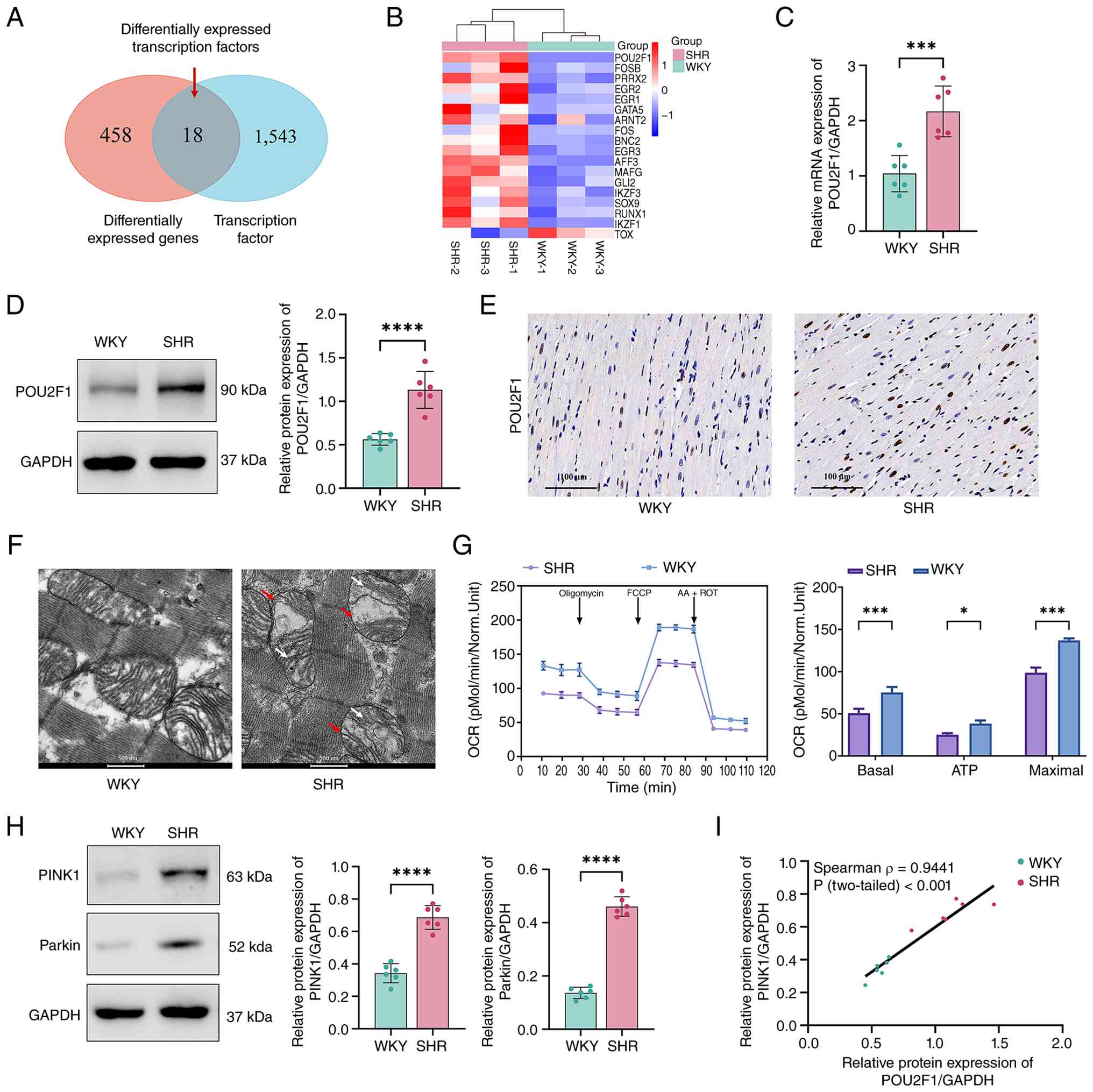 | Figure 1.POU2F1 upregulation and mitochondrial
damage in myocardial fibrosis of SHRs. (A) Overlap between
transcription factors and differentially expressed genes in cardiac
tissues of SHRs compared with WKY rats. (B) Heatmap representing
the relative levels of differentially expressed transcription
factors between WKY rats and SHRs. (C) mRNA levels of POU2F1 in
cardiac tissues were detected by reverse transcription-quantitative
PCR. (D) Protein levels of POU2F1 in cardiac tissues were detected
by western blotting (n=6). (E) Representative immunohistochemical
staining images of POU2F1 in left ventricular myocardial tissues
from WKY rats and SHR. (scale bar, 100 µm). (F) Transmission
electron microscopy was used to observe mitochondrial structure in
cardiac tissue from WKY rats and SHRs (scale bar, 500 nm). White
arrows indicate cristae disorganization; red arrows indicate
damaged mitochondria. (G) Seahorse assay profile of OCRs in CFs
following sequentially treatment with oligomycin, FCCP and AA +
ROT. Quantitative analysis of mitochondrial function parameters
(basal respiration, maximal respiration and ATP production) is
shown in the bar charts (n=3). (H) Protein levels of PINK1 and
Parkin in cardiac tissues were detected by western blotting (n=6).
(I) Protein expression levels of PINK1 were positively correlated
with POU2F1 expression (Spearman correlation, n=12). *P<0.05;
***P<0.001; ****P<0.0001. AA, antimycin A; FCCP, carbonyl
cyanide-4-trifluoromethoxy phenylhydrazone; OCR, oxygen consumption
rate; PINK1, PTEN-induced kinase 1; POU2F1, POU domain class 2
transcription factor 1; ROT, rotenone; SHR, spontaneously
hypertensive rat; WKY, Wistar-Kyoto. |
TEM demonstrated mitochondrial ultrastructural
damage in the cardiac tissues of SHRs, characterized by cristae
disorganization and an increased number of mitochondria with
abnormal morphology, including swelling and disrupted cristae
(Figs. 1F and S1E). In addition, mitochondrial
respiratory function, as assessed by OCR, was significantly
impaired in SHRs, with decreased basal respiration, maximal
respiration and ATP production-associated respiration (Fig. 1G). Notably, the expression levels
of the key mitophagy regulators PINK1 and Parkin were significantly
increased in the hearts of SHRs (Fig.
1H), suggesting dysregulation of the PINK1/Parkin-associated
mitophagy pathway during hypertensive fibrogenesis. Furthermore, a
strong positive correlation was observed between PINK1 and POU2F1
expression (r=0.9441, P<0.001; Fig.
1I).
POU2F1 upregulation and mitochondrial
damage in Ang II-induced CFs
Primary CFs were treated with Ang II (0–100 µmol/l)
for 48 h and cell viability was assessed using the CCK-8 assay. The
results showed that 1 µmol/l Ang II maintained relatively higher
cell viability. Therefore, 1 µmol/l Ang II was selected for
subsequent experiments (Fig.
S2A). Notably, the concurrent increase in EdU positivity
(Figs. 2A and S2B) and α-SMA expression (Figs. 2B and S2C) demonstrated that Ang II-induced CFs
underwent both proliferation and phenotypic activation
(myofibroblast differentiation). These coordinated cellular
responses contribute to pathological ECM accumulation during
myocardial fibrosis (42). This
finding was corroborated by significantly elevated protein levels
of both collagen I and α-SMA in Ang II-treated CFs (Fig. S2D). Consistent with the in
vivo findings, Ang II-induced CFs exhibited elevated mRNA and
protein levels of POU2F1 (Fig. 2C and
D). Furthermore, POU2F1 protein levels were positively
correlated with α-SMA fluorescence intensity (r=0.8951, P<0.001;
Fig. 2E).
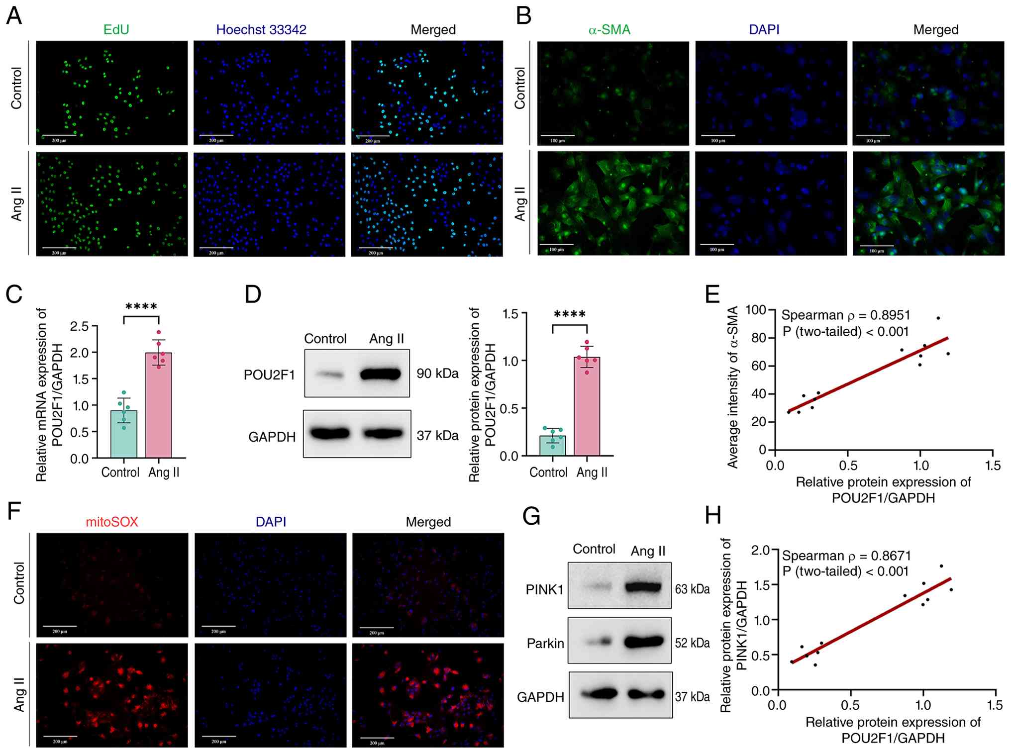 | Figure 2.POU2F1 upregulation and mitochondrial
damage in Ang II-induced CFs. (A) CF proliferation was assessed
using an EdU assay (scale bar, 200 µm). (B) Representative
immunofluorescence staining of α-SMA in CFs (scale bar, 100 µm).
(C) mRNA levels of POU2F1 in CFs were detected by reverse
transcription-quantitative PCR (n=6). (D) Protein levels of POU2F1
in CFs were detected by western blotting (n=6). (E) α-SMA
fluorescence intensity was positively correlated with the protein
expression levels of POU2F1 in CFs (Spearman correlation, n=12).
(F) MitoSOX staining showing levels of mitochondrial superoxide in
CFs (scale bar, 200 µm). (G) Protein levels of PINK1 and Parkin in
CFs were detected by western blotting (n=6). (H) Protein expression
levels of PINK1 were positively correlated with POU2F1 expression
in CFs (Spearman correlation, n=12). Data are presented as the mean
± SD. (C and D) Unpaired two-tailed Student's t-test were used for
statistical analysis. ****P<0.0001. α-SMA, α-smooth muscle
actin; Ang II, angiotensin II; CF, cardiac fibroblast; PINK1,
PTEN-induced kinase 1; POU2F1, POU domain class 2 transcription
factor 1. |
In addition to POU2F1 upregulation, mitochondrial
reactive oxygen species were assessed using mitoSOX Red staining
(Fig. 2F). In Ang II-induced CFs,
robust red fluorescence signals indicative of mitochondrial
superoxide accumulation were observed, demonstrating heightened
mitochondrial oxidative stress (Figs.
2F and S2E). Consistently,
western blotting revealed marked upregulation of PINK1 and Parkin,
suggesting potential activation of the mitophagy pathway (Figs. 2G and S2F). Furthermore, correlation analysis
revealed a strong positive correlation between POU2F1 and PINK1
protein expression (r=0.8671, P<0.001; Fig. 2H).
POU2F1 knockdown reduces
PINK1-mediated mitophagy and mitochondrial dysfunction in Ang
II-induced CFs
To investigate the role of POU2F1 in PINK1-mediated
mitophagy and myocardial fibrosis in vitro, a plasmid-based
shRNA vector was constructed to specifically knock down POU2F1
expression, and the knockdown efficiency was confirmed by RT-qPCR
analysis in CFs (Fig. S3A). In
the shPOU2F1 group, mitoSOX levels were decreased, suggesting
attenuated oxidative stress (Fig. 3A
and C), and TEM further confirmed reduced mitochondrial
ultrastructural damage (Fig. 3B and
C). In addition, mitochondrial respiratory function was
significantly increased upon POU2F1 knockdown (Fig. 3D). Concomitantly, under Ang II
stimulation, the shPOU2F1 group showed a significant decrease in
the expression levels of the mitophagy-related proteins PINK1,
Parkin and LC3B, and an increase in P62 expression, indicating that
silencing of POU2F1 inhibited the PINK1-mediated mitophagy pathway
(Fig. 3E). Additionally, POU2F1
knockdown inhibited CF proliferation and α-SMA expression (Fig. 3F-H). Western blot analysis
confirmed the decreased expression of collagen I and α-SMA in cells
with POU2F1 knockdown, thus indicating that Ang II-induced fibrotic
remodeling was attenuated (Fig.
3I).
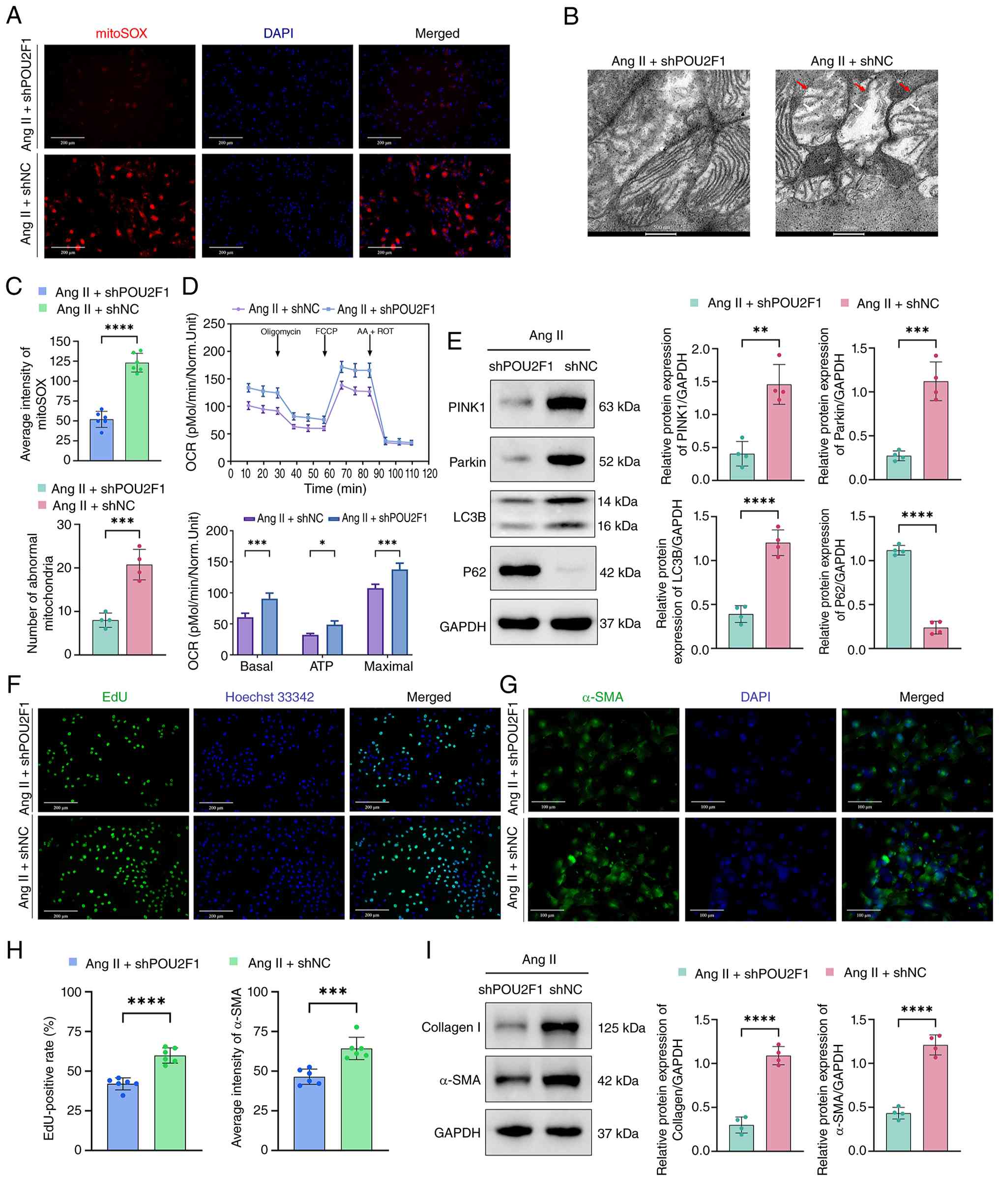 | Figure 3.POU2F1 knockdown reduces
PINK1-mediated mitophagy and mitochondrial dysfunction in Ang
II-induced CFs. (A) MitoSOX staining showing levels of
mitochondrial superoxide in Ang II-induced CFs infected with
shPOU2F1 or shNC (scale bar, 200 µm). (B) Transmission electron
microscopy was used to observe mitochondrial structure (scale bar,
500 nm). White arrows indicate cristae disorganization; red arrows
indicate damaged mitochondria. (C) Quantification of MitoSOX
fluorescence intensity was performed (n=6). Quantification of
abnormal mitochondria was performed using low-magnification TEM
images (×5,300 magnification; n=4). (D) Seahorse assay profile of
OCRs in CFs following treatment with oligomycin, FCCP and AA + ROT.
Quantitative analysis of mitochondrial function parameters (basal
respiration, maximal respiration and ATP production) is shown in
the bar charts (n=3). (E) Western blotting was utilized to
determine the protein expression levels of PINK1, Parkin, LC3B and
P62 (n=4). (F) CF proliferation was assessed using an EdU assay
(scale bar, 200 µm). (G) Representative immunofluorescence images
of α-SMA (scale bar, 100 µm). (H) Quantification of EdU-positive
cells and α-SMA expression (n=6). (I) Western blotting was utilized
to determine the protein expression levels of collagen I and α-SMA
(n=4). Data are presented as the mean ± SD. (C-E, H and I) Unpaired
two-tailed Student's t-test was used for statistical analysis.
*P<0.05; **P<0.01; ***P<0.001; ****P<0.0001. α-SMA,
α-smooth muscle actin; AA, antimycin A; Ang II, angiotensin II; CF,
cardiac fibroblast; FCCP, carbonyl cyanide-4-trifluoromethoxy
phenylhydrazone; NC, negative control; OCR, oxygen consumption
rate; PINK1, PTEN-induced kinase 1; POU2F1, POU domain class 2
transcription factor 1; ROT, rotenone; sh, short hairpin. |
PINK1 knockdown reverses POU2F1
overexpression-induced myocardial fibrosis
The current study generated plasmids to induce
POU2F1 overexpression and PINK1 knockdown, with the aim of further
investigating the role of the POU2F1/PINK1 signaling pathway in
myocardial fibrosis. The overexpression efficiency of POU2F1 and
knockdown efficiency of PINK1 were confirmed by RT-qPCR analysis in
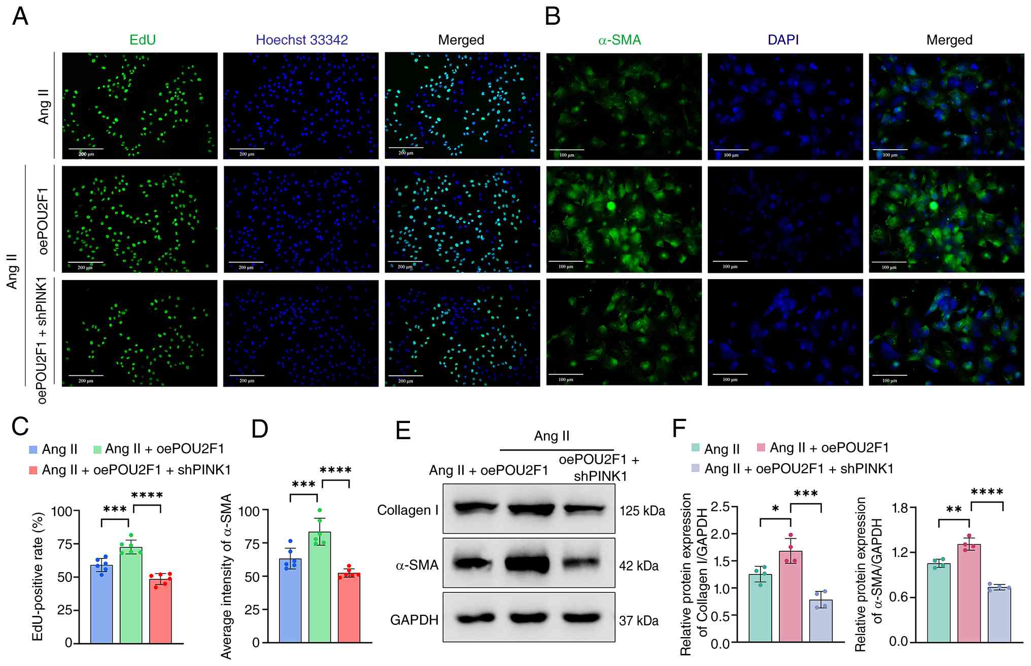
CFs (Fig. S3B). A significant
increase in both CF proliferation (Fig. 4A and C) and α-SMA expression
(Fig. 4B and D) was observed
following POU2F1 overexpression. Furthermore, consistent with the
pro-fibrotic phenotype, elevated expression levels of collagen I
and α-SMA proteins were detected in response to POU2F1
overexpression (Fig. 4E and F).
Notably, this pro-fibrotic effect was attenuated by PINK1
knockdown. These findings highlight the key role of PINK1 in the
regulation of myocardial fibrosis.
PINK1 knockdown reduces mitophagy and
myocardial fibrosis independent of POU2F1 expression in Ang
II-induced CFs
To investigate the potential interplay between
POU2F1 and PINK1, gene-specific knockdown experiments were
performed in CFs. POU2F1 knockdown significantly reduced PINK1
expression, whereas PINK1 knockdown did not alter POU2F1 protein
levels in Ang II-induced CFs (Fig. 5A
and B), suggesting a unidirectional regulatory role of POU2F1
over PINK1. These findings indicated that POU2F1 does not directly
interact with PINK1 but may function as an upstream transcriptional
regulator of PINK1 expression. Given the central role of PINK1 in
mitophagy, the current study next assessed the effects of PINK1
knockdown on the mitophagy pathway. In Ang II-induced CFs,
knockdown of PINK1 significantly decreased the expression levels of
key mitophagy-related proteins, including PINK1, Parkin and LC3B,
while inducing the accumulation of P62, collectively indicating
suppression of mitophagy (Fig.
5C). Furthermore, PINK1 knockdown resulted in an attenuation of
collagen I and α-SMA levels (Fig.
5D). Collectively, these results demonstrated that
PINK1-mediated mitophagy critically contributes to Ang II-induced
myocardial fibrosis, and POU2F1 likely exerts profibrotic effects
by modulating PINK1 transcription.
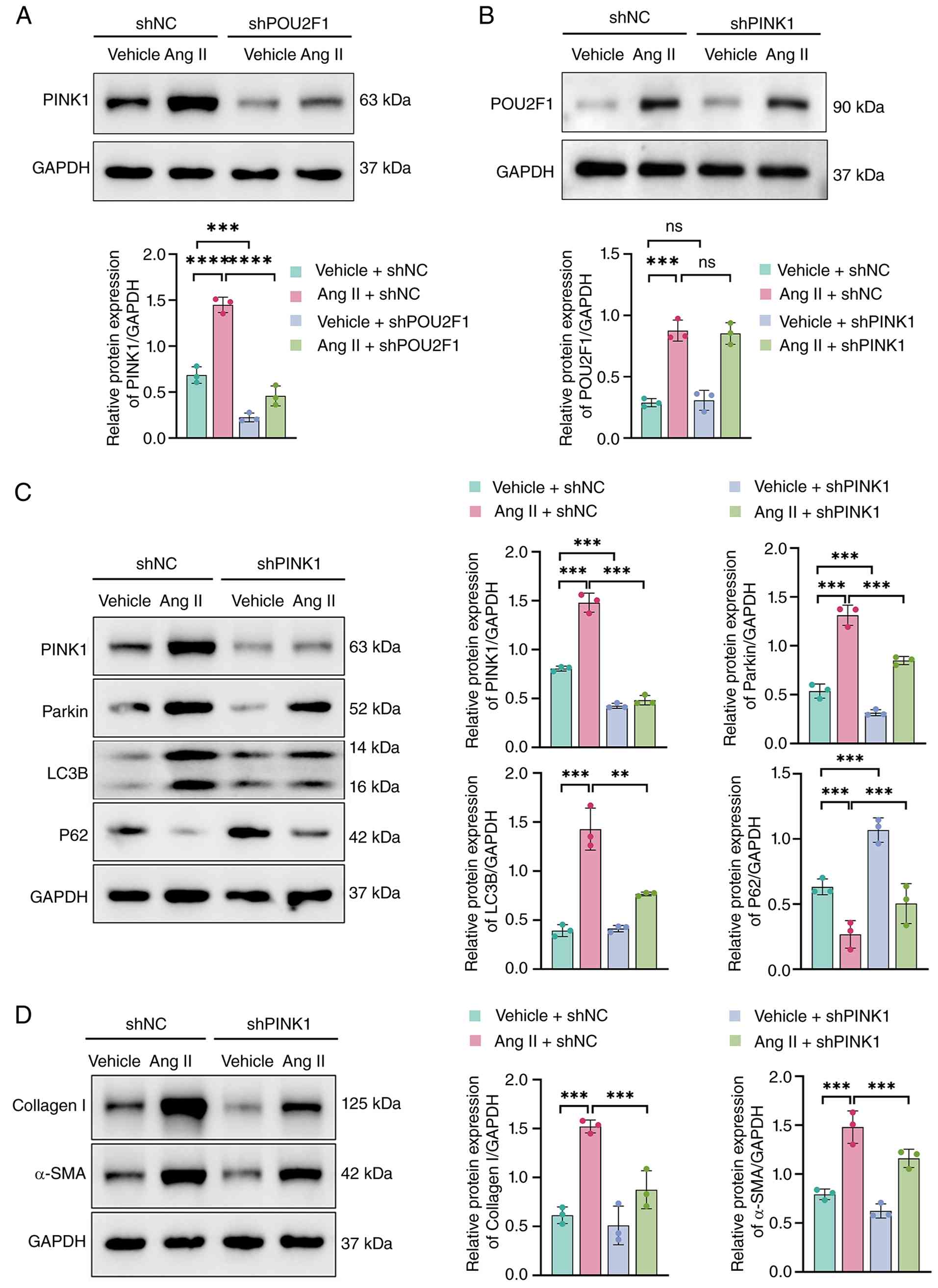 | Figure 5.PINK1 knockdown reduces mitophagy and
myocardial fibrosis independent of POU2F1 expression in Ang
II-induced cardiac fibroblasts. Western blotting was utilized to
determine the protein expression levels of (A) PINK1 and (B) POU2F1
(n=3). Expression levels of (C) PINK1, Parkin, LC3B and P62, as
well as (D) collagen I and α-SMA were determined using western
blotting (n=3). **P<0.01; ***P<0.001; ****P<0.0001. α-SMA,
α-smooth muscle actin; Ang II, angiotensin II; ns, not significant;
NC, negative control; PINK1, PTEN-induced kinase 1; POU2F1, POU
domain class 2 transcription factor 1; sh, short hairpin. |
POU2F1 directly binds to the PINK1
gene promoter to enhance PINK1 transcription
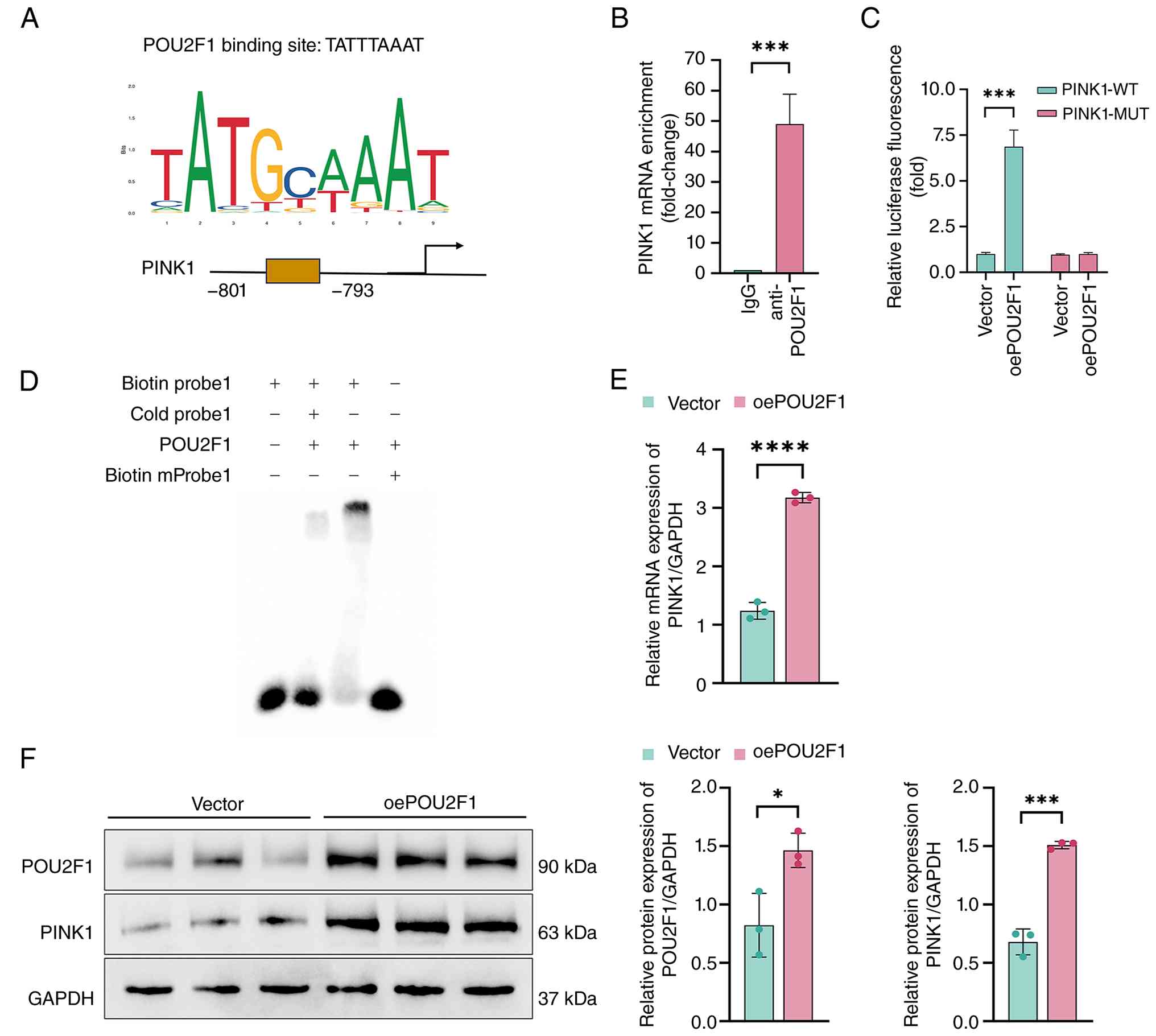
Bioinformatics analysis was next performed to
elucidate POU2F1-mediated transcriptional regulation of PINK1. The
rat PINK1 promoter sequence (−2,000 bp) was retrieved from NCBI,
with Jaspar software predicting a POU2F1-binding motif; POU2F1 was
identified as a transcription factor binding to the PINK1 promoter
at the specific motif TATTTAAAT (Fig.
6A). The specific binding of POU2F1 was validated using a
series of complementary experiments. First, ChIP assays using an
anti-POU2F1 antibody demonstrated significant enrichment of the
PINK1 promoter region compared with the IgG control, confirming the
binding of POU2F1 to the PINK1 promoter (Fig. 6B). To characterize the specificity
of this interaction, EMSA was performed. POU2F1 protein bound
specifically to a fluorescently labeled probe (lane 3) containing
the consensus wild-type sequence (TATTTAAAT). Notably, this binding
was competitively inhibited by an excess of unlabeled wild-type
probes (lane 2), but was unaffected by a mutated probe (lane 4),
underscoring the sequence-specific nature of the binding (Fig. 6D). Finally, a dual-luciferase
reporter assay was conducted to determine the functional relevance
of this binding; this confirmed that the direct binding of POU2F1
to the PINK1 promoter robustly enhanced its transcriptional
activity (Fig. 6C).
POU2F1 overexpression was utilized to validate its
regulation of PINK1. Western blotting and RT-qPCR confirmed that
the mRNA and protein expression levels of PINK1 were enhanced
following POU2F1 overexpression (Fig.
6E and F). Collectively, these findings confirmed that POU2F1
binds to specific motifs in the PINK1 promoter and drive its
transcription.
POU2F1 knockdown reduces
PINK1-mediated mitochondrial dysfunction and cardiac fibrosis in
SHRs
To clarify the effects of POU2F1 on
hypertension-induced cardiac fibrosis in SHRs, AAV9 vectors
encoding shPOU2F1 or shNC were constructed and injected into
14-week-old SHRs via the tail vein. After 4 weeks, AAV9-shPOU2F1
achieved efficient myocardial transduction, significantly reducing
POU2F1 expression (Fig. 7A).
Furthermore, compared with the AAV9-shNC group, POU2F1 knockdown in
SHRs significantly decreased myocardial PINK1 and Parkin expression
(Fig. 7B). In
AAV9-shPOU2F1-induced SHRs, TEM and Seahorse metabolic assays
confirmed that mitochondrial damage was alleviated and
mitochondrial respiratory function was improved (Fig. 7C and D). The fibrotic area of the
cardiac tissues was also markedly decreased (Fig. 7E), with a concomitant
downregulation of collagen I and α-SMA protein expression (Fig. 7F). Echocardiography demonstrated an
improvement in cardiac structure compared with in the SHR +
AAV9-shNC group, with reduced interventricular septal thickness
(IVSD and IVSS) and posterior wall thickness (LVPWD and LVPWS), and
increased left ventricular internal dimensions (LVIDD and LVIDS)
(Fig. 7G and H). Overall, these
findings demonstrated that inhibiting the expression of POU2F1 may
suppress PINK1-mediated mitochondrial dysfunction and reduce
cardiac fibrosis in SHRs.
 | Figure 7.POU2F1 knockdown reduces
PINK1-mediated mitochondrial dysfunction and cardiac fibrosis in
SHRs. Western blot analysis of (A) POU2F1, and (B) PINK1 and Parkin
in SHRs infected with AAV9-shPOU2F1 or AAV9-shNC (n=4). (C)
Transmission electron microscopy was used to observe the
mitochondrial structure in cardiac tissue from SHRs infected with
AAV9-shPOU2F1 or AAV9-shNC (scale bar, 500 nm). White arrows
indicate cristae disorganization; red arrows indicate damaged
mitochondria. (D) Seahorse assay profile of OCRs in CFs following
treatment with oligomycin, FCCP and AA + ROT. Quantitative analysis
of mitochondrial function parameters (basal respiration, maximal
respiration and ATP production) is shown in the bar charts (n=3).
(E) Interstitial myocardial fibrosis (blue) assessed via Masson's
trichrome staining (scale bar, 100 µm; n=4). (F) Western blot
analysis of collagen I and α-SMA protein expression in two groups
(n=4). (G) Representative echocardiographic images of SHRs
following different interventions. (H) Echocardiographic
parameters, including IVSD, IVSS, LVPWD, LVPWS, LVIDD and LVIDS,
were measured in the two groups (n=4). Data are presented as the
mean ± SD. (A-F and H) Unpaired two-tailed Student's t-test was
used for statistical analysis. ns, no significance; *P<0.05;
**P<0.01; ***P<0.001; ****P<0.0001. α-SMA, α-smooth muscle
actin; AA, antimycin A; AAV9, adeno-associated virus serotype 9;
FCCP, carbonyl cyanide-4-trifluoromethoxy phenylhydrazone; IVSD,
interventricular septal diastolic thickness; IVSS, interventricular
septal systolic thickness; LVIDD, left ventricular end-diastolic
diameter; LVIDS, left ventricular end-systolic diameter; LVPWD,
left ventricular posterior wall diastolic thickness; LVPWS, left
ventricular posterior wall systolic thickness; NC, negative
control; OCR, oxygen consumption rate; PINK1, PTEN-induced kinase
1; POU2F1, POU domain class 2 transcription factor 1; ROT,
rotenone; sh, short hairpin; SHR, spontaneously hypertensive
rat. |
Discussion
Hypertension is known to induce mitochondrial
damage, increase collagen deposition and drive myocardial fibrosis.
While PINK1-mediated mitophagy is a well-characterized pathway, its
upstream regulatory mechanisms in hypertension-induced cardiac
fibrosis remain incompletely understood. The present study provided
the following key insights: i) POU2F1 acts as a critical regulator,
which is significantly elevated in both SHRs and Ang II-induced
CFs, exacerbating hypertension-induced cardiac fibrosis. ii) POU2F1
is associated with enhanced PINK1/Parkin-mediated mitophagy-related
signaling, accompanied by disrupted mitochondrial homeostasis. iii)
POU2F1, as a transcription factor, directly binds to the PINK1
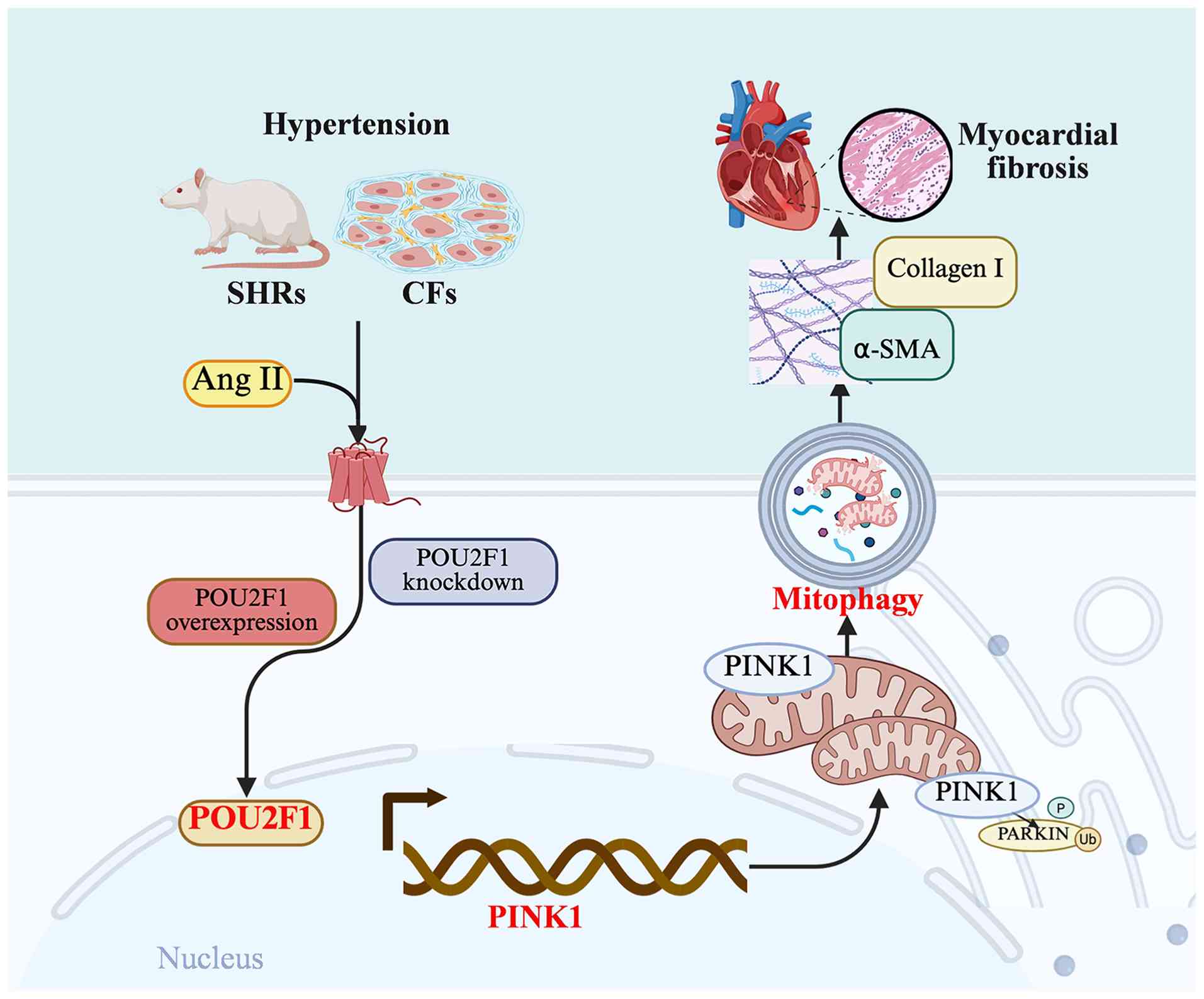
promoter to enhance PINK1 transcription and expression. These
findings indicated that POU2F1-regulated mitochondrial homeostasis
is a potential target for hypertensive cardiac remodeling therapies
(Fig. 8).
Hypertension represents a major risk factor for
cardiovascular diseases, as it not only elevates cardiac afterload
but also triggers progressive pathological cardiac remodeling,
ultimately leading to heart failure (43–45).
In the damaged heart, CFs express excessive levels of cytokines,
including transforming growth factor-β, IL-1β) and interleukin-6
(IL-6), leading to the enhanced proliferation of CFs, myofibroblast
activation and excessive secretion of ECM components (46). The present study revealed that
POU2F1 expression exhibited a positive correlation with the
severity of myocardial fibrosis in both SHRs and Ang II-induced
CFs. Knockdown of POU2F1 in vitro suppressed the expression
of α-SMA and collagen I, whereas its overexpression conversely
promoted their expression. Moreover, knockdown of POU2F1 in SHRs
attenuated cardiac fibrosis and improved cardiac function, which
further confirms the critical role of POU2F1 in
hypertension-induced cardiac fibrosis. These results extend the
findings of previous studies on transcriptional regulation in
cardiac fibrosis, which have primarily focused on factors such as
TGF-β/Smad, NF-κB and other transcription factors, by introducing
POU2F1 as a novel participant in this network (47–49).
Transcription factors bind to specific cis-acting
elements in the promoter or enhancer regions of target genes,
thereby activating or repressing transcriptional initiation and
modulating the spatiotemporal expression of downstream genes
(50,51). Previous studies have demonstrated
that POU2F1 can directly bind to the promoters of target genes such
as aldolase A and lactate dehydrogenase A to promote the
proliferation of tumor cells (52,53).
However, its function in cardiac fibrosis remains incompletely
elucidated. A key insight from the present study is the direct
transcriptional regulation of PINK1 by POU2F1. A strong positive
correlation was observed between POU2F1 and PINK1 expression in
SHRs and Ang II-induced CFs. Notably, POU2F1 overexpression
markedly promoted PINK1 expression and myocardial fibrosis, and
this effect was abrogated by PINK1 knockdown, indicating that the
pro-fibrotic effects of POU2F1 are PINK1-dependent. To demonstrate
this direct regulatory relationship, the binding sites for POU2F1
were predicted within the PINK1 promoter. Using EMSA and a
dual-luciferase reporter assay, it was established that POU2F1 can
bind directly to the PINK1 promoter, an interaction that may
enhance both PINK1 transcription and its protein expression. This
is consistent with evidence that transcription factors coordinate
the fibrotic response by regulating gene expression in CFs
(54,55). The present study therefore reveals
a novel transcriptional regulatory mechanism linked to mitophagy
modulation.
PINK1/Parkin-mediated mitophagy is crucial for
maintaining cell homeostasis, and is important in the prevention
and treatment of cardiovascular diseases (56,57).
PINK1, a serine/threonine kinase located on depolarized
mitochondria, is rapidly degraded upon entering the mitochondria
under physiological conditions (58). However, when PINK1 is upregulated,
it accumulates on depolarized mitochondria and self-activates,
recruiting Parkin to ubiquitinate mitochondrial substrates
(59). These proteins bind to
autophagosomes and are ultimately transported to lysosomes,
completing the mitophagy process. Mitophagy has emerged as a
double-edged sword in cardiac biology, with moderate activation
promoting cell survival under stress, whereas excessive or aberrant
mitophagy can contribute to tissue damage (60–62).
In the present study, elevated expression of PINK1, Parkin and LC3B
in both SHRs and Ang II-induced CFs confirmed the occurrence of
excessive mitophagy. Knockdown of PINK1 in Ang II-induced CFs
downregulated mitophagy-associated proteins and attenuated
myocardial fibrosis. Furthermore, both in vivo and in
vitro, knockdown of POU2F1 suppressed PINK1 transcription and
expression, which similarly led to the inhibition of mitophagy. A
limitation of the present study is the lack of direct TEM evidence
of mitochondria-containing autophagosomes. Given the transient
nature of mitophagy and the limited sampling scope of
ultrastructural imaging, further assessment of mitophagic flux is
warranted. Nevertheless, mitochondrial structural damage, impaired
respiratory function, and increased PINK1 and Parkin expression
suggest dysregulation of the PINK1/Parkin-mediated mitophagy
pathway.
Excessive mitophagy disrupts mitochondrial
homeostasis, thereby contributing to the pathogenesis of fibrotic
diseases (63–65). Activation of sirtuin 3 has been
shown to suppress mitochondrial oxidative stress and activate the
AMPK/Parkin axis, thereby mitigating mitochondrial damage and
improving cardiac function (66).
The hypoxia-inducible factor 1α/BCL2-interacting protein 3
signaling pathway is involved in mitophagy and serves a protective
role in renal fibrosis (67).
Furthermore, high temperature requirement protein A2 expression
regulates mitochondrial DNA damage and mitochondrial homeostasis in
hepatocytes during liver fibrogenesis (68). To the best of our knowledge, the
present study is the first to elucidate the regulation of
mitochondrial homeostasis by POU2F1. POU2F1 was knocked down in
SHRs and Ang II-induced CFs, and it was observed that the
mitophagy-related proteins PINK1, Parkin and LC3B were
downregulated, accompanied by alleviated mitochondrial structural
damage, improved mitochondrial respiratory function and a reduced
degree of myocardial fibrosis. This result confirmed that POU2F1
may promote hypertensive cardiac fibrosis by regulating
mitochondrial homeostasis.
In summary, the present study established POU2F1 as
a key upstream regulator that is significantly upregulated in
hypertensive models. POU2F1 directly bind to the PINK1 promoter to
potentiate its transcription, thereby enhancing
PINK1/Parkin-associated mitophagy-related signaling and driving
cardiac fibrotic remodeling. This finding highlights POU2F1 as a
promising therapeutic target; therefore, modulating its expression
to restore mitochondrial homeostasis may halt fibrotic
remodeling.
Supplementary Material
Supporting Data
Supporting Data
Acknowledgements
Not applicable.
Funding
The authors declare that financial support was received for the
research and/or publication of this article; this work was
supported by grants from the National Natural Science Foundation of
China (grant nos. 82174130 and 82274262), the Shanghai Science and
Technology Innovation Program for Cultivation of Lightening Stars
(Yangfan Project) (grant no. 23YF1448400), the Shanghai ‘Rising
Stars of Medical Talents’ Youth Development Program (Youth Medical
Talents-Specialist Program) (grant no. RY411.19.01.14), the
Shanghai Magnolia Talent Plan Pujiang Project (grant no. 24PJD113)
and the Shanghai Municipal Health Commission Traditional Chinese
Medicine Research Project (grant no. 2022QN031).
Availability of data and materials
The RNA-seq data generated in the present study may
be found in the OMIX (China National Center for Bioinformation)
database under accession number OMIX018255 or at the following URL:
https://ngdc.cncb.ac.cn/omix/release/OMIX018255. The
other data generated in the present study may be requested from the
corresponding author.
Authors' contributions
DYF and MZW designed the experiments. YBM, XZC and
ZXL performed the experiments and drafted the manuscript. YLM and
MTG helped with the experiments. CLH, YFL and BL analyzed the data.
All authors read and approved the final manuscript. YBM and XZC
confirm the authenticity of all the raw data.
Ethics approval and consent to
participate
The present study was performed at Yueyang Hospital
of Integrated Traditional Chinese and Western Medicine, and the
animal study was approved by the Institutional Animal Care and Use
Committee of Yueyang Hospital of Integrated Traditional Chinese and
Western Medicine (approval no. YYLAC-2023-182-1) following the
National Institutes of Health guidelines.
Patient consent for publication
Not applicable.
Competing interests
The authors declare that they have no competing
interests.
References
|
1
|
NCD Risk Factor Collaboration (NCD-RisC),
. Worldwide trends in hypertension prevalence and progress in
treatment and control from 1990 to 2019: A pooled analysis of 1201
population-representative studies with 104 million participants.
Lancet. 398:957–980. 2021. View Article : Google Scholar : PubMed/NCBI
|
|
2
|
Shinde AV, Humeres C and Frangogiannis NG:
The role of α-smooth muscle actin in fibroblast-mediated matrix
contraction and remodeling. Biochim Biophys Acta Mol Basis Dis.
1863:298–309. 2017. View Article : Google Scholar : PubMed/NCBI
|
|
3
|
Querejeta R, López B, González A, Sánchez
E, Larman M, Martínez Ubago JL and Díez J: Increased collagen type
I synthesis in patients with heart failure of hypertensive origin:
relation to myocardial fibrosis. Circulation. 110:1263–1268. 2004.
View Article : Google Scholar : PubMed/NCBI
|
|
4
|
Liu T, Song D, Dong J, Zhu P, Liu J, Liu
W, Ma X, Zhao L and Ling S: Current understanding of the
pathophysiology of myocardial fibrosis and its quantitative
assessment in heart failure. Front Physiol. 8:2382017. View Article : Google Scholar : PubMed/NCBI
|
|
5
|
Gibb AA, Lazaropoulos MP and Elrod JW:
Myofibroblasts and fibrosis: Mitochondrial and metabolic control of
cellular differentiation. Circ Res. 127:427–447. 2020. View Article : Google Scholar : PubMed/NCBI
|
|
6
|
Lin LC, Tu B, Song K, Liu ZY, Sun H, Zhou
Y, Sha JM, Yang JJ, Zhang Y, Zhao JY and Tao H: Mitochondrial
quality control in cardiac fibrosis: Epigenetic mechanisms and
therapeutic strategies. Metabolism. 145:1556262023. View Article : Google Scholar : PubMed/NCBI
|
|
7
|
Li B, Liu F, Chen X, Chen T, Zhang J, Liu
Y, Yao Y, Hu W, Zhang M, Wang B, et al: FARS2 deficiency causes
cardiomyopathy by disrupting mitochondrial homeostasis and the
mitochondrial quality control system. Circulation. 149:1268–1284.
2024. View Article : Google Scholar : PubMed/NCBI
|
|
8
|
Lin J, Chen X, Du Y, Li J, Guo T and Luo
S: Mitophagy in cell death regulation: Insights into mechanisms and
disease implications. Biomolecules. 14:12702024. View Article : Google Scholar : PubMed/NCBI
|
|
9
|
Palikaras K, Lionaki E and Tavernarakis N:
Mechanisms of mitophagy in cellular homeostasis, physiology and
pathology. Nat Cell Biol. 20:1013–1022. 2018. View Article : Google Scholar : PubMed/NCBI
|
|
10
|
Nguyen TN, Padman BS and Lazarou M:
Deciphering the molecular signals of PINK1/Parkin mitophagy. Trends
Cell Biol. 26:733–744. 2016. View Article : Google Scholar : PubMed/NCBI
|
|
11
|
Hu J, Liu T, Fu F, Cui Z, Lai Q, Zhang Y,
Yu B, Liu F, Kou J and Li F: Omentin1 ameliorates myocardial
ischemia-induced heart failure via SIRT3/FOXO3a-dependent
mitochondrial dynamical homeostasis and mitophagy. J Transl Med.
20:4472022. View Article : Google Scholar : PubMed/NCBI
|
|
12
|
Ren S, Wang Y, Zhang Y, Yan P, Xiao D,
Zhao Y, Jia W, Ding L, Dong H, Wei C, et al: Paeoniflorin
alleviates AngII-induced cardiac hypertrophy in H9c2 cells by
regulating oxidative stress and Nrf2 signaling pathway. Biomed
Pharmacother. 165:1152532023. View Article : Google Scholar : PubMed/NCBI
|
|
13
|
Li F, Fan X, Zhang Y, Zhang Y, Ma X, Kou J
and Yu B: Inhibition of myosin IIA-actin interaction prevents
ischemia/reperfusion induced cardiomyocytes apoptosis through
modulating PINK1/Parkin pathway and mitochondrial fission. Int J
Cardiol. 271:211–218. 2018. View Article : Google Scholar : PubMed/NCBI
|
|
14
|
Pan XC, Xiong YL, Hong JH, Liu Y, Cen YY,
Liu T, Yang QF, Tao H, Li YN and Zhang HG: Cardiomyocytic FoxP3 is
involved in Parkin-mediated mitophagy during cardiac remodeling and
the regulatory role of triptolide. Theranostics. 12:2483–2501.
2022. View Article : Google Scholar : PubMed/NCBI
|
|
15
|
Zhang X, Li ZL, Crane JA, Jordan KL, Pawar
AS, Textor SC, Lerman A and Lerman LO: Valsartan regulates
myocardial autophagy and mitochondrial turnover in experimental
hypertension. Hypertension. 64:87–93. 2014. View Article : Google Scholar : PubMed/NCBI
|
|
16
|
Ma Y, Zhou X, Gui M, Yao L, Li J, Chen X,
Wang M, Lu B and Fu D: Mitophagy in hypertension-mediated organ
damage. Front Cardiovasc Med. 10:13098632024. View Article : Google Scholar : PubMed/NCBI
|
|
17
|
Ma Y, Li D, Zhou X, Chen X, Hou C, Li Y,
Zhao Y, Gui M, Yao L, Li J, et al: METTL3-mediated m6A modification
of FUNDC1/IP3R2 pathway facilitates cardiac hypertrophy in obesity
hypertension. Life Sci. 377:1237802025. View Article : Google Scholar : PubMed/NCBI
|
|
18
|
Zhu HY, Cao GY, Wang SP, Chen Y, Liu GD,
Gao YJ and Hu JP: POU2F1 promotes growth and metastasis of
hepatocellular carcinoma through the FAT1 signaling pathway. Am J
Cancer Res. 7:1665–1679. 2017.PubMed/NCBI
|
|
19
|
Li F, Wang T and Huang Y: POU2F1 induces
the immune escape in lung cancer by up-regulating PD-L1. Am J
Transl Res. 13:672–683. 2021.PubMed/NCBI
|
|
20
|
Li M, Wu J, Hu G, Song Y, Shen J, Xin J,
Li Z, Liu W, Dong E, Xu M, et al: Pathological matrix stiffness
promotes cardiac fibroblast differentiation through the POU2F1
signaling pathway. Sci China Life Sci. 64:242–254. 2021. View Article : Google Scholar : PubMed/NCBI
|
|
21
|
Yang W, Li Y, Bai J, You T, Yi K, Xie D,
Zhang X and Xie X: A functional variant Rs492554 associated with
congenital heart defects modulates SESN2 expression through POU2F1.
Front Cell Dev Biol. 9:6684742021. View Article : Google Scholar : PubMed/NCBI
|
|
22
|
Feng N, Yu H, Wang Y, Zhang Y, Xiao H and
Gao W: Exercise training attenuates angiotensin II-induced cardiac
fibrosis by reducing POU2F1 expression. J Sport Health Sci.
12:464–476. 2023. View Article : Google Scholar : PubMed/NCBI
|
|
23
|
Liu N, Gong Z, Li Y, Xu Y, Guo Y, Chen W,
Sun X, Yin X and Liu W: CTRP3 inhibits myocardial fibrosis through
the P2X7R-NLRP3 inflammasome pathway in SHR rats. J Hypertens.
42:315–328. 2024. View Article : Google Scholar : PubMed/NCBI
|
|
24
|
Wang TJ, Lian GL, Lin X, Zhong HB, Xu CS,
Wang HJ and Xie LD: Hypomethylation of Agtrap is associated with
long-term inhibition of left ventricular hypertrophy in
prehypertensive losartan-treated spontaneously hypertensive rats.
Mol Med Rep. 15:839–846. 2017. View Article : Google Scholar : PubMed/NCBI
|
|
25
|
National Research Council (US) Committee
for the Update of the Guide for the Care and Use of Laboratory
Animals, . Guide for the care and use of laboratory animals. 8th
edition. National Academies Press; Washington, DC: 2011, PubMed/NCBI
|
|
26
|
Tarbit E, Singh I, Peart JN, Bivol S and
Rose Meyer RB: Increased release of serotonin from rat primary
isolated adult cardiac myofibroblasts. Sci Rep. 11:203762021.
View Article : Google Scholar : PubMed/NCBI
|
|
27
|
Feng J, Li Y, Li Y, Yin Q, Li H, Li J,
Zhou B, Meng J, Lian H, Wu M, et al: Versican promotes
cardiomyocyte proliferation and cardiac repair. Circulation.
149:1004–1015. 2024. View Article : Google Scholar : PubMed/NCBI
|
|
28
|
Mao Q, Zhang X, Yang J, Kong Q, Cheng H,
Yu W, Cao X, Li Y, Li C, Liu L and Ding Z: HSPA12A acts as a
scaffolding protein to inhibit cardiac fibroblast activation and
cardiac fibrosis. J Adv Res. 67:217–229. 2025. View Article : Google Scholar : PubMed/NCBI
|
|
29
|
Wu Y, Zhan S, Chen L, Sun M, Li M, Mou X,
Zhang Z, Xu L and Xu Y: TNFSF14/LIGHT promotes cardiac fibrosis and
atrial fibrillation vulnerability via PI3Kγ/SGK1 pathway-dependent
M2 macrophage polarisation. J Transl Med. 21:5442023. View Article : Google Scholar : PubMed/NCBI
|
|
30
|
Guo J, Hang P, Yu J, Li W, Zhao X, Sun Y,
Fan Z and Du Z: The association between RGS4 and choline in cardiac
fibrosis. Cell Commun Signal. 19:462021. View Article : Google Scholar : PubMed/NCBI
|
|
31
|
Vue Z, Garza-Lopez E, Neikirk K, Katti P,
Vang L, Beasley H, Shao J, Marshall AG, Crabtree A, Murphy AC, et
al: 3D reconstruction of murine mitochondria reveals changes in
structure during aging linked to the MICOS complex. Aging Cell.
22:e140092023. View Article : Google Scholar : PubMed/NCBI
|
|
32
|
Rabinovich-Nikitin I, Rasouli M, Reitz CJ,
Posen I, Margulets V, Dhingra R, Khatua TN, Thliveris JA, Martino
TA and Kirshenbaum LA: Mitochondrial autophagy and cell survival is
regulated by the circadian Clock gene in cardiac myocytes during
ischemic stress. Autophagy. 17:3794–3812. 2021. View Article : Google Scholar : PubMed/NCBI
|
|
33
|
Hu R, Huang Y, Geng Y, Liu Z, Li F, Zhang
Z, Ma W, Song K, Dong H, Song Y and Zhang M: Jiawei Buzhong Yiqi
decoction ameliorates polycystic ovary syndrome via
oocyte-granulosa cell communication. J Ethnopharmacol.
323:1176542024. View Article : Google Scholar : PubMed/NCBI
|
|
34
|
Stellato M, Dewenter M, Rudnik M, Hukara
A, Özsoy Ç, Renoux F, Pachera E, Gantenbein F, Seebeck P, Uhtjaerv
S, et al: The AP-1 transcription factor Fosl-2 drives cardiac
fibrosis and arrhythmias under immunofibrotic conditions. Commun
Biol. 6:1612023. View Article : Google Scholar : PubMed/NCBI
|
|
35
|
Convertini P, Santarsiero A, Todisco S,
Gilio M, Palazzo D, Pappalardo I, Iacobazzi D, Frontuto M and
Infantino V: ACLY as a modulator of liver cell functions and its
role in metabolic dysfunction-associated steatohepatitis. J Transl
Med. 21:5682023. View Article : Google Scholar : PubMed/NCBI
|
|
36
|
Zhang Y, Fan Y, Hu H, Zhang X, Wang Z, Wu
Z, Wang L, Yu X, Song X, Xiang P, et al: ZHX2 emerges as a negative
regulator of mitochondrial oxidative phosphorylation during acute
liver injury. Nat Commun. 14:75272023. View Article : Google Scholar : PubMed/NCBI
|
|
37
|
Liu M, Peng T, Hu L, Wang M, Guo D, Qi B,
Ren G, Wang D, Li Y, Song L, et al: N-glycosylation-mediated CD147
accumulation induces cardiac fibrosis in the diabetic heart through
ALK5 activation. Int J Biol Sci. 19:137–155. 2023. View Article : Google Scholar : PubMed/NCBI
|
|
38
|
Li Z, Hu O, Xu S, Lin C, Yu W, Ma D, Lu J
and Liu P: The SIRT3-ATAD3A axis regulates MAM dynamics and
mitochondrial calcium homeostasis in cardiac hypertrophy. Int J
Biol Sci. 20:831–847. 2024. View Article : Google Scholar : PubMed/NCBI
|
|
39
|
Xie SY, Liu SQ, Zhang T, Shi WK, Xing Y,
Fang WX, Zhang M, Chen MY, Xu SC, Fan MQ, et al: USP28 serves as a
key suppressor of mitochondrial morphofunctional defects and
cardiac dysfunction in the diabetic heart. Circulation.
149:684–706. 2024. View Article : Google Scholar : PubMed/NCBI
|
|
40
|
Berta DG, Kuisma H, Välimäki N, Räisänen
M, Jäntti M, Pasanen A, Karhu A, Kaukomaa J, Taira A, Cajuso T, et
al: Deficient H2A.Z deposition is associated with genesis of
uterine leiomyoma. Nature. 596:398–403. 2021. View Article : Google Scholar : PubMed/NCBI
|
|
41
|
Zhang X, Huang DX, Xuan C, Li Y, Jiang Y,
Wu X, Zhou W, Lei Y, Yang F, Ma H, et al: Aerobic exercise training
attenuates ischemia-reperfusion injury in mice by decreasing the
methylation level of METTL3-associated m6A RNA in cardiomyocytes.
Life Sci. 361:1232942025. View Article : Google Scholar : PubMed/NCBI
|
|
42
|
Frangogiannis NG: Cardiac fibrosis.
Cardiovasc Res. 117:1450–1488. 2021. View Article : Google Scholar : PubMed/NCBI
|
|
43
|
Chen YF, Qi RQ, Song JW, Wang SY, Dong ZJ,
Chen YH, Liu Y, Zhou XY, Li J, Liu XY and Zhong JC: Sirtuin 7
ameliorates cuproptosis, myocardial remodeling and heart
dysfunction in hypertension through the modulation of YAP/ATP7A
signaling. Apoptosis. 29:2161–2182. 2024. View Article : Google Scholar : PubMed/NCBI
|
|
44
|
Yeo TM, Chin CWL, Seah CWA, Cheng LJ, Lin
W, Dalakoti M, Foo R and Wang W: Global prevalence of myocardial
fibrosis among individuals with cardiometabolic conditions: A
systematic review and meta-analysis. Eur J Prev Cardiol.
32:1077–1091. 2025. View Article : Google Scholar : PubMed/NCBI
|
|
45
|
Iyer NR, Le TT, Kui MSL, Tang HC, Chin CT,
Phua SK, Bryant JA, Pua CJ, Ang B, Toh DF, et al: Markers of focal
and diffuse nonischemic myocardial fibrosis are associated with
adverse cardiac remodeling and prognosis in patients with
hypertension: the REMODEL study. Hypertension. 79:1804–1813. 2022.
View Article : Google Scholar : PubMed/NCBI
|
|
46
|
Travers JG, Kamal FA, Robbins J, Yutzey KE
and Blaxall BC: Cardiac fibrosis: The fibroblast awakens. Circ Res.
118:1021–1040. 2016. View Article : Google Scholar : PubMed/NCBI
|
|
47
|
Tuleta I, Hanna A, Humeres C, Aguilan JT,
Sidoli S, Zhu F and Frangogiannis NG: Fibroblast-specific TGF-β
signaling mediates cardiac dysfunction, fibrosis, and hypertrophy
in obese diabetic mice. Cardiovasc Res. 120:2047–2063. 2024.
View Article : Google Scholar : PubMed/NCBI
|
|
48
|
Umbarkar P, Ejantkar S, Tousif S and Lal
H: Mechanisms of fibroblast activation and myocardial fibrosis:
Lessons learned from FB-specific conditional mouse models. Cells.
10:24122021. View Article : Google Scholar : PubMed/NCBI
|
|
49
|
Wang C, Luo H, Xu Y, Tao L, Chang C and
Shen X: Salvianolic acid B-alleviated angiotensin II induces
cardiac fibrosis by suppressing NF-κB pathway in vitro. Med Sci
Monit. 24:7654–7664. 2018. View Article : Google Scholar : PubMed/NCBI
|
|
50
|
Lambert SA, Jolma A, Campitelli LF, Das
PK, Yin Y, Albu M, Chen X, Taipale J, Hughes TR and Weirauch MT:
The human transcription factors. Cell. 172:650–665. 2018.
View Article : Google Scholar : PubMed/NCBI
|
|
51
|
Lee TI and Young RA: Transcriptional
regulation and its misregulation in disease. Cell. 152:1237–1251.
2013. View Article : Google Scholar : PubMed/NCBI
|
|
52
|
Lin J, Xia L, Oyang L, Liang J, Tan S, Wu
N, Yi P, Pan Q, Rao S, Han Y, et al: The POU2F1-ALDOA axis promotes
the proliferation and chemoresistance of colon cancer cells by
enhancing glycolysis and the pentose phosphate pathway activity.
Oncogene. 41:1024–1039. 2022. View Article : Google Scholar : PubMed/NCBI
|
|
53
|
Wang H, Zhang F, Li L, Kang Z, Li J, Mao
Y, Liu K, Song L and Shan S: POU2F1 facilitates the malignant
phenotypes and aerobic glycolysis of pituitary adenoma by
activating LDHA transcription. Brain Res. 1866:1498702025.
View Article : Google Scholar : PubMed/NCBI
|
|
54
|
Hong JH and Zhang HG: Transcription
factors involved in the development and prognosis of cardiac
remodeling. Front Pharmacol. 13:8285492022. View Article : Google Scholar : PubMed/NCBI
|
|
55
|
Zhang Q, Wang L, Wang S, Cheng H, Xu L,
Pei G, Wang Y, Fu C, Jiang Y, He C and Wei Q: Signaling pathways
and targeted therapy for myocardial infarction. Signal Transduct
Target Ther. 7:782022. View Article : Google Scholar : PubMed/NCBI
|
|
56
|
Wu Y, Jiang T, Hua J, Xiong Z, Dai K, Chen
H, Li L, Peng J, Peng X, Zheng Z and Xiong W: PINK1/Parkin-mediated
mitophagy in cardiovascular disease: From pathogenesis to novel
therapy. Int J Cardiol. 361:61–69. 2022. View Article : Google Scholar : PubMed/NCBI
|
|
57
|
D'Arcy MS: Mitophagy in health and
disease. Molecular mechanisms, regulatory pathways, and therapeutic
implications. Apoptosis. 29:1415–1428. 2024. View Article : Google Scholar : PubMed/NCBI
|
|
58
|
Narendra DP and Youle RJ: The role of
PINK1-Parkin in mitochondrial quality control. Nat Cell Biol.
26:1639–1651. 2024. View Article : Google Scholar : PubMed/NCBI
|
|
59
|
Vizziello M, Borellini L, Franco G and
Ardolino G: Disruption of mitochondrial homeostasis: The role of
PINK1 in parkinson's disease. Cells. 10:30222021. View Article : Google Scholar : PubMed/NCBI
|
|
60
|
Bravo-San Pedro JM, Kroemer G and Galluzzi
L: Autophagy and mitophagy in cardiovascular disease. Circ Res.
120:1812–1824. 2017. View Article : Google Scholar : PubMed/NCBI
|
|
61
|
Li A, Gao M, Liu B, Qin Y, Chen L, Liu H,
Wu H and Gong G: Mitochondrial autophagy: Molecular mechanisms and
implications for cardiovascular disease. Cell Death Dis.
13:4442022. View Article : Google Scholar : PubMed/NCBI
|
|
62
|
Yang T, Yang Q, Lai Q, Zhao J, Nie L, Liu
S, Yang J and Chu C: AP39 inhibits ferroptosis by inhibiting
mitochondrial autophagy through the PINK1/parkin pathway to improve
myocardial fibrosis with myocardial infarction. Biomed
Pharmacother. 165:1151952023. View Article : Google Scholar : PubMed/NCBI
|
|
63
|
Lin D, Luo H, Dong B, He Z, Ma L, Wang Z,
Wu X and Sui Q: FOXO3a induces myocardial fibrosis by upregulating
mitophagy. Front Biosci (Landmark Ed). 29:562024. View Article : Google Scholar : PubMed/NCBI
|
|
64
|
Li Y, Zhao K, Hu Y, Yang F, Li P and Liu
Y: MicroRNA-142-3p alleviated high salt-induced cardiac fibrosis
via downregulating optineurin-mediated mitophagy. iScience.
27:1097642024. View Article : Google Scholar : PubMed/NCBI
|
|
65
|
Zhang YY, Zhou XT, Huang GZ, Liao WJ, Chen
X and Ma YR: The pro-fibrotic role of autophagy in renal intrinsic
cells: Mechanisms and therapeutic potential in chronic kidney
disease. Front Cell Dev Biol. 12:14994572024. View Article : Google Scholar : PubMed/NCBI
|
|
66
|
Peng F, Liao M, Jin W, Liu W, Li Z, Fan Z,
Zou L, Chen S, Zhu L, Zhao Q, et al: 2-APQC, a small-molecule
activator of Sirtuin-3 (SIRT3), alleviates myocardial hypertrophy
and fibrosis by regulating mitochondrial homeostasis. Signal
Transduct Target Ther. 9:1332024. View Article : Google Scholar : PubMed/NCBI
|
|
67
|
Li J, Lin Q, Shao X, Li S, Zhu X, Wu J,
Mou S, Gu L, Wang Q, Zhang M, et al: HIF1α-BNIP3-mediated mitophagy
protects against renal fibrosis by decreasing ROS and inhibiting
activation of the NLRP3 inflammasome. Cell Death Dis. 14:2002023.
View Article : Google Scholar : PubMed/NCBI
|
|
68
|
Hur W, Kang BY, Kim SM, Lee GW, Kim JH,
Nam MK, Rhim H and Yoon SK: Serine protease HtrA2/Omi deficiency
impairs mitochondrial homeostasis and promotes hepatic fibrogenesis
via activation of hepatic stellate cells. Cells. 8:11192019.
View Article : Google Scholar : PubMed/NCBI
|