Introduction
Programmed cell death (PCD) is an essential, tightly
regulated process with critical roles in the development and the
maintenance of cellular and tissue homeostasis. More than 20
distinct forms of PCD have been identified, with apoptosis being
the most investigated (1,2). Apoptosis is a caspase-mediated
process that plays a vital role in normal cell turnover, immune
regulation, development and tissue atrophy. Aberrations in the
apoptotic process are implicated in numerous pathologies, including
cancer, neurodegenerative diseases, ischemic injury, and autoimmune
conditions (3).
The term ‘apoptosis’ was initially introduced by
Kerr, Wyllie and Currie in 1972 (4), who described it as a process of
regulated cell deletion that serves a complementary role, yet
opposite, to mitosis in the regulation of animal cell population
balance. Over the past 50 years, research has greatly enhanced our
understanding of the molecular mechanisms underlying apoptosis.
Nonetheless, the morphological criteria originally described by
Kerr, Wyllie, and Currie, including nuclear condensation, membrane
blebbing, cellular fragmentation into apoptotic bodies and their
subsequent engulfment by phagocytic cells, are still accepted as
the basic cellular events associated with apoptotic death (5).
Historically, apoptosis was regarded as an
irreversible, one-way process which invariably led to cellular
death. However, recent discoveries have dismantled this binary view
of cellular life and death. It is now evident that cells undergoing
the early stages of apoptosis can reverse the death process and
recover their function upon the removal of the stress stimulus
(6–8). This phenomenon has been termed
‘anastasis’ (Greek for ‘rising to life’) (9). In the Christian tradition,
‘Anastasis’ specifically refers to the Resurrection of Jesus
Christ. By adopting this potent terminology, this scientific
definition underscores a reversal of destiny; just as the biblical
Anastasis represents a miraculous return from mortality, cellular
anastasis describes the unexpected ability of cells to halt the
‘falling’ trajectory of programmed death and ‘rise’ back to
functional survival.
The molecular events underlying this death-cheating
phenomenon are yet to be fully elucidated. This process becomes
highly relevant in the case of cancer cells. While anastasis serves
as a vital safeguard for tissue preservation following transient
injury of otherwise normal cells, the molecular machinery of
anastasis can be manipulated by cancer cells to enhance their
survival after chemotherapy, contributing to disease relapse and
progression. Consequently, elucidating the precise molecular
orchestration of anastasis and translating this knowledge into the
clinical practice for the therapeutic benefit of cancer patients
has garnered significant scientific attention.
N-acetyl-5-methoxytryptamine (melatonin) has emerged
as a promising candidate for the pharmacological modulation of
anastasis (10). While best known
for regulating circadian rhythms, melatonin is also a potent
antioxidant and mitochondrial guardian with profound cytoprotective
properties (11). However, the
extent to which melatonin directly influences anastasis remains
uncertain. The current review provides a comprehensive evaluation
of the molecular evidence implicating anastasis in oncogenesis,
highlighting its capacity to drive cancer cell survival and promote
a more aggressive cellular phenotype. In addition, it examines
whether the pleiotropic actions of melatonin may influence pathways
relevant to apoptotic recovery and its role as a potential
pharmacological modulator capable of intercepting the process of
anastasis.
Discussions on comparison of apoptosis and
anastasis
The molecular basis of apoptosis and
the ‘point of no return’
Apoptosis is induced by a variety of stimuli and
mechanisms, which are generally classified into two principal
routes: The extrinsic and intrinsic pathways. These distinct
signaling cascades ultimately converge on the activation of shared
executioner molecules that mediate programmed cell death (1,2). The
molecular events characterizing apoptosis are divided into three
consecutive temporal phases: The initiation phase, where an
extrinsic or an intrinsic stress signal is received, activating the
proximal components of the cell death machinery; the effector
phase, characterized by the irreversible activation of executioner
molecules; and the degradation phase, during which classical
morphological hallmarks become evident (12).
Investigators have often aimed to determine the
exact ‘point of no return’ in apoptosis, representing the specific
molecular transition at which a cell becomes irreversibly committed
to its demise (13,14). This critical juncture is widely
associated with mitochondrial outer membrane permeabilization
(MOMP), a process controlled by the proapoptotic members of the
Bcl-2 protein family (15–17). When apoptotic stimuli are
encountered, a subset of proteins translocate to the mitochondria,
compromising the integrity of the outer mitochondrial membrane and
resulting in the release of apoptosis-inducing factors such as
cytochrome c. These mitochondrial proteins initiate the
activation of downstream effector molecules, predominantly the
caspase network (18). The
subsequent activation of executioner caspases leads to irreversible
proteolysis of various cellular substrates, ultimately resulting in
the orderly disassembly of the cell. Additionally, dysfunctional
mitochondria and the associated release of cytotoxic proteins into
the cytoplasm contribute to the induction of caspase-independent
cell death pathways (18,19).
Re-evaluating the irreversibility of
apoptosis
For decades, initiation of apoptosis was regarded as
an irreversible event culminating in cellular death. There is
evidence, however, suggesting that cells can retain viability even
following MOMP or activation of executioner caspases (8,20).
Tang et al (6) demonstrated
that cancer cells subjected to cytotoxic agents and exhibiting both
morphological and biochemical markers of apoptosis, unexpectedly
survived and subsequently resumed proliferation following the
removal of the toxic inducer. These observations were confirmed
across multiple cell lines and with various cytotoxic stimuli,
supporting the existence of a previously unknown mechanism that
allows cancer cells to escape the apoptotic pathway, provided that
the cytotoxic stimulus is withdrawn (7). Notably, >90% of primary liver
cells and embryonic fibroblast (NIH 3T3) cells demonstrate the
capacity to reverse all apoptotic markers after removal of the
apoptosis-inducing stimulus, even following DNA damage. However,
these cells retained persistent genetic alterations, including an
increased frequency of micronuclei, a biological indicator of
substantial chromosomal rearrangements, aberrant micronuclei
formation during early post-apoptosis cell division and a marked
rise in karyotypic abnormalities such as chromosomal aneuploidy and
radial configurations. These findings suggest that the persistence
of genetic alteration following apoptotic reversal may confer a
transformed phenotype with potential carcinogenic consequences
(7). The phenomenon of cellular
evasion from apoptosis has also been observed by other
investigators. Roux et al (21), for example, analyzed the concept of
‘fractional cell killing’, which describes the incomplete
elimination of cells via the extrinsic pathway of apoptosis.
The term anastasis, derived from the Greek word
meaning ‘rising to life’, was introduced by Tang et al
(22) to characterize the reversal
of apoptosis (9). This concept
challenges the notion that apoptosis represents an irreversible
process, suggesting that cells retain the potential for
physiological recovery and continued viability even following
activation of executioner caspases, provided the lethal stimulus is
withdrawn.
Molecular signature of anastasis
To elucidate the transcriptional dynamics of
anastasis, Tang et al (23)
utilized a time-course microarray analysis on mouse primary liver
cells recovering from ethanol-induced apoptosis. The study compared
gene expression in apoptotic cells following treatment with 4.5%
ethanol for 5 h, recovering cells (harvested at 3, 6, 24 and 48 h
post-washout) and untreated controls. This analysis revealed
distinct, time-dependent waves of gene expression. During the early
phase of anastasis (at 3 h), a broad upregulation of genes
associated with Activator protein-1 (AP-1) transcription
factors, the transforming growth factor-β (TGF-β) signaling pathway
and its related regulators, pro-survival Bcl-2 family members
(Bag3) and p53 inhibition (Mdm2) was observed. This
upregulation also extended to pathways involved in antioxidant
responses (Hmox2), antiproliferative processes
(Btg1), DNA damage (Ddit3, Ddit4), vesicular
trafficking (Vps37b) and stress-inducible responses
(Dnajb1, Dnajb9, Herpud1, Hspb1, Hspa1b) was observed.
Following this initial response, a different set of pathways was
activated at 6 h post-recovery, characterized by significant
upregulation of genes governing cell cycle arrest (Cdkn1a),
autophagy (Atg12, Sqstm1), and cell migration (MMPs).
Reverse transcription-quantitative PCR validation in the human
liver cancer cell line HepG2 confirmed these gene expression
patterns, a finding suggesting that the core transcriptional
program of anastasis is conserved between primary and malignant
cells. Taken together, the upregulation of these genes during
anastasis indicates potential regulatory mechanisms that degrade
released cytochrome c, inhibit activated executioner
proteins and reestablish a functional mitochondrial network
essential for energy production (9,23).
In a similar study, researchers exposed HeLa cells
to ethanol in order to trigger apoptosis (24). RNAseq compared gene expression in
untreated, apoptotic and surviving cells at various time points
after removing ethanol. Analysis revealed distinct sets of early
and late response genes. Early-response genes were primarily
related to transcription, chromatin modification and stress
regulation, while late-response genes involved noncoding RNA
processing, ribosome biogenesis, focal adhesion and actin
cytoskeleton regulation, indicating a clear temporal shift in
cellular programs and a transition from a proliferative towards a
migrative phenotype during the different stages of anastasis.
Moreover, this study documented that the transcripts of some
early-response genes were already elevated in apoptotic cells
compared to untreated ones. This suggested that cells enter a
‘poised for recovery’ state, which is characterized by the
accumulation of specific survival-associated mRNAs. Translation of
these mRNAs enables rapid recovery if the stress is removed,
otherwise, these mRNAs are degraded and the cell proceeds to death
(24).
To facilitate interpretation of the findings of the
two previously mentioned studies, some of the reported gene
expression alterations observed during anastasis are summarized in
Tables I and II. Table
I presents representative genes with upregulated expression,
whereas Table II summarizes
representative genes with downregulated expression. As these
studies employed different experimental systems and methodologies,
as well as distinct sampling time points, the datasets are not
directly comparable. Therefore, the tables are intended solely as
descriptive summaries of the reported findings rather than
comparative analyses. For organizational purposes, the phases of
anastasis are conceptually grouped as Early phase (1–4 h) and Late
phase (≥5 h) following recovery initiation.
 | Table I.Representative genes with upregulated
expression during anastasis. |
Table I.
Representative genes with upregulated
expression during anastasis.
| First author/s,
year | Phase of
anastasis | Gene | Biological
role | (Refs.) |
|---|
| Tang et al,
2017; Sun et al, 2017 | Early | AP-1 family
(e.g.,Atf3, Fos, Fosb, Jun, Junb) | Transcription
factors | (23,24) |
| Tang et al,
2017; Sun et al, 2017 | Early | TGF-β signal
pathway and its regulators (Inhba, Snai1, Tgif1, Sox4, Sox9, Klf4,
Klf6, Klf9) | TGF-β
regulation/EMT/Migration | (23,24) |
| Tang et al,
2017 | Early | Bag3 | Anti-apoptotic
Bcl-2 protein | (23) |
| Tang et al,
2017 | Early | Mdm2 | Inhibitor of
p53 | (23) |
| Tang et al,
2017 | Early | Hmox1 | Anti-oxidation | (23) |
| Tang et al,
2017; Sun et al, 2017 | Early | Btg1 |
Anti-proliferation | (23,24) |
| Tang et al,
2017; Sun et al, 2017 | Early OR Late | Ddit3 and
Ddit4 | Stress responsive
genes | (23,24) |
| Tang et al,
2017; Sun et al, 2017 | Early | Vps37b | Vesicular
trafficking | (23,24) |
| Tang et al,
2017; Sun et al, 2017 | Early | VEGFA | Angiogenesis | (23,24) |
| Tang et al,
2017 | Early | Hist1h2ae | Histone | (23) |
| Tang et al,
2017; Sun et al, 2017 | Early OR Late | Cdkn1a | Cell cycle
arrest | (23,24) |
| Tang et al,
2017 | Late | Trp53inp1 | Cell cycle
arrest | (23) |
| Tang et al,
2017 | Late | Atg12 | Autophagy | (23) |
| Tang et al,
2017 | Late | Sqstm1 | Autophagy | (23) |
| Tang et al,
2017; Sun et al, 2017 | Late | H2af | Histone | (23,24) |
| Tang et al,
2017 | Throughout | Rnu6 | RNA splicing | (23) |
| Tang et al,
2017 | Early | Gadd45g | Growth arrest and
DNA repair | (23) |
| Sun et al,
2017 | Early | SNAI1, SNAI2 | Transcription
factors/EMT | (24) |
| Sun et al,
2017 | Throughout | PGF | Angiogenesis | (24) |
| Sun et al,
2017 | Throughout | EPHA2, EPHB2,
EPHB4 | Angiogenesis | (24) |
| Sun et al,
2017 | Throughout | EFNB1, EFNB2 | Angiogenesis | (24) |
| Sun et al,
2017 | Throughout | SPRY2 | Angiogenesis | (24) |
| Table II.Representative genes with
downregulated expression during anastasis. |
Table II.
Representative genes with
downregulated expression during anastasis.
| First author/s,
year | Phase of
anastasis | Gene | Biological
Role | (Refs.) |
|---|
| Tang et al,
2017 | Late | Hist1h2ak | Histone | (23) |
| Tang et al,
2017 | Late | Hist1h2ag | Histone | (23) |
| Tang et al,
2017 | Late | Fzd2 | Transmembrane
receptor | (23) |
| Tang et al,
2017 | Late | Sdpr | Cell stress
adaptation | (23) |
| Tang et al,
2017 | Late | Bmp4 | TGF-β
regulation | (23) |
| Tang et al,
2017 | Late | Gadd45g | Growth arrest and
DNA repair | (23) |
| Sun et al,
2017 | Throughout | SLFN5 | Transcriptional
repressor | (24) |
As well as transcriptional changes associated with
recovery, evidence suggests that cells surviving apoptosis may also
acquire functional phenotypic alterations. A transcriptional
analysis of melanoma cells that have undergone failed apoptosis
identified a gene expression profile enriched for pathways
associated with cell motility, including focal adhesion and cell
adhesion molecules, transendothelial migration and actin
cytoskeleton regulation. Subsequent in vitro and live
imaging studies demonstrated that these cells exhibited superior
matrix adhesion, larger focal adhesions, enhanced non-directional
migration ability, and greater invasive and migratory capacities
compared to the control cells. These findings imply that failed
apoptosis may enhance the migratory and invasive potential of
melanoma cells (25).
Collectively, these findings delineate the molecular
signature of anastasis as a highly conserved, biphasic process,
governed by a strictly orchestrated transcriptional program. The
early phase functions as an emergency survival response,
characterized by the upregulation of stress-inducible pathways,
such as AP-1 and TGF-β signaling. These modifications arrest cell
cycle progression, promote DNA repair and neutralize apoptotic
executioners. On the other hand, the late phase marks a transition
toward structural recovery and phenotypic plasticity, prioritizing
cytoskeleton reorganization, ribosomal biogenesis and motility over
proliferation. This temporal shift implies that anastasis is not
merely the cessation of apoptosis, but a distinct, biologically
conserved, adaptive mechanism that restores mitochondrial integrity
while potentially driving surviving cancer cells toward a more
migratory, and perhaps metastatic, phenotype.
The oncogenic role of anastasis
The capacity of anastasis to rescue dying cancer
cells raises critical concerns regarding its role in oncogenesis.
DNA damage is a hallmark of apoptosis, resulting from the combined
action of apoptosis-related DNases and the caspase-3 induced
cleavage and inactivation of DNA repair enzymes such as PARP-1
(26,27). The genomic damage observed during
apoptosis constitutes an aspect of cellular disassembly and does
not carry biological significance, as the cell is undergoing PCD.
However, halting the destruction process, such as in the case of
anastasis, leads to numerous mutations due to DNA damage, which
results in the accumulation of genetic alterations and chromosomal
rearrangements with unpredictable biological outcomes and a
potential association with carcinogenesis (20,28,29).
Wang et al (30) used human liver cancer cells HepG2
to investigate the consequences of apoptosis reversal following
ethanol treatment. Clones that recovered from apoptosis exhibited a
significantly higher inhibitory concentration (IC50) for
ethanol compared to controls (19.7±4.0 g/l vs. 14.8±1.7 g/l;
P<0.001). Subsequent in vitro studies demonstrated
increased motility and invasive capacity of the recovered cells,
suggesting the development of a more aggressive cancer cell
phenotype following apoptosis survival. A separate study exposed
breast cancer cells to the toxic drugs, staurosporine and
paclitaxel, to induce PCD. Following the removal of these cytotoxic
agents, a proportion of cells managed to reverse apoptosis and
demonstrated enhanced tumorigenic and stem cell-like traits. These
reversed cells formed mammospheres more readily in vitro,
which led to the formation of tumors of larger size in vivo,
faster and at lower counts compared to controls (31). In addition, the findings documented
an increased presence of CD44+/CD24-cells in populations
recovering from apoptosis, a phenotype characteristic of breast
cancer stem cells (32). This
corresponded with increased CD44 mRNA and decreased CD24 mRNA
levels. This change was attributed to an epigenetic modification,
specifically an altered methylation status of the promoter of these
genes, induced during the apoptosis reversal process. Reversal of
apoptosis might be associated with the development of cancer clones
with higher tumorigenic capacities and stem-like features (31).
Vasileva et al (33) investigated the capacity of
seemingly apoptotic metastatic melanoma cells to proliferate under
conditions of anchorage independence. The study observed that a
non-adherent, FSClowSSChigh melanoma cell subpopulation, typically
considered as non-viable in melanoma cell cultures, demonstrated a
preferential enrichment of cell surface CD24 expression, a marker
which in the case of melanoma is associated with stem-like features
and acquired resistance to targeted therapies (34–37).
Although this cellular fraction displayed apoptotic features, it
nonetheless retained metabolic activity and exhibited proliferative
capacity. These findings indicated that CD24 expression may
function as a potential surface marker for anastasis in melanoma
cells, highlighting the need for more aggressive management
(33).
Human colorectal cancer cell lines have also been
used to investigate the characteristics acquired by cells that
survive chemotherapy-induced apoptosis (38). Consistent with previous findings,
these surviving cells demonstrated enhanced migratory and
metastatic capacities, both in vitro and in vivo. RNA
sequencing of this cell population indicated an upregulation of
BIRC3, a gene that encodes the cellular Inhibitor of
Apoptosis-2 (cIAP2), a member of the IAP protein family which has
been associated with regulating cellular migration (39,40).
Subsequent investigation revealed that cIAP2 facilitates the
migration of anastatic cells by activating NF-κB signaling. These
two molecules were found to establish a positive feedback loop,
which reciprocally enhances their expression and promotes the
migratory activity of anastatic cells. Finally, the anastatic cells
acquired resistance to chemotherapeutic drugs, including 5-FU,
oxaliplatin and irinotecan. This resistance was mechanistically
linked to the upregulation of cIAP2 since knocking down the
BIRC3 gene markedly reduced the difference in
chemoresistance between cell populations (38).
Combined mathematical modeling and experimental
validation were used to demonstrate that interfering with the
regulatory interconnection between initiator and effector caspases
can induce a ‘third state’ of partial substrate cleavage (41). This condition, existing between
cell survival and death, poses significant risks, as cells may
persist with extensive DNA damage, thereby promoting genomic
instability, a core characteristic of cancer. This finding offers a
new potential pathogenic role for oncogenes such as Bcl-2; these
molecules might not only inhibit cell death but also promote the
accumulation of genetic alterations by maintaining cells in this
dysfunctional state. Supporting this observation, at sub-lethal
concentrations TNF-related apoptosis-inducing ligand promotes
mutagenesis in surviving cells, through a caspase 8-dependent
manner (42).
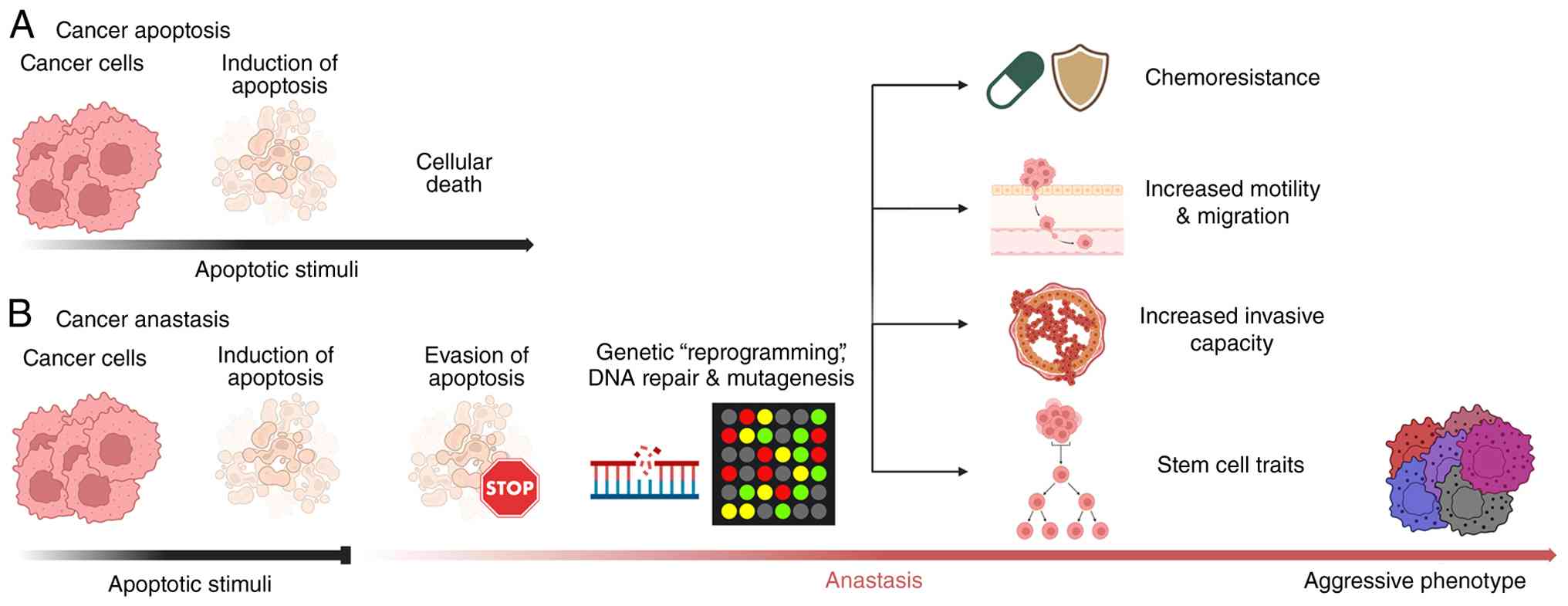
In summary, the aforementioned studies provided
evidence confirming that the rescue of dying cells through
anastasis serves as a significant factor in tumor evolution and
therapeutic resistance. This process allows for the preservation of
a subpopulation of cells exhibiting considerable genomic
instability and epigenetic modifications. The resulting ‘anastatic’
phenotype is distinguished by increased chemoresistance, as well as
the development of stem-like properties and greater metastatic
capacity. Accordingly, anastasis appears to act as a mechanism of
clonal selection, indicating that sub-lethal therapeutic stress may
unintentionally promote the emergence of more aggressive,
relapse-prone cancer variants rather than achieving complete tumor
eradication (43). Fig. 1 provides a schematic overview of
the principal events associated with anastasis, which ultimately
contribute to the development of a more aggressive cancer cell
phenotype.
Melatonin: A multipotent modulator of
cellular recovery
Biosynthesis and physiological
pleiotropy
Melatonin is an indoleamine initially discovered by
Lerner et al (44,45), following its isolation from the
bovine pineal gland. The indoleamine is synthesized within
pinealocytes from tryptophan, a process primarily driven by the
nocturnal upregulation of serotonin-N-acetyltransferase (46,47).
Notably, melatonin's nocturnal peak production is invariably
associated with the daily dark phase regardless of the activity
pattern of the species, that is, diurnal, nocturnal, or
crepuscular. In addition to the daily variation, pineal melatonin
production appears to be directly related to the length of the
night and, by extension, exhibits temporal variation throughout the
year (48).
Melatonin synthesis also occurs in multiple
extrapineal tissues and may, in fact, be ubiquitous in all somatic
cells (49–51). Moreover, the identification of
melatonin in the mitochondria of neural cells and hepatocytes, even
in pinealectomized animals, and the identification of its synthetic
enzymes in neurons has unequivocally confirmed the existence of
extrapineal sources in melatonin production (52–55).
Although the pineal gland is the primary source of circulating
circadian melatonin, it accounts for <5% of total production
(56). It is now apparent that
most melatonin is synthesized within the mitochondria of non-pineal
cells (57–59). This mitochondrial synthesis depends
on acetyl coenzyme A (acetyl-CoA) derived from pyruvate via the
action of pyruvate dehydrogenase (60). Under conditions of aerobic
glycolysis or hypoxia, decreased mitochondrial pyruvate results in
acetyl-CoA deficiency, thereby inhibiting melatonin production.
Notably, supplementation with melatonin can restore normal function
by promoting pyruvate transport into mitochondria, reestablishing
acetyl-CoA synthesis and supporting a healthy cellular physiology
(59).
Melatonin is now recognized as a functionally
pleiotropic molecule. Its roles extend well beyond the regulation
of circadian and seasonal biorhythms, encompassing functions in
retinal physiology, immune modulation, tumor suppression,
broad-spectrum antioxidant activity, and significant
cardioprotective effects (61,62).
Melatonin exerts its physiological effects through a dual mechanism
of action: Receptor-mediated and receptor-independent pathways.
Several receptor subtypes have been identified, most notably MT1
and MT2, members of the transmembrane G-protein-coupled receptor
superfamily (63). The
receptor-independent actions of melatonin primarily involve its
capacity to directly scavenge free radicals, thereby protecting
cells from oxidative damage (64).
Melatonin as a mitochondrial
guardian
The direct cytoprotective efficacy of melatonin
against oxidative stress is most pronounced in mitochondria, the
principal source of cellular reactive oxygen species (ROS). Martín
et al (65) indicated that
melatonin accumulates in mitochondria at concentrations up to
100-fold greater than those found in plasma, where it provides
protection as an endogenous antioxidant, supporting critical
glutathione redox homeostasis. Consistent with its high
intramitochondrial concentrations, melatonin enhances the activity
of respiratory chain complexes, thereby minimizing electron leakage
and free-radical generation, a protective process referred to as
‘radical avoidance’ (66).
The significant accumulation of melatonin within
mitochondria positions it at the molecular epicenter of anastasis,
where it regulates mitochondrial function and exerts essential
protective mechanisms (67–69).
Using multiphoton laser scanning microscopy, Jou et al
(70) managed to elucidate the
protective role of melatonin in cells harboring the mitochondrial
DNA ‘Common Deletion’, a defect characterized by respiratory chain
dysfunction and elevated mitochondrial ROS (mROS). The authors
demonstrated that melatonin markedly reduced the augmented mROS
formation by preventing the depolarization of the mitochondrial
membrane potential, thereby inhibiting the subsequent opening of
the mitochondrial permeability transition pore and the release of
cytochrome c.
Another important mechanism of protection is
melatonin's ability to preserve cardiolipin structure and
functionality. Cardiolipin is a phospholipid integral to the inner
mitochondrial membrane and it is essential for regulating
bioenergetics and oxidative phosphorylation. However, its high
unsaturated fatty acid content and proximity to sites of high ROS
production render it highly susceptible to peroxidation (71,72).
As a critical molecule in mitochondrial bioenergetics, any
alterations to cardiolipin, such as decreased content due to
reduced synthetase activity or changes in acyl chain composition
resulting from altered remodeling or peroxidation under oxidative
stress, can impair mitochondrial function and result in cell death
through mitophagy and apoptosis (73). Cardiolipin deficiency results in
mitochondrial enlargement and loss or disruption of cristae
structures, both of which are critical for the progression of
apoptosis. By maintaining cardiolipin integrity and supporting
mitochondrial bioenergetics, melatonin directly contributes to the
energy-intensive repair processes that underlie successful
anastasis (43).
Context-dependent modulation of DNA
repair: Cytoprotection vs. tumor sensitization
In addition to its prominent protective function
within mitochondria, melatonin serves as a critical line of defense
against oxidative DNA damage, a principal adverse effect that
occurs during anastasis. This genoprotective effect of melatonin
appears to be context-dependent, exhibiting distinctions between
normal and cancerous cells. Generally, melatonin supports genomic
integrity through its comprehensive antioxidant capacity,
functioning directly by scavenging free radicals and indirectly via
its active metabolites, stimulation of antioxidative enzymes, and
modulation of DNA repair pathways (74,75).
Liu et al (76) employed
the alkaline DNA comet assay in breast (MCF-7) and colon cancer
(HCT-15) cell lines to demonstrate that melatonin pretreatment
markedly increases repair capacity following methyl
methanesulfonate exposure. Genome-wide gene expression analyses
have suggested that this enhancement is mediated by the
upregulation of critical DNA integrity-maintenance proteins,
including centrosomal protein of 152 kDa and NEDD4-binding protein
2-like 2.
By contrast, a recent investigation on non-small
cell lung cancer identified a distinct mechanism by which melatonin
modulates genomic stability by operating as an inhibitor rather
than a facilitator of DNA repair (77). A specific subset of malignant cells
exhibiting aberrant germ cell gene expression demonstrate a notably
aggressive phenotype, characterized by accelerated DNA
double-strand break repair, rapid cellular proliferation and
enhanced resistance to radiotherapy. This aggressive behavior is
predominantly driven by the thyroid hormone receptor interactor 13
(TRIP13) (78). The study revealed
that melatonin effectively suppresses TRIP13 expression,
consequently inhibiting the critical DNA repair proteins DNA repair
protein RAD51 homolog 1 and DNA repair protein Ku80. As a result,
the capability of these cancer cells to conduct DNA repair is
markedly diminished, thereby increasing the sensitivity to
genotoxic anti-cancer interventions (77). These findings underscore the
multifaceted and context-dependent role of melatonin: While it
facilitates DNA repair and preserves genomic integrity in healthy
or stressed cells, it can selectively impair DNA repair processes
in malignant cells, thereby restricting their proliferation,
increasing their sensitivity to treatment and functioning as a
selective physiological regulator.
Tunneling nanotube (TNT)
formation
Another important aspect in melatonin's role in
promoting anastasis is the development of tunneling nanotubes
(TNTs), a form of highly sensitive nanotubular structures formed
de novo between cells to establish intricate functional
networks. Originally described by Rustom et al (79), TNTs enable the selective transfer
of membrane vesicles, cellular organelles and small molecules
between cells (80,81). Mitochondria are among the key
elements transported via TNTs, allowing recipient cells to
incorporate exogenous mitochondria into their existing networks,
thereby enhancing their energy reserves and resulting in notable
alterations in bioenergetics and overall cellular function
(82). Restoring cellular
bioenergetics is fundamental to facilitating repair mechanisms in
damaged tissue. An in vitro study targeting post-ischemic
endothelial cells confirmed that healthy mesenchymal stem cells
(MSCs) transfer functional mitochondria to cells with dysfunctional
organelles via TNTs. This process effectively rescued aerobic
respiration and protected the endothelial cells from apoptosis
(83). Additionally, in a murine
model of lipopolysaccharide-induced acute lung injury,
administration of wild-type MSCs resulted in the establishment of
gap junctions with alveolar epithelial cells, which allowed
mitochondrial transfer and led to a significant elevation of
alveolar ATP levels. Restoration of energy metabolism was critical
for restoring the physiological function of alveolar cells and
limiting mortality (84).
The role of melatonin in
ferroptosis
Recent advances in molecular biology have elucidated
novel forms of PCD, including ferroptosis, which appears to hold a
critical regulatory role in cancer progression. Ferroptosis is a
morphologically distinct form of PCD characterized by specific
mitochondrial alterations including increased membrane density,
reduced volume, loss of cristae and rupture of the outer membrane
(85).
In contrast to the reversal of apoptosis,
eliminating ferroptosis inducers (such as erastin or glutamate) is
insufficient to arrest cell death. Instead, the survival and
recovery of ferroptotic cells require the concurrent removal of the
inducer and supplementation with reduced glutathione or the
radical-trapping antioxidant ferrostatin-1 (86). As a mitochondria-mediated form of
cell death, it is plausible that ferroptosis is markedly modulated
by melatonin. By orchestrating iron chelation and antioxidant
defense within the organelle, melatonin holds significant promise
as a therapeutic intervention in the ferroptosis cascade (10,87).
In the context of cancer therapy, unravelling the interplay between
anastasis and ferroptosis is crucial for understanding
chemoresistance. Targeting the molecular mechanisms underlying
anastasis may potentiate the efficacy of ferroptosis-inducing
agents, offering a synergistic strategy to overcome drug resistance
(88).
In summary, melatonin transcends its classical role
as a circadian transducer to function as a fundamental driver of
cellular resilience. Its pleiotropic capacity is defined by a
precise context-dependence: acting as a mitochondrial guardian and
genomic protector in healthy tissue, while conversely sensitizing
malignant cells to therapeutic intervention. By orchestrating these
diverse mechanisms, melatonin secures the physiological stability
required for successful anastasis and tissue regeneration. Fig. 2 provides a schematic overview of
some of the pleiotropic effects of melatonin.
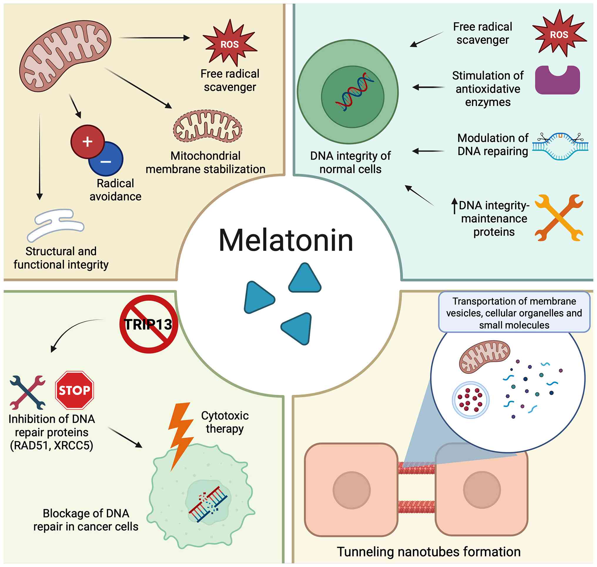 | Figure 2.The pleiotropic actions of melatonin
associated with cellular anastasis. Melatonin accumulates within
mitochondria, serving as a direct free radical scavenger while
maintaining their structural and functional integrity through
mechanisms such as radical avoidance and mitochondrial membrane
stabilization (top left). In non-malignant cells, melatonin
supports DNA integrity by stimulating antioxidative enzymes,
regulating DNA repair processes, and upregulating proteins
responsible for maintaining DNA stability (top right). Conversely,
in cancer cells, melatonin exerts context-dependent effects that
inhibit DNA repair. For example, by suppressing TRIP13 expression,
melatonin reduces the activity of repair proteins RAD51 and XRCC5,
thus enhancing cellular sensitivity to cytotoxic therapies (bottom
left). Additionally, melatonin promotes the formation of TNTs,
which facilitate the intercellular transport of membrane vesicles,
small molecules, and functional mitochondria, contributing to the
restoration of bioenergetics and supporting anastasis (bottom
right). Created in BioRender. Georgiou, A. (2026) https://BioRender.com/0f7njf4. ROS, reactive
oxygen species; TRIP13, thyroid hormone receptor interactor 13;
RAD51, DNA repair protein RAD51 homolog 1; XRCC5, DNA repair
protein Ku80; TNTs, tunneling nanotubes. |
Conclusion
The identification of anastasis has fundamentally
challenged the long-standing dogma of apoptotic irreversibility,
revealing a conserved survival mechanism that preserves tissue
integrity under transient stress but may also serve as a potent
driver of tumor evolution and therapeutic resistance. Drug
resistance is a principal factor contributing to cancer-related
mortality and constitutes a significant challenge that requires
urgent attention (89,90). The ‘resurrection’ of dying cancer
cells following sub-lethal cytotoxic stress may facilitate the
persistence of more aggressive, genetically unstable clones
exhibiting enhanced metastatic potential and stem-like
properties.
The anticancer properties of melatonin have been
extensively investigated in recent years, with research focusing on
both its potential roles in cancer prevention and treatment
(91–94). In the context of anastasis,
melatonin emerges as a compelling, mechanistically grounded
candidate pharmacological modulator whose pleiotropic biology
intersects with key molecular determinants of apoptotic recovery.
Through its capacity to preserve mitochondrial structural
integrity, regulate cellular bioenergetics, and modulate DNA repair
pathways in a context-dependent manner, melatonin may exert a dual
influence on cell fate. While its cytoprotective and
genome-stabilizing properties may support cellular recovery and
survival in normal tissues, melatonin has also been shown to exert
onco-suppressive and cytotoxic effects in malignant cells by
disrupting tumor-promoting processes, including the inhibition of
DNA repair, modulation of redox balance, and interference with
proliferative signaling pathways (10,95).
In addition, melatonin is increasingly recognized as an important
regulator of the immune system, capable of modulating both innate
and adaptive immune responses, enhancing antitumor immunity, and
influencing the tumor microenvironment. These immunomodulatory
properties suggest that melatonin may also have potential as an
adjuvant in cancer immunotherapy, possibly improving
immune-mediated tumor control while protecting normal tissues from
therapy-associated damage (94,96).
Taken together, this dual and context-dependent behavior suggests
that melatonin may simultaneously protect normal tissues from
stress or treatment-induced injury while sensitizing cancer cells
to anticancer therapies and supporting antitumor immune
responses.
Nevertheless, despite the growing body of evidence
supporting the existence of anastasis, several conceptual and
methodological challenges remain unresolved. The operational
definition of anastasis varies considerably among studies, as
differences in experimental systems, apoptotic stimuli, exposure
duration and recovery conditions may substantially influence the
observed extent of cellular recovery. Furthermore, it remains
difficult to ascertain whether cells that survive despite
exhibiting apoptotic characteristics truly undergo reversal of the
death program or merely constitute a selected population of
intrinsically resistant clones. In addition, further investigation
is warranted to elucidate the biological effects of melatonin which
appear to be highly context-dependent and influenced by factors
such as concentration, timing of administration, duration of
exposure, and cellular background. Finally, the majority of
available evidence has been derived from in vitro
experimental models and the extent to which these findings
accurately reflect the complexity of the tumor microenvironment and
their applicability to clinical settings remains uncertain.
The principal objective of the present study was to
introduce the concept of anastasis, examine its potential
implications for the development of more aggressive cancer
phenotypes and therapeutic resistance and, based on mechanistic
evidence, propose the possible involvement of melatonin in this
process. While the mechanistic rationale linking melatonin to
pathways associated with anastasis is biologically plausible, the
proposed role of melatonin in modulating pathways associated with
anastasis remains largely theoretical and requires further
experimental validation. Future progress will depend on the
development of robust in vitro and in vivo models
capable of faithfully recapitulating the tumor microenvironment
during apoptotic recovery, as well as the identification of
reliable biomarkers that can detect and monitor anastasis in
clinical settings and evaluate the translational relevance of
melatonin in this context.
Acknowledgements
Not applicable.
Funding
Funding: No funding was received.
Availability of data and materials
Not applicable.
Authors' contributions
AG performed the literature review, writing of the
manuscript and preparation of the figures and the table. DAS, VZ
and RJR contributed to the conceptualization of the study. DAS and
VZ supervised the writing process. RJR critically reviewed and
polished the manuscript. Data authentication is not applicable. All
authors read and approved the final manuscript.
Ethics approval and consent to
participate
Not applicable.
Patient consent for publication
Not applicable.
Competing interests
AG, RJR, VZ and DAS declare that they have no
competing interests. DAS is the Editor-in-Chief for the journal,
but had no personal involvement in the reviewing process, or any
influence in terms of adjudicating on the final decision, for this
article.
Use of artificial intelligence tools
During the preparation of this work, artificial
intelligence tools were used to improve the readability and
language of the manuscript or to generate images, and subsequently,
the authors revised and edited the content produced by the
artificial intelligence tools as necessary, taking full
responsibility for the ultimate content of the present
manuscript.
References
|
1
|
Elmore S: Apoptosis: A review of
programmed cell death. Toxicol Pathol. 35:495–516. 2007. View Article : Google Scholar : PubMed/NCBI
|
|
2
|
Mustafa M, Ahmad R, Tantry IQ, Ahmad W,
Siddiqui S, Alam M, Abbas K, Moinuddin Hassan MI, Habib S and Islam
S: Apoptosis: A comprehensive overview of signaling pathways,
morphological changes, and physiological significance and
therapeutic implications. Cells. 13:18382024. View Article : Google Scholar : PubMed/NCBI
|
|
3
|
Vitale I, Pietrocola F, Guilbaud E,
Aaronson SA, Abrams JM, Adam D, Agostini M, Agostinis P, Alnemri
ES, Altucci L, et al: Apoptotic cell death in disease-current
understanding of the NCCD 2023. Cell Death Differ. 30:1097–1154.
2023. View Article : Google Scholar : PubMed/NCBI
|
|
4
|
Kerr JFR, Wyllie AH and Currie AR:
Apoptosis: A basic biological phenomenon with wideranging
implications in tissue kinetics. Br J Cancer. 26:239–257. 1972.
View Article : Google Scholar : PubMed/NCBI
|
|
5
|
Nössing C and Ryan KM: 50 Years on and
still very much alive: ‘Apoptosis: A basic biological phenomenon
with wide-ranging implications in tissue kinetics’. Br J Cancer.
128:426–431. 2023. View Article : Google Scholar : PubMed/NCBI
|
|
6
|
Tang HL, Yuen KL, Tang HM and Fung MC:
Reversibility of apoptosis in cancer cells. Br J Cancer.
100:118–122. 2009. View Article : Google Scholar : PubMed/NCBI
|
|
7
|
Tang HL, Tang HM, Mak KH, Hu S, Wang SS,
Wong KM, Wong CS, Wu HY, Law HT, Liu K, et al: Cell survival, DNA
damage, and oncogenic transformation after a transient and
reversible apoptotic response. Mol Biol Cell. 23:2240–2252. 2012.
View Article : Google Scholar : PubMed/NCBI
|
|
8
|
Nano M, Mondo JA, Harwood J, Balasanyan V
and Montell DJ: Cell survival following direct executioner-caspase
activation. Proc Natl Acad Sci USA. 120:e22165311202023. View Article : Google Scholar : PubMed/NCBI
|
|
9
|
Tang HM and Tang HL: Anastasis: Recovery
from the brink of cell death. R Soc Open Sci. 5:1804422018.
View Article : Google Scholar : PubMed/NCBI
|
|
10
|
Reiter RJ, Sharma R, Manucha W, Balduini
W, Loh D, Spandidos DA, Romero A, Georgakopoulou VE and Zhu W:
Mechanistic insights into the role of melatonin in cancer cell
chemoresistance. Biocell. 49:2033–2067. 2025. View Article : Google Scholar
|
|
11
|
Reiter RJ, Mayo JC, Tan D, Sainz RM,
Alatorre-Jimenez M and Qin L: Melatonin as an antioxidant: Under
promises but over delivers. J Pineal Res. 61:253–278. 2016.
View Article : Google Scholar : PubMed/NCBI
|
|
12
|
Doonan F and Cotter TG: Morphological
assessment of apoptosis. Methods. 44:200–204. 2008. View Article : Google Scholar : PubMed/NCBI
|
|
13
|
Messam CA and Pittman RN: Asynchrony and
commitment to die during apoptosis. Exp Cell Res. 238:389–398.
1998. View Article : Google Scholar : PubMed/NCBI
|
|
14
|
Brunet CL, Gunby RH, Benson RS, Hickman
JA, Watson AJ and Brady G: Commitment to cell death measured by
loss of clonogenicity is separable from the appearance of apoptotic
markers. Cell Death Differ. 5:107–115. 1998. View Article : Google Scholar : PubMed/NCBI
|
|
15
|
Chipuk JE, Bouchier-Hayes L and Green DR:
Mitochondrial outer membrane permeabilization during apoptosis: The
innocent bystander scenario. Cell Death Differ. 13:1396–1402. 2006.
View Article : Google Scholar : PubMed/NCBI
|
|
16
|
Bhola PD and Letai A: Mitochondria-judges
and executioners of cell death sentences. Mol Cell. 61:695–704.
2016. View Article : Google Scholar : PubMed/NCBI
|
|
17
|
Suhaili SH, Karimian H, Stellato M, Lee TH
and Aguilar MI: Mitochondrial outer membrane permeabilization: A
focus on the role of mitochondrial membrane structural
organization. Biophys Rev. 9:443–457. 2017. View Article : Google Scholar : PubMed/NCBI
|
|
18
|
Tait SWG and Green DR: Mitochondrial
regulation of cell death. Cold Spring Harb Perspect Biol.
5:a0087062013. View Article : Google Scholar : PubMed/NCBI
|
|
19
|
Julien O and Wells JA: Caspases and their
substrates. Cell Death Differ. 24:1380–1389. 2017. View Article : Google Scholar : PubMed/NCBI
|
|
20
|
Ichim G, Lopez J, Ahmed SU, Muthalagu N,
Giampazolias E, Delgado ME, Haller M, Riley JS, Mason SM, Athineos
D, et al: Limited mitochondrial permeabilization causes DNA damage
and genomic instability in the absence of cell death. Mol Cell.
57:860–872. 2015. View Article : Google Scholar : PubMed/NCBI
|
|
21
|
Roux J, Hafner M, Bandara S, Sims JJ,
Hudson H, Chai D and Sorger PK: Fractional killing arises from
cell-to-cell variability in overcoming a caspase activity
threshold. Mol Syst Biol. 11:8032015. View Article : Google Scholar : PubMed/NCBI
|
|
22
|
Tang HL, Tang HM, Hardwick JM and Fung MC:
Strategies for tracking anastasis, a cell survival phenomenon that
reverses apoptosis. J Vis Exp. 519642015.PubMed/NCBI
|
|
23
|
Tang HM, Talbot CC Jr, Fung MC and Tang
HL: Molecular signature of anastasis for reversal of apoptosis.
F1000Res. 6:432017. View Article : Google Scholar : PubMed/NCBI
|
|
24
|
Sun G, Guzman E, Balasanyan V, Conner CM,
Wong K, Zhou HR, Kosik KS and Montell DJ: A molecular signature for
anastasis, recovery from the brink of apoptotic cell death. J Cell
Biol. 216:3355–3368. 2017. View Article : Google Scholar : PubMed/NCBI
|
|
25
|
Berthenet K, Castillo Ferrer C, Fanfone D,
Popgeorgiev N, Neves D, Bertolino P, Gibert B, Hernandez-Vargas H
and Ichim G: Failed apoptosis enhances melanoma cancer cell
aggressiveness. Cell Rep. 31:1077312020. View Article : Google Scholar : PubMed/NCBI
|
|
26
|
Los M, Mozoluk M, Ferrari D, Stepczynska
A, Stroh C, Renz A, Herceg Z, Wang ZQ and Schulze-Osthoff K:
Activation and caspase-mediated inhibition of PARP: A molecular
switch between fibroblast necrosis and apoptosis in death receptor
signaling. Mol Biol Cell. 13:978–988. 2002. View Article : Google Scholar : PubMed/NCBI
|
|
27
|
Wang X: The expanding role of mitochondria
in apoptosis. Genes Dev. 15:2922–1933. 2001.PubMed/NCBI
|
|
28
|
Liu X, He Y, Li F, Huang Q, Kato TA, Hall
RP and Li CY: Caspase-3 promotes genetic instability and
carcinogenesis. Mol Cell. 58:284–296. 2015. View Article : Google Scholar : PubMed/NCBI
|
|
29
|
Cao K, Riley JS, Heilig R, Montes-Gómez
AE, Vringer E, Berthenet K, Cloix C, Elmasry Y, Spiller DG, Ichim
G, et al: Mitochondrial dynamics regulate genome stability via
control of caspase-dependent DNA damage. Dev Cell. 57:1211–1225.e6.
2022. View Article : Google Scholar : PubMed/NCBI
|
|
30
|
Wang SS, Xie X, Wong CST, Choi Y and Fung
MC: HepG2 cells recovered from apoptosis show altered drug
responses and invasiveness. Hepatobiliary Pancreat Dis Int.
13:293–300. 2014. View Article : Google Scholar : PubMed/NCBI
|
|
31
|
Xu Y, So C, Lam HM, Fung MC and Tsang SY:
Apoptosis reversal promotes cancer stem cell-like cell formation.
Neoplasia. 20:295–303. 2018. View Article : Google Scholar : PubMed/NCBI
|
|
32
|
Al-Hajj M, Wicha MS, Benito-Hernandez A,
Morrison SJ and Clarke MF: Prospective identification of
tumorigenic breast cancer cells. Proc Natl Acad Sci USA.
100:3983–3978. 2003. View Article : Google Scholar : PubMed/NCBI
|
|
33
|
Vasileva MH, Bennemann A, Zachmann K,
Schön MP, Frank J and Ulaganathan VK: CD24 flags anastasis in
melanoma cells. Apoptosis. 30:1–15. 2025. View Article : Google Scholar : PubMed/NCBI
|
|
34
|
Tang MR, Guo JY, Wang D and Xu N:
Identification of CD24 as a marker for tumorigenesis of melanoma.
Onco Targets Ther. 11:3401–3406. 2018. View Article : Google Scholar : PubMed/NCBI
|
|
35
|
Li W, Ma H, Zhang J, Zhu L, Wang C and
Yang Y: Unraveling the roles of CD44/CD24 and ALDH1 as cancer stem
cell markers in tumorigenesis and metastasis. Sci Rep. 7:138562017.
View Article : Google Scholar : PubMed/NCBI
|
|
36
|
Knowles O, Doldan P, Hillier-Richardson I,
Lunetto S, Lunt S, Youssef G, Gammon L, Mackenzie IC, Philpott MP,
Rizvi H, et al: A CD24+CD271+ melanoma cancer
stem cell possesses hybrid characteristics of its single marker
counterparts and promotes invasion and therapeutic resistance. BMC
Biol. 23:2352025. View Article : Google Scholar : PubMed/NCBI
|
|
37
|
Hüser L, Altevogt P and Utikal J: Role of
STAT3 dependent SOX2 and CD24 expression in melanoma cell adaptive
resistance towards targeted therapies. Oncotarget. 10:1662–1663.
2019. View Article : Google Scholar : PubMed/NCBI
|
|
38
|
Wang R, Wang Y, Liu X, Liu M, Sun L, Pan
X, Hu H, Jiang B, Zou Y, Liu Q, et al: Anastasis enhances
metastasis and chemoresistance of colorectal cancer cells through
upregulating cIAP2/NFκB signaling. Cell Death Dis. 14:3882023.
View Article : Google Scholar : PubMed/NCBI
|
|
39
|
Mehrotra S, Languino LR, Raskett CM,
Mercurio AM, Dohi T and Altieri DC: IAP regulation of metastasis.
Cancer Cell. 17:53–64. 2010. View Article : Google Scholar : PubMed/NCBI
|
|
40
|
Kenneth NS and Duckett CS: IAP proteins:
Regulators of cell migration and development. Curr Opin Cell Biol.
24:871–875. 2012. View Article : Google Scholar : PubMed/NCBI
|
|
41
|
Albeck JG, Burke JM, Aldridge BB, Zhang M,
Lauffenburger DA and Sorger PK: Quantitative analysis of pathways
controlling extrinsic apoptosis in single cells. Mol Cell.
30:11–25. 2008. View Article : Google Scholar : PubMed/NCBI
|
|
42
|
Lovric MM and Hawkins CJ: TRAIL treatment
provokes mutations in surviving cells. Oncogene. 29:5048–5060.
2010. View Article : Google Scholar : PubMed/NCBI
|
|
43
|
Zaitceva V, Kopeina GS and Zhivotovsky B:
Anastasis: Return journey from cell death. Cancers (Basel).
13:36712021. View Article : Google Scholar : PubMed/NCBI
|
|
44
|
Lerner AB, Case JD, Takahashi Y, Lee TH
and Mori W: Isolation of melatonin, the pineal gland factor that
lightens melanocytes1. J Am Chem Soc. 80:25871958.
View Article : Google Scholar
|
|
45
|
Lerner AB, Case JD and Heinzelman RV:
Structure of melatonin. J Am Chem Soc. 81:6084–6085. 1959.
View Article : Google Scholar
|
|
46
|
Gastel JA, Roseboom PH, Rinaldi PA, Weller
JL and Klein DC: Melatonin production: Proteasomal proteolysis in
serotonin N-acetyltransferase regulation. Science. 279:1358–1360.
1998. View Article : Google Scholar : PubMed/NCBI
|
|
47
|
Cipolla-Neto J and Amaral FGD: Melatonin
as a hormone: New physiological and clinical insights. Endocr Rev.
39:990–1028. 2018. View Article : Google Scholar : PubMed/NCBI
|
|
48
|
Reiter RJ: The melatonin rhythm: Both a
clock and a calendar. Experientia. 49:654–664. 1993. View Article : Google Scholar : PubMed/NCBI
|
|
49
|
Acuña-Castroviejo D, Escames G, Venegas C,
Díaz-Casado ME, Lima-Cabello E, López LC, Rosales-Corral S, Tan DX
and Reiter RJ: Extrapineal melatonin: Sources, regulation, and
potential functions. Cell Mol Life Sci. 71:2997–3025. 2014.
View Article : Google Scholar : PubMed/NCBI
|
|
50
|
Cardinali DP and Rosner JM: Retinal
localization of the hydroxyindole-O-methyl transferase (HIOMT) in
the rat. Endocrinology. 89:301–303. 1971. View Article : Google Scholar : PubMed/NCBI
|
|
51
|
Vlahakes GJ and Wurtman RJ: A Mg
2+ dependent hydroxyindole O-methyltransferase in rat
Harderian gland. Biochim Biophys Acta. 261:194–197. 1972.
View Article : Google Scholar : PubMed/NCBI
|
|
52
|
Raikhlin NT, KvetnoyI M and Tolkschev VN:
Melatonin may be synthesised in enterochromaffin cells. Nature.
255:344–345. 1975. View Article : Google Scholar : PubMed/NCBI
|
|
53
|
Reiter RJ, Sharma R, Tan DX, Chuffa LGA,
da Silva DGH, Slominski AT, Steinbrink K and Kleszczynski K: Dual
sources of melatonin and evidence for different primary functions.
Front Endocrinol (Lausanne). 15:14144632024. View Article : Google Scholar : PubMed/NCBI
|
|
54
|
Venegas C, García JA, Escames G, Ortiz F,
López A, Doerrier C, García-Corzo L, López LC, Reiter RJ and
Acuña-Castroviejo D: Extrapineal melatonin: Analysis of its
subcellular distribution and daily fluctuations. J Pineal Res.
52:217–227. 2012. View Article : Google Scholar : PubMed/NCBI
|
|
55
|
Suofu Y, Li W, Jean-Alphonse FG, Jia J,
Khattar NK, Li J, Baranov SV, Leronni D, Mihalik AC, He Y, et al:
Dual role of mitochondria in producing melatonin and driving GPCR
signaling to block cytochrome c release. Proc Natl Acad Sci USA.
114:E7997–E8006. 2017. View Article : Google Scholar : PubMed/NCBI
|
|
56
|
Zhao D, Yu Y, Shen Y, Liu Q, Zhao Z,
Sharma R and Reiter RJ: Melatonin synthesis and function:
Evolutionary history in animals and plants. Front Endocrinol
(Lausanne). 10:2492019. View Article : Google Scholar : PubMed/NCBI
|
|
57
|
Tan D, Manchester LC, Liu X,
Rosales-Corral SA, Acuna-Castroviejo D and Reiter RJ: Mitochondria
and chloroplasts as the original sites of melatonin synthesis: A
hypothesis related to melatonin's primary function and evolution in
eukaryotes. J Pineal Res. 54:127–138. 2013. View Article : Google Scholar : PubMed/NCBI
|
|
58
|
He C, Wang J, Zhang Z, Yang M, Li Y, Tian
X, Ma T, Tao J, Zhu K, Song Y, et al: Mitochondria synthesize
melatonin to ameliorate its function and improve mice oocyte's
quality under in vitro conditions. Int J Mol Sci. 17:9392016.
View Article : Google Scholar : PubMed/NCBI
|
|
59
|
Reiter RJ, Sharma R, Rosales-Corral S,
Manucha W, Chuffa LGA and Zuccari DAPC: Melatonin and pathological
cell interactions: Mitochondrial glucose processing in cancer
cells. Int J Mol Sci. 22:124942021. View Article : Google Scholar : PubMed/NCBI
|
|
60
|
Reiter RJ, Sharma R, Rodriguez C, Martin
V, Rosales-Corral S, Zuccari DAPC and Chuffa LGA: Part-time cancers
and role of melatonin in determining their metabolic phenotype.
Life Sci. 278:1195972021. View Article : Google Scholar : PubMed/NCBI
|
|
61
|
Reiter RJ: Melatonin: Clinical relevance.
Best Pract Res Clin Endocrinol Metab. 17:273–285. 2003. View Article : Google Scholar : PubMed/NCBI
|
|
62
|
Reiter RJ, Tan DX, Paredes SD and
Fuentes-Broto L: Beneficial effects of melatonin in cardiovascular
disease. Ann Med. 42:276–285. 2010. View Article : Google Scholar : PubMed/NCBI
|
|
63
|
Liu J, Clough SJ, Hutchinson AJ,
Adamah-Biassi EB, Popovska-Gorevski M and Dubocovich ML: MT1 and
MT2 melatonin receptors: A therapeutic perspective. Annu Rev
Pharmacol Toxicol. 56:361–383. 2016. View Article : Google Scholar : PubMed/NCBI
|
|
64
|
Reiter RJ, Tan DX and Galano A: Melatonin:
Exceeding expectations. Physiology (Bethesda). 29:325–333.
2014.PubMed/NCBI
|
|
65
|
Martín M, Macías M, Escames G, León J and
Acuña-Castroviejo D: Melatonin but not vitamins C and E maintains
glutathione homeostasis in t-butyl hydroperoxide-induced
mitochondrial oxidative stress. FASEB J. 14:1677–1679. 2000.
View Article : Google Scholar : PubMed/NCBI
|
|
66
|
Hardeland R: Antioxidative protection by
melatonin: Multiplicity of mechanisms from radical detoxification
to radical avoidance. Endocrine. 27:119–130. 2005. View Article : Google Scholar : PubMed/NCBI
|
|
67
|
Leon J, Acuña-Castroviejo D, Sainz RM,
Mayo JC, Tan DX and Reiter RJ: Melatonin and mitochondrial
function. Life Sci. 75:765–790. 2004. View Article : Google Scholar : PubMed/NCBI
|
|
68
|
León J, Acuña-Castroviejo D, Escames G,
Tan DX and Reiter RJ: Melatonin mitigates mitochondrial
malfunction. J Pineal Res. 38:1–9. 2005. View Article : Google Scholar : PubMed/NCBI
|
|
69
|
Acuna-Castroviejo D, Escames G, Rodriguez
MI and Lopez LC: Melatonin role in the mitochondrial function.
Front Biosci. 12:947–963. 2007. View
Article : Google Scholar : PubMed/NCBI
|
|
70
|
Jou MJ, Peng TI, Yu PZ, Jou SB, Reiter RJ,
Chen JY, Wu HY, Chen CC and Hsu LF: Melatonin protects against
common deletion of mitochondrial DNA-augmented mitochondrial
oxidative stress and apoptosis. J Pineal Res. 43:389–403. 2007.
View Article : Google Scholar : PubMed/NCBI
|
|
71
|
Daum G: Lipids of mitochondria. Biochim
Biophys Acta. 822:1–42. 1985. View Article : Google Scholar : PubMed/NCBI
|
|
72
|
Paradies G, Paradies V, Ruggiero FM and
Petrosillo G: Role of cardiolipin in mitochondrial function and
dynamics in health and disease: Molecular and pharmacological
aspects. Cells. 8:7282019. View Article : Google Scholar : PubMed/NCBI
|
|
73
|
Fuentes J and Morcillo P: The role of
cardiolipin in mitochondrial function and neurodegenerative
diseases. Cells. 13:6092024. View Article : Google Scholar : PubMed/NCBI
|
|
74
|
Galano A, Tan DX and Reiter R: Melatonin:
A versatile protector against oxidative DNA damage. Molecules.
23:5302018. View Article : Google Scholar : PubMed/NCBI
|
|
75
|
Sliwinski T, Rozej W, Morawiec-Bajda A,
Morawiec Z, Reiter R and Blasiak J: Protective action of melatonin
against oxidative DNA damage: Chemical inactivation versus
base-excision repair. Mutat Res. 634:220–227. 2007. View Article : Google Scholar : PubMed/NCBI
|
|
76
|
Liu R, Fu A, Hoffman AE, Zheng T and Zhu
Y: Melatonin enhances DNA repair capacity possibly by affecting
genes involved in DNA damage responsive pathways. BMC Cell Biol.
14:12013. View Article : Google Scholar : PubMed/NCBI
|
|
77
|
Liu W, van Pelt AMM and Hamer G: Melatonin
suppresses cancer cell proliferation, DNA repair and expression of
the oncogene TRIP13. Cell Death Discov. 11:4892025. View Article : Google Scholar : PubMed/NCBI
|
|
78
|
Liu W, Lei Q, van Pelt AMM and Hamer G:
Repeated ionizing radiation exposure induces TRIP13 expression,
conferring radioresistance in lung cancer cells. Sci Rep.
15:9852025. View Article : Google Scholar : PubMed/NCBI
|
|
79
|
Rustom A, Saffrich R, Markovic I, Walther
P and Gerdes HH: Nanotubular highways for intercellular organelle
transport. Science. 303:1007–1010. 2004. View Article : Google Scholar : PubMed/NCBI
|
|
80
|
Abounit S and Zurzolo C: Wiring through
tunneling nanotubes-from electrical signals to organelle transfer.
J Cell Sci. 125:1089–1098. 2012. View Article : Google Scholar : PubMed/NCBI
|
|
81
|
Luchetti F, Carloni S, Nasoni MG, Reiter
RJ and Balduini W: Tunneling nanotubes and mesenchymal stem cells:
New insights into the role of melatonin in neuronal recovery. J
Pineal Res. 73:e128002022. View Article : Google Scholar : PubMed/NCBI
|
|
82
|
Shanmughapriya S, Langford D and
Natarajaseenivasan K: Inter and Intracellular mitochondrial
trafficking in health and disease. Ageing Res Rev. 62:1011282020.
View Article : Google Scholar : PubMed/NCBI
|
|
83
|
Liu K, Ji K, Guo L, Wu W, Lu H, Shan P and
Yan C: Mesenchymal stem cells rescue injured endothelial cells in
an in vitro ischemia-reperfusion model via tunneling nanotube like
structure-mediated mitochondrial transfer. Microvasc Res. 92:10–18.
2014. View Article : Google Scholar : PubMed/NCBI
|
|
84
|
Islam MN, Das SR, Emin MT, Wei M, Sun L,
Westphalen K, Rowlands DJ, Quadri SK, Bhattacharya S and
Bhattacharya J: Mitochondrial transfer from bone-marrow-derived
stromal cells to pulmonary alveoli protects against acute lung
injury. Nat Med. 18:759–765. 2012. View Article : Google Scholar : PubMed/NCBI
|
|
85
|
Wang H, Liu C, Zhao Y and Gao G:
Mitochondria regulation in ferroptosis. Eur J Cell Biol.
99:1510582020. View Article : Google Scholar : PubMed/NCBI
|
|
86
|
Tang HM and Tang HL: Cell recovery by
reversal of ferroptosis. Biol Open. 8:bio0431822019. View Article : Google Scholar : PubMed/NCBI
|
|
87
|
Zhang D, Jia X, Lin D and Ma J: Melatonin
and ferroptosis: Mechanisms and therapeutic implications. Biochem
Pharmacol. 218:1159092023. View Article : Google Scholar : PubMed/NCBI
|
|
88
|
Cao Y, Lu C, Beeraka NM, Efetov S, Enikeev
M, Fu Y, Yang X, Basappa B, He M and Li Z: Exploring the
relationship between anastasis and mitochondrial ROS-mediated
ferroptosis in metastatic chemoresistant cancers: A call for
investigation. Front Immunol. 15:14289202024. View Article : Google Scholar : PubMed/NCBI
|
|
89
|
Ingham J, Ruan JL and Coelho MA: Breaking
barriers: We need a multidisciplinary approach to tackle cancer
drug resistance. BJC Rep. 3:112025. View Article : Google Scholar : PubMed/NCBI
|
|
90
|
Dhanyamraju PK: Drug resistance mechanisms
in cancers: Execution of pro-survival strategies. J Biomed Res.
38:95–121. 2024. View Article : Google Scholar : PubMed/NCBI
|
|
91
|
Reiter RJ, Rosales-Corral SA, Tan DX,
Acuna-Castroviejo D, Qin L, Yang SF and Xu K: Melatonin, a full
service anti-cancer agent: Inhibition of initiation, progression
and metastasis. Int J Mol Sci. 18:8432017. View Article : Google Scholar : PubMed/NCBI
|
|
92
|
Li Y, Li S, Zhou Y, Meng X, Zhang JJ, Xu
DP and Li HB: Melatonin for the prevention and treatment of cancer.
Oncotarget. 8:39896–39921. 2017. View Article : Google Scholar : PubMed/NCBI
|
|
93
|
Putta CL, Eswar K and Rengan AK:
Melatonin: Avenues in cancer therapy and its nanotechnological
advancements. MedComm-Biomater Appl. 2:e582023.PubMed/NCBI
|
|
94
|
Guo R, Rao PG, Liao BZ, Luo X, Yang WW,
Lei XH and Ye JM: Melatonin suppresses PD-L1 expression and exerts
antitumor activity in hepatocellular carcinoma. Sci Rep.
15:84512025. View Article : Google Scholar : PubMed/NCBI
|
|
95
|
Mir SM, Aliarab A, Goodarzi G, Shirzad M,
Jafari SM, Qujeq D, Samavarchi Tehrani S and Asadi J: Melatonin: A
smart molecule in the DNA repair system. Cell Biochem Funct.
40:4–16. 2022. View Article : Google Scholar : PubMed/NCBI
|
|
96
|
Moslehi M, Moazamiyanfar R, Dakkali MS,
Rezaei S, Rastegar-Pouyani N, Jafarzadeh E, Mouludi K, Khodamoradi
E, Taeb S and Najafi M: Modulation of the immune system by
melatonin; implications for cancer therapy. Int Immunopharmacol.
108:1088902022. View Article : Google Scholar : PubMed/NCBI
|