Introduction
Preterm birth remains a leading cause of neonatal
mortality and long-term neurodevelopmental morbidity worldwide.
Survival rates have improved substantially, particularly for
extremely preterm infants, yet the burden of neurodevelopmental
impairment among survivors remains a major public health challenge
(1,2). Recent epidemiological data indicate
that the prevalence of adverse outcomes, including cerebral palsy,
cognitive deficits and behavioral disorders, does not decline
commensurately with improved survival, underscoring the critical
need for enhanced neuroprotective strategies (3,4).
The conceptual framework for understanding preterm
brain injury has evolved considerably in recent decades.
Historically, the predominant lesions were characterized as either
cystic periventricular leukomalacia or hemorrhagic lesions
originating in the germinal matrix. Contemporary neuroimaging and
neuropathological studies, however, have revealed a more complex
and diffuse pattern of injury. This is now conceptualized as the
encephalopathy of prematurity, which encompasses both white matter
dysmaturation and gray matter disturbances (5,6).
Panfoli et al (7)
systematically reviewed the role of free radical oxidative damage
to the newborn brain and proposed that oxidative stress (OS)
constitutes a primary risk factor for preterm brain injury. The
authors emphasized the particular maturation-dependent
vulnerability of oligodendrocyte precursors. This evolving
understanding reveals that the preterm brain possesses unique
developmental characteristics that render it selectively vulnerable
to injury. Jiang et al (8)
comprehensively examined the multifaceted aspects of OS in this
context. The authors reported that preterm infants are particularly
susceptible to inflammation and hypoxia because of their
underdeveloped antioxidant systems, high brain metabolic demands in
the brain, an immature blood-brain barrier and increased
vulnerability to inflammation and hypoxia. Multiple converging
pathways, including OS, neuroinflammation, excitotoxicity and
cerebrovascular dysregulation, interact to produce the final common
pathway of cellular injury and impaired maturation (9,10).
Among these interconnected pathological mechanisms,
OS occupies a central position as both an early initiator and an
amplifier of injury cascades. The immature brain possesses
underdeveloped antioxidant defenses, high metabolic demand and
vulnerability to inflammation. These factors converge to create a
permissive environment for free radical-mediated damage (5). Critically, the consequences of OS
extend beyond immediate macromolecular damage to encompass enduring
modifications of the epigenome. Thus, acute perinatal insults are
translated into sustained alterations in gene expression and
cellular function. Scarpato et al (11) provide an overview of how oxidative
genome damage can influence DNA methylation, thereby becoming a
determinant of pathological conditions in the newborn. This
establishes a direct molecular link between OS and epigenetic
dysregulation. This conceptual bridge between acute oxidative
injury and long-term epigenetic reprogramming offers new insights
into the pathogenesis of preterm brain injury and identifies novel
targets for intervention (12,13).
Despite the accumulating evidence, several
unresolved controversies complicate the translation of these
mechanistic insights into clinical practice. Chief among these is
the question of causality: Does OS drive epigenetic reprogramming
as a primary pathogenic event, or are the observed epigenetic
changes predominantly secondary to systemic inflammation, hypoxia
and other perinatal insults? The literature contains conflicting
findings, with some studies reporting strong associations between
oxidative biomarkers and epigenetic modifications, while others
suggest that these associations are confounded by gestational age,
nutritional status and genetic variability. Furthermore, the extent
to which cell-type-specific epigenetic responses, rather than
global changes, determine clinical outcomes remains poorly defined.
These controversies are not merely academic; they have direct
implications for biomarker selection, therapeutic targeting and
trial design. The following sections synthesized the available
evidence while explicitly highlighting areas of uncertainty and
conflicting data.
The present review was organised around a central,
unifying thesis: OS is not merely an acute cytotoxic event but a
trigger for enduring epigenetic reprogramming that shapes the
responses of all cellular players in the preterm brain.
Accordingly, the present review proceeded through four
interconnected domains. First, the sources and developmental
vulnerability of the preterm brain to OS were examined. Second, the
molecular mechanisms by which reactive species modify the
epigenome, namely DNA methylation, histone modifications and
non-coding RNAs, were integrated. Third, the cellular consequences
of this OS (OS)-to-epigenetic axis were analysed across the major
cell types (oligodendrocyte lineage, astrocytes, microglia, neurons
and the neurovascular unit), with the gut-brain axis presented as
an emerging systemic modulator. Fourth, neuroprotective strategies
were critically evaluated according to which node of the OS
epigenetic cascade they target. To guide the reader through this
integrated framework, a conceptual diagram (Fig. 1) is provided that illustrates how
OS, epigenetic regulation, cellular responses, neuroprotective
interventions, AI-based monitoring and systemic modulators
(including the gut-brain axis) converge within a unified pathogenic
cascade. Within each domain, the discussion prioritized clinically
actionable insights, biomarkers with prognostic value,
interventions with completed or ongoing clinical trials and AI
tools approaching clinical deployment, while mechanistic and
preclinical findings were presented as the basis for future
translation. Throughout the present review, the hierarchy of
evidence was observed: clinical findings (from prospective cohorts,
randomized controlled trials and meta-analyses) were presented as
the strongest evidence; case-control and cross-sectional studies
were identified as associative; and preclinical findings (animal
models and cell culture) were presented as mechanistic insights
that require clinical validation. To enhance transparency, the
evidence type (clinical or preclinical) for each study was
systematically provided in Table
I, Table II, Table III under the ‘Model system’
column. In the main text, clinical evidence was signaled by
descriptive terms (such as ‘clinical cohort’, ‘randomized
controlled trial’), whereas preclinical evidence was identified as
‘animal model’ or ‘mechanistic study’. At the end of each major
section, a summary statement highlighted the principal clinical
findings and directed the reader to the corresponding tables for
detailed source classification.
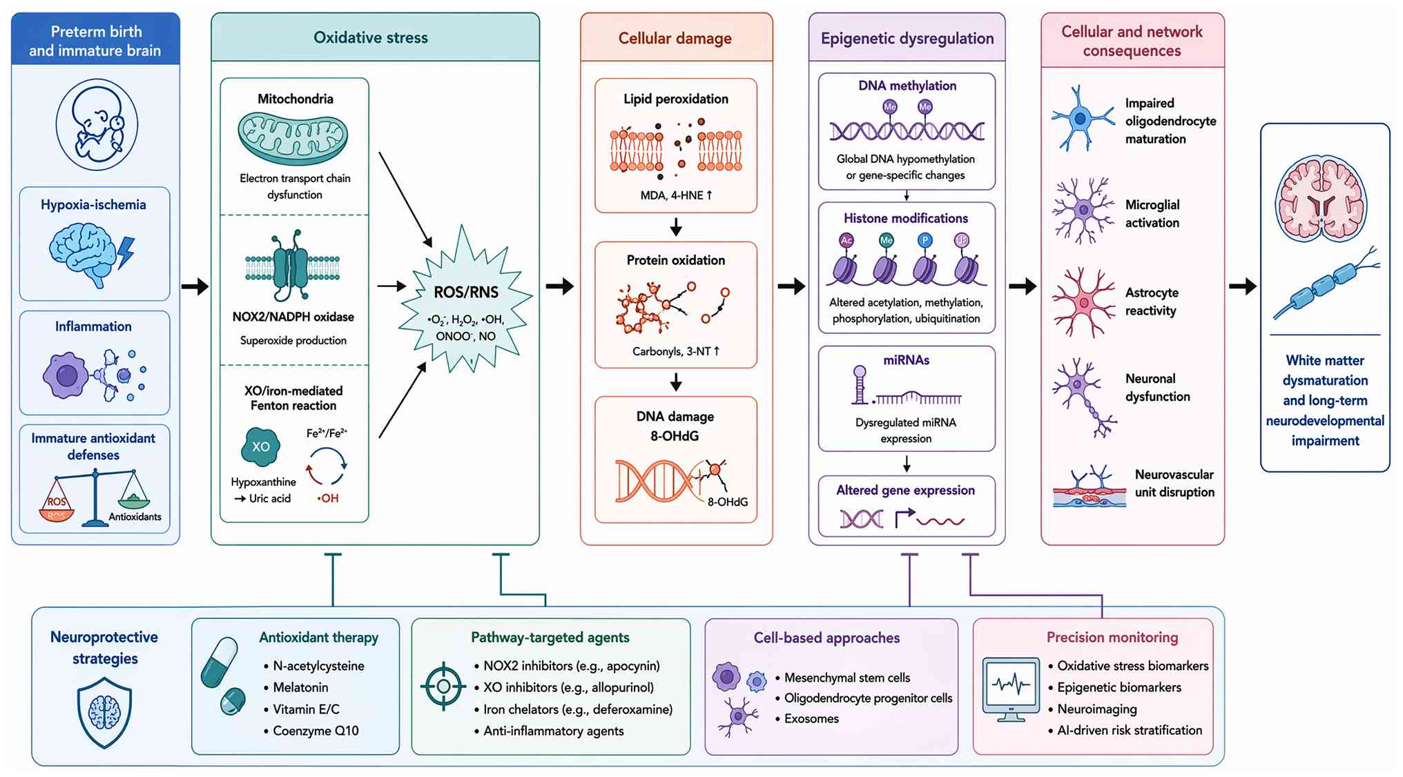 | Figure 1.Integrated conceptual framework of
preterm brain injury (www.figdraw.com; ID: TIRAT7295f). The diagram
illustrates the unifying OS-epigenetic axis connecting OS sources,
developmental vulnerabilities, epigenetic modifications, cellular
responses, therapeutic interventions targeting specific cascade
nodes and AI/monitoring tools within a unified pathogenic cascade.
Mechanistic and preclinical findings provide the foundation for
future translation, while clinically actionable elements
(biomarkers, trialed interventions, validated tools) are
prioritized. OS, oxidative stress; AI, artificial intelligence;
OPC, oligodendrocyte progenitor cell; ROS, reactive oxygen species;
RNS, reactive nitrogen species; NADPH, nicotinamide adenine
dinucleotide phosphate; NOX, NADPH oxidase; XO, xanthine oxidase;
NPBI, non-protein-bound iron; TET, ten-eleven translocation; HDAC,
histone deacetylase; DNMT, DNA methyltransferase; miRNA, microRNA;
MSC, mesenchymal stem cell; aEEG, amplitude-integrated
electroencephalography; NIRS, near-infrared spectroscopy. |
 | Table I.Cell-type specific responses and
mechanisms in preterm brain injury. |
Table I.
Cell-type specific responses and
mechanisms in preterm brain injury.
| First author/s,
year | Cell type | Model system | Mechanism | Key finding | Outcome | (Refs.) |
|---|
| Srivastava et al,
2018 | OPC | Mouse model | TLR/AKT/FoxO3
signaling | A TLR/AKT/FoxO3
immune tolerance-like pathway disrupts the repair capacity of
OPCs. | Impaired OPC
maturation and repair | (66) |
| Boccazzi et al,
2021 |
Oligodendrocyte | Murine model of
PWMI | TLR3
activation | TLR3 activation in
oligo-dendrocytes triggers a pro-inflammatory response compromising
their survival. | Pro-inflammatory
response, reduced survival | (67) |
| Al-Griw et al,
2021 | Oligodendrocyte
lineage | Ex vivo rat
ischemia model | Ionotropic GluR
signaling | Inhibition of
ionotropic glutamate receptor signaling preserves the
oligodendrocyte lineage. | Preservation of
oligoden-drocyte lineage and myelination | (68) |
| Chang et al,
2018 | OPC | Rat model | IUGR +
hyperoxia | Intrauterine growth
restriction combined with postnatal hyperoxia induces WMI via OPC
disruption. | White matter
injury | (69) |
| Lin et al,
2023 | Oligodendrocyte
lineage | Neonatal rat HI
model | Hypoxic-ischemic
injury | Characterized
dynamic changes in oligodendrogenesis after HI injury, revealing
temporal windows of vulnerability. | Limited
regenerative capacity, temporal vulnerability | (70) |
| Parfenova et al,
2018 | Astrocyte | Newborn pig
model | Carbon monoxide
production | Astrocyte-produced
carbon monoxide protects against cerebrovascular dysfunction after
neonatal asphyxia. | Cerebrovascular
protection | (71) |
| Nobuta et al,
2012 | Astrocyte | Mouse model of
neonatal brain injury | STAT3 signaling,
TGFβ-1 modulation | STAT3-mediated
astrogliosis protects myelin development by modulating microglial
TGFβ-1 expression. | Protection of
myelin development | (72) |
| Renz et al,
2024 | Astrocyte | Perinatal mouse
model | Reactive
astrogliosis | Neuroinflammatory
reactive astrocyte formation correlates with adverse outcomes in
perinatal WMI. | Adverse clinical
trajectory | (73) |
| Fang et al,
2020 | Neurovascular unit
(incl. astrocytes) | Neonatal rat HI
model | FGF10
signaling | FGF10 protects the
neurovascular unit, including astrocytes, in neonatal HI
injury. | Neurovascular
protection | (74) |
| Xu et al, 2025 | Astrocyte | Mouse WMI
model | HDAC3 inhibition,
iron homeostasis | Targeting HDAC3
suppresses ferroptosis and demyelination partly through restoring
astrocytic iron homeostasis. | Reduced ferroptosis
and demyelination | (60) |
| Yang et al,
2022 | Microglia | Neonatal rat HI
model | Microglial
polarization (toward anti-inflammatory) | Caffeine treatment
reduces HI white matter damage by regulating phenotypic microglial
polarization. | Reduced white
matter damage | (76) |
| Charriaut-Marlangue
et al, 2018 | Microglia | Mouse model of
neonatal stroke | PARP inhibition,
sex-specific responses | Identified sex
differences in microglial phenotypes following neonatal
stroke. | Differential
microglial activation | (77) |
| Zaghloul et al,
2020 | Microglia | Rat HI model | NF-κB
inhibition | Prophylactic
inhibition of NF-κB expression in microglia attenuates HI
injury. | Reduced
hypoxic-ischemic injury | (78) |
| Mairesse et al,
2019 | Microglia | Mouse model of
perinatal brain injury | Oxytocin receptor
activation | Oxytocin receptor
agonist reduces perinatal brain damage by directly targeting
microglia. | Reduced brain
damage | (79) |
| Van Steenwinckel et
al, 2019 | Microglia | Developing mouse
brain | Wnt/β-catenin
signaling | Decreased
microglial Wnt/β-catenin signaling drives pro-inflammatory
activation in the developing brain. | Pro-inflammatory
microglial activation | (80) |
| Bernis et al,
2022 | Microglia | Neonatal HI mouse
model | Temporal gene
expression dynamics | Temporally
characterized microglia-associated gene expression in a sensitized
HI model. | Inflammatory and
anti-inflammatory gene regulation | (81) |
| Sathyanesan et al,
2018 | Neuron
(Purkinje) | Mouse model of
neonatal brain injury | Cerebellar circuit
disruption | Neonatal brain
injury causes cerebellar learning deficits and Purkinje cell
dysfunction. | Learning deficits,
Purkinje cell dysfunction | (82) |
| Sathyanesan et al,
2021 | Neuron
(Purkinje) | Mouse model | Purkinje cell
dysfunction | Disruption of
neonatal Purkinje cell function underlies injury-related learning
deficits. | Learning
deficits | (83) |
| Scheuer et al,
2022 | Neuron
(interneuron) | Mouse model of
neonatal hyperoxia | OS | Neonatal OS impairs
cortical synapse formation and GABA homeostasis in
parvalbumin-expressing interneurons. | Impaired synapse
formation, GABA dysregulation | (26) |
| Lacaille et al,
2019 | Neuron
(interneuron) | Mouse model of
neonatal brain injury | Impaired
interneuron development | Demonstrated
impaired interneuron development in a novel model of neonatal brain
injury. | Long-term
neuropsychiatric sequelae | (84) |
| Sheikh et al,
2019 | Neuron
(subplate) | Mouse HI model | Circuit changes in
subplate neurons | Neonatal HI causes
functional circuit changes in subplate neurons. | Altered cortical
development | (85) |
| Northington et al,
2022 | Neuron
(cholinergic) | Mouse HI model | Cholinergic system
damage | Basal forebrain
magnocellular cholinergic systems are damaged following neonatal HI
in mice. | Long-term cognitive
deficits | (86) |
| Lee et al,
2018 | Neurovascular unit
(endothelial) | In vitro/In
vivo rat HI model | Endothelial
resilience |
Hypoxia-preconditioned human umbilical
vein endothelial cells protect against neurovascular damage after
HI. | Neurovascular
protection | (87) |
| Chand et al,
2022 | Neurovascular
unit | Growth-restricted
newborn lamb model | Hemodynamic
modulation | Neurovascular unit
alterations in growth-restricted newborns are improved following
ibuprofen treatment. | Improved
neurovascular structure | (88) |
| Fang et al,
2023 | Neurovascular unit
(BBB) | Neonatal rat HI
model | BBB protection | Chloroquine
protects against neonatal brain injury by mitigating blood-brain
barrier disruption. | Reduced brain
injury | (89) |
| Wang et al,
2020 | Oligovascular
unit | Neonatal mouse HI
model | PI3K/AKT/mTOR
signaling | Oligogenesis in the
‘oligovascular unit’ involves PI3K/AKT/mTOR signaling. | Oligogenesis | (90) |
| Seki et al,
2021 | Gut-brain axis | Human
cohort/preterm neonates |
Microbiota-immune-brain axis
dysregulation | Aberrant
gut-microbiota-immune-brain axis development occurs in premature
neonates with brain damage. | Pro-inflammatory
tone, brain damage | (91) |
| Drobyshevsky et al,
2024 | Gut-brain axis | Mouse HI model | Microbiota
modulation | Intestinal
microbiota modulates neuroinflammatory response and brain injury
after neonatal HI. | Altered
neuroinflammation and brain injury | (92) |
| Liu et al,
2023 | Gut-brain axis | Human VLBW infant
cohort |
Microbiota-gut-brain axis
dysregulation | Multi-omics
analyses reveal aberrant microbiota-gut-brain axis in VLBW infants
with white matter injury. | White matter
injury | (93) |
| Vaher et al,
2024 | Gut-brain axis | Human preterm
infant cohort | Microbiota
profiles | The neonatal gut
microbiota plays a role in the encephalopathy of prematurity. | Adverse
neurodevelopmental outcomes | (94) |
| Seki et al,
2024 | Gut-brain axis | Human preterm
infant cohort | Microbial genome
features | Identified gut
microbiota genome features associated with brain injury in
extremely premature infants. | Brain injury | (95) |
| Table II.Summary of neuroprotective strategies
for preterm brain injury by mechanism and evidence level. |
Table II.
Summary of neuroprotective strategies
for preterm brain injury by mechanism and evidence level.
| First author/s,
year | Strategy
category |
Intervention/target |
Model/population | Key findings | Evidence level | (Refs.) |
|---|
| Carloni et al,
2017 | Antioxidant | Melatonin
pharmacokinetics | Preterm
neonates | Significant
interindividual variability in absorption complicates dosing
optimization | Clinical
pharmacokinetic study | (100) |
| Domínguez Rubio et
al, 2017 | Antioxidant | Melatonin (maternal
administration) | LPS-treated mouse
model | Short- and
long-term neuroprotective effects in offspring | Preclinical | (101) |
| Garofoli et al,
2024 | Antioxidant | Melatonin
(oral) | Preterm
newborns | Reaches systemic
circulation and reduces OS markers | Clinical pilot | (102) |
| Lee et al,
2019 | Antioxidant | Melatonin | Mouse model of
preterm birth | Prevented preterm
birth and associated fetal brain injury | Preclinical | (103) |
| Ahmed et al,
2021 | Antioxidant | Melatonin
(systematic review) | Neonatal
encephalopathy | Safety favorable
but evidence insufficient; small samples, heterogeneity | Systematic
review | (104) |
| Favrais et al,
2021 | Antioxidant | Melatonin | Rat
chorioamnionitis model | Partial protective
effects only; monotherapy inadequate | Preclinical | (106) |
| Zmora et al,
2021 | Antioxidant | N-acetylcysteine
(maternal) | Rat NF-κB
model | Prevents neonatal
brain injury associated with NEC | Preclinical | (107) |
| Chavez-Valdez et
al, 2025 | Antioxidant | Vitamin C | Neonatal
encephalopathy | Shows preclinical
promise; clinical translation limited |
Commentary/Review | (108) |
| Al et al, 2020 | Antioxidant | Uridine | Neonatal rat
hyperoxic brain injury | Antioxidative
effects demonstrated | Preclinical | (109) |
| Chen et al,
2024 | Antioxidant | Nrf2 induction
targeting mitochondria | Experimental
models | Protects against
preterm birth and fetal brain injury | Preclinical | (110) |
| Fischer et al,
2017 |
Pathway-targeted | Erythropoietin
(meta-analysis) | Preterm
infants | Reduces
transfusions; neurodevelopmental evidence inconclusive | Meta-analysis | (113) |
| Juul et al,
2020 |
Pathway-targeted | High-dose
erythropoietin | Extremely preterm
infants | No significant
difference in death or severe neurodevelopmental impairment at 2
years | RCT | (114) |
| Song et al,
2021 |
Pathway-targeted | Erythropoietin | Preterm infants
with IVH | Improved outcomes
in IVH subgroup | Clinical
cohort | (116) |
| Jakab et al,
2019 |
Pathway-targeted | Early high-dose
erythropoietin | Very preterm
infants | Trophic effects on
brain connectivity | Neuroimaging
study | (117) |
| Wassink et al,
2017 |
Pathway-targeted | Prolonged
erythropoietin infusion | Preterm fetal sheep
(asphyxia) | Partial white and
grey matter protection | Preclinical (large
animal) | (118) |
| Ma and Shi,
2022 |
Pathway-targeted | Erythropoietin
(review) | Premature brain
injury | Clinical
translation requires improved patient stratification | Comprehensive
review | (119) |
| Endesfelder et al,
2017 |
Pathway-targeted | Caffeine | Neonatal rat
hyperoxia model | Neuroprotection
against hyperoxia-induced injury | Preclinical | (120) |
| Di Martino et al,
2020 |
Pathway-targeted | Caffeine
(therapeutic window) | Neonatal mouse HI
model | Early
administration critical for efficacy | Preclinical | (121) |
| Fang et al,
2024 |
Pathway-targeted | PPAR-γ
activation | Neonatal mouse HI
model | Inhibits
microglia-mediated neuroinflammation | Preclinical | (123) |
| Feng et al,
2023 |
Pathway-targeted | PPAR-γ activation
(alpha-asaronol) | OPC culture and
animal model | Promotes OPC
differentiation and myelination | Preclinical | (124) |
| Prasad et al,
2021 |
Anti-inflammatory | Various agents
(review) |
Inflammation-related preterm brain
injury | Numerous agents
show efficacy; translation limited by complexity | Comprehensive
review | (126) |
| Takahashi et al,
2023 |
Anti-inflammatory | IL-1 receptor
blockade | Preterm fetal sheep
LPS model | Suppressed
neuroinflammation | Preclinical (large
animal) | (127) |
| Bokobza et al,
2022 |
Anti-inflammatory | miR-146b | Perinatal brain
injury model | Protects against
microglia-induced hypomyelination | Preclinical | (128) |
| Rantakari et al,
2021 | Clinical care | Oxygen
saturation | Extremely preterm
infants | Both low and high
saturations associated with adverse outcomes | Clinical
cohort | (129) |
| Oei et al,
2026 | Clinical care | Targeted oxygen for
resuscitation | Preterm
infants | No significant
difference; highlights complexity of titration | RCT | (130) |
| Engur et al,
2024 | Clinical care | Supplemental
oxygen | Developing mouse
brain | Alters pentose
phosphate pathway through SIRT signaling | Preclinical | (131) |
| Milette et al,
2017 | Clinical care | Developmental care
guidelines | NICU setting | Institutional
implementation guidelines | Position
statement | (132) |
| Murthy et al,
2020 | Clinical care | Neuroprotection
care bundle | Preterm
infants | Reduced IVH rates
following implementation | Quality
improvement | (133) |
| Travis et al,
2025 | Clinical care | Skin-to-skin
holding | Preterm
infants | Associated with
improved white matter microstructure | Observational | (134) |
| Belfort and Inder,
2022 | Clinical care | Human milk | Preterm
infants | Breastfeeding
associated with improved brain development | Narrative
review | (135) |
| Brandt et al,
2025 | Clinical care | Human milk
oligosaccharides | Rat preterm brain
injury model | Improve white
matter and interneuron development | Preclinical | (136) |
| Drommelschmidt et
al, 2017 | Cell-based | MSC-derived
extracellular vesicles |
Inflammation-induced preterm brain injury
model | Ameliorate
injury | Preclinical | (141) |
| Thomi et al,
2019 | Cell-based | MSC-derived
exosomes (intranasal) | Perinatal brain
injury model | Reduce
microglia-mediated neuroinflammation | Preclinical | (142) |
| Ahn et al,
2018 | Cell-based | Mesenchymal stem
cells | Preterm infants
with severe IVH | Phase I:
feasibility and safety demonstrated | Phase I clinical
trial | (143) |
| Romantsik et al,
2023 | Cell-based | Stem cell
interventions (Cochrane review) | Preterm
infants | Preclinical
evidence promising; clinical data insufficient | Systematic
review | (144) |
| Tscherrig et al,
2024 | Cell-based | miRNAs from MSC
extracellular vesicles | White matter injury
model | Rescue white matter
injury after intranasal administration | Preclinical | (145) |
| Vaes et al,
2024 | Cell-based | Modified MSC
secretome | Encephalopathy of
prematurity model | Prolongs
regenerative treatment window | Preclinical | (146) |
| Table III.AI applications in preterm brain
injury: Summary of key studies. |
Table III.
AI applications in preterm brain
injury: Summary of key studies.
| First author/s,
year | Application
area | AI
Method/Model | Data
source/evidence type | Key findings | Primary
advantage | (Refs.) |
|---|
| Ahmad et al,
2025 | Neuroimaging
(cranial ultrasound) | Convolutional
neural networks (CNN) | Clinical (very
preterm infant CUS dataset) | DL models
classified CUS images as normal/abnormal in very preterm infants
with AUC 0.86; proof-of-concept for computer-aided detection | Automated
interpretation of routine screening tool; reduces inter-observer
variability | (154) |
| Chen et al,
2024 | Neuroimaging
(MRI) | nnU-Net (automated
segmentation) | Clinical
(multi-institutional MRI dataset) | Developed automated
neonatal brain MRI extractor trained on multi-institutional
dataset; robust generalizability across clinical settings | High-throughput,
standardized brain extraction; enables large- scale neuroimaging
studies | (155) |
| Estermann et al,
2025 | Neuroimaging
(cranial ultrasound) | CACTUS multiview
classifier | Clinical (CUS
volumes) | Detected punctate
white matter lesions in CUS volumes; addressed diagnostically
challenging task with neuro-developmental implications | Automated detection
of subtle pathology; multiview integration improves
sensitivity | (156) |
| Wang et al,
2023 | Neuromonitoring
(aEEG/EEG) | Automated
quantitative feature extraction | Clinical (10-year
cohort of preterm infants) | Quantitative aEEG
features predicted long-term neuro- developmental outcomes in
extremely preterm infants; automated extraction matched expert
interpretation | Real-time bedside
prognostication; reduces need for expert EEG interpretation | (157) |
| Abbasi et al,
2024 | Neuromonitoring
(EEG) | Deep learning with
wavelet-scalogram | Preclinical (fetal
sheep HI model) | Developed seizure
detection models for post-hypoxic-ischemic EEG in preclinical
models | High sensitivity
for seizure patterns; generalizable across species | (158) |
| Roozbehi et al,
2024 | Neuromonitoring
(EEG) | Transformer-based
architecture | Preclinical (fetal
sheep EEG data) | Refined seizure
pattern recognition using transformer models; improved accuracy
over conventional deep learning | Captures temporal
dependencies in EEG; superior pattern recognition | (159) |
| Ashoori et al,
2023 | Neuromonitoring
(NIRS) | Machine learning
classification | Clinical (preterm
infant NIRS data) | Data-driven
definitions of prolonged desaturation outper- formed
threshold-based approaches for IVH detection | Objective,
physiology- based risk stratification; identifies subtle
hemodynamic instability | (160) |
| Hibner et al,
2026 | Multimodal
monitoring | Integrated signal
analysis (echocardiography, NIRS, electrical cardiometry) | Clinical (preterm
infant cohort) | Multimodal approach
provided complementary information unavailable from any single
modality for IVH prediction | Comprehensive
hemody- namic assessment; captures multi-system physiology | (161) |
| He et al, 2025 | Predictive
modelling (brain injury risk) | CatBoost with SHAP
(PBIPred model) | Clinical
(prospective cohort of 650 infants) | Developed
explainable ML model using 7 clinical features; AUC 0.8229 for PBI
prediction; online webserver available | Interpretable
predictions at individual patient level; clinically deployable
tool | (162) |
| Yang et al,
2024 | Predictive
modelling (IVH/mortality) | Machine learning
(multiple algorithms) | Clinical
(nationwide multicenter cohort) | Nationwide
multicenter study; robust prediction of early mortality and severe
IVH in VLBW infants | Large,
representative cohort; generalizable findings | (163) |
| Han et al,
2024 | Predictive
modelling (IVH) | Time-series machine
learning | Clinical (preterm
infant physiological data) | Temporal dynamics
of physiolo- gical data improved IVH prediction compared with
static variables alone | Captures evolving
physiological instability; dynamic risk assessment | (164) |
| Song et al,
2025 | Predictive
modelling (WMI) | Ensemble machine
learning | Clinical (extremely
preterm infant cohort) | Two risk assessment
models for WMI in extremely preterm infants; ensemble methods
outperformed single algorithms | Robust methodology;
comparison of multiple modelling approaches | (165) |
| Shu et al,
2025 | Predictive
modelling (multimorbidity) | Machine learning
(multiple algorithms) | Clinical (VLBW
infant dataset) | Predicted mortality
and multiple morbidities simultaneously using routinely collected
data; comprehensive outcome prediction | Efficient use of
existing data; multi-outcome prediction | (166) |
| Li et al, 2025 | Prognostication
(neurodevelopmental outcomes) | Functional
connectivity analysis | Clinical (preterm
infant MRI cohort) | Functional
connectivity measures predicted outcomes in preterm infants without
severe brain injury; identified risk despite normal conventional
imaging | Sensitive to subtle
network alterations; identifies ‘hidden’ risk | (167) |
| Liu et al,
2024 | Prognostication
(brain age) | Graph convolutional
neural networks | Clinical (preterm
neonatal MRI data) | Predicted brain
age; deviations from normative maturation trajectories explained
outcomes more accurately than static injury assessments | Dynamic,
trajectory-based assessment; captures maturational delay | (168) |
| Marinelli et al,
2024 | Prognostication
(cerebral palsy) | Machine learning
(multiple algorithms) | Clinical (extreme
preterm cohort, ages 2 & 10) | Compared
predictions at ages 2 and 10 years; early predictions require
refinement as children mature | Long-term
follow-up; identifies temporal dynamics of prediction accuracy | (169) |
| Yuan et al,
2024 | Prognostication
(intellectual disability) | Nomogram
development | Clinical (children
with CP cohort) | Developed practical
tool for predicting intellectual disability in children with
cerebral palsy; enables early intervention targeting | Clinically
applicable tool; user-friendly format | (170) |
| van Boven et al,
2025 | Prognostication
(neurodevelopmental outcomes) | Machine learning
with oxygenation vital signs | Clinical (preterm
infant cohort) | Oxygenation
dynamics improved prediction of neurodevelopmental outcomes at ages
2 and 5 years | Incorporates
routinely monitored vital signs; long-term outcome prediction | (173) |
Molecular mechanisms of OS in the preterm
brain
The developing brain of preterm infants is
particularly susceptible to OS, a condition arising from an
imbalance between the production of reactive oxygen and nitrogen
species (ROS/RNS) and the capacity of antioxidant defense systems.
This vulnerability is not merely a consequence of one single factor
but results from a convergence of developmental, physiological and
pathological elements that collectively disrupt redox homeostasis.
The following sections detailed the primary sources of OS, the
inherent vulnerabilities of the preterm brain, the consequential
molecular damage and the intricate interplay with other injurious
pathways. A summary of the major sources and targets of OS in this
context was presented in Fig.
2.
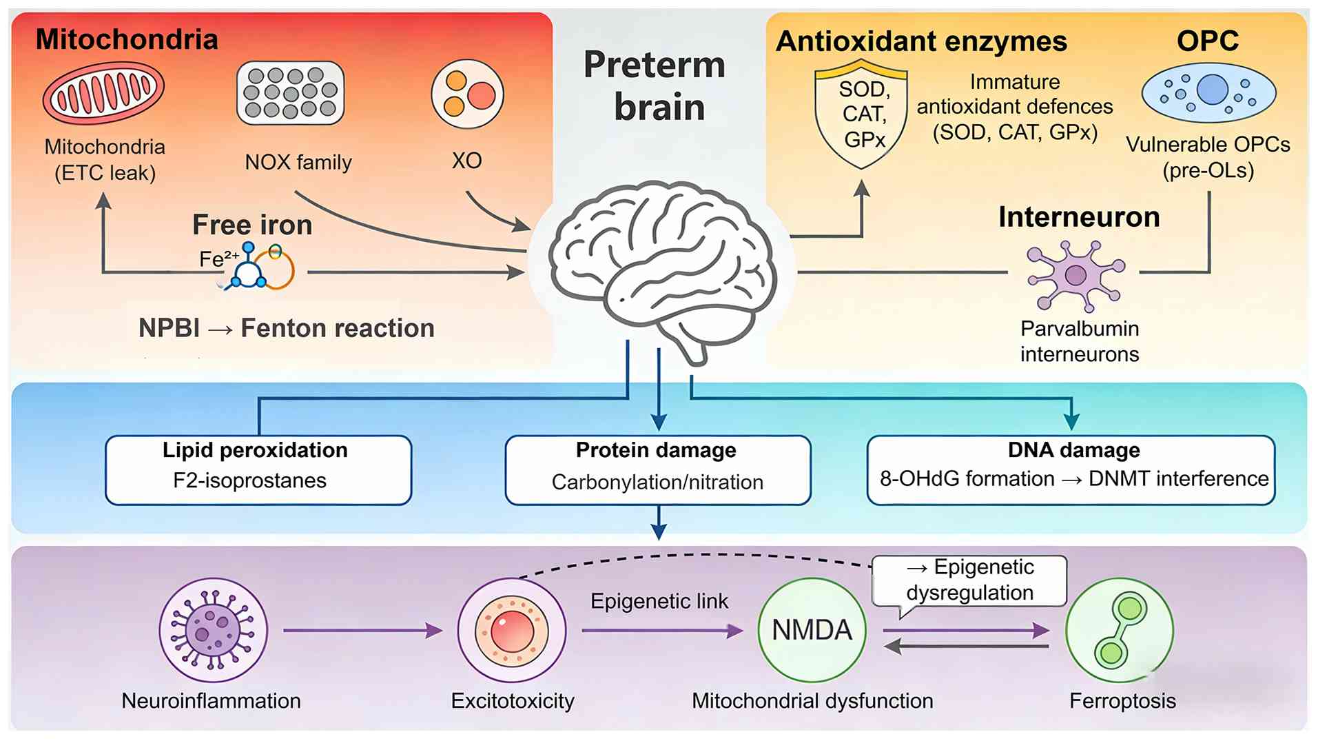 | Figure 2.Sources, vulnerability and
pathological cascades of OS in preterm brain injury (www.figdraw.com; ID: IWSTO44884). The schematic
illustrates major sources of ROS/RNS in the preterm brain,
including mitochondrial dysfunction, NOX activation, XO reaction
and iron-dependent Fenton reactions. The immature antioxidant
system and high susceptibility of OPCs render the preterm brain
vulnerable to oxidative damage. OS triggers macromolecular injuries
and interacts with neuroinflammation, excitotoxicity and
ferroptosis, forming a vicious pathological cascade that
exacerbates preterm brain damage. CAT, catalase; DNMT, DNA
methyltransferase; ETC, electron transport chain; GPx, glutathione
peroxidase; NMDA, N-methyl-D-aspartate; NOX, NADPH oxidase; NPBI,
non-protein-bound iron; 8-OHdG, 8-hydroxy-2′-deoxyguanosine; OPC,
oligodendrocyte progenitor cell; pre-OL, pre-oligodendrocyte; PV,
parvalbumin; ROS, reactive oxygen species; RNS, reactive nitrogen
species; SOD, superoxide dismutase; OS, oxidative stress; XO,
xanthine oxidase. |
Sources of ROS/RNS
The genesis of OS in the preterm brain is
multifactorial, with several key enzymatic and non-enzymatic
sources contributing to the ROS/RNS burden. Mitochondria are
primary contributors, as electron leakage from the electron
transport chain, particularly during the rapid but often
inefficient oxidative phosphorylation required for
neurodevelopment, generates superoxide anions (14,15).
This is compounded by the immature regulation of this process.
Beyond mitochondria, the nicotinamide adenine dinucleotide
phosphate (NADPH) oxidase (NOX) family of enzymes plays a
significant role. Investigations have shown that NOX enzymes are
activated in neuroinflammatory conditions, with neuronal NADPH
oxidase 1 contributing to injury in models of cerebral ischemia
(16).
Following hypoxia-ischemia, the enzyme xanthine
oxidase (XO) is another major source of ROS, generated during the
reperfusion phase when molecular oxygen is reintroduced to tissues
with accumulated hypoxanthine (17). The phase III ALBINO trial
(NCT03162653), an investigator-initiated, randomized,
placebo-controlled, double-blinded, multi-national trial, is
currently evaluating allopurinol as an adjunct to therapeutic
hypothermia for hypoxic-ischemic encephalopathy; the detailed
statistical analysis plan has been published, with the primary
endpoint being long-term outcome determined as survival with
neurodevelopmental impairment vs. death vs. non-impaired survival
at 2 years of age (18). As
Martini et al (19) and Hu
et al (20) have detailed,
free iron catalyzes the Fenton reaction, converting hydrogen
peroxide into the highly toxic hydroxyl radical, thereby amplifying
oxidative damage. The convergence of these diverse sources suggests
that effective neuroprotection may require a multi-pronged approach
rather than targeting any single enzymatic pathway, as the dominant
source of ROS likely varies with the phase of injury and individual
patient characteristics.
Developmental vulnerability of the
preterm brain
The preterm brain possesses an inherent
vulnerability to OS that distinguishes it from the mature brain. A
critical factor is the underdevelopment of its antioxidant enzyme
systems. As Tataranno et al (21) and Kletkiewicz et al
(22) have discussed, the
expression and activity of key enzymes such as superoxide
dismutase, catalase and glutathione peroxidase are low in the
preterm period, limiting the capacity to neutralize ROS. This is
further exacerbated by the interruption of the maternal-fetal
transfer of essential antioxidant cofactors like selenium, zinc and
copper (21).
The cellular composition of the preterm brain also
heightens its risk. Oligodendrocyte progenitor cells (pre-OLs),
which are abundant in the periventricular white matter during the
critical window of 24–32 weeks gestation, are exquisitely sensitive
to OS. Pandya et al (23)
demonstrated that hemoglobin exposure induces OS and mitochondrial
dysfunction in these cells, while Sunny et al (24) showed that hyperoxia-induced damage
in oligodendrocyte progenitor cells (OPCs) is mediated by specific
molecular pathways, highlighting their fragility. Wellmann et
al (25) have explored this
concept, framing the vulnerability of white matter as a consequence
of either too much or too little oxygen, both of which can disrupt
myelination. Even neuronal subpopulations are not spared; Scheuer
et al (26) provided
evidence that neonatal OS, from relative hyperoxia, impairs
cortical synapse formation and γ-aminobutyric acid (GABA)
homeostasis in parvalbumin-expressing interneurons, potentially
explaining long-term neuropsychiatric sequelae. This developmental
window of susceptibility underscores that neuroprotective
strategies must be timed precisely, as interventions effective in
the mature brain may be ineffective or even harmful when applied
during this period of unique cellular fragility and ongoing
maturation.
Oxidative damage to cellular
components
The unmitigated production of ROS and RNS in the
vulnerable preterm brain leads to widespread damage to essential
cellular macromolecules. Lipid peroxidation is a hallmark of this
injury, as the brain's high content of polyunsaturated fatty acids
makes it a prime target. The formation of F2-isoprostanes, stable
end-products of lipid peroxidation, has been extensively studied.
Negro et al (27) and
Stolwijk et al (28) have
independently shown that elevated levels of F2-isoprostanes in cord
blood or postnatally are predictive of brain damage in term and
preterm infants, respectively. Coviello et al (29) further linked higher plasma
isoprostane levels with decreased functional brain activity on
amplitude-integrated electroencephalography (aEEG) in extremely
preterm infants, providing a direct correlation between oxidative
injury and early neurophysiological dysfunction.
Proteins are also susceptible, with ROS/RNS causing
carbonylation and nitration, which can impair enzyme function and
disrupt cellular signaling. Nucleic acids are not immune; the
formation of 8-hydroxy-2′-deoxyguanosine (8-OHdG) from DNA damage
is a recognized marker of OS and can itself interfere with
methyltransferase binding, linking OS directly to epigenetic
dysregulation (14). The fact that
oxidative damage simultaneously compromises lipids, proteins and
DNA suggests that rescue strategies aimed at a single class of
macromolecules are unlikely to be sufficient, reinforcing the need
for interventions that bolster global cellular resilience rather
than targeting isolated downstream effects.
Interplay with other pathological
pathways
OS does not occur in isolation; it is intimately
interconnected with other major pathological mechanisms of preterm
brain injury. A central relationship exists between OS and
neuroinflammation. Shin et al (30) showed that tumor necrosis factor-α
(TNF-α) antagonism attenuates lipopolysaccharide (LPS)-induced
white matter injury, underscoring the role of inflammatory
cytokines. The interaction of OS with excitotoxicity is also
well-established. Overactivation of N-methyl-D-aspartate receptors,
as occurs in hypoxia-ischemia, leads to calcium influx and
mitochondrial dysfunction, further promoting ROS production
(31,32). Mitochondrial dysfunction itself is
a critical hub, as highlighted by Odorcyk et al (33), who demonstrated age-dependent
mitochondrial dysfunction and apoptosis following neonatal
hypoxia-ischemia. This bioenergetic failure not only generates more
ROS but also compromises cellular repair mechanisms.
Furthermore, OS is a key driver of ferroptosis, an
iron-dependent form of regulated cell death, which Chang et
al (34) reviewed in the
context of hypoxia-associated brain injury. The interplay between
these pathways creates a vicious cycle where OS amplifies
inflammation and excitotoxicity, which in turn exacerbate OS,
culminating in cell death and long-term neurodevelopmental
impairment (35,36).
In summary, there is broad consensus that
mitochondria, NADPH oxidase, xanthine oxidase and iron-mediated
Fenton reactions are major sources of OS in the preterm brain and
that the developmental vulnerability of OPCs and immature
antioxidant defenses are key determinants of injury. However,
significant controversies remain. The relative contribution of each
OS source likely varies with gestational age, injury phase and
clinical context, yet no study has systematically compared these
sources across the same cohort. Furthermore, the extent to which OS
acts as a primary driver vs. a secondary amplifier of injury,
particularly in relation to neuroinflammation and excitotoxicity,
remains unresolved. Key unanswered questions include the following:
Which OS source is the most tractable therapeutic target; whether
antioxidant strategies should be initiated before, during, or after
the oxidative burst; and how OS interacts with genetic and
epigenetic factors to determine individual susceptibility.
Addressing these questions will require integrated multi-omics
approaches and longitudinal cohort studies with serial
biosampling.
The clinical evidence for oxidative biomarkers (such
as F2-isoprostanes, 8-OHdG) and their association with brain injury
comes primarily from prospective cohort and case-control studies in
preterm infants (27–29), whereas the mechanistic sources of
ROS and the developmental vulnerability of OPCs are largely derived
from preclinical models (14–16,20,23,26).
From OS to epigenetic regulation: The
molecular bridge
The previous sections have detailed how OS serves as
a central hub of injury in the preterm brain, damaging cellular
components and interacting with excitotoxicity and
neuroinflammation. However, the effect of OS extends beyond
immediate macromolecular damage. A growing body of evidence
indicates that OS acts as a potent driver of enduring epigenetic
modifications, establishing a molecular bridge that translates
acute perinatal insults into sustained changes in gene expression,
cellular function and ultimately, long-term neurodevelopmental
outcomes. This section explored the epigenetic mechanisms most
relevant to neurodevelopment, examined how OS alters the epigenetic
landscape and amalgamated the evidence for specific modifications
implicated in preterm brain injury. An overview of these key
epigenetic changes is presented in Fig. 3.
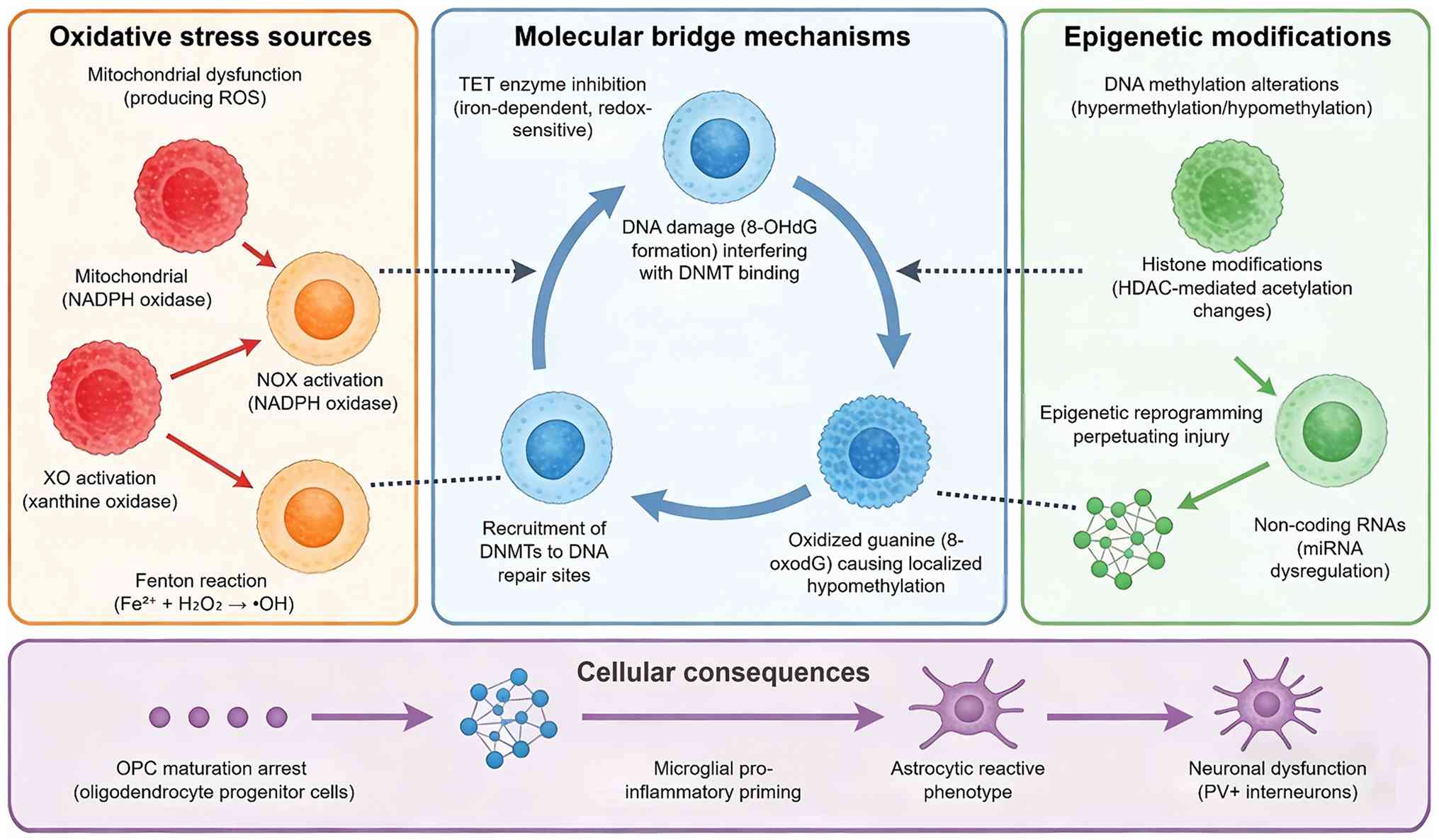 | Figure 3.OS-induced epigenetic dysregulation
and cellular pathologies in preterm brain injury (www.figdraw.com; ID: WWRAP27077). This diagram shows
the molecular linkage between OS and epigenetic modifications in
the preterm brain. ROS/RNS disrupts DNA methylation, histone
modification and microRNA regulation via altering key epigenetic
enzymes. Such persistent epigenetic reprogramming induces abnormal
gene expression, triggers cell-specific pathological injuries and
ultimately leads to long-term neurodevelopmental deficits in
preterm infants. OS, oxidative stress; ROS, reactive oxygen
species; RNS, reactive nitrogen species; 8-OHdG,
8-hydroxy-2′-deoxyguanosine; 8-oxodG,
8-oxo-7,8-dihydro-2′-deoxyguanosine; DNMT, DNA methyltransferase;
GABA, γ-aminobutyric acid; HAT, histone acetyltransferase; HDAC,
histone deacetylase; NOX, NADPH oxidase; NPBI, non-protein-bound
iron; OPC, oligodendrocyte progenitor cell; PV, parvalbumin; TET,
ten-eleven translocation; XO, xanthine oxidase. |
Introduction to epigenetic mechanisms
in neurodevelopment
Epigenetic mechanisms, including DNA methylation,
histone modifications and non-coding (nc)RNAs, are fundamental
regulators of gene expression that operate without altering the
underlying DNA sequence. During brain development, these processes
orchestrate critical events such as neural stem cell proliferation,
neuronal and glial differentiation, synaptogenesis and the
establishment of complex neural circuits (37,38).
DNA methylation, the addition of a methyl group to cytosine
residues in CpG dinucleotides, is typically associated with
transcriptional repression when it occurs in promoter regions and
is dynamically regulated by DNA methyltransferases and ten-eleven
translocation (TET) demethylases (39). Histone modifications, such as
acetylation, methylation and phosphorylation, alter chromatin
structure and gene accessibility, with writers, erasers and readers
of these marks determining the transcriptional output (40,41).
microRNAs (miRNAs) provide an additional layer of
post-transcriptional control by degrading or inhibiting the
translation of target mRNAs (42,43).
The exquisite temporal and spatial control of these processes
render the developing brain particularly susceptible to
environmental perturbations that can disrupt the normal epigenetic
trajectory.
OS as a driver of epigenetic
modifications
OS can directly and indirectly influence the
establishment and maintenance of epigenetic marks. One primary
mechanism involves the modification of enzymes that regulate the
epigenome. For instance, the activity of TET enzymes, which
catalyze DNA demethylation, is dependent on iron and
α-ketoglutarate, making them vulnerable to redox imbalance
(44,45). ROS can also cause DNA damage and
the repair process itself can lead to the recruitment of DNA
methyltransferases to the site of damage, potentially establishing
aberrant methylation patterns (11,46).
Furthermore, ROS can oxidize guanine bases in DNA to form
8-oxo-7,8-dihydroguanine, which can inhibit the binding of
methyltransferases to adjacent CpG sites, leading to localized
hypomethylation (47). Scarpato
et al (11) provided an
overview of how oxidative genome damage can be a determinant of
pathological conditions in the newborn by influencing DNA
methylation. This direct chemical interplay between ROS and the DNA
methylation machinery underscores how an acute oxidative burst can
have lasting consequences for the epigenome.
A critical question that emerges from these
observations is whether the OS-driven epigenetic changes represent
a primary pathogenic mechanism or a secondary epiphenomenon. The
evidence for causality is strongest for TET enzyme inhibition and
8-oxodG formation, where direct chemical mechanisms have been
established in vitro and in animal models. For TET enzymes,
their dioxygenase activity is iron- and α-ketoglutarate-dependent,
rendering them sensitive to redox imbalance. OS has been shown to
upregulate TET1 expression and induce active DNA demethylation in
human cells (48), while TET2
counteracts aberrant DNA methylation during OS through
acetylation-dependent mechanisms (49). For 8-oxodG, this oxidative DNA
lesion accumulates in GC-rich promoters and regulatory regions,
where it can inhibit DNMT binding to adjacent CpG sites, recruit
OGG1 to initiate repair processes and interfere with cytosine
methylation. 8-oxodG has also been shown to interplay with histone
modifications and DNA methylation through complex regulatory
networks (50). However, for most
histone modifications and non-coding RNA changes, the causal chain
from OS to epigenetic mark to functional outcome remains
inferential rather than proven. For histone modifications, OS has
been shown to modulate HDAC activity in a context-dependent manner;
activated microglia can decrease histone acetylation in astrocytes
via increased HDAC activity (51),
while specific HDAC isoforms (HDAC1-11) are expressed in glial
cells and regulate the neuroinflammatory response. For non-coding
RNAs, emerging evidence has revealed complex regulatory networks
involving long non-coding (lnc)RNAs, miRNAs and mRNAs that are
altered in association with inflammatory and OS markers in neonatal
brain injury models, although direct causal relationships remain to
be fully elucidated. Specific examples include miR-34c-5p, which
attenuates apoptosis and OS following hypoxic-ischemic brain injury
in neonatal mice by targeting Arhgap26 (52); miR-210, which promotes microglial
pro-inflammatory activation through reprogramming mitochondrial
metabolism (53); the lncRNA
EVADR, which mitigates neonatal hypoxic-ischemic injury via the
miR-145/WNT/β-catenin axis (54);
and lncRNA GAS5, which prevents mitochondrial apoptosis through the
miR-128-3p/Bax/Akt/GSK-3β axis (55). Circular RNAs (circRNAs) have also
been implicated, with distinct expression patterns observed in the
brain tissues of neonatal rats with periventricular white matter
damage, suggesting that circRNAs actively respond to
hypoxic-ischemic insults (56).
Moreover, the temporal relationship between OS exposure and
epigenetic modification is rarely examined in human studies;
cross-sectional designs cannot distinguish whether methylation
changes precede or follow injury. This ambiguity has practical
consequences: If epigenetic changes are predominantly secondary to
established injury, then targeting them therapeutically may offer
limited benefit; if they are primary drivers, they represent
compelling intervention targets. Future longitudinal studies with
serial sampling from the perinatal period are urgently needed to
resolve this question.
Key epigenetic modifications in
preterm brain injury
A substantial body of research has now linked
specific epigenetic changes to preterm brain injury. DNA
methylation is the most extensively studied mark in this context.
Lorente-Pozo et al (57)
delivered a seminal finding by demonstrating that the oxygen load
supplied during delivery room stabilization of preterm infants
modifies the DNA methylation profile. This work provided direct
clinical evidence that a major source of OS, resuscitation with
high oxygen, can rapidly alter the neonatal epigenome. Similarly,
Rzemieniec et al (44)
showed that perinatal asphyxia in a rodent model led to
hypermethylation of specific genes, an effect that was counteracted
by a neuroprotective compound. More recently, Xu et al
(58) demonstrated that maternal
exposure to multiple metals, a known provocation for OS, is
associated with an increased risk of spontaneous preterm birth, a
relationship potentially mediated by alterations in DNA
methylation. Balasubramanian et al (46) provided mechanistic depth by showing
that traumatic brain injury leads to persistent oxidative damage in
the hippocampus via DNA Methyltransferase 3b-mediated DNA
methylation and silencing of the antioxidant enzyme superoxide
dismutase2. This illustrates a vicious cycle whereby OS-induced
methylation can impair endogenous antioxidant defenses,
perpetuating oxidative injury.
Histone modifications are also emerging as critical
players. The acetylation and deacetylation of histones, controlled
by histone acetyltransferases and deacetylases (HDACs),
respectively, are sensitive to cellular energy and redox states. Ye
et al (41), in a
comprehensive review, highlighted the potential of HDAC inhibitors
as a therapeutic strategy in traumatic brain injury. Experimental
studies support this concept. Liu et al (59) demonstrated that HDAC2 regulates
nuclear factor erythroid 2-related factor 2 (Nrf2) acetylation,
influencing neuronal ferroptosis in neonatal rats with
hypoxic-ischemic brain injury. Furthermore, Xu et al
(60) showed that targeting HDAC3
suppresses ferroptosis and demyelination in white matter injury by
restoring pyruvate dehydrogenase kinase 4-mediated iron
homeostasis. These studies point towards a crucial role for histone
deacetylases in orchestrating the cellular response to OS and
injury. Beyond acetylation, other histone marks are implicated. For
instance, Xiang et al (61)
found that exposure to fluoride, which can induce OS, exacerbates
cognitive deficits in diabetic rats, potentially through mechanisms
involving the poly (ADP-ribose) polymerase-1 pathway, a sensor of
DNA damage that also modifies chromatin structure.
ncRNAs, particularly miRNAs, add another layer of
complexity. These small RNAs can be dysregulated by OS and, in
turn, regulate the expression of genes involved in antioxidant
defense, inflammation and cell survival. For example, Zhou et
al (43) demonstrated that
suppression of the lncRNA metastasis-associated lung adenocarcinoma
transcript 1 alleviated neuronal apoptosis and ROS production
through the miR-499-5p/SRY-box transcription factor 6 axis in a
model of subarachnoid hemorrhage. Similarly, Chen et al
(42) reported that depleting
SRY-box transcription factor 2 improved ischemic stroke outcomes
via the lncRNA plasmacytoma variant translocation
1/microRNA-24-3p/signal transducer and activator of transcription 3
(STAT3) axis. The rapid responsiveness of miRNAs to cellular stress
makes them attractive candidates as both early biomarkers and
therapeutic targets (62).
Cell-type specific epigenetic
responses
It is crucial to recognize that epigenetic responses
to OS are not uniform across all brain cell types. The specific
vulnerability of OPCs, discussed in the previous section, likely
has an epigenetic basis. Kulkarni et al (63) demonstrated that brain trauma
induces DNA methylation-mediated regulation of the Mitofusin 2
gene, which is critical for mitochondrial dynamics, in the brain,
but the cell-specific nature of this regulation requires further
elucidation. Astrocytes also undergo significant epigenetic
reprogramming in response to injury. Li et al (64) showed that alcohol exposure, which
induces OS, activates the Nrf2 pathway and influences the
expression of genes related to glutathione metabolism in
astrocytes, a process with potential epigenetic underpinnings.
Microglia, the brain's resident immune cells, are particularly
prone to epigenetically mediated priming, where an initial insult
leads to a sensitized state and an exaggerated response to a
subsequent challenge. Chen et al (65) recently reported that HDAC1
dysregulation promotes pro-inflammatory microglial activation and
aggravates post-stroke neuroinflammation. This suggests that
OS-induced epigenetic changes in microglia could perpetuate a
chronic neuroinflammatory state, contributing to ongoing white
matter dysmaturation. While much work remains to be conducted, it
is evident that the epigenetic landscape of each cell type dictates
its unique response to the OS caused by preterm birth. The
convergence of evidence from clinical cohorts demonstrating
oxygen-induced methylation changes (57) and preclinical models elucidating
the pathways of HDAC-mediated ferroptosis (59,60)
strongly supports the concept that OS serves as a direct molecular
bridge to enduring epigenetic dysregulation. This paradigm shifts
from viewing OS as merely an acute insult to recognizing it as a
trigger for long-term epigenetic reprogramming opens new avenues
for biomarkers and therapies aimed at mitigating the lifelong
neurodevelopmental consequences of preterm birth.
The direct clinical evidence linking oxygen exposure
to DNA methylation changes in preterm infants is provided by a
prospective cohort study (57),
while most mechanistic insights into histone modifications and
ncRNAs originate from preclinical models (41–44,59–61).
Cellular and molecular players in preterm
brain injury
The cellular responses that define preterm brain
injury, from OPCs vulnerability to microglial activation and
astrocyte reactivity, are not independent phenomena. They are
unified by a common upstream driver, the OS-epigenetic axis
described in the preceding sections. This section therefore
examined each major cell type not as an isolated actor but as a
node within this integrated pathological network. Understanding how
the OS epigenetic cascade manifests in each cell population and how
cell-specific responses in turn feedback to amplify or contain the
injury, is essential for developing targeted neuroprotective
strategies. Table I summarizes
current evidence on the roles of oligodendrocyte lineage cells,
astrocytes, microglia, neurons and the emerging contribution of the
gut-brain axis, providing a cellular framework.
Oligodendrocyte lineage: Primary
victims
OPCs represent the most vulnerable cellular
population in the preterm brain, with their susceptibility
underpinning the predominant white matter injury phenotype.
Srivastava et al (66)
demonstrated that a Toll-like receptor (TLR)/protein kinase B
(AKT)/forkhead box O3 immune tolerance-like pathway specifically
disrupts the repair capacity of OPCs, providing mechanistic insight
into why these cells fail to mature after injury. This maturational
arrest is further exacerbated by inflammatory signals; Boccazzi
et al (67) showed that
TLR3 activation in oligodendrocytes triggers a pro-inflammatory
response that compromises their survival in a murine model of
preterm white matter injury.
The susceptibility extends beyond inflammatory
mediators to excitotoxic mechanisms, as Al-Griw et al
(68) demonstrated that inhibition
of ionotropic glutamate receptor signaling preserves the
oligodendrocyte lineage in an ex vivo model of ischemic
injury. Importantly, the fate of OPCs is not uniformly determined.
Chang et al (69) provided
evidence that intrauterine growth restriction combined with
postnatal hyperoxia induces white matter injury through mechanisms
involving OPCs disruption, while Lin et al (70) characterized the dynamic changes in
oligodendrogenesis following hypoxic-ischemic injury, revealing
temporal windows of vulnerability and limited regenerative
capacity. These findings collectively indicate that OPCs injury
involves multiple converging pathways and the failure of endogenous
repair mechanisms, rather than simply cell death, may be the
critical determinant of long-term dysmyelination. The observation
that OPCs can survive but fail to mature suggests that therapeutic
strategies should target differentiation blockade rather than
solely preventing acute cell loss.
Astrocytes: Double-edged players
Astrocytes exhibit context-dependent responses that
can either contain injury or amplify damage in the preterm brain.
Parfenova et al (71)
demonstrated a protective role, showing that astrocyte-produced
carbon monoxide protects against cerebrovascular dysfunction
following neonatal asphyxia. This beneficial capacity is consistent
with findings from Nobuta et al (72), who reported that STAT3-mediated
astrogliosis protects myelin development by modulating microglial
transforming growth factor beta-1 (TGFβ-1) expression, thereby
preventing impaired oligodendrocyte maturation.
Conversely, astrocytes can adopt pathogenic
phenotypes under certain conditions. Renz et al (73) demonstrated that neuroinflammatory
reactive astrocyte formation correlates with adverse outcomes in
perinatal white matter injury, suggesting that the nature of
astrocytic reactivity determines clinical trajectory. The signaling
pathways governing these divergent responses have been increasingly
characterized. Fang et al (74) showed that fibroblast growth
factor-10 protects the neurovascular unit, including astrocytes, in
neonatal hypoxic-ischemic injury, while Xu et al (60) demonstrated that targeting HDAC3
suppresses ferroptosis and demyelination partly through restoring
PDK4-mediated iron homeostasis. The duality of astrocyte function
(protective under some conditions but detrimental under others)
highlights the need for nuanced therapeutic approaches that
preserve beneficial functions while limiting maladaptive responses.
This balance may be particularly critical in the preterm brain,
where astrocyte development is ongoing and thus highly susceptible
to perturbation.
Microglia: mediators of
neuroinflammation
Microglial cells serve as central orchestrators of
the neuroinflammatory response, with their activation state
critically influencing injury progression and repair (75). Activated microglia produce large
quantities of ROS via NADPH oxidase 2 (NOX2), which can in turn
perpetuate inflammatory signaling, establishing a feed-forward loop
between OS and neuroinflammation (8). Yang et al (76) demonstrated that caffeine treatment,
initiated before injury, reduces hypoxic-ischemic white matter
damage by regulating phenotypic microglial polarization, shifting
cells toward a less inflammatory state. This concept of microglial
plasticity is supported by Charriaut-Marlangue et al
(77), who identified sex
differences in microglial phenotypes following neonatal stroke,
with females showing distinct responses to poly (ADP-ribose)
polymerase inhibition compared with males.
The signaling pathways regulating microglial
reactivity have been extensively investigated. Zaghloul et
al (78) reported that
prophylactic inhibition of NF-κB expression in microglia attenuates
hypoxic-ischemic injury, while Mairesse et al (79) showed that oxytocin receptor agonist
reduces perinatal brain damage by directly targeting microglia. Van
Steenwinckel et al (80)
provided mechanistic depth by demonstrating that decreased
microglial Wnt/β-catenin signaling drives pro-inflammatory
activation in the developing brain. The convergence of these
studies suggests that microglial responses are not binary but
represent a spectrum of activation states and the capacity to
modulate these states, either pharmacologically or through
endogenous pathways, offers therapeutic potential. Importantly, the
work of Bernis et al (81),
which temporally characterized microglia-associated gene expression
in a sensitized hypoxia-ischemia model, emphasizes that the timing
of microglial modulation may be as critical as the specific
intervention.
Neurons: Targets of injury
Although white matter injury predominates in preterm
encephalopathy, neuronal populations are also markedly affected,
contributing to long-term cognitive and motor deficits. Sathyanesan
et al (82) demonstrated
that neonatal brain injury causes cerebellar learning deficits and
Purkinje cell dysfunction, revealing that the cerebellum is a
previously underappreciated target. This finding aligns with
subsequent work from the same group showing that disruption of
neonatal Purkinje cell function underlies injury-related learning
deficits (83). Cortical neuronal
populations are similarly vulnerable. Scheuer et al
(26) provided evidence that
neonatal OS impairs cortical synapse formation and GABA homeostasis
in parvalbumin-expressing interneurons, a finding extended by
Lacaille et al (84) who
demonstrated impaired interneuron development in a novel model of
neonatal brain injury. Sheikh et al (85) showed that neonatal hypoxia-ischemia
causes functional circuit changes in subplate neurons, which serve
as crucial relay stations during cortical development.
The cholinergic system also appears susceptible;
Northington et al (86)
reported that basal forebrain magnocellular cholinergic systems are
damaged following neonatal hypoxia-ischemia in mice. These diverse
neuronal vulnerabilities suggest that preterm brain injury
represents a multi-system disorder affecting multiple
neurotransmitter systems and brain regions. The recognition that
interneurons, particularly parvalbumin-positive subtypes, are
consistently affected provides a plausible cellular basis for the
long-term neuropsychiatric sequelae observed in preterm
survivors.
Neurovascular unit
The concept of the neurovascular unit emphasizes
that injury to any component (endothelium, pericytes, astrocytes,
or neurons) affects the integrity of the whole. Lee et al
(87) demonstrated that
hypoxia-preconditioned human umbilical vein endothelial cells
protect against neurovascular damage following hypoxic ischemia,
suggesting that enhancing endothelial resilience confers broader
neuroprotection. Chand et al (88) showed that neurovascular unit
alterations in growth-restricted newborns are improved following
ibuprofen treatment, linking hemodynamic modulation to structural
protection. Fang et al (89) provided additional evidence that
chloroquine protects against neonatal brain injury by mitigating
blood-brain barrier disruption, while Wang et al (90) demonstrated that oligogenesis in the
‘oligovascular unit’ involves PI3K/AKT/mTOR signaling. The
interdependence of these cellular components has important
therapeutic implications: Interventions targeting a single cell
type may fail if other components remain compromised, suggesting
that multimodal approaches addressing the entire neurovascular unit
may be necessary.
Gut-brain axis in preterm brain
injury
Emerging evidence implicates the gut microbiota as a
significant modulator of brain injury in preterm infants. Seki
et al (91) provided
seminal findings by demonstrating that aberrant
gut-microbiota-immune-brain axis development occurs in premature
neonates with brain damage, with Klebsiella overgrowth
highly predictive of injury and associated with a pro-inflammatory
immunological tone. This clinical observation is supported by
experimental work from Drobyshevsky et al (92), who showed that intestinal
microbiota modulates neuroinflammatory response and brain injury
following neonatal hypoxia-ischemia. Liu et al (93) extended these findings using
multi-omics analyses, demonstrating aberrant microbiota-gut-brain
axis in very low birth weight infants with white matter injury.
Vaher et al (94) recently
confirmed that the neonatal gut microbiota plays a role in the
encephalopathy of prematurity, with specific microbial profiles
associated with adverse neurodevelopmental outcomes. Seki et
al (95) subsequently
identified gut microbiota genome features associated with brain
injury, suggesting that the mechanistic basis for these
associations likely involves microbial metabolites and immune
modulation. This evolving field opens novel therapeutic
possibilities, including probiotic interventions and dietary
modulation, although causal relationships require further
validation. The recognition that the gut-brain axis operates during
the neonatal period challenges traditional conceptualizations of
brain injury as a purely CNS-limited process and suggests that
systemic interventions may have central consequences.
The cellular landscape of preterm brain injury
reveals a highly interconnected system in which the response of any
single cell type influences and is influenced by others. This
complexity probably explains why single-target interventions have
shown limited clinical success and supports the need for approaches
that consider the multicellular nature of injury and repair.
Critically, the OS epigenetic axis provides the mechanistic thread
that connects these disparate cellular responses: OS modifies the
epigenome of each cell type in a context-specific manner and these
epigenetic changes, in turn, dictate divergent outcomes ranging
from OPCs maturational arrest to astrocyte reactivity and
microglial priming. Thus, the cellular diversity of preterm brain
injury is not a random assortment of pathologies but a spectrum of
cell-type-specific manifestations of a common molecular
derangement. However, the cell-type specificity of epigenetic
responses also poses a major translational challenge: Interventions
that modify the epigenome globally may have unintended consequences
in cell types where the same epigenetic mark serves a protective
rather than pathogenic function. For example, HDAC inhibition may
benefit oligodendrocyte survival while simultaneously promoting
astrocyte reactivity or microglial priming, depending on the
cellular context and timing of intervention. The responses of
astrocytes and microglia to HDAC inhibition are highly
context-dependent, varying with the specific HDAC inhibitor used,
the cell type and the inflammatory milieu. In pure microglial
cultures, HDAC inhibitors such as trichostatin A (TSA) and
suberoylanilide hydroxamic acid can potentiate LPS-induced
pro-inflammatory cytokine and nitric oxide responses (96). Conversely, in astrocyte-rich
cultures exposed to activated microglial factors, TSA and valproic
acid restored histone acetylation, upregulated the Nrf2-inducible
antioxidant defense and conferred protection against OS (51). In a neonatal rat model of white
matter injury, the selective HDAC2/3 inhibitor MI192 administered
post-insult markedly reduced oligodendrocyte loss and suppressed
microglial activation (97). These
findings collectively illustrate that the timing of HDAC inhibition
and the specific cell types targeted critically determine
therapeutic outcomes, whether the inhibitor is administered
prophylactically, immediately after injury, or during the repair
phase. This duality underscores the need for cell-type-specific
delivery strategies or interventions targeted to the relevant
epigenetic enzymes in a temporally restricted manner. It must be
emphasized, however, that the majority of mechanistic insights
discussed in this section derive from preclinical models (rodent
and ex vivo systems). While these findings are invaluable
for understanding pathogenesis, their translation to the human
preterm brain requires confirmation in clinical cohorts.
Clinical evidence for the gut-brain axis in preterm
brain injury comes from prospective cohorts and case-control
studies (91,93–95),
whereas the majority of cellular response data (oligodendrocyte,
astrocyte, microglia, neuron) are derived from animal models. See
Table I for full
classification.
Intercellular crosstalk in the preterm
brain
The pathological responses of individual cell types
in preterm brain injury do not occur in isolation; rather, they are
shaped by dynamic intercellular communication networks that
collectively determine injury progression and repair capacity.
Microglia and astrocytes engage in bidirectional crosstalk that
profoundly influences the neuroinflammatory milieu. Activated
microglia can decrease histone acetylation in astrocytes via
increased HDAC activity, thereby impairing astrocytic antioxidant
defenses and perpetuating OS (51). Conversely, astrocytes modulate
microglial phenotype through STAT3-dependent mechanisms; Nobuta
et al (72) demonstrated
that STAT3-mediated astrogliosis protects myelin development by
preventing pathological TGFβ-1 expression in microglia, which would
otherwise inhibit oligodendrocyte progenitor maturation. This
protective astrocyte-microglia interaction is critical, as TGFβ-1
directly and dose-dependently inhibits the maturation of purified
oligodendrocyte progenitors (11).
Astrocytes also exert direct effects on
oligodendrocyte lineage cells. Shiow et al (98) showed that reactive astrocytes in
neonatal white matter injury develop ‘A2’ reactivity and produce
COX2-derived prostaglandin E, which acts directly on
oligodendrocyte progenitor cells via the EP1 receptor to block
their maturation. In vivo inhibition of COX2 rescues
hypomyelination and behavioral impairment, demonstrating that
astrocyte-derived inflammatory mediators are functionally
significant drivers of OPC maturation arrest (98).
Microglia-oligodendrocyte interactions are equally
critical. Inflammatory activation of microglia releases cytokines
and ROS that directly impair oligodendrocyte survival and
maturation (8,30). Conversely, protective microglial
phenotypes can support oligodendrocyte development; M2
microglia-derived exosomal miR-144-5p has been shown to promote OPC
growth and reduce white matter injury by regulating the PTEN/AKT
pathway (99). The work of Bernis
et al (81) further
emphasizes that the temporal dynamics of microglial activation,
shifting from pro-inflammatory to anti-inflammatory gene expression
over time, determine whether microglia exert harmful or beneficial
effects on oligodendrocyte lineage cells.
The neurovascular unit represents another critical
interface for intercellular crosstalk. Endothelial cells,
pericytes, astrocytes and neurons communicate through reciprocal
signaling that maintains blood-brain barrier integrity and
regulates cerebral blood flow. Lee et al (87) demonstrated that
hypoxia-preconditioned endothelial cells confer broader
neuroprotection, suggesting that enhancing the resilience of one
cellular component benefits the entire unit. Wang et al
(90) showed that oligogenesis in
the ‘oligovascular unit’ involves PI3K/AKT/mTOR signaling,
illustrating how vascular and glial compartments coordinately
regulate white matter repair. Collectively, these findings
establish that preterm brain injury is not a cell-autonomous
process but a network disorder in which the responses of microglia,
astrocytes, oligodendrocytes and vascular cells are intimately
interconnected through reciprocal signaling pathways. This
interconnectedness provides a mechanistic explanation for why
single-cell-type interventions have shown limited success and
supports the rationale for multimodal therapeutic strategies that
target the broader cellular network.
Neuroprotective strategies: Current evidence
and emerging approaches
The preceding sections have elucidated the complex
cascade from OS to epigenetic dysregulation in preterm brain
injury, involving multiple cellular populations and interconnected
pathological pathways. To impose conceptual order on the diverse
array of neuroprotective strategies currently under investigation,
this section organised interventions according to which node of the
OS epigenetic cascade they target: Prevention of OS generation
(antioxidants, oxygen management); blockade of epigenetic
modification (HDAC inhibitors, miRNA modulators); or modulation of
downstream cellular responses (anti-inflammatory agents, cell-based
therapies). This complexity poses significant challenges for
neuroprotection, as interventions targeting single mechanisms may
prove insufficient. It is important to distinguish between
preclinical promise and clinical evidence: while numerous agents
demonstrate efficacy in animal models, the number that have been
rigorously tested in adequately powered human trials remains small
and the translational gap is substantial. The following sections
critically evaluate current and emerging neuroprotective
strategies, with particular attention to the strength of evidence
and areas of ongoing controversy. A summary of these strategies,
categorized by mechanism and evidence level, is presented in
Table II.
Antioxidant-based strategies
Melatonin has been extensively investigated as an
antioxidant neuroprotectant. Carloni et al (100) characterized melatonin
pharmacokinetics following oral administration in preterm neonates,
demonstrating significant interindividual variability that
complicates dosing optimization. Domínguez Rubio et al
(101) reported that maternal
melatonin administration exerted both short- and long-term
neuroprotective effects in offspring from
lipopolysaccharide-treated mice. Garofoli et al (102) recently confirmed that orally
administered melatonin reaches systemic circulation and reduces OS
markers in preterm newborns, although clinical impact on
neurodevelopmental outcomes awaits definitive trials. Lee et
al (103) demonstrated in a
mouse model that melatonin administration prevented preterm birth
and associated fetal brain injury. However, the systematic review
by Ahmed et al (104)
concluded that while safety appears favorable, evidence for
neuroprotection remains insufficient due to small sample sizes and
heterogeneity in dosing and timing. A more recent review by Häusler
et al (105)
systematically examined the pathomechanisms underlying preterm
brain injury and correlated them with melatonin's neuroprotective
potential, while emphasizing significant pharmacokinetic and
pharmacodynamic uncertainties that must be addressed before
clinical translation can be realized. Favrais et al
(106) showed only partial
protective effects of melatonin in a rat model of chorioamnionitis,
suggesting that monotherapy may be inadequate against
multifactorial injury. Other antioxidants, including
N-acetylcysteine (107), vitamin
C (108) and uridine (109), have shown preclinical promise but
limited clinical translation. Chen et al (110) recently demonstrated that
targeting mitochondrial OS through Nrf2 induction protects against
preterm birth and fetal brain injury in experimental models,
although clinical validation is awaited. Beyond these agents,
emerging preclinical evidence continues to expand the repertoire of
potential antioxidant neuroprotectants. Gunduz et al
(111) recently demonstrated that
polydatin, a natural polyphenolic compound, exerts dose-dependent
protective effects against sodium fluoride-induced oxidative and
inflammatory damage in rats, as evidenced by restored antioxidant
enzyme activities (glutathione, catalase, superoxide dismutase),
reduced lipid peroxidation (malondialdehyde) and suppressed
pro-inflammatory gene expression (TNF-α, NF-κB, IFN-γ). Although
this study was conducted in a fluoride-toxicity model rather than a
perinatal brain injury context, it reinforces the broader principle
that pharmacological agents capable of restoring redox balance and
modulating inflammatory pathways hold therapeutic potential for
conditions characterized by OS. Importantly, polydatin has also
been shown to exert neuroprotective effects in neonatal rat models
of hypoxic-ischemic brain injury, improving learning and memory
impairments through upregulation of brain-derived neurotrophic
factor (112). These findings
suggest that polydatin warrants further investigation as a
candidate neuroprotective agent in the preterm population, although
rigorous preclinical optimization and clinical translation studies
are required before any clinical recommendations can be made.
Targeting specific molecular
pathways
Erythropoietin (EPO) represents the most
extensively studied pathway-targeted agent. Fischer et al
(113) performed a meta-analysis
concluding that while EPO reduces transfusions, evidence for
improved neurodevelopmental outcomes was inconclusive. The landmark
Preterm Erythropoietin Neuroprotection Trial (PENUT) trial by Juul
et al (114) randomized
941 extremely preterm infants to high-dose EPO or placebo and found
no significant difference in the primary outcome of mortality or
severe neurodevelopmental impairment at 2 years between the two
groups. Ehrenreich et al (115) emphasized that negative findings
highlight the importance of patient selection and dosing rather
than refuting EPO's potential. By contrast, Song et al
(116) reported that EPO improved
outcomes specifically in preterm infants with intraventricular
hemorrhage, suggesting subgroup-specific benefits. Jakab et
al (117) used network-based
statistics to reveal trophic effects of early high-dose EPO on
brain connectivity in very preterm infants. Wassink et al
(118) showed that prolonged EPO
infusion provided partial white and grey matter protection after
asphyxia in preterm fetal sheep. Ma and Shi (119) comprehensively reviewed cellular
mechanisms of EPO neuroprotection, concluding that clinical
translation requires improved patient stratification. The
discrepancy between the neutral PENUT trial and positive
preclinical and subgroup studies likely reflects several factors:
The PENUT trial administered EPO at 24 h of life, whereas
preclinical data suggest earlier administration (within 6 h) may be
more effective; the extremely high baseline rate of favourable
outcomes in the control group (79.6% survival without impairment)
limited statistical power to detect a benefit; and the trial
excluded infants with significant prior brain injury, potentially
removing the population most likely to respond (114,119). These observations suggest that
EPO may benefit specific subgroups, particularly infants with
established injury or those treated earlier, rather than providing
universal neuroprotection and that future trials should stratify by
injury severity and timing of intervention. The conflicting results
between consistently positive preclinical models and largely
neutral clinical trials underscore the limitations of animal models
and the critical importance of adequately powered trials.
Caffeine has emerged as a candidate neuroprotective
agent. Endesfelder et al (120) demonstrated that caffeine provides
neuroprotection against hyperoxia-induced neonatal brain injury in
rats. Yang et al (76)
showed that caffeine treatment reduced hypoxic-ischemic white
matter damage through regulation of phenotypic microglia
polarization. Di Martino et al (121) defined a therapeutic window for
caffeine neuroprotection, showing that early administration is
critical for efficacy. A recent network meta-analysis by Wang et
al (122) comparing caffeine,
erythropoietin, magnesium sulfate and thyroxine found that caffeine
showed the most promising effect in reducing the risk of
neurodevelopmental impairment [relative risk (RR): 0.43, 95%
confidence interval (CI): 0.22–0.86] and cerebral palsy (RR: 0.55,
95% CI: 0.43–0.70) in preterm infants. These preclinical data,
combined with caffeine's established safety profile, support its
continued use, although prospective trials with neurodevelopmental
primary outcomes are lacking. Emerging pathway-targeted strategies
include HDAC modulation, with Liu et al (59) demonstrating that HDAC2 regulates
Nrf2 acetylation and thereby influences neuronal ferroptosis.
Peroxisome proliferator-activated receptor gamma (PPAR-γ)
activation has also been investigated, with Fang et al
(123) demonstrating inhibition
of microglia-mediated neuroinflammation and Feng et al
(124) showing promotion of
oligodendrocyte precursor cell differentiation. All remain at
preclinical stages.
Anti-inflammatory strategies
Beyond direct antioxidant and pathway-targeted
approaches, modulating the inflammatory cascade itself represents a
distinct and complementary therapeutic avenue, given that
neuroinflammation both amplifies oxidative injury and operates
through partially independent molecular networks (125). Shin et al (30) demonstrated that TNF-α antagonist
attenuated lipopolysaccharide-induced white matter injury in
neonatal rats. Prasad et al (126) comprehensively reviewed
anti-inflammatory therapies, examining agents including TNF-α
antagonists (such as etanercept), minocycline, COX-2 inhibitors,
melatonin and IL-1 receptor antagonists. While numerous agents show
preclinical efficacy, clinical translation has been limited by the
complexity of the inflammatory cascade and the difficulty of timing
interventions appropriately. The inflammatory cascade involves
sequential activation of multiple mediators: peripheral immune
responses trigger central neuroinflammation characterized by
proinflammatory cytokine release (TNF-α, IL-1β, IL-6), microglial
activation and reactive gliosis, which in turn activate NADPH
oxidase enzymes generating ROS, establishing a feed-forward loop
between OS and inflammation (8,30).
The temporal heterogeneity of microglial responses, shifting from
pro-inflammatory to anti-inflammatory gene expression over time,
further complicates therapeutic targeting (80). Different anti-inflammatory agents
require distinct therapeutic windows: etanercept administered at
three days after hypoxia-ischemia ameliorates white matter injury
(30); early postnatal caffeine
treatment confers improved neuroprotection than late treatment
(76,120,121); minocycline requires early
post-insult administration to target secondary neuroinflammation;
and dexamethasone exhibits a developmentally sensitive therapeutic
window (126). These temporal
constraints, combined with the difficulty of predicting the onset
and progression of inflammation in individual preterm infants, pose
substantial barriers to clinical translation.
IL-1 has emerged as a promising target, with
Takahashi et al (127)
demonstrating that pharmacological IL-1 receptor blockade
suppressed lipopolysaccharide-induced neuroinflammation in preterm
fetal sheep. Van Steenwinckel et al (80) identified decreased microglial
Wnt/β-catenin signaling as a driver of pro-inflammatory activation.
Bokobza et al (128)
demonstrated that miR-146b protects against microglia-induced
hypomyelination, suggesting that microRNA-based therapies could
modulate neuroinflammation more selectively. The temporal
heterogeneity of microglial responses, emphasized by Bernis et
al (81), implies that
anti-inflammatory interventions must be precisely timed to avoid
interfering with beneficial microglial functions.
Clinical care strategies to reduce
OS
Oxygen management is paramount. Rantakari et
al (129) demonstrated that
both low and high oxygen saturations are associated with adverse
outcomes. The targeted oxygen for resuscitation of preterm infants
and their developmental outcomes (TORPIDO) 30/60 trial randomized
1,469 preterm infants and found no significant association between
initial FiO2 concentration (0.6 vs. 0.3) for resuscitation and
death or brain injury at 36 weeks' corrected gestational age
(130). Engur et al
(131) provided mechanistic
insight by demonstrating that supplemental oxygen alters the
pentose phosphate pathway through sirtuin signaling.
Developmental care practices have been codified
into neuroprotective care bundles. Milette et al (132) published guidelines for
institutional implementation of developmental neuroprotective care.
Murthy et al (133)
evaluated a neuroprotection care bundle, reporting reduced
intraventricular hemorrhage rates following implementation. Travis
et al (134) demonstrated
that skin-to-skin holding is associated with improved white matter
microstructure. Nutritional strategies have also been investigated.
Belfort and Inder (135) reviewed
the relationship between human milk and brain development,
concluding that breastfeeding is associated with improved outcomes.
Brandt et al (136)
demonstrated that human milk oligosaccharides improve white matter
and interneuron development in a rat model. These strategies offer
low cost and wide availability, but definitive evidence of
neuroprotective efficacy remains limited.
Multimodal neurocritical care
Multimodal neuromonitoring has become a cornerstone
of neurocritical care in preterm infants, enabling early detection
of physiological instability and evolving brain injury before
irreversible damage occurs. El-Dib et al (137) provided a comprehensive framework
for integrating multiple monitoring modalities, including aEEG,
near-infrared spectroscopy (NIRS) and magnetic resonance imaging
(MRI), to detect cerebral dysfunction across different
physiological domains. Among these, aEEG has emerged as a
particularly valuable bedside tool. Variane et al (138) demonstrated that early aEEG
monitoring effectively identifies neonates at high risk for brain
injury, facilitating timely intervention. Griesmaier et al
(139) further validated the
prognostic utility of aEEG by demonstrating significant
associations between aEEG patterns and MRI defined injury severity
at term equivalent age, establishing a correlation between
functional and structural assessments that enhances clinical
interpretation. Beyond electrophysiological assessment, Lin et
al (140) integrated aEEG
with General Movements assessment, showing that combining
neurophysiological and motor function evaluations provides a more
comprehensive prediction of neural recovery than either modality
alone. Despite these advances, the clinical value of multimodal
monitoring ultimately depends on the availability of effective,
evidence-based interventions to act upon the detected
abnormalities. The integration of real time neuromonitoring data
with AI-driven decision support systems and biomarker panels
represents a promising avenue for translating early detection into
improved outcomes, although prospective validation in large cohorts
is still needed.
Cell-based therapies represent an emerging
frontier. Drommelschmidt et al (141) demonstrated that mesenchymal stem
cell-derived extracellular vesicles ameliorate inflammation-induced
preterm brain injury. Thomi et al (142) showed that intranasally
administered exosomes reduce microglia-mediated neuroinflammation.
Ahn et al (143) conducted
a phase I dose-escalation clinical trial of mesenchymal stem cells
for severe intraventricular hemorrhage, demonstrating feasibility
and safety. However, the Cochrane systematic review by Romantsik
et al (144) sought to
include randomized controlled trials comparing stem cell-based
interventions vs. control for the prevention (in infants <28
weeks' gestation, ≤24 h of age, without ultrasound diagnosis) or
treatment (in infants <37 weeks' gestation, with
ultrasound-confirmed germinal matrix-intraventricular hemorrhage or
encephalopathy of prematurity) of these conditions. However, no
completed studies met the inclusion criteria; only three ongoing
trials were identified. The authors concluded that no evidence is
currently available to evaluate the benefits and harms of stem
cell-based interventions for treatment or prevention of these
conditions in preterm infants (144). Tscherrig et al (145) identified specific miRNAs from
mesenchymal stromal cell extracellular vesicles that rescue white
matter injury. Vaes et al (146) demonstrated that modifying the
secretome prolongs the regenerative treatment window. Despite
extensive preclinical literature, the gap between experimental
promise and clinical evidence remains substantial, necessitating
rigorous trial design before cell-based therapies enter routine
practice. Viewed through the OS epigenetic lens, the therapeutic
landscape becomes coherent: Antioxidants and oxygen management aim
to reduce the upstream trigger; HDAC inhibitors and miRNA-based
approaches target the epigenetic machinery that translates
oxidative insults into persistent cellular dysfunction; and
cell-based therapies seek to modulate the downstream cellular
responses that determine repair vs. injury. This conceptual
taxonomy underscores that effective neuroprotection will likely
require multimodal strategies that address multiple nodes of the
integrated OS epigenetic cellular pathway, timed to the
developmental window of vulnerability.
In synthesizing the neuroprotective evidence,
several points of consensus emerge: Melatonin and erythropoietin
have been the most extensively studied agents, both showing
favourable safety profiles but inconsistent efficacy; caffeine has
a strong preclinical rationale and established clinical safety; and
cell-based therapies hold substantial promise but remain
experimental. However, major controversies and unresolved questions
persist. The most critical is the translational gap between
consistently positive preclinical findings and largely neutral or
inconclusive clinical trials, a discrepancy that may reflect
differences in timing, dosing, patient selection, or the inherent
limitations of animal models. There is also no consensus on whether
combination therapy (such as antioxidants plus anti-inflammatory
agents) is superior to monotherapy, or whether interventions should
be targeted to specific injury subtypes. Emerging preclinical
evidence has begun to explore multimodal combination strategies.
For instance, combination therapy with sulforaphane (an Nrf2
activator with NF-κB inhibitory properties) and deferiprone (an
iron chelator) has been shown to mitigate M1 microglial
infiltration, suppress inflammation and reduce ferroptosis in a
neonatal rabbit model of intraventricular hemorrhage (147). Combined omega-3 polyunsaturated
fatty acid and folic acid supplementation has demonstrated enhanced
neuroprotection against neonatal hypoxic-ischemic brain injury
through synergistic anti-inflammatory and anti-apoptotic mechanisms
(148). The combination of
erythropoietin and melatonin has shown sustained repair of
functional deficits in a rat model of cerebral palsy following
preterm brain injury (149), with
ongoing clinical trials evaluating the safety of this combinatorial
approach in very preterm infants with intraventricular hemorrhage
or white matter injury. N-acetylcysteine combined with vitamin D
has been shown to decrease OS in plasma and central nervous system
and is associated with favorable developmental outcomes in neonatal
hypoxic-ischemic encephalopathy models (150). Furthermore, allopurinol (a
xanthine oxidase inhibitor) is currently being evaluated in the
phase III ALBINO trial as an adjunct to therapeutic hypothermia for
hypoxic-ischemic brain injury (17). These emerging combination
approaches, although primarily at preclinical or early clinical
stages, highlight the potential for multimodal strategies targeting
both oxidative and inflammatory pathways to achieve superior
neuroprotection compared with monotherapy. Furthermore, the optimal
window for intervention, before, during, or after the oxidative
insult, has not been established for most agents. Future research
must prioritise adequately powered, biomarker-stratified trials
with long-term neurodevelopmental follow-up to resolve these
uncertainties and translate preclinical promise into clinical
reality.
The clinical evidence base for neuroprotective
agents is strongest for caffeine [network meta-analysis of
randomized controlled trials (RCTs)], erythropoietin (PENUT RCT and
cohort studies) and oxygen titration (TORPIDO RCT), whereas
melatonin, HDAC inhibitors, PPAR-γ agonists and cell-based
therapies remain largely at preclinical or early-phase clinical
stages (see Table II for detailed
evidence levels and model systems). Several systematic reviews and
meta-analyses have amalgamated the evidence for neuroprotective
agents in preterm infants (105,122,144). A Cochrane review on stem
cell-based interventions concluded that no completed RCTs met
inclusion criteria, highlighting the substantial gap between
preclinical promise and clinical evidence (144). Network meta-analysis data suggest
caffeine may be the most promising agent for reducing
neurodevelopmental impairment and cerebral palsy risk (122), although this finding derives from
indirect comparisons and requires confirmation in prospective
trials. For melatonin, a systematic review consistently identified
small sample sizes and dosing heterogeneity as major limitations
precluding definitive conclusions (105). Formal guidelines from
international bodies provide specific recommendations for
neuroprotective interventions. The American Academy of Pediatrics
recommends therapeutic hypothermia for neonates with
moderate-to-severe hypoxic-ischemic encephalopathy born at ≥36
weeks' gestation, initiated within 6 h of birth and continued for
72 h (151). UK national
guidelines recommend antenatal magnesium sulfate for mothers in
preterm labour (<30 weeks' gestation) to reduce the risk of
cerebral palsy (152). The
European Resuscitation Council 2025 guidelines provide
evidence-based recommendations for newborn life support, including
neuroprotective strategies during transition at birth (153). Additionally, structured
neuroprotection care bundles have been shown to markedly reduce
death or severe brain injury in extremely preterm infants (133). Despite these established
guidelines, formal consensus from the American Academy of
Pediatrics or European bodies specifically addressing
pharmacological neuroprotection for preterm brain injury beyond
therapeutic hypothermia and magnesium sulfate remains limited.
Artificial intelligence (AI) in preterm
brain injury: Prediction, diagnosis and monitoring
AI has emerged as a transformative tool in neonatal
care, offering novel approaches to predict, diagnose and monitor
preterm brain injury. As summarised in Table III, AI applications now span
neuroimaging analysis, electrophysiological signal processing and
multimodal risk stratification, each contributing uniquely to the
evolving landscape of neurocritical care.
AI in neuroimaging analysis
AI has substantially enhanced neuroimaging
interpretation accuracy and efficiency. Ahmad et al
(154) demonstrated that deep
learning models could classify cranial ultrasound images for brain
injury detection in very preterm infants, achieving performance
comparable to expert clinicians. Chen et al (155) developed an automated neonatal
nnU-Net brain MRI extractor trained on a large multi-institutional
dataset, showing robust generalizability across different clinical
settings. For subtle pathology, Estermann et al (156) introduced CACTUS, a multiview
classifier for detecting punctate white matter lesions in cranial
ultrasound volumes, addressing a diagnostically challenging task
with significant neurodevelopmental implications. These advances
suggest AI-based image analysis has substantial potential to
enhance neuroimaging interpretation, although protocol variability
across institutions, limited external validation and the absence of
prospective clinical trials remain important barriers to clinical
deployment.
AI-enhanced neuromonitoring
Continuous neuromonitoring generates vast
physiological data ideally suited for AI-based analysis. Wang et
al (157) conducted a
landmark cohort study demonstrating that quantitative features from
early amplitude-integrated electroencephalography predicted
long-term neurodevelopmental outcomes in extremely preterm infants,
with automated feature extraction performing equivalently to
combined qualitative and quantitative assessments. Abbasi et
al (158) developed deep
learning models for seizure detection using wavelet-scalogram
approaches in preclinical models, subsequently refined by Roozbehi
et al (159) using
transformer-based architectures. Ashoori et al (160) applied machine learning to NIRS
signals, demonstrating that data-driven definitions of prolonged
desaturation outperformed traditional threshold-based approaches
for detecting intraventricular hemorrhage. Hibner et al
(161) extended this concept by
developing a multimodal approach combining echocardiography, NIRS
and electrical cardiometry, showing integrated signal analysis
provides complementary information unavailable from any single
modality.
Predictive modeling for brain injury
risk
AI-based predictive models have shown considerable
promise for early risk stratification. He et al (162) developed PBIPred, an explainable
machine learning model using seven clinical features that achieved
an area under the curve of 0.8229 for predicting preterm brain
injury, with Shapley Additive Explanations rendering predictions
interpretable at individual patient level. For intraventricular
hemorrhage specifically, Yang et al (163) conducted a nationwide multicenter
study demonstrating robust prediction using machine learning. Han
et al (164) incorporated
time-series analysis of physiological data, showing that temporal
dynamics improve prediction compared with static variables alone.
For white matter injury, Song et al (165) developed two risk assessment
models in extremely preterm infants, demonstrating that ensemble
methods may outperform single algorithms. Shu et al
(166) applied machine learning
to predict mortality and multiple morbidities simultaneously,
showing comprehensive outcome prediction is feasible using
routinely collected data.
AI in prognostication and outcome
prediction
Long-term neurodevelopmental prognostication
represents the most clinically challenging AI application. Li et
al (167) demonstrated that
functional connectivity measures predict outcomes in preterm
infants without severe brain injury, suggesting subtle network
alterations captured by AI analysis identify risk even with normal
conventional imaging. Liu et al (168) used graph convolutional neural
networks to predict brain age, showing deviations from normative
maturation trajectories explain outcomes more accurately than
static injury assessments. For cerebral palsy, Marinelli et
al (169) compared
predictions at ages 2 and 10 years, revealing that early
predictions require refinement as children mature. Yuan et
al (170) developed a
nomogram for predicting intellectual disability, providing a
practical tool for early intervention targeting. Despite these
advances, two recent systematic reviews identify critical
limitations in AI applications for neonatal care. Ortega-Leon et
al (171) reported that
>70% of studies lacked a validation procedure, highlighting a
critical gap in methodological rigor in their systematic review of
machine learning techniques for predicting neurodevelopmental
impairments in premature infants. Similarly, Martinez-Millana et
al (172) found that among AI
studies in the neonatal intensive care unit setting, the majority
lacked external validation, limiting generalisability to new
clinical settings. These limitations suggest that while AI
applications have progressed from proof-of-concept to clinical
validation, generalisability remains the principal barrier to
widespread adoption.
Despite the proliferation of AI studies, several
fundamental limitations impede clinical translation. First,
external validation, the critical step for demonstrating
generalizability, is performed in <30% of published models and
performance consistently declines in cross-institutional settings
(154,173). Second, heterogeneity in imaging
protocols, EEG configurations and clinical data definitions across
centers creates substantial challenges for model generalization
(155,157). Third, the ‘black-box’ nature of
numerous deep learning algorithms, combined with poor clinical
interpretability, undermines clinician trust and hinders bedside
adoption (162). Fourth, the
absence of prospective impact studies, trials demonstrating that
AI-assisted decision-making actually improves patient outcomes
rather than merely achieving statistical accuracy, represents the
most critical evidence gap (173). These limitations suggest that
while AI applications have progressed from proof-of-concept to
clinical validation, generalizability and implementation remain the
principal barriers to widespread adoption.
Integration and future directions
The convergence of mechanistic insights into OS and
epigenetic dysregulation with advances in neuroimaging,
electrophysiological monitoring and AI heralds a transformative era
in the care of preterm infants at risk for brain injury. This
convergence is not coincidental but logically coherent: The OS
epigenetic axis provides the mechanistic rationale that unifies the
diverse cellular pathologies and therapeutic targets discussed in
preceding sections, while AI and multimodal monitoring provide the
tools to detect, quantify and track this axis in real time at the
bedside. In this framework, molecular pathophysiology supplies the
biological targets and explanatory mechanisms; AI-based predictive
tools enable early identification of at-risk infants and dynamic
risk stratification; and clinical translation operationalizes these
insights into preventive and therapeutic strategies. The synergy
among these three components, mechanism, prediction and
intervention, constitutes the unifying conceptual framework that
transforms a descriptive understanding of preterm brain injury into
an actionable, precision-based clinical paradigm. Specifically, the
OS-epigenetic axis and its cell-type-specific consequences (such as
OPC maturational arrest, microglial priming) serve as the core
biological targets supplied by molecular pathophysiology; AI tools
such as the PBIPred model (CatBoost with SHAP explanations),
convolutional neural networks for aEEG maturational age estimation
and multimodal monitoring integration (NIRS, aEEG,
echocardiography) provide predictive and monitoring capabilities;
these have been clinically translated into early risk
stratification, personalized bedside monitoring and
decision-support tools, although prospective impact validation
remains ongoing (162,174). The recognition that early-life
perturbations can programme long-term neurological outcomes,
potentially extending even to neurodegenerative risk in adulthood,
underscores the imperative for precision-based, developmentally
timed interventions (175).
AI has emerged as a pivotal tool for synthesizing
the high-dimensional data generated by contemporary neonatal
intensive care. Mader et al (174) demonstrated that convolutional
neural networks applied to amplitude-integrated
electroencephalography (aEEG) recordings accurately estimate EEG
maturational age in preterm infants, with predicted age difference
scores correlating markedly with cognitive outcomes at two years.
This automated approach enables real-time, bedside tracking of
brain maturation and holds promise for early identification of
infants requiring targeted neuroprotective interventions.
Similarly, He et al (162)
developed and validated PBIPred, an explainable machine learning
model incorporating seven clinical features that achieved robust
prediction of preterm brain injury (area under the curve 0.8229) in
a prospective cohort of 650 infants, with Shapley Additive
Explanations rendering individual-level predictions interpretable
for clinical decision-making. These advances align with broader
efforts to develop multimodal, AI-driven diagnostic frameworks that
integrate imaging, electrophysiological and clinical data.
Parallel progress in neuromonitoring technologies
is refining the ability to detect evolving injury before
irreversible damage occurs. The integration of cerebral NIRS with
aEEG provides complementary information on hemodynamic stability
and electrophysiological function. Wang et al (157), in a landmark 10-year cohort
study, demonstrated that both qualitative and quantitative aEEG
features predict long-term neurodevelopmental outcomes in extremely
preterm infants, with automated feature extraction achieving
accuracy comparable to expert interpretation. Griesmaier et
al (139) further validated
this approach by demonstrating significant associations between
aEEG patterns and MRI-defined injury severity at term-equivalent
age, reinforcing that functional and structural assessments provide
complementary prognostic information. The SafeBoosC-III trial,
while not demonstrating a significant reduction in death or severe
brain injury with cerebral oximetry monitoring in the primary
analysis, has generated important ancillary data and ongoing
Bayesian re-analyses that may identify subgroups most likely to
benefit (176). The neutral
primary outcome of SafeBoosC-III merits critical interpretation.
The absence of a significant benefit may reflect the heterogeneous
nature of preterm brain injury, the fact that cerebral oximetry
provides physiological data but does not itself constitute a
therapeutic intervention and the challenge of translating
monitoring-derived insights into consistent clinical actions across
participating centers (175).
Importantly, the trial demonstrated that cerebral oximetry is
feasible and safe and ancillary MRI analyses have revealed
potential benefits in specific injury patterns, suggesting that the
value of such monitoring lies not in universal application but in
enabling targeted interventions for physiologically defined
subgroups (175). Future trials
should integrate cerebral oximetry with other neuromonitoring
modalities and biomarker data to identify infants most likely to
benefit from physiology-guided care. Emerging technologies such as
functional ultrasound imaging offer the potential for non-invasive,
bedside assessment of cerebral blood flow and vascular
abnormalities, further expanding the multimodal monitoring
armamentarium.
Cell-based therapies represent one of the most
promising frontiers for neurorepair, moving beyond neuroprotection
toward active regeneration. The recently completed PREMSTEM
consortium, a six-year multinational effort, systematically
evaluated human mesenchymal stem cell (MSC) therapy in preclinical
models of perinatal brain injury, establishing that intranasal
administration of umbilical cord-derived MSCs yields optimal
therapeutic effects when delivered shortly after injury (177). Despite this promising preclinical
foundation, it must be acknowledged that cell-based therapies
remain experimental in the context of preterm brain injury. The
number of treated infants in published trials remains small,
long-term safety data are sparse and optimal dosing, timing and
cell source have not been established. Thus, while the therapeutic
potential is substantial, clinical readiness is not yet achieved
and current evidence does not support routine clinical use. This
route exploits the olfactory nerve-cerebrospinal fluid pathway,
enabling rapid and targeted delivery to injured brain regions while
avoiding systemic side effects. Mechanistically, MSCs exert their
effects not through direct cellular replacement but via paracrine
signaling, releasing anti-inflammatory cytokines (hepatocyte growth
factor, IL-1RA) that suppress microglial activation and
neurotrophic factors (granulocyte colony-stimulating factor,
leukemia inhibitory factor) that promote neurogenesis and
oligodendrocyte maturation (177,178). Importantly, combination with
therapeutic hypothermia extends the treatment window and produces
synergistic neuroprotection, addressing a critical clinical
limitation of hypothermia alone (178). Clinical translation is now
accelerating, with multiple phase I trials demonstrating
feasibility and safety of cord blood-derived cell therapies in
extremely preterm infants (179).
Zhou et al (179) recently
reported secondary analyses from the CORD-SaFe trial, showing
encouraging early neurodevelopmental outcomes in infants receiving
autologous cord blood cell administration, although larger
randomized controlled trials are required to establish
efficacy.
A critical limitation of current AI applications,
however, is their dissociation from biological mechanism.
Predictive models based on clinical or imaging features can
identify infants at risk with considerable accuracy, but they do
not reveal why those infants are at risk or which molecular
pathways are driving the injury. A systematic review of machine
learning applications for predicting neurodevelopmental impairments
in premature infants (26 studies; 2018–2023) identified that most
models rely on clinical and neuroimaging data, while the inclusion
of multi-omics or molecular data remains largely absent (171). The present review emphasized that
current approaches predominantly utilize static clinical variables
without integrating dynamic biological pathway information,
limiting their ability to provide mechanistic insights into injury
pathogenesis (171). Furthermore,
the ‘black box’ nature of numerous deep learning algorithms,
combined with heterogeneity in data sources and imaging protocols
across institutions, presents a significant barrier to
understanding the biological drivers underlying predictions. This
creates a translational bottleneck: risk prediction without
mechanistic understanding cannot guide targeted intervention.
Conversely, the mechanistic insights reviewed in Sections 2–4, the
OS epigenetic axis and its cellular consequences, lack the
real-time, individualized assessment that AI can provide. The
integration of these two domains, mechanistic biology and
computational prediction, represents the next frontier. Emerging
evidence supports this integrative paradigm. Pammi et al
(180) demonstrated that
leveraging AI and machine learning tools for the integration of
multiomics data (genomics, epigenomics, metabolomics, proteomics)
with clinical information enables the development of predictive
models that identify risk before the condition is clinically
apparent, thereby facilitating early interventions and paving the
way for precision medicine in perinatology. Reiss et al
(181) further proposed that
systems biology approaches incorporating ante- and post-natal risk
factors and analyzing omic and multiomic data using machine
learning are promising methodologies for elucidating the biologic
mechanisms of fetal and neonatal brain injury. Collectively, these
frameworks illustrate how molecular pathophysiology supplies the
biological targets and explanatory mechanisms, AI-based predictive
tools enable early identification and dynamic risk stratification
and clinical translation operationalizes these insights into
preventive and therapeutic strategies. For instance, incorporating
epigenetic biomarkers (such as methylation patterns) or metabolomic
signatures into AI models could bridge the gap, enabling both
accurate risk stratification and biological insight into the
underlying pathology. Such integrative approaches remain rare but
hold transformative potential.
Looking forward, several interconnected priorities
emerge. Foremost among these is the need to translate the OS
epigenetic framework from a conceptual model into a clinically
actionable paradigm. First, the integration of AI-driven predictive
models with real-time neuromonitoring data should enable dynamic,
personalized risk stratification that adapts to an infant's
evolving clinical trajectory (162). Second, the identification of
robust molecular biomarkers, including epigenetic signatures,
extracellular vesicle cargo and metabolomic profiles, will
facilitate early diagnosis and monitoring of treatment response
(175). Third, rigorous
preclinical optimization of cell therapy parameters (dose, timing,
route and combination with hypothermia) must be followed by
adequately powered multicenter trials with long-term
neurodevelopmental follow-up (177). Finally, a lifespan perspective
acknowledging that early neurodevelopmental disturbances may
predispose to later neurodegenerative processes reinforce the need
for sustained surveillance and early intervention strategies
extending well beyond the neonatal period.
Three priority research areas are identified: i)
Prospective validation of epigenetic, OS and miRNA biomarkers for
early outcome prediction (57,62);
ii) optimization of therapeutic windows for candidate agents
through pharmacokinetic studies; and iii) development of integrated
multimodal risk prediction algorithms (162). Regarding translational readiness,
caffeine is ready for phase III trials with neurodevelopmental
primary outcomes (120,122); melatonin and erythropoietin
require further dose-optimization and patient-selection studies
(114,119); and HDAC inhibitors, miRNA-based
approaches and cell-based therapies remain at preclinical stages
requiring safety and target-engagement validation (128,144). Future trials should incorporate
biomarker-enriched enrolment, adaptive designs, long-term
neurodevelopmental follow-up (≥2 years) and biomarkers requiring
validation include F2-isoprostanes, 8-OHdG, DNA methylation
signatures and circulating miRNAs. The OS epigenetic framework
provides a unifying thread that connects these diverse priorities:
it offers a mechanistic rationale for biomarker selection, a
biological basis for AI-based risk stratification and a molecular
target for therapeutic development. The convergence of these
diverse but complementary approaches offers genuine hope that the
paradigm is shifting from reactive neuroprotection to predictive,
personalized neurorestoration for the most vulnerable patients.
Conclusions
Preterm brain injury arises from converging
mechanisms wherein OS not only causes acute damage but also
triggers enduring epigenetic modifications that program long-term
neurodevelopmental outcomes. This OS epigenetic axis serves as the
common pathogenic denominator that unifies the diverse cellular
responses, from OPC maturational arrest to microglial activation
and astrocyte reactivity and provides a coherent framework for
understanding why single-target interventions have had limited
success. The complex cellular interplay involving oligodendrocytes,
microglia and astrocytes underpins this pathophysiology. Emerging
AI applications now enable early risk stratification and
personalized monitoring. Future neuroprotective strategies must
integrate multimodal approaches targeting oxidative-epigenetic
pathways with precisely timed, cell-specific interventions to
translate mechanistic insights into improved clinical outcomes for
this vulnerable population.
Acknowledgements
Not applicable.
Funding
Funding: No funding was received.
Availability of data and materials
Not applicable.
Authors' contributions
YG and XK led the project's conception and critical
revision. YL and LW contributed substantially to the design,
literature synthesis and drafting of major sections. FS, HW and XS
provided specialized comments and suggestions on methodological and
technical content. All authors participated in the intellectual
development, manuscript revision, final approval and assumes public
accountability for their contributions to the present review. Data
authentication is not applicable. All authors read and approved the
final manuscript.
Ethics approval and consent to
participate
Not applicable.
Patient consent for publication
Not applicable.
Competing interests
The authors declare that they have no competing
interests.
Glossary
Abbreviations
Abbreviations:
|
8-OHdG
|
8-hydroxy-2′-deoxyguanosine
|
|
aEEG
|
amplitude-integrated
electroencephalography
|
|
AI
|
artificial intelligence
|
|
AKT
|
protein kinase B
|
|
EPO
|
erythropoietin
|
|
GABA
|
γ-aminobutyric acid
|
|
HDAC
|
histone deacetylase
|
|
LPS
|
lipopolysaccharide
|
|
MSC
|
mesenchymal stem cell
|
|
NADPH
|
nicotinamide adenine dinucleotide
phosphate
|
|
NIRS
|
near-infrared spectroscopy
|
|
NOX
|
NADPH oxidase
|
|
NPBI
|
non-protein-bound iron
|
|
Nrf2
|
nuclear factor erythroid 2-related
factor 2
|
|
8-OHdG
|
8-hydroxy-2′-deoxyguanosine
|
|
OPC
|
oligodendrocyte progenitor cell
|
|
OS
|
oxidative stress
|
|
PBI
|
preterm brain injury
|
|
PENUT
|
preterm erythropoietin
neuroprotection trial
|
|
PPAR-γ
|
peroxisome proliferator-activated
receptor gamma
|
|
pre-OL
|
pre-oligodendrocyte
|
|
RNS
|
reactive nitrogen species
|
|
ROS
|
reactive oxygen species
|
|
SOD
|
superoxide dismutase
|
|
STAT3
|
signal transducer and activator of
transcription 3
|
|
TET
|
ten-eleven translocation
|
|
TGFβ-1
|
transforming growth factor-beta 1
|
|
TLR
|
Toll-like receptor
|
|
TNF-α
|
tumor necrosis factor-alpha
|
|
TORPIDO
|
targeted oxygen for resuscitation of
preterm infants and their developmental outcomes
|
|
XO
|
xanthine oxidase
|
References
|
1
|
Siffel C, Hirst AK, Sarda SP, Chen H,
Ferber J, Kuzniewicz MW and Li DK: The clinical burden of extremely
preterm birth in a large medical records database in the United
States: Complications, medication use, and healthcare resource
utilization. J Matern Fetal Neonatal Med. 35:10271–10278. 2022.
View Article : Google Scholar : PubMed/NCBI
|
|
2
|
Siffel C, Hirst AK, Sarda SP, Kuzniewicz
MW and Li DK: The clinical burden of extremely preterm birth in a
large medical records database in the United States: Mortality and
survival associated with selected complications. Early Hum Dev.
171:1056132022. View Article : Google Scholar : PubMed/NCBI
|
|
3
|
Horbar JD, Greenberg LT, Buzas JS, Ehret
DEY, Soll RF and Edwards EM: Trends in Mortality and morbidities
for infants born 24 to 28 weeks in the US: 1997–2021. Pediatrics.
153:e20230641532024. View Article : Google Scholar : PubMed/NCBI
|
|
4
|
Wang LW, Chu CH, Lin YC and Huang CC:
Severe brain injury and trends of gestational-age-related
neurodevelopmental outcomes in infants born very preterm: A
population cohort study. Dev Med Child Neurol. 67:59–67. 2025.
View Article : Google Scholar : PubMed/NCBI
|
|
5
|
Baş EK, Bülbül A, Şirzai H, Arslan S, Uslu
S, Baş V, Zubarioglu U, Celik M, Dursun M, Güran Ö and Kuran B: The
long-term impacts of preterm birth and associated morbidities on
bone health in preschool children: A prospective cross-sectional
study from Turkey. J Matern Fetal Neonatal Med. 35:677–684. 2022.
View Article : Google Scholar : PubMed/NCBI
|
|
6
|
Parmentier CEJ, El Bakkali L, Verhagen EA,
Steggerda SJ, Alderliesten T, Lequin MH, van de Pol LA, Benders
MJNL, van Bel F, Koopman-Esseboom C, et al: Brain MRI injury
patterns across gestational age among preterm infants with
perinatal asphyxia. Neonatology. 121:616–626. 2024. View Article : Google Scholar : PubMed/NCBI
|
|
7
|
Panfoli I, Candiano G, Malova M, De
Angelis L, Cardiello V, Buonocore G and Ramenghi LA: Oxidative
stress as a primary risk factor for brain damage in preterm
newborns. Front Pediatr. 6:3692018. View Article : Google Scholar : PubMed/NCBI
|
|
8
|
Jiang Q, Wen J, Ding Y and Cui H: From
vulnerability to resilience: Unraveling the role of oxidative
stress in preterm brain injury. Ital J Pediatr. 51:2322025.
View Article : Google Scholar : PubMed/NCBI
|
|
9
|
Beksac MS, Korkmaz A, Kasapoglu T, Ozdemir
P, Cosgun E and Tanacan A: Antenatal corticosteroids for women at
risk of preterm delivery: The ‘emperor's new clothes’ tale in
medical practice. J Matern Fetal Neonatal Med. 35:705–712. 2022.
View Article : Google Scholar : PubMed/NCBI
|
|
10
|
Garvey AA, Walsh BH and Inder TE:
Pathogenesis and prevention of intraventricular hemorrhage. Semin
Perinatol. 46:1515922022. View Article : Google Scholar : PubMed/NCBI
|
|
11
|
Scarpato R, Testi S, Colosimo V, Garcia
Crespo C, Micheli C, Azzarà A, Tozzi MG and Ghirri P: Role of
oxidative stress, genome damage and DNA methylation as determinants
of pathological conditions in the newborn: an overview from
conception to early neonatal stage. Mutat Res Rev Mutat Res.
783:1082952020. View Article : Google Scholar : PubMed/NCBI
|
|
12
|
Benlamri A, Murthy P, Zein H, Thomas S,
Scott JN, Abou Mehrem A, Esser MJ, Lodha A, Noort J, Tang S, et al:
Neuroprotection care bundle implementation is associated with
improved long-term neurodevelopmental outcomes in extremely
premature infants. J Perinatol. 42:1380–1384. 2022. View Article : Google Scholar : PubMed/NCBI
|
|
13
|
Shukla VV, Klinger A, Yazdi S, Rahman
AKMF, Wright S, Barganier A, Ambalavanan N, Carlo WA and Ramani M:
Prevention of severe brain injury in very preterm neonates: A
quality improvement initiative. J Perinatol. 42:1417–1423. 2022.
View Article : Google Scholar : PubMed/NCBI
|
|
14
|
Torres-Cuevas I, Corral-Debrinski M and
Gressens P: Brain oxidative damage in murine models of neonatal
hypoxia/ischemia and reoxygenation. Free Radic Biol Med. 142:3–15.
2019. View Article : Google Scholar : PubMed/NCBI
|
|
15
|
Samaiya PK, Krishnamurthy S and Kumar A:
Mitochondrial dysfunction in perinatal asphyxia: Role in
pathogenesis and potential therapeutic interventions. Mol Cell
Biochem. 476:4421–4434. 2021. View Article : Google Scholar : PubMed/NCBI
|
|
16
|
Choi DH, Kim JH, Lee KH, Kim HY, Kim YS,
Choi WS and Lee J: Role of neuronal NADPH oxidase 1 in the
peri-infarct regions after stroke. PLoS One. 10:e01168142015.
View Article : Google Scholar : PubMed/NCBI
|
|
17
|
Annink KV, Franz AR, Derks JB, Rudiger M,
Bel FV and Benders MJNL: Allopurinol: Old drug, new indication in
neonates? Curr Pharm Des. 23:5935–5942. 2017. View Article : Google Scholar : PubMed/NCBI
|
|
18
|
Engel C, Rüdiger M, Benders MJNL, van Bel
F, Allegaert K, Naulaers G, Bassler D, Klebermaß-Schrehof K, Vento
M, Vilan A, et al: Detailed statistical analysis plan for ALBINO:
Effect of Allopurinol in addition to hypothermia for
hypoxic-ischemic brain injury on neurocognitive outcome-a blinded
randomized placebo-controlled parallel group multicenter trial for
superiority (phase III). Trials. 25:812024. View Article : Google Scholar : PubMed/NCBI
|
|
19
|
Martini S, Austin T, Aceti A, Faldella G
and Corvaglia L: Free radicals and neonatal encephalopathy:
Mechanisms of injury, biomarkers, and antioxidant treatment
perspectives. Pediatr Res. 87:823–833. 2020. View Article : Google Scholar : PubMed/NCBI
|
|
20
|
Hu DW, Zhang G, Lin L, Yu XJ, Wang F and
Lin Q: Dynamic changes in brain iron metabolism in neonatal rats
after hypoxia-ischemia. J Stroke Cerebrovasc Dis. 31:1063522022.
View Article : Google Scholar : PubMed/NCBI
|
|
21
|
Tataranno ML, Perrone S, Longini M and
Buonocore G: New antioxidant drugs for neonatal brain injury. Oxid
Med Cell Longev. 2015:1082512015. View Article : Google Scholar : PubMed/NCBI
|
|
22
|
Kletkiewicz H, Klimiuk M, Woźniak A,
Mila-Kierzenkowska C, Dokladny K and Rogalska J: How to improve the
antioxidant defense in asphyxiated newborns-lessons from animal
models. Antioxidants (Basel). 9:8982020. View Article : Google Scholar : PubMed/NCBI
|
|
23
|
Pandya CD, Vekaria H, Joseph B, Slone SA,
Gensel JC, Sullivan PG and Miller BA: Hemoglobin induces oxidative
stress and mitochondrial dysfunction in oligodendrocyte progenitor
cells. Transl Res. 231:13–23. 2021. View Article : Google Scholar : PubMed/NCBI
|
|
24
|
Sunny DE, Hammer E, Strempel S, Joseph C,
Manchanda H, Ittermann T, Hübner S, Weiss FU, Völker U and Heckmann
M: Nup133 and ERα mediate the differential effects of
hyperoxia-induced damage in male and female OPCs. Mol Cell Pediatr.
7:102020. View Article : Google Scholar : PubMed/NCBI
|
|
25
|
Wellmann S, Bührer C and Schmitz T: Focal
necrosis and disturbed myelination in the white matter of newborn
infants: A tale of too much or too little oxygen. Front Pediatr.
2:1432015. View Article : Google Scholar : PubMed/NCBI
|
|
26
|
Scheuer T, Endesfelder S, Auf dem Brinke
E, Bührer C and Schmitz T: Neonatal oxidative stress impairs
cortical synapse formation and GABA homeostasis in
parvalbumin-expressing interneurons. Oxid Med Cell Longev.
2022:84697562022. View Article : Google Scholar : PubMed/NCBI
|
|
27
|
Negro S, Benders MJNL, Tataranno ML,
Coviello C, de Vries LS, van Bel F, Groenendaal F, Longini M,
Proietti F, Belvisi E, et al: Early Prediction of hypoxic-ischemic
brain injury by a new panel of biomarkers in a population of term
newborns. Oxid Med Cell Longev. 2018:76081082018. View Article : Google Scholar : PubMed/NCBI
|
|
28
|
Stolwijk LJ, Lemmers PMA, van Herwaarden
MYA, van der Zee DC, van Bel F, Groenendaal F, Tataranno ML,
Calderisi M, Longini M, Bazzini F, et al: Predictive role of
F2-isoprostanes as biomarkers for brain damage after
neonatal surgery. Dis Markers. 2017:27281032017. View Article : Google Scholar : PubMed/NCBI
|
|
29
|
Coviello C, Perrone S, Buonocore G, Negro
S, Longini M, Groenendaal F, Vijlbrief DC, Dani C, Benders MJNL and
Tataranno ML: Oxidative stress biomarkers and early brain activity
in extremely preterm infants: A prospective cohort study. Children
(Basel). 9:13762022.PubMed/NCBI
|
|
30
|
Shin SH, Kim EK, Lee KY and Kim HS: TNF-α
antagonist attenuates systemic lipopolysaccharide-induced brain
white matter injury in neonatal rats. BMC Neurosci. 20:452019.
View Article : Google Scholar : PubMed/NCBI
|
|
31
|
Albrecht M, Zitta K, Groenendaal F, van
Bel F and Peeters-Scholte C: Neuroprotective strategies following
perinatal hypoxia-ischemia: Taking aim at NOS. Free Radic Biol Med.
142:123–131. 2019. View Article : Google Scholar : PubMed/NCBI
|
|
32
|
Fang M, Yuan J, Jiang S, Hu Y, Pan S, Zhu
J, Fu X, Jiang H, Lin J, Li P and Lin Z: Dl-3-n-butylphthalide
attenuates hypoxic-ischemic brain injury through inhibiting
endoplasmic reticulum stress-induced cell apoptosis and alleviating
blood-brain barrier disruption in newborn rats. Brain Res.
1747:1470462020. View Article : Google Scholar : PubMed/NCBI
|
|
33
|
Odorcyk FK, Ribeiro RT, Roginski AC,
Duran-Carabali LE, Couto-Pereira NS, Dalmaz C, Wajner M and Netto
CA: Differential age-dependent mitochondrial dysfunction, oxidative
stress, and apoptosis induced by neonatal hypoxia-ischemia in the
immature rat brain. Mol Neurobiol. 58:2297–2308. 2021. View Article : Google Scholar : PubMed/NCBI
|
|
34
|
Chang XW, Zhao AP, Yao WT, Li WB and Wang
R: Research progress of ferroptosis in hypoxia-associated brain
injury. Sheng Li Xue Bao. 75:255–268. 2023.(In Chinese). PubMed/NCBI
|
|
35
|
Nair S, Rocha-Ferreira E, Fleiss B,
Nijboer CH, Gressens P, Mallard C and Hagberg H: Neuroprotection
offered by mesenchymal stem cells in perinatal brain injury: Role
of mitochondria, inflammation, and reactive oxygen species. J
Neurochem. 158:59–73. 2021. View Article : Google Scholar : PubMed/NCBI
|
|
36
|
Jamali Z, Salimi A, Khezri S, Norozi P,
Garmabi B and Khaksari M: Protective role of ellagic acid against
ethanol-induced neurodevelopmental disorders in newborn male rats:
Insights into maintenance of mitochondrial function and inhibition
of oxidative stress. J Stud Alcohol Drugs. 86:95–105. 2025.
View Article : Google Scholar : PubMed/NCBI
|
|
37
|
Rodríguez-Cano AM, Calzada-Mendoza CC,
Estrada-Gutierrez G, Mendoza-Ortega JA and Perichart-Perera O:
Nutrients, mitochondrial function, and perinatal health. Nutrients.
12:21662020. View Article : Google Scholar : PubMed/NCBI
|
|
38
|
Chao MW, Chen CP, Yang YH, Chuang YC, Chu
TY and Tseng CY: N-acetylcysteine attenuates
lipopolysaccharide-induced impairment in lamination of Ctip2-and
Tbr1-expressing cortical neurons in the developing rat fetal brain.
Sci Rep. 6:323732016. View Article : Google Scholar : PubMed/NCBI
|
|
39
|
Choi DH, Choi IA and Lee J: The role of
DNA methylation in stroke recovery. Int J Mol Sci. 23:103732022.
View Article : Google Scholar : PubMed/NCBI
|
|
40
|
Díaz-Pérez A, Pérez B, Manich G,
García-Aranda J, Navarro X, Penas C and Jiménez-Altayó F: Histone
deacetylase inhibition by suberoylanilide hydroxamic acid during
reperfusion promotes multifaceted brain and vascular protection in
spontaneously hypertensive rats with transient ischaemic stroke.
Biomed Pharmacother. 172:1162872024. View Article : Google Scholar : PubMed/NCBI
|
|
41
|
Ye L, Li W, Tang X, Xu T and Wang G:
Emerging neuroprotective strategies: Unraveling the potential of
HDAC inhibitors in traumatic brain injury management. Curr
Neuropharmacol. 22:2298–2313. 2024. View Article : Google Scholar : PubMed/NCBI
|
|
42
|
Chen Z, Fan T, Zhao X and Zhang Z:
Depleting SOX2 improves ischemic stroke via lncRNA
PVT1/microRNA-24-3p/STAT3 axis. Mol Med. 27:1072021. View Article : Google Scholar : PubMed/NCBI
|
|
43
|
Zhou X, Zheng B, Pang L, Che Y and Qi X:
Suppression of MALAT1 alleviates neurocyte apoptosis and reactive
oxygen species production through the miR-499-5p/SOX6 axis in
subarachnoid hemorrhage. J Mol Histol. 53:85–96. 2022. View Article : Google Scholar : PubMed/NCBI
|
|
44
|
Rzemieniec J, Bratek E, Wnuk A,
Przepiórska K, Salińska E and Kajta M: Neuroprotective effect of
3,3′-Diindolylmethane against perinatal asphyxia involves
inhibition of the AhR and NMDA signaling and hypermethylation of
specific genes. Apoptosis. 25:747–762. 2020. View Article : Google Scholar : PubMed/NCBI
|
|
45
|
Morris-Blanco KC, Chokkalla AK, Kim T,
Bhatula S, Bertogliat MJ, Gaillard AB and Vemuganti R: High-dose
vitamin C prevents secondary brain damage after stroke via
epigenetic reprogramming of neuroprotective genes. Transl Stroke
Res. 13:1017–1036. 2022. View Article : Google Scholar : PubMed/NCBI
|
|
46
|
Balasubramanian N, Sagarkar S, Choudhary
AG, Kokare DM and Sakharkar AJ: Epigenetic blockade of hippocampal
SOD2 via DNMT3b-mediated DNA methylation: Implications in mild
traumatic brain injury-induced persistent oxidative damage. Mol
Neurobiol. 58:1162–1184. 2021. View Article : Google Scholar : PubMed/NCBI
|
|
47
|
Alexander BT: The impact of nutritional
insults during fetal life on blood pressure. J Nutr Sci Vitaminol
(Tokyo). 61 (Suppl 1):S5–S6. 2015. View Article : Google Scholar : PubMed/NCBI
|
|
48
|
Zuo M, He J, Yin Q, He X, Liu Y, Liu Y,
Liu J, Liu S and Ma L: Oxidative stress-induced TET1 upregulation
mediates active DNA demethylation in human gastric epithelial
cells. J Toxicol Sci. 48:273–283. 2023. View Article : Google Scholar : PubMed/NCBI
|
|
49
|
Zhang YW, Wang Z, Xie W, Cai Y, Xia L,
Easwaran H, Luo J, Yen RC, Li Y and Baylin SB: Acetylation enhances
TET2 function in protecting against abnormal DNA methylation during
oxidative stress. Mol Cell. 65:323–335. 2017. View Article : Google Scholar : PubMed/NCBI
|
|
50
|
Hahm JY, Park J, Jang ES and Chi SW:
8-Oxoguanine: From oxidative damage to epigenetic and
epitranscriptional modification. Exp Mol Med. 54:1626–1642. 2022.
View Article : Google Scholar : PubMed/NCBI
|
|
51
|
Correa F, Mallard C, Nilsson M and
Sandberg M: Activated microglia decrease histone acetylation and
Nrf2-inducible anti-oxidant defence in astrocytes: Restoring
effects of inhibitors of HDACs, p38 MAPK and GSK3β. Neurobiol Dis.
44:142–151. 2011. View Article : Google Scholar : PubMed/NCBI
|
|
52
|
Song Y, Xing X, Xiao Y, Luo Q, Gu B, Cheng
Y, Zhao Y, Liu W, Liu D and Wang Z: Liposome-loaded miR-34c-5p
attenuates apoptosis and oxidative stress following
hypoxia-ischemia brain damage in neonatal mice by targeting
Arhgap26. Eur J Pharmacol. 998:1774712025. View Article : Google Scholar : PubMed/NCBI
|
|
53
|
Hu S, Lopez-Robles Y, Shen G, Liu E, Zhang
L and Ma Q: MicroRNA210 suppresses mitochondrial metabolism and
promotes microglial activation in neonatal hypoxic-ischemic brain
injury. Cells. 14:12022025. View Article : Google Scholar : PubMed/NCBI
|
|
54
|
Lu S, Bao M and Hu J: EVADR lncRNA
mitigates neonatal hypoxic-ischemic brain injury via the
miR-145→WNT/β-catenin axis and exhibits biomarker potential.
Neurochem Res. 51:532026. View Article : Google Scholar : PubMed/NCBI
|
|
55
|
Wang L, Zhang Z and Wang H: Downregulation
of lncRNA GAS5 prevents mitochondrial apoptosis and
hypoxic-ischemic brain damage in neonatal rats through the
microRNA-128-3p/Bax/Akt/GSK-3β axis. Neuroreport. 32:1395–1402.
2021. View Article : Google Scholar : PubMed/NCBI
|
|
56
|
Zhu L, Zhao R, Huang L, Mo S, Yu Z, Jiang
L and Qiao L: Circular RNA expression in the brain of a neonatal
rat model of periventricular white matter damage. Cell Physiol
Biochem. 49:2264–2276. 2018. View Article : Google Scholar : PubMed/NCBI
|
|
57
|
Lorente-Pozo S, Parra-Llorca A,
Núñez-Ramiro A, Cernada M, Hervás D, Boronat N, Sandoval J and
Vento M: The oxygen load supplied during delivery room
stabilization of preterm infants modifies the DNA methylation
profile. J Pediatr. 202:70–76.e2. 2018. View Article : Google Scholar : PubMed/NCBI
|
|
58
|
Xu R, Meng X, Pang Y, An H, Wang B, Zhang
L, Ye R, Ren A, Li Z and Gong J: Associations of maternal exposure
to 41 metals/metalloids during early pregnancy with the risk of
spontaneous preterm birth: Does oxidative stress or DNA methylation
play a crucial role? Environ Int. 158:1069662022. View Article : Google Scholar : PubMed/NCBI
|
|
59
|
Liu X, Tao X, Xiong Z, Wang H and Zeng L:
Mechanism of HDAC2 regulating Nrf2 acetylation level in neuronal
ferroptosis of neonatal rats with hypoxic-ischemic brain injury.
Brain Inj. 39:635–645. 2025. View Article : Google Scholar : PubMed/NCBI
|
|
60
|
Xu T, Ye L, Li W, Liu F, Wei T, Gu W, Xu
L, Fang M, Luo Q, Wu C and Wang G: Targeting HDAC3 suppresses
ferroptosis and demyelination in white matter injury by restoring
PDK4-mediated iron homeostasis. CNS Neurosci Ther. 31:e704712025.
View Article : Google Scholar : PubMed/NCBI
|
|
61
|
Xiang J, Qi XL, Cao K, Ran LY, Zeng XX,
Xiao X, Liao W, He WW, Hong W, He Y and Guan ZZ: Exposure to
fluoride exacerbates the cognitive deficit of diabetic patients
living in areas with endemic fluorosis, as well as of rats with
type 2 diabetes induced by streptozotocin via a mechanism that may
involve excessive activation of the poly(ADP ribose)
polymerase-1/P53 pathway. Sci Total Environ. 912:1695122024.
View Article : Google Scholar : PubMed/NCBI
|
|
62
|
Bamahel AS, Sun X, Wu W, Mu C, Liu J, Bi S
and Xu H: Regulatory roles and therapeutic potential of miR-122-5p
in hypoxic-ischemic brain injury: Comprehensive review. Cell
Biochem Biophys. 83:2651–2668. 2025. View Article : Google Scholar : PubMed/NCBI
|
|
63
|
Kulkarni PG, Balasubramanian N, Manjrekar
R, Banerjee T and Sakharkar A: DNA methylation-mediated Mfn2 gene
regulation in the brain: A role in brain trauma-induced
mitochondrial dysfunction and memory deficits. Cell Mol Neurobiol.
43:3479–3495. 2023. View Article : Google Scholar : PubMed/NCBI
|
|
64
|
Li C, Fan J, Sun G, Zhao H, Zhong X, Huang
X, Zhu X and Qi X: Nrf2 pathway activation promotes the expression
of genes related to glutathione metabolism in alcohol-exposed
astrocytes. PeerJ. 12:e175412024. View Article : Google Scholar : PubMed/NCBI
|
|
65
|
Chen JS, Wang HK, Su YT, Ho YC, Liang CL,
Lee YK, Chu TH, Lin YS and Wu CC: HDAC1 dysregulation promotes
pro-inflammatory microglial activation and aggravates post-stroke
neuroinflammation. Ann Med. 57:25976242025. View Article : Google Scholar : PubMed/NCBI
|
|
66
|
Srivastava T, Diba P, Dean JM, Banine F,
Shaver D, Hagen M, Gong X, Su W, Emery B, Marks DL, et al: A
TLR/AKT/FoxO3 immune tolerance-like pathway disrupts the repair
capacity of oligodendrocyte progenitors. J Clin Invest.
128:2025–2041. 2018. View Article : Google Scholar : PubMed/NCBI
|
|
67
|
Boccazzi M, Van Steenwinckel J, Schang AL,
Faivre V, Le Charpentier T, Bokobza C, Csaba Z, Verderio C,
Fumagalli M, Mani S and Gressens P: The immune-inflammatory
response of oligodendrocytes in a murine model of preterm white
matter injury: The role of TLR3 activation. Cell Death Dis.
12:1662021. View Article : Google Scholar : PubMed/NCBI
|
|
68
|
Al-Griw MA, Salter MG and Wood IC:
Inhibition of ionotropic GluR signaling preserves oligodendrocyte
lineage and myelination in an ex vivo rat model of white matter
ischemic injury. Acta Neurobiol Exp (Wars). 81:233–248. 2021.
View Article : Google Scholar : PubMed/NCBI
|
|
69
|
Chang JL, Bashir M, Santiago C, Farrow K,
Fung C, Brown AS, Dettman RW and Dizon MLV: Intrauterine growth
restriction and hyperoxia as a cause of white matter injury. Dev
Neurosci. 40:344–357. 2018. View Article : Google Scholar : PubMed/NCBI
|
|
70
|
Lin Q, Lin L, Li L, Zheng YF, Hu DW and
Zhang G: Dynamic changes of oligodendrogenesis in neonatal rats
with hypoxic-ischemic white matter injury. Brain Res.
1817:1484952023. View Article : Google Scholar : PubMed/NCBI
|
|
71
|
Parfenova H, Pourcyrous M, Fedinec AL, Liu
J, Basuroy S and Leffler CW: Astrocyte-produced carbon monoxide and
the carbon monoxide donor CORM-A1 protect against cerebrovascular
dysfunction caused by prolonged neonatal asphyxia. Am J Physiol
Heart Circ Physiol. 315:H978–H988. 2018. View Article : Google Scholar : PubMed/NCBI
|
|
72
|
Nobuta H, Ghiani CA, Paez PM, Spreuer V,
Dong H, Korsak RA, Manukyan A, Li J, Vinters HV, Huang EJ, et al:
STAT3-mediated astrogliosis protects myelin development in neonatal
brain injury. Ann Neurol. 72:750–765. 2012. View Article : Google Scholar : PubMed/NCBI
|
|
73
|
Renz P, Steinfort M, Haesler V, Tscherrig
V, Huang EJ, Chavali M, Liddelow S, Rowitch DH, Surbek D,
Schoeberlein A and Brosius Lutz A: Neuroinflammatory reactive
astrocyte formation correlates with adverse outcomes in perinatal
white matter injury. Glia. 72:1663–1673. 2024. View Article : Google Scholar : PubMed/NCBI
|
|
74
|
Fang M, Jiang S, Zhu J, Fu X, Hu Y, Pan S,
Jiang H, Lin J, Yuan J, Li P and Lin Z: Protective effects of FGF10
on neurovascular unit in a rat model of neonatal hypoxic-ischemic
brain injury. Exp Neurol. 332:1133932020. View Article : Google Scholar : PubMed/NCBI
|
|
75
|
Van Steenwinckel J, Bokobza C, Laforge M,
Shearer IK, Miron VE, Rua R, Matta SM, Hill-Yardin EL, Fleiss B and
Gressens P: Key roles of glial cells in the encephalopathy of
prematurity. Glia. 72:475–503. 2024. View Article : Google Scholar : PubMed/NCBI
|
|
76
|
Yang L, Yu X, Zhang Y, Liu N, Xue X and Fu
J: Caffeine treatment started before injury reduces
hypoxic-ischemic white-matter damage in neonatal rats by regulating
phenotypic microglia polarization. Pediatr Res. 92:1543–1554. 2022.
View Article : Google Scholar : PubMed/NCBI
|
|
77
|
Charriaut-Marlangue C, Leconte C, Csaba Z,
Chafa L, Pansiot J, Talatizi M, Simon K, Moretti R, Marchand-Leroux
C, Baud O and Besson VC: Sex differences in the effects of PARP
inhibition on microglial phenotypes following neonatal stroke.
Brain Behav Immun. 73:375–389. 2018. View Article : Google Scholar : PubMed/NCBI
|
|
78
|
Zaghloul N, Kurepa D, Bader MY, Nagy N and
Ahmed MN: Prophylactic inhibition of NF-κB expression in microglia
leads to attenuation of hypoxic ischemic injury of the immature
brain. J Neuroinflammation. 17:3652020. View Article : Google Scholar : PubMed/NCBI
|
|
79
|
Mairesse J, Zinni M, Pansiot J,
Hassan-Abdi R, Demene C, Colella M, Charriaut-Marlangue C, Rideau
Batista Novais A, Tanter M, Maccari S, et al: Oxytocin receptor
agonist reduces perinatal brain damage by targeting microglia.
Glia. 67:345–359. 2019. View Article : Google Scholar : PubMed/NCBI
|
|
80
|
Van Steenwinckel J, Schang AL, Krishnan
ML, Degos V, Delahaye-Duriez A, Bokobza C, Csaba Z, Verdonk F,
Montané A, Sigaut S, et al: Decreased microglial Wnt/β-catenin
signalling drives microglial pro-inflammatory activation in the
developing brain. Brain. 142:3806–3833. 2019. View Article : Google Scholar : PubMed/NCBI
|
|
81
|
Bernis ME, Schleehuber Y, Zweyer M, Maes
E, Felderhoff-Müser U, Picard D and Sabir H: Temporal
characterization of microglia-associated pro- and anti-inflammatory
genes in a neonatal inflammation-sensitized hypoxic-ischemic brain
injury model. Oxid Med Cell Longev. 2022:24796262022. View Article : Google Scholar : PubMed/NCBI
|
|
82
|
Sathyanesan A, Kundu S, Abbah J and Gallo
V: Neonatal brain injury causes cerebellar learning deficits and
Purkinje cell dysfunction. Nat Commun. 9:32352018. View Article : Google Scholar : PubMed/NCBI
|
|
83
|
Sathyanesan A, Kratimenos P and Gallo V:
Disruption of neonatal Purkinje cell function underlies
injury-related learning deficits. Proc Natl Acad Sci USA.
118:e20178761182021. View Article : Google Scholar : PubMed/NCBI
|
|
84
|
Lacaille H, Vacher CM, Bakalar D, O'Reilly
JJ, Salzbank J and Penn AA: Impaired interneuron development in a
novel model of neonatal brain injury. eNeuro.
6:ENEURO.0300–18.2019. 2019. View Article : Google Scholar : PubMed/NCBI
|
|
85
|
Sheikh A, Meng X, Liu J, Mikhailova A, Kao
JPY, McQuillen PS and Kanold PO: Neonatal hypoxia-ischemia causes
functional circuit changes in subplate neurons. Cereb Cortex.
29:765–776. 2019. View Article : Google Scholar : PubMed/NCBI
|
|
86
|
Northington FJ, Kratimenos P, Turnbill V,
Flock DL, Asafu-Adjaye D, Chavez-Valdez R and Martin LJ: Basal
forebrain magnocellular cholinergic systems are damaged in mice
following neonatal hypoxia-ischemia. J Comp Neurol. 530:1148–1163.
2022. View Article : Google Scholar : PubMed/NCBI
|
|
87
|
Lee YC, Chang YC, Wu CC and Huang CC:
Hypoxia-preconditioned human umbilical vein endothelial cells
protect against neurovascular damage after hypoxic ischemia in
neonatal brain. Mol Neurobiol. 55:7743–7757. 2018. View Article : Google Scholar : PubMed/NCBI
|
|
88
|
Chand KK, Miller SM, Cowin GJ, Mohanty L,
Pienaar J, Colditz PB, Bjorkman ST and Wixey JA: Neurovascular unit
alterations in the growth-restricted newborn are improved following
ibuprofen treatment. Mol Neurobiol. 59:1018–1040. 2022. View Article : Google Scholar : PubMed/NCBI
|
|
89
|
Fang M, Liu J, Zhang Z, Li Y, Zhu J and
Lin Z: Chloroquine protects hypoxia/ischemia-induced neonatal brain
injury in rats by mitigating blood-brain barrier disruption. ACS
Chem Neurosci. 14:1764–1773. 2023. View Article : Google Scholar : PubMed/NCBI
|
|
90
|
Wang X, Wang Y, Wang L, Shi S, Yang C,
Jiang W, Luan Z, Liu L and Yao R: Oligogenesis in the
‘oligovascular unit’ involves PI3K/AKT/mTOR signaling in
hypoxic-ischemic neonatal mice. Brain Res Bull. 155:81–91. 2020.
View Article : Google Scholar : PubMed/NCBI
|
|
91
|
Seki D, Mayer M, Hausmann B, Pjevac P,
Giordano V, Goeral K, Unterasinger L, Klebermaß-Schrehof K, De
Paepe K, Van de Wiele T, et al: Aberrant
gut-microbiota-immune-brain axis development in premature neonates
with brain damage. Cell Host Microbe. 29:1558–1572.e6. 2021.
View Article : Google Scholar : PubMed/NCBI
|
|
92
|
Drobyshevsky A, Synowiec S, Goussakov I,
Fabres R, Lu J and Caplan M: Intestinal microbiota modulates
neuroinflammatory response and brain injury after neonatal
hypoxia-ischemia. Gut Microbes. 16:23338082024. View Article : Google Scholar : PubMed/NCBI
|
|
93
|
Liu L, Xiang M, Cai X, Wu B, Chen C, Cai N
and Ao D: Multi-omics analyses of gut microbiota via 16S rRNA gene
sequencing, LC-MS/MS and diffusion tension imaging reveal aberrant
microbiota-gut-brain axis in very low or extremely low birth weight
infants with white matter injury. BMC Microbiol. 23:3872023.
View Article : Google Scholar : PubMed/NCBI
|
|
94
|
Vaher K, Cabez MB, Parga PL, Binkowska J,
van Beveren GJ, Odendaal ML, Sullivan G, Stoye DQ, Corrigan A,
Quigley AJ, et al: The neonatal gut microbiota: A role in the
encephalopathy of prematurity. Cell Rep Med. 5:1018452024.
View Article : Google Scholar : PubMed/NCBI
|
|
95
|
Seki D, Kirkegaard R, Osvatic J, Hausmann
B, Séneca J, Pjevac P, Berger A, J Hall L, Wisgrill L and Berry D:
Gut microbiota genome features associated with brain injury in
extremely premature infants. Gut Microbes. 16:24104792024.
View Article : Google Scholar : PubMed/NCBI
|
|
96
|
Suuronen T, Huuskonen J, Pihlaja R,
Kyrylenko S and Salminen A: Regulation of microglial inflammatory
response by histone deacetylase inhibitors. J Neurochem.
87:407–416. 2003. View Article : Google Scholar : PubMed/NCBI
|
|
97
|
Al-Griw MA, Shmela ME, Elhensheri MM and
Bennour EM: HDAC2/3 inhibitor MI192 mitigates oligodendrocyte loss
and reduces microglial activation upon injury: A potential role of
epigenetics. Open Vet J. 11:447–457. 2021. View Article : Google Scholar : PubMed/NCBI
|
|
98
|
Shiow LR, Favrais G, Schirmer L, Schang
AL, Cipriani S, Andres C, Wright JN, Nobuta H, Fleiss B, Gressens P
and Rowitch DH: Reactive astrocyte COX2-PGE2 production inhibits
oligodendrocyte maturation in neonatal white matter injury. Glia.
65:2024–2037. 2017. View Article : Google Scholar : PubMed/NCBI
|
|
99
|
Zhu Z, Meng M, Mo S, Wang X and Qiao L: M2
microglia-derived exosomal miR-144-5p attenuates white matter
injury in preterm infants by regulating the PTEN/AKT pathway
through KLF12. Mol Biotechnol. 68:277–294. 2026. View Article : Google Scholar : PubMed/NCBI
|
|
100
|
Carloni S, Proietti F, Rocchi M, Longini
M, Marseglia L, D'Angelo G, Balduini W, Gitto E and Buonocore G:
Melatonin pharmacokinetics following oral administration in preterm
neonates. Molecules. 22:21152017. View Article : Google Scholar : PubMed/NCBI
|
|
101
|
Domínguez Rubio AP, Correa F, Aisemberg J,
Dorfman D, Bariani MV, Rosenstein RE, Zorrilla Zubilete M and
Franchi AM: Maternal administration of melatonin exerts short- and
long-term neuroprotective effects on the offspring from
lipopolysaccharide-treated mice. J Pineal Res. 63:2017. View Article : Google Scholar : PubMed/NCBI
|
|
102
|
Garofoli F, Franco V, Accorsi P, Albertini
R, Angelini M, Asteggiano C, Aversa S, Ballante E, Borgatti R,
Cabini RF, et al: Fate of melatonin orally administered in preterm
newborns: Antioxidant performance and basis for neuroprotection. J
Pineal Res. 76:e129322024. View Article : Google Scholar : PubMed/NCBI
|
|
103
|
Lee JY, Song H, Dash O, Park M, Shin NE,
McLane MW, Lei J, Hwang JY and Burd I: Administration of melatonin
for prevention of preterm birth and fetal brain injury associated
with premature birth in a mouse model. Am J Reprod Immunol.
82:e131512019. View Article : Google Scholar : PubMed/NCBI
|
|
104
|
Ahmed J, Pullattayil S AK, Robertson NJ
and More K: Melatonin for neuroprotection in neonatal
encephalopathy: A systematic review & meta-analysis of clinical
trials. Eur J Paediatr Neurol. 31:38–45. 2021. View Article : Google Scholar : PubMed/NCBI
|
|
105
|
Häusler S, Robertson NJ, Golhen K, van den
Anker J, Tucker K and Felder TK: Melatonin as a therapy for preterm
brain injury: What is the evidence? Antioxidants (Basel).
12:16302023. View Article : Google Scholar : PubMed/NCBI
|
|
106
|
Favrais G, Saliba E, Savary L, Bodard S,
Gulhan Z, Gressens P and Chalon S: Partial protective effects of
melatonin on developing brain in a rat model of chorioamnionitis.
Sci Rep. 11:221672021. View Article : Google Scholar : PubMed/NCBI
|
|
107
|
Zmora O, Gutzeit O, Segal L, Boulos S,
Millo Z, Ginsberg Y, Khatib N, Fainaru O, Ross MG, Weiner Z and
Beloosesky R: Maternal N-acetyl-cysteine prevents neonatal brain
injury associated with necrotizing enterocolitis in a rat model.
Acta Obstet Gynecol Scand. 100:979–987. 2021. View Article : Google Scholar : PubMed/NCBI
|
|
108
|
Chavez-Valdez R, Kuter N and Jayakumar S:
Vitamin C for neuroprotection in neonatal encephalopathy. Pediatr
Res. 98:1181–1183. 2025. View Article : Google Scholar : PubMed/NCBI
|
|
109
|
Al N, Çakir A, Koç C, Cansev M and Alkan
T: Antioxidative effects of uridine in a neonatal rat model of
hyperoxic brain injury. Turk J Med Sci. 50:2059–2066. 2020.
View Article : Google Scholar : PubMed/NCBI
|
|
110
|
Chen C, Zhu S, Fu T, Chen Y, Bai L and
Chen D: Targeting mitochondrial oxidative stress to protect against
preterm birth and fetal brain injury via Nrf2 induction. Antioxid
Redox Signal. 41:597–615. 2024. View Article : Google Scholar : PubMed/NCBI
|
|
111
|
Gunduz ÖC, Acaroz U and Demirel HH:
Polydatin attenuates sodium fluoride-induced oxidative and
inflammatory damage in rats. Istanus J Appl Biol Sci. 1:e52025.
View Article : Google Scholar
|
|
112
|
Sun J, Qu Y, He H, Fan X, Qin Y, Mao W and
Xu L: Protective effect of polydatin on learning and memory
impairments in neonatal rats with hypoxic-ischemic brain injury by
up-regulating brain-derived neurotrophic factor. Mol Med Rep.
10:3047–3051. 2014. View Article : Google Scholar : PubMed/NCBI
|
|
113
|
Fischer HS, Reibel NJ, Bührer C and Dame
C: Prophylactic early erythropoietin for neuroprotection in preterm
infants: A meta-analysis. Pediatrics. 139:e201643172017. View Article : Google Scholar : PubMed/NCBI
|
|
114
|
Juul SE, Comstock BA, Wadhawan R, Mayock
DE, Courtney SE, Robinson T, Ahmad KA, Bendel-Stenzel E, Baserga M,
LaGamma EF, et al: A randomized trial of erythropoietin for
neuroprotection in preterm infants. N Engl J Med. 382:233–243.
2020. View Article : Google Scholar : PubMed/NCBI
|
|
115
|
Ehrenreich H, Neubauer AP and Miskowiak K:
Erythropoietin in preterm infants. N Engl J Med. 382:18622020.
View Article : Google Scholar : PubMed/NCBI
|
|
116
|
Song J, Wang Y, Xu F, Sun H, Zhang X, Xia
L, Zhang S, Li K, Peng X, Li B, et al: Erythropoietin improves poor
outcomes in preterm infants with intraventricular hemorrhage. CNS
Drugs. 35:681–690. 2021. View Article : Google Scholar : PubMed/NCBI
|
|
117
|
Jakab A, Ruegger C, Bucher HU, Makki M,
Huppi PS, Tuura R and Hagmann C; Swiss EPO Neuroprotection Trial
Group, : Network based statistics reveals trophic and
neuroprotective effect of early high dose erythropoetin on brain
connectivity in very preterm infants. Neuroimage Clin.
22:1018062019. View Article : Google Scholar : PubMed/NCBI
|
|
118
|
Wassink G, Davidson JO, Dhillon SK, Fraser
M, Galinsky R, Bennet L and Gunn AJ: Partial white and grey matter
protection with prolonged infusion of recombinant human
erythropoietin after asphyxia in preterm fetal sheep. J Cereb Blood
Flow Metab. 37:1080–1094. 2017. View Article : Google Scholar : PubMed/NCBI
|
|
119
|
Ma X and Shi Y: Whether erythropoietin can
be a neuroprotective agent against premature brain injury: Cellular
mechanisms and clinical efficacy. Curr Neuropharmacol. 20:611–629.
2022. View Article : Google Scholar : PubMed/NCBI
|
|
120
|
Endesfelder S, Weichelt U, Strauß E,
Schlör A, Sifringer M, Scheuer T, Bührer C and Schmitz T:
Neuroprotection by caffeine in hyperoxia-induced neonatal brain
injury. Int J Mol Sci. 18:1872017. View Article : Google Scholar : PubMed/NCBI
|
|
121
|
Di Martino E, Bocchetta E, Tsuji S, Mukai
T, Harris RA, Blomgren K and Ådén U: Defining a time window for
neuroprotection and glia modulation by caffeine after neonatal
hypoxia-ischaemia. Mol Neurobiol. 57:2194–2205. 2020. View Article : Google Scholar : PubMed/NCBI
|
|
122
|
Wang Y, Wang S, Dou H, Zhang Y and Yang L:
Neuroprotective effects of caffeine, erythropoietin, magnesium
sulfate, and thyroxine in preterm infants: A network meta-analysis.
Transl Pediatr. 14:2640–2656. 2025. View Article : Google Scholar : PubMed/NCBI
|
|
123
|
Fang M, Yu Q, Ou J, Lou J, Zhu J and Lin
Z: The neuroprotective mechanisms of PPAR-γ: Inhibition of
microglia-mediated neuroinflammation and oxidative stress in a
neonatal mouse model of hypoxic-ischemic white matter injury. CNS
Neurosci Ther. 30:e700812024. View Article : Google Scholar : PubMed/NCBI
|
|
124
|
Feng Z, Gao Z, Kong R, Zhuang T, Liu J,
Liu T, Zheng X, Bai Y and Yao R: Alpha-asaronol promoted
oligodendrocyte precursor cell differentiation and improved
myelination as an activator PPARγ. Biomed Pharmacother.
163:1148152023. View Article : Google Scholar : PubMed/NCBI
|
|
125
|
Zhou KQ and Davidson JO: Targeting
neuroinflammation after therapeutic hypothermia for perinatal
hypoxic-ischemic brain injury. Neural Regen Res. 18:1261–1262.
2023. View Article : Google Scholar : PubMed/NCBI
|
|
126
|
Prasad JD, Gunn KC, Davidson JO, Galinsky
R, Graham SE, Berry MJ, Bennet L, Gunn AJ and Dean JM:
Anti-inflammatory therapies for treatment of inflammation-related
preterm brain injury. Int J Mol Sci. 22:40082021. View Article : Google Scholar : PubMed/NCBI
|
|
127
|
Takahashi Y, Takahashi T, Usuda H, Carter
S, Fee EL, Furfaro L, Chemtob S, Olson DM, Keelan JA, Kallapur S
and Kemp MW: Pharmacological blockade of the interleukin-1 receptor
suppressed Escherichia coli lipopolysaccharide-induced
neuroinflammation in preterm fetal sheep. Am J Obstet Gynecol MFM.
5:1011242023. View Article : Google Scholar : PubMed/NCBI
|
|
128
|
Bokobza C, Joshi P, Schang AL, Csaba Z,
Faivre V, Montané A, Galland A, Benmamar-Badel A, Bosher E, Lebon
S, et al: miR-146b protects the perinatal brain against
microglia-induced hypomyelination. Ann Neurol. 91:48–65. 2022.
View Article : Google Scholar : PubMed/NCBI
|
|
129
|
Rantakari K, Rinta-Koski OP, Metsäranta M,
Hollmén J, Särkkä S, Rahkonen P, Lano A, Lauronen L, Nevalainen P,
Leskinen MJ and Andersson S: Early oxygen levels contribute to
brain injury in extremely preterm infants. Pediatr Res. 90:131–139.
2021. View Article : Google Scholar : PubMed/NCBI
|
|
130
|
Oei JL, Kirby A, Travadi J, Davis P,
Wright I, Ghadge A, Yeung C, Cruz M, Keech A, Hague W, et al:
Targeted oxygen for initial resuscitation of preterm infants: The
TORPIDO 30/60 randomized clinical trial. JAMA. 335:523–530. 2026.
View Article : Google Scholar : PubMed/NCBI
|
|
131
|
Engur D, Cilaker Micili S, Soy S, Bilici
G, Tufekci KU, Kiser C, Ercan İ, Kumral A and Genc S: Supplemental
oxygen alters the pentose phosphate pathway in the developing mouse
brain through SIRT signaling. Neurochem Int. 180:1058862024.
View Article : Google Scholar : PubMed/NCBI
|
|
132
|
Milette I, Martel MJ, Ribeiro da Silva M
and Coughlin McNeil M: Guidelines for the institutional
implementation of developmental neuroprotective care in the
neonatal intensive care unit. Part A: Background and rationale. A
joint position statement from the CANN, CAPWHN, NANN, and COINN.
Can J Nurs Res. 49:46–62. 2017. View Article : Google Scholar : PubMed/NCBI
|
|
133
|
Murthy P, Zein H, Thomas S, Scott JN, Abou
Mehrem A, Esser MJ, Lodha A, Metcalfe C, Kowal D, Irvine L, et al:
Neuroprotection care bundle implementation to decrease acute brain
injury in preterm infants. Pediatr Neurol. 110:42–48. 2020.
View Article : Google Scholar : PubMed/NCBI
|
|
134
|
Travis KE, Lazarus MF, Scala M, Marchman
VA, Bruckert L, Velasco Poblaciones R, Dubner SE and Feldman HM:
Skin-to-skin holding in relation to white matter microstructure in
infants born preterm. Neurology. 105:e2141382025. View Article : Google Scholar : PubMed/NCBI
|
|
135
|
Belfort MB and Inder TE: Human milk and
preterm infant brain development: A narrative review. Clin Ther.
44:612–621. 2022. View Article : Google Scholar : PubMed/NCBI
|
|
136
|
Brandt MJV, van Steenwinckel J, van Emst
BL, Lohr J, Mank M, Schipper L, Harvey L, Benders MJNL and de
Theije CGM: Human milk oligosaccharides improve white matter and
interneuron development in a double-hit rat model for preterm brain
injury. Neuropharmacology. 276:1105072025. View Article : Google Scholar : PubMed/NCBI
|
|
137
|
El-Dib M, Abend NS, Austin T, Boylan G,
Chock V, Cilio MR, Greisen G, Hellström-Westas L, Lemmers P,
Pellicer A, et al: Neuromonitoring in neonatal critical care part
II: Extremely premature infants and critically ill neonates.
Pediatr Res. 94:55–63. 2023. View Article : Google Scholar : PubMed/NCBI
|
|
138
|
Variane GFT, Magalhães M, Gasperine R,
Alves HCBR, Scoppetta TLPD, Figueredo RJG, Rodrigues FPM, Netto A,
Mimica MJ and Gallacci CB: Early amplitude-integrated
electroencephalography for monitoring neonates at high risk for
brain injury. J Pediatr (Rio J). 93:460–466. 2017. View Article : Google Scholar : PubMed/NCBI
|
|
139
|
Griesmaier E, Schreiner C, Winkler I,
Posod A, Sappler M, Kiechl-Kohlendorfer U and Neubauer V:
Association of aEEG and brain injury severity on MRI at
term-equivalent age in preterm infants. Acta Paediatr. 113:229–238.
2024. View Article : Google Scholar : PubMed/NCBI
|
|
140
|
Lin C, Chen B, Wang Z, Zou A and Ke M:
Assessment of neural function recovery in premature infants at high
risk of brain injury using amplitude integrated
electroencephalography and GMs scales. J Neurosci Methods.
410:1102462024. View Article : Google Scholar : PubMed/NCBI
|
|
141
|
Drommelschmidt K, Serdar M, Bendix I, Herz
J, Bertling F, Prager S, Keller M, Ludwig AK, Duhan V, Radtke S, et
al: Mesenchymal stem cell-derived extracellular vesicles ameliorate
inflammation-induced preterm brain injury. Brain Behav Immun.
60:220–232. 2017. View Article : Google Scholar : PubMed/NCBI
|
|
142
|
Thomi G, Surbek D, Haesler V,
Joerger-Messerli M and Schoeberlein A: Exosomes derived from
umbilical cord mesenchymal stem cells reduce microglia-mediated
neuroinflammation in perinatal brain injury. Stem Cell Res Ther.
10:1052019. View Article : Google Scholar : PubMed/NCBI
|
|
143
|
Ahn SY, Chang YS, Sung SI and Park WS:
Mesenchymal stem cells for severe intraventricular hemorrhage in
preterm infants: Phase I dose-escalation clinical trial. Stem Cells
Transl Med. 7:847–856. 2018. View Article : Google Scholar : PubMed/NCBI
|
|
144
|
Romantsik O, Moreira A, Thébaud B, Ådén U,
Ley D and Bruschettini M: Stem cell-based interventions for the
prevention and treatment of intraventricular haemorrhage and
encephalopathy of prematurity in preterm infants. Cochrane Database
Syst Rev. 2:CD0132012023.PubMed/NCBI
|
|
145
|
Tscherrig V, Steinfort M, Haesler V,
Surbek D, Schoeberlein A and Joerger-Messerli MS: All but small:
miRNAs from Wharton's Jelly-mesenchymal stromal cell small
extracellular vesicles rescue premature white matter injury after
intranasal administration. Cells. 13:5432024. View Article : Google Scholar : PubMed/NCBI
|
|
146
|
Vaes JEG, Onstwedder SM, Trayford C,
Gubbins E, Maas M, van Rijt SH and Nijboer CH: Modifying the
secretome of mesenchymal stem cells prolongs the regenerative
treatment window for encephalopathy of prematurity. Int J Mol Sci.
25:64942024. View Article : Google Scholar : PubMed/NCBI
|
|
147
|
Castro Diaz V, Sunshine M, Hu F, Shah S,
Huang W, Thompson CI, Wolin MS, Subbian S, LaGamma EF and Vinukonda
G: Combination drug therapy reduces iron accumulation and
microglia-mediated pathologies in neonatal intraventricular
hemorrhage: A biochemical and transcriptomic analysis. Front Cell
Neurosci. 20:18125292026. View Article : Google Scholar : PubMed/NCBI
|
|
148
|
Sun W, Ojha T, Kumar Verma V and Mishra
SK: Combined omega-3 fatty acid and folic acid supplementation
reduces neonatal hypoxic-ischemic brain injury via
anti-inflammatory and anti-apoptotic mechanisms. Iran J Pharm Res.
24:e1639432025. View Article : Google Scholar : PubMed/NCBI
|
|
149
|
Jantzie LL, Oppong AY, Conteh FS,
Yellowhair TR, Kim J, Fink G, Wolin AR, Northington FJ and Robinson
S: Repetitive neonatal erythropoietin and melatonin combinatorial
treatment provides sustained repair of functional deficits in a rat
model of cerebral palsy. Front Neurol. 9:2332018. View Article : Google Scholar : PubMed/NCBI
|
|
150
|
Jenkins DD, Moss HG, Brown TR, Yazdani M,
Thayyil S, Montaldo P, Vento M, Kuligowski J, Wagner C, Hollis BW
and Wiest DB: NAC and vitamin D improve CNS and plasma oxidative
stress in neonatal HIE and are associated with favorable long-term
outcomes. Antioxidants (Basel). 10:13442021. View Article : Google Scholar : PubMed/NCBI
|
|
151
|
Zanelli SA, Wusthoff CJ, Lucke AM and
Kaufman DA; Committee on Fetus, : Newborn Section on Neurology:
Therapeutic hypothermia for neonatal hypoxic-ischemic
encephalopathy: Clinical report. Pediatrics. 157:e20250736272026.
View Article : Google Scholar : PubMed/NCBI
|
|
152
|
Edwards HB, Sillero-Rejon C, McLeod H,
Hill EM, Opmeer BC, Peters C, Odd D, de Vocht F and Luyt K:
Implementation of national guidelines on antenatal magnesium
sulfate for neonatal neuroprotection: Extended evaluation of the
effectiveness and cost-effectiveness of the national PReCePT
programme in England. BMJ Qual Saf. 35:95–105. 2026. View Article : Google Scholar : PubMed/NCBI
|
|
153
|
Hogeveen M, Monnelly V, Binkhorst M,
Cusack J, Fawke J, Kardum D, Roehr CC, Rüdiger M, Schwindt E,
Solevåg AL, et al: European resuscitation council guidelines 2025
newborn resuscitation and support of transition of infants at
birth. Resuscitation. 215 (Suppl 1):S1107662025. View Article : Google Scholar
|
|
154
|
Ahmad T, Guida A, Stewart S, Barrett N,
Jiang X, Vincer M and Afifi J: Can deep learning classify cerebral
ultrasound images for the detection of brain injury in very preterm
infants? Eur Radiol. 35:1948–1958. 2025. View Article : Google Scholar : PubMed/NCBI
|
|
155
|
Chen JV, Li Y, Tang F, Chaudhari G, Lew C,
Lee A, Rauschecker AM, Haskell-Mendoza AP, Wu YW and Calabrese E:
Automated neonatal nnU-Net brain MRI extractor trained on a large
multi-institutional dataset. Sci Rep. 14:45832024. View Article : Google Scholar : PubMed/NCBI
|
|
156
|
Estermann F, Kaftandjian V, Guy P, Quetin
P and Delachartre P: CACTUS: Multiview classifier for punctate
white matter lesions detection & segmentation in cranial
ultrasound volumes. Comput Biol Med. 197:1110852025. View Article : Google Scholar : PubMed/NCBI
|
|
157
|
Wang X, Trabatti C, Weeke L, Dudink J,
Swanenburg de Veye H, Eijsermans RMJC, Koopman-Esseboom C, Benders
MJNL and Tataranno ML: Early qualitative and quantitative
amplitude-integrated electroencephalogram and raw
electroencephalogram for predicting long-term neurodevelopmental
outcomes in extremely preterm infants in the Netherlands: A 10-year
cohort study. Lancet Digit Health. 5:e895–e904. 2023. View Article : Google Scholar : PubMed/NCBI
|
|
158
|
Abbasi H, Davidson JO, Dhillon SK, Zhou
KQ, Wassink G, Gunn AJ and Bennet L: Deep learning for generalized
EEG seizure detection after hypoxia-ischemia-preclinical
validation. Bioengineering (Basel). 11:2172024. View Article : Google Scholar : PubMed/NCBI
|
|
159
|
Roozbehi A, Abbasi H, Dhillon SK, Davidson
J, Gunn AJ and Bennet L: Transformer-based wavelet-scalogram deep
learning for improved seizure pattern recognition in
post-hypoxic-ischemic fetal sheep EEG. Annu Int Conf IEEE Eng Med
Biol Soc. 2024:1–4. 2024.PubMed/NCBI
|
|
160
|
Ashoori M, O'Toole JM, O'Halloran KD,
Naulaers G, Thewissen L, Miletin J, Cheung PY, El-Khuffash A, Van
Laere D, Straňák Z, et al: Machine learning detects
intraventricular haemorrhage in extremely preterm infants. Children
(Basel). 10:9172023.PubMed/NCBI
|
|
161
|
Hibner AM, Tong K, Liu L, Morales A,
Sanjay S, Lee HC and Katheria A: Multimodal approach to
intraventricular hemorrhage using echocardiography, near-infrared
spectroscopy, and electrical cardiometry in preterm infants. J
Perinatol. 46:947–953. 2026. View Article : Google Scholar : PubMed/NCBI
|
|
162
|
He Z, Zhang R, Qu P, Meng Y, Jia J, Wang
Z, Wang P, Ni Y, Shan L, Liao M and Li Y: Development and
validation of an explainable model of brain injury in premature
infants: A prospective cohort study. Comput Methods Programs
Biomed. 260:1085592025. View Article : Google Scholar : PubMed/NCBI
|
|
163
|
Yang YH, Wang TT, Su YH, Chu WY, Lin WT,
Chen YJ, Chang YS, Lin YC, Lin CH and Lin YJ: Predicting early
mortality and severe intraventricular hemorrhage in very-low birth
weight preterm infants: A nationwide, multicenter study using
machine learning. Sci Rep. 14:108332024. View Article : Google Scholar : PubMed/NCBI
|
|
164
|
Han HJ, Ji H, Choi JE, Chung YG, Kim H,
Choi CW, Kim K and Jung YH: Development of a machine learning model
to identify intraventricular hemorrhage using time-series analysis
in preterm infants. Sci Rep. 14:237402024. View Article : Google Scholar : PubMed/NCBI
|
|
165
|
Song S, Zhu Z, Zhang K, Xiao M, Gao R, Li
Q, Chen X, Mei H, Zeng L, Wei Y, et al: Two risk assessment models
for predicting white matter injury in extremely preterm infants.
Pediatr Res. 97:246–252. 2025. View Article : Google Scholar : PubMed/NCBI
|
|
166
|
Shu CH, Zebda R, Espinosa C, Reiss J,
Debuyserie A, Reber K, Aghaeepour N and Pammi M: Early prediction
of mortality and morbidities in VLBW preterm neonates using machine
learning. Pediatr Res. 97:2056–2064. 2025. View Article : Google Scholar : PubMed/NCBI
|
|
167
|
Li YT, Chen LW, Koh CL, Lin YC and Huang
CC: Functional connectivity as a prognostic biomarker for
neurodevelopmental outcomes in preterm infants without severe brain
injury. Brain Commun. 7:fcaf4762025. View Article : Google Scholar : PubMed/NCBI
|
|
168
|
Liu M, Lu M, Kim SY, Lee HJ, Duffy BA,
Yuan S, Chai Y, Cole JH, Wu X, Toga AW, et al: Brain age predicted
using graph convolutional neural network explains
neurodevelopmental trajectory in preterm neonates. Eur Radiol.
34:3601–3611. 2024. View Article : Google Scholar : PubMed/NCBI
|
|
169
|
Marinelli T, Yi JX, O'Shea TM, Joseph RM,
Hooper SR, Kuban KCK, Sakai C, Msall ME, Fry R and Singh R;
ELGAN-ECHO Study Investigators, : Cerebral palsy and motor
impairment after extreme prematurity: Prediction of diagnoses at
ages 2 and 10 years. J Pediatr. 271:1140372024. View Article : Google Scholar : PubMed/NCBI
|
|
170
|
Yuan J, Wang G, Li M, Zhang L, He L, Xu Y,
Zhu D, Yang Z, Xin W, Sun E, et al: Development and validation of a
nomogram for predicting intellectual disability in children with
cerebral palsy. Int J Clin Health Psychol. 24:1004932024.
View Article : Google Scholar : PubMed/NCBI
|
|
171
|
Ortega-Leon A, Urda D, Turias IJ,
Lubián-López SP and Benavente-Fernández I: Machine learning
techniques for predicting neurodevelopmental impairments in
premature infants: A systematic review. Front Artif Intell.
8:14813382025. View Article : Google Scholar : PubMed/NCBI
|
|
172
|
Millana AM, Solaz-García Á, Montaner AG,
Portolés-Morales M, Xiao L, Sun Y, Traver V, Vento M and
Sáenz-González P: A systematic review on the use of artificial
intelligence in the neonatal intensive care unit: Far beyond the
potential impact. Semin Fetal Neonatal Med. 31:1016902026.
View Article : Google Scholar : PubMed/NCBI
|
|
173
|
van Boven M, Bennis F, Onland W,
Aarnoudse-Moens C, Katz T, Romijn M, Hoogendoorn M, Leemhuis A, van
Kaam A, Konigs M and Oosterlaan J: The value of oxygenation vital
signs in machine learning prediction of neurodevelopmental outcomes
in preterm infants. IEEE J Biomed Health Inform. 29:5622–5630.
2025. View Article : Google Scholar : PubMed/NCBI
|
|
174
|
Mader J, Hartmann M, Klebermass-Schrehof
K, Werther T, Dressler A, Oberdorfer L, Pointner N, Fuiko R, Berger
A, Kluge T and Giordano V: Automated estimation of EEG maturity in
preterm neonates and its association with long-term outcome. Clin
Neurophysiol. 181:21114322026. View Article : Google Scholar : PubMed/NCBI
|
|
175
|
Neonatologist Society, Chinese Medical
Doctor Association and Editorial Board of Chinese Journal of
Contemporary Pediatrics, . Expert consensus on the clinical
practice of neonatal brain magnetic resonance imaging. Zhongguo
Dang Dai Er Ke Za Zhi. 24:14–25. 2022.(In English, Chinese).
PubMed/NCBI
|
|
176
|
Alsina-Casanova M, Lühr-Hansen M,
Aldecoa-Bilbao V, Del Rio R, Maton P, Sarafidis K, Zafra-Rodriguez
P, Vesoulis ZA, Mastretta E, Bresesti I, et al: Effect of cerebral
oximetry-guided treatment on brain injury in preterm infants as
assessed by magnetic resonance imaging at term equivalent age: An
ancillary SafeBoosC-III study. Neonatology. 122:38–45.
2025.PubMed/NCBI
|
|
177
|
Bonifacio SL, Chalak LF, Van Meurs KP,
Laptook AR and Shankaran S: Neuroprotection for hypoxic-ischemic
encephalopathy: Contributions from the neonatal research network.
Semin Perinatol. 46:1516392022. View Article : Google Scholar : PubMed/NCBI
|
|
178
|
Charpak N, Tessier R, Ruiz JG, Uriza F,
Hernandez JT, Cortes D and Montealegre-Pomar A: Kangaroo mother
care had a protective effect on the volume of brain structures in
young adults born preterm. Acta Paediatr. 111:1004–1014. 2022.
View Article : Google Scholar : PubMed/NCBI
|
|
179
|
Zhou L, McDonald CA, Yawno T, Razak A,
Connelly K, Novak I, Miller SL, Jenkin G and Malhotra A:
Feasibility and safety of autologous cord blood derived cell
administration in extremely preterm infants: A single-centre,
open-label, single-arm, phase I trial (CORD-SaFe study).
EBioMedicine. 111:1054922025. View Article : Google Scholar : PubMed/NCBI
|
|
180
|
Pammi M, Aghaeepour N and Neu J:
Multiomics, artificial intelligence, and precision medicine in
perinatology. Pediatr Res. 93:308–315. 2023. View Article : Google Scholar : PubMed/NCBI
|
|
181
|
Reiss JD, Peterson LS, Nesamoney SN, Chang
AL, Pasca AM, Marić I, Shaw GM, Gaudilliere B, Wong RJ, Sylvester
KG, et al: Perinatal infection, inflammation, preterm birth, and
brain injury: A review with proposals for future investigations.
Exp Neurol. 351:1139882022. View Article : Google Scholar : PubMed/NCBI
|

















