Introduction
Obesity is a complex chronic metabolic disorder
related to a plethora of comorbidities, such as metabolic syndrome
(1), insulin resistance (2), hypertension and diabetes (3). Excessive accumulation of body fat in
adipose tissue (AT) is a major feature of obesity and occurs
primarily through triglyceride storage, leading to adipocyte
hypertrophy and/or hyperplasia. Although AT inflammation is
typically low grade, continuous stimulation by inflammatory
mediators is sufficient to cause major organ dysfunction and lead
to the onset of metabolic obesity-derived complications (4,5).
Synthetic anti-obesity agents currently available include orlistat,
an anti-obesity agent that inhibits gastrointestinal and pancreatic
lipases, thereby decreasing intestinal fat absorption (6), and liraglutide, an agent that
preferentially decreases fat mass but increases lean body mass
(7,8). Pharmacological interventions
represent an increasingly promising avenue for the treatment of
obesity. However, adverse drug reactions remain a notable public
health concern and a major barrier to anti-obesity drug
development. Notable examples include the serious adverse events
associated with naltrexone/bupropion (such as cardiovascular
events, seizures, severe psychiatric symptoms including suicidal
ideation, hepatotoxicity and severe hypersensitivity) and the
dose-dependent gastrointestinal events (nausea, vomiting and
diarrhea) frequently observed with semaglutide (9,10).
Consequently, identifying specific molecular targets that regulate
adipose tissue function and developing targeted therapies are of
considerable relevance.
Methyltransferase 3 (METTL3) serves as the core
catalytic subunit of the m6A methyltransferase complex
(11–13) and plays a central role in numerous
biological processes, including cell proliferation, migration
(14,15) and energy metabolism (16–18).
As the most abundant reversible chemical modification in eukaryotic
mRNA (19,20), m6A methylation is
dynamically regulated by ‘writer’ (for example METTL3) (21,22),
‘eraser’ (for example fat mass and obesity-associated protein)
(23) and ‘reader’ proteins (for
example YTH domain family proteins 1/2/3) (24–26),
which collectively modulate mRNA processing and fate (27,28).
A previous study has demonstrated that METTL3-mediated
m6A modification promotes the induction of beige
adipocytes within white AT (WAT), a process associated with
improved glucose and lipid metabolism and protection against
obesity (29). Moreover, METTL3
upregulates glycolytic genes via m6A modification during
cold-induced activation of beige adipocytes (30). These findings highlight the
potential of targeting METTL3-mediated m6A modification
to restore AT homeostasis.
Traditional Chinese medicine (TCM) has emerged as a
promising source of anti-obesity agents, with accumulating evidence
highlighting the therapeutic potential of herbal formulations in
modulating energy homeostasis and AT function (31,32).
Given their structural diversity and potential for multitarget
modulation, natural products warrant continued exploration as
sources of anti-obesity lead compounds. Among these, Fangji-Huangqi
Decoction (FJHQT), a classic formula originally documented in Zhang
Zhongjing's Jin Gui Yao Lue (Synopsis of the Golden Chamber), has
garnered particular interest for its anti-obesity properties
(33,34). FJHQT comprises four medicinal
herbs, Stephaniae tetrandrae Radix (Fangji), Astragali Radix
(Huangqi), Atractylodis macrocephalae Rhizoma (Baizhu) and
Glycyrrhizae Radix (Gancao), and has been reported to enhance
glucose and lipid metabolism and induce considerable weight loss,
representing one of the earliest TCM formulas with recognized
anti-obesity efficacy that has gained international attention
(35–37). A growing body of evidence supports
the anti-obesity effects of FJHQT and its constituent herbs. FJHQT
is identified as a TCM therapy that affects appetite-regulating
pathways, primarily by regulating the hypothalamic AgRP/NPY and
POMC/CART neuropeptide systems and the leptin signaling pathway
(33). Notably, each component of
FJHQT independently exhibits anti-obesity or metabolic regulatory
effects. Fangji and its alkaloid tetrandrine have shown
anti-obesity potential by reducing leptin expression and inhibiting
adipogenesis through downregulation of CCAAT/enhancer-binding
protein α and peroxisome proliferator-activated receptor-γ
(38). Huangqi is highly relevant
to lipid metabolism and lipolysis pathways, and is widely reported
to improve insulin resistance and dyslipidemia (39,40).
Baizhu, in combination with other herbs, is reported to improve
high-fat diet (HFD)-induced obesity by suppressing adipogenesis
(41). Gancao and its flavonoids
reduce obesity and restore metabolic homeostasis by inducing AT
browning and suppressing lipogenesis (42,43).
Based on this body of evidence, FJHQT has been established as a
rationally designed TCM formula whose anti-obesity effects are well
supported by the literature, making it an ideal source for
identifying bioactive compounds that regulate METTL3 expression and
AT function. However, the specific active components of FJHQT that
mediate its metabolic benefits, as well as the underlying molecular
mechanisms, remain largely unknown.
Therefore, we hypothesized that FJHQT contains
bioactive compounds capable of regulating METTL3 expression and
ameliorating AT dysfunction. Identifying such compounds may suggest
new therapeutic approaches to obesity-associated metabolic
syndrome. Based on the molecular docking screening of compounds
from the four herbal ingredients of FJHQT, Licoisoflavone A
(LIC-A), a principal natural isoflavone in Glycyrrhizae Radix
(Gancao, one of the four constituent herbs of FJHQT), was
identified as a top-ranked candidate. To the best of our knowledge,
its role in obesity has not previously been investigated.
Accordingly, in the present study, the effects of LIC-A on
adipokine secretion and inflammatory responses in TNF-α or LPS
stimulated 3T3-L1 adipocytes were evaluated, and its metabolic
effects assessed in a high-fat diet (HFD)-induced obese mouse
model.
Materials and methods
Animal experiments
The animal study was conducted according to a
protocol approved by the Institutional Animal Care and Use
Committee of Guilin Medical University, Guilin, China (approval no.
IACUC-202510137) and adhered to the National Institutes of Health
guidelines for laboratory animal welfare. A total of 12 male
C57BL/6J mice (aged 4 weeks; body weight 12–15 g at arrival) were
obtained from Cyagen Biosciences, Inc. Male mice were selected to
minimize the potential confounding effects of estrous
cycle-associated hormonal fluctuations on metabolic parameters,
including body weight, insulin sensitivity and AT inflammation,
thereby ensuring more stable and reproducible readouts for the
assessment of LIC-A efficacy. The experimental mice were housed
under standard specific-pathogen-free conditions, with a 12 h
light-dark cycle, at a temperature of 24±2°C and a humidity of
50±10°C, and were allowed ad libitum access to a HFD
(manufactured by Biotech HD Biotechnology Co., Ltd.). This purified
diet was specifically formulated for diet-induced obesity studies
with the following approximate composition: 34.9% fat (primarily
lard), 23.5% protein (casein) and 26.2% carbohydrates (maltodextrin
and sucrose), providing 60% of the total calories from fat, 20%
from protein and 20% from carbohydrates, with an energy density of
5.24 kcal/g.
Throughout the experimental period, all mice were
monitored daily for general health status, behavioral changes and
body weight. To minimize animal suffering and ensure compliance
with ethical guidelines, the following humane endpoints were
predefined in accordance with the IACUC-approved protocol (approval
no. GLMU-IACUC-202510137): i) Body weight loss exceeding 20% of
baseline body weight (measured at the start of dietary
intervention); ii) persistent clinical signs of severe illness or
distress lasting >48 h, including but not limited to hunched
posture, piloerection, lethargy, immobility or markedly reduced
responsiveness to external stimuli; iii) severe neurological
symptoms (for example convulsions, circling behavior and/or
paralysis) or persistent respiratory distress (for example labored
breathing and/or cyanosis) that did not resolve spontaneously
within 2 h; and iv) severe abdominal distension or ulcerative
dermatitis that interfered with normal movement or caused visible
bleeding. Animals meeting any of these criteria would be
immediately euthanized under deep anesthesia (sodium pentobarbital,
100 mg/kg, intraperitoneal injection) prior to the scheduled
endpoint to prevent unnecessary suffering. During the entire study,
no animals in either the control group or the LIC-A treatment group
reached any of these predefined humane endpoints, confirming that
the experimental interventions were well tolerated. All animals
were therefore maintained until the scheduled termination at week
16, at which point they were euthanized for tissue collection as
described below.
At week 4 after the start of the HFD intervention,
the mice were randomly divided into two groups (n=6 per group): i)
Control group (0.5% carboxymethyl cellulose sodium by oral gavage);
and ii) LIC-A treatment group (60 mg/kg body weight LIC-A dissolved
in 0.5% carboxymethyl cellulose sodium by oral gavage). From week 4
to 16 after the start of the dietary intervention, treatment was
administered twice weekly (for a total of 24 doses). Body weight
was recorded weekly. At week 15, metabolic assessments including
glucose tolerance tests (GTT) and insulin tolerance tests (ITT)
were performed with a 3-day recovery interval between tests. After
the metabolic assessments, the mice were allowed to recover and
continued to receive the respective treatments until the end of
week 16. At the end of week 16, mice were deeply anesthetized by
intraperitoneal injection of 1% sodium pentobarbital (50 mg/kg body
weight). Blood was collected from the retro-orbital venous plexus
using a heparinized glass capillary tube (inner diameter 0.9–1.0
mm) and immediately transferred to a sterile tube without
anticoagulant. In total, 0.6–0.8 ml of blood was obtained and after
blood collection, gentle pressure was applied to the orbit to
ensure hemostasis. Mice were then euthanized by cervical
dislocation while still under deep anesthesia. The blood samples
were allowed to clot at room temperature for 30 min and
subsequently centrifuged at 1,500 × g for 15 min at 4°C. The
resulting serum supernatant was carefully aspirated, aliquoted and
stored at −80°C for subsequent biochemical and ELISA analyses.
Epididymal WAT (eWAT) and inguinal WAT (iWAT) were rapidly
dissected and collected for subsequent analysis.
Molecular docking
To identify potential upstream regulators of METTL3
in FJHQT, a multistep virtual screening strategy was employed.
First, using the Traditional Chinese Medicine Systems Pharmacology
Database and Analysis Platform (TCMSP), compounds were screened
from the chemical constituents of four herbal ingredients:
Stephaniae Tetrandrae Radix (Fangji), Astragali Radix (Huangqi),
Atractylodis macrocephalae Rhizoma (Baizhu) and Glycyrrhizae Radix
(Gancao), to construct a compound library. The initial library
comprised 528 compounds.
Second, to identify compounds with favorable
pharmacokinetic properties and drug-like characteristics, the
compound library was screened using Lipinski's rule of five, which
comprises four criteria related to oral bioavailability: Molecular
weight <500 Da, ≤5 hydrogen bond donors, ≤10 hydrogen bond
acceptors and a calculated logP value <5. Compounds that did not
meet any of these criteria were excluded. This step ensured that
the selected compounds possessed sufficient oral absorption and
bioavailability to meet the requirements for subsequent in
vivo evaluation.
Third, the three-dimensional (3D) structure of
METTL3 was retrieved from the Protein Data Bank (PDB; https://www.rcsb.org; PDB ID: 5TEY) and processed
using Discovery Studio 2021 software (Dassault Systèmes Americas
Corp.). The binding pocket was defined within a 5 Å radius around
the native ligand. Flexible docking analysis was performed using
the LibDock module and the ligand-protein binding affinities were
ranked based on LibDock score values. Ultimately, the 16 compounds
with the highest docking scores were selected for further
experimental validation (Table
SI). LIC-A exhibited the strongest METTL3 expression-promoting
activity in the subsequent experiments.
Cell culture
3T3-L1 cells (Wuhan Pricella Biotechnology Co.,
Ltd.) were maintained in DMEM (Beijing Solarbio Science &
Technology Co., Ltd.) supplemented with 10% heat-inactivated fetal
bovine serum (FBS: Sangon Biotech Co., Ltd.) and 1%
penicillin-streptomycin (MilliporeSigma) at 37°C in a humidified
atmosphere containing 5% CO2.
For experimental assays, 3T3-L1 cells were seeded at
the following densities: i) 5×10³ cells/well in 96-well
plates; ii) 2×10⁵ cells/well in 6-well plates; and iii)
1×10⁶ cells per T25 flask. The cells were allowed to
adhere overnight prior to treatment.
Cocktail induction method
To induce adipogenic differentiation, 3T3-L1 cells
were cultured in T25 flasks or 6-well plates at 37°C in a
humidified atmosphere with 5% CO2 until they reached
100% confluency. The standard medium was then replaced with
adipogenic differentiation medium supplemented with 0.5 mM
3-isobutyl-1-methylxanthine (IBMX; Beijing Solarbio Science &
Technology Co., Ltd.), 1 µM dexamethasone (Beyotime Biotechnology)
and 1 µg/ml insulin (Wuhan Pricella Biotechnology Co., Ltd.), and
the cells were incubated under the same conditions. After 3 days of
induction, the medium was switched to maintenance medium (DMEM
containing 10% FBS and 1 µg/ml insulin). Subsequently, the
maintenance medium was refreshed every 2 days until day 8 to
support complete differentiation.
After differentiation, 3T3-L1 adipocytes were
treated in the presence or absence of TNF-α (10 ng/ml; Wuhan
Pricella Biotechnology Co., Ltd.) or LPS (100 ng/ml; Wuhan Pricella
Biotechnology Co., Ltd.), with or without LIC-A (Beijing Kangrun
Chengye Biotechnology Co., Ltd.). Based on the optimal
concentrations determined by MTT assay, LIC-A was used at 8 µmol/l
for TNF-α-stimulated experiments and 6 µmol/l for LPS-stimulated
experiments. Cells were pretreated with LIC-A for 6 h, followed by
24 h of co-culture with TNF-α or LPS. The control group was treated
with an equal volume of vehicle (0.1% DMSO, the solvent for LIC-A)
and maintained under the same culture conditions without any drug
exposure. The experimental groups included: i) Control group; ii)
group treated with LIC-A alone; iii) group treated with TNF-α
alone; iv) group treated with LPS alone; v) group treated with the
combination of TNF-α and LIC-A; and vi) group treated with the
combination of LPS and LIC-A. In the METTL3 knockdown experiment,
cells transduced with short hairpin (sh) METTL3 were subjected to
the same treatment protocol and compared with the sh negative
control (NC) group. Each experiment was performed at least three
times to ensure reproducibility.
MTT detection of cell viability
Cells were treated with 5 mg/ml MTT solution
(MedChemExpress; cat. no. HY-15924) and incubated for 4 h at 37°C.
After removing the supernatant, the resulting formazan crystals
were solubilized by adding DMSO and agitating the plates for 10
min. The optical density (OD) of each well was subsequently
measured at 490 nm using a microplate reader.
RNA extraction and quantification
Total RNA was extracted from cultured 3T3-L1 or AT
cells using the TRIzol reagent (Invitrogen; Thermo Fisher
Scientific, Inc.) according to the manufacturer's instructions.
After extraction, the RNA concentration and purity were determined
using a multifunctional microplate reader (BioTek; Agilent
Technologies, Inc.), and the RNA purity was assessed by calculating
the absorbance ratios, A260/A280 and A260/A230. Only samples with
an A260/A280 ratio between 1.8 and 2.0 and an A260/A230 ratio
>2.0 were selected as purified samples for subsequent
experiments. The RNA concentration was then adjusted to 1 µg/µl
using RNase-free water and the samples were stored at −80°C until
use.
RNA m6A quantitative
assay
Total RNA was extracted and quantified as described
in the RNA extraction and quantification subsection. Relative m6A
levels in total RNA were measured using an m6A RNA
Methylation Quantification Kit (Wuhan Aimeijie Technology Co.,
Ltd.; cat. no. P-9008-48) according to the manufacturer's
instructions. For the assay, 200 ng of extracted RNA was added to
each well and incubated in the provided binding solution at 37°C
for 90 min to facilitate RNA immobilization. Following the washing
steps, the captured RNA was incubated sequentially with capture and
detection antibodies. Colorimetric signals were developed and
quantified by measuring the OD at 450 nm. Each reaction was
performed in triplicate to calculate relative m6A
levels.
m6A dot blot assays
The total RNA was harvested as described in the RNA
extraction and quantification subsection. Equal amounts of total
RNA (concentration determined by spectrophotometry) were
heat-denatured at 95°C for 10 min and then snap-cooled on ice.
Samples were blotted onto an N+ nylon membrane. The RNA was
immobilized by UV cross-linking. The membrane was blocked with 5%
(w/v) non-fat dry milk in PBST [phosphate-buffered saline
containing 0.1% (v/v) Tween-20] for 1 h at room temperature,
followed by overnight incubation at 4°C with a primary antibody
against m6A (1:2,000; Proteintech Group, Inc.; cat. no.
68055-1-Ig). After extensive washing with PBST, the membrane was
probed with a horseradish peroxidase (HRP)-conjugated goat
anti-rabbit IgG secondary antibody (1:5,000; Proteintech Group,
Inc.; cat. no. RGAR001-50ul)) for 1 h at room temperature on a
shaker. Signals were developed using an enhanced chemiluminescence
(ECL) kit (Shanghai Epizyme Biomedical Technology Co., Ltd.; cat.
no. SQ201) and captured using a chemiluminescence imaging
system.
ELISA for IL-6, IL-1β, adiponectin,
resistin and leptin
The levels of inflammatory cytokines (IL-6 and
IL-1β) and adipokines (adiponectin, resistin and leptin) secreted
into the culture medium by 3T3-L1 cells following LIC-A treatment
were determined by ELISA. The following ELISA kits were purchased
from Wuhan Elabscience Biotechnology Co., Ltd.: IL-6 (cat. no.
E-AB-F1207D), IL-1β (cat. no. E-EL-M0037), leptin (cat. no.
E-EL-M3008-96T), adiponectin (cat. no. E-EL-M0002-96T) and resistin
(cat. no. E-EL-M3056-96T). The assays were performed strictly
following the manufacturer's instructions and the OD was measured
at 450 nm on a microplate reader. All measurements were performed
in triplicate for each sample.
Reverse transcription-quantitative
polymerase chain reaction (RT-qPCR)
Total RNA was extracted from 3T3-L1 cells or AT and
quantified as described in the RNA extraction and quantification
subsection. RT was performed using PrimeScript II RT Mix with gDNA
Remover (Beijing Kangrun Chengye Biotechnology Co., Ltd.; cat. no.
A224) according to the manufacturer's instructions. Briefly, 1 µg
of total RNA was mixed with 2 µl of 5X gDNA Remover Reaction buffer
and 1 µl of gDNA Remover in DEPC-treated water (total volume 10
µl), then incubated at 37°C for 5 min to remove genomic DNA.
Subsequently, 10 µl of the gDNA-removed RNA sample was added to a
mixture containing 4 µl of 5X RT Reaction Mix, 1 µl of PrimeScript
II RT and DEPC-treated water to a final volume of 20 µl. The RT
reaction was carried out at 42°C for 15 min, followed by
inactivation at 85°C for 5 min. The resulting cDNA was placed on
ice for immediate use or stored at −20°C for subsequent
experiments.
Following RT, the resulting cDNA was subjected to
qPCR with 2X Taq PCR Master Mix (Wuhan Huarui Kang Biotechnology
Co., Ltd.) using a real-time PCR system (Thermo Fisher Scientific,
Inc.). The thermal cycling program was as follows: Initial
denaturation at 95°C for 3 min, followed by 40 cycles of
denaturation at 95°C for 15 sec, annealing at 54°C for 30 sec and
extension at 72°C for 30 sec. To verify amplification specificity,
melting curve analysis was performed from 65°C to 95°C in 0.5°C
increments. Gene expression levels were normalized to β-actin and
calculated using the 2−ΔΔCq method (44). Primer sequences and their
corresponding annealing temperatures are listed in Table SII.
Western blotting
Total protein was extracted from 3T3-L1 cells or AT
using precooled RIPA lysis buffer (Beijing Solarbio Science &
Technology Co., Ltd.) supplemented with a 1% protease inhibitor
cocktail (MilliporeSigma). After centrifugation at 12,000 × g for
15 min at 4°C, the supernatant was collected. Protein concentration
was determined using a BCA protein assay kit (Beyotime
Biotechnology) according to the manufacturer's instructions.
Briefly, the working reagent was prepared by mixing solutions A and
B at a 50:1 ratio (v/v). A 25 µl aliquot of either the bovine serum
albumin (BSA) standard or the diluted protein sample (the original
sample was diluted 10-fold with ultrapure water) was added to
individual wells of a 96-well plate. Subsequently, 200 µl of the
working reagent was added to each well and the plate was incubated
at 37°C for 30 min with gentle shaking. The absorbance was measured
at 562 nm using a microplate reader. Following quantification,
equal aliquots of protein (25–30 µg per lane) were resolved by 10%
sodium dodecyl sulfate-polyacrylamide gel electrophoresis and
electrotransferred onto 0.45 µm PVDF membranes (Merck KGaA). The
membranes were blocked with 5% non-fat milk in Tris-buffered saline
containing 0.1% (v/v) Tween-20 (TBST) for 2 h at room temperature
and then probed overnight at 4°C with primary antibodies targeting
METTL3 (1:10,000; Proteintech Group, Inc.; cat. no. 15073-1-AP),
monocyte chemoattractant protein-1 (1:2,000; Proteintech Group,
Inc.; cat. no. HZ-1334), IL-6 (1:2,000; Proteintech Group, Inc.;
cat. no. 83747-5-RR) and β-actin (1:5,000; Proteintech Group, Inc.;
cat. no. 66009-1-Ig). After extensive washing with TBST, the
membranes were incubated with appropriate HRP-conjugated secondary
antibodies (goat anti-mouse or goat anti-rabbit IgG; 1:4,000;
OriGene Technologies, Inc.) for 2 h at 4°C. Immunoreactive bands
were detected using an ECL substrate (Shanghai Epizyme Biomedical
Technology Co., Ltd.; cat. no. SQ201) and imaged using a
Tanon-5200SF chemiluminescence imaging system (Tanon Science and
Technology Co., Ltd.). Densitometric analysis was performed using
the Tanon-5200SF system software (version ALLDOC; Tanon Science
& Technology Co., Ltd.).
Immunofluorescence
Mouse AT samples were fixed with 4% paraformaldehyde
at 4°C for 2 h, dehydrated, embedded in paraffin and cut into 7
µm-thick sections. The sections were deparaffinized, rehydrated and
then permeabilized with 0.5% Triton X-100 in PBS and blocked
overnight with 2% BSA in PBS at 4°C. The tissues were then
incubated overnight at 4°C with primary antibodies against METTL3
(1:200; Proteintech Group, Inc.; cat. no. 15073-1-AP), F4/80
(1:200; Proteintech Group, Inc.; cat. no. 28463-1-AP) and Plin1
(1:300; Proteintech Group, Inc.; cat. no. 83905-4-RR). After
washing, the sections were incubated with fluorophore-conjugated
secondary antibodies for 1 h at room temperature as follows:
Cy3-conjugated goat anti-rabbit IgG (1:400; Proteintech Group,
Inc.) for METTL3, Cy3-conjugated goat anti-rabbit IgG (1:400;
Proteintech Group, Inc.) for F4/80 and FITC-conjugated goat
anti-rabbit IgG (1:400; Proteintech Group, Inc.) for Plin1. Nuclei
were counterstained with a DAPI-containing antifade mounting medium
(Beijing Solarbio Science & Technology Co., Ltd.) at room
temperature for 5 min. Fluorescent images were acquired using a
confocal microscope.
Lentiviral transduction
Three independent shRNA sequences targeting mouse
METTL3 (NM_019721; CDS region 138-1,880 bp) were designed and
inserted into the GV493 lentiviral vector (Shanghai GeneChem Co.,
Ltd.). The resulting plasmids were designated as follows:
LV-METTL3-RNAi (cat. no. 112185-1; shMETTL3-1), LV-METTL3-RNAi
(cat. no. 112186-1; shMETTL3-2) and LV-METTL3-RNAi (cat. no.
112187-1; shMETTL3-3). The core targeting sequences (guide strand,
5′-3′) were: shMETTL3-1, GAAGGAACACTGCTTGGTTGG; shMETTL3-2,
GAACCAACAGTCAACGAAAGA; and shMETTL3-3, GCCAAGGAACAGTCCATTGTT. A
non-targeting shRNA with the sequence 5′-TTCTCCGAACGTGTCACGT-3′
(cat. no. CON313; Shanghai GeneChem Co., Ltd.) was used as a
negative control (shNC). Seed-region BLAST analysis (nucleotides
2–8 of the guide strand) confirmed that these sequences
specifically target METTL3 with no perfect matches to other known
mouse genes.
Lentiviral particles were generated using a
second-generation self-inactivating packaging system. Briefly, 293T
cells (Chinese Academy of Sciences) were co-transfected with the
GV493 vector plasmid (20 µg per 10 cm dish), the packaging plasmid
pHelper 1.0 (15 µg; containing gag, pol and rev genes), and the
envelope plasmid pHelper 2.0 (10 µg; encoding VSV-G), at a ratio of
~4:3:2. Transfection was performed at 37°C for 6 h, after which the
medium was replaced with fresh complete medium. Cells were further
cultured at 37°C for 48–72 h, and lentiviral supernatants were
collected. Among the three shRNAs, shMETTL3-1 was selected for all
functional experiments based on its validated knockdown
efficiency.
To establish stable knockdown cell lines, 3T3-L1
cells at ~60% confluency were transduced with shMETTL3-1 or shNC
lentivirus at a multiplicity of infection of 8 for 16 h, after
which the virus-containing medium was replaced with fresh complete
medium. At 48 h post-transduction, stably transduced cells were
selected with 3 µg/ml puromycin for 24 h. Following selection, the
puromycin concentration was reduced to 1 µg/ml for maintenance
culture. Cells were used for subsequent experiments after stable
knockdown was confirmed.
ITTs and GTTs
At the 16-week time point, metabolic phenotyping of
the mice was performed using glucose and insulin tolerance tests.
For the GTT, mice were subjected to a 12 h fast, followed by an
intraperitoneal injection of D-glucose (2 g/kg body weight)
prepared as a 400 mg/ml solution. Blood glucose levels were
measured using blood collected from the tail vein using a glucose
meter (Kefu Industrial Co., Ltd.) at 0, 15, 30, 60, 90 and 120 min
after injection.
An ITT was conducted after a 3-day recovery period
following the GTT. Mice were fasted for 4 h and then
intraperitoneally injected with insulin at a dose of 0.75 U/kg body
weight. Blood glucose concentrations were monitored at 0, 15, 30,
60, 90 and 120 min after insulin administration. All glucose
measurements were recorded and subjected to statistical
analysis.
Histological analysis and hematoxylin
and eosin (H&E) staining
Mouse adipose tissue (eWAT and iWAT) samples were
fixed with 4% paraformaldehyde at 4°C for 2 h, then dehydrated
through a graded ethanol series (70, 80, 95 and 100%), cleared in
xylene and embedded in paraffin wax. The paraffin-embedded tissues
were sectioned at 7 µm thickness using a microtome. For H&E
staining, the sections were dewaxed in xylene and rehydrated
through a descending ethanol series to distilled water. The
sections were then stained with Harris hematoxylin solution
(Beijing Solarbio Science & Technology Co., Ltd.) for 8 min at
room temperature (25°C), followed by washing in running tap water
for 5 min to allow blueing. Subsequently, the sections were
counterstained with eosin Y solution (0.5%, w/v) for 2 min at room
temperature. After dehydration through graded ethanol and clearing
in xylene, the sections were mounted with neutral balsam mounting
medium. Images were captured using a light microscope (Olympus
Corporation) at ×200 magnification. For adipocyte size
quantification, at least three randomly selected non-overlapping
fields per section and six mice per group were analyzed using
ImageJ software (version 1.54p; National Institutes of Health), and
the cross-sectional area of adipocytes was measured.
Statistical analyses
Data are presented as mean ± SD. Normality and
variance homogeneity were verified. For multi-group comparisons,
one-way or repeated-measures ANOVA was applied as appropriate,
followed by Tukey's post hoc test for all pairwise comparisons. For
direct two-group comparisons, an unpaired two-tailed Student's
t-test was used. All analyses were performed using GraphPad Prism
10 (Dotmatics). P<0.05 was considered statistically
significant.
Results
LIC-A monomer in FJHQT increases
METTL3 expression in adipogenic 3T3-L1 cells
To identify candidate compounds that regulate METTL3
expression and AT homeostasis, compounds from FJHQT were retrieved
using the TCMSP, and molecular docking was performed against METTL3
(PDB ID: 5TEY) using Discovery Studio. LibDock was used to sort the
docking scores into binding free energies and molecular affinities
(Table SII) to select 16
drug-like monomers (Fig. S1).
The optimal non-cytotoxic treatment concentration
for each monomer in adipogenic 3T3-L1 cells was first determined
using MTT assays across a range of concentrations (Fig. 1A). The cells were then treated with
each monomer at the selected concentrations for 48 h. Western blot
analysis revealed that, among all candidates, LIC-A significantly
upregulated METTL3 protein expression compared with other drug
monomers and the DMSO control (Fig. 1B
and C). Moreover, dot blotting and colorimetric assays showed
that LIC-A treatment markedly increased global RNA m6A modification
levels (Fig. 1D and E). Together,
these results indicated that LIC-A enhanced METTL3-mediated m6A
modification in 3T3-L1 preadipocytes.
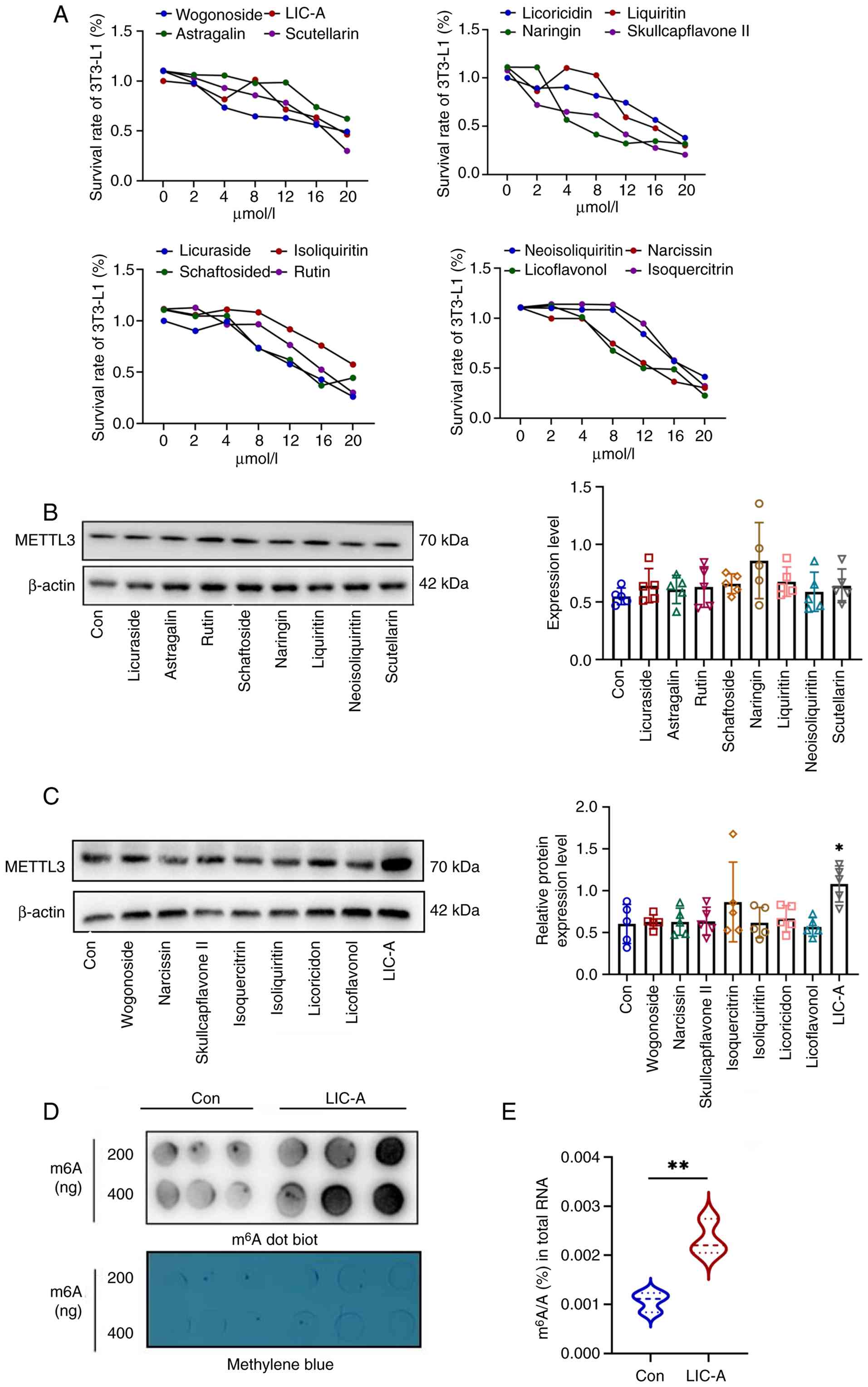 | Figure 1.LIC-A monomer in FJHQT promotes
METTL3 expression in adipogenic 3T3-L1 cells. (A) Viability of
3T3-L1 cells treated with the indicated drugs at various
concentrations, as determined by MTT assay. (B) Western blot
analysis of METTL3 protein expression in 3T3-L1 cells following
treatment with individual compounds, with corresponding
densitometric semi-quantification. (C) An independent western blot
analysis of METTL3 protein expression in 3T3-L1 cells following
treatment with individual compounds, with corresponding
densitometric semi-quantification. (D) Global m6A levels
in mRNA from LIC-A-treated 3T3-L1 cells, measured by m6A
dot blot using an anti-m6A antibody. (E) Quantitative
analysis of global mRNA m6A levels in LIC-A-treated
cells using a colorimetric ELISA kit. For western blot analysis in
(B) and (C), data are presented as mean ± SD from five independent
experiments (n=3 per group), and statistical significance was
determined by one-way ANOVA followed by Tukey's post hoc test for
multi-group comparisons. For MTT assay (A), data are presented as
mean ± SD from three independent experiments (n=3 per group), also
analyzed by one-way ANOVA with Tukey's post hoc test. For
m6A dot blot in (D) and colorimetric ELISA in (E), which
involve direct two-group comparisons, data are presented as mean ±
SD from three independent experiments (n=3 per group) and were
analyzed using unpaired two-tailed Student's t-test. *P<0.05,
**P<0.01. FJHQT, Fangji-Huangqi Decoction; LIC-A, licoisoflavone
A; METTL3, methyltransferase 3; m6A,
N6-methyladenosine. |
LIC-A ameliorates TNF-α- and
LPS-induced adipokine dysregulation and inflammatory responses in
differentiating 3T3-L1 adipocytes
To explore the effects of LIC-A on adipocyte
function, adipokine and inflammatory factor expression in TNF-α- or
LPS-stimulated adipogenic 3T3-L1 adipocytes was examined. The
optimal LIC-A concentration was first determined under acute TNF-α
stimulation (Fig. 2A) and 8 µmol/l
was used in all subsequent cotreatment experiments (Fig. 2B-H). Differentiated adipogenic
3T3-L1 cells were challenged with the obesity-mimicking cocktail
and followed by TNF-α treatment. ELISA showed that LIC-A treatment
could significantly enhance adiponectin secretion and inhibit
leukin secretion (leptin and resistin) when compared with the group
treated by TNF-α alone (Fig. 2B and
C). LIC-A also inhibited the release of inflammatory cytokines,
such as IL-6 and IL-1β, induced by TNF-α (Fig. 2D) and elevated expression of
insulin-sensitizing genes (Fig.
2E). At the transcriptional level, LIC-A significantly promoted
the expression of adiponectin mRNA while suppressing leptin and
resistin mRNA compared with the TNF-α group (Fig. 2F). Consistent with these findings,
LIC-A significantly decreased the mRNA and protein levels of
TNF-α-induced inflammatory factors (Fig. 2G and H).
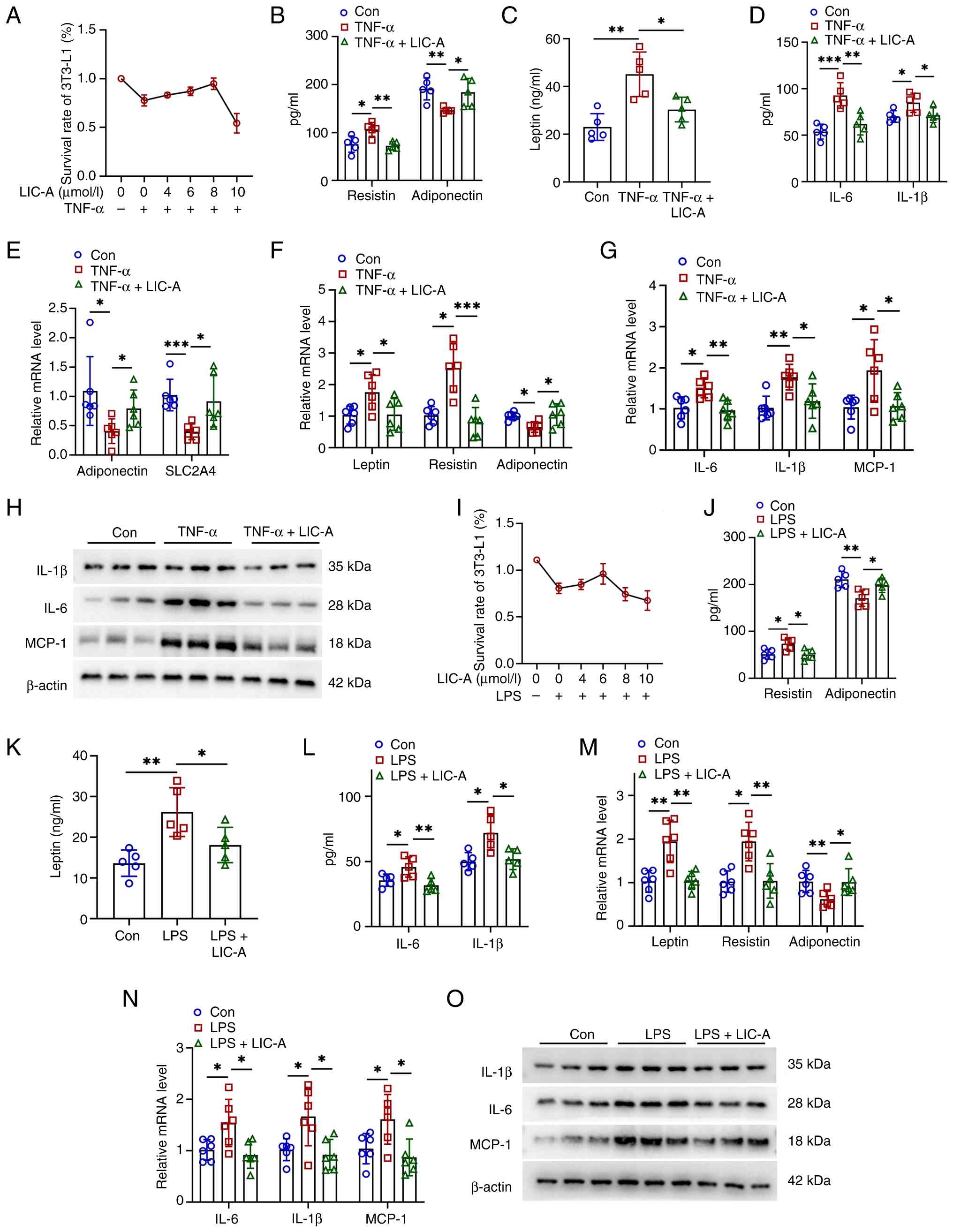 | Figure 2.LIC-A ameliorates TNF-α- and
LPS-induced adipokine dysregulation and inflammatory responses in
differentiating 3T3-L1 adipocytes. (A) Determining optimal LIC-A
concentration under TNF-α stimulation by MTT assay. (B-H)
Differentiated 3T3-L1 adipocytes were stimulated with TNF-α (10
ng/ml) in the presence or absence of LIC-A (8 µmol/l) for the
indicated periods. (B) ELISA measuring secreted levels of
adiponectin and resistin. (C) ELISA measuring secreted levels of
leptin. (D) ELISA measuring secreted levels of pro-inflammatory
cytokines IL-6 and IL-1β. (E) Analyzing mRNA expression of insulin
sensitivity genes by qPCR. (F) qPCR analysis of mRNA expression of
adipokines (adiponectin, leptin and resistin). (G) qPCR analysis of
mRNA expression of inflammatory cytokines (IL-6, IL-1β and MCP-1).
(H) Detecting protein levels of inflammatory cytokines by western
blotting. (I) Determining optimal LIC-A concentration under LPS
stimulation by MTT assay. (J-O) Differentiated 3T3-L1 adipocytes
were stimulated with LPS (100 ng/ml) in the presence or absence of
LIC-A (6 µmol/l) for the indicated periods. (J) ELISA measuring
secreted levels of adiponectin and resistin. (K) ELISA measuring
secreted levels of leptin. (L) ELISA measuring secreted levels of
IL-6 and IL-1β. (M) qPCR analysis of mRNA expression of adipokines
(adiponectin, leptin and resistin). (N) qPCR analysis of mRNA
expression of inflammatory cytokines (IL-6, IL-1β and MCP-1). (O)
Detecting protein levels of inflammatory cytokines by western
blotting. For western blot analysis, data are presented as mean ±
SD from three independent experiments (n=3 per group). For MTT,
ELISA and qPCR assays, data are presented as mean ± SD from five
independent experiments (n=5 per group). Statistical significance
was determined by one-way ANOVA followed by Tukey's post hoc test.
*P<0.05, **P<0.01, ***P<0.001. Con, control; LIC-A,
licoisoflavone A; LPS, lipopolysaccharides; METTL3,
methyltransferase 3; qPCR, quantitative polymerase chain reaction;
MCP-1, monocyte chemoattractant protein-1. |
Whether LIC-A exerts similar regulatory effects
under acute LPS stimulation was further evaluated. The optimal
LIC-A concentration under LPS stimulation was determined (Fig. 2I) and this concentration was used
consistently (6 µmol/l) in all subsequent co-treatment experiments
(Fig. 2J-O). It was observed that
LIC-A treatment notably ameliorated LPS-induced alterations in
adipokine secretion and inflammatory cytokine production (Fig. 2J-L). Moreover, LIC-A regulated mRNA
and protein expression of adipokines and inflammatory factors in a
manner highly consistent with its effects under TNF-α stimulation
(Fig. 2M-O). Together, these data
indicated that LIC-A alleviated TNF-α- and LPS-induced
dysregulation of adipokine expression and inflammatory responses in
adipogenic 3T3-L1 cells.
METTL3 deficiency abolishes the
effects of LIC-A on adipokine dysregulation and inflammatory
responses in TNF-α- or LPS-stimulated adipogenic 3T3-L1 cells
Since METTL3 upregulation may mediate the effects of
LIC-A on TNF-α- and LPS-induced adipokine dysregulation and
inflammatory responses, METTL3 protein levels in adipogenic 3T3-L1
cells were examined. Acute stimulation with TNF-α or LPS markedly
decreased METTL3 levels, whereas LIC-A treatment restored them
(Fig. 3A). Next, adipogenic 3T3-L1
cells were transduced with a METTL3-knockdown lentivirus. Knockdown
efficiency was confirmed at the protein and mRNA levels (Fig. 3B and C). ELISA revealed that, in
response to acute TNF-α stimulation, METTL3 knockdown abolished the
beneficial effects of LIC-A. This was supported by the lack of
increase in adiponectin secretion, lack of a significant decrease
in leptin and resistin expression and lack of an effect on the
secreted levels of key proinflammatory cytokines in comparison to
the empty vector group (Fig.
3D-F). In accordance with these changes, METTL3 knockdown
blocked the LIC-A upregulation of adiponectin mRNA and the
downregulation of leptin, resistin and proinflammatory factor
transcripts (Fig. 3G and H).
Whether LIC-A could ameliorate secretory dysfunction under LPS
challenge in METTL3 deficient cells was also investigated.
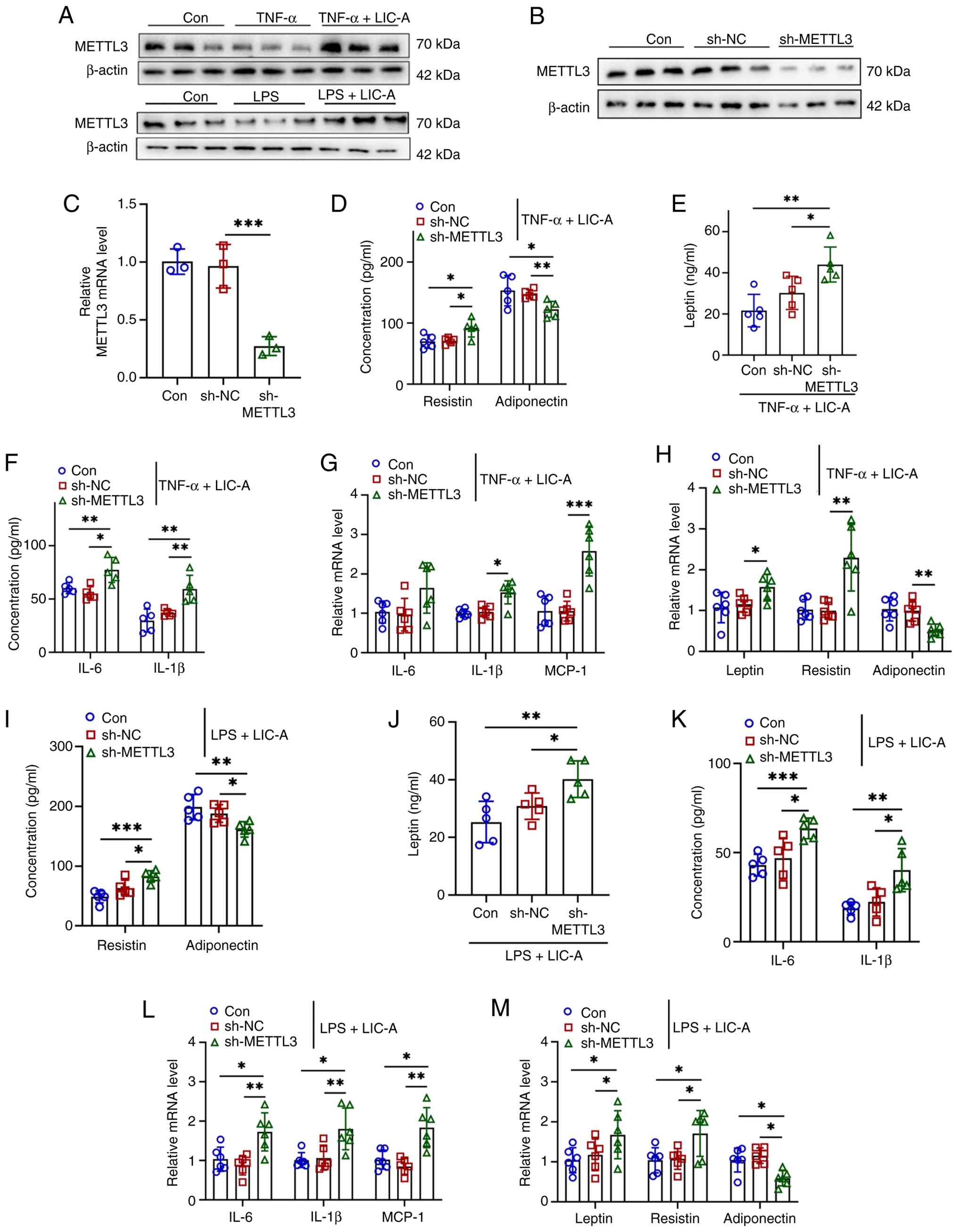 | Figure 3.METTL3 deficiency abolishes the
effects of LIC-A on adipokine dysregulation and inflammatory
responses in TNF-α- or LPS-stimulated adipogenic 3T3-L1. (A)
Western blot analysis of METTL3 protein expression during
adipogenesis in 3T3-L1 cells under acute TNF-α or LPS stimulation.
(B) Western blot analysis verifying the knockdown efficiency of
METTL3 in adipogenic 3T3-L1 cells. (C) qPCR analysis verifying the
knockdown efficiency of METTL3 in adipogenic 3T3-L1 cells. (D-H)
METTL3-knockdown 3T3-L1 adipocytes were stimulated with TNF-α (10
ng/ml) in the presence or absence of LIC-A (8 µmol/l) for the
indicated periods. (D) ELISA measuring secreted levels of
adiponectin and resistin. (E) ELISA measuring secreted levels of
leptin. (F) ELISA measuring secreted levels of pro-inflammatory
cytokines IL-6 and IL-1β. (G) qPCR analysis of mRNA expression of
inflammatory cytokines IL-6 and IL-1β. (H) qPCR analysis of mRNA
expression of Adipoq, leptin, resistin and MCP-1. (I-M)
METTL3-knockdown 3T3-L1 adipocytes were stimulated with LPS (100
ng/ml) in the presence or absence of LIC-A (6 µmol/l) for the
indicated periods. (I) ELISA measuring secreted levels of
adiponectin and resistin. (J) ELISA measuring secreted levels of
leptin. (K) ELISA measuring secreted levels of IL-6 and IL-1β. (L)
qPCR analysis of mRNA expression of inflammatory cytokines Il6,
IL-1β and MCP-1. (M) qPCR analysis of mRNA expression of adipokines
Adipoq, leptin and resistin. For western blot analysis data are
presented as mean ± SD from three independent experiments (n=3 per
group). For MTT, ELISA and qPCR assays, data are presented as mean
± SD from five independent experiments (n=5 per group). Statistical
significance was determined by one-way ANOVA followed by Tukey's
post hoc test. *P<0.05, **P<0.01, ***P<0.001. Con,
control; LIC-A, licoisoflavone A; LPS, lipopolysaccharides; METTL3,
methyltransferase 3; qPCR, quantitative polymerase chain reaction;
MCP-1, monocyte chemoattractant protein-1; sh, short hairpin RNA;
NC, negative control. |
ELISA confirmed that METTL3 knockdown abrogated the
protective effects of LIC-A under LPS challenge, as shown by the
persistently low adiponectin secretion alongside unabated levels of
leptin, resistin and inflammatory mediators. Consistent with this
profile, qPCR analysis further revealed a parallel failure in the
transcriptional regulation of these key metabolic and inflammatory
genes (Fig. 3I-M). Collectively,
these results indicated that the ability of LIC-A to counteract
TNF-α- and LPS-induced adipokine dysregulation and inflammation
depends on its promotion of METTL3 expression.
LIC-A alleviates HFD-induced metabolic
disorders and AT dysfunction in obese mice
To assess the in vivo anti-obesity effects of
LIC-A, obesity was induced in C57BL/6J mice who were fed a HFD over
a 12-week period. Experimental animals (4–16 weeks of age) were
treated with LIC-A (60 mg/kg) by oral gavage (twice a week)
(Fig. 4A). The body weight of the
experimental group treated with LIC-A decreased compared with that
of the HFD control group (Fig. 4B and
C). These changes were associated with improved insulin
sensitivity and glucose homeostasis (Fig. S2A and B) and reduced hyperglycemia
and dyslipidemia (Fig. S2C). In
addition, LIC-A efficiently attenuated WAT mass enlargement and
impeded the pathological remodeling of this tissue (Fig. 4D and E). A notable reduction in the
size of adipocytes in the eWAT of LIC-A-treated animals was
observed (Fig. 4F, 4G and S2D), which reflects the inhibition of
adipocyte hypertrophy, a hallmark of dysfunctional eWAT in
obesity.
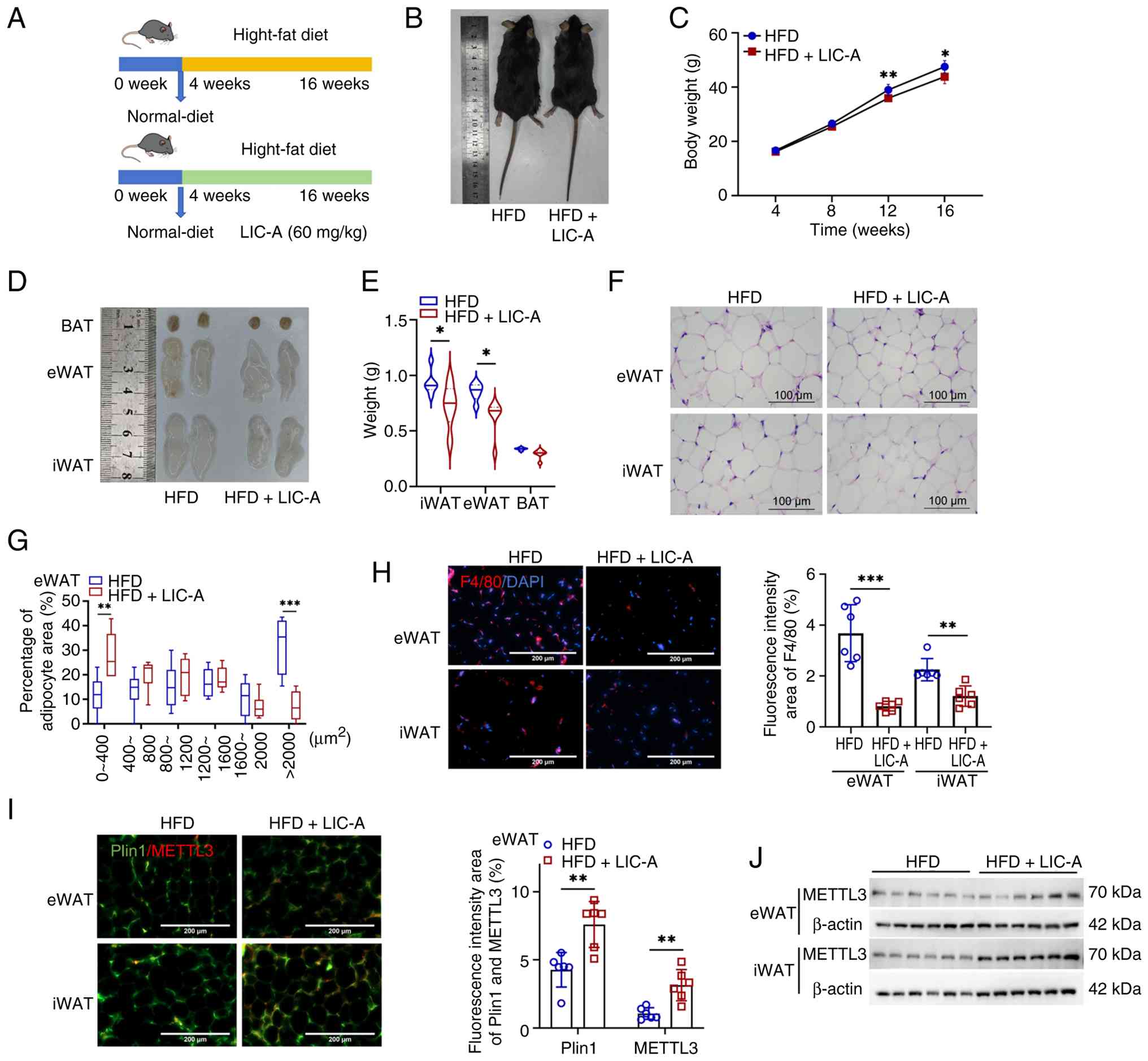 | Figure 4.LIC-A alleviates HFD-induced
metabolic disorders and AT dysfunction in obese mice. (A) Schematic
of the HFD experimental strategy. (B) Representative gross
morphology of mice after 16 weeks. (C) Monitoring of body weight
changes. (D and E) Examination of eWAT morphology and weight. (F)
Histological assessment of AT by hematoxylin and eosin staining.
(G) Quantification of adipocyte size frequency distribution in
eWAT. (H) Assessing macrophage infiltration by F4/80/DAPI
immunofluorescence and quantitative analysis of inflammatory areas.
(I) Analyzing METTL3 localization by Plin1/METTL3
immunofluorescence and quantitative analysis of METTL3-positive
areas. (J) Detecting METTL3 protein levels in WAT by western blot.
All data are presented as mean ± SD (n=6 mice per group for both
HFD control and LIC-A treatment groups). Statistical significance
was determined by one-way ANOVA followed by Tukey's post hoc test.
ns, not significant; *P<0.05, **P<0.01, ***P<0.001. AT,
adipose tissue; BAT, brown adipose tissue; Con, control; eWAT,
epididymal white adipose tissue; HFD, high-fat diet; iWAT, inguinal
white adipose tissue; LIC-A, licoisoflavone A; LPS,
lipopolysaccharides; METTL3, methyltransferase 3. |
Notably, LIC-A treatment markedly attenuated eWAT
inflammation. Immunofluorescence staining for the macrophage marker
F4/80 revealed a significant reduction in macrophage infiltration
(quantified as F4/80-positive area) in the eWAT of LIC-A-treated
mice compared with HFD controls (Fig.
4H). Consistently, the entire panel of adipokines showed a
metabolically favorable shift. ELISA of eWAT homogenates further
showed increased adiponectin levels and decreased levels of leptin,
resistin and inflammatory factors. Furthermore, qPCR analysis of
inflammatory gene expression at the transcript level confirmed the
downregulation of proinflammatory genes (Fig. S2E-H).
Notably, LIC-A treatment also elevated METTL3
expression in the AT of mice, thereby recapitulating the in
vitro findings and confirming that LIC-A promotes METTL3
expression in both physiological and experimental settings
(Fig. 4I and J). Taken together,
these results demonstrated that LIC-A supplementation effectively
alleviated metabolic dysregulation and pathological AT changes in
diet-induced obese mice, suggesting that the beneficial effects of
LIC-A may be mediated, at least in part, through METTL3
upregulation in AT.
Discussion
Obesity and its metabolic comorbidities are
associated with dysregulated AT, which becomes pathologically
remodeled and chronically inflamed and exhibits altered adipokine
secretion (45–47). The present study identified LIC-A
as a new upstream regulator of the RNA methyltransferase METTL3 and
showed that LIC-A alleviated the metabolic disturbances associated
with obesity by stimulating METTL3-mediated m6A RNA
methylation in the AT.
The present study combined virtual screening with
functional validation. That is, molecular docking analysis, based
on LibDock scores, identified LIC-A as exhibiting a favorable
predicted binding affinity for METTL3 among the 16 candidate
compounds screened from the FJHQT database. This prediction guided
the selection of LIC-A for experimental validation. The in
vitro experimental results confirmed the hypothesis derived
from the docking analysis, demonstrating that LIC-A promoted METTL3
protein expression and induced m6A modification.
Furthermore, in vivo experimental data from HFD mice further
demonstrated that LIC-A upregulated METTL3 expression in the AT;
this observation was not limited to the cellular level but was also
validated in a physiologically relevant context. Through these
steps, beginning with computational predictions and validation of
cellular mechanisms and culminating in metabolic assessments in
whole animals, a relatively comprehensive body of evidence was
established that supports the identification of LIC-A as a compound
capable of regulating METTL3 expression.
Collectively, the data from the present study
indicated that LIC-A upregulated METTL3 expression and increased
global m6A modification in adipogenic 3T3-L1 adipocytes,
suggesting that LIC-A affects adipocyte metabolism through an
epitranscriptomic mechanism. LIC-A treatment significantly reversed
TNF-α- and LPS-induced deregulation of adipokines (leptin,
adiponectin and resistin) and inhibited production of the
pro-inflammatory cytokines. This protective effect was lost upon
METTL3 knockdown, establishing an etiological link between
anti-inflammatory and metabolic effects of LIC-A and METTL3. In
vivo, LIC-A effectively improved systemic glucose homeostasis,
insulin sensitivity and lipid profiles in HFD-fed C57BL/6J mice.
These improvements were associated with decreased adipocyte
hypertrophy, reduced macrophage infiltration and ameliorated
inflammation in eWAT.
The findings from the present study align with
accumulating evidence that METTL3 is a key regulator of AT
plasticity and metabolic health (48). Previous studies have demonstrated
that METTL3 participates in beige adipogenesis and enhances
glycolytic gene expression through m6A modification,
thereby promoting energy expenditure and glucose tolerance
(29,30). In the present study, a basis for
this therapeutic approach was provided by identifying a natural
compound that may enhance METTL3 expression.
Additionally, endocrine dysfunction in the AT may be
the primary mechanism by which LIC-A exerts its anti-obesity
effects. The restoration of adipokine homeostasis and suppression
of AT inflammation noted in the present study may partly explain
the observed improvements in insulin resistance and dyslipidemia,
which are manifestations of systemic AT dysfunction. Notably,
decreased levels of leptin and resistin, coupled with the elevated
levels of adiponectin, indicated that the role of LIC-A in
improving endocrine dysfunction in AT is crucial for maintaining
metabolic homeostasis.
The following limitations of the present study
merits mentioning. First, LIC-A was identified as a METTL3 enhancer
by molecular docking and functional assays, but direct binding of
LIC-A to METTL3 has yet to be confirmed by biophysical approaches.
Although the docking analysis revealed a favorable predicted
binding mode of LIC-A within the METTL3 active pocket, this in
silico observation served as a hypothesis-generating tool
rather than a direct measure of the physical interaction. The
subsequent in vitro and in vivo data collectively
support the functional modulation of METTL3 by LIC-A, but do not
unequivocally establish a direct ligand-protein binding event.
Second, the downstream targets of METTL3 that mediate the
LIC-A-induced improvement in metabolism need to be explored
further, as RNA-sequencing and m6A-sequencing could
detect the individual transcripts whose methylation and expression
are regulated by LIC-A. Third, the translational relevance of the
data from the present study warrants systematic evaluation,
including validation in human cells, comprehensive pharmacokinetic
and toxicological profiling, and rigorous clinical testing. These
studies will collectively determine whether LIC-A, a natural METTL3
agonist, could serve as an effective therapy for treating obesity
and its associated metabolic complications.
Based on the findings and limitations of the present
study, we propose a general framework for developing LIC-A as a
potential drug for the treatment of obesity and related metabolic
diseases. First, at the molecular level, biophysical techniques
should be used to definitively identify the precise molecular
interaction sites between LIC-A and METTL3. Second, at the
transcriptomic level, an integrated analysis of m6A sequencing and
RNA-sequencing data from METTL3-deficient adipocytes is needed to
systematically identify downstream m6A-modified
transcripts that mediate the metabolic benefits of LIC-A, thereby
revealing the complete LIC-A/METTL3/m6A target gene
regulatory axis. Third, at the physiological level, tissue-specific
METTL3 conditional knockout mice will be used (for example
achieving adipocyte-specific METTL3 deficiency using AdipoqCre) to
definitively determine whether the in vivo anti-obesity
effects of LIC-A stem directly from its action on METTL3 in AT or
involve effects in other metabolic organs. Fourth, at the
translational medicine level, rigorous pharmacokinetic and
toxicological evaluations must be conducted in preclinical models,
followed by validation in human primary adipocytes, and ultimately
through carefully designed clinical trials to comprehensively
assess the safety, efficacy and therapeutic potential of LIC-A in
humans. In summary, we aim to systematically establish a
progressive research pathway from molecular mechanisms to clinical
translation with the goal of rigorously validating the therapeutic
potential of LIC-A.
In the present study, it was confirmed that LIC-A is
a first-in-class, natural agonist of METTL3, which is promising
therapeutically for both obesity and metabolic disorders.
Reconciling LIC-A with METTL3-catalyzed m6A methylation
offers a mechanistic basis for how TCM compounds can influence
epigenomic composition and reverse metabolic imbalances. The future
framework aforementioned, which encompasses everything from
biophysical binding assays and transcriptomic analysis to
validation in human cells and translational pharmacology, provides
a clear roadmap for consolidating the preclinical potential of
LIC-A and advancing its development toward clinical
applications.
Supplementary Material
Supporting Data
Supporting Data
Acknowledgements
Not applicable.
Funding
This study was supported by the National Natural Sciences
Foundation of China (grant no. 82370463), Natural Science
Foundation of the Guangxi Zhuang Autonomous Region (grant no.
2024GXNSFAA010154), Guangdong Basic and Applied Basic Research
Foundation (grant no. 2025A1515012522) and the China Postdoctoral
Science Foundation (grant no. 2024M761313).
Availability of data and materials
The data generated in the present study may be
requested from the corresponding author.
Authors' contributions
ST was responsible for data acquisition and
analysis, adipose tissue examination and writing the original
draft. XW was responsible for the formal statistical analysis and
interpretation of all quantitative data (including ELISA, qPCR,
GTT/ITT metabolic parameters and serum biochemical indices), and
contributed to the methodological optimization and quality control
of the in vitro and in vivo experimental protocols.
ZW was responsible for data curation and validation, including the
molecular docking screening, MTT assay data collection and
quantitative analysis of ELISA and qPCR results. HT was responsible
for investigation and supervision, including conducting the cell
culture and adipogenic differentiation experiments, and overseeing
the daily operations of the animal experiments. KY was responsible
for conceptualization and design, project administration, and
critical revision of the manuscript. XZ completed conceptualization
and contributed to writing, reviewing, and editing the manuscript.
ST and XW confirm the authenticity of all the raw data. All authors
read and approved the final version of the manuscript.
Ethics approval and consent to
participate
The animal experiments of the project met the
requirements of animal welfare ethics and passed the animal welfare
ethics investigation by IACUC of Guilin Medical University
(approval no. GLMU-IACUC-202510137).
Patient consent for publication
Not applicable.
Competing interests
The authors declare that they have no competing
interests.
References
|
1
|
Neeland IJ, Lim S, Tchernof A, Gastaldelli
A, Rangaswami J, Ndumele CE, Powell-Wiley TM and Despres JP:
Metabolic syndrome. Nat Rev Dis Primers. 10:772024. View Article : Google Scholar : PubMed/NCBI
|
|
2
|
Wang J, Li L, Zhang Z, Zhang X, Zhu Y,
Zhang C and Bi Y: Extracellular vesicles mediate the communication
of adipose tissue with brain and promote cognitive impairment
associated with insulin resistance. Cell Metab. 34:1264–1279.
e82022. View Article : Google Scholar : PubMed/NCBI
|
|
3
|
Tasali E, Pamidi S, Covassin N and Somers
VK: Obstructive sleep apnea and cardiometabolic disease: Obesity,
hypertension, and diabetes. Circ Res. 137:764–787. 2025. View Article : Google Scholar : PubMed/NCBI
|
|
4
|
Kawai T, Autieri MV and Scalia R: Adipose
tissue inflammation and metabolic dysfunction in obesity. Am J
Physiol Cell Physiol. 320:C375–C391. 2021. View Article : Google Scholar : PubMed/NCBI
|
|
5
|
Marcelin G, Gautier EL and Clement K:
Adipose tissue fibrosis in obesity: Etiology and challenges. Annu
Rev Physiol. 84:135–155. 2022. View Article : Google Scholar : PubMed/NCBI
|
|
6
|
Xue Y, Zou H, Ruan Z, Chen X, Lai Y, Yao
D, Ung COL and Hu H: Pharmacoeconomic evaluation of anti-obesity
drugs for chronic weight management: A systematic review of
literature. Front Endocrinol (Lausanne). 14:12543982023. View Article : Google Scholar : PubMed/NCBI
|
|
7
|
Astrup A, Carraro R, Finer N, Harper A,
Kunesova M, Lean ME, Niskanen L, Rasmussen MF, Rissanen A, Rossner
S, et al: Safety, tolerability and sustained weight loss over 2
years with the once-daily human GLP-1 analog, liraglutide. Int J
Obes (Lond). 36:843–854. 2012. View Article : Google Scholar : PubMed/NCBI
|
|
8
|
Silver HJ, Olson D, Mayfield D, Wright P,
Nian H, Mashayekhi M, Koethe JR, Niswender KD, Luther JM and Brown
NJ: Effect of the glucagon-like peptide-1 receptor agonist
liraglutide, compared to caloric restriction, on appetite, dietary
intake, body fat distribution and cardiometabolic biomarkers: A
randomized trial in adults with obesity and prediabetes. Diabetes
Obes Metab. 25:2340–2350. 2023. View Article : Google Scholar : PubMed/NCBI
|
|
9
|
McGowan B, Ciudin A, Baker JL, Busetto L,
Dicker D, Fruhbeck G, Goossens GH, Monami M, Sbraccia P,
Martinez-Tellez B, et al: A systematic review and meta-analysis of
the efficacy and safety of pharmacological treatments for obesity
in adults. Nat Med. 31:3317–3329. 2025. View Article : Google Scholar : PubMed/NCBI
|
|
10
|
Nauck MA, Petrie JR, Sesti G, Mannucci E,
Courreges JP, Lindegaard ML, Jensen CB and Atkin SL; Study 1821
Investigators, : A phase 2, randomized, dose-finding study of the
novel once-weekly human glp-1 analog, semaglutide, compared with
placebo and open-label liraglutide in patients with type 2
diabetes. Diabetes Care. 39:231–241. 2016. View Article : Google Scholar : PubMed/NCBI
|
|
11
|
Chen XY, Zhang J and Zhu JS: The role of
m6A RNA methylation in human cancer. Mol Cancer.
18:1032019. View Article : Google Scholar : PubMed/NCBI
|
|
12
|
Yue Y, Liu J and He C: RNA
N6-methyladenosine methylation in post-transcriptional gene
expression regulation. Genes Dev. 29:1343–1355. 2015. View Article : Google Scholar : PubMed/NCBI
|
|
13
|
Oerum S, Meynier V, Catala M and Tisne C:
A comprehensive review of m6A/m6Am RNA methyltransferase
structures. Nucleic Acids Res. 49:7239–7255. 2021. View Article : Google Scholar : PubMed/NCBI
|
|
14
|
Wang T, Li X, Tao Y, Wang X, Li L and Liu
J: METTL3-mediated NDUFB5 m6A modification promotes cell migration
and mitochondrial respiration to promote the wound healing of
diabetic foot ulcer. J Transl Med. 22:6432024. View Article : Google Scholar : PubMed/NCBI
|
|
15
|
Hou X, Yang Y, Wang C, Huang Z, Zhang M,
Yang J, Li N, Yang H, Yang L and Wu K: H3K36 methyltransferase
SMYD2 affects cell proliferation and migration in Hirschsprung's
disease by regulating METTL3. J Cell Physiol. 239:e314022024.
View Article : Google Scholar : PubMed/NCBI
|
|
16
|
Yuan W, Ouyang S, Lv Q and Liao L: The
effect of m6A methyltransferase METTL3 mediated TMEM30A regulation
on tumor energy metabolism and cisplatin anti-tumor activity in
oral squamous cell carcinoma. Life Sci. 358:1231222024. View Article : Google Scholar : PubMed/NCBI
|
|
17
|
Li Y, Wang J, Huang C, Shen M, Zhan H and
Xu K: RNA N6-methyladenosine: A promising molecular target in
metabolic diseases. Cell Biosci. 10:192020. View Article : Google Scholar : PubMed/NCBI
|
|
18
|
Wang C, Tanizawa H, Hill C, Havas A, Zhang
Q, Liao L, Hao X, Lei X, Wang L, Nie H, et al: METTL3-mediated
chromatin contacts promote stress granule phase separation through
metabolic reprogramming during senescence. Nat Commun. 15:54102024.
View Article : Google Scholar : PubMed/NCBI
|
|
19
|
Tuck MT: The formation of internal
6-methyladenine residues in eucaryotic messenger RNA. Int J
Biochem. 24:379–386. 1992. View Article : Google Scholar : PubMed/NCBI
|
|
20
|
Qiu L, Jing Q, Li Y and Han J: RNA
modification: Mechanisms and therapeutic targets. Mol Biomed.
4:252023. View Article : Google Scholar : PubMed/NCBI
|
|
21
|
Liu J, Yue Y, Han D, Wang X, Fu Y, Zhang
L, Jia G, Yu M, Lu Z, Deng X, et al: A METTL3-METTL14 complex
mediates mammalian nuclear RNA N6-adenosine methylation. Nat Chem
Biol. 10:93–95. 2014. View Article : Google Scholar : PubMed/NCBI
|
|
22
|
Jiang X, Liu B, Nie Z, Duan L, Xiong Q,
Jin Z, Yang C and Chen Y: The role of m6A modification in the
biological functions and diseases. Signal Transduct Target Ther.
6:742021. View Article : Google Scholar : PubMed/NCBI
|
|
23
|
Wei J, Yu X, Yang L, Liu X, Gao B, Huang
B, Dou X, Liu J, Zou Z, Cui XL, et al: FTO mediates LINE1
m6A demethylation and chromatin regulation in mESCs and
mouse development. Science. 376:968–973. 2022. View Article : Google Scholar : PubMed/NCBI
|
|
24
|
Wang X, Zhao BS, Roundtree IA, Lu Z, Han
D, Ma H, Weng X, Chen K, Shi H and He C: N(6)-methyladenosine
modulates messenger RNA translation efficiency. Cell.
161:1388–1399. 2015. View Article : Google Scholar : PubMed/NCBI
|
|
25
|
Shi H, Wang X, Lu Z, Zhao BS, Ma H, Hsu
PJ, Liu C and He C: YTHDF3 facilitates translation and decay of
N6-methyladenosine-modified RNA. Cell Res. 27:315–328.
2017. View Article : Google Scholar : PubMed/NCBI
|
|
26
|
Zhang H, Lu W, Tang H, Chen A, Gao X, Zhu
C and Zhang J: Novel insight of N6-methyladenosine in
cardiovascular system. Medicina (Kaunas). 61:2222025. View Article : Google Scholar : PubMed/NCBI
|
|
27
|
Zaccara S, Ries RJ and Jaffrey SR:
Reading, writing and erasing mRNA methylation. Nat Rev Mol Cell
Biol. 20:608–624. 2019. View Article : Google Scholar : PubMed/NCBI
|
|
28
|
Song B, Zeng Y, Cao Y, Zhang J, Xu C, Pan
Y, Zhao X and Liu J: Emerging role of METTL3 in inflammatory
diseases: Mechanisms and therapeutic applications. Front Immunol.
14:122169092023. View Article : Google Scholar
|
|
29
|
Xie R, Yan S, Zhou X, Gao Y, Qian Y, Hou
J, Chen Z, Lai K, Gao X and Wei S: Activation of METTL3 promotes
white adipose tissue beiging and combats obesity. Diabetes.
72:1083–1094. 2023. View Article : Google Scholar : PubMed/NCBI
|
|
30
|
Li Y, Zhang Y, Zhang T, Ping X, Wang D,
Chen Y, Yu J, Liu C, Liu Z, Zheng Y, et al: Rna M6 a
methylation regulates glycolysis of beige fat and contributes to
systemic metabolic homeostasis. Adv Sci (Weinh). 10:e23004362023.
View Article : Google Scholar : PubMed/NCBI
|
|
31
|
Perez-Torres I, Castrejon-Tellez V, Soto
ME, Rubio-Ruiz ME, Manzano-Pech L and Guarner-lans V: Oxidative
stress, plant natural antioxidants, and obesity. Int J Mol Sci.
22:17862021. View Article : Google Scholar : PubMed/NCBI
|
|
32
|
Li SZ, Zeng SL and Liu EH: Anti-obesity
natural products and gut microbiota. Food Res Int. 151:1108192022.
View Article : Google Scholar : PubMed/NCBI
|
|
33
|
Wang Y, Qi F, Li M, Xu Y, Dong L and Cai
P: Traditional Chinese medicine modulates hypothalamic
neuropeptides for appetite regulation: A comprehensive review.
Biosci Trends. 19:281–295. 2025. View Article : Google Scholar : PubMed/NCBI
|
|
34
|
Wang Y, Ma Y, Ke Y, Jiang X, Liu J, Xiao
Y, Zheng H, Wang C, Chen X and Shi M: Fangji Huangqi decoction
ameliorates membranous nephropathy through the upregulation of
BNIP3-mediated mitophagy. J Ethnopharmacol. 324:1177342024.
View Article : Google Scholar : PubMed/NCBI
|
|
35
|
Li D, Yang C, Zhu JZ, Lopez E, Zhang T,
Tong Q, Peng C and Lin LG: Berberine remodels adipose tissue to
attenuate metabolic disorders by activating sirtuin 3. Acta
Pharmacol Sin. 43:1285–1298. 2022. View Article : Google Scholar : PubMed/NCBI
|
|
36
|
Sen S, Tiwari AK, Binwal M, Israr M, Kumar
A, Pathak N, Washimkar KR, Ratnasekhar CH, Mugale MN, Negi AS and
Bawankule DU: Steroidal glycoside-rich extract of fenugreek seeds
mitigates obesity-induced insulin resistance via attenuation of
low-grade inflammation. Food Res Int. 220:1171272025. View Article : Google Scholar : PubMed/NCBI
|
|
37
|
Noh JW, Jun MS, Yang HK and Lee BC:
Cellular and molecular mechanisms and effects of berberine on
obesity-induced inflammation. Biomedicines. 10:17392022. View Article : Google Scholar : PubMed/NCBI
|
|
38
|
Jang BC: Tetrandrine has anti-adipogenic
effect on 3T3-L1 preadipocytes through the reduced expression
and/or phosphorylation levels of C/EBP-α, PPAR-γ, FAS, perilipin A,
and STAT-3. Biochem Biophys Res Commun. 494:422–423. 2017.
View Article : Google Scholar : PubMed/NCBI
|
|
39
|
Zhu Y, Su Y, Zhang J, Zhang Y, Li Y, Han
Y, Dong X and Li W and Li W: Astragaloside IV alleviates liver
injury in type 2 diabetes due to promotion of AMPK/mTOR-mediated
autophagy. Mol Med Rep. 23:4372021. View Article : Google Scholar : PubMed/NCBI
|
|
40
|
Wang L, Zheng W, Yang J, Ali A and Qin H:
Mechanism of Astragalus membranaceus alleviating acquired
hyperlipidemia induced by high-fat diet through regulating lipid
metabolism. Nutrients. 14:9542022. View Article : Google Scholar : PubMed/NCBI
|
|
41
|
Kim HL, Lee SK, Min DE, Choi BK and Lee
DR: Anti-Obesity Effects of a mixture of atractylodes macrocephala
and amomum villosum extracts on 3T3-L1 adipocytes and high-fat
diet-induced obesity in mice. Molecules. 27:9062022. View Article : Google Scholar : PubMed/NCBI
|
|
42
|
Lee HE, Yang G, Han SH, Lee JH, An TJ,
Jang JK and Lee JY: Anti-obesity potential of Glycyrrhiza uralensis
and licochalcone A through induction of adipocyte browning. Biochem
Biophys Res Commun. 503:2117–2123. 2018. View Article : Google Scholar : PubMed/NCBI
|
|
43
|
Hong MK, Han Y, Park HJ, Shin MR, Roh SS
and Kwon EY: The synergistic action of metformin and glycyrrhiza
uralensis fischer extract alleviates metabolic disorders in mice
with diet-induced obesity. Int J Mol Sci. 24:9362023. View Article : Google Scholar : PubMed/NCBI
|
|
44
|
Livak KJ and Schmittgen TD: Analysis of
relative gene expression data using real-time quantitative PCR and
the 2(−Delta Delta C(T)) method. Methods. 25:402–408. 2001.
View Article : Google Scholar : PubMed/NCBI
|
|
45
|
Younossi ZM, Kalligeros M and Henry L:
Epidemiology of metabolic dysfunction-associated steatotic liver
disease. Clin Mol Hepatol. 31 (Suppl):S32–S50. 2025. View Article : Google Scholar : PubMed/NCBI
|
|
46
|
Miranda AMA, McAllan L, Mazzei G, Andrew
I, Davies I, Ertugrul M, Kenkre J, Kudo H, Carrelha J, Patel B, et
al: Scott, Selective remodelling of the adipose niche in obesity
and weight loss. Nature. 644:769–779. 2025. View Article : Google Scholar : PubMed/NCBI
|
|
47
|
An SM, Cho SH and Yoon JC: Adipose tissue
and metabolic health. Diabetes Metab J. 47:595–611. 2023.
View Article : Google Scholar : PubMed/NCBI
|
|
48
|
Ronningen T, Dahl MB, Valderhaug TG, Cayir
A, Keller M, Tonjes A, Bluher M and Bottcher Y: m6A regulators in
human adipose tissue-depot-specificity and correlation with
obesity. Front Endocrinol (Lausanne). 12:7788752021. View Article : Google Scholar : PubMed/NCBI
|

















