Introduction
Esophageal cancer (EC) is the sixth leading cause of
cancer-related mortality worldwide, with an estimated 511,054 new
cases and 445,391 deaths in 2022, ~50% of which were expected to
occur in China (1,2). Histologically, >90% of EC cases in
China are esophageal squamous cell carcinoma (ESCC), which has a
poor 5-year overall survival rate of 20–30%. Notably, most patients
with ESCC are diagnosed at advanced stages, for which effective
targeted therapeutic strategies remain limited (3,4).
Accumulating evidence has indicated that metabolic reprogramming,
especially enhanced aerobic glycolysis (the Warburg effect), is a
hallmark of ESCC progression and drives malignant phenotypes
including proliferation, invasion and distant metastasis, which
represents a promising therapeutic vulnerability (5). Multiple natural small molecule
compounds, such as baicalin, have been reported to exert antitumor
activity through inhibiting hyperactivated Wnt signaling in
epithelial tumors, offering promising candidate agents for tumor
intervention (6). The poor
prognosis and limited therapeutic options for ESCC drive the urgent
need to identify novel prognostic biomarkers and therapeutic
targets.
Kinesin family member 4A (KIF4A) is an
evolutionarily conserved microtubule motor protein, which is
primarily involved in mitotic spindle organization and chromosomal
segregation during cell division (7,8).
Emerging evidence has validated the oncogenic roles of KIF4A across
diverse malignancies, including non-small cell lung cancer,
colorectal cancer, breast cancer and hepatocellular carcinoma
(9–13). KIF4A modulates multiple malignant
phenotypes, including cell proliferation, invasion, stemness
maintenance and therapeutic resistance, by regulating diverse
oncogenic signaling cascades such as the Akt, TGF-β/Smad3 and DNA
damage response pathways (14–19).
Clinically, KIF4A upregulation is associated with unfavorable
prognosis in multiple types of cancer, including non-small cell
lung cancer, colorectal cancer, breast cancer and hepatocellular
carcinoma, and has been developed as a potential therapeutic
target, with small-molecule inhibitors such as WZ-3146 exhibiting
antitumor efficacy in preclinical models (12,13,16,17,20).
Notably, our previous study identified KIF4A as a prognostic
biomarker for ESCC (21), but the
functional mechanisms and downstream regulatory pathways of KIF4A
in ESCC progression remain largely undefined.
The Wnt signaling pathway is an evolutionarily
conserved cascade divided into the canonical β-catenin-dependent
pathway and non-canonical branches (including the
Wnt/Ca2+ and planar cell polarity pathways), which
collectively regulate cellular proliferation, differentiation,
migration and tissue homeostasis (22–27).
Among them, the canonical Wnt/β-catenin signaling pathway is the
most well-characterized branch in tumor biology, with aberrant
activation closely implicated in the tumorigenesis and progression
of multiple solid malignancies, including breast cancer, liver
cancer and gastrointestinal tumors such as ESCC (28–32).
In the resting state, β-catenin is phosphorylated and degraded by
the destruction complex; upon pathway activation, stabilized
β-catenin translocates to the nucleus, forms a complex with T-cell
factor/lymphoid enhancer-binding factor (TCF/LEF) transcription
factors, and drives the expression of pro-oncogenic target genes
such as c-Myc, cyclin D1 and MMP-7 (28,30).
A recent study confirmed that the transcription factor E2F1
participates in the malignant progression and sorafenib resistance
of EC, and multiple transcription factors, including E2F1, can
synergistically interact with the Wnt cascade to remodel tumor cell
phenotypes and drug responsiveness (33). Previous studies have reported that
abnormal activation of the Wnt/β-catenin pathway is highly
prevalent in ESCC, and that it is associated with tumor progression
and poor prognosis (34,35). However, to the best of our
knowledge, whether KIF4A exerts its oncogenic effects in ESCC by
regulating the Wnt/β-catenin pathway has not yet been reported.
The current study systematically investigated the
expression pattern, prognostic value, biological functions and
downstream regulatory mechanisms of KIF4A in ESCC. Accordingly, the
present study aimed to systematically investigate the expression
profile, clinical prognostic value and biological functions of
KIF4A in ESCC, and to further explore whether KIF4A drives ESCC
progression by regulating the canonical Wnt/β-catenin signaling
pathway. This work may provide a novel potential prognostic
biomarker and therapeutic target for ESCC management.
Materials and methods
Bioinformatics analysis
The Cancer Genome Atlas (TCGA) pan-cancer
transcriptomic dataset (2,900 samples) and ESCC transcriptomic
dataset (195 samples) with corresponding clinical data were
downloaded and analyzed via the cBioPortal (https://www.cbioportal.org/) and UALCAN (https://ualcan.path.uab.edu/) online platforms.
Kaplan-Meier survival analysis and differential expression analysis
of KIF4A were performed using the UALCAN database (http://ualcan.path.uab.edu) based on the TCGA
esophageal carcinoma (ESCA) cohort. The correlation between KIF4A
expression and overall survival was evaluated by the log-rank test.
Gene expression levels were normalized using the
\(\log_2(\text{RSEM}+1)\) transformation. The association between
KIF4A expression and pTNM stage grouping (I–IV) in ESCC, as well as
the co-expression correlation between KIF4A and CTNNB1 (encoding
β-catenin)/TCF7, were analyzed. Heatmap visualization of Wnt
signaling pathway-related genes was constructed based on normalized
expression values using Heatmapper 2 (version 2.0; http://www.heatmapper.ca), a freely available online
platform for gene expression heatmap generation.
Clinical tissue specimens
The present study involved two independent sets of
in-house human tissue specimens, all obtained from patients with
pathologically confirmed esophageal squamous cell carcinoma (ESCC)
undergoing routine radical esophagectomy at Xiangyang No. 1
People's Hospital (Xiangyang, China). No patients received
neoadjuvant chemoradiotherapy before surgery, and no additional
invasive procedures were performed specifically for research
purposes. Inclusion and exclusion criteria were as follows:
Patients with pathologically confirmed primary ESCC who underwent
complete radical esophagectomy with complete clinicopathological
data. Exclusion criteria were as follows: Patients with a history
of other primary malignancies, confirmed distant metastasis at
diagnosis, incomplete follow-up records or severe systemic
comorbidities that affect survival endpoints. The main research
cohort, comprising 43 matched pairs of fresh ESCC tissues and
paired adjacent non-malignant esophageal tissues, was surgically
collected between August 2023 and September 2025. Patients in this
cohort had a mean age of 63 years (range, 56–74 years) and included
29 males and 14 females. The present study was reviewed and
approved by the Institutional Review Board of Xiangyang First
People's Hospital (approval no. 2025KY082) in September 2025. The
second set of specimens, dedicated to patient-derived organoid
culture and functional validation assays, was prospectively
collected from December 2025 to March 2026. All specimens were
freshly resected tumor tissues obtained from 15 patients with
pathologically confirmed primary ESCC undergoing routine radical
esophagectomy. This cohort was reviewed and approved under a
separate independent ethics approval (approval no. 2025KY142,
officially approved in December 2025) by the Institutional Review
Board of Xiangyang No. 1 People's Hospital. Sample processing,
organoid establishment, routine passaging and relevant follow-up
experiments were performed immediately after surgical resection
following standardized operating procedures. Written informed
consent was obtained from all enrolled patients at the time of
specimen collection, and all procedures involving human tissues
were conducted in strict compliance with The Declaration of
Helsinki Patients were stratified into low- and high-KIF4A
expression groups according to KIF4A expression levels. Baseline
clinical characteristics including age, sex, clinical stage,
drinking history and lymph node metastasis status were compared
between the two groups. The detailed clinicopathological
characteristics of the main research cohort are summarized in
Table SI.
Immunohistochemistry (IHC)
Tissues were fixed in 4% paraformaldehyde for 24 h,
embedded in paraffin and cut into 4-µm sections. The sections were
then baked at 60°C for 2 h, deparaffinized with eco-friendly
deparaffinization solution (Wuhan Servicebio Technology Co., Ltd.)
for 130 min at room temperature (25°C) and rehydrated through a
gradient ethanol series to distilled water. Antigen retrieval was
performed using Tris-EDTA buffer under high pressure at 121°C for
20 min and endogenous peroxidase activity was blocked with 3%
H2O2 for 10 min at room temperature. The
sections were then incubated with anti-KIF4A primary antibody
(1:200; catalog no. A9080; ABclonal Biotech Co., Ltd.) overnight at
4°C, followed by incubation with a horseradish peroxidase
(HRP)-conjugated secondary antibody for 1 h at room temperature.
The chromogenic reaction was developed with DAB for 1–3 min at room
temperature, followed by hematoxylin counterstaining for 2 min at
room temperature, gradient ethanol dehydration and xylene
clearance. Slides were mounted with neutral resin and images were
captured under a light microscope.
KIF4A staining was scored semi-quantitatively by two
independent pathologists in a blinded manner, based on staining
intensity (0, negative; 1, weak; 2, moderate; 3, strong) and the
percentage of positive cells (0, 0%; 1, 1–25%; 2, 26–50%; 3,
51–75%; 4, 76–100%). The ‘staining intensity × positive cell
percentage’ scoring system (intensity 0–3, positive rate 0–4, total
score range 0–12) with a cutoff of 6 for KIF4A IHC (≥6, high; <6
low) has been widely adopted in multiple published studies on KIF4A
in solid tumors, which ensures the comparability of the present
results with those of previous reports (36,37).
Cell culture and transfection
Human ESCC cell lines (KYSE-150, KYSE-180, TE-10 and
TE-1) and the normal human esophageal epithelial cell line HET-1A
were used in the present study. All cell lines were obtained from
the American Type Culture Collection, authenticated via short
tandem repeat profiling and routinely tested for mycoplasma
contamination (negative throughout the study). ESCC cell lines were
cultured in RPMI-1640 medium (Pricella; Elabscience Bionovation
Inc.) supplemented with 10% fetal bovine serum (FBS; Pricella;
Elabscience Bionovation Inc) and 1% penicillin/streptomycin, in a
humidified incubator containing 5% CO2 at 37°C. HET-1A
cells were maintained in specialized epithelial cell medium
(Shanghai Jinyuan Biotechnology Co., Ltd.) under the same culture
conditions.
For KIF4A silencing, KYSE-150 and TE-1 cells were
seeded into 6-well plates (5×10⁴ cells/well). Four
KIF4A-targeting small interfering RNAs (si-KIF4A-1, si-KIF4A-2,
si-KIF4A-3 and si-KIF4A-4) were synthesized by Shanghai GeneChem
Co., Ltd. Cells were transfected with 50 nM corresponding siRNA
using Lipo8000™ transfection reagent (Beyotime Biotechnology) at
37°C in a humidified 5% CO2 incubator for 6 h, followed
by complete medium replacement. The knockdown efficiency at the
protein level was verified by western blot, and si-KIF4A-1 with the
highest interference efficiency was selected for all subsequent
functional experiments. The full sequences of all siRNAs are listed
in the supplementary materials. For KIF4A overexpression, KYSE-150
and TE-10 cells (the same lines used for gene silencing) were
transfected with 5 µg of KIF4A overexpression plasmid (GV712 vector
backbone; Shanghai GeneChem Co., Ltd.) using the same transfection
reagent. Overexpression efficiency was verified by western blotting
at 48 h post-transfection. The sequences of KIF4A siRNAs are
provided in Table I.
 | Table I.siRNA sequences. |
Table I.
siRNA sequences.
| siRNA | Sense (5′-3′) | Anti-sense
(5′-3′) |
|---|
| KIF4A si-1 |
GCAACAGAAUACCAAGAAATT |
UUUCUUGGUAUUCUGUUGCTT |
| KIF4A si-2 |
UAUUUGAUCCCUCUACUGATT |
UCAGUAGAGGGAUCAAAUATT |
| KIF4A si-3 |
CAGGUCAGCAAACUUGAAATT |
UUUCAAGUUUGCUGACCUGTT |
| KIF4A si-4 |
GCUGCUGAAACUUGAAAGATT |
UCUUUCAAGUUUAGCAGCTT |
| si-NC |
UUCUCCGAACGUGUCACGUTT |
ACGUGACACGUUCGGAGAATT |
Western blot analysis
Total protein was extracted from KYSE-150 and TE-10
human ESCC cell lines and surgically resected ESCC tissue
specimens, using pre-chilled RIPA lysis buffer (Beyotime
Biotechnology) supplemented with a protease inhibitor cocktail and
1 mM phenylmethanesulfonyl fluoride (both Beyotime Biotechnology).
All samples were fully lysed on ice for 30 min, and the
supernatants were collected by high-speed centrifugation for
subsequent BCA protein quantification. Total protein was extracted
by sonication at a frequency of 20 kHz in an ice bath (4°C), with
3-sec on/5-sec off pulses for a total effective duration of 30 sec,
followed by centrifugation at 14,000 × g for 15 min at 4°C. Protein
concentration was determined using the BCA protein assay kit
(Beyotime Biotechnology). Subsequently, equal amounts of protein
(30 µg/lane) were separated on 10% gels using SDS-PAGE and
transferred to polyvinylidene fluoride membranes (Wuhan Servicebio
Technology Co., Ltd.) via wet transfer. The membranes were then
blocked with 5% non-fat milk in Tris-buffered saline containing
0.1% (v/v) Tween-20 (TBST) for 1 h at room temperature, then
incubated with the following primary antibodies overnight at 4°C:
Anti-KIF4A (1:1,000; catalog no. A9080; ABclonal Biotech Co.,
Ltd.), anti-β-catenin (1:1,000; catalog no. A19657; ABclonal
Biotech Co., Ltd.) and anti-TCF7 (1:1,000; catalog no. A20835;
ABclonal Biotech Co., Ltd.), and the loading controls
anti-α-tubulin (1:10,000; catalog no. 66031-1-Ig; Wuhan Sanying
Biotechnology), anti-GAPDH (1:10,000; catalog no. 60004-1-Ig; Wuhan
Sanying Biotechnology), anti-β-actin (1:10,000; catalog no.
60008-1-Ig; Wuhan Sanying Biotechnology) and anti-Histone H3
(1:10,000; catalog no. 17168-1-AP; Wuhan Sanying Biotechnology).
After three washes with TBST, the membranes were incubated with
HRP-conjugated secondary antibodies for 1 h at room temperature.
Protein bands were visualized using the SuperSignal™ West Pico PLUS
ECL kit (Wuhan Servicebio Technology Co., Ltd.) on the ChemiDoc
XRS+ system (Bio-Rad Laboratories, Inc.). Semi-quantitative
grayscale analysis of protein bands was performed using ImageJ
software (version 1.53t; National Institutes of Health).
Reverse transcription-quantitative PCR
(RT-qPCR) and RNA sequencing
Total RNA was extracted from cells using
TRIzol® reagent (Invitrogen; Thermo Fisher Scientific,
Inc.). RT was performed using the AbScript First Strand cDNA
Synthesis Kit (catalog no. RK20300; ABclonal Biotech Co., Ltd.)
according to the manufacturer's instructions. qPCR was conducted
using the Universal SYBR Green Fast qPCR Mix (ABclonal Biotech Co.,
Ltd.) on the ABI 7500 Real-Time PCR System (Applied Biosystems;
Thermo Fisher Scientific, Inc.). The thermal cycling conditions
were as follows: Initial denaturation at 95°C for 30 sec, followed
by 40 cycles of denaturation at 95°C for 5 sec and
annealing/extension at 60°C for 30 sec. Melting curve analysis was
performed after amplification to verify the specificity of PCR
products. The relative mRNA expression levels were calculated using
the 2−ΔΔCq method (38), with GAPDH as the internal
reference. Detailed information of all primers used for RT-qPCR,
including sequences, amplicon lengths annealing temperatures and
NCBI Gene IDs, is summarized in Table
SII.
For RNA sequencing, total RNA was extracted from
KIF4A-knockdown TE-1 cells and TE-1 cells transfected with negative
control siRNA (si-NC), with 1 µg of high-quality RNA used for
library preparation per sample. RNA sequencing was performed by
Shanghai Jikai Gene Chemical Technology Co., Ltd. Differential
expression analysis was performed using DESeq2 v1.16.1
(Bioconductor) (39), with
|log2(fold change)|≥1 and false discovery rate (FDR) <0.05 set
as the threshold for differentially expressed genes (DEGs). Gene
Ontology (GO) and Kyoto Encyclopedia of Genes and Genomes (KEGG)
pathway enrichment analyses were performed using clusterProfiler
v3.4.4 (40), with FDR-adjusted
P<0.05 defined as significant enrichment (41–43).
Colony formation assay
Transfected cells were seeded into 6-well plates at
a density of 500 cells/well, and cultured for 7–14 days (when
visible colonies of proper size formed) at 37°C in an atmosphere
containing 5% CO2, with the medium replaced every 3–4
days. Colonies were fixed with pre-chilled 100% methanol at room
temperature for 15 min, stained with 0.1% crystal violet (Shanghai
Acmec Biochemical Technology Co., Ltd.) at room temperature for 20
min, and then washed with PBS. The number of colonies (≥50
cells/colony) was counted under a light microscope. All experiments
were performed in triplicate with three biological replicates.
Wound healing assay
Transfected cells were seeded into 6-well plates and
cultured to 90% confluence. Subsequently, a straight scratch was
made in the cell monolayer using a sterile 200-µl pipette tip. The
cells were then washed with PBS to remove floating cells and
cultured in serum-free RPMI-1640 medium. Images of the scratch were
captured at 0 and 24 h post-scratching under an inverted light
microscope (Olympus IX73; Olympus Corporation). The wound healing
rate (%) was calculated as: (0 h wound width-24 h wound width)/0 h
wound width ×100. All experiments were performed in triplicate with
three biological replicates.
Transwell migration assay
ESCC cells were harvested 48 h post-transfection,
resuspended in serum-free RPMI-1640 medium and seeded into the
upper chambers of 24-well Transwell plates (pore size, 8 µm;
Shanghai Acmec Biochemical Technology Co., Ltd.) at a density of
4×10⁴ cells/well. The lower chambers were filled with
600 µl RPMI-1640 medium supplemented with 20% FBS as a
chemoattractant. After 24 h incubation at 37°C, non-migrated cells
on the upper surface of the membrane were removed with cotton
swabs. Cells that had migrated to the lower surface were fixed with
4% paraformaldehyde at room temperature for 15 min, stained with
0.1% crystal violet at room temperature for 10 min, and images were
captured under an inverted light microscope. The number of migrated
cells was counted in five random fields per well. All experiments
were performed in triplicate with three biological replicates.
EdU staining assay
Cell proliferation was detected using the EdU-488
Cell Proliferation Imaging Detection Kit (Hubei Pumei Biotechnology
Co., Ltd.) according to the manufacturer's instructions.
Transfected cells were seeded into 96-well plates (2×10⁴
cells/well) and cultured for 24 h. Cells were then incubated with
EdU working solution at 37°C for 2 h, fixed with 4%
paraformaldehyde at room temperature for 15 min, permeabilized with
0.5% Triton X-100 at room temperature for 10 min, and stained with
the click reaction solution in the dark at room temperature for 30
min. Nuclei were counterstained with DAPI in the dark at room
temperature for 5 min. Images were captured under a fluorescence
microscope and the percentage of EdU-positive cells was calculated.
All experiments were performed in triplicate with three biological
replicates.
Co-immunoprecipitation (co-IP)
assay
TE-1 and KYSE-150 cells were lysed in pre-chilled
non-denaturing lysis buffer (Beyotime Biotechnology) supplemented
with protease inhibitor cocktail on ice for 5 min. After
centrifugation at 12,000 × g for 20 min at 4°C, the supernatant was
collected as total protein extract. For co-immunoprecipitation
assays, two independent sets of reactions were performed with
different capture antibodies for distinct verification purposes:
Anti-KIF4A antibody was used to capture the KIF4A protein complex
to verify the endogenous binding between KIF4A and core Wnt pathway
proteins, while anti-β-catenin antibody was used to capture the
β-catenin protein complex in negative control and KIF4A-knockdown
TE-1 cells to evaluate the effect of KIF4A knockdown on the
stability of the β-catenin/TCF7 transcriptional complex, and normal
mouse IgG was set as the negative control for both
immunoprecipitation systems. For each reaction, 500 µg of total
protein lysate was incubated with the corresponding antibody or
isotype control overnight at 4°C with gentle rotation, followed by
2 h of incubation with 20–40 µl Protein A/G magnetic beads (Abmart
Pharmaceutical Technology Co., Ltd.) at 4°C. After incubation, the
tubes were briefly centrifuged at 3,000 × g for 1 min at 4°C to
collect beads from the tube wall, and the immune complexes were
then isolated using a magnetic stand before the supernatant was
discarded. The magnetic beads were washed three times with
pre-chilled lysis buffer; after each wash, beads were collected via
brief centrifugation at 3,000 × g for 1 min at 4°C followed by
magnetic separation, and the supernatant was completely
removed.
The precipitated protein complexes were eluted with
2X SDS loading buffer, boiled at 100°C for 10 min for denaturation,
and centrifuged at 10,000 × g for 10 sec at room temperature to
collect all eluate to the tube bottom. The supernatant was
subsequently analyzed by western blotting. Semi-quantitative
grayscale analysis of protein bands was performed using ImageJ
software (version 1.53t; National Institutes of Health).
The detailed antibody information for the
co-immunoprecipitation assay is listed as follows: Mouse monoclonal
anti-KIF4A antibody (cat. no. sc-365144; Santa Cruz Biotechnology),
mouse monoclonal anti-β-catenin antibody (cat. no. sc-7963; Santa
Cruz Biotechnology) and normal mouse IgG isotype control (cat. no.
B900620; Proteintech Group, Inc.). For each immunoprecipitation
reaction, 2 µg of target antibody or isotype control was added.
ESCC organoid culture
Fresh ESCC surgical specimens were cut into small
pieces, and digested in AdDF++++ medium containing collagenase II
(Sigma-Aldrich; Merck KGaA) and DNase I (Sigma-Aldrich; Merck KGaA)
for 3–4 h at 37°C in a water bath. The cell suspension was filtered
through a 70-µm cell strainer and centrifuged at 300 × g for 5 min
at 4°C. The cell pellet was washed three times with D-PBS. Red
blood cells were lysed using red blood cell lysis buffer
(Invitrogen; Thermo Fisher Scientific, Inc.) when the cell pellet
appeared visibly red or abundant erythrocytes were observed under
light microscopy. The cell pellet was mixed with Matrigel (Corning,
Inc.) at a 1:1 volume ratio, and 50 µl droplets of the
cell-Matrigel mixture were seeded into pre-warmed 24-well plates.
After Matrigel gelation at 37°C for 30 min, 500 µl ESCC organoid
medium (prepared in-house) was added to each well, with the medium
changed every 3 days. Organoids were passaged every 1–2 weeks using
TrypLE Express (Gibco; Thermo Fisher Scientific, Inc.). Detailed
medium components are provided in Table SIII.
In vivo xenograft tumor model
All animal experiments were performed in strict
accordance with the ARRIVE guidelines and the National Institutes
of Health Guide for the Care and Use of Laboratory Animals
(44,45). The study protocol was approved by
the Institutional Animal Care and Use Committee of Xiangyang No. 1
People's Hospital (approval no. XYYYE20240197). A total of 20 male
BALB/c nude mice (aged 4–6 weeks) were purchased from Wuhan Zi
Keheng Biotechnology Co., Ltd. All mice were housed in a specific
pathogen-free facility at 22±2°C and 50±10% relative humidity, with
a 12-h light/dark cycle and free access to sterile food and
water.
Mice were randomly divided into two
groups (n=10 per group)
A total of 1×10⁵ control (si-NC transfected) or
KIF4A-knockdown KYSE-150 cells resuspended in 100 µl sterile PBS
were subcutaneously injected into the right flank of each mouse.
Tumor volume was measured every 3 days using a vernier caliper and
calculated with the formula: Volume=(length ×
width2)/2.
Mice were euthanized 5 weeks after injection via
inhalation of an overdose of carbon dioxide at a precise flow rate
of 30% chamber volume per minute for at least 5 min, followed by
cervical dislocation to confirm death. Subcutaneous xenograft
tumors were excised, weighed and subjected to immunohistochemical
staining with Ki-67 rabbit polyclonal antibody (catalog no.
GB121141; Wuhan Servicebio Technology Co., Ltd.).
Nuclear-cytoplasmic fractionation
assay
The subcellular distribution of β-catenin was
detected by nuclear-cytoplasmic fractionation using a Nuclear and
Cytoplasmic Protein Extraction Kit (cat. no. G3741-50T; Wuhan
Servicebio Technology Co., Ltd.) following the manufacturer's
standard protocol. In brief, TE-1 cells transfected with siRNA-NC
or si-KIF4A were harvested, washed with ice-cold PBS, and
resuspended in pre-chilled cytoplasmic extraction buffer
supplemented with protease inhibitor cocktail. After incubation on
ice for 15 min, samples were centrifuged at 12,000 × g for 5 min at
4°C, and the supernatant was collected as the cytoplasmic protein
fraction. The remaining nuclear pellets were resuspended in nuclear
extraction buffer, incubated on ice for 30 min with intermittent
vortexing, and centrifuged at 12,000 × g for 10 min at 4°C to
obtain nuclear protein extracts. β-actin and histone H3 were used
as specific loading controls for cytoplasmic and nuclear fractions,
respectively, and the protein levels of β-catenin in both fractions
were determined by western blotting to evaluate the effect of KIF4A
silencing on β-catenin nuclear translocation. Semi-quantitative
grayscale analysis of protein bands was performed using ImageJ
software (version 1.53t; National Institutes of Health).
Wnt/β-catenin pathway rescue
assay
Rescue experiments were performed to verify that
KIF4A exerts its oncogenic effects via the Wnt/β-catenin signaling
pathway, using Wnt agonist 1 as a specific activator of the
canonical Wnt pathway. Wnt agonist 1 was purchased from Selleck
Chemicals (cat. no. S8178), dissolved in DMSO to prepare a 10 mM
stock solution, and stored at −20°C protected from light. An equal
volume of DMSO was added to the control groups to eliminate solvent
interference. TE-1 cells were allocated into four experimental
groups: Negative control group, KIF4A-knockdown group, Wnt agonist
1 single-treatment group and KIF4A-knockdown + Wnt agonist 1
combined-treatment group. For the combined treatment, Wnt agonist 1
was added to the culture medium at a final concentration of 10 µM
at 6 h after siRNA transfection, and cells were continuously
incubated for 48 h before subsequent detection. The protein
expression level of β-catenin in each group was determined by
western blotting to confirm the molecular activation effect of the
agonist. Cell proliferation capacity was assessed via EdU
incorporation assay. Briefly, cells in each group were incubated
with EdU working solution for 2 h, then fixed with 4%
paraformaldehyde, permeabilized with 0.5% Triton X-100, and
subjected to fluorescent staining following the manufacturer's
protocol. Nuclei were counterstained with DAPI, and the proportion
of EdU-positive cells was quantified under a fluorescence
microscope to evaluate cell proliferation activity. Cell migration
capacity was evaluated by wound healing assay. Cells from each
group were seeded in 6-well plates and cultured until reaching
90–100% confluence. A uniform linear wound was generated across the
cell monolayer using a sterile 200-µl pipette tip, and floating
cells were removed by gentle washing with pre-warmed PBS. Cells
were then cultured in serum-free medium, and microscopic images of
the identical wound regions were captured at 0 and 24 h after
scratching. The wound closure rate was calculated to compare the
migration ability among different groups.
Statistical analysis
All in vitro experiments were performed in
triplicate with three independent biological replicates.
Statistical analysis was performed using GraphPad Prism 8.0
(Dotmatics). For normally distributed data, values are presented as
the mean ± standard deviation; comparisons between two independent
groups were performed using two-sided independent samples Student's
t-test; paired comparisons, such as tumor and matched adjacent
non-tumor tissues from the same patient, were analyzed using paired
Student's t-test; and pairwise comparisons among multiple groups
were performed using one-way analysis of variance followed by
Tukey's post hoc test. For non-normally distributed categorical
data, such as immunohistochemical scoring results, data are
presented as the median (interquartile range); the Mann-Whitney U
test was used for comparisons between two independent groups; and
the Wilcoxon signed-ranks test was used for paired comparisons. For
categorical variables in contingency tables, including
clinicopathological characteristics of patients, between-group
comparisons were analyzed using two-tailed Fisher's exact test.
Survival analysis was performed using the Kaplan-Meier method with
log-rank test. The correlation between gene expression levels was
analyzed using Pearson correlation coefficient. P<0.05 was
considered to indicate a statistically significant difference.
Results
KIF4A is highly expressed in ESCC and
is associated with poor prognosis
To explore the expression and genomic alteration
patterns of KIF4A in human malignancies, the TCGA pan-cancer
dataset was first analyzed. As shown in Fig. 1A, KIF4A mRNA was widely upregulated
in 32 out of 33 cancer types, including esophageal carcinoma.
Fig. 1B further presents the
frequency of genomic alterations of KIF4A (including amplification,
mutation and deep deletion) across these cancer types, providing
genomic-level evidence for the aberrant activation of KIF4A in
tumors. KIF4A expression was tightly regulated by copy number
variation, with gene amplification associated with upregulated mRNA
expression, and deep deletion associated with transcriptional
repression (Fig. 1C).
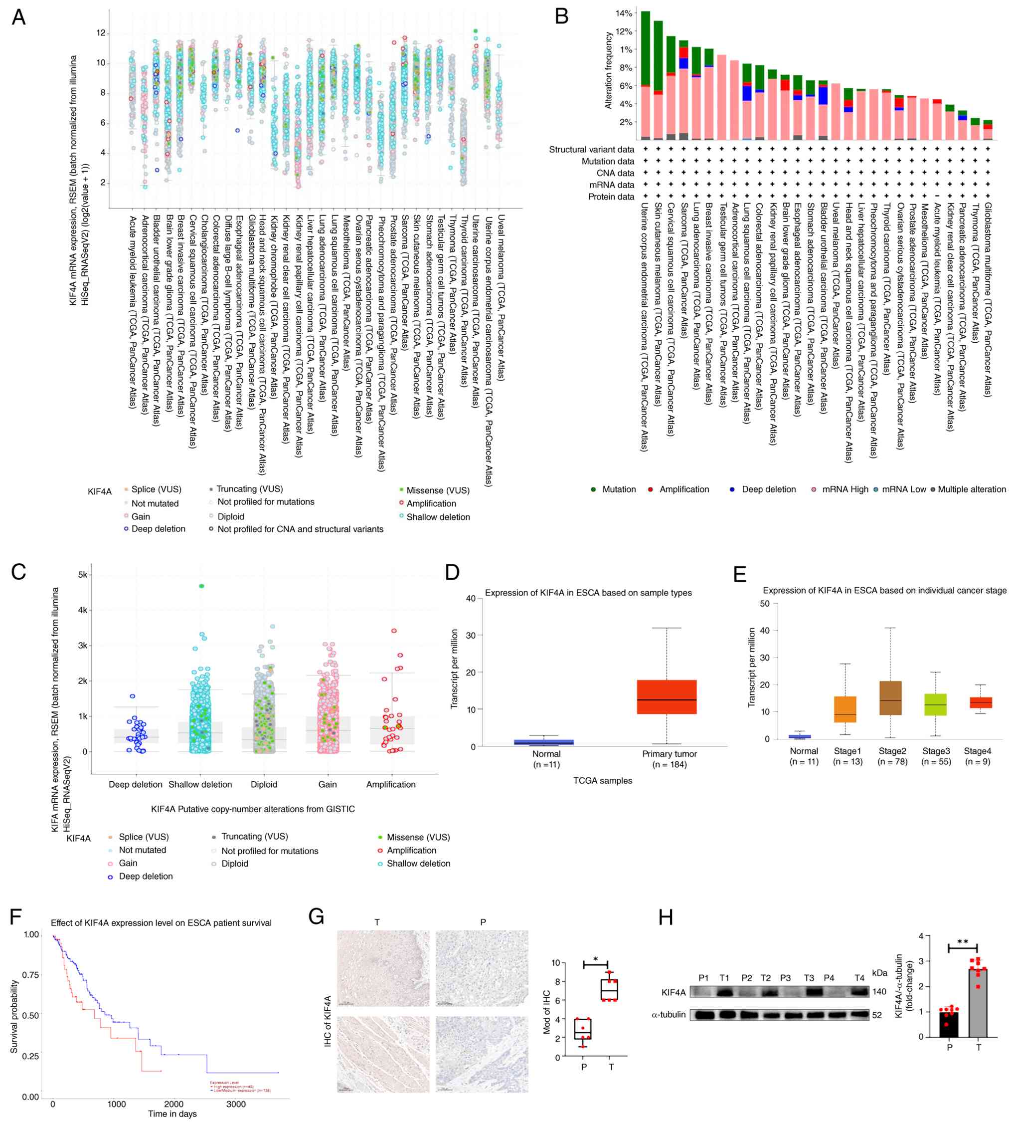 | Figure 1.KIF4A is highly expressed in ESCC and
is associated with poor prognosis. (A) mRNA expression levels of
KIF4A across multiple cancer types from TCGA pan-cancer dataset
(cBioPortal). (B) Genomic variant characteristics of KIF4A across
multiple cancer types (cBioPortal). (C) Association between KIF4A
copy number variation and its mRNA expression (cBioPortal). (D)
KIF4A mRNA expression in normal esophageal tissues and primary ESCC
tissues. (E) KIF4A expression across different tumor stages in ESCC
(UALCAN). (F) Kaplan-Meier survival analysis of overall survival in
patients with ESCC stratified by KIF4A expression (UALCAN, log-rank
P=0.045). (G) IHC staining of KIF4A in ESCC and adjacent
non-malignant tissues, with semi-quantitative statistical analysis.
Scale bar, 100 µm, MOD, semi-quantitative immunohistochemical
score, calculated as the product of staining intensity score and
positive cell proportion score. (H) Western blot analysis of KIF4A
protein expression in four paired ESCC (T) and adjacent
non-malignant (P) tissues. *P<0.05, **P<0.01. ESCC,
esophageal squamous cell carcinoma; ESCA, esophageal carcinoma;
IHC, immunohistochemistry; KIF4A, kinesin family member 4A; TCGA,
The Cancer Genome Atlas. |
Further analysis of TCGA ESCC dataset revealed that
KIF4A expression was significantly higher in primary ESCC tissues
than in adjacent normal esophageal tissues (Fig. 1D). KIF4A expression increased
progressively from stage I to stage II, and remained elevated in
advanced stages, indicating its potential role in early ESCC
tumorigenesis (Fig. 1E).
Kaplan-Meier survival analysis based on the TCGA ESCA dataset via
the UALCAN platform showed that high KIF4A expression was
significantly associated with poorer 5-year overall survival rate
in patients with esophageal carcinoma (log-rank P=0.045; Fig. 1F).
KIF4A expression was further validated in the
43-pair clinical ESCC cohort. IHC staining further validated the
tumor-specific upregulation of KIF4A in ESCC tissues (Fig. 1G). Western blot analysis confirmed
that KIF4A protein was significantly upregulated in ESCC tissues
compared with in paired adjacent non-malignant tissues (Fig. 1H). Clinicopathological analysis
showed that high KIF4A expression was closely associated with lymph
node metastasis and advanced TNM stage (46) in patients with ESCC (Table SI). These results indicated that
KIF4A is upregulated in ESCC and may serve as a promising
prognostic biomarker.
KIF4A knockdown inhibits ESCC cell
proliferation and migration in vitro
Consistent with the clinical tissue findings, KIF4A
protein expression was first detected in a panel of ESCC cell lines
(KYSE-150, KYSE-180, TE-10 and TE-1) and the normal esophageal
epithelial cell line HET-1A. The results showed that KIF4A was
highly expressed in all ESCC cell lines, with the highest
expression in TE-1 and KYSE-150 cells, which were selected for
subsequent loss-of-function assays (Fig. 2A).
 | Figure 2.KIF4A knockdown inhibits ESCC cell
proliferation and migration in vitro. (A) Western blot
analysis of KIF4A protein expression in ESCC cell lines (KYSE-150,
KYSE-180, TE-10, TE-1) and the normal esophageal epithelial cell
line HET-1A. (B) Western blot analysis of KIF4A knockdown
efficiency in TE-1 and KYSE-150 cells transfected with KIF4A
siRNAs. (C) Wound healing assay evaluating the effect of KIF4A
knockdown on ESCC cell migration, with quantitative analysis of
wound healing rate. Scale bar, 200 µm. (D) EdU staining assay
evaluating the effect of KIF4A knockdown on ESCC cell
proliferation, with quantitative analysis of EdU-positive cell
rate. Scale bar, 100 µm. (E) Transwell migration assay evaluating
the effect of KIF4A knockdown on ESCC cell migration, with
quantitative analysis of the number of migratory cells. Scale bar,
100 µm. (F) Colony formation assay evaluating the effect of KIF4A
knockdown on ESCC cell clonogenic capacity. Scale bar, 5 mm.
*P<0.05, **P<0.01, ***P<0.001 and ****P<0.0001 vs. the
NC group; ns, not significant. ESCC, esophageal squamous cell
carcinoma; KIF4A, kinesin family member 4A; NC, negative control;
si, small interfering RNA. |
TE-1 and KYSE-150 cells were transfected with four
different KIF4A siRNAs respectively. Western blot analysis
confirmed effective knockdown of KIF4A protein expression, among
which si-KIF4A-1 showed the highest efficiency and was used for
subsequent experiments (Fig. 2B).
The EdU staining assay showed that KIF4A knockdown significantly
impaired the proliferative capacity of ESCC cells (Fig. 2D). The colony formation assay
further confirmed that KIF4A silencing resulted in fewer and
smaller colonies in both TE-1 and KYSE-150 cells (Fig. 2F). The wound healing assay showed
that KIF4A knockdown significantly reduced the migratory capacity
of ESCC cells (Fig. 2C), which was
further validated by Transwell migration assay (Fig. 2E). Together, these results
demonstrated that KIF4A knockdown inhibits the proliferation and
migration of ESCC cells in vitro.
KIF4A overexpression promotes ESCC
cell proliferation and migration in vitro
Conversely, to further validate the oncogenic role
of KIF4A in ESCC, a KIF4A overexpression model was established in
KYSE-150 cells, a classic well-characterized ESCC cell line with
stable transfection efficiency and consistent genetic background
with the knockdown model to ensure result comparability. The
overexpression efficiency confirmed via western blotting (Fig. 3A). The EdU staining assay showed
that KIF4A overexpression significantly enhanced the proliferative
capacity of ESCC cells (Fig. 3C).
The colony formation assay showed that KIF4A-overexpressing cells
formed significantly more and larger colonies than control cells
(Fig. 3E). Furthermore, the wound
healing and Transwell migration assays consistently showed that
KIF4A overexpression significantly accelerated the migration of
ESCC cells (Fig. 3B and D). These
results confirmed that KIF4A overexpression promotes the
proliferation and migration of ESCC cells in vitro.
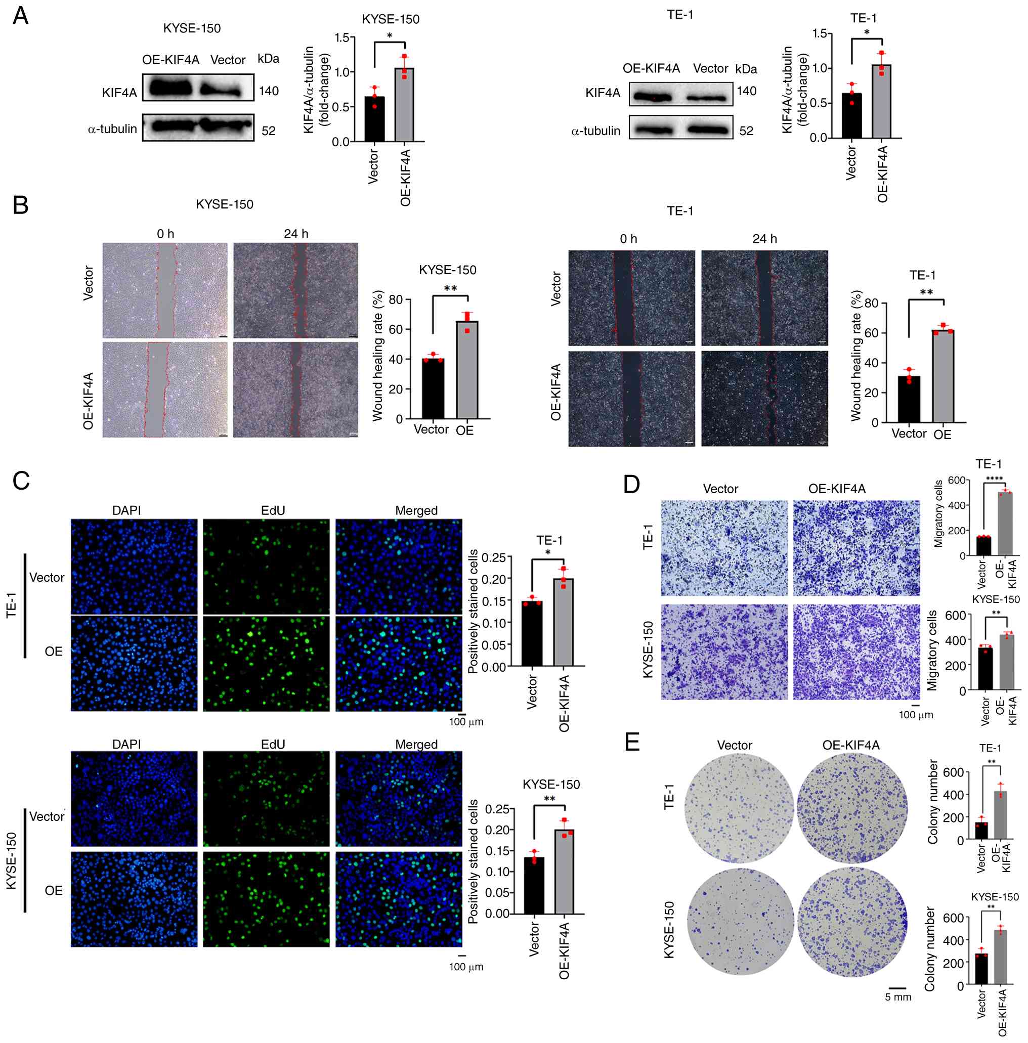 | Figure 3.KIF4A OE promotes ESCC cell
proliferation and migration in vitro. (A) Western blot
analysis of KIF4A OE efficiency in KYSE150 cells. (B) Wound healing
assay evaluating the effect of KIF4A overexpression on ESCC cell
migration, with quantitative analysis of wound healing rate. Scale
bar, 200 µm. (C) EdU staining assay evaluating the effect of KIF4A
overexpression on ESCC cell proliferation, with quantitative
analysis of EdU-positive cell rate. Scale bar, 100 µm. (D)
Transwell migration assay evaluating the effect of KIF4A
overexpression on ESCC cell migration, with quantitative analysis
of the number of migratory cells. Scale bar, 100 µm. (E) Colony
formation assay evaluating the effect of KIF4A overexpression on
ESCC cell clonogenic capacity. Scale bar, 5 mm. *P<0.05,
**P<0.01, ****P<0.0001. ESCC, esophageal squamous cell
carcinoma; KIF4A, kinesin family member 4A; OE, overexpression. |
Identification of the Wnt/β-catenin
pathway as a downstream target of KIF4A
Having confirmed the oncogenic phenotypes of KIF4A
in vitro, the current study next explored its downstream
regulatory mechanism by performing RNA sequencing on
KIF4A-knockdown and control TE-1 cells. A total of 11,068
coexpressed genes were identified in both groups (Fig. 4A). Differential expression analysis
identified 1,697 significant DEGs, including 647 upregulated and
1,050 downregulated genes in the KIF4A-knockdown group (Fig. 4B and C).
Gene Ontology (GO) enrichment analysis showed that
the downregulated DEGs were mainly enriched in the following
biological process terms: ‘Regulation of transcription,
DNA-templated’, ‘protein complex assembly’ and ‘regulation of cell
cycle’ (Fig. 4D). KEGG pathway
enrichment analysis of downregulated DEGs revealed that the ‘basal
cell carcinoma’ pathway, which is tightly associated with the
canonical Wnt signaling cascade, was among the top enriched
pathways (Fig. 4E) (38,40).
Key components of the Wnt/β-catenin pathway, including WNT2B,
WNT5B, WNT8B, TCF7 and FZD2, were significantly downregulated upon
KIF4A knockdown. Heatmap visualization further confirmed that most
Wnt pathway-related genes were positively associated with KIF4A
expression (Fig. 4F). These
results indicated that KIF4A may exert its oncogenic effects in
ESCC by regulating the Wnt/β-catenin signaling pathway.
KIF4A activates the Wnt/β-catenin
pathway by stabilizing the β-catenin-TCF7 complex
To validate the regulatory effect of KIF4A on the
Wnt/β-catenin pathway, western blot analysis was performed. The
results showed that KIF4A knockdown significantly reduced the
protein expression of β-catenin and TCF7 in ESCC cells (Fig. 5A). In paired clinical ESCC
specimens, the protein levels of KIF4A, β-catenin and TCF7 were
consistently elevated in tumor tissues compared with adjacent
normal tissues (Fig. 5B), which
further corroborated the positive association between KIF4A and Wnt
pathway activation at the clinical level. RT-qPCR analysis further
confirmed that KIF4A silencing significantly downregulated the mRNA
expression levels of β-catenin, TCF7, and the canonical Wnt pathway
downstream target genes, including AXIN2, MMP-7, cyclin D1 and
c-Myc (Fig. 5C).
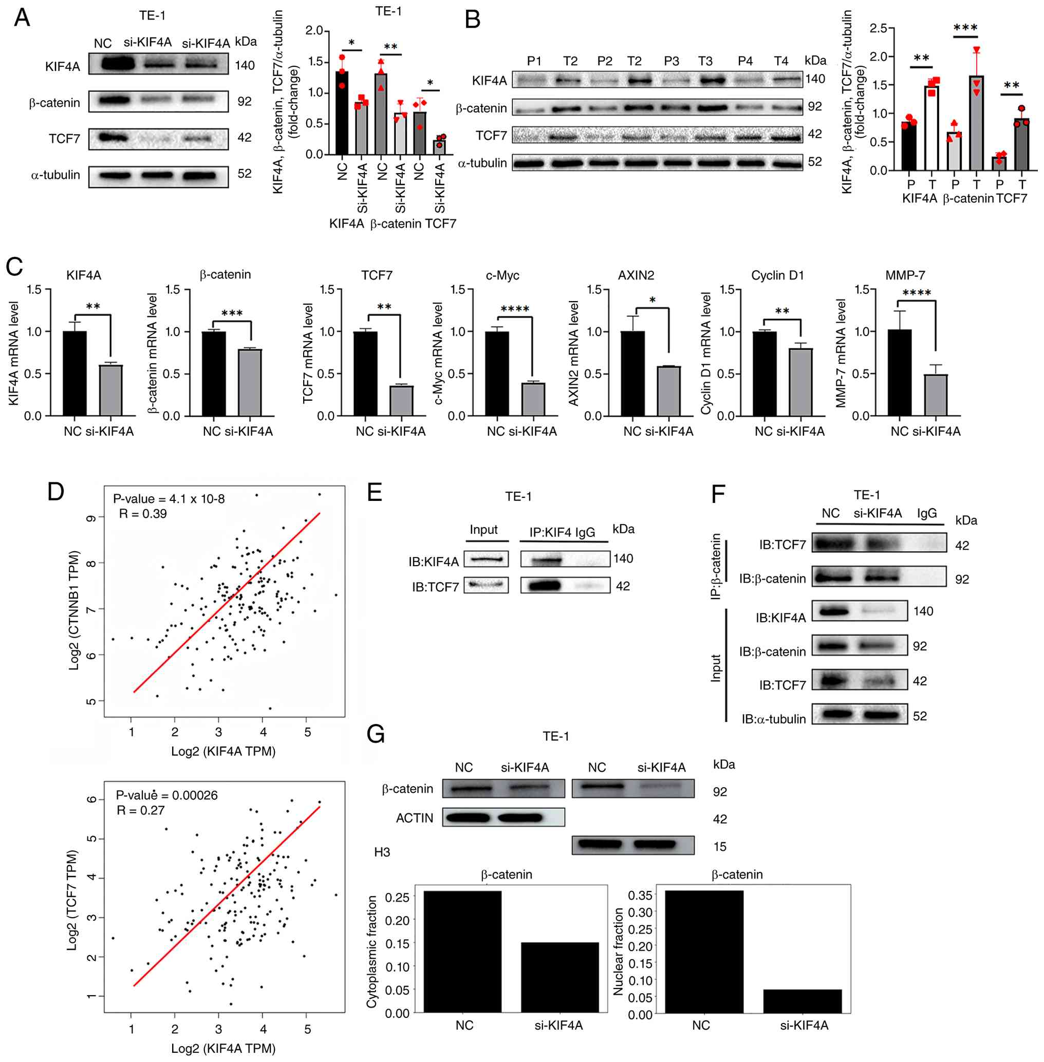 | Figure 5.KIF4A activates the Wnt/β-catenin
pathway by stabilizing the β-catenin-TCF7 complex. Western blot
analysis of β-catenin and TCF7 protein expression in (A)
KIF4A-knockdown ESCC cells and (B) paired ESCC/adjacent tissues.
(C) Reverse transcription-quantitative PCR analysis of the mRNA
expression levels of Wnt/β-catenin pathway components and
downstream target genes in KIF4A-knockdown ESCC cells. (D) Pearson
correlation analysis between KIF4A and CTNNB1/TCF7 expression in
ESCC tissues (The Cancer Genome Atlas dataset). (E) Co-IP assay
confirming the direct interaction between KIF4A and TCF7 in TE-1
cells. (F) Co-IP assay showing that KIF4A knockdown disrupts the
interaction between β-catenin and TCF7 in TE-1 cells. (G) Relative
protein levels of β-catenin in nuclear and cytoplasmic fractions of
ESCC cells with or without KIF4A silencing. The
nuclear-to-cytoplasmic ratio of β-catenin was markedly reduced upon
KIF4A knockdown. *P<0.05, **P<0.01, ***P<0.001 and
****P<0.0001 vs. the NC group. CTNNB1, β-catenin; ESCC,
esophageal squamous cell carcinoma; IB, immunoblot; IP,
immunoprecipitation; KIF4A, kinesin family member 4A; NC, negative
control; si, small interfering; TCF7, T-cell factor 7; T, tumor
tissue; P, peritumoral normal tissue. |
Correlation analysis using TCGA ESCC dataset showed
that KIF4A expression was positively but weakly correlated with the
expression of β-catenin (r=0.39, P=4.1×10⁻⁸) and TCF7
(r=0.27, P=0.00026) in ESCC tissues (Fig. 5D). A co-IP assay confirmed that
KIF4A directly interacted with TCF7 in ESCC cells (Fig. 5E). Furthermore, the co-IP assay
showed that KIF4A knockdown markedly disrupted the interaction
between β-catenin and TCF7 in ESCC cells (Fig. 5F). Nuclear-cytoplasmic
fractionation showed that KIF4A knockdown markedly reduced nuclear
β-catenin (5.1-fold downregulation) and moderately decreased
cytoplasmic β-catenin (1.7-fold reduction). The
nuclear-to-cytoplasmic ratio of β-catenin was decreased by 65.9%
upon KIF4A silencing, indicating that KIF4A promotes β-catenin
nuclear translocation and canonical Wnt pathway activation
(Fig. 5G). Based on these
findings, it may be hypothesized that KIF4A facilitates activation
of the Wnt/β-catenin pathway, at least in part, by stabilizing the
β-catenin-TCF7 transcription complex via direct interaction with
TCF7. However, the possibility that KIF4A may also indirectly
modulate this complex through other intermediate proteins or
post-translational modifications cannot be excluded; therefore,
this requires further validation.
KIF4A is essential for the pro-tumor
effects of Wnt/β-catenin pathway activation
To further confirm the functional association
between KIF4A and the Wnt/β-catenin pathway, rescue experiments
were performed using a Wnt pathway agonist (Wnt agonist 1). Western
blot analysis showed that Wnt agonist 1 significantly upregulated
β-catenin expression in control TE-1 cells, but failed to
effectively restore β-catenin protein level in KIF4A-knockdown
cells; no statistically significant difference was detected between
the si-KIF4A group and the Wnt agonist 1 + si-KIF4A group (Fig. 6A). Functional assays showed
consistent results: Wnt agonist 1 markedly enhanced the
proliferation and migration abilities of control cells, but could
not significantly reverse the inhibitory effects induced by KIF4A
knockdown (Fig. 6B and C). These
results demonstrated that KIF4A is an essential regulator required
for full activation of the canonical Wnt/β-catenin signaling
pathway, and thus drives the malignant phenotype of ESCC cells.
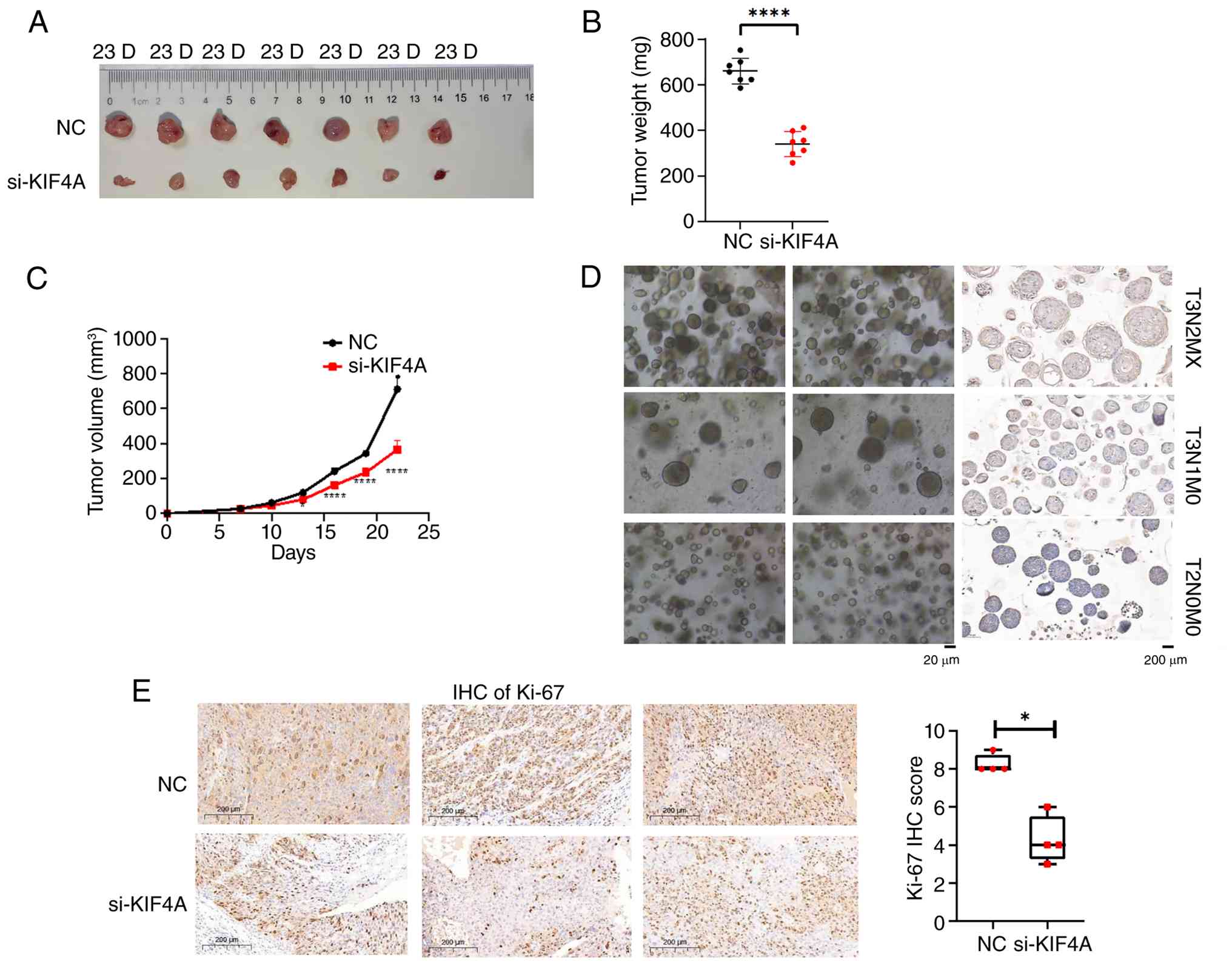
KIF4A promotes ESCC xenograft tumor
growth in vivo
To validate the in vivo oncogenic role of
KIF4A, a subcutaneous xenograft tumor model was established in nude
mice. The results showed that KIF4A knockdown significantly
inhibited the growth of ESCC xenograft tumors, with markedly
reduced tumor volume and weight in the KIF4A-knockdown group
compared with the control group (Fig.
7A-C). Furthermore, IHC staining showed that the expression of
Ki-67, a proliferation marker, was significantly reduced in
xenograft tumors from the KIF4A-knockdown group (Fig. 7E). In addition, IHC staining of
ESCC patient-derived organoids showed that KIF4A expression was
positively associated with tumor stage progression (Fig. 7D). These results confirmed that
KIF4A promotes ESCC tumorigenesis in vivo.
Discussion
ESCC is the predominant histological subtype of EC
in China, with high morbidity and mortality rates, and limited
targeted therapeutic options (47,48).
The identification of novel prognostic biomarkers and therapeutic
targets is critical to improve the clinical outcomes of patients
with ESCC. In the present study, the expression, prognostic value,
biological functions and molecular mechanisms of KIF4A were
systematically investigated in ESCC. The findings demonstrated that
KIF4A was upregulated in ESCC, predicted a poor prognosis, and
drove ESCC cell proliferation and migration by activating the
canonical Wnt/β-catenin signaling pathway.
In terms of molecular function, KIF4A is a member of
the kinesin superfamily, which serves critical roles in mitotic
progression and intracellular transport (7,8,13,49).
Previous studies have validated the oncogenic roles of KIF4A in
multiple malignancies. For example, KIF4A drives hepatocellular
carcinoma proliferation by activating the Akt signaling pathway
(13), promotes endometrial cancer
progression by regulating TPX2 protein degradation (11), and enhances colorectal cancer cell
invasion by upregulating MMP2/MMP9 expression (17). KIF4A was identified in our previous
study (50) as a prognostic
biomarker for ESCC, but its functional mechanisms in ESCC remain
largely unknown. In the current study, it was confirmed that KIF4A
was significantly upregulated in ESCC tissues and cell lines, and
its high expression was associated with advanced tumor stage and
poor overall survival in patients with ESCC. Loss- and
gain-of-function assays demonstrated that KIF4A may exert strong
oncogenic effects, promoting ESCC cell proliferation, migration and
in vivo xenograft tumor growth, which is consistent with its
oncogenic roles in other malignancies.
Moving beyond functional phenotypes, the current
study further elucidated the downstream regulatory mechanism of
KIF4A in ESCC via RNA sequencing analysis, which revealed that the
Wnt/β-catenin signaling pathway was the key downstream target of
KIF4A. The canonical Wnt/β-catenin pathway is a well-characterized
oncogenic cascade, which is aberrantly activated in 30–50% of ESCC
cases, and is associated with tumor progression, therapeutic
resistance and poor prognosis (34,35).
In the canonical Wnt pathway, stabilized β-catenin translocates to
the nucleus, forms a transcription complex with TCF/LEF family
members, and drives the expression of pro-oncogenic target genes,
such as c-Myc, cyclin D1 and MMP-7 (51,52).
In the present study, it was demonstrated that KIF4A knockdown
significantly downregulated the expression of β-catenin, TCF7 and
the downstream target genes of the Wnt/β-catenin pathway, whereas
KIF4A overexpression exerted the opposite effects. Rescue
experiments further demonstrated that the pro-proliferative and
pro-migratory effects of Wnt/β-catenin pathway activation are
largely dependent on KIF4A expression in ESCC cells., indicating
that KIF4A drives ESCC progression via activating the Wnt/β-catenin
pathway.
Notably, the mRNA-level correlation between KIF4A
and core Wnt pathway molecules (β-catenin and TCF7) in the TCGA
ESCC cohort was at a weak-to-moderate level. This is consistent
with the universal characteristics of solid tumor transcriptome
studies: Clinical tumor tissues are highly heterogeneous and
influenced by multiple confounding factors, including individual
genetic background, tumor microenvironment, pathological stage and
differentiation status. In solid tumor research, where intratumor
heterogeneity may dilute correlation signals observed from
single-biopsy sampling, modest correlation coefficients with
statistical significance may still reflect biologically relevant
relationships, although the threshold for ‘biological meaning’
remains context-dependent and requires independent validation
(53,54). Mechanistically, KIF4A, as a kinesin
family protein, regulates the Wnt/β-catenin pathway mainly at the
post-translational level rather than directly driving the
transcription of CTNNB1 and TCF7.
Notably, the present study further revealed the
mechanistic basis of KIF4A-mediated Wnt pathway activation. The
co-IP assay confirmed that KIF4A directly interacted with TCF7, and
KIF4A knockdown disrupted the interaction between β-catenin and
TCF7. As summarized in Fig. 8,
these findings collectively support a model in which KIF4A
stabilizes the β-catenin-TCF7 transcription complex via direct
binding to TCF7, thus enhancing the transcriptional output of the
Wnt/β-catenin pathway. Notably, this mechanism does not rule out
potential indirect regulatory effects mediated by additional
cofactors, and the exact structural basis underlying this
regulation remains to be fully elucidated. This finding provides
novel insights into the regulatory mechanism of the Wnt/β-catenin
pathway in ESCC, and establishes the KIF4A-Wnt/β-catenin axis as a
key oncogenic cascade in ESCC progression.
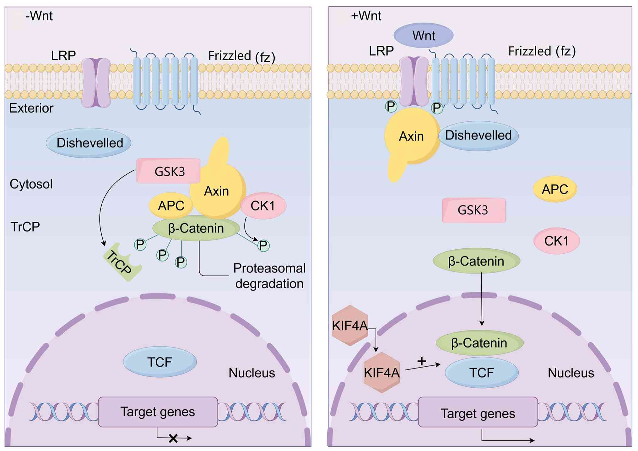 | Figure 8.Schematic diagram of the mechanism by
which KIF4A drives esophageal squamous cell carcinoma progression
via activating the canonical Wnt/β-catenin signaling pathway. KIF4A
stabilizes the β-catenin-TCF7 transcription complex via direct
interaction with TCF7, thereby enhancing the transcriptional
activity of the Wnt/β-catenin pathway and promoting the expression
of downstream pro-oncogenic target genes, ultimately driving ESCC
cell proliferation, migration and tumorigenesis. KIF4A, kinesin
family member 4A; TCF, T-cell factor. APC, adenomatous polyposis
coli; CK1, casein kinase 1; GSK3, glycogen synthase kinase 3; LRP,
low-density lipoprotein receptor-related protein; TrCP,
β-transducin repeat-containing protein. |
From a clinical perspective, the current study has
two important translational values for ESCC management. First,
KIF4A was validated as a promising prognostic biomarker for ESCC.
High KIF4A expression was closely associated with advanced tumor
stage and poor overall survival, which may help clinicians stratify
high-risk ESCC patients at initial diagnosis, optimize personalized
follow-up strategies and guide adjuvant treatment decisions.
Second, the KIF4A-Wnt/β-catenin axis was identified as a potential
therapeutic target for ESCC. Small-molecule inhibitors targeting
KIF4A, such as WZ-3146, have shown significant antitumor efficacy
in preclinical glioma models (20), which provides a preclinical basis
for the development of KIF4A-targeted therapies for ESCC. For
patients with ESCC, and with KIF4A upregulation and aberrant
Wnt/β-catenin activation, combination therapy with KIF4A inhibitors
and Wnt pathway inhibitors may improve therapeutic efficacy and
overcome drug resistance, which warrants further preclinical and
clinical investigation. However, the clinical translation of KIF4A
inhibitors faces potential specificity challenges. Given that KIF4A
is also expressed in actively dividing normal cells and shares
structural homology with other kinesin family members, off-target
effects on normal proliferative tissues and non-specific inhibition
of other kinesins may occur. Further structural optimization of the
ATP-binding pocket of inhibitors and the development of
ESCC-specific delivery strategies (such as antibody-drug
conjugates) are needed to minimize off-target toxicity in future
studies.
The current study has several limitations that need
to be addressed in future research. First, although the co-IP assay
showed that KIF4A knockdown reduced the amount of TCF7
co-precipitated with β-catenin, we acknowledge that the total
protein level of TCF7 in the input was also decreased after KIF4A
silencing. Therefore, the reduced TCF7 in the immunoprecipitated
complex cannot be solely attributed to the weakened interaction
between β-catenin and TCF7; the downregulation of total TCF7
expression may also contribute to this observation. The exact
effect of KIF4A on the binding affinity between β-catenin and TCF7,
independent of total protein expression changes, needs to be
further validated via in vitro pull-down assays with
normalized protein input and yeast two-hybrid systems. Notably, the
current work has not fully delineated whether KIF4A modulates
β-catenin protein stability through regulating its ubiquitination
degradation, and the in situ spatial colocalization of KIF4A
with β-catenin/TCF7 in clinical ESCC tissues remains to be
visualized at the single-cell level. These post-translational
regulatory details and histological spatial evidence will be
systematically investigated in our follow-up studies. Second, the
prognostic value of KIF4A was validated in a single-center cohort,
and further large-scale, multi-center clinical studies are needed
to confirm its clinical application value. Third, the in
vivo antitumor efficacy of KIF4A inhibitors needs to be
validated in patient-derived xenograft models and ESCC organoid
models to lay a more solid foundation for clinical translation.
In conclusion, the present study systematically
indicated that KIF4A is significantly upregulated in ESCC, and its
high expression is associated with advanced tumor stage and poor
overall survival in patients with ESCC. Functional experiments
confirmed that KIF4A can drive ESCC cell proliferation, migration
and in vivo tumorigenesis. Mechanistically, the present
findings suggested that KIF4A exerts its oncogenic effects in ESCC,
at least in part, through activation of the canonical Wnt/β-catenin
signaling pathway, potentially via stabilization of the
β-catenin-TCF7 transcription complex. These findings identify KIF4A
as a promising prognostic biomarker and therapeutic target for
ESCC, and provide novel insights into the molecular mechanisms of
ESCC progression.
Supplementary Material
Supporting Data
Acknowledgements
The authors would like to thank Professor Shanshan
Liang (Zhongshan Hospital of Dalian University, Dalian, China) for
providing technical guidance and support.
Funding
This work was supported by the Educational Commission of Hubei
Province of China (grant no. B2023106), the Natural Science
Foundation of Hubei Province for Young Scholars (grant no.
2022CFB930), the Natural Science Foundation of Hubei Province
(grant no. 2026AFB087) and the Key Project of Science and
Technology Bureau of Xiangyang City (grant no. 2022YL28A).
Availability of data and materials
The sequencing data generated in the present study
may be found in the Open Archive for Miscellaneous Data (OMIX),
China National Center for Bioinformation/Beijing Institute of
Genomics, Chinese Academy of Sciences, under accession number
OMIX018645 or at the following URL: https://ngdc.cncb.ac.cn/omix/preview/cWRNNvxO. Access
to the data is subject to restrictions due to privacy. The other
data generated in the present study may be requested from the
corresponding author.
Authors' contributions
LZ and XS conceptualized the study and designed the
overall research framework. JC and TW developed the research
methodology, including the design and optimization of experimental
protocols for cellular function assays and molecular detection. JC
and DW performed the formal data analysis and statistical
interpretation. JC, TW and DW conducted the experimental
investigations. WL, CL and YY participated in the conception and
design of the clinical research protocol, performed the acquisition
and pathological verification of clinical tissue specimens,
analyzed and interpreted the clinical patient data, and critically
reviewed and revised the clinical content of the manuscript for
important intellectual content. They also provided essential
research resources, including the clinical tissue biobank and
experimental platform support. JC and DW drafted the original
manuscript. LZ, XS, WL, CL and YY critically reviewed and edited
the manuscript. LZ and XS supervised the whole study and acquired
the research funding. LZ and XS confirm the authenticity of all the
raw data, and WL, CL and YY confirm the authenticity and integrity
of the clinical data and tissue specimens. All authors have read
and approved the final version of the manuscript.
Ethics approval and consent to
participate
All studies involving human participants were
reviewed and approved by the Institutional Review Board of
Xiangyang No.1 People's Hospital (approval nos. 2025KY082 and
2025KY142). Written informed consent was obtained from all enrolled
participants, and all experiments involving human tissues were
performed in accordance with The Declaration of Helsinki. All
animal experiments were reviewed and approved by the Institutional
Animal Care and Use Committee of Xiangyang No.1 People's Hospital
(approval no. XYYYE20240197), and performed in strict accordance
with the ARRIVE guidelines and national guidelines for the care and
use of laboratory animals.
Patient consent for publication
Written informed consent for the publication of
clinical data, pathological images and related research results was
obtained from all enrolled participants.
Competing interests
The authors declare that they have no competing
interests.
References
|
1
|
Bray F, Laversanne M, Sung H, Ferlay J,
Siegel RL, Soerjomataram I and Jemal A: Global cancer statistics
2022: GLOBOCAN estimates of incidence and mortality worldwide for
36 cancers in 185 countries. CA Cancer J Clin. 74:229–263.
2024.PubMed/NCBI
|
|
2
|
Zheng Y, Teng Y, He S, et al:
Epidemiological characteristics of esophageal cancer worldwide and
in China, 2022. China Cancer. 34:165–170. 2025.(In Chinese).
|
|
3
|
Dawsey SM and Duits LC: A substantial
advance for screening of oesophageal cancer. Lancet Gastroenterol
Hepatol. 8:393–395. 2023. View Article : Google Scholar : PubMed/NCBI
|
|
4
|
Mwachiro MM and Dawsey SM: The use of
questionnaire-based risk-stratification tools in screening for
esophageal squamous cell carcinoma. Gastrointest Endosc.
93:119–121. 2021. View Article : Google Scholar : PubMed/NCBI
|
|
5
|
Luo Q, Xiao S, Xu S, Qian F, Jiaying T,
Yao X and Hui L: Recent advances in the glycolytic processes linked
to tumor metastasis. Curr Mol Pharmacol. 17:e187614293083612024.
View Article : Google Scholar : PubMed/NCBI
|
|
6
|
Pourhanifeh MH, Farrokhi-Kebria H,
Mostanadi P, Farkhondeh T and Samarghandian S: Anticancer
properties of baicalin against breast cancer and other
gynecological cancers: Therapeutic opportunities based on
underlying mechanisms. Curr Mol Pharmacol. 17:e187614292630632024.
View Article : Google Scholar : PubMed/NCBI
|
|
7
|
Zhang R, Liu S, Gong B, Xie W, Zhao Y, Xu
L, Zheng Y, Jin S, Ding C, Xu C and Dong Z: Kif4A mediates
resistance to neoadjuvant chemoradiotherapy in patients with
advanced colorectal cancer via regulating DNA damage response. Acta
Biochim Biophys Sin (Shanghai). 54:940–951. 2022. View Article : Google Scholar : PubMed/NCBI
|
|
8
|
Lan Z, Wang J, Wang J, et al: Expression
of KIF4A in hepatocellular carcinoma and its prognostic value.
Chinese Journal of General Surgery. 31:55–63. 2022.(In
Chinese).
|
|
9
|
Sheng L, Hao SL, Yang WX and Sun Y: The
multiple functions of kinesin-4 family motor protein KIF4 and its
clinical potential. Gene. 678:90–99. 2018. View Article : Google Scholar : PubMed/NCBI
|
|
10
|
Yang K, Li D, Jia W, Song Y, Sun N, Wang
J, Li H and Yin C: MiR-379-5p inhibits the proliferation,
migration, and invasion of breast cancer by targeting KIF4A. Thorac
Cancer. 13:1916–1924. 2022. View Article : Google Scholar : PubMed/NCBI
|
|
11
|
Zhang J, An L, Zhao R, Shi R, Zhou X, Wei
S, Zhang Q, Zhang T, Feng D, Yu Z and Wang H: KIF4A promotes
genomic stability and progression of endometrial cancer through
regulation of TPX2 protein degradation. Mol Carcinog. 62:303–318.
2023. View
Article : Google Scholar : PubMed/NCBI
|
|
12
|
Zhang D, Lu W and Samadi N: KIF4A
knockdown inhibits tumor progression and promotes chemo-sensitivity
via induction of P21 in lung cancer cells. Chem Biol Drug Des.
101:1042–1047. 2023. View Article : Google Scholar : PubMed/NCBI
|
|
13
|
Huang Y, Wang H, Lian Y, Wu X, Zhou L,
Wang J, Deng M and Huang Y: Upregulation of kinesin family member
4A enhanced cell proliferation via activation of Akt signaling and
predicted a poor prognosis in hepatocellular carcinoma. Cell Death
Dis. 9:1412018. View Article : Google Scholar : PubMed/NCBI
|
|
14
|
Zhang C, Wang M, Ying Y, Meng F, Gao H,
Zeng S, Zhu Y, Liu A, Zhang Z and Xu C: Knockdown of kinesin family
member 4A inhibits cell proliferation, migration, and invasion
while promoting apoptosis of urothelial bladder carcinoma cells.
Cancer Med. 12:12581–12592. 2023. View Article : Google Scholar : PubMed/NCBI
|
|
15
|
Cho SY, Kim S, Kim G, Singh P and Kim DW:
Integrative analysis of KIF4A, 9, 18A, and 23 and their clinical
significance in low-grade glioma and glioblastoma. Sci Rep.
9:45992019. View Article : Google Scholar : PubMed/NCBI
|
|
16
|
Kahm YJ, Kim IG, Jung U, Lee JH and Kim
RK: Impact of KIF4A on cancer stem cells and EMT in lung cancer and
glioma. Cancers (Basel). 15:55232023. View Article : Google Scholar : PubMed/NCBI
|
|
17
|
Tang AQ, Jiang T and Bai J: Effects of
KIF4A on migration and invasion of colorectal cancer cells and its
mechanisms. J Xuzhou Med Univ. 40:710–714. 2020.
|
|
18
|
Wandke C, Barisic M, Sigl R, Rauch V, Wolf
F, Amaro AC, Tan CH, Pereira AJ, Kutay U, Maiato H, et al: Human
chromokinesins promote chromosome congression and spindle
microtubule dynamics during mitosis. J Cell Biol. 198:847–863.
2012. View Article : Google Scholar : PubMed/NCBI
|
|
19
|
Zhang H, Jing M and Du X: KIF4A on
TGF-β1/Smad3 pathway: A preliminary investigation on the
proliferation and immune response in in-vitro cultured breast
cancer cells. Comb Chem High Throughput Screen. 28:2959–2968. 2025.
View Article : Google Scholar : PubMed/NCBI
|
|
20
|
Yan T, Jiang Q, Ni G, Ma H, Meng Y, Kang
G, Xu M, Peng F, Li H, Chen X and Wang M: WZ-3146 acts as a novel
small molecule inhibitor of KIF4A to inhibit glioma progression by
inducing apoptosis. Cancer Cell Int. 24:2212024. View Article : Google Scholar : PubMed/NCBI
|
|
21
|
Wang L, Liu G, Bolor-Erdene E, Li Q, Mei Y
and Zhou L: Identification of KIF4A as a prognostic biomarker for
esophageal squamous cell carcinoma. Aging (Albany NY).
13:24050–24070. 2021. View Article : Google Scholar : PubMed/NCBI
|
|
22
|
Nusse R and Varmus HE: Many tumors induced
by the mouse mammary tumor virus contain a provirus integrated in
the same region of the host genome. Cell. 31:99–109. 1982.
View Article : Google Scholar : PubMed/NCBI
|
|
23
|
Bugter JM, Fenderico N and Maurice MM:
Mutations and mechanisms of WNT pathway tumour suppressors in
cancer. Nat Rev Cancer. 21:5–21. 2021. View Article : Google Scholar : PubMed/NCBI
|
|
24
|
Koni M, Pinnarò V and Brizzi MF: The Wnt
signalling pathway: A tailored target in cancer. Int J Mol Sci.
21:76972020. View Article : Google Scholar : PubMed/NCBI
|
|
25
|
De A: Wnt/Ca2+ signaling
pathway: A brief overview. Acta Biochim Biophys Sin (Shanghai).
43:745–756. 2011. View Article : Google Scholar : PubMed/NCBI
|
|
26
|
Akoumianakis I, Polkinghorne M and
Antoniades C: Non-canonical WNT signalling in cardiovascular
disease: Mechanisms and therapeutic implications. Nat Rev Cardiol.
19:783–797. 2022. View Article : Google Scholar : PubMed/NCBI
|
|
27
|
Rim EY, Clevers H and Nusse R: The Wnt
pathway: From signaling mechanisms to synthetic modulators. Annu
Rev Biochem. 91:571–598. 2022. View Article : Google Scholar : PubMed/NCBI
|
|
28
|
Clevers H and Nusse R: Wnt/β-catenin
signaling and disease. Cell. 149:1192–1205. 2012. View Article : Google Scholar : PubMed/NCBI
|
|
29
|
Gao R, Zheng X, Jiang A, He W and Liu T:
Modulating β-catenin/BCL9 interaction with
cell-membrane-camouflaged carnosic acid to inhibit Wnt pathway and
enhance tumor immune response. Front Immunol. 14:12742232023.
View Article : Google Scholar : PubMed/NCBI
|
|
30
|
Liu J, Xiao Q, Xiao J, Niu C, Li Y, Zhang
X, Zhou Z, Shu G and Yin G: Wnt/β-catenin signalling: Function,
biological mechanisms, and therapeutic opportunities. Signal
Transduct Target Ther. 7:32022. View Article : Google Scholar : PubMed/NCBI
|
|
31
|
Zou G and Park JI: Wnt signaling in liver
regeneration, disease, and cancer. Clin Mol Hepatol. 29:33–50.
2023. View Article : Google Scholar : PubMed/NCBI
|
|
32
|
Xu X, Zhang M, Xu F and Jiang S: Wnt
signaling in breast cancer: Biological mechanisms, challenges and
opportunities. Mol Cancer. 19:1652020. View Article : Google Scholar : PubMed/NCBI
|
|
33
|
Ma Z, Zhu T, Wang H, Wang B, Fu L and Yu
G: E2F1 Reduces Sorafenib's sensitivity of esophageal carcinoma
cells via modulating the miR-29c-3p/COL11A signaling axis. Curr Mol
Pharmacol. 16:e0603232023.
|
|
34
|
Wang W, Liu HT, Cai YR, et al: Expression
and significance of relevant factors of Wnt2, β-catenin, c-myc and
cyclin D1 in esophageal squamous cell carcinoma tissue. Cancer
Research on Prevention and Treatment. 36:213–215. 2009.(In
Chinese).
|
|
35
|
Zhan Z, An Z, Ren M, Zhao B and Hou X:
Correlation between classic Wnt signaling pathway and
radioresistance of esophageal cancer cells. Chin J Radiat Oncol.
30:614–618. 2021.(In Chinese).
|
|
36
|
Hou PF, Jiang T, Chen F, Shi PC, Li HQ,
Bai J and Song J: KIF4A facilitates cell proliferation via
induction of p21-mediated cell cycle progression and promotes
metastasis in colorectal cancer. Cell Death Dis. 9:4772018.
View Article : Google Scholar : PubMed/NCBI
|
|
37
|
Hu G, Yan Z, Zhang C, Cheng M, Yan Y, Wang
Y, Deng L, Lu Q and Luo S: FOXM1 promotes hepatocellular carcinoma
progression by regulating KIF4A expression. J Exp Clin Cancer Res.
38:1882019. View Article : Google Scholar : PubMed/NCBI
|
|
38
|
Livak KJ and Schmittgen TD: Analysis of
relative gene expression data using real-time quantitative PCR and
the 2(−Delta Delta C(T)) method. Methods. 25:402–408. 2001.
View Article : Google Scholar : PubMed/NCBI
|
|
39
|
Love MI, Huber W and Anders S: Moderated
estimation of fold change and dispersion for RNA-seq data with
DESeq2. Genome Biol. 15:5502014. View Article : Google Scholar : PubMed/NCBI
|
|
40
|
Yu G, Wang LG, Han Y and He QY:
clusterProfiler: An R package for comparing biological themes among
gene clusters. OMICS. 16:284–287. 2012. View Article : Google Scholar : PubMed/NCBI
|
|
41
|
Kanehisa M, Furumichi M, Sato Y, Matsuura
Y and Ishiguro-Watanabe M: KEGG: Biological systems database as a
model of the real world. Nucleic Acids Res. 53:D672–D677. 2025.
View Article : Google Scholar : PubMed/NCBI
|
|
42
|
Kanehisa M: Toward understanding the
origin and evolution of cellular organisms. Protein Sci.
28:1947–1951. 2019. View Article : Google Scholar : PubMed/NCBI
|
|
43
|
Kanehisa M and Goto S: KEGG: Kyoto
encyclopedia of genes and genomes. Nucleic Acids Res. 28:27–30.
2000. View Article : Google Scholar : PubMed/NCBI
|
|
44
|
Kilkenny C, Browne WJ, Cuthill IC, Emerson
M and Altman DG: Improving bioscience research reporting: The
ARRIVE guidelines for reporting animal research. PLoS Biol.
8:e10004122010. View Article : Google Scholar : PubMed/NCBI
|
|
45
|
National Research Council (US) Committee
for the Update of the Guide for the Care and Use of Laboratory
Animals, . Guide for the Care and Use of Laboratory Animals. 8th
edition. National Academies Press; Washington, DC: 2011
|
|
46
|
Amin MB, Edge SB, Greene FL, Byrd DR,
Brookland RK, Washington MK, Gershenwald JE, Compton CC, Hess KR,
Sullivan DC, et al: AJCC Cancer Staging Manual. 8th edition. New
York: Springer; 2017
|
|
47
|
Arnold M, Abnet CC, Neale RE, Vignat J,
Giovannucci EL, McGlynn KA and Bray F: Global burden of 5 major
types of gastrointestinal cancer. Gastroenterology.
159:335–349.e15. 2020. View Article : Google Scholar : PubMed/NCBI
|
|
48
|
He J, Chen WQ, Li ZS, et al: China
guideline for the screening, early detection and early treatment of
esophageal cancer (2022, Beijing). China Cancer. 31:401–436.
2022.(In Chinese).
|
|
49
|
Hirokawa N and Noda Y: Intracellular
transport and kinesin superfamily proteins, KIFs: Structure,
function, and dynamics. Physiol Rev. 88:1089–1118. 2008. View Article : Google Scholar : PubMed/NCBI
|
|
50
|
Wang L, Liu G, Bolor-Erdene E, Li Q, Mei Y
and Zhou L: Identification of KIF4A as a prognostic biomarker for
esophageal squamous cell carcinoma. Aging (Albany NY).
13:24050–24070. 2021. View Article : Google Scholar : PubMed/NCBI
|
|
51
|
Xue C, Chu Q, Shi Q, Zeng Y, Lu J and Li
L: Wnt signaling pathways in biology and disease: Mechanisms and
therapeutic advances. Signal Transduct Target Ther. 10:1062025.
View Article : Google Scholar : PubMed/NCBI
|
|
52
|
Fang H, Shi X, Gao J, Yan Z, Wang Y, Chen
Y, Zhang J and Guo W: TMEM209 promotes hepatocellular carcinoma
progression by activating the Wnt/β-catenin signaling pathway
through KPNB1 stabilization. Cell Death Discov. 10:4382024.
View Article : Google Scholar : PubMed/NCBI
|
|
53
|
Gerlinger M, Rowan AJ, Horswell S, Math M,
Larkin J, Endesfelder D, Gronroos E, Martinez P, Matthews N,
Stewart A, et al: Intratumor heterogeneity and branched evolution
revealed by multiregion sequencing. N Engl J Med. 366:883–892.
2012. View Article : Google Scholar : PubMed/NCBI
|
|
54
|
The ICGC/TCGA Pan-Cancer Analysis of Whole
Genomes Consortium, . Pan-Cancer Analysis of Whole Genomes. Nature.
578:82–93. 2020. View Article : Google Scholar : PubMed/NCBI
|