Introduction
Over the past five years, research on cancer
immunometabolism has shifted from characterizing isolated metabolic
abnormalities to establishing nutrient availability as a dynamic
determinant of immune-cell fitness and therapeutic responses.
Arginine has become a core regulatory node, as its local
availability is jointly controlled by tumor-cell biosynthesis and
uptake, arginase 1 (ARG1)/arginase 2 (ARG2) activity, nitric oxide
synthase (NOS)-dependent metabolic diversion and stromal-vascular
transport. Experimentally, using engineered bacteria to increase
intratumoral L-arginine enhanced T-cell infiltration and produced a
synergistic effect with programmed death-ligand 1 (PD-L1) blockade,
while low-arginine conditions trigger metabolic and transcriptional
reprogramming in activated CD4+ T cells via the activating
transcription factor 4 (ATF4)-solute carrier family 7 member 11
(SLC7A11)-GSH axis, ultimately endowing T cells with Treg-like
immunosuppressive properties (1,2).
Meanwhile, studies have linked arginine-derived
polyamine metabolism to ferroptosis and immune remodeling.
Arginine-derived polyamines can enhance ferroptosis through
H2O2-dependent lipid peroxidation and an iron
overload-WNT/MYC proto-oncogene (MYC)-ornithine decarboxylase 1
(ODC1)-polyamine positive-feedback circuit (3). However, ferroptosis is not uniformly
immunostimulatory. Ferroptotic cancer cells may impair
dendritic-cell maturation and antigen cross-presentation and the
immunological consequences of ferroptosis vary depending on the
affected cell populations, the stage of cell death and the
surrounding microenvironment (4,5).
These advances reveal major unresolved issues: Arginine metabolism,
polyamine homeostasis, ferroptosis susceptibility and immune
feedback are still generally regarded as independent processes and
static measurements of individual enzymes or metabolites cannot
adequately explain the differential responses among spatially
heterogeneous tumors. Therefore, a comprehensive framework is
required to connect these processes with biomarker-guided patient
stratification and rational treatment sequences.
In this context, two recurring challenges remain.
First, explanations based on single pathways are insufficient to
account for why the same metabolic perturbation can produce
different or even opposite immune outcomes in different tumor types
and spatial microenvironments. Second, although a number of
individual mechanisms are biologically plausible, the field still
lacks a transferable framework that connects metabolic states with
pharmacological interventions, treatment sequences and
biomarker-guided patient stratification.
To address these challenges, the present study
conceptualized the tumor microenvironment (TME) as a state-matched
immunopharmacology system organized around four interrelated
layers, as illustrated in Fig. 1.
The Input layer includes the availability of extracellular arginine
and its local regulation through biosynthesis, membrane transport,
ARG1/ARG2-mediated depletion and NOS-dependent metabolic diversion,
thereby defining the nutritional and metabolic pressures imposed on
tumor and immune cells. The State layer describes how these
upstream inputs are redistributed through the synthesis of
ornithine and polyamines, the uptake-efflux cycling, polyamine
catabolism and antioxidant buffering, collectively establishing a
polyamine-dominated suppressive state or an oxidative state
approaching the ferroptosis threshold. The Output layer represents
the executable biological phenotypes generated by these metabolic
states, including tumor-cell survival or ferroptosis, as well as
the preservation or impairment of effector T-cell function. The
Feedback layer captures how tumor-cell death, interferon-γ (IFN-γ)
signaling, antigen presentation and myeloid remodeling subsequently
reshape arginine consumption, redox defenses and therapeutic
sensitivity.
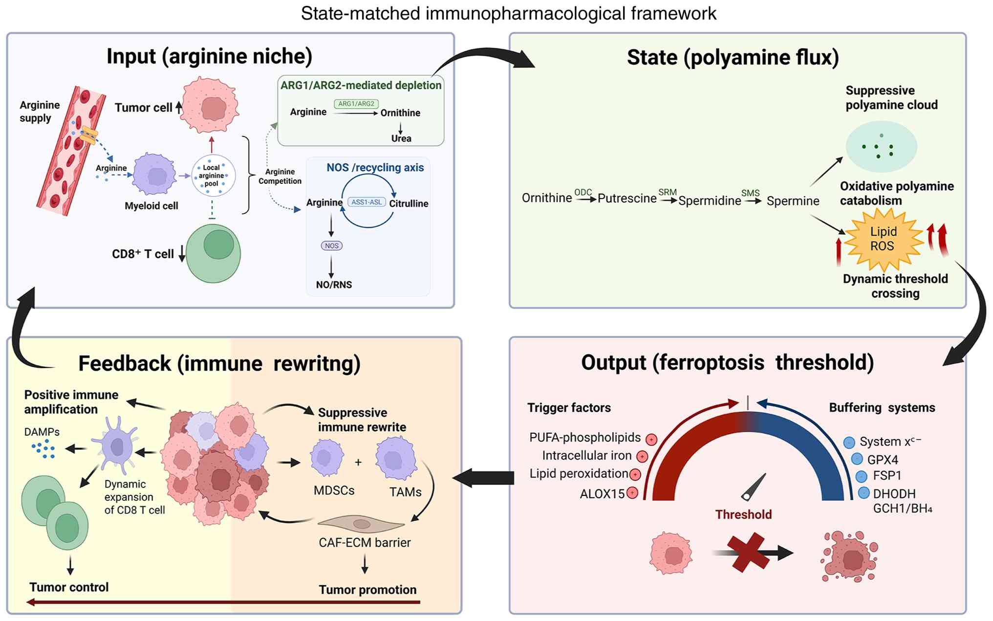 | Figure 1.State-matched immunopharmacological
framework integrating the arginine niche, polyamine flux,
ferroptosis threshold and immune feedback. The arginine niche acts
as the Input layer, polyamine flux as the State layer, ferroptosis
threshold as the therapeutic output and immune rewiring as the
Feedback layer. ARG1, arginase 1; ARG2, arginase 2; NOS, nitric
oxide synthase; NO/RNS, nitric oxide/reactive nitrogen species;
CD8, cluster of differentiation 8; ASS1, argininosuccinate synthase
1; ASL, argininosuccinate lyase; ODC, ornithine decarboxylase; SRM,
spermidine synthase; SMS, spermine synthase; ROS, reactive oxygen
species; DAMPs, damage-associated molecular patterns; MDSCs,
myeloid-derived suppressor cells; TAMs, tumor-associated
macrophages; CAF, cancer-associated fibroblast; ECM, extracellular
matrix; PUFA, polyunsaturated fatty acid; ALOX15, arachidonate
15-lipoxygenase; system xc−,
cystine/glutamate antiporter; GPX4, glutathione peroxidase 4; FSP1,
ferroptosis suppressor protein 1; DHODH, dihydroorotate
dehydrogenase; GCH1, GTP cyclohydrolase 1; BH4,
tetrahydrobiopterin. |
From this perspective, the key question is not
merely whether a particular marker is increased or decreased, but
whether the therapeutic intervention aligns with the relevant
functional thresholds, such as the effector T-cell functional
threshold or the ferroptosis execution threshold. Equally important
is whether the output of resulting tumor-cell death translates into
enhanced antitumor immunity or is redirected toward suppressive
immune remodeling. Together, these four layers provide the primary
organizational logic for the subsequent sections.
The arginine niche as an immunometabolic
input for pharmacological intervention
Defining the niche: Supply-side
processes of biosynthesis, uptake and recycling
The core of the arginine niche concept lies in
transcending the view of ‘arginine availability’ as a single
concentration-based indicator, instead defining it as a
system-level balance of local exchangeable flux. Within any given
tumor microdomain, the net availability of arginine is determined
by the relative strengths of two sets of processes. The first set
consists of supply-side processes that determine how arginine
enters cells or the local exchangeable pool. These processes
include compensatory biosynthesis, transmembrane uptake and
recycling within tumor cells, as well as processes in selected
stromal or immune-cell populations. The second set comprises
demand-side processes that drive the consumption of arginine or its
conversion into chemical stress-inducing mediators. These processes
include myeloid-dominated depletion through ARG1/ARG2 and
NOS-driven diversion through the NOS-nitric oxide (NO)/reactive
nitrogen species (RNS) axis (6–9).
This supply-demand framework is important because it
establishes strict consistency requirements for subsequent
sections. Any discussion regarding polyamine homeostasis or
ferroptosis thresholds must first define the upstream arginine
background in a given microdomain: Whether the state is primarily
driven by supply deficiency, excessive consumption, or the
superimposition of both processes (7–9).
This control node is intrinsically spatially
heterogeneous. Within the same tumor tissue, different microdomain
states may coexist, such as regions with limited supply but
moderate consumption, or regions with relatively preserved supply
but strong consumption pressure. These heterogeneous microdomains
may exert different or even opposing regulatory effects on the
polyamine homeostasis at the State layer and the ferroptosis
threshold at the Output layer, leading to different biological
predictions. This spatially resolved interpretation provides a
conceptual basis for the subsequent development of state-based
tumor stratification strategies (6–11).
Tumor-cell compartment:
biosynthesis-dependent axis and transport competition involving
argininosuccinate synthase 1 (ASS1)/argininosuccinate lyase (ASL)
and cationic amino acid transporters (CATs)
To avoid simplifying arginine regulation to a mere
upregulation or downregulation of a single gene, the present study
described the arginine handling in tumor cells as a
biosynthesis-dependent axis. One end of this axis represents a
relatively self-sufficient state, where tumor cells maintain
effective arginine replenishment through endogenous synthesis or
citrulline-to-arginine recycling. The other end represents an
auxotrophic state, where the replenishment capacity of tumor cells
is limited and highly dependent on exogenous arginine (12,13).
This axis constitutes the core coordinates of the Input layer, as
it not only determines whether tumor cells are susceptible to
extracellular arginine-deprivation strategies but also dictates
their competitive intensity for local arginine against immune cells
in the TME. When tumor cells lack sufficient endogenous arginine
self-sufficiency, they may compensate by enhancing arginine uptake
and recycling programs. This adaptive response shifts the
niche-level competitive pressure onto immune cells, reducing the
residual arginine flux available to effector T cells, thereby
pushing these cells below the functional threshold required for
activation, proliferation and cytotoxicity (14,15).
At the molecular level, compensatory arginine
biosynthesis is primarily mediated by ASS1 and ASL, which cooperate
to recycle citrulline back into arginine and establish a buffering
loop to cope with fluctuations in extracellular arginine
availability (16). In parallel,
CATs, particularly context-dependent members of the solute carrier
family 7 (SLC7), support the uptake of extracellular arginine,
thereby determining the efficiency with which tumor cells utilize
the local exchangeable arginine pool. The ASS1/ASL-dependent
recycling and CAT-mediated uptake together determine whether tumor
cells act as replenishment-competent competitors or
arginine-dependent consumers within the niche.
However, in multiple types of tumors, ASS1 may be
epigenetically silenced or transcriptionally suppressed. This
change should not be interpreted merely as an incidental metabolic
defect; in some cases, it represents an adaptive response to
metabolic pressure in the TME. The inhibition of ASS1 may be
related to metabolic redistribution between the urea cycle and
de novo pyrimidine synthesis, increasing dependence on
extracellular arginine and providing a selective growth advantage
under specific nutritional conditions (17–19).
In this context, CAT-mediated uptake becomes particularly
important, as the loss of endogenous replenishment increases
reliance on the extracellular arginine pool. Thus, tumors with low
ASS1 expression may become vulnerable to arginine deprivation
treatment while imposing stronger arginine competition on
infiltrating immune cells.
More importantly, these biosynthetic and transport
processes often collaborate with stress-responses and oncogenic
pathways, including the integrated stress response/ATF4 axis, MYC-
or KRAS proto-oncogene, GTPase (KRAS)-driven metabolic
reprogramming and stromal constraints. These interactions can form
a self-reinforcing adaptive chain: insufficient arginine
replenishment enhances dependence on transporter-mediated uptake
and recycling processes; increased consumption by tumor cells
reduces immune-accessible arginine flux below the functional
threshold for effector T cells; and the resulting immunosuppressive
microenvironment further supports tumor-cell survival in areas of
inadequate perfusion or spatial constraints (20,21).
Therefore, before discussing polyamine homeostasis or ferroptosis
thresholds, it is necessary to define the position of a given tumor
state on this axis: a state of sufficient replenishment and
relative self-sufficiency, or a dependence-driven,
competition-dominated state. These two configurations imply
fundamentally different pharmacological vulnerabilities and
combination treatment strategies.
Myeloid compartment:
ARG1/ARG2-mediated depletion and T-cell threshold crossing
In a number of solid tumors, the dominant regulators
of the L-arginine niche are not necessarily the tumor cells
themselves, but myeloid populations, including myeloid-derived
suppressor cells (MDSCs), tumor-associated macrophages (TAMs) and
tumor-associated neutrophils (TANs). These compartments converge on
a common functional endpoint, reducing the extracellular arginine
pool and impairing T-cell activity, but differ in their
developmental state, tissue localization, deployment of ARG and
accompanying suppressive mediators. Therefore, MDSCs, TAMs and TANs
should not be viewed as interchangeable sources of ARG activity.
MDSCs represent a heterogeneous population, including
polymorphonuclear MDSCs (PMN-MDSCs) and monocytic MDSCs (M-MDSCs).
PMN-MDSCs typically couple arginine depletion dependent on ARG1
with the generation of reactive oxygen species (ROS) or
peroxynitrite produced by nicotinamide adenine dinucleotide
phosphate (NADPH) oxidase 2 (NOX2), while M-MDSCs more frequently
utilize NO dependent on nitric oxide synthase 2 (NOS2) and
context-dependent suppressive cytokines. These distinctions are not
absolute, as the TME can functionally reprogram these myeloid
subsets. In particular, hypoxia and hypoxia-inducible factor 1
alpha (HIF-1α) signaling can increase the expression of ARG1 and
NOS2, expanding the suppressive activity of tumor-infiltrating
MDSCs and promoting their differentiation into macrophage-like
states (22,23). Therefore, MDSCs serve as a plastic
suppressive compartment, where arginine depletion is combined with
oxidative, nitrosative and cytokine-mediated inhibition (22,23).
TAMs represent a more differentiated and spatially persistent
myeloid compartment. In the suppressive TAM state, ARG1-mediated
arginine hydrolysis not only limits the substrate available to
infiltrating T cells but also increases the availability of
ornithine for downstream polyamine synthesis and tissue-remodeling
pathways (24). Importantly, ARG1
should not be viewed solely as a marker of macrophage polarization.
In pancreatic cancer models, macrophage-specific Arg1 deletion
delayed the development of aggressive tumors and increased the
infiltration of CD8+ T cells, while pharmacological ARG inhibition
further enhanced tumor sensitivity to programmed cell death protein
1 (PD-1) blockade (24). Arg1
deletion also induced compensatory Arg2 expression in a subset of
macrophages, indicating that mitochondrial ARG2 may serve as a
context-dependent intracellular adaptive pathway rather than a
unified extracellular depletion mechanism for all myeloid cells
(24). TANs and PMN-MDSCs share
similarities in morphological and functional characteristics, and
the distinctions between these populations may become blurred
within tumors (22,25). A characteristic of human
neutrophils is the storage of ARG1 in intracellular granules. Upon
activation and degranulation, ARG1 can be released into the
extracellular space, where it rapidly consumes local L-arginine and
inhibits T-cell proliferation. This release-based mechanism differs
from the more persistent transcriptional and metabolic programs
observed in TAMs and provides a rapid pathway for neutrophil-rich
tumor regions to establish local arginine deprivation (25). However, not every
tumor-infiltrating neutrophil is immunosuppressive; therefore, the
functional identification of TANs or PMN-MDSCs relies on their
immunosuppressive activity and metabolic profiles, rather than
solely on granulocytic phenotypic markers.
Despite these differences, all three compartments
can cooperate to maintain a low-flux arginine niche. MDSCs provide
a flexible combination of ARG, ROS, RNS and cytokine-dependent
suppression; TAMs establish sustained tissue-resident metabolic and
polyamine-remodeling programs, while TANs can rapidly release
extracellular ARG1 through degranulation. Their common downstream
effects include reduced expression of the T-cell receptor CD3ζ,
impaired proliferation and decreased production of cytokines and
cytotoxic effectors. Therefore, distinguishing these cell origins
is important for biomarker selection and determining whether ARG
inhibition should be combined with MDSC-directed,
macrophage-directed or neutrophil-directed interventions.
This depletion has significant immunological
consequences, as T-cell function is highly dependent on the
availability of arginine. When local arginine flux falls below the
functional threshold required for effector T-cell activation,
proliferation and cytotoxicity, the effector program may experience
a switch-like collapse. This collapse is characterized by impaired
clonal expansion, reduced production of cytotoxic molecules and
cytokines and a shift in metabolic and signaling programs toward a
maintenance state or dysfunctional state (26,27).
Therefore, the ecological effects of the myeloid ARG axis are
threshold-amplifying. It does not require uniformly high ARG
expression throughout the entire tumor mass. Sustained arginine
depletion in selected microdomains, such as regions distant from
blood vessels, regions behind cancer-associated fibroblast
(CAF)-extracellular matrix (ECM) barriers, or peri-necrotic zones,
may be sufficient to push local immunity below the execution
threshold and establish a suppressive state where immune-mediated
tumor clearance becomes inefficient (28).
This connection links the Input layer, represented
by the arginine niche, to the State layer, represented by polyamine
homeostasis. A stronger ARG axis increases the conversion of
arginine to ornithine, thereby expanding the precursor supply for
polyamine biosynthesis and favoring a state characterized by high
polyamine flux and stabilized immunosuppression. Under specific
stress conditions, the involvement of the polyamine catabolic axis
may further reshape this state. The catabolism mediated by
polyamine oxidase (PAOX) and spermine oxidase (SMOX) can generate
ROS, including H2O2, thereby increasing
lipid-peroxidation pressure and weakening residual anti-ferroptotic
buffering capacity. This may create a therapeutic window in which
ARG inhibition, polyamine blockade, ferroptosis sensitization and
ICB can be rationally combined. In this sense, the myeloid ARG axis
is not an auxiliary mechanism but a key bridge that transmits the
arginine depletion of the Input layer to downstream state
remodeling and the output-layer's ferroptosis vulnerability
(29–31).
Diversion axis: NOS-NO/RNS converts
nutrient limitation into oxidative and nitrosative chemical
stress
If ARG1/ARG2-mediated depletion primarily suppresses
antitumor immunity by depleting the local exchangeable arginine
pool below the functional threshold required for effector T-cell
activity, then NOS-mediated arginine diversion represents a form of
chemical amplification of niche perturbation. It not only consumes
arginine but also converts this substrate into diffusible stress
mediators, including NO and downstream RNS. Therefore, the
diversion axis should not be viewed merely as another mode of
arginine consumption. Instead, it transforms nutritional limitation
from a substrate supply issue into a broader chemical stress state
within the TME.
Under comparable conditions of reduced local
arginine, a diversion-dominant niche imposes an additional
NO/RNS-driven oxidative and nitrosative baseline. This baseline can
suppress immune execution, reset redox homeostasis and increase
lipid-peroxidation pressure, thereby preconditioning the system for
subsequent changes in the threshold of ferroptosis (32,33).
In this sense, NOS-mediated diversion not only links the Input
layer to T-cell functional impairment but also associates with
downstream redox remodeling and susceptibility to ferroptosis.
The clinical significance of NOS-mediated diversion
lies not merely in the simple suppression or enhancement of NO
production, but in identifying the flux- and context-dependent
switches that determine its biological output. In MCF7 cells under
defined aerobic conditions, low steady-state NO concentrations
(<50 nM) activate extracellular signal-regulated kinase (ERK)
through soluble guanylate cyclase, moderate concentrations (>100
nM) promote the accumulation of HIF-1α, while higher concentrations
(>300 nM) induce p53 Ser15 phosphorylation. These values should
not be interpreted as universal clinical cutoffs. Instead, they
provide experimental evidence that NO concentration and exposure
time can participate in different signaling programs, while oxygen
consumption, cell density and ROS production can alter effective
thresholds (34).
In immune-cold or myeloid-dominated
microenvironments, the balance between NO and superoxide acts as a
critical molecular switch. M-MDSCs predominantly suppress T-cell
responses through inducible nitric oxide synthase (iNOS)-derived
NO, whereas granulocytic MDSCs can generate peroxynitrite through
the simultaneous production of NO and superoxide. Experimental
inhibition of iNOS-derived NO preferentially impairs monocytic
MDSC-mediated suppression, while the clearance of peroxynitrite or
the disruption of endothelial nitric oxide synthase (eNOS)- and
NOX2-dependent oxidant production reduces the suppressive activity
of granulocyte MDSCs (22). Thus,
NO-dominant and peroxynitrite-dominant tumor niches induce T-cell
dysfunction via different chemical mechanisms.
The regulatory relationship between NO signaling and
ferroptosis is bidirectional and context-dependent. In activated
macrophages, iNOS-derived NO can inhibit the generation of
ferroptotic phospholipid signals. Genetic or pharmacological
inhibition of iNOS renders M1 macrophages sensitive to
GPX4-inhibitor-induced ferroptosis, while NO donors increase the
ferroptosis resistance of M2 macrophages. This protective effect is
related to the nitroxidative interference with
15-lipoxygenase-dependent phospholipid oxidation and oxidized
phosphatidylethanolamine (PE) intermediates (35).
Conversely, when NO and superoxide are co-generated
in an iron-rich environment with weakened GSH-GPX4 buffering, the
formation of peroxynitrite can increase lipid-peroxidation pressure
and facilitate ferroptosis. In an engineered NO-generating tumor
model, the production of NO promoted GSH consumption, reduced GPX4
expression, increased heme oxygenase 1-associated Fe2+
release and enhanced lipid peroxidation (36). This evidence should be interpreted
as a context-dependent pro-ferroptotic configuration rather than as
a universal property of NO. Therefore, the functional consequences
of the axis shift depend on NO concentration and exposure time, the
NO-to-superoxide balance, cellular identity, oxygen tension, labile
iron availability and the capacity of GPX4-dependent and
GPX4-independent antioxidant systems. These variables determine
whether NOS-NO/RNS signaling primarily suppresses immune execution,
protects specific myeloid cells from ferroptosis or facilitates
ferroptosis in tumor-cell death.
Stromal and vascular compartments:
Spatial barriers as amplifiers of arginine depletion
The arginine niche should be viewed as a niche, not
just a metabolic pathway, because its determining factors are not
limited to the intrinsic activity of metabolic enzymes. They also
include the spatial organization of transport, perfusion and
diffusion. In most solid tumors, abnormal vasculature,
heterogeneous perfusion, elevated interstitial pressure and dense
CAF-ECM matrix collectively create a geographically constrained
supply environment. Once nutrients diffuse out of the vasculature
into tumor tissues, their effective diffusion range is markedly
restricted. Therefore, microdomains located far from functional
blood vessels may be chronically exposed to low-flux arginine
states.
Under these conditions, myeloid ARG-mediated
depletion or NOS-driven diversion are amplified, not necessarily
because the enzymatic activity in these regions is higher, but
because the replenishment of arginine is more difficult to deliver.
Slower delivery kinetics, elongated diffusion trajectories and
stromal barriers transform transient local nutrient depletion into
persistent substrate scarcity (37–40).
In this sense, the stromal and vascular compartments act as
depletion amplifiers. They stabilize transient arginine consumption
into spatially fixed low-arginine microdomains, increasing the
likelihood of impaired T-cell function. Therefore, the inhibitory
consequences of arginine limitation cannot be fully understood
without considering the physical context embedded in nutrient
delivery, cellular consumption and metabolic competition.
The essence of a low-perfusion region is input
limitation. Even with normal plasma arginine levels, insufficient
perfusion or vascular leakage can reduce effective local delivery,
causing the exchangeable arginine pool within selected microdomains
to remain at low levels (37–40).
More importantly, the restricted supply and enhanced consumption do
not simply linearly add up. Instead, they are more likely to
produce nonlinear amplification near functional thresholds. When
abnormal perfusion and diffusion limitations reduce local arginine
supply to near the critical requirements needed for effector T-cell
activity, even slight additional ARG-mediated depletion or
tumor-cell uptake competition may be sufficient to push local
arginine flux below the threshold required for CD3ζ expression,
cell-cycle progression and cytotoxic function (26,27,37–40).
By contrast, in relatively well-perfused regions, the same degree
of arginine consumption may only lead to partial inhibition,
insufficient to trigger sustained T-cell functional inactivation
(26,27,37–40).
Stromal and vascular compartments can also shape
niche plasticity through changes in tissue architecture and
perfusion (38,39). Therefore, structural interventions,
such as stromal decompression or vascular normalization, should be
understood as strategies to alter system boundary conditions. By
increasing supply flux, shortening diffusion distances, and
reducing the amplification factors of depletion, these
interventions may restore the availability of local arginine above
the functional threshold for effector T-cell activity and create
space for subsequent combination therapies (38,39).
Overall, the strength of the arginine niche is determined not only
by metabolism but also by transport. In tumors characterized by low
perfusion or a dense stromal matrix, transport bottlenecks act as a
multiplicative factor, amplifying arginine depletion (37–40).
The spatial organization of the arginine niche, including vascular
supply, tumor-cell uptake, myeloid-mediated depletion, NOS-driven
diversion and stromal barriers, is summarized in Fig. 2.
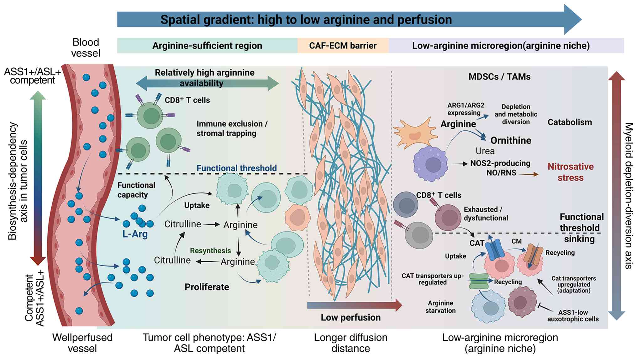 | Figure 2.Spatial organization of the arginine
niche. Vascular supply, ASS1/ASL-dependent biosynthesis,
CAT-mediated uptake, ARG1/ARG2-mediated depletion, NOS-driven
diversion and CAF-ECM barriers generate microdomain-specific
arginine availability and immune functional thresholds. ASS1,
argininosuccinate synthase 1; ASL, argininosuccinate lyase; ARG1,
arginase 1; ARG2, arginase 2; CAF, cancer-associated fibroblast;
CAT, cationic amino acid transporter; CM, cell membrane; ECM,
extracellular matrix; CD8, cluster of differentiation 8; NOS2,
nitric oxide synthase 2; NO/RNS, nitric oxide/reactive nitrogen
species; L-Arg, L-arginine; MDSCs, myeloid-derived suppressor
cells; TAMs, tumor-associated macrophages. |
Polyamine flux as a targetable state
variable
Polyamine metabolism represents a State layer
through which arginine availability is translated into
proliferative, immunosuppressive and redox-adaptive programs. In
this framework, polyamine flux is viewed not only as a downstream
product of arginine catabolism but also as a targetable state
variable that determines whether the system remains in a
suppressive buffering state or shifts towards oxidative stress and
ferroptosis sensitivity.
As illustrated in Fig.
3, the buffering branch is maintained by ODC1- and
adenosylmethionine decarboxylase 1 (AMD1)-dependent polyamine
synthesis, as well as uptake and efflux cycling. This branch
maintains the availability of polyamines, supports tumor-cell
proliferation and stress tolerance, and reinforces the suppressive
polyamine cloud. By contrast, the oxidative-triggered branch is
mediated by spermidine/spermine N1-acetyltransferase 1
(SAT1)-mediated acetylation and back-conversion, PAOX-dependent
oxidation of acetylated polyamines and SMOX-dependent oxidation of
spermine. These catabolic pathways produce
H2O2, aldehydes and carbonyl stress, thereby
increasing lipid-peroxidation pressure and lowering the ferroptosis
threshold. Thus, Fig. 3
distinguishes between polyamine retention and recycling that favors
buffering and oxidative polyamine catabolism that may prepare
Output layers for ferroptosis.
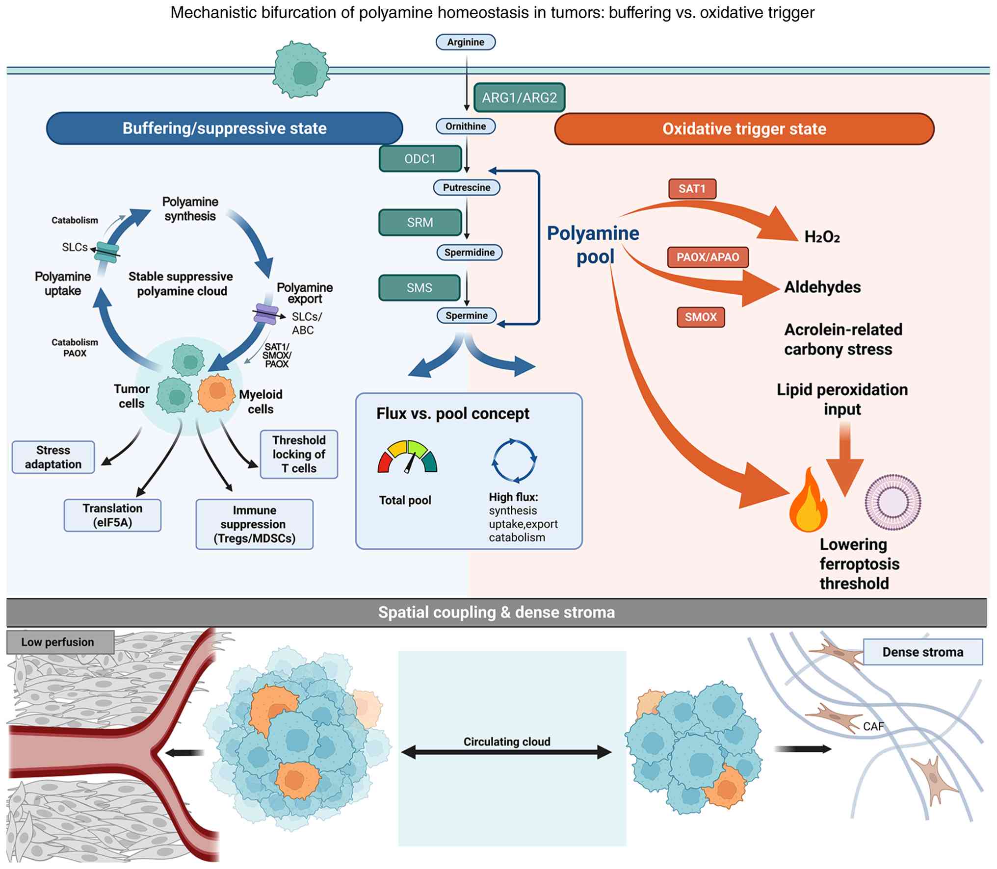 | Figure 3.Mechanistic bifurcation of polyamine
homeostasis. Polyamine synthesis and uptake-efflux cycling can
stabilize a buffering or suppressive state, whereas SAT1-, PAOX-
and SMOX-associated catabolism can generate
H2O2, aldehydes and carbonyl stress that
lower the ferroptosis threshold. SAT1, spermidine/spermine
N1-acetyltransferase 1; PAOX, polyamine oxidase; APAO,
acetylpolyamine oxidase; SMOX, spermine oxidase; ARG1, arginase 1;
ARG2, arginase 2; ODC1, ornithine decarboxylase 1; SRM, spermidine
synthase; SMS, spermine synthase; SLCs, solute carrier family
transporters; ABC, ATP-binding cassette transporters; eIF5A,
eukaryotic translation initiation factor 5A; Tregs, regulatory T
cells; MDSCs, myeloid-derived suppressor cells; CAF,
cancer-associated fibroblast; H2O2, hydrogen
peroxide. |
The synthetic axis: From
arginine-derived ornithine to the ODC1-AMD1 program
The polyamine synthetic axis provides a direct
pathway through which the arginine niche is converted into cellular
states. Arginine is converted to ornithine via ARG1/ARG2, which is
then decarboxylated by ODC1 to produce putrescine. Putrescine is
further converted to spermidine and spermine through spermidine
synthase (SRM)- and spermine synthase (SMS)-mediated
aminopropyl-transfer reactions, while AMD1 generates decarboxylated
S-adenosylmethionine, which is the required aminopropyl donor for
maintaining this process (41).
The importance of this axis lies not only in the
enzymatic reactions. Enhanced ARG-mediated arginine catabolism can
channel substrates towards ornithine-dependent polyamine
biosynthesis, thereby linking the arginine niche at the Input layer
to polyamine homeostasis at the State layer. This connection can be
summarized through three dimensions: Ornithine supply,
ODC1-SRM-SMS-driven biosynthetic activity and AMD1-dependent
aminopropyl-donor capacity. Together, these features determine
whether the system enters a polyamine high-buffering state that
supports tumor-cell proliferation, stress tolerance and immune
suppression (30,41,42).
The core state characteristics of the polyamine synthetic axis,
including ornithine availability, synthetic driving force and
methyl-donor restriction, are summarized in Table I.
 | Table I.Minimal state dimensions of the
polyamine synthetic axis. |
Table I.
Minimal state dimensions of the
polyamine synthetic axis.
| Core dimension | Variable
composition | Biological
significance |
|---|
| Ornithine
supply | Intensity of
ARG-mediated depletion/tumor metabolic phenotype | Determines the
substrate basis for polyamine biosynthesis. High ARG activity or
tumor metabolic reprogramming can provide sufficient
precursors. |
| Synthetic driving
capacity | ODC1-AMD1 activity
axis | Determines the
driving force and rate of polyamine biosynthesis. ODC1 represents
the rate-limiting step, whereas AMD1 supports sustained progression
of the synthetic chain. |
| Methyl-donor
constraint | SAM/dcSAM
balance | Determines the
resource boundary of polyamine biosynthesis and its coupling with
epigenetic regulation. The direction of this balance shapes the
interaction between metabolism and epigenetic state. |
Polyamine levels and polyamine flux:
Why flux language is necessary
Polyamine homeostasis can only be treated as a
central State-layer processor when defined by flux rather than
static pool size. The total polyamine levels measured by
metabolomics largely reflect cellular storage, as a number of
intracellular polyamines are bound to nucleic acids, proteins or
other macromolecular structures. Therefore, total abundance does
not necessarily represent the exchangeable pool involved in
signaling, stress adaptation, redox reactions or ferroptosis
regulation. What determines immunosuppressive buffering and
susceptibility for ferroptosis is not merely how much polyamine is
stored, but how polyamines are synthesized, imported, exported and
catabolized (42–45).
This flux-based perspective can be summarized
through three operational dimensions: Synthetic drive, reflected by
ODC1/AMD1/SRM/SMS activity and ornithine supply; state
externalization, reflected by acetylated polyamines that indicate
readiness for efflux or catabolic entry; and oxidative input,
reflected by SAT1/PAOX/SMOX-associated catabolism, which redirects
the polyamine pools toward ROS-generating flux. Methodologically,
using labeled arginine or ornithine for stable isotope tracing can
directly estimate the flux of polyamine biosynthesis. In more
clinically accessible settings, acetylated-to-total polyamine
ratios, combined with the characteristics of synthetic and
catabolic genes, may serve as a practical alternative readout.
These readouts should be interpreted alongside spatial features
such as hypoxia, low perfusion and myeloid enrichment, aligning
polyamine state variables with the actual architecture of the TME
(32,33).
Uptake-efflux cycling: Polyamines as a
diffusible ‘polyamine cloud’
Once polyamine homeostasis is defined as a
flux-based state rather than a static pool, uptake-efflux cycling
becomes a key determinant of whether polyamine metabolism acts
solely within individual cells or extends to tissue scale.
Intercellular transfer allows polyamines to shape the
microenvironment beyond their synthesizing cells, thereby linking
intracellular metabolic states with population-level immune and
redox regulation (46).
The present review defined the spatially persistent,
polyamine-rich inhibitory environment maintained by uptake, efflux
and local retention as the polyamine cloud. This concept refers not
merely to an increase in polyamine abundance at a given time point.
Instead, it describes a continuous background flux state that
relies on ongoing polyamine exchange and is particularly relevant
in low-perfusion or CAF-ECM-dense regions, where diffusion and
clearance are constrained (3,47).
The polyamine cloud should be viewed as a conceptual
framework for evidence of organizational dispersion rather than a
unified mechanism fully validated across all cancer types. Its
primary value lies in providing operational standards for treatment
sequences. When a stable polyamine cloud exists, directly enhancing
the Output layer through ferroptosis sensitization may not be the
best choice. Instead, disrupting the cloud-maintaining cycle may
need to occur prior to ferroptosis induction, especially in
myeloid-dominant tumors with active polyamine uptake-efflux
cycling. Otherwise, treatment may induce inflammatory stress
without sufficient tumor-cell killing, thereby favoring suppressive
myeloid recruitment and limiting therapeutic efficacy.
The catabolic axis: SAT1-PAOX/SMOX as
oxidative triggers linking polyamine state to lipid
peroxidation
The core significance of polyamine catabolism lies
in its ability to convert the State-layer polyamine program into
directed oxidative inputs. Polyamine oxidation generates
H2O2 and aldehyde byproducts, which can
increase local oxidative pressure and push membrane
lipid-peroxidation kinetics toward the ferroptosis execution
threshold. Therefore, the catabolic axis is not merely about
reducing polyamine abundance; it determines whether buffering
polyamine status is redirected toward the amplification of
oxidative stress.
Spermidine/spermine
n1-acetyltransferase (SSAT)/SAT1-PAOX Axis: Acetylation-guided flux
routing and peroxisomal oxidative output
In mammalian cells, the canonical polyamine
catabolic pathway involves SAT1-mediated acetylation, followed by
PAOX-mediated oxidative back-conversion. SAT1, also known as SSAT1,
acetylates spermine and spermidine, generating N¹-acetylated
polyamines that can be exported or oxidized by the peroxisomal
PAOX, also known as acetylpolyamine oxidase (APAO). PAOX converts
these acetylated substrates into lower-order polyamines while
generating H2O2 and aldehyde-derived stress
signals (48,49).
Functionally, SAT1 acts as a flux gate. It shifts
the polyamine pool from structural binding or buffering to efflux
and oxidative catabolism. PAOX serves as a peroxisomal execution
module, converting these redirected substrates into oxidative
products. The SAT1-PAOX axis links polyamine turnover to redox
stress and establishes a mechanistic bridge between polyamine
status and ferroptosis vulnerability (50).
The SMOX axis: Direct spermine
oxidation, H2O2 production and
acrolein-related carbonyl stress
In parallel with the SAT1-PAOX pathway, SMOX
directly oxidizes spermine to spermidine while producing
H2O2 and 3-aminopropanal. The latter can
further convert into reactive carbonyl species, including acrolein,
thereby exacerbating protein damage, membrane-lipid injury and
oxidative stress (51–53). In inflammation-associated tumors,
increased SMOX activity may thus convert inflammatory inputs into
oxidative and carbonyl stress. This is particularly relevant in
myeloid-dominated microenvironments where inflammation is present
but immune execution is inefficient. Under these conditions,
SMOX-mediated catabolism may elevate basal lipid-peroxidation
pressure and bring the system closer to the ferroptosis threshold
(41,54,55).
SAT1-arachidonate 15-lipoxygenase
(ALOX15) coupling: From catabolic flux to ferroptosis
execution
Among the links between polyamine catabolism and
ferroptosis, the SAT1-ALOX15 axis provides one of the clearest
mechanistic examples. Ou et al (56) demonstrated that p53
transcriptionally activates SAT1, while SAT1 induction promotes
lipid peroxidation, ferroptosis and tumor-growth suppression under
oxidative stress conditions. Mechanistically, SAT1 upregulation is
accompanied by increased ALOX15 expression, and pharmacological
inhibition of ALOX15 rescues the ferroptosis phenotype induced by
SAT1. These findings suggest that SAT1 not only increases polyamine
turnover; it couples catabolic flux to membrane phospholipid
peroxidation through a lipoxygenase-dependent amplifier, thereby
promoting lethal lipid-peroxidation accumulation and ferroptosis
execution.
Polyamine catabolism-ferroptosis
amplification in cancer therapy
Beyond the canonical p53-SAT1-ALOX15 pathway, recent
studies further support the notion that polyamine metabolism can
amplify the vulnerability to ferroptosis in cancer therapy. A 2024
study proposed polyamine-mediated ferroptosis amplification as a
targetable vulnerability, indicating that polyamine metabolism can
expose therapeutically relevant susceptibility to ferroptosis
(3). A more recent 2025 study
linked this principle to KRAS-targeted therapy, showing that
polyamines sensitize KRAS-mutant tumors to KRAS inhibition, and
this sensitivity depends on the status of kelch-like ECH-associated
protein 1 (KEAP1), with SAT1-mediated polyamine catabolism being
essential for this ferroptosis-related response (57).
These findings collectively establish a universal
mechanistic principle: Polyamine catabolism is not merely
correlated with ferroptosis but dictates whether therapeutic
interventions induce lethal lipid-peroxidation stress. The
executability of this axis depends on whether SAT1-mediated flux
routing is involved and whether downstream oxidative outputs,
including PAOX- or SMOX-associated pathways, are sufficient to push
the system beyond the ferroptosis threshold. Therefore, the
polyamine catabolism-ferroptosis axis provides a state-dependent
vulnerability that can be exploited in rational combination
therapies.
Summary: Catabolic flux maps polyamine
state onto the ferroptosis threshold
Overall, the polyamine catabolic axis contains two
major oxidative-output pathways: The SAT1-guided
acetylation/back-conversion pathway through PAOX and the
SMOX-mediated direct oxidation of spermine. Although these two
pathways differ in substrate preference, subcellular localization
and byproduct characteristics, both generate
H2O2 and aldehyde-induced carbonyl stress,
thereby elevating lipid-peroxidation pressure.
Importantly, polyamine catabolism should not be
viewed merely as a terminal clearance step of polyamine metabolism.
In the p53-SAT1-ALOX15 model, catabolic flux can couple with
membrane phospholipid peroxidation through amplifiers dependent on
lipoxygenases, thereby making the turnover of polyamines to
ferroptosis execution more efficient. Thus, the catabolic axis
serves as a critical interface that converts the State layer into
the Output layer. The strength and direction of this mapping depend
on SAT1-mediated flux routing, PAOX- or SMOX-associated oxidative
outputs, subcellular localization and the availability of
lipid-peroxidation amplifiers such as ALOX15.
Polyamines and immunosuppression:
Myeloid polarization and T-cell functional recovery
Polyamines are not merely growth-associated
metabolites of tumor cells; they also act as immunoregulatory state
variables in the TME. Changes in the synthesis, uptake, efflux and
catabolism of polyamines can reshape myeloid polarization, impair
antigen presentation and limit T-cell effector differentiation. In
this manner, polyamine flux locks the T-cell functional impairment
induced by Input-layer arginine depletion into a sustained
immunosuppressive state, helping explain why T cells struggle to
recover once local arginine availability drops below a functional
threshold (58,59).
This suppressive effect can be understood through
three interconnected mechanisms. First, polyamine homeostasis is
coupled with an inhibitory myeloid phenotype. ARG-driven myeloid
cells consume arginine and generate ornithine, thereby supporting
polyamine biosynthesis; in turn, a polyamine-rich environment may
help maintain or polarize myeloid cells into an immunosuppressive
state, including M2-like TAMs and MDSCs (42). Second, polyamines may weaken the
initiation of adaptive immunity by impairing dendritic-cell
maturation and antigen-presenting capacity, thereby reducing
sustained antigenic stimulation and costimulatory support for T
cells (59).
Third, extracellular polyamines can directly limit
the metabolic plasticity of T cells. In pancreatic ductal
adenocarcinoma, tumor-derived spermine reduced cytosolic and
mitochondrial Ca2+ availability in CD8+ T cells and
inhibited the phosphorylation of mechanistic target of rapamycin,
ribosomal protein S6 kinase (S6K) and eukaryotic translation
initiation factor 4E-binding protein 1. These changes were
accompanied by reductions in glycolysis, tricarboxylic acid cycle
activity and oxidative phosphorylation, impaired mitochondrial
fitness, decreased adenosine triphosphate production and reduced
T-cell proliferation and cytotoxic-effector production. Spermine
exposure also decreased the expression of granzyme B, IFN-γ, tumor
necrosis factor-α and perforin, providing a direct mechanistic link
between a spermine-rich TME, mechanistic target of rapamycin
complex 1 (mTORC1) inhibition and CD8+ T-cell dysfunction (60). Experimental disruption of tumor
spermidine production restored CD8+ T-cell activity and improved
the efficacy of immune-checkpoint blockade in pancreatic cancer
models.
Polyamine-dependent T-cell state regulation also
extends to epigenetic programming. In CD4+ T cells, disruption of
ODC1-dependent polyamine synthesis altered histone acetylation and
led to a loss of fidelity in helper-T-cell lineage. Spermidine also
provides the aminobutyl substrate required for eukaryotic
translation initiation factor 5A (eIF5A) hypusination, linking
polyamine availability to mitochondrial metabolism, protein
translation and the maintenance of appropriate transcriptional
programs (43). Thus, polyamine
flux can influence not only short-term nutrient sensing but also
the chromatin and transcriptional states that determine T-cell
differentiation and functional plasticity.
Importantly, existing evidence does not demonstrate
that every extracellular polyamine-rich niche induces the same
epigenetic program. Currently, the strongest direct causal evidence
is in pancreatic cancer, where spermine-mediated mTORC1 inhibition
and mitochondrial metabolism are observed, while the findings of
histone acetylation and eIF5A-hypusination primarily showcase how
intracellular polyamine availability regulates T-cell lineage
identity. Thus, the polyamine cloud should be interpreted as a
source of sustained polyamine exposure at the tissue level, which
can translate into metabolic and epigenetic effects after cellular
uptake, rather than as a single universally validated inhibitory
pathway. These mechanisms collectively explain how Input-layer
arginine depletion can be stabilized at the State layer through
myeloid polarization, impaired antigen presentation, reduced T-cell
metabolic fitness and altered lineage programming.
The ferroptosis threshold as an executable
therapeutic output
Ferroptosis is not a binary present-or-absent
phenotype, but rather a critical state transition determined by the
balance between lipid-peroxidation pressure and anti-ferroptotic
buffering capacity (44). In the
present review, the ferroptosis threshold referred to the minimum
net perturbation required to drive lipid peroxidation from a
reversible adaptive zone into irreversible membrane damage and
cell-death execution under a given cellular state and
microenvironmental context.
A lower threshold indicates that endogenous stress
or therapeutic interventions can more easily trigger ferroptosis,
while a higher threshold allows tumor cells to tolerate oxidative
pressure and maintain fitness. Therefore, therapies targeting
ferroptosis should not be simplified to merely adding ferroptosis
inducers. Instead, it requires defining whether the intervention
strengthens the triggering arm, weakens the buffering arm or
bypasses compensatory ferroptosis defenses that may mask the true
vulnerability of the system. Only when the Output layer aligns with
the Input and State layers, by considering oxidative baselines,
substrate allocation, polyamine flux and spatial nutrient supply,
can ferroptosis induction become a predictable and stratifiable
therapeutic window. Fig. 4
summarizes the balance between ferroptosis triggers and buffering
systems.
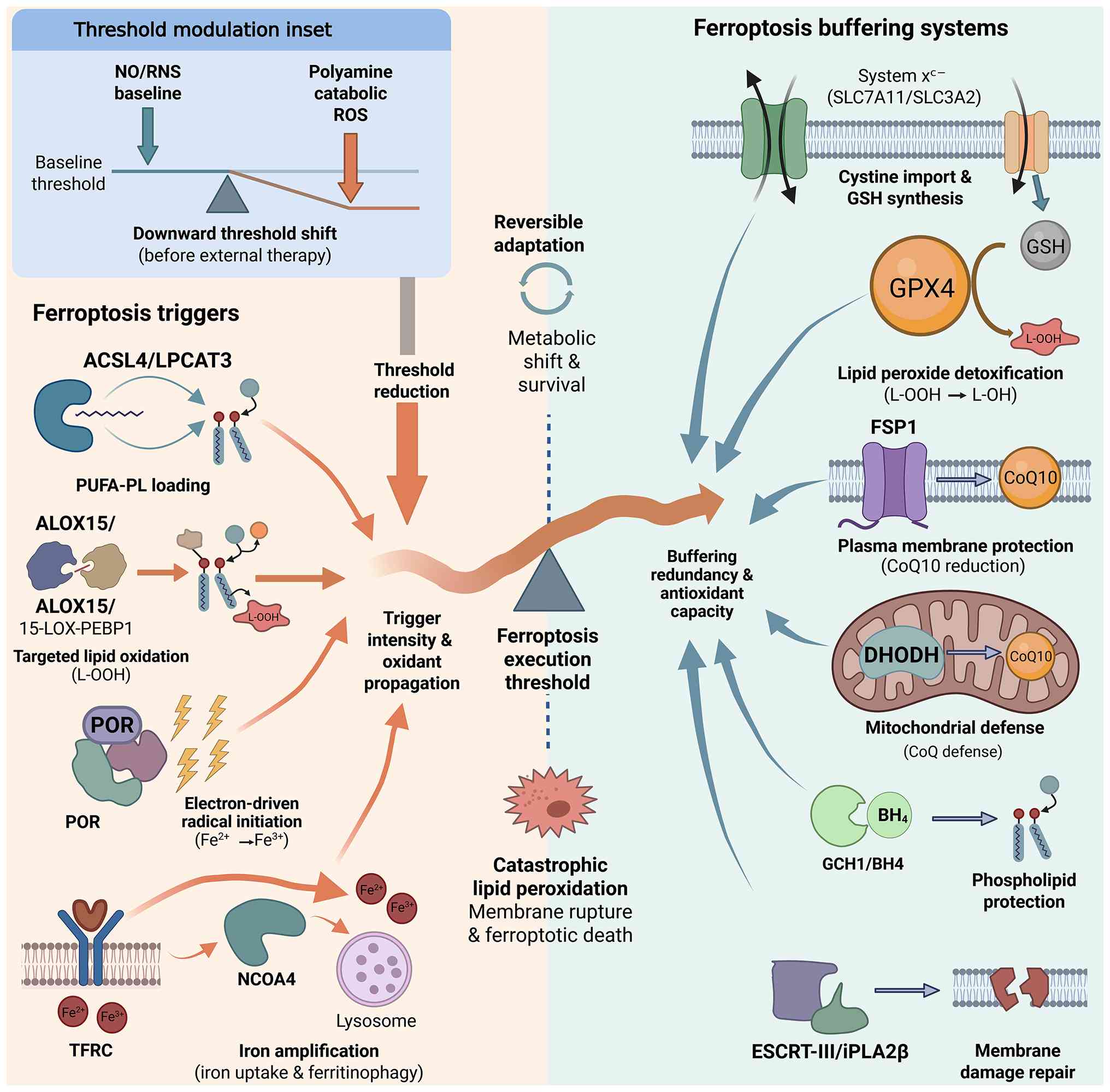 | Figure 4.Ferroptosis threshold defined by
trigger modules and buffering systems. ACSL4/LPCAT3, ALOX15/PEBP1,
POR and iron-loading modules promote lipid-peroxidation
propagation, whereas the system xc−-GSH-GPX4
axis, FSP1-CoQ10, DHODH, GCH1/BH4 and membrane-repair systems
provide redundant buffering. ACSL4, acyl-CoA synthetase long-chain
family member 4; LPCAT3, lysophosphatidylcholine acyltransferase 3;
ALOX15, arachidonate 15-lipoxygenase; 15-LOX, 15-lipoxygenase;
PEBP1, phosphatidylethanolamine-binding protein 1; POR, cytochrome
P450 oxidoreductase; NO/RNS, nitric oxide/reactive nitrogen
species; ROS, reactive oxygen species; PUFA-PL, polyunsaturated
fatty acid-containing phospholipids; TFRC, transferrin receptor;
NCOA4, nuclear receptor coactivator 4; system
xc−, cystine/glutamate antiporter; SLC7A11,
solute carrier family 7 member 11; SLC3A2, solute carrier family 3
member 2; GSH, glutathione; GPX4, glutathione peroxidase 4; FSP1,
ferroptosis suppressor protein 1; CoQ10, coenzyme Q10; DHODH,
dihydroorotate dehydrogenase; GCH1, GTP cyclohydrolase 1;
BH4, tetrahydrobiopterin; ESCRT-III, endosomal sorting
complex required for transport III; iPLA2β, calcium-independent
phospholipase A2 β; L-OOH, lipid hydroperoxide; L-OH, lipid
alcohol. |
Trigger modules: Polyunsaturated fatty
acid-containing phospholipid (PUFA-PL) loading and lipoxygenase
(LOX)-mediated lipid-peroxidation amplification
The ferroptosis triggering module refers to the
initiation and amplification of lipid-peroxidation chain reactions.
It is determined by two interrelated requirements: The availability
of peroxidizable membrane substrates, primarily PUFA-PLs, and the
presence of catalytic systems that ignite and propagate oxidative
damage. acyl-CoA synthetase long-chain family member 4 (ACSL4) and
lysophosphatidylcholine acyltransferase 3 (LPCAT3) facilitate the
integration of PUFAs into membrane phospholipids, while ALOX15,
cytochrome P450 oxidoreductase (POR) and unstable iron pools
provide directional oxidation, free radical initiation and
Fenton-driven amplification (61,62).
Therefore, the triggering module is not a single molecular switch
but a dynamic process composed of substrate loading, oxidative
ignition and chain propagation.
This concept provides operational standards for
stratification. In tumors with abundant PUFA-PL substrates and
complete amplification chains, the inhibition of the
cystine/glutamate antiporter system xc− or an
increase in ROS input is more likely to translate into executable
ferroptosis. By contrast, in tumors with substrate scarcity, weak
lipid-peroxidation mechanisms, or limited iron supply, merely
adding a ferroptosis inducer may be insufficient to cross the
execution threshold. In such contexts, ferroptosis-targeted therapy
should be aligned with upstream Input- and State-layer
interventions that reshape substrate loading, iron availability or
oxidative baseline, thereby creating a more predictable therapeutic
window.
Substrate loading: The ACSL4-LPCAT3
axis and ferroptosis-competent membranes
Ferroptosis sensitivity is strongly influenced by
the composition of membrane lipids. PUFAs only become lethal
substrates for lipid-peroxidation chain reactions when incorporated
into membrane phospholipids, particularly PE and
phosphatidylcholine (63). ACSL4
activates ω-6 PUFAs, such as arachidonic acid and adrenic acid,
into acyl-CoA derivatives and promotes their incorporation into
phospholipid species. Genetic and multi-omics evidence suggests
that ACSL4 is a key determinant of ferroptosis susceptibility, and
the loss of ACSL4 reduces PUFA-phospholipid loading and oxidized
lipid death signals, thereby conferring ferroptosis resistance
(45,63,64).
LPCAT3 further esterifies activated PUFA-CoA into
membrane phospholipids, generating a pool of peroxidizable
substrates rich in PUFAs, located on organelle membranes and the
plasma membrane (65). Together,
ACSL4 and LPCAT3 act as a substrate-programming module that
determines whether the membrane is ferroptosis-competent. High
levels of ACSL4-LPCAT3 activity increase the density of
peroxidizable sites and reduce the perturbation required to execute
ferroptosis, while weak substrate loading raises the execution
threshold. This may help explain why tumors, or even distinct
microdomains within a single tumor, exhibit variable sensitivities
to ferroptosis inducers despite comparable basal antioxidant
defenses (66).
Directional oxidation: The
phosphatidylethanolamine-binding protein 1 (PEBP1)-ALOX15 axis
directs peroxidation toward PE
Ferroptosis is not equivalent to random lipid
oxidation. In multiple models, lipoxygenase-mediated oxidation
generates specific oxidized PUFA-containing PE species, which serve
as executable ferroptotic signals. Wenzel et al (62) demonstrated that PEBP1 can form a
complex with 15-LOX and alter its substrate preference toward PE,
thereby promoting pro-ferroptotic PE oxidation. This mechanism
transforms lipid peroxidation from nonspecific oxidative noise into
a more directional execution program.
This directional oxidation also connects the Output
layer with the aforementioned polyamine catabolic axis. The
SAT1-mediated catabolic routing can increase oxidative pressure
through polyamine turnover and is associated with ALOX15-dependent
lipid-peroxidation amplification (56). Therefore, ALOX15/PEBP1 should not
only be interpreted as a marker of oxidative stress but also as an
indicator of directional ferroptosis execution capacity. For
therapeutic stratification, ALOX15/PEBP1 can be interpreted
alongside SAT1, PAOX and SMOX to identify tumors where catabolic
flux and lipid-peroxidation mechanisms are consistent, thereby
making it more likely for the system to cross the ferroptosis
threshold.
Non-enzymatic propagation:
POR-mediated radical priming and chain reaction amplification
In addition to LOX-dependent oxidation, ferroptotic
lipid peroxidation can also propagate through non-enzymatic
free-radical processes. Using a clustered regularly interspaced
short palindromic repeats (CRISPR) inhibition screening, Zou et
al (67) identified NADPH-POR
as a common requirement for ferroptosis across multiple cell
lineages. POR can transfer electrons from NADPH to oxygen-dependent
redox reactions, generating radical-priming signals that promote
PUFA-phospholipid peroxidation.
Functionally, POR enhances the ‘spark intensity’ of
the triggering module. Its effect depends on the redox baseline
established by the Input and State layers. In tumor states
characterized by elevated NO/RNS, mitochondrial ROS leakage or
oxidative inputs derived from polyamine metabolism, POR-mediated
radical priming may more readily initiate lipid-peroxidation chain
reactions and reduce the additional perturbation required to
execute ferroptosis. For stratification, POR and related
NADPH-dependent redox flux indicators may help distinguish tumors
with strong oxidative ignition capabilities from those primarily
driven by downstream buffering systems that confer ferroptosis
resistance.
Iron supply and amplification:
Transferrin receptor 1 (TFRC)-mediated uptake and nuclear receptor
coactivator 4 (NCOA4)-dependent ferritinophagy
Labile iron availability promotes lipid-radical
chain reactions through Fenton chemistry and supports
iron-dependent enzymes involved in directed phospholipid
peroxidation, including members of the lipoxygenase family
(68). The size of the labile iron
pool is determined by the coordinated balance among iron uptake,
intracellular storage and release and cellular export.
TFRC-mediated endocytosis increases the import of transferrin-bound
iron, while NCOA4-dependent ferritinophagy releases
ferritin-sequestered iron into the exchangeable intracellular pool.
Ferroportin 1 (FPN1), encoded by solute carrier family 40 member 1
(SLC40A1), balances these processes by exporting intracellular iron
and is currently considered the primary cellular iron-efflux
transporter. Therefore, increased TFRC activity, enhanced
NCOA4-dependent ferritin degradation or reduced FPN1 abundance can
increase the labile Fe2+ pool and promote susceptibility
to ferroptosis, while retention or increased efflux mediated by
FPN1 reduces the availability of intracellular iron and may confer
ferroptosis resistance.
FPN1 expression and functional activity are tightly
regulated at both transcriptional and post-translational levels.
Hepcidin binds to FPN1 and promotes its internalization and
degradation, thereby limiting iron export and increasing
intracellular iron retention. In tumor models, additional
regulatory programs can modify FPN1 expression or stability. For
example, sterol O-acyltransferase 1-dependent maintenance of
SLC40A1 expression increased iron efflux and reduced glioma
sensitivity to ferroptosis, while loss of anterior gradient 2
activated a p53-dependent program that reduced FPN1 expression,
leading to intracellular iron accumulation and making pancreatic
cancer cells sensitive to ferroptosis. Post-translational
ubiquitination of SLC40A1 has also been shown to reduce FPN1
protein stability and increase ferroptosis sensitivity in glioma
stem-like cells. These findings suggest that FPN1 is not merely a
passive iron-export marker but a dynamically regulated ferroptosis
checkpoint located downstream of oncogenic, stress-response and
protein-degradation pathways.
At the Output layer, the combination of increased
iron uptake, enhanced ferritin iron release and impaired iron
export strengthens the executability of ferroptosis by combining
Fenton-driven free radical amplification with iron-dependent lipid
oxidation. However, the biological consequences depend on the cell
type. In tumor cells, loss of FPN1 may create a therapeutic
vulnerability to ferroptosis, whereas in tumor-infiltrating immune
cells, excessive iron retention may instead promote ferroptosis and
impair antitumor function (68).
Recent experimental evidence suggests that tumor-derived hepcidin
and chronic T-cell receptor stimulation reduce SLC40A1 in CD8+
tumor-infiltrating lymphocytes, leading to intracellular iron
accumulation, lipid peroxidation, ferroptosis and functional
exhaustion, while restoration of SLC40A1 protects these cells and
maintains cytotoxic activity (69).
The susceptibility to ferroptosis may thus become
spatially heterogeneous, with iron-rich and oxidative-stress-high
microdomains being more readily pushed across the execution
threshold. For stratification, TFRC and NCOA4 should be assessed
alongside FPN1/SLC40A1 and related hepcidin-related markers, rather
than interpreted independently. These iron-handling markers can
further be integrated with NOS2, ARG1/ARG2 and SAT1/SMOX to
identify regions where iron influx, ferritin iron release and
restricted export converge, thereby creating an executable
ferroptosis window. Additionally, cell-type-resolved assessment is
required to distinguish a desirable tumor-cell ferroptosis
vulnerability from potentially harmful ferroptosis of CD8+ T cells
or other antitumor immune populations (68,69).
Buffering modules: GPX4 and parallel
anti-ferroptotic defenses
If the trigger module determines whether lipid
peroxidation can be initiated, the buffering module determines
whether this damage can be neutralized before ferroptosis
execution. Ferroptosis defense is not governed solely by the system
xc−-GSH-GPX4 axis, but is organized as a
redundant, compartmentalized network that includes
FSP1-DHODH-coenzyme Q (CoQ)10 on the plasma membrane, CoQ on the
mitochondrial inner membrane, pathways for oxidized phospholipid
repair or clearance and membrane-repair mechanisms (68).
These buffering systems collectively define the
effective ferroptosis threshold. Robust buffering capacity allows
tumor cells to withstand high oxidative pressure without undergoing
ferroptosis, while impaired or bypassed buffering makes
lipid-peroxidation stress more likely to become executable
(70). Therefore, stratifying
ferroptosis sensitivity requires assessing not only the trigger
intensity but also the availability of parallel ferroptosis
defenses that may mask therapeutic vulnerabilities.
The core axis: System
xc−-GSH-GPX4 axis as a common threshold
pivot
The system xc−, composed of
SLC7A11 and solute carrier family 3 member 2 (SLC3A2), imports
cystine and thereby supports GSH synthesis. GPX4 then utilizes GSH
to reduce phospholipid hydroperoxides, preventing the propagation
of lipid-peroxidation chains and serving as a central pivot for the
ferroptosis threshold (70).
However, GPX4 is not a tumor-specific vulnerability.
Inducible Gpx4 deletion in adult mice leads to acute renal failure
and early death, indicating that renal tissue largely relies on
GPX4-mediated control of lipid peroxidation and defining the major
safety boundary for systemic, non-selective GPX4 inhibition
(71). This evidence comes from
genetic mouse models and should not be presented as clinical
adverse events for GPX4 inhibitors. Direct GPX4 inhibitors remain
largely in the preclinical research stage and their tolerability,
dose-limiting toxicities and therapeutic windows in patients have
yet to be determined.
Therefore, risk mitigation strategies focus on
reducing the intensity and duration of GPX4 inhibition while
limiting its distribution in normal tissues. Rather than starting
with sustained maximal systemic GPX4 blockade, a state-matched
sequence could first lower the tumor ferroptosis threshold through
immune-derived IFN-γ signaling, system
xc−-GSH disruption or inhibition of a
dominant bypass pathway such as FSP1 or DHODH. Short-term,
intermittent or windowed GPX4 inhibition should only be introduced
when pharmacodynamic evidence indicates that the tumor has entered
a ferroptosis-sensitive state. This sequence may reduce the
exposure required for tumor-cell killing, although its safety
advantages still need prospective validation.
Tumor-selective delivery provides an additional
mitigation strategy. Tumor microenvironment-activated conjugates,
prodrugs, nanoparticles and cell-type-selective degraders may
restrict GPX4 inhibition to malignant tissues and reduce exposure
in kidneys and other normal organs that depend on GPX4 (72). As an instance, a preclinical
tumor-microenvironment-activated peptide-ferriporphyrin conjugate,
for example, improved tumor accumulation and locally enhanced GPX4
inhibition in multiple tumor models (72). Clinical translation also requires
gradual dose escalation, baseline and during-treatment renal
function assessments and treatment interruption upon evidence of
renal injury or excessive systemic lipid peroxidation. These
approaches should be viewed as recommended safety principles rather
than clinically validated dosing standards.
Parallel defense I: FSP1-CoQ10 and
vitamin K-linked GPX4-independent protection
FSP1, also known as apoptosis-inducing factor
mitochondria-associated 2, has been identified as a potent
GPX4-independent ferroptosis inhibitor. It reduces CoQ10 to
ubiquinol using NAD(P)H, which is a lipophilic radical-trapping
antioxidant that can prevent lipid-peroxidation propagation across
the plasma membrane (73,74). A study has further associated
FSP1-dependent reductive activity with vitamin K-mediated
ferroptosis resistance, expanding the scope of GPX4-independent
buffering (75).
This finding has direct implications for
state-matched therapies. The standalone GPX4 or system
xc− status may be insufficient to predict
ferroptosis sensitivity, as FSP1-CoQ10 or related reductive bypass
defenses can prevent lipid-peroxidation stress from becoming
executable. If these bypass systems are not assessed, interventions
targeting the system xc−-GSH-GPX4 axis may
produce molecular perturbation without cell-death execution.
Therefore, FSP1 should be included in the minimal
ferroptosis-threshold panel to identify tumor states that require
the removal of bypasses to achieve effective ferroptosis
output.
Parallel defense II: DHODH-CoQ
provides mitochondrial ferroptosis buffering
DHODH provides compartment-specific ferroptosis
defense at the mitochondrial inner membrane. Mao et al
(76) showed that DHODH promotes
CoQ reduction and cooperates with mitochondrial GPX4 to inhibit
ferroptosis. Pharmacological inhibition of DHODH by brequinar
selectively induces ferroptosis in GPX4-low tumors, while in
GPX4-high environments, DHODH inhibition synergizes with system
xc− inhibition, such as sulfasalazine.
From a state-stratification perspective, DHODH
reveals that ferroptosis buffering is compartmentalized. If
FSP1-mediated protection is weak, tumor cells may appear vulnerable
at the plasma membrane, but are still protected through
DHODH-dependent CoQ reduction in the mitochondrial compartment.
This hidden mitochondrial buffering may explain the specific
differences in tissue or model responses to ferroptosis-inducing
strategies. Therefore, Output-layer stratification should assess
DHODH along with GPX4 and FSP1 to distinguish insufficient
triggering intensity from strong compartment-specific bypass
protection.
Parallel defense III: GCH1-BH4
provides substrate-level antioxidant protection
The GCH1-BH4 axis represents another
GPX4-independent buffering mechanism. Kraft et al (77) showed that GCH1-driven BH4
production protects cells from ferroptosis by acting as a potent
antioxidant system and reshaping phospholipid composition,
particularly by protecting highly peroxidizable PUFA-containing
phospholipids. Through this substrate-level antioxidant protection
and lipid remodeling, the GCH1-BH4 axis enhances the buffering
capacity against ferroptosis and makes lipid-peroxidation stress
less likely to become executable.
This mechanism has direct implications for
therapeutic design. Sensitivity to ferroptosis can be altered not
only by targeting GPX4 or parallel reductive systems but also by
modifying the oxidizability of membrane substrates. In tumors, high
GCH1-BH4 activity may indicate substrate-protective buffering and
reduced ferroptosis sensitivity, suggesting that sensitization at
the substrate level is needed, rather than just targeting the GPX4
axis. Conversely, in cases where it is desirable to protect normal
tissues from ferroptosis damage, enhancing this axis may be
beneficial. Therefore, for Output-layer stratification,
GCH1/BH4-related markers may complement GPX4, FSP1 and DHODH to
define effective ferroptosis thresholds.
Terminal defense: Membrane repair and
peroxidized-lipid clearance
The physical endpoint of ferroptosis is the loss of
membrane integrity. The endosomal sorting complex required for
transport III (ESCRT-III) is a membrane-remodeling mechanism
involved in repairing damaged plasma membranes, which can delay
ferroptosis during lipid-peroxidation stress. Nanopores and
Ca2+ influx associated with ferroptosis further activate
ESCRT-III repair, thereby modulating death kinetics and potentially
affecting the immunological consequences of ferroptotic tumor-cell
death (78,79).
Meanwhile, calcium-independent phospholipase A2 beta
(iPLA2β), encoded by PLA2G6, is an enzyme that hydrolyzes and
detoxifies peroxidized phospholipids, including oxidized PE species
such as 15-hydroperoxyeicosatetraenoyl-phosphatidylethanolamine.
This mechanism provides an additional layer of protection under
GPX4-deficient or p53-driven ferroptosis pressure, indicating that
terminal membrane repair and peroxidized-lipid clearance can buffer
the final execution phase of ferroptosis (80,81).
Overall, the buffering module consists of a
segregated and compensatory defense network, including the
canonical system xc−-GSH-GPX4 axis,
FSP1-CoQ10 protection at the plasma membrane, DHODH-CoQ activity at
the mitochondrial inner membrane, GCH1-BH4-mediated substrate
protection and terminal damage-control mechanisms involving
ESCRT-III and iPLA2β. For clinically actionable stratification, the
minimal Output-layer panel should include SLC7A11/GPX4, FSP1 and
DHODH, with additional assessment of GCH1-BH4 activity and markers
of membrane repair or peroxidized-lipid clearance as appropriate.
This panel may distinguish truly ferroptosis-sensitive tumors from
those with parallel defenses that could lead to intrinsic or
adaptive treatment resistance.
Compensatory crosstalk and therapeutic
resistance
These ferroptosis-defense pathways do not operate
independently. Instead, they provide partially redundant protection
across different subcellular compartments and stages of ferroptosis
execution. The system xc−-GSH-GPX4 axis
directly removes phospholipid hydroperoxides, while FSP1-CoQ10 and
DHODH-CoQ maintain radical-trapping ubiquinol pools at the plasma
membrane and mitochondrial inner membrane, respectively.
GCH1-derived BH4 reduces membrane oxidation by trapping radicals
and remodeling phospholipids, while iPLA2β and ESCRT-III further
act downstream by removing oxidized phospholipids or repairing
damaged membranes (73,74,76–81).
Therefore, disrupting one arm of defense may increase functional
dependence on another without necessarily causing uniform
transcriptional upregulation.
GPX4 inhibition provides the clearest example of
this compensatory organization. High FSP1 activity can maintain
plasma-membrane protection and confer resistance to GPX4 inhibition
through GPX4-independent CoQ10 reduction (73,74).
In KEAP1-inactivated mutant lung cancer, nuclear factor erythroid
2-related factor 2-dependent transcriptional activation of FSP1
further enhances this bypass pathway and promotes resistance to
ferroptosis-inducing therapies (82). However, this adaptive increase in
FSP1 is genotype-dependent and should not be interpreted as a
universal response to GPX4 inhibition.
DHODH provides a related but compartment-specific
form of compensation. DHODH-mediated CoQ reduction cooperates with
mitochondrial GPX4 to inhibit mitochondrial lipid peroxidation.
Experimental DHODH inactivation preferentially induces ferroptosis
in GPX4 low-expressing cancer cells, while in the case of high GPX4
expression, DHODH inhibition combined with system
xc− blockade is necessary (76). Therefore, DHODH-mediated resistance
is more accurately described as an increased functional dependence
on mitochondrial CoQ reduction rather than a consistent
compensatory upregulation of DHODH expression.
Compensation may also occur at the substrate and
terminal-damage levels. The reduced activity of GCH1-BH4 decreases
the abundance and oxidation of phospholipids sensitive to
ferroptosis, thereby reducing the oxidative burden on GPX4, FSP1
and DHODH (77). Even after lipid
peroxidation has been initiated, iPLA2β-mediated phospholipid
clearance and ESCRT-III-mediated membrane repair can delay or
prevent irreversible membrane failure (78–81).
These layered defensive mechanisms explain why SLC7A11 or GPX4
inhibition can induce detectable oxidative stress at the
biochemical level without complete ferroptosis execution.
This compensatory network has a direct effect on
treatment resistance. Therefore, the expression of GPX4 or SLC7A11
alone may be insufficient to predict sensitivity to ferroptosis.
Tumors with high FSP1 activity, strong mitochondrial DHODH
dependence, active GCH1-BH4 metabolism, or effective terminal
membrane-damage control may still maintain resistance despite
disrupting the canonical GPX4 pathway. Therefore, rational
combination therapies should target the major compensatory defenses
identified in each tumor state, rather than indiscriminately
suppressing all ferroptosis-protective pathways.
State interface: NO/RNS and polyamine
catabolism reprogram the ferroptosis threshold baseline
The Output-layer ferroptosis threshold is not
determined in isolation. It depends on the redox baseline and
substrate-allocation state established by the Input and State
layers. Two upstream modulators are particularly important:
NOS-mediated NO/RNS production, which converts arginine into
nitrosamines and oxidative stress, and polyamine catabolism, which
generates oxidative inputs related to H2O2
and aldehydes through the SAT1-PAOX/SMOX-related pathways.
This leads to a layered operational principle: The
ferroptosis threshold is not a single-variable function of
ferroptosis-pathway expression, but a joint function of the
oxidative baseline, substrate loading and buffering redundancy.
Therefore, Output-layer assessments should integrate upstream
Input/State markers with downstream triggering and buffering
modules. NOS2, ARG1/ARG2 and SAT1/SMOX can serve as explanatory
variables for threshold shifts, while ACSL4/LPCAT3 and TFRC/NCOA4
define substrate- and iron-dependent triggering capabilities. In
parallel, SLC7A11/GPX4, FSP1 and DHODH indicate the strength of
ferroptosis buffering. This coupled layered framework provides a
basis for therapeutic sequences: the Input and State layers should
be calibrated first, either by reducing inhibitory baseline stress
or by disrupting state-locking circuits, before applying the
Output-layer ferroptosis amplification. Only through this alignment
can molecular perturbation more reliably translate into tumor-cell
death and effective immune feedback.
Direct molecular cross-talk and
context-dependent effects
The relationship among arginine metabolism,
polyamine homeostasis and ferroptosis is supported by several
direct regulatory axes, not merely metabolic coexistence. ARG1/ARG2
converts arginine into ornithine, which subsequently participates
in polyamine synthesis via ODC1. The polyamine catabolism
associated with SAT1, PAOX and SMOX can generate
H2O2 and aldehyde-related oxidative stress,
thereby increasing lipid-peroxidation pressure. In addition, iron
overload has been shown to activate WNT/MYC-dependent ODC1
transcription, establishing an
iron-WNT/MYC-ODC1-polyamine-H2O2
positive-feedback loop that amplifies ferroptosis in specific
experimental models (3).
Transcriptional and epigenetic regulation further
links the availability of arginine to ferroptosis and immune-cell
function. The p53-SAT1 axis connects stress-responsive
transcription with polyamine catabolism and ALOX15-related lipid
peroxidation (56). Under arginine
starvation, specific cancer cells can induce ASS1 through ATF4 and
CCAAT/enhancer-binding protein beta (C/EBPβ) and utilize citrulline
to restore arginine synthesis. Activated T cells, however, show
reduced chromatin accessibility and increased repressive histone
modifications under arginine starvation, limiting the same adaptive
response (20). Low arginine can
also activate the ATF4-SLC7A11-GSH program in activated CD4+ T
cells and promote Treg-like immunosuppressive properties (2).
Importantly, these regulatory effects are not
uniform and unidirectional. While SAT1 induction is associated with
ALOX15-dependent lipid peroxidation and ferroptosis, there are also
reports that SAT1 activation increases glutamine utilization and
GSH synthesis in lung cancer cells, thereby supporting antioxidant
adaptation and tumor-cell survival (83). Similarly, arginine deprivation may
create therapeutic vulnerability in ASS1-deficient tumor cells
while impairing the proliferation and cytokine production of
effector T cells. The output of ferroptotic tumor cells may promote
antitumor immunity in certain experimental systems, but in others,
it may impair dendritic-cell maturation and antigen
cross-presentation (4,84).
These apparently different observations suggest
that the arginine-polyamine-ferroptosis network should be
interpreted as a set of conditionally regulatory axes rather than
as a single linear cascade. Biological outcomes depend on cell
identity, tumor genotype, nutrient availability, polyamine routing,
iron status, antioxidant capacity, drug concentration, exposure
duration and the stage of ferroptosis. Accordingly, the expression
of individual markers such as ARG1, ODC1, SAT1 or GPX4 cannot
independently establish metabolic flux or predict treatment
response. Mechanistic interpretations require cell-type-resolved
measurements and, where feasible, metabolite, flux and functional
validation.
Immune feedback determines the consequences
of ferroptotic tumor-cell output
The immune system can either mediate therapeutic
benefits or trigger adverse feedback responses in
ferroptosis-targeted cancer therapies. Ferroptotic tumor-cell
output, including oxidized lipids, damage-associated molecular
patterns (DAMPs) and metabolic debris, may elicit different immune
consequences depending on timing, spatial context and pre-existing
immune status. In one direction, it may promote immune
amplification through dendritic-cell maturation, improved
cross-presentation and T-cell expansion. In the opposite direction,
it may drive myeloid-mediated immunosuppressive remodeling,
characterized by impaired dendritic-cell function, suppressive
inflammation and the expansion of myeloid-derived suppressive
populations.
Thus, the core question of the Feedback layer is
not merely whether ferroptosis is immunogenic, but rather which
feedback basin the Output layer may enter under a given state-map
configuration. Fig. 5 illustrates
how ferroptotic tumor-cell output may be converted into productive
immune amplification or suppressive myeloid remodeling.
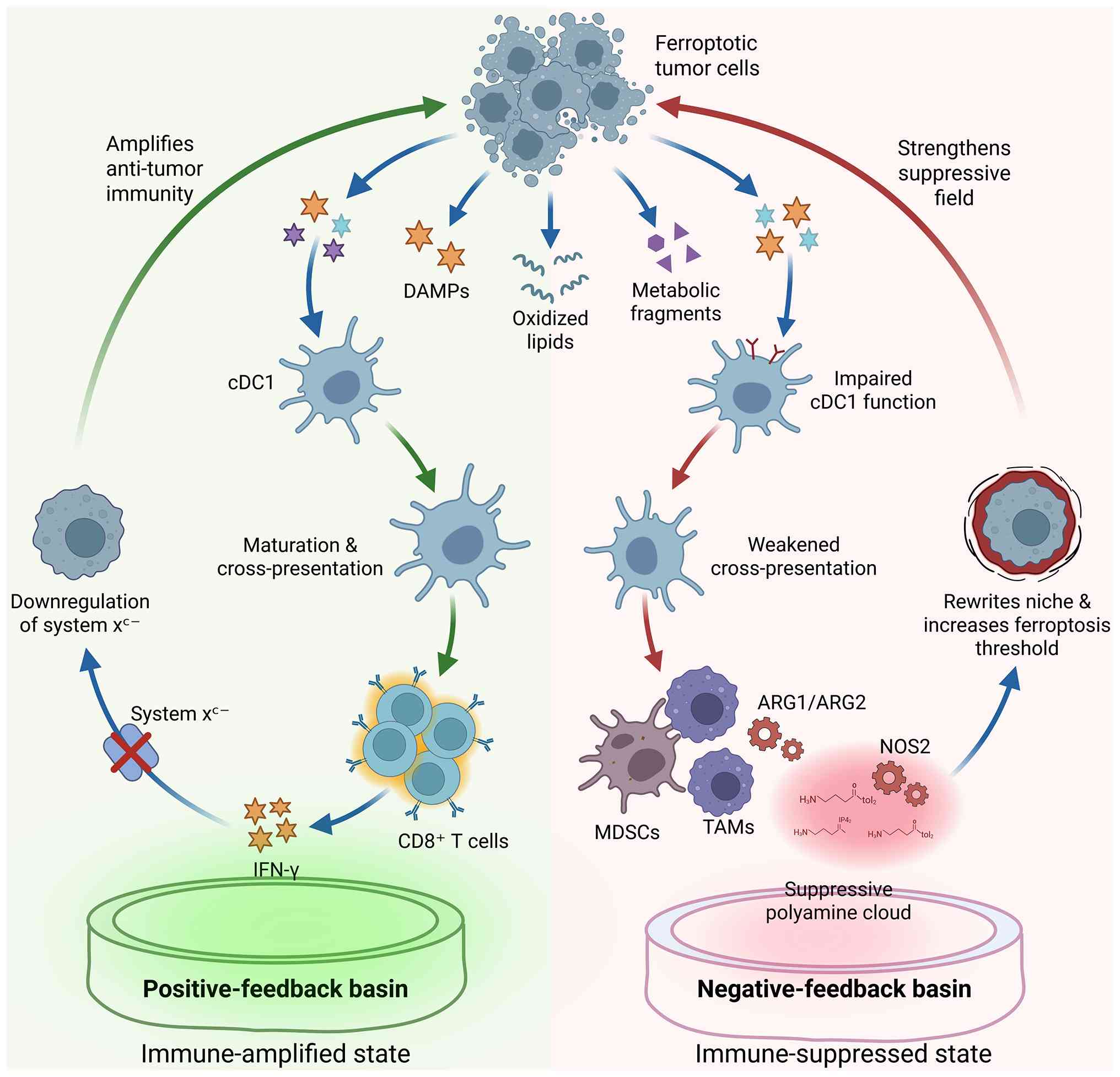 | Figure 5.Immune feedback routes after
ferroptotic tumor-cell output. Processable ferroptotic signals may
support dendritic-cell activation, IFN-γ production and positive
feedback, whereas excessive or spatially disconnected output may
impair antigen presentation and promote suppressive myeloid
rewriting. DAMPs, damage-associated molecular patterns; cDC1,
conventional type 1 dendritic cell; CD8, cluster of differentiation
8; IFN-γ, interferon-γ; system xc−,
cystine/glutamate antiporter; ARG1, arginase 1; ARG2, arginase 2;
MDSCs, myeloid-derived suppressor cells; TAMs, tumor-associated
macrophages; NOS2, nitric oxide synthase 2. |
Positive feedback: IFN-gamma links
t-cell effector activity to the ferroptosis threshold
The clearest template for positive immune feedback
is the loop of ICB-induced CD8+ T-cell activation, IFN-γ
production, system xc− suppression, reduced
antioxidant capacity and enhanced ferroptosis. Wang et al
(85) showed that IFN-γ released
by CD8+ T cells downregulates SLC7A11 and SLC3A2 in tumor cells,
thereby reducing cystine uptake, weakening GSH-dependent
antioxidant defenses and promoting lipid peroxidation and
ferroptosis. Consistently, cyst(e)ine depletion by cyst(e)inase can
synergize with ICB to enhance antitumor immunity and ferroptotic
tumor-cell death (85,86).
This feedback loop provides a strategic sequence
for state-matching therapies. When T-cell execution remains viable,
the immune response itself can act as an Output-layer threshold
regulator. Sustained IFN-γ signaling weakens the system
xc−-GSH-GPX4 axis, reducing the additional
perturbations required to execute ferroptosis. This explains why
restoring T-cell activity prior to strong ferroptosis amplification
may be more effective than directly forcing ferroptosis in
immune-inflamed tumors or tumors that can be converted into a ‘hot’
immune state. Such restoration may require improving arginine
availability, dismantling suppressive polyamines or myeloid
constraints, or alleviating spatial exclusion before applying
Output-layer amplification (87,88).
Negative feedback: Ferroptotic
products may impair dendritic-cell processing and
cross-presentation
Although ferroptosis can promote antitumor
immunity, increasing evidence indicates that ferroptotic tumor-cell
products may also impair antigen presentation under specific timing
and compositional contexts. Wiernicki et al (4) showed that ferroptotic tumor cells can
inhibit dendritic-cell maturation, hinder cross-presentation, and
fail to induce effective tumor protection when used for
dendritic-cell loading (5). These
findings indicate that ferroptosis is not synonymous with
immunogenic cell death; its immune consequences depend on the
generation, processing, and presentation of ferroptotic
materials.
In immune rejection or immune-desert tumors,
negative feedback is more likely to occur, especially in
environments dominated by myeloid cells. In these environments,
dendritic cells and T cells are spatially separated or functionally
restricted and excessive oxidized lipids or membrane-peroxidation
products may exceed the antigen-processing capacity and promote
suppressive inflammatory remodeling (29,89).
The execution kinetics of ferroptosis may further influence this
output. ESCRT-III-mediated membrane repair can delay ferroptosis
and regulate membrane-pore formation, Ca2+ signaling and
immune visibility, thereby altering the immune system's perception
of dying tumor cells (78,79). Therefore, negative feedback does
not imply that ferroptosis is universally detrimental; rather, it
reflects a mismatch between ferroptosis output and the immune
system's processing capacity.
What determines whether death output
enters positive or negative feedback?
Within a state-matched immunopharmacological
framework, ferroptosis should not be assumed to be automatically
immunogenic. The same ferroptosis output may enter opposing
feedback states depending on whether it can be processed by the
immune system, coupled with the effects circuit at the tissue level
and whether it is released from the dominant suppressive network.
Based on current evidence, the present study proposed an
evidence-based but hypothesis-generating feedback-gating framework
organized around three conditions that may favor positive feedback:
Sufficient immune-processing capacity, spatial accessibility and
lack of dominant myeloid- or polyamine-mediated suppression. Each
condition has different experimental or human-tumor evidence
supporting it, as described below. However, their simultaneous
requirements have yet to be prospectively validated in a single
tumor cohort, and thus should be interpreted as testable gating
variables rather than universally established necessary and
sufficient criteria. The failure of any one condition may prevent
the conversion of ferroptosis output into immune amplification.
Conversely, the system may enter processing failure, suppressive
myeloid remodeling and stable therapeutic tolerance. These gating
conditions should not be viewed as strict categories but as an
operational framework for predicting feedback direction. Their
value lies in helping determine whether a given tumor state is
ready for Output-layer amplification, or whether treatment should
first shift upstream toward Input-layer correction, State-layer
disruption or spatial preconditioning.
Is immune-processing capacity
sufficient: Processable output or processing failure?
The premise of positive feedback is that the output
of ferroptotic tumor cells can be captured and processed by
dendritic cells, promoting their maturation and supporting antigen
cross-presentation, thereby translating into sustained T-cell
expansion and IFN-γ-driven amplification. Experimental evidence
suggests that this process largely depends on the stage and method
of ferroptosis induction. In murine MCA205 fibrosarcoma and GL261
glioma models, early RSL3-induced ferroptotic cells were
effectively engulfed by bone-marrow-derived dendritic cells,
promoting dendritic-cell maturation and generating protective
immunity in preventive vaccination experiments, while late
ferroptotic cells failed to produce comparable protection (84).
However, ferroptosis does not uniformly produce
processable immune output. Using inducible GPX4 inhibition and
pharmacological ferroptosis models, Wiernicki et al
(4) reported that ferroptotic
cancer cells impaired dendritic-cell maturation, reduced antigen
cross-presentation and failed to protect mice from subsequent
MCA205 fibrosarcoma or B16-OVA melanoma growth. These seemingly
disparate findings suggest that immune-processing capacity is
determined not by ferroptosis alone, but by the inducers, death
stage, DAMP-release kinetics, oxidized-lipid burden, antigen cargo
and the functional state of receiving dendritic cells. Early,
controlled and immune-processable cell death outputs facilitate
immune amplification, whereas prolonged or highly oxidized output
may become a processing burden and promote negative feedback.
The immune-processing capacity can thus be viewed
as the first feedback-gating condition. Surrogate indicators may
include the abundance of conventional type 1 dendritic cell (cDC1),
such as the basic leucine zipper ATF-like transcription factor 3,
X-C motif chemokine receptor 1 and C-type lectin domain family 9
member A; dendritic-cell maturation markers, including cluster of
differentiation 86, C-C motif chemokine receptor 7 and interleukin
12; as well as antigen-presentation modules, including transporter
associated with antigen processing 1/2, beta-2-microglobulin and
major histocompatibility complex class I-related gene sets
(90–92). T-cell-side indicators include
clonal expansion and IFN-γ-axis activation, with representative
genes being interferon gamma gene, C-X-C motif chemokine ligand 9
and C-X-C motif chemokine ligand 10, as well as the restoration of
cytotoxic and proliferative programs (93,94).
Output-side readings may include lipid-peroxidation burden and
oxidized-lipid species, such as 4-hydroxynonenal and oxidized PE.
Insufficient immune processing capacity results in robust molecular
perturbations from direct ferroptosis amplification but no
effective immune activation. In this case, enhancing dendritic-cell
processing capacity or reducing unprocessable oxidative outputs
should precede Output-layer amplification.
Is spatial accessibility present: can
death output be coupled to the dendritic cell (DC)/T-cell
circuit?
Even if ferroptosis outputs can be processed, if
they are spatially decoupled from dendritic-cell and T-cell
circuits, they may not enter positive feedback. This is
particularly relevant in immune-excluded or immune-desert tumors,
where CAF-ECM barriers, low perfusion and diffusion bottlenecks
limit antigen capture and T-cell entry. In this case, ferroptosis
may occur within the inhibitory tumor core, which has poor
accessibility to immune cells. The output may not drive adaptive
immune amplification but instead convert to local inflammation and
myeloid-recruitment signals, thereby increasing the likelihood of
negative feedback (95,96).
Although direct spatial studies linking ferroptotic
tumor-cell outputs to dendritic-cell-T-cell coupling remain
limited, evidence from human tumor immunology supports spatial
accessibility as a biologically relevant threshold. In advanced
melanoma patients receiving immune checkpoint inhibitors, the
spatial proximity and interaction of cDC1s with CD8+ T cells
positively correlated with therapeutic response (97). By contrast, analyses of metastatic
urothelial cancers treated with atezolizumab showed that
transforming growth factor β (TGF-β) signaling, collagen-rich
stroma and the exclusion of CD8+ T cells from the tumor parenchyma
were associated with treatment resistance. In immune-excluded mouse
models, combined TGF-β and PD-L1 blockade restored T-cell
penetration into the tumor center and promoted tumor regression
(98). These studies did not
directly manipulate ferroptosis, but they provide human-tumor and
experimental evidence, indicating that the biological effects of
local death signals depend on whether antigen-presenting cells and
effector T cells are spatially connected to the area where the
signal is generated.
Therefore, spatial accessibility is the second
feedback-gating condition. Whether the Output layer can be
perceived and written back by the Feedback layer depends not only
on the composition of death output but also on whether death occurs
at a location connected to immune-processing and effector circuits.
If spatial accessibility is lacking, therapeutic priorities should
shift from direct Output-layer amplification to coupling repairs,
such as improving perfusion, reducing stromal barriers or relieving
immune rejection. Otherwise, the system may enter a state of
structural failure, where tumor-cell death occurs, but the immune
circuit remains disconnected.
Is the myeloid/polyamine suppressive
network dominant: Has negative-feedback gain been
preconfigured?
The third gating condition is whether the TME has
been dominated by the suppression network established by myeloid
activity and externalized polyamine flux. When the depletion
mediated by ARG1/ARG2 and the metastasis driven by NOS continue to
limit the availability of arginine, and polyamine uptake-efflux
cycling sustains a suppressive cloud, the output of ferroptosis may
primarily be interpreted as an additional damage signal. This can
promote myeloid recruitment, reinforce ARG/NOS activity, deepen
nutrient-dependent immune dysfunction and drive suppressive
inflammatory remodeling. In this case, Output-layer amplification
may have adverse effects: Tumor-cell killing remains insufficient,
whereas immune effector cells are further depressed below their
functional threshold (99,100).
Direct experimental evidence from pancreatic cancer
indicates that the output of ferroptosis can be converted into
tumor-promoting myeloid remodeling. Oxidative stress and
autophagy-dependent ferroptosis facilitate the release of oncogenic
KRAS^G12D from pancreatic cancer cells into extracellular vesicles.
Macrophages internalize extracellular KRAS^G12D through advanced
glycosylation end-product specific receptor (AGER), subsequently
polarizing towards an M2-like tumor-promoting state via signal
transducer and activator of transcription 3 (STAT3)-dependent
fatty-acid oxidation. Disruption of KRAS^G12D release or macrophage
uptake reduces macrophage-mediated pancreatic tumor growth, while
detection of KRAS^G12D in macrophages correlates with poor survival
in patients with pancreatic cancer (99). This provides a representative
example in which ferroptotic tumor-cell output directly enters a
negative myeloid-feedback circuit.
Hepatocellular carcinoma (HCC) provides a
complementary example of polyamine-mediated suppressive feedback.
N1-acetylspermidine is enriched in human HCC tissues and paired
plasma samples. Inflammatory macrophages induce the expression of
SAT1 in hepatoma cells, promoting SLC3A2-dependent
N1-acetylspermidine efflux. Extracellular metabolites activate SRC
proto-oncogene, non-receptor tyrosine kinase (SRC) signaling,
inducing C-C motif chemokine ligand 1 (CCL1)+ macrophage
polarization and inducing C-C motif chemokine receptor 8+
regulatory T cells, thereby hindering immune-checkpoint blockade.
Targeting SAT1, SLC3A2 or CCL1 improved the antitumor effects of
checkpoint therapy in experimental models (100). Although this study did not
specifically initiate the circuit through ferroptosis, it provides
human-tumor and mechanistic evidence that an established
polyamine-efflux network can preconfigure the TME toward
suppressive feedback.
Therefore, the key issue is not whether a single
suppressive molecule is upregulated, but whether the
myeloid/polyamine network has gained sufficient system-level gain
to prioritize the conversion of death output into suppressive
remodeling rather than adaptive immune amplification. Surrogate
indicators may include MDSC/TAM enrichment, ARG1/ARG2 and NOS2
expression, polyamine-related modules such as ODC1/AMD1 and
SAT1/SMOX, acetylated-polyamine ratios, and additional stress
signals such as cyst(e)ine limitation or antioxidant-substrate
competition (32,46,54,101–105). If this suppressive network is
dominant, ferroptosis amplification should be positioned as a
later-stage intervention. Upfront therapy should instead prioritize
dismantling myeloid/polyamine suppression, restoring Input-layer
nutrient availability, disrupting State-layer locking and reducing
negative-feedback gain.
Summary: Three gating conditions
determine the feedback basin and therapeutic sequence
Overall, the immunological consequences of
ferroptosis output should be interpreted as a gated circuit rather
than an automatic result. Output-layer amplification is more likely
to produce effective immune feedback only when all three conditions
are simultaneously met: Sufficient immune-processing capacity,
spatial accessibility and absence of dominant myeloid/polyamine
suppressive-network control. If any condition is missing, the
treatment sequence should shift upstream toward Input-layer
correction, State-layer disruption or spatial preconditioning,
rather than simply increasing ferroptosis stress.
These gating conditions should be viewed as an
evidence-anchored and operational hypothesis rather than a
universal rule validated across all cancer types and clinical
settings. Immune-processing capacity is supported by
ferroptosis-cell vaccination and dendritic-cell experiments;
spatial accessibility is supported by spatially resolved human
tumor studies and immune-exclusion models, while the
suppressive-network dominance is supported by studies on
ferroptosis-driven macrophage polarization and polyamine-mediated
immune remodeling mechanisms. Nevertheless, these three conditions
have primarily been studied in different experimental systems and
tumor types. Their combined predictive value, relative weight, and
potential cancer-specific thresholds need to be prospectively
validated through spatial profiling, cell-type-resolved metabolic
measurements and longitudinal treatment-response cohorts.
Therefore, the current framework provides a testable decision
scaffold for determining when to repair upstream conditions, when
to amplify ferroptosis output and when to switch therapeutic
routes.
Feedback rewriting of the input and
state layers: Myeloid expansion, ARG/NOS activation and
polyamine-cloud restabilization
When the aforementioned gating conditions are not
met, ferroptosis output may enter a negative-feedback basin. A
typical niche remodeling trajectory is initiated by oxidative
stress or damage-associated signals that drive myeloid cell
recruitment and expansion. These myeloid cell populations can
further upregulate ARG1/ARG2 and NOS2, intensifying arginine
depletion and diversion within the arginine niche and pushing local
arginine flux further below the threshold required for effector
T-cell activity.
Meanwhile, increased ornithine supply can reinforce
polyamine synthesis and uptake-efflux cycles, leading to the
restabilization of the suppressive polyamine cloud. Through this
circuit, short-term death-output events may be converted into
long-term niche remodeling. This feedback rewriting may contribute
to post-treatment immune cooling, tumor recurrence and therapeutic
tolerance.
State-matched immunopharmacological
strategies and therapeutic sequencing
Based on the Input-State-Output-Feedback framework,
cancer therapy should not be designed solely by targeting isolated
metabolic nodes. A more feasible strategy is to first define the
state coordinates of the tumor system and then apply sequential or
combinatorial interventions to reshape the vulnerability to
ferroptosis and immune execution capacity. From this perspective,
treatment design shifts from node-centric inhibition to
state-matching threshold modulation, where the timing, order and
combination of interventions are determined by the availability of
arginine, polyamine flux, ferroptosis-buffering capacity and
immune-feedback potential. Fig. 6
translates this framework into a state-matching therapeutic
decision diagram.
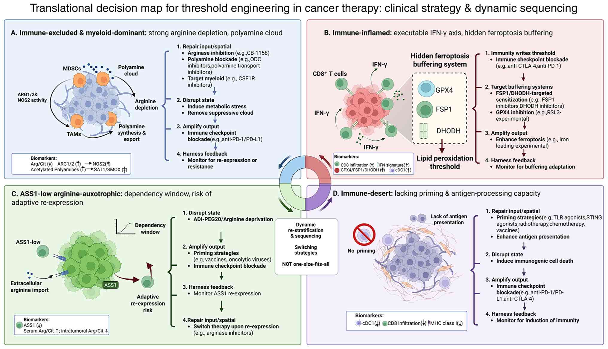 | Figure 6.State-matched therapeutic decision
map. Four tumor-immune prototypes are organized by dominant
bottleneck, preferred therapeutic sequence, pharmacodynamic
readouts and switch triggers: immune-excluded/myeloid-dominant,
immune-inflamed/IFN-gamma-executable, ASS1-low/arginine-dependent
and immune-desert/priming-deficient states. MDSCs, myeloid-derived
suppressor cells; TAMs, tumor-associated macrophages; ARG1/2,
arginase 1/arginase 2; NOS2, nitric oxide synthase 2; ODC,
ornithine decarboxylase; CSF1R, colony-stimulating factor 1
receptor; Arg/Cit, arginine-to-citrulline ratio; SAT1,
spermidine/spermine N1-acetyltransferase 1; SMOX, spermine oxidase;
IFN-γ, interferon-γ; CD8, cluster of differentiation 8; GPX4,
glutathione peroxidase 4; FSP1, ferroptosis suppressor protein 1;
DHODH, dihydroorotate dehydrogenase; cDC1, conventional type 1
dendritic cell; PD-1, programmed cell death protein 1; PD-L1,
programmed death-ligand 1; CTLA-4, cytotoxic
T-lymphocyte-associated protein 4; ASS1, argininosuccinate synthase
1; ADI-PEG20, pegylated arginine deiminase; TLR, toll-like
receptor; STING, stimulator of interferon genes; MHC class I, major
histocompatibility complex class I. |
Why state mapping should precede
pharmacological combination
State-matching immunopharmacological therapy should
not be simplified to ferroptosis induction or immune activation
alone. Identical metabolic perturbations can induce opposing
biological outcomes across different tumor types, spatial
microdomains and treatment stages. For example, arginine
restriction may impair T-cell effector function by reducing local
arginine flux below the immune functional threshold, but under
specific redox and buffering conditions, it may also increase
susceptibility to lipid peroxidation. Similarly, ferroptosis
induction may promote immune amplification in one context while
leading to dendritic-cell processing failure, suppressive myeloid
remodeling or ineffective molecular perturbation in another. These
different outcomes should not merely be viewed as experimental
inconsistencies; rather, they reflect different state coordinates
that determine the direction and executability of therapeutic
output.
Based on the Input-State-Output-Feedback logic, the
present study proposed state mapping as a practical framework for
organizing these differences. Here, ‘state mapping’ does not refer
to a validated pan-cancer classification system, but rather to an
operational four-dimensional coordinate model that identifies the
main bottlenecks of a given tumor system: Input-layer arginine
availability, State-layer polyamine flux, Output-layer ferroptosis
vulnerability or Feedback-layer immune-processing capacity. The aim
is to guide therapeutic sequencing; what should be corrected first,
what should be amplified next, when treatment should be escalated
and when a therapeutic route should be switched. Thus, state
mapping is intended to organize decision logic rather than replace
disease-specific biological details.
Pharmacological leverage points across
the Input-State-Output-Feedback layers
Therapy can be viewed as a targeted reconfiguration
of the Input-State-Output-Feedback system, rather than an isolated
single-node inhibition. The stability of therapeutic efficacy
depends not only on the potency of individual drugs but also on
whether the intervention aligns with the current state coordinates
and is delivered in a sequence that pushes the system toward an
executable therapeutic window.
To enhance operational applicability, treatment
strategies are organized around six decision dimensions: initial
state, dominant bottleneck, priority leverage point, combination
sequence, dynamic pharmacodynamic readouts and escape or
state-drift trajectories, as summarized in Table II.
| Table II.Prototype-based state-matching matrix
for therapeutic sequencing, pharmacodynamic monitoring and
treatment switching. |
Table II.
Prototype-based state-matching matrix
for therapeutic sequencing, pharmacodynamic monitoring and
treatment switching.
| Prototype | Decision
dimension | Details |
|---|
| A.
Immune-excluded/myeloid-dominant | State criteria | Low Arg/Cit; high
ARG1/ARG2 or NOS2; CAF-ECM barrier; marginal CD8+ T cells; strong
polyamine uptake-efflux cycling |
|
| Dominant
bottleneck | Spatial exclusion;
ARG-driven depletion; suppressive polyamine cloud |
|
| Preferred
sequence | Input/spatial
repair → polyamine blockade → bypass-informed ferroptosis
sensitization |
|
| Example
combinations | Arginase inhibition
± spatial repair; DFMO + AMXT1501; later FSP1/DHODH/BH4-informed
sensitization |
|
| Minimal PD
readouts | Arg/Cit;
ARG1/ARG2/NOS2; CD8+ entry; acetylated polyamines;
cDC1/cross-presentation; FSP1/DHODH/BH4 |
|
| Switch
triggers | No improvement in
CD8+ entry or DC processing → return to repair; bypass buffering
increases → add bypass-targeted sensitization; myeloid rewriting
increases → reduce output pressure |
| B.
Immune-inflamed/IFN-γ-executable | State criteria | Active
IFN-γ/cytotoxic programs; preserved DC processing; SLC7A11/system
xc− vulnerability; variable bypass
buffering |
|
| Dominant
bottleneck | Hidden
anti-ferroptotic buffering limits immune-driven amplification |
|
| Preferred
sequence | ICB-driven immune
activation →system xc− weakening →
bypass-stratified ferroptosis sensitization |
|
| Example
combinations | ICB; ICB + system
xc−pressure; ICB + DHODH/FSP1/BH4-informed
sensitization |
|
| Minimal PD
readouts | IFN-γ signature;
CXCL9/CXCL10; SLC7A11/GPX4; FSP1/DHODH/BH4; DC maturation |
|
| Switch
triggers | DC processing
declines → use pulsed/windowed pressure; bypass buffering increases
→ shift to bypass dismantling; inflamed-to-excluded drift → follow
Prototype A |
| C.
ASS1-low/arginine-dependent | State criteria | Low or defective
ASS1; exogenous arginine dependence; sensitivity to arginine
deprivation; risk of ASS1 re-expression |
|
| Dominant
bottleneck | Dependency window,
but excessive deprivation may impair immune execution |
|
| Preferred
sequence | ADI-PEG20 induction
→ immune/substrate support → output sensitization after state
reassessment |
|
| Example
combinations |
ADI-PEG20/pegargiminase ± chemotherapy or
ICB; later ferroptosis sensitization according to buffering
status |
|
| Minimal PD
readouts | ASS1
expression/methylation; Arg/Cit; immune phenotype; IFN-γ axis;
FSP1/DHODH/BH4 |
|
| Switch
triggers | ASS1 re-expression
→ stop relying on deprivation; IFN-γ or DC function declines →
reduce deprivation intensity; state shifts to excluded/inflamed →
follow A or B |
| D.
Immune-desert/priming-deficient | State criteria | Low cDC1; weak
antigen presentation; poor CD8+ infiltration; absent
IFN-γ/cytotoxic programs |
|
| Dominant
bottleneck | No effective immune
receiver for ferroptotic output |
|
| Preferred
sequence | Build immune
priming/processing → repair access if needed → introduce output
amplification only after immune conversion |
|
| Example
combinations | DC-priming
strategies; antigen-presentation enhancement; spatial repair; later
ICB or ferroptosis sensitization according to new state |
|
| Minimal PD
readouts | cDC1 markers;
MHC-I/TAP1/2/B2M; DC maturation; CD8+ infiltration; myeloid
suppression index |
|
| Switch
triggers | Priming fails →
avoid output amplification; desert-to-excluded transition → follow
A; desert-to-inflamed transition → follow B |
Four tumor-immune prototypes for
therapeutic sequencing
The following four prototypes are not intended to
define a strict classification system for all solid tumors.
Instead, they provide a minimal translational framework for
applying state matching to therapeutic sequencing. Each prototype
is organized around five questions: Where the dominant bottleneck
lies, which leverage point should be prioritized, how the
intervention sequence should be arranged, which pharmacodynamic
panel should be monitored and when the therapeutic route should be
switched.
This framework does not override disease-specific
biological characteristics with fixed classification categories.
Instead, it provides a conceptual scaffold for comparing tumor
states across different models and cancer types, translating
Input-State-Output-Feedback logic into executable therapeutic
rules. To operationalize these prototypes, we define candidate
marker combinations and assay-based reference standards, rather
than single-marker diagnostic thresholds (Table II). Prototype assignments should
require concordance across at least two orthogonal evidence layers,
including at least one spatial or functional measurement when
feasible. ‘High’ and ‘low’ states should be prospectively defined
using disease-specific reference cohorts or validated assay
thresholds, rather than arbitrary post hoc median splits.
Importantly, Prototypes A, B and D primarily describe
immune-spatial states, whereas Prototype C describes a tumor-cell
metabolic dependency. Prototype C may therefore coexist with
Prototype A, B or D and should be reported as a composite state,
such as C-A or C-B, when appropriate.
Prototype A: Immune-excluded,
myeloid-dominant tumors with high ARG activity and a strong
polyamine cloud
In this prototype, the dominant bottleneck is
typically not initially located at the Output layer. Instead,
therapeutic failure arises from poor spatial accessibility and high
inhibitory feedback gain, which prevents ferroptosis output from
combining with effective immune amplification. Thus, Prototype A
represents a state that should prioritize early repair rather than
immediate Output-layer amplification. A more appropriate sequence
is to first restore Input-layer arginine availability and improve
spatial accessibility, then disrupt the State-layer polyamine-cloud
cycle. Only after immune entry and processing capacity improve
should ferroptosis amplification be introduced, with additional
sensitization guided by the status of anti-ferroptosis bypass
defenses (98,101,106).
Prototype A is not intended as a universal
definition for all immune-excluded tumors but rather as a working
model for those where the primary failure point lies in the
decoupling of death output from the immune circuit. A minimal
pharmacodynamic panel should include arginine/citrulline (Arg/Cit)
dynamics, ARG/NOS-axis activity, spatial redistribution of CD8+ T
cells from the tumor margin to the parenchyma, acetylated-polyamine
ratios or other polyamine-flux surrogates, DC/cDC1 processing
capacity and anti-ferroptosis bypass markers. If immune entry and
processing capacity fail to improve, or if bypass buffering becomes
dominant, treatment should shift to further spatial repair,
suppressive-network dismantling or schedule adjustments, rather
than premature ferroptosis amplification. Operational assignments
for Prototype A should simultaneously require evidence of
immune-exclusion patterns and myeloid/ARG-polyamine dominance. High
expression of a single marker, such as ARG1 or cluster of
differentiation 163, is insufficient. The immune-exclusion
components should be demonstrated by the preferential localization
of CD8+ T cells at the invasive margin rather than their
distribution within the tumor parenchyma, while suppressive-network
dominance should be supported by concordant myeloid-cell
enrichment, ARG/NOS activity and arginine/polyamine metabolic
measurements.
Prototype B: Immune-inflamed tumors
with an executable IFN-γ axis
In this prototype, the system already incorporates
baseline positive-feedback potential, with the dominant bottleneck
typically shifting from spatial inaccessibility to hidden
anti-ferroptotic buffering. Despite the presence of immune
activity, Output-layer amplification may still be limited by bypass
defenses such as FSP1, DHODH and GCH1/BH4, which elevate the
effective ferroptosis threshold (74,85).
Therefore, Prototype B represents a state where immune activity
should first be written into the threshold. ICB may activate CD8+ T
cells and IFN-γ signaling, suppress the system
xc−-GSH antioxidant axis and weaken the
tumor-cell buffering capacity. Subsequently, ferroptosis
sensitization should be adjusted based on the status of bypass
defenses, further amplifying the existing immune-ferroptosis
window.
The key implication is that therapeutic failure in
immune-inflamed tumors does not necessarily indicate insufficient
immune activation. Rather, it may reflect persistent hidden
buffering that prevents immune-derived oxidative pressure from
translating into executable ferroptosis output. Therefore, the
minimal pharmacodynamic panel should include IFN-γ/cytotoxicity
signatures, system xc−-related indicators,
anti-ferroptotic bypass markers including FSP1, DHODH and GCH1/BH4
and dendritic-cell processing capacity. If dendritic-cell function
declines, treatment should shift from continuous pressure to a
windowed or pulsed schedule. If bypass buffering increases, the
strategy should shift from single-axis amplification to bypass
dismantling. If the immune-inflammatory state drifts toward immune
exclusion, the therapeutic logic should revert to the Prototype A
sequence: repair first, amplify later (4,94).
Operational assignments for Prototype B should require intratumoral
effector-cell infiltration, combined with active IFN-γ and
antigen-presentation programs. Markers such as FSP1, DHODH, GCH1
and SLC7A11/GPX4 should be used to define the dominant
ferroptosis-buffering mechanism, rather than establishing the
immune-inflamed phenotype itself.
Prototype C: ASS1-low auxotrophic
tumors with exogenous arginine dependence
The core feature of this prototype is a strong
Input-layer dependency window arising from limited endogenous
arginine-replenishment capacity. In ASS1-low tumors,
arginine-deprivation strategies such as pegylated arginine
deiminase (ADI-PEG20) can induce acute Input crisis and show
clinical activity in specific disease contexts (107). However, the dominant bottleneck
is dual. Arginine deprivation may inhibit tumor growth but may also
further reduce local arginine availability below the threshold
required for effector T-cell function, thereby weakening immune
execution. Additionally, therapeutic pressure may induce ASS1
re-expression or alternative replenishment programs, leading to
state-coordinate drift (108,109).
Therefore, Prototype C should not be interpreted as
a linear scenario where continuous arginine deprivation alone is
sufficient. Rather, arginine deprivation should be viewed as an
initiating Input-layer perturbation that must be combined with
immune support, pharmacodynamic monitoring and adaptive sequences.
A practical sequence could be formulated as follows:
ADI-PEG20-based Input perturbation, followed by immune-supportive
or threshold-protective interventions and then Output-layer
ferroptosis amplification guided by trigger capacity and
bypass-buffering status. If ASS1 re-expression, enhanced
arginine-replenishment capacity, or simultaneous declines in IFN-γ
signaling and dendritic-cell processing capacity are observed,
treatment should shift to reducing perturbation intensity, window
interventions or reclassifying to Prototype A or B pathways, rather
than further intensifying arginine-deprivation pressure (110,111). Prototype C should not be assigned
solely based on reduced ASS1 expression. Functional arginine
auxotrophy should be confirmed by impaired growth under
arginine-depleted conditions, failure of citrulline supplementation
to fully restore proliferation and/or restoration of resistance
following ASS1 re-expression. Sensitivity to ADI-PEG20 may provide
additional functional readouts but should be interpreted alongside
treatment-induced ASS1 re-expression and alternative
arginine-replenishment pathways.
Prototype D: Immune-desert tumors with
deficient processing capacity
In this prototype, the main bottleneck is initially
not the threshold for ferroptosis, but rather the lack of an
established Feedback circuit. When immune-initiating gain and
dendritic-cell processing capacity are deficient, the amplified
ferroptotic outputs cannot be effectively captured and presented to
trigger adaptive immune amplification. Instead, it may promote the
recruitment of myeloid cells and negative-feedback remodeling
(112). Therefore, Prototype D
represents a state where Feedback-layer construction should precede
Output-layer amplification. The priority is to establish immune
priming and processing capacity, while repairing spatial
accessibility when necessary. Amplification of ferroptosis should
only be introduced after the system transitions from an
immune-desert state to a manageable immune state.
The key implication is that not all cold tumors are
suitable for intensive ferroptosis induction. When
immune-processing capacity is absent, Output-layer enhancement may
lack an effective immune receiver and may instead feed negative
feedback. Thus, the minimal pharmacodynamic panel should initially
prioritize cDC1 abundance, dendritic-cell maturation,
cross-presentation modules, and spatial immune-phenotype
transition, rather than ferroptosis markers alone. Once the
immune-desert state shifts toward an immune-excluded or
immune-inflamed phenotype, subsequent sequencing should follow the
logic of Prototype A or Prototype B, respectively (4,113,114).
Operational assignment and reference
standards
Operational assignment should follow a two-step
procedure. First, tumor-cell arginine dependency should be assessed
to determine whether there is a functional ASS1-low nutritional
deficiency coverage. Second, the immune-spatial state should be
classified as Prototype A, B or D based on the distribution of CD8+
T cells and cDC1s, IFN-γ and antigen-presentation activity, and the
dominance of myeloid/ARG-polyamine suppression. Tumors that do not
satisfy a single dominant pattern should be reported as mixed or
transitional states, rather than being forcibly categorized into
one category.
Reference standards should be specific to the
detection. The spatial immune phenotype should be prioritized for
assessment using multiplex immunohistochemistry, multiplex
immunofluorescence or spatial transcriptomics. The arginine and
polyamine status should be supported by targeted metabolomics,
rather than solely relying on transcriptional expression. The
ferroptosis-buffering state should be assessed using combined
expression and activity-related indicators, as the abundance of
GPX4, FSP1 or DHODH does not necessarily reflect pathway flux.
Functional auxotrophy should be confirmed through
arginine-deprivation assays, citrulline-rescue experiments or
ASS1-restoration experiments. These measurements should be
calibrated against disease-specific control tissues, independent
validation cohorts or prospectively defined assay thresholds.
The proposed criteria are intended to serve as
candidate operational standards rather than clinically validated
diagnostic thresholds. Their reproducibility, relative weight and
predictive value need to be validated in spatially annotated
patient cohorts, in vitro tumor models and prospective
pharmacodynamic studies.
Summary: The four prototypes provide a
sequential decision scaffold
These four prototypes are not intended to support a
universal therapeutic combination or clinically validated
diagnostic classification. Instead, they emphasize that a rational
combination therapy should first identify the main failure points
in the Input-State-Output-Feedback system, and then match the
therapeutic sequence, combination structure and amplification
timing accordingly. Prototype assignment should be based on
composite molecular, metabolic, spatial and functional evidence
rather than on individual markers.
Prototypes A, B and D represent dominant
immune-spatial states, whereas Prototype C represents an orthogonal
metabolic dependency that may coexist with any of the three
immune-spatial states. Therefore, these prototypes should be
understood as rule-level and potential composite representations
within a state-matching decision framework. Their reproducibility,
clinical relevance thresholds and predictive value need to be
validated through spatially resolved, longitudinal and
pharmacodynamic studies.
State drift, switch triggers and
dynamic reassessment
Therapeutic resistance can be defined as
system-level drift between distinct tumor-immune states, rendering
the original therapeutic sequence mismatched with the dominant
bottleneck. Therefore, switch triggers should be incorporated into
dynamic decision rules and the Input-State-Output-Feedback
coordinates should be reassessed longitudinally during the
treatment process.
Input drift includes ASS1 re-expression, altered
arginine uptake or enhanced ARG/NOS activity, indicating a
restoration of the tumor-cell replenishment capacity or progression
toward a myeloid-dominant arginine-restricted state. State drift
includes increased ODC1/AMD1-driven synthesis,
SAT1/PAOX/SMOX-associated catabolic flux or restabilization of
polyamine uptake-efflux cycling. Output drift is reflected as an
increase in ferroptosis buffering, such as SLC7A11/GPX4, FSP1,
DHODH or GCH1/BH4, or insufficient triggering capacity involving
ACSL4/LPCAT3, ALOX15 or TFRC/NCOA4. Feedback drift includes the
loss of IFN-γ/cytotoxicity signatures, impaired dendritic-cell
processing, reduced antigen presentation or a transition towards
immune exclusion or immune desert.
These drift triggers should define the switching
routes in Table II. Dynamic
reassessment helps distinguish true therapeutic resistance from
state mismatch and determines whether the next step should be
Input-layer repair, State-layer disruption, Output-layer
amplification or Feedback-layer restoration.
Safety boundaries for
ferroptosis-directed combinations
Given that GPX4 and its associated
anti-lipid-peroxidation networks are essential for normal tissue
homeostasis, ferroptosis-targeted combination therapies require
both precise state matching and strict safety boundaries. The
guiding principle should be to stratify first and then intensify.
Prior to Output-layer amplification, the core ferroptosis-defense
axis and parallel bypass systems, including system
xc−-GSH-GPX4 axis, FSP1-CoQ10, DHODH-CoQ and
GCH1/BH4, should be evaluated to avoid ineffective or harmful
exposure in tumors that do not rely on the target buffering
axis.
Safety should also influence therapeutic
sequencing. Input-layer interventions may provide upstream leverage
but require monitoring for systemic metabolic effects and
unintended T-cell suppression. State-layer polyamine blockade may
disrupt suppressive buffering, but long-term exposure should be
carefully evaluated because polyamines support normal tissue
renewal and immune-cell function. Thus, ferroptosis-directed
combinations should use stepwise escalation guided by
pharmacodynamic evidence of tumor vulnerability, bypass-buffering
status and preserved immune competence.
Future perspectives and conclusions
To make this state-matching immunopharmacological
framework practically applicable in translation, the key challenge
lies not in further expanding the list of node-level mechanisms but
in transforming the proposed Input-State-Output-Feedback
coordinates into clinically accessible, reproducible and updateable
readout systems. Thus, in the present review, the framework was
presented as a conceptual yet testable working model. Its value
lies not in replacing disease-specific evidence, but in providing a
unified language to define what should be monitored, how
therapeutic states should be validated, how sequential trials
should be designed and when treatment routes should be
switched.
At the monitoring level, future work should
establish a minimal variable set that supports dynamic state
reassessment rather than relying on single static indicators. At
the Input layer, monitoring may include Arg/Cit dynamics, ASS1
status, ARG/NOS-axis activity and spatial flux surrogates. At the
State layer, priority should be given to surrogate readouts of
polyamine flux, such as acetylated-polyamine ratios, paired
synthetic and catabolic signatures and spatial-omics constraints.
At the Output layer, ACSL4/LPCAT3, iron-metabolism surrogates and
buffering markers including GPX4, FSP1 and DHODH should be
interpreted jointly. At the Feedback layer, longitudinal panels
should assess dendritic-cell processing capacity, spatial
accessibility and myeloid-mediated suppressive remodeling. A useful
monitoring system should not be defined by the number of variables
included, but by whether it can determine, during treatment,
whether the tumor system has undergone a state transition.
Spatial heterogeneity should be assessed by
combining tissue-resolved mapping with repeatable whole-tumor
imaging. Spatial transcriptomics and multiplex immunostaining of
pretreatment or on-treatment biopsies can identify CAF-rich and
ECM-dense regions, their proximity to functional vasculature, local
myeloid accumulation and exclusion of CD8+ T cells. However, these
tissue-based methods provide spatial snapshots and become
longitudinal only when applied to serial, image-guided biopsies
(115). Dynamic contrast-enhanced
magnetic resonance imaging can non-invasively quantify regional
perfusion and vascular permeability, including parameters such as
K^trans, while serial ^18F-fluoromisonidazole positron emission
tomography/computed tomography can map and track hypoxic tumor
subvolumes during treatment (116,117). Therefore, a practical clinical
workflow would co-register imaging at baseline and during
treatment, obtain biopsies from radiographically distinct regions
when feasible, and combine these findings with spatial omics and
circulating metabolite measurements. This multimodal strategy may
distinguish persistent transport barriers from reversible metabolic
states and determine whether interventions improve nutrient
delivery and immune accessibility before ferroptosis amplification.
Table III summarizes
representative intervention leverage points classified according to
the Input-State-Output-Feedback framework.
| Table III.Representative pharmacological agents
and investigational compounds targeting the
Input-State-Output-Feedback framework. |
Table III.
Representative pharmacological agents
and investigational compounds targeting the
Input-State-Output-Feedback framework.
| Layer and molecular
target | Representative
agents or drug classes | Principal
pharmacological action | Development
stage | Key
limitations |
|---|
| Input:
ARG1/ARG2 | Numidargistat/CB
1158; OATD-02 | Inhibit arginase
activity and preserve extracellular L-arginine availability for
antitumor immune cells | Preclinical and
early clinical investigation | Limited activity
may occur in spatially excluded tumors or in the presence of
compensatory myeloid suppressive pathways |
| Input: ASS1-low
arginine dependency |
ADI-PEG20/pegargiminase | Enzymatically
deplete extracellular arginine in arginine-auxotrophic tumors | Clinical
investigation | May impair effector
T-cell function and select for ASS1 re-expression or alternative
arginine-replenishment pathways |
| State: Polyamine
synthesis and transport | Eflornithine/DFMO;
DFMO plus AMXT1501 | Inhibit
ODC1-dependent polyamine synthesis and block compensatory
extracellular polyamine uptake | Preclinical and
early clinical investigation | Uptake
compensation, metabolic adaptation and normal-tissue exposure may
limit efficacy |
| Output: system
xc−-GSH-GPX4 axis | Sulfasalazine and
other system xc− inhibitors; experimental
direct GPX4 inhibitors | Restrict
cystine-dependent GSH synthesis or directly impair
phospholipid-hydroperoxide detoxification | Drug repurposing
and preclinical investigation | Non-selective
systemic GPX4 inhibition may cause severe normal-tissue toxicity,
including renal injury. Direct GPX4 inhibitors remain preclinical
and require tumor-selective delivery, intermittent dosing and renal
safety monitoring |
| Output: FSP1-CoQ10
defense | iFSP1 and other
experimental FSP1 inhibitors | Disrupt
GPX4-independent CoQ10 reduction and plasma-membrane radical
trapping | Preclinical | Lack of clinically
validated inhibitors, tumor-selective delivery systems and
established pharmacodynamic biomarkers |
| Output: DHODH-CoQ
defense | Brequinar and other
DHODH inhibitors | Inhibit
mitochondrial CoQ reduction and weaken mitochondrial ferroptosis
buffering | Preclinical
ferroptosis investigation | Activity depends on
GPX4 abundance, mitochondrial dependence and compensatory extra
mitochondrial defenses |
| Feedback:
PD-1/PD-L1 immune-checkpoint axis | Anti-PD-1 and
anti-PD-L1 antibodies | Restore CD8+ T-cell
activity and IFN-γ-dependent suppression of tumor antioxidant
defenses | Clinically
established drug class | Limited efficacy in
immune-excluded, immune-desert or
dendritic-cell-processing-deficient tumors |
Acknowledgements
Not applicable.
Funding
The present study was supported by the Nantong Municipal Science
and Technology Plan Project (grant no. MSZ2025012), the Nantong
Natural Science Foundation (grant no. JC2025056), the Nantong
Municipal Health Commission Directive Project-General Program
(grant no. MS2025037), the Health Development Promotion Project
(grant no. KM-20250805-01) and the Nantong University Clinical
Medicine Special Funding Project (grant no. 2024JY040).
Availability of data and materials
Not applicable.
Authors' contributions
XD and YH contributed to conceptualization,
literature review, manuscript drafting and figure preparation. JS
contributed to literature organization and manuscript revision. HZ
and JL supervised the work, contributed to conceptual refinement
and critically revised the manuscript. All authors read and
approved the final manuscript. Data authentication is not
applicable.
Ethics approval and consent to
participate
Not applicable.
Patient consent for publication
Not applicable.
Competing interests
The authors declare that they have no competing
interests.
Use of artificial intelligence tools
During the preparation of this work, artificial
intelligence tools were used to improve the readability and
language of the manuscript or to generate images, and subsequently,
the authors revised and edited the content produced by the
artificial intelligence tools as necessary, taking full
responsibility for the ultimate content of the present manuscript.
No generative AI tools were used to create scientific figures.
Glossary
Abbreviations
Abbreviations:
|
ASS1
|
argininosuccinate synthase 1
|
|
ARG
|
arginase
|
|
CAF
|
cancer-associated fibroblast
|
|
DFMO
|
difluoromethylornithine
|
|
DHODH
|
dihydroorotate dehydrogenase
|
|
FSP1
|
ferroptosis suppressor protein 1
|
|
GCH1
|
GTP cyclohydrolase 1
|
|
GPX4
|
glutathione peroxidase 4
|
|
GSH
|
glutathione
|
|
ICB
|
immune checkpoint blockade
|
|
NOS
|
nitric oxide synthase
|
|
PAOX
|
polyamine oxidase
|
|
ROS
|
reactive oxygen species
|
|
SAT1
|
spermidine/spermine
N1-acetyltransferase 1
|
|
SMOX
|
spermine oxidase
|
|
TME
|
tumor microenvironment
|
References
|
1
|
Canale FP, Basso C, Antonini G, Perotti M,
Li N, Sokolovska A, Neumann J, James MJ, Geiger S, Jin W, et al:
Metabolic modulation of tumours with engineered bacteria for
immunotherapy. Nature. 598:662–666. 2021. View Article : Google Scholar : PubMed/NCBI
|
|
2
|
Zou Z, Cheng Q, Zhou J, Guo C,
Hadjinicolaou AV, Salio M, Liang X, Yang C, Du Y, Yao W, et al:
ATF4-SLC7A11-GSH axis mediates the acquisition of immunosuppressive
properties by activated CD4+ T cells in low arginine
condition. Cell Rep. 43:1139952024. View Article : Google Scholar : PubMed/NCBI
|
|
3
|
Bi G, Liang J, Bian Y, Shan G, Huang Y, Lu
T, Zhang H, Jin X, Chen Z, Zhao M, et al: Polyamine-mediated
ferroptosis amplification acts as a targetable vulnerability in
cancer. Nat Commun. 15:24612024. View Article : Google Scholar : PubMed/NCBI
|
|
4
|
Wiernicki B, Maschalidi S, Pinney J,
Adjemian S, Vanden Berghe T, Ravichandran KS and Vandenabeele P:
Cancer cells dying from ferroptosis impede dendritic cell-mediated
anti-tumor immunity. Nat Commun. 13:36762022. View Article : Google Scholar : PubMed/NCBI
|
|
5
|
Catanzaro E, Demuynck R, Naessens F,
Galluzzi L and Krysko DV: Immunogenicity of ferroptosis in cancer:
A matter of context? Trends Cancer. 10:407–416. 2024. View Article : Google Scholar : PubMed/NCBI
|
|
6
|
Lim AR, Rathmell WK and Rathmell JC: The
tumor microenvironment as a metabolic barrier to effector T cells
and immunotherapy. Elife. 9:e551852020. View Article : Google Scholar : PubMed/NCBI
|
|
7
|
DePeaux K and Delgoffe GM: Metabolic
barriers to cancer immunotherapy. Nat Rev Immunol. 21:785–797.
2021. View Article : Google Scholar : PubMed/NCBI
|
|
8
|
Matos A, Carvalho M, Bicho M and Ribeiro
R: Arginine and arginases modulate metabolism, tumor
microenvironment and prostate cancer progression. Nutrients.
13:45032021. View Article : Google Scholar : PubMed/NCBI
|
|
9
|
Bai D, Zhou Y, Jing L, Guo C and Yang Q:
Arginine metabolism in cancer biology and immunotherapy. Immune
Netw. 25:e302025. View Article : Google Scholar : PubMed/NCBI
|
|
10
|
Missiaen R, Lesner NP and Simon MC: HIF: A
master regulator of nutrient availability and metabolic cross-talk
in the tumor microenvironment. EMBO J. 42:e1120672023. View Article : Google Scholar : PubMed/NCBI
|
|
11
|
Wu Y, Chen Z, Chen X, Hu Y and Ma Y:
Recent advances in the investigation of the regulatory network
underlying reactive nitrogen species-mediated tumorigenesis:
Molecular mechanisms and targeted therapeutic strategies. Redox
Rep. 30:25645932025. View Article : Google Scholar : PubMed/NCBI
|
|
12
|
Wetzel TJ, Erfan SC, Figueroa LD, Wheeler
LM and Ananieva EA: Crosstalk between arginine, glutamine, and the
branched chain amino acid metabolism in the tumor microenvironment.
Front Oncol. 13:11865392023. View Article : Google Scholar : PubMed/NCBI
|
|
13
|
Dunlap KN, Bender A, Bowles A, Bott AJ,
Tay J, Grossmann AH, Rutter J and Ducker GS: SLC7A5 is required for
cancer cell growth under arginine-limited conditions. Cell Rep.
44:1151302025. View Article : Google Scholar : PubMed/NCBI
|
|
14
|
Wu Y, Wu Y, Gao Z, Yu W, Zhang L and Zhou
F: Revitalizing T cells: Breakthroughs and challenges in overcoming
T cell exhaustion. Signal Transduct Target Ther. 11:22026.
View Article : Google Scholar : PubMed/NCBI
|
|
15
|
Grzybowski MM, Uçal Y, Muchowicz A,
Rejczak T, Kikulska A, Głuchowska KM, Szostakowska-Rodzoś M,
Zagożdżon A, Bausbacher T, Tkaczyk A, et al: Metabolomic
reprogramming of the tumor microenvironment by dual arginase
inhibitor OATD-02 boosts anticancer immunity. Sci Rep.
15:187412025. View Article : Google Scholar : PubMed/NCBI
|
|
16
|
Engel K, Höhne W and Häberle J: Mutations
and polymorphisms in the human argininosuccinate synthetase (ASS1)
gene. Hum Mutat. 30:300–307. 2009. View Article : Google Scholar : PubMed/NCBI
|
|
17
|
Lim LQJ, Adler L, Hajaj E, Soria LR, Perry
RBT, Darzi N, Brody R, Furth N, Lichtenstein M, Bab-Dinitz E, et
al: ASS1 metabolically contributes to the nuclear and cytosolic
p53-mediated DNA damage response. Nat Metab. 6:1294–1309. 2024.
View Article : Google Scholar : PubMed/NCBI
|
|
18
|
Ding Q, Li R, Wang Q, Yu L and Zi F: A
pan-cancer analysis of the role of argininosuccinate synthase 1 in
human tumors. Front Oncol. 13:10491472023. View Article : Google Scholar : PubMed/NCBI
|
|
19
|
Rabinovich S, Adler L, Yizhak K, Sarver A,
Silberman A, Agron S, Stettner N, Sun Q, Brandis A, Helbling D, et
al: Diversion of aspartate in ASS1-deficient tumours fosters de
novo pyrimidine synthesis. Nature. 527:379–383. 2015. View Article : Google Scholar : PubMed/NCBI
|
|
20
|
Crump NT, Hadjinicolaou AV, Xia M,
Walsby-Tickle J, Gileadi U, Chen JL, Setshedi M, Olsen LR, Lau IJ,
Godfrey L, et al: Chromatin accessibility governs the differential
response of cancer and T cells to arginine starvation. Cell Rep.
35:1091012021. View Article : Google Scholar : PubMed/NCBI
|
|
21
|
Gai X, Liu Y, Lan X, Chen L, Yuan T, Xu J,
Li Y, Zheng Y, Yan Y, Yang L, et al: Oncogenic KRAS induces
arginine auxotrophy and confers a therapeutic vulnerability to
SLC7A1 inhibition in non-small cell lung cancer. Cancer Res.
84:1963–1977. 2024. View Article : Google Scholar : PubMed/NCBI
|
|
22
|
Raber PL, Thevenot P, Sierra R,
Wyczechowska D, Halle D, Ramirez ME, Ochoa AC, Fletcher M, Velasco
C, Wilk A, et al: Subpopulations of myeloid-derived suppressor
cells impair T cell responses through independent nitric
oxide-related pathways. Int J Cancer. 134:2853–2864. 2014.
View Article : Google Scholar : PubMed/NCBI
|
|
23
|
Corzo CA, Condamine T, Lu L, Cotter MJ,
Youn JI, Cheng P, Cho HI, Celis E, Quiceno DG, Padhya T, et al:
HIF-1α regulates function and differentiation of myeloid-derived
suppressor cells in the tumor microenvironment. J Exp Med.
207:2439–2453. 2010. View Article : Google Scholar : PubMed/NCBI
|
|
24
|
Menjivar RE, Nwosu ZC, Du W, Donahue KL,
Hong HS, Espinoza C, Brown K, Velez-Delgado A, Yan W, Lima F, et
al: Arginase 1 is a key driver of immune suppression in pancreatic
cancer. Elife. 12:e807212023. View Article : Google Scholar : PubMed/NCBI
|
|
25
|
Rotondo R, Bertolotto M, Barisione G,
Astigiano S, Mandruzzato S, Ottonello L, Dallegri F, Bronte V,
Ferrini S and Barbieri O: Exocytosis of azurophil and arginase
1-containing granules by activated polymorphonuclear neutrophils is
required to inhibit T lymphocyte proliferation. J Leukoc Biol.
89:721–727. 2011. View Article : Google Scholar : PubMed/NCBI
|
|
26
|
Martí I, Líndez AA and Reith W:
Arginine-dependent immune responses. Cell Mol Life Sci.
78:5303–5324. 2021. View Article : Google Scholar : PubMed/NCBI
|
|
27
|
Raynor JL and Chi H: Nutrients: Signal 4
in T cell immunity. J Exp Med. 221:e202218392024. View Article : Google Scholar : PubMed/NCBI
|
|
28
|
Sattiraju A, Kang S, Giotti B, Chen Z,
Marallano VJ, Brusco C, Ramakrishnan A, Shen L, Tsankov AM,
Hambardzumyan D, et al: Hypoxic niches attract and sequester
tumor-associated macrophages and cytotoxic T cells and reprogram
them for immunosuppression. Immunity. 56:1825–1843.e6. 2023.
View Article : Google Scholar : PubMed/NCBI
|
|
29
|
Arpinati L, Carradori G and Scherz-Shouval
R: CAF-induced physical constraints controlling T cell state and
localization in solid tumours. Nat Rev Cancer. 24:676–693. 2024.
View Article : Google Scholar : PubMed/NCBI
|
|
30
|
Pegg AE: Mammalian polyamine metabolism
and function. IUBMB Life. 61:880–894. 2009. View Article : Google Scholar : PubMed/NCBI
|
|
31
|
Smirnova OA, Bartosch B, Zakirova NF,
Kochetkov SN and Ivanov AV: Polyamine metabolism and oxidative
protein folding in the ER as ROS-producing systems neglected in
virology. Int J Mol Sci. 19:12192018. View Article : Google Scholar : PubMed/NCBI
|
|
32
|
DeFelice BC and Fiehn O: Rapid LC-MS/MS
quantification of cancer related acetylated polyamines in human
biofluids. Talanta. 196:415–419. 2019. View Article : Google Scholar : PubMed/NCBI
|
|
33
|
Cerrada-Gimenez M, Häkkinen MR,
Vepsäläinen J, Auriola S, Alhonen L and Keinänen TA: Polyamine flux
analysis by determination of heavy isotope incorporation from 13C,
15N-enriched amino acids into polyamines by LC-MS/MS. Amino Acids.
42:451–460. 2012. View Article : Google Scholar : PubMed/NCBI
|
|
34
|
Thomas DD, Espey MG, Ridnour LA, Hofseth
LJ, Mancardi D, Harris CC and Wink DA: Hypoxic inducible factor
1alpha, extracellular signal-regulated kinase, and p53 are
regulated by distinct threshold concentrations of nitric oxide.
Proc Natl Acad Sci USA. 101:8894–8899. 2004. View Article : Google Scholar : PubMed/NCBI
|
|
35
|
Kapralov AA, Yang Q, Dar HH, Tyurina YY,
Anthonymuthu TS, Kim R, St Croix CM, Mikulska-Ruminska K, Liu B,
Shrivastava IH, et al: Redox lipid reprogramming commands
susceptibility of macrophages and microglia to ferroptotic death.
Nat Chem Biol. 16:278–290. 2020. View Article : Google Scholar : PubMed/NCBI
|
|
36
|
Zhu L, Leng D, Guo Z, Zhao Y, Leung KT,
Dai Y, Li J and Zhao Q: Self-catalyzed nitric oxide nanocomplexes
induce ferroptosis for cancer immunotherapy. J Control Release.
377:524–539. 2025. View Article : Google Scholar : PubMed/NCBI
|
|
37
|
Cognet G and Muir A: Identifying metabolic
limitations in the tumor microenvironment. Sci Adv.
10:eadq73052024. View Article : Google Scholar : PubMed/NCBI
|
|
38
|
Henke E, Nandigama R and Ergün S:
Extracellular matrix in the tumor microenvironment and its impact
on cancer therapy. Front Mol Biosci. 6:1602020. View Article : Google Scholar : PubMed/NCBI
|
|
39
|
Jain RK: Normalization of tumor
vasculature: An emerging concept in antiangiogenic therapy.
Science. 307:58–62. 2005. View Article : Google Scholar : PubMed/NCBI
|
|
40
|
Apiz Saab JJ, Dzierozynski LN, Jonker PB,
AminiTabrizi R, Shah H, Menjivar RE, Scott AJ, Nwosu ZC, Zhu Z,
Chen RN, et al: Pancreatic tumors exhibit myeloid-driven amino acid
stress and upregulate arginine biosynthesis. Elife. 12:e812892023.
View Article : Google Scholar : PubMed/NCBI
|
|
41
|
Babbar N and Casero RA Jr: Tumor necrosis
factor-alpha increases reactive oxygen species by inducing spermine
oxidase in human lung epithelial cells: A potential mechanism for
inflammation-induced carcinogenesis. Cancer Res. 66:11125–11130.
2006. View Article : Google Scholar : PubMed/NCBI
|
|
42
|
Van den Bossche J, Lamers WH, Koehler ES,
Geuns JMC, Alhonen L, Uimari A, Pirnes-Karhu S, Van Overmeire E,
Morias Y, Brys L, et al: Pivotal advance: Arginase-1-independent
polyamine production stimulates the expression of IL-4-induced
alternatively activated macrophage markers while inhibiting
LPS-induced expression of inflammatory genes. J Leukoc Biol.
91:685–699. 2012. View Article : Google Scholar : PubMed/NCBI
|
|
43
|
Puleston DJ, Baixauli F, Sanin DE,
Edwards-Hicks J, Villa M, Kabat AM, Kamiński MM, Stanckzak M, Weiss
HJ, Grzes KM, et al: Polyamine metabolism is a central determinant
of helper T cell lineage fidelity. Cell. 184:4186–4202.e20. 2021.
View Article : Google Scholar : PubMed/NCBI
|
|
44
|
Dixon SJ, Lemberg KM, Lamprecht MR, Skouta
R, Zaitsev EM, Gleason CE, Patel DN, Bauer AJ, Cantley AM, Yang WS,
et al: Ferroptosis: an iron-dependent form of nonapoptotic cell
death. Cell. 149:1060–1072. 2012. View Article : Google Scholar : PubMed/NCBI
|
|
45
|
Doll S, Proneth B, Tyurina YY, Panzilius
E, Kobayashi S, Ingold I, Irmler M, Beckers J, Aichler M, Walch A,
et al: ACSL4 dictates ferroptosis sensitivity by shaping cellular
lipid composition. Nat Chem Biol. 13:91–98. 2017. View Article : Google Scholar : PubMed/NCBI
|
|
46
|
Holbert CE, Cullen MT, Casero RA Jr and
Stewart TM: Polyamines in cancer: Integrating organismal metabolism
and antitumour immunity. Nat Rev Cancer. 22:467–480. 2022.
View Article : Google Scholar : PubMed/NCBI
|
|
47
|
Miska J, Rashidi A, Lee-Chang C, Gao P,
Lopez-Rosas A, Zhang P, Burga R, Castro B, Xiao T, Han Y, et al:
Polyamines drive myeloid cell survival by buffering intracellular
pH to promote immunosuppression in glioblastoma. Sci Adv.
7:eabc89292021. View Article : Google Scholar : PubMed/NCBI
|
|
48
|
Wu T, Yankovskaya V and McIntire WS:
Cloning, sequencing, and heterologous expression of the murine
peroxisomal flavoprotein, N1-acetylated polyamine oxidase. J Biol
Chem. 278:20514–20525. 2003. View Article : Google Scholar : PubMed/NCBI
|
|
49
|
Pegg AE:
Spermidine/spermine-N(1)-acetyltransferase: A key metabolic
regulator. Am J Physiol Endocrinol Metab. 294:E995–E1010. 2008.
View Article : Google Scholar : PubMed/NCBI
|
|
50
|
Kramer DL, Diegelman P, Jell J, Vujcic S,
Merali S and Porter CW: Polyamine acetylation modulates polyamine
metabolic flux, a prelude to broader metabolic consequences. J Biol
Chem. 283:4241–4251. 2008. View Article : Google Scholar : PubMed/NCBI
|
|
51
|
Kashiwagi K and Igarashi K: Molecular
characteristics of toxicity of acrolein produced from spermine.
Biomolecules. 13:2982023. View Article : Google Scholar : PubMed/NCBI
|
|
52
|
Uemura T, Takasaka T, Igarashi K and
Ikegaya H: Spermine oxidase promotes bile canalicular lumen
formation through acrolein production. Sci Rep. 7:148412017.
View Article : Google Scholar : PubMed/NCBI
|
|
53
|
Narayanan SP, Shosha E and Palani CD:
Spermine oxidase: A promising therapeutic target for
neurodegeneration in diabetic retinopathy. Pharmacol Res.
147:1042992019. View Article : Google Scholar : PubMed/NCBI
|
|
54
|
Diaz E, Adhikary S, Tepper AWJW, Riley D,
Ortiz-Meoz R, Krosky D, Buyck C, Lamenca CM, Llaveria J, Fang L, et
al: Structure of human spermine oxidase in complex with a highly
selective allosteric inhibitor. Commun Biol. 5:7872022. View Article : Google Scholar : PubMed/NCBI
|
|
55
|
Yang H, Tai F, Wang T, Zheng X, Ge C, Qin
Y and Fu H: Hydrogen peroxide and iron ions can modulate lipid
peroxidation, apoptosis, and the cell cycle, but do not have a
significant effect on DNA double-strand break. Biochem Biophys Res
Commun. 651:121–126. 2023. View Article : Google Scholar : PubMed/NCBI
|
|
56
|
Ou Y, Wang SJ, Li D, Chu B and Gu W:
Activation of SAT1 engages polyamine metabolism with p53-mediated
ferroptotic responses. Proc Natl Acad Sci USA. 113:E6806–E6812.
2016. View Article : Google Scholar : PubMed/NCBI
|
|
57
|
Bian Y, Shan G, Bi G, Xu Z, Liang J, Yan
Y, Guo W, Sui Q, Yi Y, Shi H, et al: Targeting polyamine metabolism
and ferroptosis enhances the efficacy of KRAS-targeted therapy
depending on KEAP1 status. Nat Commun. 16:99232025. View Article : Google Scholar : PubMed/NCBI
|
|
58
|
Hibino S, Eto S, Hangai S, Endo K,
Ashitani S, Sugaya M, Osawa T, Soga T, Taniguchi T and Yanai H:
Tumor cell-derived spermidine is an oncometabolite that suppresses
TCR clustering for intratumoral CD8+ T cell activation.
Proc Natl Acad Sci USA. 120:e23052451202023. View Article : Google Scholar : PubMed/NCBI
|
|
59
|
Li G, Ding H, Yu X, Meng Y, Li J, Guo Q,
Zhou H and Shen N: Spermidine suppresses inflammatory DC function
by activating the FOXO3 pathway and counteracts autoimmunity.
iScience. 23:1008072020. View Article : Google Scholar : PubMed/NCBI
|
|
60
|
Yang H, Zhang X, Zhang S, Yang Y, Chen Y,
Jiang Y, Lu Q, Liu L, Lao M, Du W, et al: Targeting spermine
metabolism to overcome immunotherapy resistance in pancreatic
cancer. Nat Commun. 16:78272025. View Article : Google Scholar : PubMed/NCBI
|
|
61
|
Ru Q, Li Y, Chen L, Wu Y, Min J and Wang
F: Iron homeostasis and ferroptosis in human diseases: Mechanisms
and therapeutic prospects. Signal Transduct Target Ther. 9:2712024.
View Article : Google Scholar : PubMed/NCBI
|
|
62
|
Wenzel SE, Tyurina YY, Zhao J, St Croix
CM, Dar HH, Mao G, Tyurin VA, Anthonymuthu TS, Kapralov AA,
Amoscato AA, et al: PEBP1 wardens ferroptosis by enabling
lipoxygenase generation of lipid death signals. Cell.
171:628–641.e26. 2017. View Article : Google Scholar : PubMed/NCBI
|
|
63
|
Kagan VE, Mao G, Qu F, Angeli JPF, Doll S,
Croix CS, Dar HH, Liu B, Tyurin VA, Ritov VB, et al: Oxidized
arachidonic and adrenic PEs navigate cells to ferroptosis. Nat Chem
Biol. 13:81–90. 2017. View Article : Google Scholar : PubMed/NCBI
|
|
64
|
Viswanathan VS, Ryan MJ, Dhruv HD, Gill S,
Eichhoff OM, Seashore-Ludlow B, Kaffenberger SD, Eaton JK, Shimada
K, Aguirre AJ, et al: Dependency of a therapy-resistant state of
cancer cells on a lipid peroxidase pathway. Nature. 547:453–457.
2017. View Article : Google Scholar : PubMed/NCBI
|
|
65
|
Jain S, Zhang X, Khandelwal PJ, Saunders
AJ, Cummings BS and Oelkers P: Characterization of human
lysophospholipid acyltransferase 3. J Lipid Res. 50:1563–1570.
2009. View Article : Google Scholar : PubMed/NCBI
|
|
66
|
Dixon SJ, Winter GE, Musavi LS, Lee ED,
Snijder B, Rebsamen M, Superti-Furga G and Stockwell BR: Human
haploid cell genetics reveals roles for lipid metabolism genes in
nonapoptotic cell death. ACS Chem Biol. 10:1604–1609. 2015.
View Article : Google Scholar : PubMed/NCBI
|
|
67
|
Zou Y, Li H, Graham ET, Deik AA, Eaton JK,
Wang W, Sandoval-Gomez G, Clish CB, Doench JG and Schreiber SL:
Cytochrome P450 oxidoreductase contributes to phospholipid
peroxidation in ferroptosis. Nat Chem Biol. 16:302–309. 2020.
View Article : Google Scholar : PubMed/NCBI
|
|
68
|
Hassannia B, Vandenabeele P and Vanden
Berghe T: Targeting ferroptosis to iron out cancer. Cancer Cell.
35:830–849. 2019. View Article : Google Scholar : PubMed/NCBI
|
|
69
|
Lin Z, Chen H, Ke Y, Xiao H, Li C, Wu Z,
Gao H, Huang N, Lu L, Sun P and Bian Y: Iron overload in the tumor
microenvironment induces CD8+ T cell ferroptosis and
dysfunction. Nat Commun. 17:67542026. View Article : Google Scholar : PubMed/NCBI
|
|
70
|
Yang WS and Stockwell BR: Ferroptosis:
Death by lipid peroxidation. Trends Cell Biol. 26:165–176. 2016.
View Article : Google Scholar : PubMed/NCBI
|
|
71
|
Friedmann Angeli JP, Schneider M, Proneth
B, Tyurina YY, Tyurin VA, Hammond VJ, Herbach N, Aichler M, Walch
A, Eggenhofer E, et al: Inactivation of the ferroptosis regulator
Gpx4 triggers acute renal failure in mice. Nat Cell Biol.
16:1180–1191. 2014. View Article : Google Scholar : PubMed/NCBI
|
|
72
|
Hou DY, Cheng DB, Zhang NY, Wang ZJ, Hu
XJ, Li X, Lv MY, Li XP, Jian LR, Ma JP, et al: In vivo assembly
enhanced binding effect augments tumor specific ferroptosis
therapy. Nat Commun. 15:4542024. View Article : Google Scholar : PubMed/NCBI
|
|
73
|
Doll S, Freitas FP, Shah R, Aldrovandi M,
da Silva MC, Ingold I, Goya Grocin A, Xavier da Silva TN, Panzilius
E, Scheel CH, et al: FSP1 is a glutathione-independent ferroptosis
suppressor. Nature. 575:693–698. 2019. View Article : Google Scholar : PubMed/NCBI
|
|
74
|
Bersuker K, Hendricks JM, Li Z, Magtanong
L, Ford B, Tang PH, Roberts MA, Tong B, Maimone TJ, Zoncu R, et al:
The CoQ oxidoreductase FSP1 acts parallel to GPX4 to inhibit
ferroptosis. Nature. 575:688–692. 2019. View Article : Google Scholar : PubMed/NCBI
|
|
75
|
Mishima E, Ito J, Wu Z, Nakamura T, Wahida
A, Doll S, Tonnus W, Nepachalovich P, Eggenhofer E, Aldrovandi M,
et al: A non-canonical vitamin K cycle is a potent ferroptosis
suppressor. Nature. 608:778–783. 2022. View Article : Google Scholar : PubMed/NCBI
|
|
76
|
Mao C, Liu X, Zhang Y, Lei G, Yan Y, Lee
H, Koppula P, Wu S, Zhuang L, Fang B, et al: DHODH-mediated
ferroptosis defence is a targetable vulnerability in cancer.
Nature. 593:586–590. 2021. View Article : Google Scholar : PubMed/NCBI
|
|
77
|
Kraft VAN, Bezjian CT, Pfeiffer S,
Ringelstetter L, Müller C, Zandkarimi F, Merl-Pham J, Bao X,
Anastasov N, Kössl J, et al: GTP cyclohydrolase
1/tetrahydrobiopterin counteract ferroptosis through lipid
remodeling. ACS Cent Sci. 6:41–53. 2020. View Article : Google Scholar : PubMed/NCBI
|
|
78
|
Dai E, Meng L, Kang R, Wang X and Tang D:
ESCRT-III-dependent membrane repair blocks ferroptosis. Biochem
Biophys Res Commun. 522:415–421. 2020. View Article : Google Scholar : PubMed/NCBI
|
|
79
|
Pedrera L, Espiritu RA, Ros U, Weber J,
Schmitt A, Stroh J, Hailfinger S, von Karstedt S and García-Sáez
AJ: Ferroptotic pores induce Ca2+ fluxes and ESCRT-III
activation to modulate cell death kinetics. Cell Death Differ.
28:1644–1657. 2021. View Article : Google Scholar : PubMed/NCBI
|
|
80
|
Chen D, Chu B, Yang X, Liu Z, Jin Y, Kon
N, Rabadan R, Jiang X, Stockwell BR and Gu W: iPLA2β-mediated lipid
detoxification controls p53-driven ferroptosis independent of GPX4.
Nat Commun. 12:36442021. View Article : Google Scholar : PubMed/NCBI
|
|
81
|
Sun WY, Tyurin VA, Mikulska-Ruminska K,
Shrivastava IH, Anthonymuthu TS, Zhai YJ, Pan MH, Gong HB, Lu DH,
Sun J, et al: Phospholipase iPLA2β averts ferroptosis by
eliminating a redox lipid death signal. Nat Chem Biol. 17:465–476.
2021. View Article : Google Scholar : PubMed/NCBI
|
|
82
|
Koppula P, Lei G, Zhang Y, Yan Y, Mao C,
Kondiparthi L, Shi J, Liu X, Horbath A, Das M, et al: A targetable
CoQ-FSP1 axis drives ferroptosis- and radiation-resistance in KEAP1
inactive lung cancers. Nat Commun. 13:22062022. View Article : Google Scholar : PubMed/NCBI
|
|
83
|
Han X, Wang D, Yang L, Wang N, Shen J,
Wang J, Zhang L, Chen L, Gao S, Zong WX and Wang Y: Activation of
polyamine catabolism promotes glutamine metabolism and creates a
targetable vulnerability in lung cancer. Proc Natl Acad Sci USA.
121:e23194291212024. View Article : Google Scholar : PubMed/NCBI
|
|
84
|
Efimova I, Catanzaro E, Van der Meeren L,
Turubanova VD, Hammad H, Mishchenko TA, Vedunova MV, Fimognari C,
Bachert C, Coppieters F, et al: Vaccination with early ferroptotic
cancer cells induces efficient antitumor immunity. J Immunother
Cancer. 8:e0013692020. View Article : Google Scholar : PubMed/NCBI
|
|
85
|
Wang W, Green M, Choi JE, Gijón M, Kennedy
PD, Johnson JK, Liao P, Lang X, Kryczek I, Sell A, et al: CD8+ T
cells regulate tumour ferroptosis during cancer immunotherapy.
Nature. 569:270–274. 2019. View Article : Google Scholar : PubMed/NCBI
|
|
86
|
Cramer SL, Saha A, Liu J, Tadi S, Tiziani
S, Yan W, Triplett K, Lamb C, Alters SE, Rowlinson S, et al:
Systemic depletion of L-cyst(e)ine with cyst(e)inase increases
reactive oxygen species and suppresses tumor growth. Nat Med.
23:120–127. 2017. View Article : Google Scholar : PubMed/NCBI
|
|
87
|
Feig C, Jones JO, Kraman M, Wells RJB,
Deonarine A, Chan DS, Connell CM, Roberts EW, Zhao Q, Caballero OL,
et al: Targeting CXCL12 from FAP-expressing carcinoma-associated
fibroblasts synergizes with anti-PD-L1 immunotherapy in pancreatic
cancer. Proc Natl Acad Sci USA. 110:20212–20217. 2013. View Article : Google Scholar : PubMed/NCBI
|
|
88
|
Shen J, Choi YL, Lee T, Kim H, Chae YK,
Dulken BW, Bogdan S, Huang M, Fisher GA, Park S, et al: Inflamed
immune phenotype predicts favorable clinical outcomes of immune
checkpoint inhibitor therapy across multiple cancer types. J
Immunother Cancer. 12:e0083392024. View Article : Google Scholar : PubMed/NCBI
|
|
89
|
Mellman I, Chen DS, Powles T and Turley
SJ: The cancer-immunity cycle: Indication, genotype, and
immunotype. Immunity. 56:2188–2205. 2023. View Article : Google Scholar : PubMed/NCBI
|
|
90
|
Silva Barcelos EC, Ricciuti D, Mondanelli
G and Gargaro M: Rewriting the dendritic cell code in cancer-from
subset identity to immunotherapeutic design. FEBS Lett.
599:2060–2083. 2025. View Article : Google Scholar : PubMed/NCBI
|
|
91
|
Gilmour BC, Corthay A and Øynebråten I:
High production of IL-12 by human dendritic cells stimulated with
combinations of pattern-recognition receptor agonists. NPJ
Vaccines. 9:832024. View Article : Google Scholar : PubMed/NCBI
|
|
92
|
López-Albaitero A, Mailliard R, Hackman T,
Filho PAA, Wang X, Gooding W, Ferrone S, Kalinski P and Ferris RL:
Maturation pathways of dendritic cells determine TAP1 and TAP2
levels and cross-presenting function. J Immunother. 32:465–473.
2009. View Article : Google Scholar : PubMed/NCBI
|
|
93
|
Ayers M, Lunceford J, Nebozhyn M, Murphy
E, Loboda A, Kaufman DR, Albright A, Cheng JD, Kang SP, Shankaran
V, et al: IFN-γ-related mRNA profile predicts clinical response to
PD-1 blockade. J Clin Invest. 127:2930–2940. 2017. View Article : Google Scholar : PubMed/NCBI
|
|
94
|
Grasso CS, Tsoi J, Onyshchenko M,
Abril-Rodriguez G, Ross-Macdonald P, Wind-Rotolo M, Champhekar A,
Medina E, Torrejon DY, Shin DS, et al: Conserved interferon-γ
signaling drives clinical response to immune checkpoint blockade
therapy in melanoma. Cancer Cell. 38:500–515.e3. 2020. View Article : Google Scholar : PubMed/NCBI
|
|
95
|
Tiwari A, Oravecz T, Dillon LA, Italiano
A, Audoly L, Fridman WH and Clifton GT: Towards a consensus
definition of immune exclusion in cancer. Front Immunol.
14:10848872023. View Article : Google Scholar : PubMed/NCBI
|
|
96
|
Joyce JA and Fearon DT: T cell exclusion,
immune privilege, and the tumor microenvironment. Science.
348:74–80. 2015. View Article : Google Scholar : PubMed/NCBI
|
|
97
|
Gobbini E, Hubert M, Doffin AC, Eberhardt
A, Hermet L, Li D, Duplouye P, Ghamry-Barrin S, Berthet J,
Benboubker V, et al: The spatial organization of cDC1 with CD8+ T
cells is critical for the response to immune checkpoint inhibitors
in patients with melanoma. Cancer Immunol Res. 13:517–526. 2025.
View Article : Google Scholar : PubMed/NCBI
|
|
98
|
Mariathasan S, Turley SJ, Nickles D,
Castiglioni A, Yuen K, Wang Y, Kadel EE III, Koeppen H, Astarita
JL, Cubas R, et al: TGFβ attenuates tumour response to PD-L1
blockade by contributing to exclusion of T cells. Nature.
554:544–548. 2018. View Article : Google Scholar : PubMed/NCBI
|
|
99
|
Dai E, Han L, Liu J, Xie Y, Kroemer G,
Klionsky DJ, Zeh HJ, Kang R, Wang J and Tang D: Autophagy-dependent
ferroptosis drives tumor-associated macrophage polarization via
release and uptake of oncogenic KRAS protein. Autophagy.
16:2069–2083. 2020. View Article : Google Scholar : PubMed/NCBI
|
|
100
|
Liu ZY, Wu CY, Wu RQ, Wang JC, Huang CX,
Wang XY, Zhang Y, Zheng L, Chen Y, Lao XM, et al: Efflux of
N1-acetylspermidine from hepatoma fosters macrophage-mediated
immune suppression to dampen immunotherapeutic efficacy. Immunity.
58:1572–1585.e10. 2025. View Article : Google Scholar : PubMed/NCBI
|
|
101
|
Hayes CS, Shicora AC, Keough MP, Snook AE,
Burns MR and Gilmour SK: Polyamine-blocking therapy reverses
immunosuppression in the tumor microenvironment. Cancer Immunol
Res. 2:274–285. 2014. View Article : Google Scholar : PubMed/NCBI
|
|
102
|
Murray Stewart T, Dunston TT, Woster PM
and Casero RA Jr: Polyamine catabolism and oxidative damage. J Biol
Chem. 293:18736–18745. 2018. View Article : Google Scholar : PubMed/NCBI
|
|
103
|
Tcyganov EN, Sanseviero E, Marvel D, Beer
T, Tang HY, Hembach P, Speicher DW, Zhang Q, Donthireddy LR,
Mostafa A, et al: Peroxynitrite in the tumor microenvironment
changes the profile of antigens allowing escape from cancer
immunotherapy. Cancer Cell. 40:1173–1189.e6. 2022. View Article : Google Scholar : PubMed/NCBI
|
|
104
|
Srivastava MK, Sinha P, Clements VK,
Rodriguez P and Ostrand-Rosenberg S: Myeloid-derived suppressor
cells inhibit T cell activation by depleting cystine and cysteine.
Cancer Res. 70:68–77. 2010. View Article : Google Scholar : PubMed/NCBI
|
|
105
|
Nagaraj S, Gupta K, Pisarev V, Kinarsky L,
Sherman S, Kang L, Herber DL, Schneck J and Gabrilovich DI: Altered
recognition of antigen is a mechanism of CD8+ T cell tolerance in
cancer. Nat Med. 13:828–835. 2007. View
Article : Google Scholar : PubMed/NCBI
|
|
106
|
Rodriguez PC, Quiceno DG, Zabaleta J,
Ortiz B, Zea AH, Piazuelo MB, Delgado A, Correa P, Brayer J,
Sotomayor EM, et al: Arginase I production in the tumor
microenvironment by mature myeloid cells inhibits T-cell receptor
expression and antigen-specific T-cell responses. Cancer Res.
64:5839–5849. 2004. View Article : Google Scholar : PubMed/NCBI
|
|
107
|
Szlosarek PW, Steele JP, Nolan L, Gilligan
D, Taylor P, Spicer J, Lind M, Mitra S, Shamash J, Phillips MM, et
al: Arginine deprivation with pegylated arginine deiminase in
patients with argininosuccinate synthase 1-deficient malignant
pleural mesothelioma: A randomized clinical trial. JAMA Oncol.
3:58–66. 2017. View Article : Google Scholar : PubMed/NCBI
|
|
108
|
Rodriguez PC, Zea AH, Culotta KS, Zabaleta
J, Ochoa JB and Ochoa AC: Regulation of T cell receptor CD3zeta
chain expression by L-arginine. J Biol Chem. 277:21123–21129. 2002.
View Article : Google Scholar : PubMed/NCBI
|
|
109
|
Chang KY, Chiang NJ, Wu SY, Yen CJ, Chen
SH, Yeh YM, Li CF, Feng X, Wu K, Johnston A, et al: Phase 1b study
of pegylated arginine deiminase (ADI-PEG 20) plus pembrolizumab in
advanced solid cancers. Oncoimmunology. 10:19432532021. View Article : Google Scholar : PubMed/NCBI
|
|
110
|
Zea AH, Rodriguez PC, Culotta KS,
Hernandez CP, DeSalvo J, Ochoa JB, Park HJ, Zabaleta J and Ochoa
AC: L-Arginine modulates CD3zeta expression and T cell function in
activated human T lymphocytes. Cell Immunol. 232:21–31. 2004.
View Article : Google Scholar : PubMed/NCBI
|
|
111
|
Phillips MM, Pavlyk I, Allen M, Ghazaly E,
Cutts R, Carpentier J, Berry JS, Nattress C, Feng S, Hallden G, et
al: A role for macrophages under cytokine control in mediating
resistance to ADI-PEG20 (pegargiminase) in ASS1-deficient
mesothelioma. Pharmacol Rep. 75:570–584. 2023. View Article : Google Scholar : PubMed/NCBI
|
|
112
|
Fuertes MB, Kacha AK, Kline J, Woo SR,
Kranz DM, Murphy KM and Gajewski TF: Host type I IFN signals are
required for antitumor CD8+ T cell responses through CD8{alpha}+
dendritic cells. J Exp Med. 208:2005–2016. 2011. View Article : Google Scholar : PubMed/NCBI
|
|
113
|
Conche C, Finkelmeier F, Pešić M, Nicolas
AM, Böttger TW, Kennel KB, Denk D, Ceteci F, Mohs K, Engel E, et
al: Combining ferroptosis induction with MDSC blockade renders
primary tumours and metastases in liver sensitive to immune
checkpoint blockade. Gut. 72:1774–1782. 2023. View Article : Google Scholar : PubMed/NCBI
|
|
114
|
Sánchez-Paulete AR, Teijeira Á, Quetglas
JI, Rodríguez-Ruiz ME, Sánchez-Arráez Á, Labiano S, Etxeberria I,
Azpilikueta A, Bolaños E, Ballesteros-Briones MC, et al:
Intratumoral immunotherapy with XCL1 and sFlt3L encoded in
recombinant Semliki Forest virus-derived vectors fosters dendritic
cell-mediated T-cell cross-priming. Cancer Res. 78:6643–6654. 2018.
View Article : Google Scholar : PubMed/NCBI
|
|
115
|
Wu SZ, Al-Eryani G, Roden DL, Junankar S,
Harvey K, Andersson A, Thennavan A, Wang C, Torpy JR, Bartonicek N,
et al: A single-cell and spatially resolved atlas of human breast
cancers. Nat Genet. 53:1334–1347. 2021. View Article : Google Scholar : PubMed/NCBI
|
|
116
|
Little RA, Barjat H, Hare JI, Jenner M,
Watson Y, Cheung S, Holliday K, Zhang W, O'Connor JPB, Barry ST, et
al: Evaluation of dynamic contrast-enhanced MRI biomarkers for
stratified cancer medicine: How do permeability and perfusion vary
between human tumours? Magn Reson Imaging. 46:98–105. 2018.
View Article : Google Scholar : PubMed/NCBI
|
|
117
|
Tachibana I, Nishimura Y, Shibata T,
Kanamori S, Nakamatsu K, Koike R, Nishikawa T, Ishikawa K, Tamura M
and Hosono M: A prospective clinical trial of tumor hypoxia imaging
with 18F-fluoromisonidazole positron emission tomography and
computed tomography (F-MISO PET/CT) before and during radiation
therapy. J Radiat Res. 54:1078–1084. 2013. View Article : Google Scholar : PubMed/NCBI
|

















