Introduction
Osteoporosis (OP) is a systemic skeletal disorder
resulting from impaired bone remodeling, and is characterized by
decreased bone mass, the deterioration of bone microarchitecture
and an enhanced risk of fragility fractures (1). This disease arises from an imbalance
between osteoclast-mediated bone resorption and osteoblast-driven
bone formation, resulting in the progressive development of
skeletal fragility and notable morbidity, particularly in older
adults (2,3). As population aging continues
worldwide, OP remains a notable public health challenge because of
its association with increased fracture risk, disability, reduced
patient quality of life and increased healthcare burden (4). Despite the availability of
antiresorptive and anabolic therapies, numerous patients with OP
continue to experience suboptimal therapeutic responses or adverse
effects during treatment, underscoring the requirement for improved
understanding of the molecular mechanisms governing bone
homeostasis (5).
Bone remodeling is coordinated through dynamic
interactions among osteoblasts, osteoclasts, osteocytes,
endothelial cells, immune cells and the extracellular matrix (ECM)
(6). A previous study suggests
that ECM-cell communication plays a role in maintaining skeletal
integrity, with integrins serving as key mediators that couple
extracellular mechanical and biochemical cues to intracellular
responses (7). Among members of
the integrin family, integrin αVβ3 has emerged as an important
regulator of bone homeostasis due to its broad expression in
multiple bone-associated cell types and its ability to integrate
signaling pathways involved in cellular adhesion, migration,
mechanotransduction, survival and differentiation (8). Through interactions with ECM proteins
containing the Arg-Gly-Asp (RGD) motif, integrin αVβ3 has been
shown to coordinate multiple biological processes that collectively
influence bone remodeling (9).
Accumulating experimental evidence has indicated
that integrin αVβ3 participates in several interconnected aspects
of skeletal biology. In osteoclasts, integrin αVβ3 has been shown
to regulate cytoskeletal organization, adhesion to the bone
surface, migration and bone-resorptive activity (10). In osteoblasts and bone marrow
mesenchymal stem cells (BMSCs), integrin αVβ3 has been shown to
contribute to osteogenic differentiation and mechanosensitive
signaling through pathways involving focal adhesion kinase (FAK),
PI3K/AKT, MAPK and Wnt-associated signaling (11–13).
A study further demonstrated that integrin αVβ3 influences
osteocyte-mediated mechanotransduction, angiogenesis within the
bone microenvironment, inflammatory signaling and
estrogen-associated skeletal adaptation (14). Notably, as discussed in the
subsequent sections on osteoblasts, osteoclasts, osteocytes and the
estrogen-deficient bone microenvironment, the functions of integrin
αVβ3 appear to vary according to cell type and microenvironmental
context, suggesting that αVβ3 may function as a multifunctional
signaling node rather than as a uniformly beneficial or detrimental
regulator of bone homeostasis (15).
Although studies have investigated the individual
functions of integrin αVβ3 in specific bone cell populations,
current knowledge remains fragmented and, to the best of our
knowledge, the interplay among its diverse biological roles has not
been comprehensively integrated and critically evaluated in
relation to OP pathogenesis (16,17).
In particular, the mechanisms by which integrin αVβ3 coordinates
communication between pathways for bone remodeling,
mechanotransduction, angiogenesis, inflammatory regulation and
estrogen signaling remain incompletely elucidated. Furthermore, the
context-dependent nature of integrin αVβ3 signaling has important
implications for targeted therapy, highlighting the need to
carefully evaluate therapeutic strategies that modulate αVβ3
activity (18–23).
The present review systematically summarizes current
evidence regarding the molecular mechanisms involved in the
integrin αVβ3-mediated regulation of bone homeostasis, with
particular emphasis on its roles in osteoblasts, osteoclasts,
osteocytes, angiogenesis, inflammation and estrogen-associated
skeletal remodeling. The present review also: i) Discusses the
context-dependent functions of integrin αVβ3 in OP pathogenesis;
ii) critically evaluates the current evidence supporting integrin
αVβ3-targeted therapeutic approaches; and iii) highlights the major
challenges and future research directions required for the clinical
translation of such therapies.
Integrin αVβ3 in the cellular regulation of
bone remodeling
Bone homeostasis is maintained by the continuous
remodeling of bone, in which osteoblast-mediated bone formation and
osteoclast-mediated bone resorption are coordinated by osteocytes,
ECM cues, vascular signals and immune mediators (24). Disruption of this coupling
contributes to progressive bone loss and OP (25). Fig.
1 summarizes the coordinated roles of osteoblasts, osteoclasts
and osteocytes, together with the principal signaling mediators
that regulate bone formation and resorption. Since integrin αVβ3 is
expressed in several skeletal and bone-associated cell populations,
a concise overview of the cellular bone-remodeling network is
necessary to fully elucidate its functions in a biological context
(26,27).
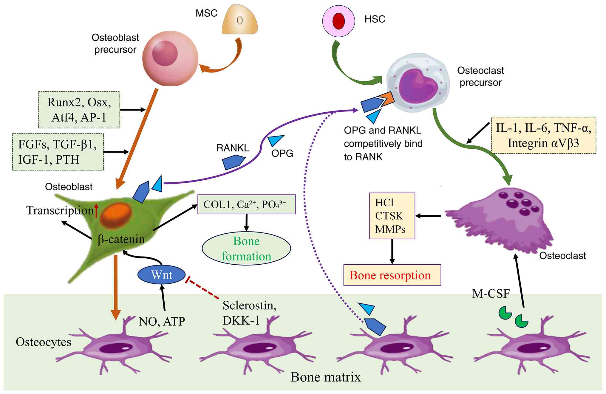 | Figure 1.Overview of physiological bone
remodeling. The figure summarizes the coordinated interactions
among osteoblasts, osteoclasts, osteocytes and their progenitor
cells during bone formation and resorption. Major regulatory
factors and signaling pathways involved in osteoblast
differentiation, osteoclastogenesis and osteocyte-mediated
regulation of bone remodeling are shown. Solid arrows indicate
activation, differentiation or promotion; blunt-ended lines
indicate inhibition; and dashed arrows indicate indirect or
paracrine regulation. AP-1, activator protein 1; Atf4, activating
transcription factor 4; COL1, type I collagen; CTSK, cathepsin K;
DKK-1, Dickkopf-1; FGF, fibroblast growth factor; HCl, hydrochloric
acid; HSC, hematopoietic stem cell; IGF-1, insulin-like growth
factor-1; M-CSF, macrophage colony-stimulating factor; NO, nitric
oxide; OPG, osteoprotegerin; Osx, osterix; PTH, parathyroid
hormone; RANK, receptor activator of NF-κB; RANKL, receptor
activator of NF-κB ligand; Runx2, runt-related transcription factor
2. |
Osteoblasts arise from BMSCs and are responsible for
bone mineralization and ECM synthesis. Their differentiation is
regulated by osteogenic transcription factors, including
runt-related transcription factor 2 (RUNX2) and osterix (OSX), as
well as signaling via the bone morphogenetic protein (BMP),
Wnt/β-catenin and growth factor signaling pathways (28,29).
Osteoblasts also influence osteoclastogenesis through the receptor
activator of NF-κB ligand (RANKL)/osteoprotegerin (OPG) axis,
thereby linking bone formation to resorption (30). Within the context of OP, integrin
αVβ3-mediated cell adhesion and mechanotransduction have been shown
to modulate osteoblast differentiation and matrix-responsive
signaling (31,32).
Osteoclasts are multinucleated cells of
hematopoietic origin whose differentiation depends primarily on
macrophage colony-stimulating factor (M-CSF) and receptor activator
of NF-κB ligand (RANKL) signaling. Mature osteoclasts attach to the
mineralized bone surface, organize specialized cytoskeletal
structures and resorb the bone matrix through acid-mediated mineral
dissolution and proteolytic degradation of its organic components
(33). Integrin αVβ3 is
particularly relevant in osteoclasts because its recognition of
RGD-containing ECM proteins supports adhesion, migration and
cytoskeletal polarization, thereby facilitating formation of the
actin-rich sealing zone required to establish the resorption
compartment and enable efficient bone resorption (34,35).
These functions provide the strongest mechanistic basis for
considering integrin αVβ3 as a therapeutic target in OP-associated
bone loss.
Osteocytes are terminally differentiated cells that
are derived from the osteoblast cell lineage; these cells are
embedded within the mineralized bone matrix and serve as important
mechanosensors within the bone tissue. Through their
lacuna-canalicular network, these cells regulate both osteoblast
and osteoclast activity by modulating sclerostin, dickkopf-1,
RANKL, OPG, nitric oxide and ATP signaling (36,37).
Integrin αVβ3 has been shown to contribute to osteocyte-mediated
mechanotransduction and may therefore influence how mechanical
loads are translated into adaptive bone remodeling responses. This
role is particularly relevant under estrogen-deficient conditions,
in which impaired αVβ3-mediated mechanosensation in osteocytes has
been associated with altered focal adhesion organization, an
increased RANKL/OPG ratio and reduced cyclooxygenase-2 (COX-2)
responses to mechanical stimulation (38).
Bone lining cells, which are osteoblast-lineage
cells located on quiescent bone surfaces, including the endosteal
surface, also contribute to local bone remodeling through ECM
turnover and regulation of the RANKL/OPG axis (39). However, direct evidence defining
integrin αVβ3-specific functions in endosteal cells remains
limited. Bone lining cells contribute to the local remodeling
microenvironment through interactions with the bone matrix and
paracrine regulation of neighboring bone cells. Although
αVβ3-specific functions in bone lining cells remain insufficiently
characterized and are therefore not discussed further in the
present review, these cells are included here to provide the
cellular context in which bone remodeling occurs. Bone remodeling
depends on coordinated interactions among osteoblasts, osteoclasts,
osteocytes and other cells within the local microenvironment.
Within this multicellular system, αVβ3 functions in a cell type-
and context-dependent manner, contributing to ECM recognition,
cytoskeletal organization and mechanotransduction in different
skeletal cell populations (40).
This framework provides the basis for the more detailed mechanistic
discussion of integrin αVβ3 activity in the following sections.
Structure, activation mechanisms and
biological properties of integrin αVβ3
Integrins are heterodimeric transmembrane receptors
that facilitate bidirectional communication between cells and the
ECM, thereby regulating cellular adhesion, migration,
mechanotransduction, survival and differentiation. In mammals, 18 α
and 8 β subunits assemble into 24 distinct integrin heterodimers
with different ligand-binding properties. Ligand recognition is
determined by the extracellular domains of the αβ heterodimer,
whereas the β-subunit cytoplasmic tail has a prominent role in
recruiting cytoskeletal and adaptor proteins involved in
intracellular signaling (41–43).
Among members of the integrin family, integrin αVβ3
belongs to the RGD-binding subfamily and recognizes ECM proteins
containing the RGD motif, including vitronectin, osteopontin (OPN),
fibronectin and bone sialoprotein (44). Ligand binding induces
conformational activation and receptor clustering, which promotes
the assembly of focal adhesion complexes and recruitment of adaptor
and signaling proteins, including FAK and Src, thereby enabling
integrin αVβ3 to couple extracellular biochemical and mechanical
cues to intracellular signaling pathways (45). Consequently, integrin αVβ3 is able
to regulate diverse cellular processes, including adhesion,
migration, proliferation, survival and mechanotransduction, through
signaling networks involving FAK, Src family kinase, the PI3K/AKT
signaling pathway, MAPK and Rho GTPases (46–49).
These signaling events enable cells to continuously sense and
respond to changes in their extracellular microenvironment
(50).
Structurally, integrin αVβ3 is a heterodimeric
glycoprotein composed of non-covalently associated αV and β3
subunits (Fig. 2A). The αV subunit
contains: i) A β-propeller domain; ii) a thigh domain; iii) two
calf domains; iv) a transmembrane helix; and v) a short cytoplasmic
tail. On the other hand, the β3 subunit consists of: i) A βI
domain; ii) a hybrid domain; iii) a plexin-semaphorin-integrin
domain; iv) four epidermal growth factor-like repeats; v) a β-tail
domain; vi) a transmembrane helix; and vii) a cytoplasmic tail
(51,52). The ligand-binding interface of
integrin αVβ3 is formed by the β-propeller domain of the αV subunit
together with the βI domain of the β3 subunit. The β3 βI domain
contains three divalent metal ion-binding sites: The metal
ion-dependent adhesion site (MIDAS), the adjacent to MIDAS and the
ligand-associated metal-binding site (also termed the synergistic
metal ion-binding site). These sites contribute to ligand binding
and its regulation, with the MIDAS metal ion directly coordinating
the Asp residue of RGD-containing ligands (53,54).
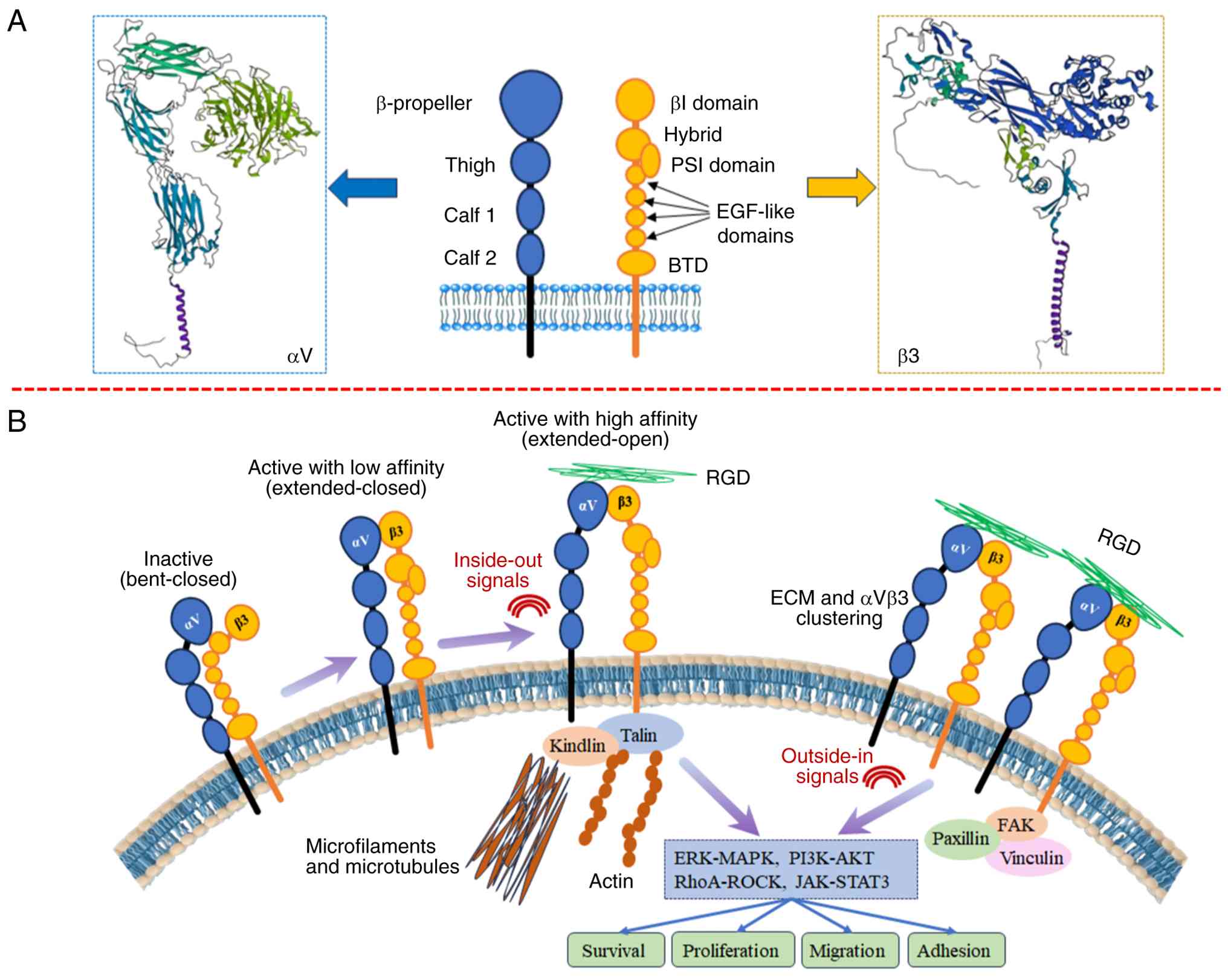 | Figure 2.Structural organization,
conformational activation and bidirectional signaling of integrin
αVβ3. (A) Structural organization of the αV and β3 subunits and
their principal extracellular, transmembrane and cytoplasmic
domains. (B) Schematic representation of integrin αVβ3 activation
from the bent-closed to extended-closed and extended-open
conformations and the associated inside-out and outside-in
signaling. Talin- and kindlin-mediated inside-out signaling
promotes receptor activation, whereas ECM ligand binding and
integrin αVβ3 clustering promote focal-adhesion assembly and
downstream ERK/MAPK, PI3K/AKT, RhoA/ROCK and JAK/STAT3 signaling.
Blue and orange structures represent the αV and β3 subunits,
respectively; purple arrows indicate conformational transitions;
green strands represent ECM ligands; and red arrows/icons indicate
intracellular signaling events. ECM, extracellular matrix; EGF,
epidermal growth factor; FAK, focal adhesion kinase; JAK, Janus
kinase; PSI, plexin-semaphorin-integrin; BTD, β-tail domain; RGD,
Arg-Gly-Asp; RhoA, Ras homolog family member A; ROCK,
Rho-associated protein kinase. |
Similar to other integrins, integrin αVβ3 undergoes
dynamic conformational transitions among three major states: i) A
bent-closed conformation associated with low ligand affinity; ii)
an extended-closed conformation that generally retains low ligand
affinity; and iii) an extended-open conformation associated with
high ligand affinity. These conformational transitions are coupled
to bidirectional inside-out and outside-in signaling mechanisms
(Fig. 2B) (55). During inside-out signaling,
intracellular adaptor proteins, particularly talin and kindlin,
bind to the cytoplasmic tail of the β3 subunit, inducing
conformational changes that increase ligand-binding affinity
(53). Following ligand
engagement, outside-in signaling promotes receptor clustering and
the assembly of focal adhesion complexes, involving proteins such
as FAK, Src, paxillin and vinculin, which activate downstream
pathways regulating cytoskeletal organization, gene transcription
and other cellular responses to the extracellular environment
(56–58).
Within the skeletal system, integrin αVβ3 is
expressed in multiple bone-associated cell populations, including
osteoclasts, osteoblasts, osteocytes, endothelial cells and several
immune cell types. Through interactions with ECM components,
integrin αVβ3 contributes to adhesion and cytoskeletal organization
in osteoclasts, differentiation and mechanosensitive signaling in
osteoblast-lineage cells, mechanotransduction in osteocytes,
angiogenic signaling in endothelial cells and inflammatory
signaling in immune and stromal cells (59,60).
For example, inhibition of integrin αVβ3 has been shown to
attenuate mechanosensitive calcium signaling in osteocytes and to
impair mechanically induced COX-2 expression and prostaglandin E2
(PGE2) release. Because COX-2 induction and PGE2 production are
established biochemical responses of osteocytes to mechanical
stimulation, these findings support the involvement of αVβ3 in
coupling mechanical cues to downstream cellular signaling (61). In addition, integrin αVβ3 exhibits
functional crosstalk with growth factor receptors, including EGFR
and transforming growth factor-β receptor II, thereby coordinating
signaling pathways, such as the Ras/MEK/MAPK, PI3K/AKT and
RhoA/Rho-associated protein kinase pathways, which influence cell
migration, proliferation and survival (62–64).
Collectively, evidence from osteoclast and osteocyte
studies indicates that integrin αVβ3 links ECM interactions to
intracellular signaling in multiple bone cell populations. Its
biological effects are cell type- and context-dependent, reflecting
differences in ECM ligand availability, mechanical stimulation and
local hormonal or inflammatory conditions. These factors represent
components of the local bone microenvironment that can modify αVβ3
expression, activation and downstream signaling. This context
dependence may contribute to the diverse functions of αVβ3 in bone
remodeling and OP (65,66).
Integrin αVβ3 as a context-dependent
regulator of bone remodeling
The functional effects of integrin αVβ3 in bone
remodeling arise from the coordinated activity of the αVβ3
heterodimer. Although the αV and β3 subunits make distinct
structural and signaling contributions, ligand recognition and
signal transmission depend on their assembly as a functional
receptor. In αVβ3, the extracellular domains of both subunits
contribute to ligand binding, whereas the β3 cytoplasmic domain has
a prominent role in recruiting intracellular adaptor and signaling
proteins. These coordinated properties provide the basis for the
cell type-specific effects of αVβ3 subsequently discussed in
osteoblast-lineage cells, osteoclasts and osteocytes (67,68).
Therefore, elucidating the complimentary roles of these subunits is
key to understanding how integrin αVβ3 affects bone remodeling.
The primary function of the αV subunit is to
facilitate recognition of the ECM via interactions with
RGD-containing ligands, such as fibronectin, vitronectin, OPN and
bone sialoprotein (44). Such
interactions play important roles in mediating cell adhesion and
transducing extracellular signals that regulate osteogenic
differentiation and skeletal organization. Experimental evidence
has shown that αV subunit-mediated signaling is involved in
osteogenic differentiation by stimulating the activation of various
signaling cascades, such as the BMP2/SMAD and ERK/STAT pathways,
which induce the differentiation of BMSCs into osteoblasts
(69,70). In addition to its involvement in
bone development, the αV subunit, as part of the αVβ3 heterodimer,
contributes to osteoclast attachment and migration through
recognition of RGD-containing matrix ligands, including OPN and
vitronectin. These interactions facilitate osteoclast adhesion and
cytoskeletal organization required for subsequent bone resorption
(71). The implication of these
findings is that the αV subunit of integrin acts as an interface
for bone cells in relation to the ECM during bone resorption and
formation (72).
On the other hand, signaling within cells is
mediated by the β3 integrin subunit after receptor-ligand binding.
In osteoclasts, the interaction of the β3 subunit with molecules
such as talin-1 and kindlin-3 is important for the activation of
integrins and the formation of focal adhesions, which are necessary
for cytoskeletal reorganization and cellular attachment to the bone
matrix (73). β3 subunit-mediated
intracellular signaling is characterized by the recruitment and
activation of Src family kinases and focal adhesion-associated
proteins, which interact with downstream signaling networks,
including JNK and NF-κB pathways, to regulate osteoclast function
(74). Further evidence indicates
that β3-integrin signaling interacts with the microRNA-17
(miR-17)/osteoclastic protein-tyrosine phosphatase (PTP-oc)/ephrin
type-A receptor 4 (EphA4) regulatory axis during osteoclast
activation. In this axis, miR-17 negatively regulates PTP-oc
expression, whereas PTP-oc activates Src signaling by
dephosphorylating the inhibitory Tyr527 residue and suppresses the
inhibitory signaling of EphA4. These interconnected mechanisms
modulate β3-integrin signaling, cytoskeletal organization and
osteoclast activity (34,75). Although these signal transduction
pathways have been predominantly studied in osteoclasts, they have
also been shown to serve roles in osteoclast-osteoblast
communication during coupled bone remodeling (76,77).
Collectively, the biological functions of the αV and
β3 subunits are best understood in the context of the functional
αVβ3 heterodimer rather than as independent activities. Both
subunits contribute to the extracellular ligand-binding interface,
whereas the β3 cytoplasmic domain has a prominent role in
recruiting adaptor and signaling proteins that transmit
ligand-dependent signals to the cell interior (78). Skeletal and microenvironmental cell
populations further indicate that the functional consequences of
αVβ3 signaling vary according to cellular context. These include
regulation of osteogenic and mechanosensitive responses in
osteoblast-lineage cells and osteocytes, adhesion and cytoskeletal
organization in osteoclasts, angiogenic signaling in endothelial
cells and inflammatory responses in immune and stromal cells. Such
effects may also be influenced by local factors, including ECM
ligand availability, mechanical stimulation, hormonal status and
inflammatory conditions. Thus, αVβ3 is more appropriately
considered a context-dependent regulator of bone remodeling than a
receptor that exerts a uniform biological effect across skeletal
cell populations (79–81).
Although there are differences in the structure and
signaling properties of the αV and β3 integrin subunits, their
functional roles are merged through the binding of the integrin
αVβ3 dimeric complex. It is therefore necessary to consider that
the biological activities regulated by integrin αVβ3 that are
described in the following sections, such as the differentiation of
osteoblasts, osteoclast function, angiogenesis and bone
microenvironmental signaling, represent the combined activity of
both subunits of integrin αVβ3 (82).
Integrin αVβ3 regulates osteoblast
function and bone formation
Bone formation is a well-coordinated process
comprising the proliferation, differentiation, matrix secretion and
mechanoresponsiveness of osteoblast-lineage cells (83). A growing body of work has indicated
that the integrin αVβ3 receptor serves a notable role in various
steps of osteoblast biology by integrating mechanical and
biochemical information from the ECM into intracellular signals
that regulate osteogenesis (84–86).
Rather than acting as an independent osteogenic stimulus, integrin
αVβ3 appears to function as a signaling mediator that coordinates
cell adhesion, cytoskeletal organization, mechanotransduction and
growth factor signaling. These processes can influence osteogenic
differentiation and bone formation by regulating downstream
pathways such as FAK/MAPK and PI3K/AKT and osteogenic
transcriptional programs, including RUNX2-dependent responses
(87).
The process of osteogenic differentiation
necessitates the formation of stable associations between
osteoblasts or BMSCs and the ECM (88). Integrin αVβ3 recognizes
RGD-containing ECM ligands, including fibronectin, vitronectin and
OPN, and contributes to the assembly of focal adhesion complexes
that physically couple the ECM to the actin cytoskeleton. These
complexes recruit adaptor and signaling proteins, including talin,
vinculin and FAK, thereby enabling extracellular adhesive and
mechanical cues to regulate cytoskeletal organization and
downstream osteogenic signaling. In experimental models,
αVβ3-dependent focal adhesion formation and FAK activation have
been associated with the regulation of osteogenic markers,
including RUNX2 and alkaline phosphatase (ALP) (44). The binding of ligands to integrin
αVβ3 also induces the recruitment of proteins associated with focal
adhesions, such as FAK, Src-family tyrosine kinases, paxillin,
talin and vinculin, leading to the activation of downstream
signaling pathways that regulate cell survival and differentiation
(89). Therefore, the
aforementioned events regulating integrin αVβ3-mediated adhesion
provide the molecular foundations on which the osteogenic signaling
cascade takes place.
Regarding pathways downstream of integrin αVβ3,
activation of the FAK/PI3K/AKT pathway has been established as one
of the key signaling processes by which the integrin αVβ3-ECM
interaction triggers the osteogenic differentiation of osteoblasts.
Previous studies have indicated that integrin αVβ3 activation leads
to FAK phosphorylation, which in turn induces PI3K/AKT signaling
and the expression of osteogenic transcription factors, such as
Runx2 and Osx (15,23). These transcription factors regulate
the expression of osteoblast-associated genes, including ALP,
osteocalcin, OPN and type I collagen. Osteogenic differentiation
indicates that the contribution of these signaling responses to
osteogenesis varies according to cell type, differentiation stage
and the nature of the mechanical stimulus applied to the
extracellular environment (90,91).
Further evidence has suggested that rather than
employing a signaling mechanism based on a one-dimensional pathway,
integrin αVβ3 interacts with several osteogenic signaling pathways.
Integrin αVβ3-associated signaling can modulate MAPK pathways,
including ERK and JNK, although the resulting effects on osteogenic
differentiation depend on the cellular and experimental context. In
addition, αVβ3 participates in crosstalk with BMP-2 signaling;
β3-integrin-dependent adhesion and cytoskeletal organization can
facilitate BMP-2-induced SMAD signaling, thereby contributing to
osteoblast differentiation (92,93).
Taken together, these results indicate that integrin αVβ3 is a
signaling coordinator that facilitates the integration of upstream
ECM-based signals with conventional osteogenesis-related signaling
pathways, such as the BMP and Wnt/β-catenin pathways (94,95).
Another important physiological mechanism that
regulates bone formation, in which integrin αVβ3 serves an
important role, is mechanical stimulation. In order to maintain
bone homeostasis, it is necessary to effectively convert mechanical
loads into biochemical responses, since osteoblasts are
continuously subjected to tensile strain, fluid shear stress and
matrix deformation. As a mechanosensitive receptor, integrin αVβ3
is involved in this process by connecting extracellular mechanical
signals to intracellular signaling responses via focal adhesions
complexes (96,97). Experimental evidence indicates that
fluid shear stress activates integrin αVβ3 in osteocytes,
particularly at dendritic processes, leading to PI3K/AKT signaling
and subsequent opening of connexin-43 hemichannels. These
hemichannels facilitate the release of signaling molecules such as
PGE2, thereby contributing to osteocyte-mediated communication with
neighboring bone cells and the anabolic response to mechanical
loading. Additionally, cyclic tensile stress stimulates
cytoskeletal reorganization and the maturation of focal adhesions
via integrin αVβ3-dependent FAK activation and yes-associated
protein nuclear translocation, thus promoting early osteogenesis
(98). In conditions where gravity
has been altered, integrin αVβ3-mediated PI3K signaling also
controls the activity of the osteogenic transcription factor Runx2,
implying that mechanical insensitivity due to defects in
mechanotransduction could be a reason for the bone loss observed in
cases of mechanical unloading or in microgravity environments
(99). Taken together, these
observations suggest that integrin αVβ3 represents an important
mechanotransducer that acts as a bridge between physical stimuli
and genes that control bone development. However, the role of
integrin αVβ3 in bone mechanobiology appears to depend on the
characteristics of the mechanical stimulus, including its type,
magnitude, frequency and duration, as well as the responding cell
type and differentiation state (92). These variables can influence the
extent and nature of αVβ3-mediated mechanotransduction and its
downstream effects on osteogenic responses.
Emerging research has indicated an even wider scope
of action for integrin αVβ3 outside of mature osteoblasts. In a
recent study (100),
osteocyte-specific deletion of the β3 integrin subunit resulted in
impaired osteoblast-mediated bone formation, reduced osteogenic
capacity of BMSCs, and decreased bone mass in load-bearing long
bones. These changes were accompanied by impaired biomechanical
properties, including reductions in maximum load-bearing capacity,
maximum displacement, stiffness and bone hardness, indicating
compromised bone mechanical strength and material properties. This
suggests that osteocyte-dependent integrin αVβ3-mediated signaling
has a notable effect on osteoblast activity via intercellular
communication during bone remodeling. Similarly, a study by
Mas-Moruno et al (101)
revealed that elevated integrin αVβ3 levels were associated with
increased osteoblast proliferation and differentiation, whereas the
pharmacological inhibition of integrin αVβ3 led to decreased levels
of ALP, Runx2 and nuclear yes-associated protein.
Despite consistent results indicating the
involvement of integrin αVβ3 in osteogenic regulation, the detailed
molecular mechanisms involved in integrin αVβ3-mediated regulation
remain ambiguous; this is especially true in the context of
osteocyte-to-osteoblast regulatory signals. Collectively, these
findings indicate that integrin αVβ3 contributes to the coupling of
ECM adhesion and mechanical cues with intracellular pathways that
regulate osteogenic differentiation and bone formation. This
mechanistic relationship may be relevant to bone tissue
engineering, as biomaterial properties and RGD-containing ligands
can be designed to modulate integrin-mediated adhesion, focal
adhesion signaling and osteogenic responses. Thus, αVβ3 represents
a potential interface through which biomaterial-based strategies
could influence bone regeneration; however, their therapeutic
applicability to OP requires further validation in disease-relevant
models. The signaling pathways responsible for osteogenesis that
are mediated by integrin αVβ3 are shown in Fig. 3.
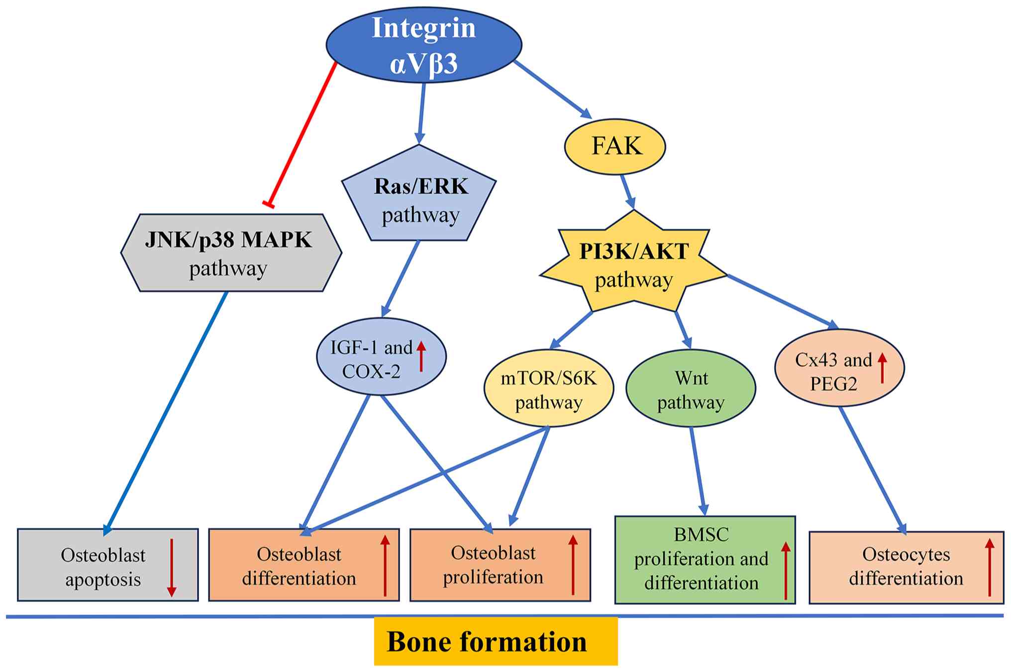 | Figure 3.Proposed integrin αVβ3-associated
signaling network in osteogenic regulation. The diagram summarizes
major signaling pathways associated with integrin αVβ3 regulation
of osteoblast/BMSC survival, proliferation and differentiation,
together with osteocyte mechanotransduction and adaptive bone
responses. Integrin αVβ3-associated pathways include FAK/PI3K/AKT,
Ras/ERK, mTOR/S6K, Wnt/β-catenin and context-dependent JNK/p38 MAPK
signaling. Mechanotransduction-associated responses include
regulation of Cx43, COX-2 and PGE2. Solid blue arrows indicate
activation or positive regulation; red blunt-ended lines indicate
inhibition; and upward- and downward-facing arrows indicate
increased and decreased expression or activity, respectively. BMSC,
bone marrow mesenchymal stem cell; COX-2, cyclooxygenase-2; Cx43,
connexin-43; FAK, focal adhesion kinase; IGF-1, insulin-like growth
factor-1; PGE2, prostaglandin E2; S6K, ribosomal protein S6
kinase. |
Integrin αVβ3 regulates osteoclast
function and bone resorption
Bone resorption is an important part of the process
of bone remodeling, and facilitates bone turnover by replacing old
or damaged bone tissue without disturbing the mineral balance of
the body. The process of bone resorption is controlled by
osteoclasts, which are large multinucleated cells belonging to the
monocyte/macrophage cell lineage. The differentiation and function
of osteoclasts are precisely regulated by external ECM molecules
and cytokines (102,103). Among the adhesion molecules
expressed by osteoclasts, integrin αVβ3 is a major receptor
mediating interactions with ECM proteins within the bone matrix.
Through these interactions, αVβ3 contributes to cytoskeletal
reorganization, mechanotransduction, osteoclast motility,
polarization and bone resorption. Integrin αVβ3 can therefore also
be considered to be a signaling molecule, rather than a simple
adhesion molecule (104,105).
Initially, osteoclast-mediated resorption of bone
involves the firm attachment of mature osteoclasts to the
mineralized surface of the bone. Integrin αVβ3 helps to achieve
this step by recognizing RGD motifs present in various ECM
proteins, such as OPN, vitronectin and bone sialoprotein, which are
abundant in the bone matrix (73,106,107). Ligand binding enhances integrin
αVβ3 clustering and the assembly of adhesion complexes, thereby
promoting osteoclast attachment, migration and polarization. These
processes are essential for organizing the actin-rich sealing zone
and ruffled border, which establish an isolated resorption
compartment where acid and proteolytic enzymes are released to
dissolve the mineral phase and degrade the organic bone matrix
(108). Adhesion events also lead
to intracellular signal transduction mediated by adaptor proteins,
such as talin, kindlin-3, FAK, paxillin and Src family kinases,
linking the recognition of ECM proteins to cytoskeletal
rearrangement (109,110).
Cytoskeletal rearrangement is essential for
osteoclast resorptive function because dynamic actin remodeling
organizes individual podosomes into higher-order structures,
including podosome clusters, rings and belts. On mineralized bone
surfaces, these actin-rich adhesion structures reorganize into the
sealing zone, which anchors the osteoclast to bone and isolates the
resorption lacuna from the surrounding extracellular environment.
Thus, the formation and remodeling of podosome rings represent
intermediate stages in the cytoskeletal organization required to
establish and maintain the functional sealing zone during bone
resorption (111). Based on
experimental research, the role of integrin αVβ3 signaling in the
formation of podosome rings has been shown to be mediated by FAK,
Src, proline-rich tyrosine kinase 2 (Pyk2), phospholipase C,
p130Cas and other proteins found in focal adhesions (106,112). This signaling promotes formation
of the ruffled border, facilitates osteoclast anchorage to the
mineralized bone surface, and enables polarized secretion of
hydrochloric acid and proteolytic enzymes, including cathepsin K
and MMPs, which collectively mediate demineralization and
degradation of the bone matrix (33). Thus, integrin αVβ3 not only
mediates osteoclast adhesion, but is also important for organizing
the architecture required for bone resorption.
In addition to being involved in the activity of
fully developed osteoclasts, integrin αVβ3 is also responsible for
the development and maturation of osteoclasts. Osteoclast formation
is predominantly regulated by the RANKL-mediated activation of
NF-κB, MAPK and nuclear factor of activated T cells 1 (NFATc1)
(113,114). According to the available
evidence, integrin αVβ3 signaling acts in coordination with
established osteoclastogenic pathways rather than functioning
independently, with signaling crosstalk contributing to osteoclast
differentiation and resorptive function. Activation of integrin
αVβ3 promotes Src-dependent signaling and the formation of
signaling complexes involving immunoreceptor tyrosine-based
activation motif-containing adaptor proteins, including
DNAX-activating protein of 12 and Fc receptor γ. These complexes
recruit downstream signaling molecules such as Syk and interact
with osteoclast regulatory pathways that influence cytoskeletal
organization and signaling through MAPKs and NFATc1 (44,115,116). This coordinated signaling
contributes to the expression of osteoclast-associated genes,
including tartrate-resistant acid phosphatase, cathepsin K and
MMP-9, which are associated with osteoclast differentiation and
bone-resorptive function (117).
Thus, rather than acting as an independent driver of
osteoclastogenesis, integrin αVβ3 cooperates with
RANKL/RANK-dependent signaling and is particularly important for
the cytoskeletal organization, polarization and resorptive function
of mature osteoclasts.
Further research has highlighted additional
crosstalk between integrin αVβ3 and M-CSF signaling in the
osteoclastogenic process (118,119). Interactions between M-CSF and
M-CSF receptor (c-FMS) increase the survival and proliferation of
osteoclast precursor cells and affect integrin activation.
Experimental evidence has shown that signaling via c-FMS elevates
the ligand-binding capacity of integrin αVβ3 and induces
Rac-dependent cytoskeletal reorganization (120). By contrast, the levels of M-CSF
in integrin β3-deficient mice have been shown to be increased, thus
indicating that the M-CSF/c-FMS interaction represents a
compensatory mechanism against reduced osteoclast differentiation
and supporting the presence of feedback loops between the c-FMS
pathway and the integrin αVβ3 pathway. These findings highlight the
importance of signaling crosstalk in osteoclast biology, whereby
integrin αVβ3 cooperates with other receptor-mediated pathways to
coordinate osteoclast differentiation, cytoskeletal organization
and resorptive function (121).
Integrin αVβ3 activity is also modulated by the
microenvironment within the bone. Acidification of this environment
during bone resorption facilitates the activation of NFATc1,
leading to higher expression levels of integrin αVβ3 and increased
migration of osteoclasts via the Pyk2/Src signaling pathway
(122). Furthermore, OPN has not
only been shown to act as an adhesion protein but also to represent
an important signaling molecule that stimulates the process of
podosome assembly. The binding of OPN to the receptor integrin αVβ3
leads to the activation of the Src/FAK pathway, as well as the
lowering of intracellular calcium levels by activating plasma
membrane Ca2+-ATPases (123). These findings illustrate how ECM
composition and local biochemical conditions dynamically regulate
integrin αVβ3-dependent osteoclast activity.
Research has also indicated the involvement of
integrin αVβ3 in communication between osteoclasts and the bone
microenvironment. Inflammatory cytokines and immune mediators have
been shown to increase the activation of osteoclasts via integrin
αVβ3-dependent mechanisms, whereas reciprocal communications
between osteoclasts and cells derived from the osteoblast lineage
have been shown to serve a notable role in bone remodeling
processes (10,124). Although this topic is considered
in more detail in the following sections, it should be noted that
integrin αVβ3 operates in a larger regulatory system outside of
osteogenic cells, involving ECM molecules, inflammatory agents and
biomechanical factors.
Since the expression levels of integrin αVβ3 are
elevated in mature osteoclasts, pharmacological targeting of
integrin αVβ3 has received notable attention as a potential
therapeutic approach to decrease bone resorption (125,126). Targeting integrin αVβ3 or its
associated downstream signaling components, including Pyk2 and Src,
has been shown to impair osteoclast adhesion, migration and
cytoskeletal organization, thereby reducing bone-resorptive
activity (108). Furthermore,
modulation of αVβ3-associated signaling may influence osteoclast
formation in inflammatory bone microenvironments through effects on
Th17-associated inflammatory responses. Reduced Th17 activity can
attenuate osteoclastogenesis by decreasing IL-17-mediated induction
of RANKL in osteoclast-supporting cells and by limiting other
pro-osteoclastogenic inflammatory signals. Thus, any effect of αVβ3
modulation on the Th17 response could indirectly influence
RANKL-dependent osteoclast formation (127). However, these findings should be
interpreted with caution because integrin αVβ3 is not restricted to
osteoclasts and is also expressed in other cell populations within
the bone microenvironment, including osteoblast-lineage cells,
osteocytes, endothelial cells and certain immune cells.
Consequently, therapeutic strategies targeting αVβ3 should consider
its cell type- and context-dependent functions, and approaches that
preferentially modulate osteoclast-associated αVβ3 signaling may
help limit unintended effects on other αVβ3-expressing cells
(128–130). The molecular mechanisms of
osteoclast-mediated bone resorption that are regulated by integrin
αVβ3 are illustrated in Fig.
4.
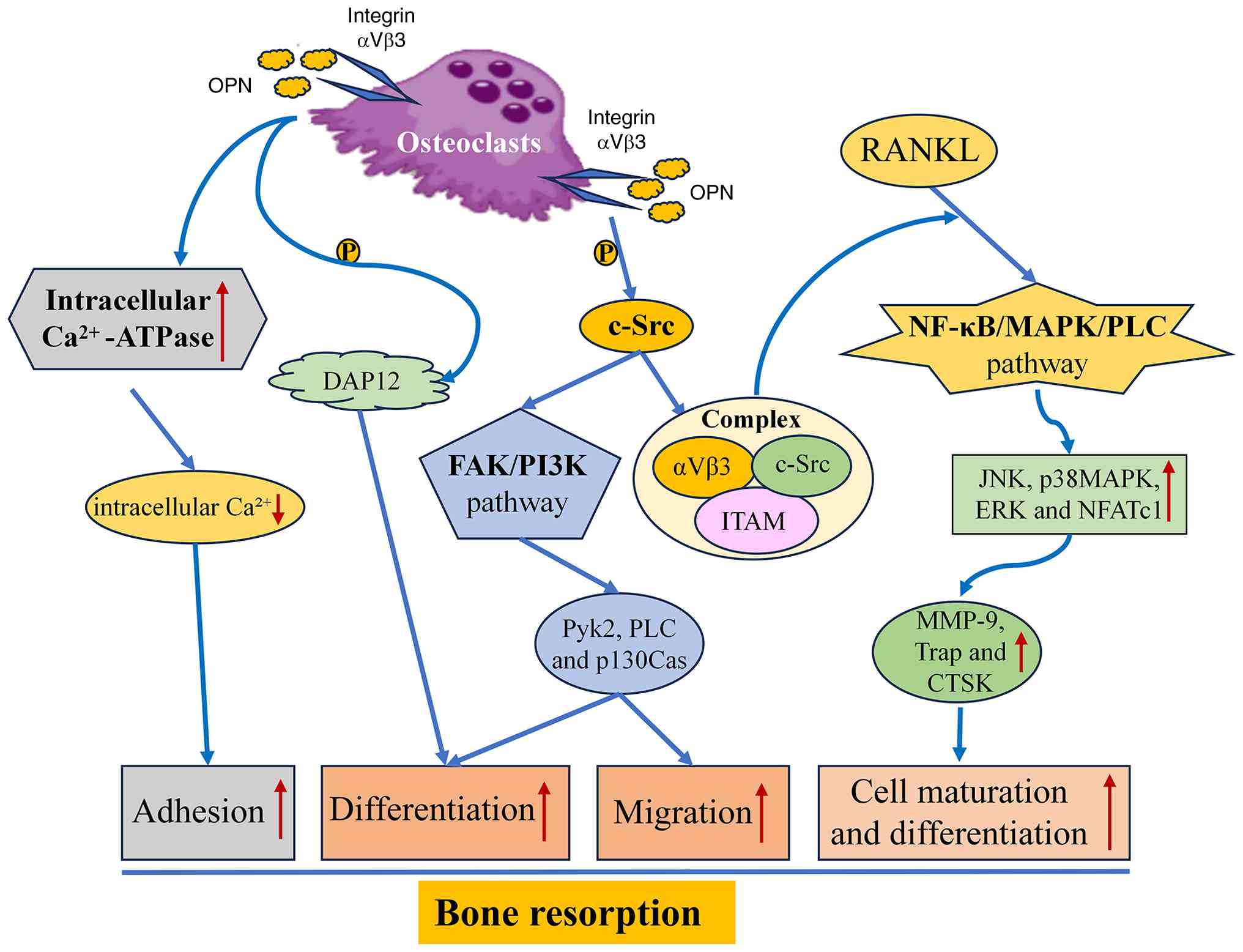 | Figure 4.Integrin αVβ3-associated signaling in
osteoclast function and bone resorption. The schematic summarizes
integrin αVβ3-mediated signaling involved in osteoclast adhesion,
migration, cytoskeletal organization, differentiation and bone
resorption. Ligand-dependent integrin αVβ3 activation engages
c-Src, FAK/PI3K and DAP12/ITAM-associated signaling and downstream
mediators, including Pyk2, PLC and p130Cas. Crosstalk with
RANKL-dependent signaling promotes activation of NF-κB, MAPK and
NFATc1 and the expression of osteoclast-associated genes, including
MMP-9, TRAP and CTSK. Solid arrows indicate activation or signaling
progression; upward- and downward-facing arrows indicate increased
and decreased activity or expression, respectively. c-Src, cellular
Src tyrosine kinase; CTSK, cathepsin K; DAP12, DNAX-activating
protein of 12 kDa; FAK, focal adhesion kinase; ITAM, immunoreceptor
tyrosine-based activation motif; NFATc1, nuclear factor of
activated T cells 1; OPN, osteopontin; P, phosphate group; PLC,
phospholipase C; Pyk2, protein tyrosine kinase 2; RANKL, receptor
activator of NF-κB ligand; TRAP, tartrate-resistant acid
phosphatase. |
The available evidence supports an important role
for integrin αVβ3 in osteoclast function by coupling bone-matrix
adhesion to intracellular signaling that regulates cytoskeletal
organization, polarization and resorptive activity. Rather than
acting as a single dominant regulator of bone resorption, αVβ3
functions in coordination with other osteoclast regulatory systems,
including RANKL/RANK and M-CSF/c-FMS signaling. Crosstalk among
these pathways enables ECM engagement and cytokine-derived signals
to converge on cytoskeletal regulatory mechanisms required for
effective bone resorption. Consequently, the functional
contribution of αVβ3 depends on the broader signaling context of
the osteoclast rather than on αVβ3 activity alone (131–133). Future research utilizing
cell-specific genetic systems, intravital microscopy and
transcriptomic analysis will prove useful in elucidating the
specific role of integrin αVβ3 in osteoclast-mediated bone
resorption and will guide future development of effective
treatments for OP.
Integrin αVβ3 regulates bone
angiogenesis
The process of bone remodeling is tightly linked to
angiogenesis, since newly developed blood vessels are required to
deliver oxygen, nutrients, osteoprogenitor cells and other
molecules that regulate bone remodeling and growth to the site of
remodeling. Integrin αVβ3 has been demonstrated to mediate the
processes involved in this angiogenic response by regulating
endothelial cell migration, adhesion, survival, and interactions
between osteogenic and endothelial cells (96,134).
However, integrin αVβ3 is not an angiogenic growth
factor per se; rather, it functions as an adhesion and
signaling receptor that integrates ECM-derived signals with growth
factor receptor signaling in endothelial cells. αVβ3 expression is
relatively low in quiescent endothelium but is increased in
activated endothelial cells during angiogenesis. Within bone,
angiogenesis is an integral component of processes such as bone
formation, fracture repair and remodeling, where newly formed
vessels support tissue regeneration and osteogenic activity.
Experimental studies have shown that pharmacological blockade of
αVβ3 can inhibit neovascularization in several angiogenesis models,
with comparatively limited effects on established vessels (118,123). These findings support a role for
αVβ3 in endothelial activation, migration and survival during
vascular remodeling (115,116,118,123).
However, genetic and pharmacological studies have produced
context-dependent findings; therefore, αVβ3 should be regarded as a
modulator of angiogenic signaling rather than as an indispensable
regulator of vascular development or maintenance (22,106,121,135).
From a molecular perspective, interactions between
integrin αVβ3 and VEGFR2 take place with notable frequency. This
signaling interface helps to coordinate the reactions of
endothelial cells to angiogenic stimuli. When integrin αVβ3 is
activated, FAK and Src family kinases are recruited, which results
in the phosphorylation of VEGFR2 and subsequent activation of the
PI3K/AKT and MAPK pathways. This in turn contributes to endothelial
cell migration, proliferation and survival, and also leads to new
blood vessel network formation (136–138). It is thus evident that integrin
αVβ3 and VEGFR2 work cooperatively to support bone
angiogenesis.
ECM remodeling may facilitate αVβ3-dependent
endothelial responses during angiogenesis by modifying the
accessibility of integrin-binding sites within the local matrix.
Proteolytic remodeling can expose RGD-containing binding sites and
alter the availability of αVβ3 ligands, thereby supporting
endothelial cell adhesion and migration. In addition, αVβ3
recognizes ECM proteins such as OPN and vitronectin, interactions
that can promote endothelial adhesion and migratory signaling.
MMP-2 further contributes to pericellular ECM degradation and
remodeling, thereby facilitating endothelial invasion through the
surrounding matrix (139,140).
It has been demonstrated that integrin αVβ3-mediated
regulation of angiogenesis is also affected by interactions between
osteogenic cells and immune cells. Specifically, alternatively
activated M2 macrophages have been shown to stimulate angiogenesis
via integrin αVβ3-related pathways (141). Angiogenesis and osteogenesis are
closely coupled during bone development, remodeling and repair, as
illustrated by specialized bone vasculature, particularly type H
vessels, which are spatially associated with osteoprogenitor cells
and participate in reciprocal signaling between endothelial and
osteogenic cell populations (29,96).
Within this broader framework of angiogenic-osteogenic coupling,
the aforementioned findings suggest that αVβ3 may contribute to
communication between endothelial cells and bone-associated cells
by integrating ECM-dependent adhesion with angiogenic signaling.
However, the specific contribution of αVβ3 to this intercellular
crosstalk remains less well defined than its established role in
endothelial adhesion and angiogenic signaling.
Integrin αVβ3 regulates the bone
microenvironment
Bone remodeling takes place in a notably dynamic
microenvironment, in which immunological signaling, hormonal
control, ECM structure and mechanical forces combine to regulate
the homeostasis of the skeleton. A number of studies have shown
that integrin αVβ3 serves a notable role in the regulatory
mechanisms involved in interactions among bone cells, immune cells,
endothelial cells and the ECM. Thus, αVβ3 may influence bone
remodeling not only through its direct effects on bone-resorbing
and bone-forming cells, but also through its actions in other
components of the local bone microenvironment, including
endothelial and immune cells (142,143). Collectively, current evidence
indicates that αVβ3 participates in bone remodeling through cell
type- and context-dependent mechanisms operating across the local
bone microenvironment. In osteoclasts, αVβ3-mediated matrix
adhesion is coupled to cytoskeletal organization and resorptive
function; in osteocytes, αVβ3 contributes to mechanotransduction
and mechanically induced biochemical responses; and in endothelial
cells, αVβ3 participates in adhesion and signaling associated with
angiogenesis. αVβ3-dependent responses may also be modified by
inflammatory and hormonal conditions, including estrogen
deficiency. These interconnected processes illustrate how αVβ3
signaling can link matrix-derived, mechanical, vascular and
microenvironmental cues to cellular responses relevant to bone
remodeling (Fig. 5).
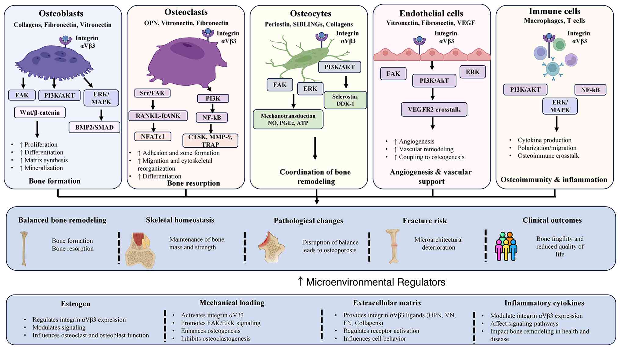 | Figure 5.Context-dependent functions and
therapeutic modulation of integrin αVβ3 in bone remodeling and
osteoporosis. The schematic summarizes the cell type-specific
functions of integrin αVβ3 in osteoblasts, osteoclasts, osteocytes,
endothelial cells and immune cells and their contributions to bone
formation, bone resorption, mechanotransduction, angiogenesis and
inflammatory regulation. The figure also illustrates
microenvironmental factors that modulate integrin αVβ3 signaling
and potential strategies for selectively targeting αVβ3-associated
pathological bone remodeling. Colored panels represent the
different bone and bone-associated cell populations. AKT, protein
kinase B; BMP, bone morphogenetic protein; CTSK, cathepsin K;
DKK-1, Dickkopf-1; FAK, focal adhesion kinase; FN, fibronectin;
NF-κB, nuclear factor-κB; NFATc1, nuclear factor of activated T
cells 1; NO, nitric oxide; OPG, osteoprotegerin; OPN, osteopontin;
PGE2, prostaglandin E2; RANK, receptor activator of NF-κB; RANKL,
receptor activator of NF-κB ligand; SIBLINGs, small
integrin-binding ligand N-linked glycoproteins; TRAP,
tartrate-resistant acid phosphatase; VN, vitronectin. |
Osteoimmune regulation
Inflammation has been widely acknowledged as a
notable cause of bone loss and OP. In inflammatory bone
microenvironments, integrin αVβ3 is expressed by several cell
populations, including osteoclasts, activated macrophages,
angiogenic endothelial cells and certain stromal cells, where it
contributes to cell-ECM interactions, migration and inflammatory or
resorptive signaling (125,131). Through these cell type-specific
functions, αVβ3 may contribute to crosstalk between inflammatory
responses, angiogenesis and osteoclast-mediated bone resorption
(144,145). Experimental evidence indicates
that pro-inflammatory cytokines, including TNF-α, IL-1β, IL-6 and
IL-17, promote osteoclastogenesis primarily by enhancing
RANKL/RANK-dependent signaling and by modifying the inflammatory
microenvironment. These cytokine-driven mechanisms can converge
with αVβ3-associated signaling during osteoclast differentiation
and activation; however, αVβ3 should not be considered the
principal mediator of their osteoclastogenic effects. In
inflammatory stromal cells, particularly synovial fibroblasts,
increased production of RANKL and MMPs contributes to osteoclast
formation and tissue destruction. Furthermore, Th17 cells promote
osteoclastogenesis through IL-17 production and RANKL-associated
mechanisms, while OPN-integrin interactions may modulate Th17-cell
adhesion and inflammatory responses (145–147).
Role of macrophages in αVβ3-regulated
inflammatory signaling
Macrophages have also been shown to serve a role in
integrin αVβ3-regulated inflammatory signaling pathways. In human
monocyte-derived macrophages, ligation of integrin αVβ3 has been
shown to activate PI3K/Akt-dependent NF-κB signaling and to enhance
NF-κB responses to pro-inflammatory stimuli, including
lipopolysaccharide and TNF-α. This response is accompanied by
increased expression of NF-κB-dependent pro-inflammatory mediators,
indicating that αVβ3 signaling can modulate macrophage inflammatory
activation (131). Neutrophils
may also contribute to inflammation-associated bone remodeling by
accumulating at inflammatory sites and releasing pro-inflammatory
mediators, reactive oxygen species and neutrophil extracellular
traps, which can enhance osteoclastogenic signaling and thereby
favor bone resorption. Integrins, including αVβ3 in specific
contexts, may contribute to neutrophil adhesion and migration;
however, the direct contribution of neutrophil αVβ3 to inflammatory
bone loss remains less well established than its role in
osteoclasts. Evidence from rheumatoid arthritis further supports
the involvement of αVβ3 in inflammatory bone resorption, where
increased αVβ3 expression has been reported in osteoclasts and
activated macrophages within affected joints (148). In osteoclasts, αVβ3-dependent
signaling promotes migration, cytoskeletal organization and
resorptive activity, whereas αVβ3-associated macrophage responses
may contribute to the inflammatory environment that supports
osteoclast activation and local bone erosion (149). Despite the different pathological
natures of rheumatoid arthritis and OP, these findings support the
potential impact of integrin αVβ3-mediated immune responses on bone
remodeling.
Estrogen-dependent regulation of
integrin αVβ3
Estrogen deficiency is a notable factor in the
development of OP after menopause, and its influence on bone
turnover has been related to its impact on osteoblasts,
osteoclasts, osteocytes and immune cells (150,151). Recent research has supported the
involvement of integrin αVβ3 in the estrogen-dependent regulation
of bone homeostasis (152).
Experimental studies using ovariectomy (OVX) models have
demonstrated apparently divergent effects of β3 integrin signaling.
Global β3 integrin deficiency protects mice against OVX-induced
bone loss, consistent with the requirement for functional β3
integrin in osteoclast-mediated bone resorption. These apparently
contrasting findings likely reflect the cell type-specific
functions of αVβ3. In osteoclasts, αVβ3 supports cytoskeletal
organization and bone-resorptive activity, whereas in osteocytes it
contributes to mechanosensation and the signaling required for
adaptive bone formation. Conversely, estrogen deficiency has been
associated with reduced αVβ3 expression or localization in
osteocytes and impaired skeletal adaptation to mechanical loading,
characterized by defective osteocyte mechanotransduction and
attenuated mechanically induced osteogenic responses (153,154). For example, genetic ablation of
the integrin β3 subunit has predominantly been shown to hamper
osteoclast adhesion, organization of the cytoskeleton and
resorption activities, and hence diminish the rate of bone loss
induced by ovariectomy (155). On
the other hand, estrogen deficiency has been demonstrated to impact
the mechanotransduction process in osteocytes, where decreased
levels of integrin αVβ3 disrupt focal adhesions, reduce actomyosin
contraction and affect downstream signaling mechanisms required for
bone homeostasis (139). In
osteocytes, estrogen withdrawal reduces αVβ3 localization at
mechanosensitive focal adhesions and is accompanied by increased
RANKL expression and decreased OPG expression, resulting in an
elevated RANKL/OPG ratio. This shift enhances osteocyte-derived
paracrine signals that favor RANKL-dependent differentiation and
activation of osteoclast precursors; it does not imply that reduced
αVβ3 expression within osteoclasts directly promotes their
differentiation. Estrogen withdrawal also impairs the mechanically
induced COX-2 response in osteocytes, an effect similarly observed
following αVβ3 antagonism. Because COX-2 induction is part of the
osteocyte response to fluid shear stress and contributes to
prostaglandin-mediated signaling involved in adaptive bone
remodeling, loss of this response indicates impaired
mechanotransduction and a reduced capacity to translate mechanical
loading into appropriate remodeling signals (95,96).
Thus, the consequences of reduced αVβ3 function are cell
type-dependent: Impaired αVβ3 function in osteocytes can alter
mechanosensation and osteoclastogenic paracrine signaling, whereas
β3 deficiency in osteoclasts compromises the adhesion and
cytoskeletal organization required for efficient bone resorption
(156,157). Therefore, the effects of integrin
β3 deficiency on bone homeostasis are notably different from those
arising from estrogen deficiency.
The apparently divergent skeletal outcomes observed
across β3-integrin deficiency and estrogen-deficiency models may
reflect differences in the cell populations affected and in the
mechanisms by which αVβ3 signaling is altered. Global or
lineage-specific deletion of β3 integrin may influence several
αVβ3-dependent cellular functions, whereas estrogen deficiency can
alter αVβ3 localization and mechanotransduction in osteocytes
together with broader hormonal effects on the bone
microenvironment. Notably, osteocyte-specific β3 integrin deletion
has been shown to reduce bone mass and impair mechanically induced
bone formation, supporting a direct role for osteocyte β3 integrin
in skeletal mechanotransduction. Further studies integrating cell
type-specific conditional genetic models with spatial and
single-cell approaches may help distinguish the contributions of
αVβ3 signaling in osteocytes, osteoclasts and other bone-associated
cells and clarify how estrogen deficiency modifies these
cell-specific mechanisms.
Therapeutic potential and translational
challenges of targeting integrin αVβ3 in OP
Increasing numbers of scientific studies have
supported the importance of integrin αVβ3 as an effective
therapeutic target for OP because of the role of this integrin in
several aspects of bone remodeling (158,159). Although traditional
antiresorptive agents work on the basis of targeting osteoclasts,
the modulation of integrin αVβ3 activity can have a notable impact
on multiple signaling pathways associated with the maintenance of
bone homeostasis. At the same time, the wide distribution of
integrin αVβ3 in different types of tissue and its various roles in
these tissues pose certain challenges for developing novel
treatments for OP.
Evidence supporting integrin αVβ3 as a potential
therapeutic target in OP is derived predominantly from preclinical
studies using cellular and animal models. Pharmacological
inhibition of αVβ3 has been shown to impair osteoclast adhesion and
cytoskeletal organization, including disruption of actin-ring and
sealing-zone formation, thereby reducing bone-resorptive activity
(64,69,160). For example, HSA-ARLDDL, a highly
selective αVβ3 antagonist, inhibited RANKL-induced osteoclast
formation and attenuated cancellous bone loss in the tibia and
femur of ovariectomized mice, while having no detectable effect on
osteoblast differentiation or calcium deposition under the
experimental conditions examined (69). In addition to direct αVβ3
antagonism, experimental inhibition of αVβ3-associated
intracellular signaling components, particularly Pyk2 and Src, has
been associated with disruption of osteoclast cytoskeletal
organization, migration and resorptive function (64,161). Collectively, these preclinical
findings identify αVβ3 and its associated signaling machinery as
potential antiresorptive targets in conditions characterized by
excessive osteoclast activity; however, their therapeutic efficacy,
selectivity and safety in human OP remain to be established
(1,162). Taken together, these studies
provide evidence that the involvement of integrin αVβ3 in
pathological bone resorption could be targeted for the treatment of
conditions with excess osteoclastic activity.
Several approaches have since been employed to study
the pharmacology of integrin αVβ3 signaling. Dual blockade of c-FMS
and integrin αVβ3 has been shown to provide improved suppression of
osteoclast differentiation compared with their individual blockade
in preclinical models (141).
This suggests that targeting multiple signal transduction pathways
may be more effective therapeutically than targeting individual
osteogenic pathways. Additionally, studies have shown promising
results regarding the use of natural inhibitors. For example,
tablysin-15 has been shown to block osteoclast differentiation
predominantly through the inhibition of integrin αVβ3-induced
activation of the FAK pathway, whereas cilengitide has been shown
to prevent integrin αVβ3-dependent cell adhesion and bone
resorption in osteoclasts (139,163). These results support the
therapeutic efficacy of integrin αVβ3-targeting therapy to mitigate
osteoclastic activity; however, additional research is required to
establish the efficacy and safety of such therapies (160).
Clinical data for therapies targeting integrin αVβ3
remain relatively sparse. In a study by Murphy et al
(140), administration of
L-000845704, an inhibitor of integrin αVβ3, markedly decreased the
levels of biochemical indicators of bone resorption and increased
bone mineral density in postmenopausal women. Although these
findings suggest potential therapeutic relevance, clinical evidence
supporting integrin αVβ-targeted therapies remains limited.
Adequately powered randomized controlled trials with longer
follow-up periods are therefore required to determine their
efficacy in reducing fracture risk and to establish their long-term
safety. Therefore, integrin αVβ3-based approaches should be
considered as investigational, rather than clinically proven
therapies for OP.
Notably, the use of integrin αVβ3 as a target for OP
therapy poses certain biological problems. Although targeting
osteoclast-associated αVβ3 signaling may provide an approach to
suppress excessive bone resorption, αVβ3 is not restricted to
osteoclasts and is also expressed in osteoblast-lineage cells,
osteocytes, endothelial cells, macrophages and other immune cell
populations. Consequently, systemic αVβ3 inhibition may affect
αVβ3-dependent functions in non-osteoclast cells, highlighting the
importance of cell-selective targeting when developing
αVβ3-directed therapies for bone disorders. Thus, systematic
blockage may also affect various physiological processes outside of
bone resorption. For example, as the role of integrin αVβ3 is
important in endothelial cell function and angiogenesis, long-term
inhibition may negatively affect vascular remodeling and tissue
repair (160). Furthermore, since
integrin αVβ3 is present in platelets and immune cells, there
remains a potential risk that integrin αVβ3 inhibition may
negatively affect immune function and wound healing, although this
issue has not been extensively investigated in clinical trials for
OP (93).
The challenges with targeting integrin αVβ3 also
include the context-dependent nature of its activity. Experimental
data have shown that the inhibition of integrin αVβ3 signaling
leads to reduced osteoclast-mediated bone resorption (64,69).
However, in the case of estrogen deficiency, study has shown that
reduced integrin αVβ3 expression in osteocytes is associated with
impaired mechanotransduction and notable bone loss (139). Despite their contradictory
nature, these two observations may be explained by the fact that
integrin αVβ3 serves different roles in distinct cells in the
skeletal system (155). The
skeletal effects of altered integrin β3/αVβ3 signaling appear to
depend on the cell type and experimental context. In osteocytes, β3
integrin deletion impairs mechanotransduction and mechanically
induced bone formation, whereas estrogen withdrawal alters αVβ3
localization at focal adhesions, disrupts mechanosensitive
signaling and modifies the RANKL/OPG ratio (164,165). Additional factors that may
contribute to differences among experimental findings include the
cell population targeted, the use of global vs. cell type-specific
genetic deletion, developmental or compensatory responses to gene
deletion, the duration and severity of estrogen deficiency and
differences between genetic, pharmacological and hormonal models.
These variables should therefore be considered when interpreting
apparently divergent effects of αVβ3/β3-integrin perturbation on
bone homeostasis. Collectively, these observations support a cell
type- and context-dependent role for integrin αVβ3 in skeletal
homeostasis rather than a uniformly beneficial or detrimental
effect on bone remodeling. Accordingly, therapeutic development
should consider the distinct functions of αVβ3 across
bone-associated cell populations rather than relying solely on
systemic receptor blockade. Preclinical studies have demonstrated
that selective αVβ3 antagonism can suppress osteoclast-mediated
bone resorption and attenuate ovariectomy-induced bone loss
(153,166). Future strategies aimed at
improving therapeutic selectivity may include preferential
targeting of osteoclast-associated αVβ3 signaling or selective
modulation of downstream signaling components while preserving
αVβ3-dependent functions in other skeletal cells. However, these
more selective approaches remain largely investigational and
require further experimental and clinical validation. Furthermore,
advancements in the fields of spatial transcriptomics, single-cell
multi-omics and conditional genetics will be important for
elucidating the roles of integrin αVβ3 on a cell-specific level and
identifying patients who would benefit the most from integrin
αVβ3-targeted therapy.
Summary and perspectives
Bone remodeling is a highly coordinated process
that depends on continuous communication among osteoblasts,
osteoclasts, osteocytes, endothelial cells, immune cells and the
ECM. Throughout the present review, integrin αVβ3 has been
highlighted as an important molecular interface linking ECM
interactions with intracellular signaling pathways that
collectively regulate skeletal homeostasis. Rather than functioning
solely as an adhesion receptor, integrin αVβ3 integrates
biochemical and mechanical signals to influence osteoblast
differentiation, osteoclast activation, osteocyte-mediated
mechanotransduction, angiogenesis and immune-skeletal interactions.
These diverse biological functions position integrin αVβ3 as an
important regulator of bone remodeling and contribute to its
involvement in the pathogenesis of OP.
One of the most notable takeaways from previous
research regarding the activity of integrin αVβ3 is that the
biological effects of integrin αVβ3 are context-dependent.
Experimental studies have indicated that integrin αVβ3 contributes
to osteogenic differentiation through the coordinated regulation of
focal adhesion signaling, cytoskeletal organization and
mechanotransduction, while simultaneously facilitating osteoclast
adhesion, migration and bone-resorptive activity through
interactions with ECM ligands and RANKL-associated signaling
pathways. In addition, integrin αVβ3 has been shown to participate
in angiogenesis and osteoimmune communication by integrating
endothelial, inflammatory and ECM-derived signals within the bone
microenvironment. Notably, studies have indicated that the function
of integrin αVβ3 differs among osteoblasts, osteoclasts,
osteocytes, endothelial cells and immune cells, emphasizing that
the biological activity of integrin αVβ3 should be interpreted with
regards to its specific cellular and microenvironmental context,
rather than considered as uniformly beneficial or detrimental in
bone homeostasis.
Current evidence has also indicated that integrin
αVβ3-targeting may offer potential as a novel treatment strategy
for diseases associated with increased bone resorption. In one
preclinical study, selective inhibition of integrin αVβ3 with
HSA-ARLDDL suppressed RANKL-induced osteoclastogenesis and
attenuated ovariectomy-induced cancellous bone loss, supporting the
potential of αVβ3 as an antiresorptive therapeutic target in OP
(69). However, clinical data
supporting the efficacy and long-term safety of αVβ3-targeted
therapies for OP remain limited, and the expression of αVβ3 across
multiple cell types raises additional concerns regarding
therapeutic specificity and potential off-target effects. As such,
the effects of treatment on physiological angiogenesis, wound
healing, immunomodulation and platelets need to be considered in
the development of any novel drug that targets the integrin αVβ3
receptor. Thus, the selective modulation of integrin αVβ3 probably
represents a more promising therapeutic option for the treatment of
skeletal disorders than treatments involving all-out integrin αVβ3
blockade.
Although substantial progress has been made in
understanding integrin αVβ3 signaling in bone, several important
questions remain unresolved. First, the molecular mechanisms
underlying the cell type-specific functions of αVβ3 require further
clarification, particularly how αVβ3-dependent responses in
osteoblasts, osteoclasts, osteocytes, endothelial cells and immune
cells collectively influence intercellular communication during
physiological and pathological bone remodeling. Second, the
apparently divergent skeletal outcomes observed in β3-integrin
genetic-deficiency and estrogen-deficiency models highlight the
need to distinguish the effects of cell type-specific loss of
β3/αVβ3 function from the broader effects of estrogen deficiency on
osteoclast activity, osteocyte mechanotransduction and
RANKL/OPG-mediated remodeling. Finally, although interactions
between αVβ3 and signaling pathways including Wnt/β-catenin, BMP,
PI3K/AKT, MAPK and RANKL/RANK have been described, their relative
contributions, temporal coordination and cell type-specific
integration during bone remodeling remain incompletely understood.
Defining these context-dependent signaling relationships will be
important for determining when and in which cellular compartments
αVβ3 represents an appropriate therapeutic target. Therefore,
future research should focus on the application of sophisticated
experimental techniques that can elucidate the complex
spatiotemporal nature of the integrin αVβ3 signaling pathway in
bone remodeling. Techniques such as conditional cell-specific
knockout studies, lineage tracing, spatial omics, single cell
multi-omics and intravital imaging may contribute to an improved
understanding of the processes involved in integrin αVβ3 signaling
in the bone microenvironment. Simultaneously, the generation of
selective drug delivery systems, allosteric modulators and
combinatorial therapies may help to enhance the therapeutic
potential of integrin αVβ3-targeting while maintaining the
physiological role of integrin αVβ3 outside of skeletal
tissues.
In conclusion, current evidence indicates that
integrin αVβ3 contributes to bone remodeling by coupling
interactions with ECM ligands to intracellular signaling that
regulates cell adhesion, cytoskeletal organization,
mechanotransduction and other cell type-specific responses in
bone-associated cells. Its functional consequences vary among
osteoclasts, osteoblast-lineage cells, osteocytes, endothelial
cells and immune cells and depend on the local signaling and
microenvironmental context. Although substantial progress has been
made in defining these functions, important questions remain
regarding the molecular determinants of cell type-specific αVβ3
signaling, its integration with other bone-remodeling pathways and
the consequences of selectively modulating this receptor in
different skeletal cell populations. Addressing these knowledge
gaps will improve understanding of αVβ3-dependent mechanisms in OP
and help determine whether cell-selective or pathway-specific
modulation of αVβ3 can be translated into safe and effective
therapeutic strategies for OP and other disorders characterized by
abnormal bone remodeling.
Acknowledgements
Not applicable.
Funding
The present work was supported by the Association Foundation
Program of the Yunnan Provincial Science and Technology Department
and Kunming Medical University (grant nos. 202401AY070001-176,
202401AY070001-180 and 202601AY070001-116), and the Yunnan
University Medical Research Foundation (grant nos. YDYXJJ2024-0029,
YDYXJJ2024-0040 and YDYXJJ2024-0017).
Availability of data and materials
Not applicable.
Authors' contributions
CC contributed to conceptualization, development of
the review methodology, literature investigation and
interpretation, preparation of figures and visualization, and
drafting of the manuscript. XH contributed to literature
investigation and evaluation, development of the review
methodology, preparation of figures and visualization, and drafting
of the manuscript. SW, YD, QZ, GC and ZC contributed to literature
identification and evaluation, synthesis and interpretation of the
evidence and critical revision of the manuscript for important
intellectual content. JJ and JG contributed to conceptualization,
supervision, project administration, funding acquisition and
critical review and editing of the manuscript. All authors read and
approved the final version of the manuscript. Data authentication
is not applicable.
Ethics approval and consent to
participate
Not applicable.
Patient consent for publication
Not applicable.
Competing interests
The authors declare that they have no competing
interests.
References
|
1
|
Feng R, Wang Q, Yu T, Hu H, Wu G, Duan X,
Jiang R, Xu Y and Huang Y: Quercetin ameliorates bone loss in OVX
rats by modulating the intestinal flora-SCFAs-inflammatory
signaling axis. Int Immunopharmacol. 136:1123412024. View Article : Google Scholar : PubMed/NCBI
|
|
2
|
Subarajan P, Arceo-Mendoza RM and Camacho
PM: Postmenopausal osteoporosis: A review of latest guidelines.
Endocrinol Metab Clin North Am. 53:497–512. 2024. View Article : Google Scholar : PubMed/NCBI
|
|
3
|
Yang J, Guo X, Cui Z, Guo H and Dong JN:
Efficacy and safety of denosumab and teriparatide versus oral
bisphosphonates to treat postmenopausal osteoporosis: A systematic
review and meta-analysis. Front Endocrinol (Lausanne).
15:14316762024. View Article : Google Scholar : PubMed/NCBI
|
|
4
|
Ma J, Wang XR, Zhou YX, Zhou WJ, Zhang JN
and Sun CY: Navigating the gut-bone axis: The pivotal role of
Coprococcus3 in osteoporosis prevention through Mendelian
randomization. Medicine (Baltimore). 103:e388612024. View Article : Google Scholar : PubMed/NCBI
|
|
5
|
Wright NC, Looker AC, Saag KG, Curtis JR,
Delzell ES, Randall S and Dawson-Hughes B: The recent prevalence of
osteoporosis and low bone mass in the United States based on bone
mineral density at the femoral neck or lumbar spine. J Bone Miner
Res. 29:2520–2526. 2014. View Article : Google Scholar : PubMed/NCBI
|
|
6
|
Xiao H, Li W, Qin Y, Lin Z, Qian C, Wu M,
Xia Y, Bai J and Geng D: Crosstalk between lipid metabolism and
bone homeostasis: Exploring Intricate signaling relationships.
Research (Wash DC). 7:04472024.PubMed/NCBI
|
|
7
|
Morin SN, Feldman S, Funnell L,
Giangregorio L, Kim S, McDonald-Blumer H, Santesso N, Ridout R,
Ward W, Ashe MC, et al: Clinical practice guideline for management
of osteoporosis and fracture prevention in Canada: 2023 update.
CMAJ. 195:E1333–E1348. 2023. View Article : Google Scholar : PubMed/NCBI
|
|
8
|
Sözen T, Özışık L and Başaran NC: An
overview and management of osteoporosis. Eur J Rheumatol. 4:46–56.
2017. View Article : Google Scholar : PubMed/NCBI
|
|
9
|
Qiu Z, Park A, Wang L, Wilsey R and Lee M:
The RGD (Arg-Gly-Asp) is a potential cell-binding motif of
UNC-52/PERLECAN. Biochem Biophys Res Commun. 586:143–149. 2022.
View Article : Google Scholar : PubMed/NCBI
|
|
10
|
Chen S, Tang M, Yu X, Qian W, Xu Y, Li J,
Wu G and Zhang S: A microprotein encoded by LINC00263 promotes
breast cancer osteolytic bone metastasis by inducing
osteoclastogenesis and inhibiting osteoclast ferroptosis. Oncogene.
44:2201–2216. 2025. View Article : Google Scholar : PubMed/NCBI
|
|
11
|
Abrahamsen B, van Staa T, Ariely R, Olson
M and Cooper C: Excess mortality following hip fracture: A
systematic epidemiological review. Osteoporos Int. 20:1633–1650.
2009. View Article : Google Scholar : PubMed/NCBI
|
|
12
|
Cauley JA, Thompson DE, Ensrud KC, Scott
JC and Black D: Risk of mortality following clinical fractures.
Osteoporos Int. 11:556–561. 2000. View Article : Google Scholar : PubMed/NCBI
|
|
13
|
Morin S, Lix LM, Azimaee M, Metge C,
Caetano P and Leslie WD: Mortality rates after incident
non-traumatic fractures in older men and women. Osteoporos Int.
22:2439–2448. 2011. View Article : Google Scholar : PubMed/NCBI
|
|
14
|
Inacio MC, Weiss JM, Miric A, Hunt JJ,
Zohman GL and Paxton EW: A community-based hip fracture registry:
Population, methods, and outcomes. Perm J. 19:29–36. 2015.
View Article : Google Scholar : PubMed/NCBI
|
|
15
|
Thi MM, Suadicani SO, Schaffler MB,
Weinbaum S and Spray DC: Mechanosensory responses of osteocytes to
physiological forces occur along processes and not cell body and
require αVβ3 integrin. Proc Natl Acad Sci USA. 110:21012–21017.
2013. View Article : Google Scholar : PubMed/NCBI
|
|
16
|
Park S, Heo JS, Mizuno S, Kim M, An H,
Hong E, Kang MG, Kim J, Yun R, Park H, et al: Tm4sf19 deficiency
inhibits osteoclast multinucleation and prevents bone loss.
Metabolism. 151:1557462024. View Article : Google Scholar : PubMed/NCBI
|
|
17
|
Chellaiah MA: L-Plastin phosphorylation:
Possible regulation by a TNFR1 signaling cascade in osteoclasts.
Cells. 10:24322021. View Article : Google Scholar : PubMed/NCBI
|
|
18
|
Dai Z, Guo F, Wu F, Xu H, Yang C, Li J,
Liang P, Zhang H, Qu L, Tan Y, et al: Integrin αvβ3 mediates the
synergetic regulation of core-binding factor α1 transcriptional
activity by gravity and insulin-like growth factor-1 through
phosphoinositide 3-kinase signaling. Bone. 69:126–132. 2014.
View Article : Google Scholar : PubMed/NCBI
|
|
19
|
Lowin T and Straub RH: Integrins and their
ligands in rheumatoid arthritis. Arthritis Res Ther. 13:2442011.
View Article : Google Scholar : PubMed/NCBI
|
|
20
|
Peng Y, Qu R, Feng Y, Huang X, Yang Y, Fan
T, Sun B, Khan AU, Wu S, Dai J and Ouyang J: Regulation of the
integrin αVβ3- actin filaments axis in early osteogenesis of human
fibroblasts under cyclic tensile stress. Stem Cell Res Ther.
12:5232021. View Article : Google Scholar : PubMed/NCBI
|
|
21
|
Su JL, Chiou J, Tang CH, Zhao M, Tsai CH,
Chen PS, Chang YW, Chien MH, Peng CY, Hsiao M, et al: CYR61
regulates BMP-2-dependent osteoblast differentiation through the
{alpha}v{beta}3 integrin/integrin-linked kinase/ERK pathway. J Biol
Chem. 285:31325–31336. 2010. View Article : Google Scholar : PubMed/NCBI
|
|
22
|
Weis SM and Cheresh DA: αV integrins in
angiogenesis and cancer. Cold Spring Harb Perspect Med.
1:a0064782011. View Article : Google Scholar : PubMed/NCBI
|
|
23
|
Yu L, Hou Y, Xie W, Cuellar-Camacho JL,
Wei Q and Haag R: Self-strengthening adhesive force promotes cell
mechanotransduction. Adv Mater. 32:e20069862020. View Article : Google Scholar : PubMed/NCBI
|
|
24
|
Kim BJ and Koh JM: Coupling factors
involved in preserving bone balance. Cell Mol Life Sci.
76:1243–1253. 2019. View Article : Google Scholar : PubMed/NCBI
|
|
25
|
Fan J, Du G, Ba T and Sun H: NLRP3
Inflammasome-mediated pyroptosis in osteoporosis: Osteoimmune
Mechanisms and therapeutic targeting. J Cell Mol Med.
29:e707982025. View Article : Google Scholar : PubMed/NCBI
|
|
26
|
Compston JE, McClung MR and Leslie WD:
Osteoporosis. Lancet. 393:364–376. 2019. View Article : Google Scholar : PubMed/NCBI
|
|
27
|
Hattner R, Epker BN and Frost HM:
Suggested sequential mode of control of changes in cell behaviour
in adult bone remodelling. Nature. 206:489–490. 1965. View Article : Google Scholar : PubMed/NCBI
|
|
28
|
Ponzetti M and Rucci N: Osteoblast
differentiation and signaling: Established concepts and emerging
topics. Int J Mol Sci. 22:66512021. View Article : Google Scholar : PubMed/NCBI
|
|
29
|
Salhotra A, Shah HN, Levi B and Longaker
MT: Mechanisms of bone development and repair. Nat Rev Mol Cell
Biol. 21:696–711. 2020. View Article : Google Scholar : PubMed/NCBI
|
|
30
|
Batoon L, Koh AJ, Millard SM, Grewal J,
Choo FM, Kannan R, Kinnaird A, Avey M, Teslya T, Pettit AR, et al:
Induction of osteoblast apoptosis stimulates macrophage
efferocytosis and paradoxical bone formation. Bone Res. 12:432024.
View Article : Google Scholar : PubMed/NCBI
|
|
31
|
Blair HC, Robinson LJ, Huang CL, Sun L,
Friedman PA, Schlesinger PH and Zaidi M: Calcium and bone disease.
Biofactors. 37:159–167. 2011. View Article : Google Scholar : PubMed/NCBI
|
|
32
|
Ito S and Nagata K: Quality control of
procollagen in cells. Annu Rev Biochem. 90:631–658. 2021.
View Article : Google Scholar : PubMed/NCBI
|
|
33
|
Omi M and Mishina Y: Roles of osteoclasts
in alveolar bone remodeling. Genesis. 60:e234902022. View Article : Google Scholar : PubMed/NCBI
|
|
34
|
Park JH, Lee NK and Lee SY: Current
understanding of RANK signaling in osteoclast differentiation and
maturation. Mol Cells. 40:706–713. 2017. View Article : Google Scholar : PubMed/NCBI
|
|
35
|
Sun Y, Li J, Xie X, Gu F, Sui Z, Zhang K
and Yu T: Recent advances in osteoclast biological behavior. Front
Cell Dev Biol. 9:7886802021. View Article : Google Scholar : PubMed/NCBI
|
|
36
|
Delgado-Calle J and Bellido T: The
osteocyte as a signaling cell. Physiol Rev. 102:379–410. 2022.
View Article : Google Scholar : PubMed/NCBI
|
|
37
|
Robling AG and Bonewald LF: The osteocyte:
New insights. Annu Rev Physiol. 82:485–506. 2020. View Article : Google Scholar : PubMed/NCBI
|
|
38
|
Azzi DV, de Jesus Pereira AN, de Oliveira
Silva V, de Carvalho Foureaux R, Lima ARV, Barducci RS, Albuquerque
AS, Reis GL, de Oliveira RR, Andrade EF, et al: Dose-response
effect of prebiotic ingestion (β-glucans isolated from
Saccharomyces cerevisiae) in diabetic rats with periodontal
disease. Diabetol Metab Syndr. 13:1112021. View Article : Google Scholar : PubMed/NCBI
|
|
39
|
Manou D, Caon I, Bouris P,
Triantaphyllidou IE, Giaroni C, Passi A, Karamanos NK, Vigetti D
and Theocharis AD: The complex interplay between extracellular
matrix and cells in tissues. Methods Mol Biol. 1952:1–20. 2019.
View Article : Google Scholar : PubMed/NCBI
|
|
40
|
Van Dijk Christiansen P, Andreasen CM,
El-Masri BM, Laursen KS, Delaisse JM and Andersen TL:
Osteoprogenitor recruitment and differentiation during
intracortical bone remodeling of adolescent humans. Bone.
177:1168962023. View Article : Google Scholar : PubMed/NCBI
|
|
41
|
Ellert-Miklaszewska A, Poleszak K,
Pasierbinska M and Kaminska B: Integrin signaling in glioma
pathogenesis: From biology to therapy. Int J Mol Sci. 21:8882020.
View Article : Google Scholar : PubMed/NCBI
|
|
42
|
Haugh MG, Vaughan TJ and McNamara LM: The
role of integrin α(V)β(3) in osteocyte mechanotransduction. J Mech
Behav Biomed Mater. 42:67–75. 2015. View Article : Google Scholar : PubMed/NCBI
|
|
43
|
Lee DY, Li YS, Chang SF, Zhou J, Ho HM,
Chiu JJ and Chien S: Oscillatory flow-induced proliferation of
osteoblast-like cells is mediated by alphavbeta3 and beta1
integrins through synergistic interactions of focal adhesion kinase
and Shc with phosphatidylinositol 3-kinase and the Akt/mTOR/p70S6K
pathway. J Biol Chem. 285:30–42. 2010. View Article : Google Scholar : PubMed/NCBI
|
|
44
|
Mitra SK, Hanson DA and Schlaepfer DD:
Focal adhesion kinase: In command and control of cell motility. Nat
Rev Mol Cell Biol. 6:56–68. 2005. View Article : Google Scholar : PubMed/NCBI
|
|
45
|
Yao M, Goult BT, Klapholz B, Hu X,
Toseland CP, Guo Y, Cong P, Sheetz MP and Yan J: The mechanical
response of talin. Nat Commun. 7:119662016. View Article : Google Scholar : PubMed/NCBI
|
|
46
|
Gu Y, Dong B, He X, Qiu Z, Zhang J, Zhang
M, Liu H, Pang X and Cui Y: The challenges and opportunities of
αvβ3-based therapeutics in cancer: From bench to clinical trials.
Pharmacol Res. 189:1066942023. View Article : Google Scholar : PubMed/NCBI
|
|
47
|
Le S, Hu X, Yao M, Chen H, Yu M, Xu X,
Nakazawa N, Margadant FM, Sheetz MP and Yan J: Mechanotransmission
and mechanosensing of human alpha-Actinin 1. Cell Rep.
21:2714–2723. 2017. View Article : Google Scholar : PubMed/NCBI
|
|
48
|
Tadokoro S, Shattil SJ, Eto K, Tai V,
Liddington RC, de Pereda JM, Ginsberg MH and Calderwood DA: Talin
binding to integrin beta tails: A final common step in integrin
activation. Science. 302:103–106. 2003. View Article : Google Scholar : PubMed/NCBI
|
|
49
|
Von Wichert G, Jiang G, Kostic A, De Vos
K, Sap J and Sheetz MP: RPTP-alpha acts as a transducer of
mechanical force on alphav/beta3-integrin-cytoskeleton linkages. J
Cell Biol. 161:143–153. 2003. View Article : Google Scholar : PubMed/NCBI
|
|
50
|
Wang DH, Chen JS, Hou R, Li Y, An JH, He
P, Cai ZG, Liang XH and Liu YL: Comparison of transcriptome
profiles of mesenchymal stem cells derived from umbilical cord and
bone marrow of giant panda (Ailuropoda melanoleuca). Gene.
845:1468542022. View Article : Google Scholar : PubMed/NCBI
|
|
51
|
Kwakwa KA and Sterling JA: Integrin αvβ3
signaling in tumor-induced bone disease. Cancers (Basel). 9:842017.
View Article : Google Scholar : PubMed/NCBI
|
|
52
|
Yeh YY, Chiao CC, Kuo WY, Hsiao YC, Chen
YJ, Wei YY, Lai TH, Fong YC and Tang CH: TGF-beta1 increases
motility and alphavbeta3 integrin up-regulation via PI3K, Akt and
NF-kappaB-dependent pathway in human chondrosarcoma cells. Biochem
Pharmacol. 75:1292–1301. 2008. View Article : Google Scholar : PubMed/NCBI
|
|
53
|
Galliher AJ and Schiemann WP: Beta3
integrin and Src facilitate transforming growth factor-beta
mediated induction of epithelial-mesenchymal transition in mammary
epithelial cells. Breast Cancer Res. 8:R422006. View Article : Google Scholar : PubMed/NCBI
|
|
54
|
Zheng Z, Wei Y, Wang G, Gong Y and Zhang
X: Surface characterization and cytocompatibility of three
chitosan/polycation composite membranes for guided bone
regeneration. J Biomater Appl. 24:209–229. 2009. View Article : Google Scholar : PubMed/NCBI
|
|
55
|
Takagi J, Petre BM, Walz T and Springer
TA: Global conformational rearrangements in integrin extracellular
domains in outside-in and inside-out signaling. Cell. 110:599–511.
2002. View Article : Google Scholar : PubMed/NCBI
|
|
56
|
Hamidouche Z, Fromigué O, Ringe J, Häupl T
and Marie PJ: Crosstalks between integrin alpha 5 and IGF2/IGFBP2
signalling trigger human bone marrow-derived mesenchymal stromal
osteogenic differentiation. BMC Cell Biol. 11:442010. View Article : Google Scholar : PubMed/NCBI
|
|
57
|
Lau KW and Sheng MH: A novel miR17/protein
tyrosine phosphatase-oc/EphA4 regulatory axis of osteoclast
activity. Arch Biochem Biophys. 650:30–38. 2018. View Article : Google Scholar : PubMed/NCBI
|
|
58
|
Purdue PE, Crotti TN, Shen Z, Swantek J,
Li J, Hill J, Hanidu A, Dimock J, Nabozny G, Goldring SR and McHugh
KP: Comprehensive profiling analysis of actively resorbing
osteoclasts identifies critical signaling pathways regulated by
bone substrate. Sci Rep. 4:75952014. View Article : Google Scholar : PubMed/NCBI
|
|
59
|
Shiratori T, Kyumoto-Nakamura Y, Kukita A,
Uehara N, Zhang J, Koda K, Kamiya M, Badawy T, Tomoda E, Xu X, et
al: IL-1β induces pathologically activated osteoclasts bearing
extremely high levels of resorbing activity: A possible
pathological subpopulation of osteoclasts, accompanied by
suppressed expression of kindlin-3 and talin-1. J Immunol.
200:218–228. 2018. View Article : Google Scholar : PubMed/NCBI
|
|
60
|
Xue Y, Hu S, Chen C, He J, Sun J, Jin Y,
Zhang Y, Zhu G, Shi Q and Rui Y: Myokine Irisin promotes
osteogenesis by activating BMP/SMAD signaling via αV integrin and
regulates bone mass in mice. Int J Biol Sci. 18:572–584. 2022.
View Article : Google Scholar : PubMed/NCBI
|
|
61
|
Vahrenbrink M, Coleman CD, Kuipers S,
Lurje I, Hammerich L, Kunkel D, Keye J, Dittrich S, Schjeide BM,
Hiß R, et al: Dynamic changes in macrophage populations and
resulting alterations in Prostaglandin E(2) sensitivity in mice
with diet-induced MASH. Cell Commun Signal. 23:2272025. View Article : Google Scholar : PubMed/NCBI
|
|
62
|
Amoui M, Sheng MH, Chen ST, Baylink DJ and
Lau KH: A transmembrane osteoclastic protein-tyrosine phosphatase
regulates osteoclast activity in part by promoting osteoclast
survival through c-Src-dependent activation of NFkappaB and JNK2.
Arch Biochem Biophys. 463:47–59. 2007. View Article : Google Scholar : PubMed/NCBI
|
|
63
|
Arias-Salgado EG, Lizano S, Sarkar S,
Brugge JS, Ginsberg MH and Shattil SJ: Src kinase activation by
direct interaction with the integrin beta cytoplasmic domain. Proc
Natl Acad Sci USA. 100:13298–13302. 2003. View Article : Google Scholar : PubMed/NCBI
|
|
64
|
Qiu Z, Li L, Huang Y, Shi K, Zhang L,
Huang C, Liang J, Zeng Q, Wang J, He X, et al: Puerarin
specifically disrupts osteoclast activation via blocking
integrin-β3 Pyk2/Src/Cbl signaling pathway. J Orthop Translat.
33:55–69. 2022. View Article : Google Scholar : PubMed/NCBI
|
|
65
|
Li ZL, Kumar S, Yang SN, Chang TC, Huang
LY, Ko CC, Yang YN, Yao CJ, Crawford DR, Gionfra F, et al: Integrin
αvβ3-dependent pathogenic effect and therapeutic effects of DL-N2
combined with EGFR inhibitors in pancreatic adenocarcinoma. J
Transl Med. 24:3212026. View Article : Google Scholar : PubMed/NCBI
|
|
66
|
Qiu Z, Fan Y, Qin J, Qi H, Miao Q, Zheng
S, Wang Z, Sun Z, Wu Y, Yang W, et al: Vitronectin-driven amyloid
deposition disrupts mitochondrial homeostasis via integrin-mediated
pathway in a humanized mouse model of transthyretin cardiac
amyloidosis. Eur Heart J. Aug 11–2026.(Epub ahead of print).
View Article : Google Scholar
|
|
67
|
Zur Y, Katchkovsky S, Itzhar A,
Abramovitch-Dahan CV, Stepensky D, Papo N and Levaot N: Preventing
osteoporotic bone loss in mice by promoting balanced bone
remodeling through M-CSF(RGD), a dual antagonist to c-FMS and αvβ3
receptors. Int J Biol Macromol. 282:1368212024. View Article : Google Scholar : PubMed/NCBI
|
|
68
|
Ao Y, Guo Y, Zhang Y, Xie L, Xia R, Xu J,
Shi M, Gao X, Yu X and Chen Z: Hypoxia-mimicking mediated
macrophage-elimination of erythrocytes promotes bone regeneration
via regulating integrin α(v)β(3)/Fe(2+)-glycolysis-inflammation.
Adv Sci (Weinh). 11:e24039212024. View Article : Google Scholar : PubMed/NCBI
|
|
69
|
Lin TH, Yang RS, Tu HJ, Liou HC, Lin YM,
Chuang WJ and Fu WM: Inhibition of osteoporosis by the αvβ3
integrin antagonist of rhodostomin variants. Eur J Pharmacol.
804:94–101. 2017. View Article : Google Scholar : PubMed/NCBI
|
|
70
|
Qin J, Vinogradova O and Plow EF: Integrin
bidirectional signaling: A molecular view. PLoS Biol. 2:e1692004.
View Article : Google Scholar : PubMed/NCBI
|
|
71
|
Zhang Y, Mo Y, Zhang Y, Yuan J and Zhang
Q: MMP-3-mediated cleavage of OPN is involved in copper oxide
nanoparticle-induced activation of fibroblasts. Part Fibre Toxicol.
20:222023. View Article : Google Scholar : PubMed/NCBI
|
|
72
|
Li X, Ye JX, Xu MH, Zhao MD and Yuan FL:
Evidence that activation of ASIC1a by acidosis increases osteoclast
migration and adhesion by modulating integrin/Pyk2/Src signaling
pathway. Osteoporos Int. 28:2221–2231. 2017. View Article : Google Scholar : PubMed/NCBI
|
|
73
|
Faccio R, Grano M, Colucci S, Villa A,
Giannelli G, Quaranta V and Zallone A: Localization and possible
role of two different alpha v beta 3 integrin conformations in
resting and resorbing osteoclasts. J Cell Sci. 115:2919–2929. 2002.
View Article : Google Scholar : PubMed/NCBI
|
|
74
|
Icer MA and Gezmen-Karadag M: The multiple
functions and mechanisms of osteopontin. Clin Biochem. 59:17–24.
2018. View Article : Google Scholar : PubMed/NCBI
|
|
75
|
Chellaiah MA and Hruska KA: The integrin
alpha(v)beta(3) and CD44 regulate the actions of osteopontin on
osteoclast motility. Calcif Tissue Int. 72:197–205. 2003.
View Article : Google Scholar : PubMed/NCBI
|
|
76
|
Yang D, Gong G, Song J, Chen J, Wang S, Li
J and Wang G: Ferroptosis-mediated osteoclast-osteoblast crosstalk:
Signaling pathways governing bone remodeling in osteoporosis. J
Orthop Surg Res. 20:8882025. View Article : Google Scholar : PubMed/NCBI
|
|
77
|
He S and Zhang K: Cadmium-induced bone
toxicity: Deciphering the osteoclast-osteoblast crosstalk. Biology
(Basel). 14:10512025.PubMed/NCBI
|
|
78
|
Nakamura I, Duong LT, Rodan SB and Rodan
GA: Involvement of alpha(v)beta3 integrins in osteoclast function.
J Bone Miner Metab. 25:337–344. 2007. View Article : Google Scholar : PubMed/NCBI
|
|
79
|
Ma L, Meng Y, An Y, Han P, Zhang C, Yue Y,
Wen C, Shi X, Jin J, Yang G and Li X: Single-cell RNA-seq reveals
novel interaction between muscle satellite cells and
fibro-adipogenic progenitors mediated with FGF7 signalling. J
Cachexia Sarcopenia Muscle. 15:1388–1403. 2024. View Article : Google Scholar : PubMed/NCBI
|
|
80
|
Wang Z, Li X, Yang J, Gong Y, Zhang H, Qiu
X, Liu Y, Zhou C, Chen Y, Greenbaum J, et al: Single-cell RNA
sequencing deconvolutes the in vivo heterogeneity of human bone
marrow-derived mesenchymal stem cells. Int J Biol Sci.
17:4192–4206. 2021. View Article : Google Scholar : PubMed/NCBI
|
|
81
|
Schüler SC, Liu Y, Dumontier S, Grandbois
M, Le Moal E, Cornelison D and Bentzinger CF: Extracellular matrix:
Brick and mortar in the skeletal muscle stem cell niche. Front Cell
Dev Biol. 10:10565232022. View Article : Google Scholar : PubMed/NCBI
|
|
82
|
Bauer A, Puglisi M, Nagl D, Schick JA,
Werner T, Klingl A, El Andari J, Hornung V, Kessler H, Götz M, et
al: Molecular signature of astrocytes for gene delivery by the
synthetic adeno-associated viral vector rAAV9P1. Adv Sci (Weinh).
9:e21049792022. View Article : Google Scholar : PubMed/NCBI
|
|
83
|
Gao Y and Bu T: Piezo1, Integrins, and
YAP/TAZ in osteoporotic mechanotransduction: Key pathways,
crosstalk, and therapeutic implications. Calcif Tissue Int.
117:1232026. View Article : Google Scholar : PubMed/NCBI
|
|
84
|
Duong LT, Lakkakorpi P, Nakamura I and
Rodan GA: Integrins and signaling in osteoclast function. Matrix
Biol. 19:97–105. 2000. View Article : Google Scholar : PubMed/NCBI
|
|
85
|
Miyauchi A, Gotoh M, Kamioka H, Notoya K,
Sekiya H, Takagi Y, Yoshimoto Y, Ishikawa H, Chihara K,
Takano-Yamamoto T, et al: AlphaVbeta3 integrin ligands enhance
volume-sensitive calcium influx in mechanically stretched
osteocytes. J Bone Miner Metab. 24:498–504. 2006. View Article : Google Scholar : PubMed/NCBI
|
|
86
|
Teti A, Blair HC, Schlesinger P, Grano M,
Zambonin-Zallone A, Kahn AJ, Teitelbaum SL and Hruska KA:
Extracellular protons acidify osteoclasts, reduce cytosolic
calcium, and promote expression of cell-matrix attachment
structures. J Clin Invest. 84:773–780. 1989. View Article : Google Scholar : PubMed/NCBI
|
|
87
|
Sun Y, Li Y, Zhang Y, Wang T, Lin K and
Liu J: A polydopamine-assisted strontium-substituted apatite
coating for titanium promotes osteogenesis and angiogenesis via
FAK/MAPK and PI3K/AKT signaling pathways. Mater Sci Eng C Mater
Biol Appl. 131:1124822021. View Article : Google Scholar : PubMed/NCBI
|
|
88
|
Liu Y, Da W, Xu MJ, Xiao CX, Deng T, Zhou
SL, Chen XT, Zhou YJ, Tang L, Nie Y, et al: Single-cell
transcriptomics reveals novel chondrocyte and osteoblast subtypes
and their role in knee osteoarthritis pathogenesis. Signal
Transduct Target Ther. 10:402025. View Article : Google Scholar : PubMed/NCBI
|
|
89
|
Couchman JR, Badley RA and Rees DA:
Redistribution of microfilament-associated proteins during the
formation of focal contacts and adhesions in chick fibroblasts. J
Muscle Res Cell Motil. 4:647–661. 1983. View Article : Google Scholar : PubMed/NCBI
|
|
90
|
Liu F, Wu M, Wu X, Chen D, Xie M and Pan
H: TGM2 accelerates migration and differentiation of BMSCs by
activating Wnt/β-catenin signaling. J Orthop Surg Res. 18:1682023.
View Article : Google Scholar : PubMed/NCBI
|
|
91
|
Yu Y, Rong K, Yao D, Zhang Q, Cao X, Rao
B, Xia Y, Lu Y, Shen Y, Yao Y, et al: The structural pathology for
hypophosphatasia caused by malfunctional tissue non-specific
alkaline phosphatase. Nat Commun. 14:40482023. View Article : Google Scholar : PubMed/NCBI
|
|
92
|
Ezzo M, Spindler K, Wang JB, Lee D,
Pecoraro G, Cowen J, Pakshir P and Hinz B: Acute contact with
profibrotic macrophages mechanically activates fibroblasts via αvβ3
integrin-mediated engagement of Piezo1. Sci Adv. 10:eadp47262024.
View Article : Google Scholar : PubMed/NCBI
|
|
93
|
Pan L, Bai P, Weng X, Liu J, Chen Y, Chen
S, Ma X, Hu K, Sun A and Ge J: Legumain is an endogenous modulator
of integrin αvβ3 triggering vascular degeneration, dissection, and
rupture. Circulation. 145:659–674. 2022. View Article : Google Scholar : PubMed/NCBI
|
|
94
|
Lin GL and Hankenson KD: Integration of
BMP, Wnt, and notch signaling pathways in osteoblast
differentiation. J Cell Biochem. 112:3491–3501. 2011. View Article : Google Scholar : PubMed/NCBI
|
|
95
|
Mao B, Wu W, Davidson G, Marhold J, Li M,
Mechler BM, Delius H, Hoppe D, Stannek P, Walter C, et al: Kremen
proteins are Dickkopf receptors that regulate Wnt/beta-catenin
signalling. Nature. 417:664–667. 2002. View Article : Google Scholar : PubMed/NCBI
|
|
96
|
Chen W, Wu P, Yu F, Luo G, Qing L and Tang
J: HIF-1α regulates bone homeostasis and angiogenesis,
participating in the occurrence of bone metabolic diseases. Cells.
11:35522022. View Article : Google Scholar : PubMed/NCBI
|
|
97
|
Yuan G, Lin X, Liu Y, Greenblatt MB and Xu
R: Skeletal stem cells in bone development, homeostasis, and
disease. Protein Cell. 15:559–574. 2024. View Article : Google Scholar : PubMed/NCBI
|
|
98
|
Saltel F, Chabadel A, Bonnelye E and
Jurdic P: Actin cytoskeletal organisation in osteoclasts: A model
to decipher transmigration and matrix degradation. Eur J Cell Biol.
87:459–468. 2008. View Article : Google Scholar : PubMed/NCBI
|
|
99
|
Li W, Li Z, Guo Z, Guo F, Tang K, Zhang Y,
Sun KY, Liu X and Li X: Periostin drives rheumatoid arthritis
progression by regulating integrin αvβ3-mediated transforming
growth factor-β1/SMAD signaling. Int Immunopharmacol.
169:1159752026. View Article : Google Scholar : PubMed/NCBI
|
|
100
|
Qin L, Chen Z, Yang D, He T, Xu Z, Zhang
P, Chen D, Yi W and Xiao G: Osteocyte β3 integrin promotes bone
mass accrual and force-induced bone formation in mice. J Orthop
Translat. 40:58–71. 2023. View Article : Google Scholar : PubMed/NCBI
|
|
101
|
Mas-Moruno C, Dorfner PM, Manzenrieder F,
Neubauer S, Reuning U, Burgkart R and Kessler H: Behavior of
primary human osteoblasts on trimmed and sandblasted Ti6Al4V
surfaces functionalized with integrin αvβ3-selective cyclic RGD
peptides. J Biomed Mater Res A. 101:87–97. 2013. View Article : Google Scholar : PubMed/NCBI
|
|
102
|
Xie Q, Du X, Liang J, Shen Y, Ling Y,
Huang Z, Ke Z, Li T, Song B, Wu T, et al: FABP4 inhibition
suppresses bone resorption and protects against postmenopausal
osteoporosis in ovariectomized mice. Nat Commun. 16:44372025.
View Article : Google Scholar : PubMed/NCBI
|
|
103
|
Guo Q, Chen N, Qian C, Qi C, Noller K, Wan
M, Liu X, Zhang W, Cahan P and Cao X: Sympathetic innervation
regulates osteocyte-mediated cortical bone resorption during
lactation. Adv Sci (Weinh). 10:e22076022023. View Article : Google Scholar : PubMed/NCBI
|
|
104
|
Faccio R, Takeshita S, Zallone A, Ross FP
and Teitelbaum SL: c-Fms and the alphavbeta3 integrin collaborate
during osteoclast differentiation. J Clin Invest. 111:749–758.
2003. View Article : Google Scholar : PubMed/NCBI
|
|
105
|
Zou W, Reeve JL, Liu Y, Teitelbaum SL and
Ross FP: DAP12 couples c-Fms activation to the osteoclast
cytoskeleton by recruitment of Syk. Mol Cell. 31:422–431. 2008.
View Article : Google Scholar : PubMed/NCBI
|
|
106
|
Demircioglu F and Hodivala-Dilke K: αvβ3
Integrin and tumour blood vessels-learning from the past to shape
the future. Curr Opin Cell Biol. 42:121–127. 2016. View Article : Google Scholar : PubMed/NCBI
|
|
107
|
Sakai H, Chen Y, Itokawa T, Yu KP, Zhu ML
and Insogna K: Activated c-Fms recruits Vav and Rac during
CSF-1-induced cytoskeletal remodeling and spreading in osteoclasts.
Bone. 39:1290–1301. 2006. View Article : Google Scholar : PubMed/NCBI
|
|
108
|
Faccio R, Novack DV, Zallone A, Ross FP
and Teitelbaum SL: Dynamic changes in the osteoclast cytoskeleton
in response to growth factors and cell attachment are controlled by
beta3 integrin. J Cell Biol. 162:499–509. 2003. View Article : Google Scholar : PubMed/NCBI
|
|
109
|
Raizada S, Obukhov AG, Bharti S, Wadhonkar
K and Baig MS: Pharmacological targeting of adaptor proteins in
chronic inflammation. Inflamm Res. 73:1645–1656. 2024. View Article : Google Scholar : PubMed/NCBI
|
|
110
|
Cowan DB, Wu H and Chen H: Epsin endocytic
adaptor proteins in angiogenic and lymphangiogenic signaling. Cold
Spring Harb Perspect Med. 14:a0411652024. View Article : Google Scholar : PubMed/NCBI
|
|
111
|
Takito J, Inoue S and Nakamura M: The
sealing zone in osteoclasts: A self-organized structure on the
bone. Int J Mol Sci. 19:9842018. View Article : Google Scholar : PubMed/NCBI
|
|
112
|
Bussolati B, Deambrosis I, Russo S,
Deregibus MC and Camussi G: Altered angiogenesis and survival in
human tumor-derived endothelial cells. FASEB J. 17:1159–1161. 2003.
View Article : Google Scholar : PubMed/NCBI
|
|
113
|
Li Y, Zhuang Q, Tao L, Zheng K, Chen S,
Yang Y, Feng C, Wang Z, Shi H, Shi J, et al: Urolithin B suppressed
osteoclast activation and reduced bone loss of osteoporosis via
inhibiting ERK/NF-kappaB pathway. Cell Prolif. 55:e132912022.
View Article : Google Scholar : PubMed/NCBI
|
|
114
|
Wang YH, Wu YY, Tsai CH, Fong YC, Ko CY,
Chen HT, Law YY and Tang CH: Apelin promotes RANKL-mediated
osteoclastogenesis by activating MAPK and NF-κB pathways. Mol Med
Rep. 33:412026.PubMed/NCBI
|
|
115
|
Mahabeleshwar GH, Feng W, Reddy K, Plow EF
and Byzova TV: Mechanisms of integrin-vascular endothelial growth
factor receptor cross-activation in angiogenesis. Circ Res.
101:570–580. 2007. View Article : Google Scholar : PubMed/NCBI
|
|
116
|
Soldi R, Mitola S, Strasly M, Defilippi P,
Tarone G and Bussolino F: Role of alphavbeta3 integrin in the
activation of vascular endothelial growth factor receptor-2. EMBO
J. 18:882–892. 1999. View Article : Google Scholar : PubMed/NCBI
|
|
117
|
Hu Y, Wang Q, Yu J, Zhou Q, Deng Y, Liu J,
Zhang L, Xu Y, Xiong W and Wang Y: Tartrate-resistant acid
phosphatase 5 promotes pulmonary fibrosis by modulating β-catenin
signaling. Nat Commun. 13:1142022. View Article : Google Scholar : PubMed/NCBI
|
|
118
|
Brooks PC, Clark RA and Cheresh DA:
Requirement of vascular integrin alpha v beta 3 for angiogenesis.
Science. 264:569–751. 1994. View Article : Google Scholar : PubMed/NCBI
|
|
119
|
Rocha LA, Learmonth DA, Sousa RA and
Salgado AJ: αvβ3 and α5β1 integrin-specific ligands: From tumor
angiogenesis inhibitors to vascularization promoters in
regenerative medicine? Biotechnol Adv. 36:208–227. 2018. View Article : Google Scholar : PubMed/NCBI
|
|
120
|
Watson SS, Zomer A, Fournier N, Lourenco
J, Quadroni M, Chryplewicz A, Nassiri S, Aubel P, Avanthay S, Croci
D, et al: Fibrotic response to anti-CSF-1R therapy potentiates
glioblastoma recurrence. Cancer Cell. 42:1507–1527.e11. 2024.
View Article : Google Scholar : PubMed/NCBI
|
|
121
|
Robinson SD and Hodivala-Dilke KM: The
role of β3-integrins in tumor angiogenesis: Context is everything.
Curr Opin Cell Biol. 23:630–637. 2011. View Article : Google Scholar : PubMed/NCBI
|
|
122
|
Cui X, Morales RT, Qian W, Wang H, Gagner
JP, Dolgalev I, Placantonakis D, Zagzag D, Cimmino L, Snuderl M, et
al: Hacking macrophage-associated immunosuppression for regulating
glioblastoma angiogenesis. Biomaterials. 161:164–178. 2018.
View Article : Google Scholar : PubMed/NCBI
|
|
123
|
Brooks PC, Montgomery AM, Rosenfeld M,
Reisfeld RA, Hu T, Klier G and Cheresh DA: Integrin alpha v beta 3
antagonists promote tumor regression by inducing apoptosis of
angiogenic blood vessels. Cell. 79:1157–1164. 1994. View Article : Google Scholar : PubMed/NCBI
|
|
124
|
Anwar A, Sapra L, Gupta N, Ojha RP, Verma
B and Srivastava RK: Fine-tuning osteoclastogenesis: An insight
into the cellular and molecular regulation of osteoclastogenesis. J
Cell Physiol. 238:1431–1464. 2023. View Article : Google Scholar : PubMed/NCBI
|
|
125
|
Cheon YH, Kwak SC, Chung CH, Lee CH, Lee
MS and Kim JY: Swiprosin-1 negatively regulates osteoclast
differentiation and bone resorption via Akt/MAPK/NF-κB pathway and
αvβ3 integrin-dependent signaling. Int J Mol Sci. 26:86132025.
View Article : Google Scholar : PubMed/NCBI
|
|
126
|
Liu CJ, Yang X, Shi JW, Shi CY, Wu XT, Lu
FX, Fan YB and Sun LW: Impacts of nonenzymatic glycation crosslinks
in bone matrix on the mechanosensitivity of osteocyte. Calcif
Tissue Int. 116:1032025. View Article : Google Scholar : PubMed/NCBI
|
|
127
|
Cabahug-Zuckerman P, Stout RF Jr, Majeska
RJ, Thi MM, Spray DC, Weinbaum S and Schaffler MB: Potential role
for a specialized β(3) integrin-based structure on osteocyte
processes in bone mechanosensation. J Orthop Res. 36:642–652. 2018.
View Article : Google Scholar : PubMed/NCBI
|
|
128
|
Iantomasi T, Romagnoli C, Palmini G,
Donati S, Falsetti I, Miglietta F, Aurilia C, Marini F, Giusti F
and Brandi ML: Oxidative stress and inflammation in osteoporosis:
Molecular mechanisms involved and the relationship with microRNAs.
Int J Mol Sci. 24:37722023. View Article : Google Scholar : PubMed/NCBI
|
|
129
|
Monti M, Iommelli F, De Rosa V, Carriero
MV, Miceli R, Camerlingo R, Di Minno G and Del Vecchio S:
Integrin-dependent cell adhesion to neutrophil extracellular traps
through engagement of fibronectin in neutrophil-like cells. PLoS
One. 12:e01713622017. View Article : Google Scholar : PubMed/NCBI
|
|
130
|
Wang L, Xia J, Li J, Hagemann TL, Jones
JR, Fraenkel E, Weitz DA, Zhang SC, Messing A and Feany MB: Tissue
and cellular rigidity and mechanosensitive signaling activation in
Alexander disease. Nat Commun. 9:18992018. View Article : Google Scholar : PubMed/NCBI
|
|
131
|
Antonov AS, Antonova GN, Munn DH, Mivechi
N, Lucas R, Catravas JD and Verin AD: αVβ3 integrin regulates
macrophage inflammatory responses via PI3 kinase/Akt-dependent
NF-κB activation. J Cell Physiol. 226:469–476. 2011. View Article : Google Scholar : PubMed/NCBI
|
|
132
|
Lainer-Carr D and Brahn E: Angiogenesis
inhibition as a therapeutic approach for inflammatory synovitis.
Nat Clin Pract Rheumatol. 3:434–442. 2007. View Article : Google Scholar : PubMed/NCBI
|
|
133
|
Wilder RL: Integrin alpha V beta 3 as a
target for treatment of rheumatoid arthritis and related rheumatic
diseases. Ann Rheum Dis. 61 (Suppl 2):ii96–ii99. 2002. View Article : Google Scholar : PubMed/NCBI
|
|
134
|
Bixel MG, Sivaraj KK, Timmen M,
Mohanakrishnan V, Aravamudhan A, Adams S, Koh BI, Jeong HW, Kruse
K, Stange R and Adams RH: Angiogenesis is uncoupled from
osteogenesis during calvarial bone regeneration. Nat Commun.
15:45752024. View Article : Google Scholar : PubMed/NCBI
|
|
135
|
Avraamides CJ, Garmy-Susini B and Varner
JA: Integrins in angiogenesis and lymphangiogenesis. Nat Rev
Cancer. 8:604–617. 2008. View Article : Google Scholar : PubMed/NCBI
|
|
136
|
Emori T, Hirose J, Ise K, Yomoda JI,
Kasahara M, Shinkuma T, Yoshitomi H, Ito H, Hashimoto M, Sugahara
S, et al: Constitutive activation of integrin α9 augments
self-directed hyperplastic and proinflammatory properties of
fibroblast-like synoviocytes of rheumatoid arthritis. J Immunol.
199:3427–3346. 2017. View Article : Google Scholar : PubMed/NCBI
|
|
137
|
Van Hamburg JP and Tas SW: Molecular
mechanisms underpinning T helper 17 cell heterogeneity and
functions in rheumatoid arthritis. J Autoimmun. 87:69–81. 2018.
View Article : Google Scholar : PubMed/NCBI
|
|
138
|
Voisin M and McNamara LM: Differential β3
and β1 integrin expression in bone marrow and cortical bone of
estrogen deficient rats. Anat Rec (Hoboken). 298:1548–1559. 2015.
View Article : Google Scholar : PubMed/NCBI
|
|
139
|
Geoghegan IP, Hoey DA and McNamara LM:
Estrogen deficiency impairs integrin α(v)β(3)-mediated
mechanosensation by osteocytes and alters osteoclastogenic
paracrine signalling. Sci Rep. 9:46542019. View Article : Google Scholar : PubMed/NCBI
|
|
140
|
Murphy MG, Cerchio K, Stoch SA,
Gottesdiener K, Wu M and Recker R; L-000845704 Study Group, :
Effect of L-000845704, an alphaVbeta3 integrin antagonist, on
markers of bone turnover and bone mineral density in postmenopausal
osteoporotic women. J Clin Endocrinol Metab. 90:2022–2028. 2005.
View Article : Google Scholar : PubMed/NCBI
|
|
141
|
Zur Y, Rosenfeld L, Keshelman CA, Dalal N,
Guterman-Ram G, Orenbuch A, Einav Y, Levaot N and Papo N: A
dual-specific macrophage colony-stimulating factor antagonist of
c-FMS and αvβ3 integrin for osteoporosis therapy. PLoS Biol.
16:e20029792018. View Article : Google Scholar : PubMed/NCBI
|
|
142
|
Jackett KN, Browne AT, Aber ER, Clements M
and Kaplan RN: How the bone microenvironment shapes the
pre-metastatic niche and metastasis. Nat Cancer. 5:1800–1814. 2024.
View Article : Google Scholar : PubMed/NCBI
|
|
143
|
Yang N and Liu Y: The role of the immune
microenvironment in bone regeneration. Int J Med Sci. 18:3697–3707.
2021. View Article : Google Scholar : PubMed/NCBI
|
|
144
|
Liu C, Yang J, Dong Z, Zhao S, Tian ZH, Li
YY, Hao YK and Wang M: The central mechanotransducer in
osteoporosis pathogenesis and therapy. Front Endocrinol.
16:16589672025. View Article : Google Scholar : PubMed/NCBI
|
|
145
|
Tao R, Liu C, Wong PY, Huang T, Alt V,
Rupp M, Jantsch J, Thebault P, Labat B, Ladam G, et al: Advances in
immune mechanisms and developing immune-targeted therapies for
osteoporosis: A systematic review. Pharmacol Res. 218:1078352025.
View Article : Google Scholar : PubMed/NCBI
|
|
146
|
Zhao X, Zhou X and Cao Y: Macromolecular
model construction and pore structure distribution of coals with
different ranks. Molecules. 31:12892026. View Article : Google Scholar : PubMed/NCBI
|
|
147
|
Xi X, Ye Q, Li X, Lu X, Fan D, Xia Y and
Xiao C: Xiong Fu powder regulates the intestinal microenvironment
to protect bones against destruction in collagen-induced arthritis
rat models. Front Cell Infect Microbiol. 12:8549402022. View Article : Google Scholar : PubMed/NCBI
|
|
148
|
Rauner M and Bozec A: Mechanisms of
osteoclast activation in inflammatory bone loss in rheumatoid
arthritis. Nat Rev Rheumatol. 22:42–61. 2026. View Article : Google Scholar : PubMed/NCBI
|
|
149
|
Ghasempour S, Warner N, Guan R, Rodari MM,
Ivanochko D, Whittaker Hawkins R, Marwaha A, Nowak JK, Liang Y,
Mulder DJ, et al: Human ITGAV variants are associated with immune
dysregulation, brain abnormalities, and colitis. J Exp Med.
221:e202405462024. View Article : Google Scholar : PubMed/NCBI
|
|
150
|
Yao Y, Cai X, Chen Y, Zhang M and Zheng C:
Estrogen deficiency-mediated osteoimmunity in postmenopausal
osteoporosis. Med Res Rev. 45:561–575. 2025. View Article : Google Scholar : PubMed/NCBI
|
|
151
|
Fischer V and Haffner-Luntzer M:
Interaction between bone and immune cells: Implications for
postmenopausal osteoporosis. Semin Cell Dev Biol. 123:14–21. 2022.
View Article : Google Scholar : PubMed/NCBI
|
|
152
|
Chen C, Chen R, Gu J, Yang F, Wen L, Liu
Z, Yang C, Geng B and Xia Y: Integrin αVβ3 mediates estrogen to
enhance osteoblast proliferation, differentiation, and alleviate
OVX-induced postmenopausal osteoporosis. J Steroid Biochem Mol
Biol. 252:1068002025. View Article : Google Scholar : PubMed/NCBI
|
|
153
|
Zhang YW, Cao MM, Li YJ, Lu PP, Dai GC,
Zhang M, Wang H and Rui YF: Fecal microbiota transplantation
ameliorates bone loss in mice with ovariectomy-induced osteoporosis
via modulating gut microbiota and metabolic function. J Orthop
Translat. 37:46–60. 2022. View Article : Google Scholar : PubMed/NCBI
|
|
154
|
Zhang X, Ma MF, Zong R, Liu HB, Wang TX,
Liu C, Cui YY, Liu LJ, Wu MM and Shen LX: Quercetin ameliorates
learning and memory in OVX/D-gal-induced Alzheimer's disease in
rats by inhibiting neuroinflammation via cGAS-STING signal pathway.
Am J Chin Med. 53:1477–1499. 2025. View Article : Google Scholar : PubMed/NCBI
|
|
155
|
Zhao H, Kitaura H, Sands MS, Ross FP,
Teitelbaum SL and Novack DV: Critical role of beta3 integrin in
experimental postmenopausal osteoporosis. J Bone Miner Res.
20:2116–2123. 2005. View Article : Google Scholar : PubMed/NCBI
|
|
156
|
Cheng CH, Chen LR and Chen KH:
Osteoporosis due to hormone imbalance: An overview of the effects
of estrogen deficiency and glucocorticoid overuse on bone turnover.
Int J Mol Sci. 23:13762022. View Article : Google Scholar : PubMed/NCBI
|
|
157
|
Khalaf RM and Almudhi AA: The effect of
vitamin D deficiency on the RANKL/OPG ratio in rats. J Oral Biol
Craniofac Res. 12:228–232. 2022. View Article : Google Scholar : PubMed/NCBI
|
|
158
|
Chen C, Gu J, Yang C, Yang F, Liu Z, Wen
L, Chen R, Geng B and Xia Y: Targeting integrin αVβ3-Ptgs2-mTOR
signaling rescues bone formation in osteoporosis: From molecular
mechanism toward therapy. Cell Mol Biol Lett. 31:172026. View Article : Google Scholar : PubMed/NCBI
|
|
159
|
Pang X, He X, Qiu Z, Zhang H, Xie R, Liu
Z, Gu Y, Zhao N, Xiang Q and Cui Y: Targeting integrin pathways:
Mechanisms and advances in therapy. Signal Transduct Target Ther.
8:12023. View Article : Google Scholar : PubMed/NCBI
|
|
160
|
Guo DY, Chen ZH, Fu YF, Li YY, Chen MN, Wu
JJ, Yuan ZD, Ye JX, Li X and Yuan FL: Cilengitide inhibits
osteoclast adhesion through blocking the αVβ3-mediated FAK/Src
signaling pathway. Heliyon. 9:e178412023. View Article : Google Scholar : PubMed/NCBI
|
|
161
|
Gil-Henn H, Destaing O, Sims NA, Aoki K,
Alles N, Neff L, Sanjay A, Bruzzaniti A, De Camilli P, Baron R and
Schlessinger J: Defective microtubule-dependent podosome
organization in osteoclasts leads to increased bone density in
Pyk2-/- mice. J Cell Biol. 178:1053–1064. 2007. View Article : Google Scholar : PubMed/NCBI
|
|
162
|
Zhong J, Mao X, Li H, Shen G, Cao X, He N,
Wang J, Xu L, Chen J, Song X, et al: Single-cell RNA sequencing
analysis reveals the relationship of bone marrow and osteopenia in
STZ-induced type 1 diabetic mice. J Adv Res. 41:145–158. 2022.
View Article : Google Scholar : PubMed/NCBI
|
|
163
|
Zeng Q, Lu W, Deng Z, Wu J, Guo R and Xu
X: Tablysin-15 inhibits osteoclastogenesis and LPS-induced bone
loss via attenuating the integrin αVβ3
pathway. Chem Biol Interact. 327:1091792020. View Article : Google Scholar : PubMed/NCBI
|
|
164
|
Skalny AV, Aschner M, Tsatsakis A, Rocha
JBT, Santamaria A, Spandidos DA, Martins AC, Lu R, Korobeinikova
TV, Chen W, et al: Role of vitamins beyond vitamin D(3) in bone
health and osteoporosis (Review). Int J Mol Med. 53:92024.
View Article : Google Scholar : PubMed/NCBI
|
|
165
|
Corrado A, Neve A, Macchiarola A, Gaudio
A, Marucci A and Cantatore FP: RANKL/OPG ratio and DKK-1 expression
in primary osteoblastic cultures from osteoarthritic and
osteoporotic subjects. J Rheumatol. 40:684–694. 2013. View Article : Google Scholar : PubMed/NCBI
|
|
166
|
Chen C, Cao Z, Lei H, Zhang C, Wu M, Huang
S, Li X, Xie D, Liu M, Zhang L and Chen G: Microbial tryptophan
metabolites ameliorate ovariectomy-induced bone loss by repairing
intestinal AhR-mediated gut-bone signaling pathway. Adv Sci
(Weinh). 11:e24045452024. View Article : Google Scholar : PubMed/NCBI
|

















