Introduction
Atherosclerosis is a complex pathophysiological
process that can lead to severe complications such as plaque
growth, erosion, or rupture (1,2).
Advanced plaques, characterized by a high density of macrophages
and a low collagen content, are particularly susceptible to
rupture. The subsequent release of inflammatory mediators from
lipid-rich cores can trigger ischemic events, including myocardial
infarction (3). Despite advances
in prevention and treatment, coronary heart disease poses a
significant global health burden. Patients experiencing acute
coronary syndromes face considerable mortality risks, primarily due
to thrombosis resulting from the rupture of unstable or
erosion-prone plaques. Although intravascular ultrasound and
optical coherence tomography are considered the gold standards for
assessing plaque vulnerability, their invasive nature and
procedural complexity restrict their practicality. Thus, the
development of non-invasive strategies for the early identification
of unstable plaques remains a critical clinical goal. Assessing
plaque burden and lipid content is, therefore, fundamental for
evaluating the progression and severity of underlying diseases.
N6-methyladenosine (m6A), the most
prevalent RNA modification in eukaryotes, serves as a key
epigenetic regulator that influences mRNA processing, metabolism,
secondary structure, subcellular localization, translation
efficiency and degradation. Dysregulation of m6A
methylation has been observed in atherosclerotic lesions (4) and epidemiological studies have linked
polymorphisms in the FTO gene (such as rs1421085, rs17817449 and
rs1121980) to an increased risk of atherosclerosis (5). These variants correlate with
established risk factors such as dyslipidemia and hypertension
(6). However, a direct causal
relationship between FTO dysfunction and plaque instability has yet
to be established.
Autophagy, a conserved eukaryotic mechanism, is
essential for maintaining cellular homeostasis and its
dysregulation has been implicated in various pathologies, including
cardiovascular disease (7,8). Macroautophagy, the predominant form,
is regulated by epigenetic mechanisms, including m6A RNA
methylation (9). Specifically, FTO
depletion enhances autophagy by suppressing MTORC1 signaling
(10), although its ectopic
expression reportedly does not influence starvation-induced
autophagy (11). Furthermore, FTO
has been found to increase ULK1 protein abundance and promote
autophagy in an m6A-dependent manner (12), emphasizing the context-specific
regulatory roles of FTO. Thus, the mechanistic interplay among
m6A, FTO and autophagy necessitates further
exploration.
The present study investigated the role of
FTO-regulated m6A demethylation in mediating autophagy
and lipid metabolism. Findings indicated that FTO critically
modulates autophagy and lipid homeostasis by fine-tuning ATG5
expression through an m6A-YTH domain-containing family
(YTHDF) 1-dependent mechanism. Additionally, adipose-specific FTO
knockdown impairs ATG5-dependent autophagy in mice. These results
revealed the involvement of m6A in autophagy regulation
and identified a novel pathway through which m6A
methylation influences lipid metabolism via autophagy.
Materials and methods
All procedures adhered to the NIH guidelines
(Publication No. 85-23, 1996 revision) (13) and received approval from the Animal
Ethics Committee of Zhengzhou No. 7 People's Hospital (approval no.
2024-017). Male apolipoprotein E-deficient (ApoE−/−)
mice, aged six weeks and weighing 20 g, on a C57BL/6J genetic
background (n=20), were obtained from the Laboratory Animal Center
at Peking University. The mice were maintained in an environment
with a controlled temperature of 23±1°C and a relative humidity of
60±5%, under a 12-h light/dark cycle, with unrestricted access to
food and water. To investigate the development of atherosclerosis,
the ApoE−/− mice were randomly assigned to the following
groups, with allocation concealment: i) Adeno-associated virus
(AAV)-mock; ii) AAV-sh-FTO (n=10 per group). The high-fat diet
(HFD) administered comprised 20% protein, 40% carbohydrate, and 40%
fat (cat. no. D12108C; OpenSource diets; Research Diets, Inc.). The
anesthetic administered was 1% pentobarbital sodium, with the
dosage adjusted according to the animal's body weight (40 mg/kg)
(13). Blood samples were obtained
from the retro-orbital venous plexus utilizing a capillary tube.
Subsequently, the mice were sacrificed by cervical dislocation, and
tissues were harvested for histological and immunohistochemical
analyses. These analyses were conducted in a blinded manner with
respect to the group allocations of the animals.
Cell culture
THP-1 cells were cultured in RPMI 1640 medium
(Beijing Solarbio Science & Technology Co., Ltd.) supplemented
with 10% fetal bovine serum (FBS) and 2% penicillin-streptomycin,
and maintained at 37°C in a humidified incubator with 5%
CO2. To induce differentiation from monocytes to
macrophages, the cells were treated with 160 nM phorbol
12-myristate 13-acetate for 24 h. Following differentiation, the
macrophages were exposed to 50 µg/ml oxidized low-density
lipoprotein (ox-LDL) for 48 h to facilitate foam cell formation.
THP-1-derived foam cells were seeded into 24-well plates at a
density of 1–3×10⁵ cells per well and cultured overnight to achieve
60–80% confluence prior to transduction. The cells were transduced
with either an empty vector control (LV-Mock) or LV-FTO, both
obtained from Genechem Co., Ltd., Shanghai, China, at a
multiplicity of infection (MOI) of 100, in the presence of 8 µg/ml
polybrene (cat. no. PP101; Genechem). The viral transduction
mixture was prepared using serum-free RPMI 1640 medium and
subsequently added to the cells, which were then incubated at 37°C
in a 5% CO2 humidified incubator for 24 h. Following the
transduction process, the medium was replaced with fresh RPMI 1640
supplemented with 10% FBS, and the cells were cultured for an
additional 48 h. The efficiency of transduction was verified
through quantitative PCR analysis.
High-performance liquid chromatography
(HPLC)
Cholesterol quantification was conducted utilizing a
reversed-phase HPLC system. This system comprised a PerkinElmer
Flexar™ (PerkinElmer, Inc.) liquid chromatography
apparatus, which was equipped with a UV–Vis detector and employed
TotalChrom software (version 6.2; PerkinElmer, Inc.) for data
acquisition and processing. Chromatographic separation was achieved
using a Zorbax ODS column (dimensions: 4.6×250 mm, particle size: 5
µm; Agilent Technologies, Inc.). The column was maintained at
ambient temperature (~25°C). The mobile phase was composed of
acetonitrile and isopropanol in a 50:50 volume ratio, delivered at
an isocratic flow rate of 1.0 ml/min. An injection volume of 20 µl
was utilized. The eluate was monitored at a UV wavelength of 210
nm. Cholesteryl heptadecanoate (17:0 cholesteryl ester) served as
the internal standard. For the quantification of free cholesterol
(FC), 0.4 units of cholesterol oxidase were added to each sample.
Total cholesterol (TC) was measured by incubating samples with 0.4
units of cholesterol oxidase in conjunction with 0.4 units of
cholesterol esterase. The content of cholesteryl esters (CE) was
calculated as the difference between TC and FC (CE=TC-FC).
Cell transfection and RNA
knockdown
Recombinant adenoviruses expressing mCherry-EGFP-LC3
(Ad-mCherry-EGFP-LC3) were obtained from Hanheng Biotechnology
(Shanghai) Co., Ltd. For the study of adenoviral infections, cells
were cultured in 6-well plates and exposed to the respective
viruses for a duration of 48 h. In a similar experimental setup,
short hairpin RNA (shRNA) transfections were performed utilizing
the Lipofectamine® 3000 reagent (Thermo Fisher
Scientific, Inc.), followed by an incubation period of 48 h. The
shRNAs targeting FTO (shFTO), ATG5 (shATG5), YTHDF1 (shYTHDF1),
YTHDF2 (shYTHDF2), and YTHDF3 (shYTHDF3), as well as a scrambled
negative control shRNA (Scr-shRNA), were procured from Guangzhou
RiboBio Co., Ltd. in the form of shRNA-expressing plasmid vectors.
For the transfection process, cells were seeded in 6-well plates
and transfected with 500 ng of shRNA plasmid per well, employing
the Lipofectamine® 3000 reagent in accordance with the
manufacturer's instructions. The transfection complexes were
subsequently introduced to the cells and maintained at 37°C for 48
h prior to further analysis. All procedures were carried out in
strict accordance with the manufacturer's instructions.
The following primes (human) were used: Scr-shRNA:
5′-TTCTCCGAACGTGTCACGTTT-3′; shFTO: 5′-GGACTTAAGGAATCCAGAATT-3′;
shATG5: 5′-GCTTCGAGATGTGTGGTTTTT-3′; shYTHDF1:
5′-GGACATTGGTACTTGGGATT-3′; shYTHDF2: 5′-GGGATTGACTTCTCAGCATT-3′;
shYTHDF3: 5′-TAAGTCAAAGAAGACGTATTATT-3′.
Western blot analysis
Cell and tissue proteins were extracted utilizing
RIPA lysis buffer (P0013B; Beyotime Biotechnology), augmented with
a protease and phosphatase inhibitor cocktail (cP1045; Beyotime
Biotechnology) while maintained on ice. Protein concentrations were
quantified using a BCA Protein Assay Kit (P0012S; Beyotime
Biotechnology). Total protein (~30 µg per sample) was subjected to
separation via 10% SDS-PAGE, followed by transfer onto
polyvinylidene difluoride membranes (MilliporeSigma; IPVH00010).
After blocking with 5% bovine serum albumin (BSA; Beyotime
Biotechnology) for 1 h at room temperature, the membranes were
incubated overnight at 4°C with primary antibodies: anti-FTO (cat.
no. ab124892; Abcam; 1:1,000), anti-ABCA1 (cat. no. ab18180; Abcam;
1:200), anti-ABCG1 (cat. no. ab52617; Abcam; 1:1,000), anti-CD36
(cat. no. ab133625; Abcam; 1:1,000), anti-scavenger receptor A
(SR-A) (cat. no. ab217843; Abcam; 1:500), anti-LC3 (ab192890;
Abcam; 1:2,000), anti-p62 (cat. no. ab109012; Abcam; 1:5,000),
anti-ATG7 (cat. no. ab52472; Abcam; 1:1,000), and anti-ATG5 (cat.
no. ab108327; Abcam; 1:1,000). After three washes with TBST,
membranes were incubated with horseradish peroxidase
(HRP)-conjugated goat anti-rabbit secondary antibodies (cat. no.
ab6721; Abcam; 1:10,000 dilution,) for 1 h at room temperature.
Protein bands were visualized using an enhanced chemiluminescence
(ECL) detection system (ChemiDoc; Bio-Rad Laboratories, Inc.). The
protein expression levels were assessed in a semi-quantitative
manner using ImageJ software (version 2.14.0/1.54f; National
Institutes of Health), employing GAPDH as a reference control.
Immunofluorescence analysis
The sections underwent deparaffinization using
xylene and a descending series of anhydrous ethanol, followed by
rinsing with PBS. Antigen retrieval was performed using a sodium
citrate buffer. Sections were incubated overnight at 4°C with
primary antibodies: The following primary antibodies were utilized:
anti-CD31 (rabbit; cat. no. ab9498; Abcam) at a dilution of 1:100,
anti-CD68 (rabbit; cat. no. ab283654; Abcam) at a dilution of
1:200, α-SMA (mouse; cat. no. A5228, MilliporeSigma) at a dilution
of 1:500, and anti-FTO (rabbit; cat. no. ab280081; Abcam) at a
dilution of 1:1,000. Subsequently, the sections were incubated with
Alexa Fluor™ 488-conjugated goat anti-rabbit IgG (H+L)
secondary antibody (A-11008, Thermo Fisher Scientific, Inc.) at a
1:500 dilution and Alexa Fluor™ 594-conjugated goat
anti-mouse IgG (H+L) secondary antibody (cat. no. A-11005, Thermo
Fisher Scientific, Inc.) at a 1:500 dilution for 1 h at room
temperature. The nuclei were counterstained with DAPI (cat. no.
D9542; MilliporeSigma) at a concentration of 1 µg/ml for 5 min at
room temperature. Imaging was conducted using a Nikon A1R confocal
microscope (Nikon Corporation), and subsequent analysis was
performed using ImageJ software (version 2.14.0/1.54f, National
Institutes of Health).
RNA immunoprecipitation-quantitative
PCR (RIP-qPCR) analysis
RNA Immunoprecipitation (RIP) was conducted
utilizing the PureBinding® RNA Immunoprecipitation Kit
(P0101; Guangzhou Geneseed Biotech. Co., Ltd.) following the
manufacturer's instructions. In summary, approximately
2×10⁷ cells were lysed in 405 µl of RIP Lysis Buffer,
which was supplemented with protease and RNase inhibitors. A
fraction of the lysate (10%) was set aside as the input control and
stored at −80°C. The remaining lysate was allocated into two
groups: The immunoprecipitation (IP) group and the negative control
IgG group. Protein A/G magnetic beads were incubated with 3–5 µg of
anti-YTHDF1 antibody or normal rabbit IgG (as a control) in IP
buffer at 4°C overnight with gentle rotation. Following incubation,
the beads underwent three washes with RIP Wash Buffer. The
immunoprecipitated RNA and Input RNA were both subsequently
purified via a column-based extraction technique and subjected to
analysis through reverse transcription-quantitative PCR (RT-qPCR).
The RT-qPCR was performed using the PowerUp™
SYBR™ Green Master Mix (A25742, Applied Biosystems;
Thermo Fisher Scientific, Inc.) on an Applied
Biosystems™ 7500 Real-Time PCR System (Thermo Fisher
Scientific, Inc.). The thermocycling protocol included an initial
denaturation step at 95°C for 2 min, followed by 40 cycles of
denaturation at 95°C for 15 sec, annealing at 60°C for 30 sec and
extension at 72°C for 30 sec. The relative enrichment of the target
RNA was quantified using the 2−ΔΔCq method (14), with normalization to the Input
RNA.
Analysis of mRNA stability
Following a 36-h transfection period with either
Scr-shRNA or shRNA targeting YTHDF1/FTO, cells were treated with 4
µM actinomycin D (MedChemExpress) to inhibit global transcription.
After incubation for the specified durations, cells were harvested
and total RNA was extracted for reverse transcription. The
resulting cDNA was subjected to RT-qPCR to quantify target mRNA
levels.
Assessment of atherosclerotic
lesion
Paraffin-embedded heart valve tissues were sliced
into 6–8 µm thick sections, stained with hematoxylin-eosin and
Masson staining at room temperature for 5 min, and assessed for
necrotic core areas and collagen content in atherosclerotic lesions
using ImageJ software (version 2.14.0/1.54f; National Institutes of
Health).
Immunohistochemistry (IHC)
For the purpose of immunohistochemical staining,
formalin-fixed, paraffin-embedded sections (4 µm thick) derived
from mouse atherosclerotic plaques treated with AAV-sh-FTO or from
cardiac arterial valve specimens transfected with a negative
control were subjected to deparaffinization in xylene and
subsequently rehydrated through a graded ethanol series. Antigen
retrieval was conducted by heating the sections in citrate buffer
(pH 6.0) at a temperature range of 95–100°C for 20 min. Following a
cooling period to room temperature, the sections were treated with
3% H2O2 in methanol for 10 min at room
temperature to inhibit endogenous peroxidase activity.
Subsequently, the sections were permeabilized using 0.1% Triton
X-100 in PBS for 10 min at room temperature. To prevent
non-specific binding, the sections were incubated with 10% normal
goat serum (cat. no. 16210064; Gibco; Thermo Fisher Scientific,
Inc.) in PBS for 1 h at room temperature. The sections were
incubated overnight at 4°C with the following primary antibodies:
Anti-LC3 (rabbit polyclonal; cat. no. ab63817; Abcam; 1 µg/ml),
anti-p62 (rabbit polyclonal; cat. no. ab155686; Abcam; 1:500),
anti-ATG5 (rabbit recombinant monoclonal; cat. no. ab108327; Abcam;
1:100), anti-ABCA1 (mouse monoclonal; cat. no. ab18180; Abcam;
1:200), and anti-ABCG1 (rabbit polyclonal; cat. no. 13578-1-AP,
Proteintech Group, Inc.; 1:200). Following incubation, the sections
were washed with PBS and subsequently incubated with horseradish
peroxidase (HRP)-conjugated secondary antibodies for 1 h at room
temperature. The specific secondary antibodies employed were
HRP-conjugated goat anti-rabbit IgG (H+L) (for rabbit primary
antibodies; cat. no. 31460; Invitrogen; Thermo Fisher Scientific,
Inc.) at a dilution of 1:200, and HRP-conjugated goat anti-mouse
IgG (H+L) (for the mouse primary antibody, cat. no. 31430,
Invitrogen; Thermo Fisher Scientific, Inc.) also at a dilution of
1:200. Immunoreactivity was visualized employing a
3,3′-diaminobenzidine (DAB) chromogen detection kit (cat. no.
GK500705; Gene Tech Biotechnology Co., Ltd.). Image acquisition was
performed using an Olympus BX50 (Olympus Corporation) microscope
equipped with a digital color camera, and quantitative analysis was
conducted using ImageJ software (version 1.8.0; National Institutes
of Health).
AAV construction
AAV production and tail vein injection. AAV serotype
9 (AAV9) vectors expressing shRNA targeting FTO (AAV-sh-FTO) or a
negative control shRNA (AAV-sh-NC) were packaged in AAV-293 cells
using the triple-plasmid transfection system, following the
manufacturer's standard protocol (Shanghai GenePharma Co., Ltd.).
Briefly, the AAV vector plasmid containing the shRNA sequence
(targeting FTO or negative control) under an appropriate promoter,
the helper plasmid encoding AAV9 Rep/Cap genes, and the adenovirus
helper plasmid were co-transfected into AAV-293 cells using the
proprietary transfection reagent RNAi-Mate (GenePharma). At 72 h
following transfection, both the cells and supernatant were
collected and subjected to three freeze-thaw cycles. Subsequently,
the samples were centrifuged at 2,000 rpm (304 × g) for 5 min at
4°C to eliminate cellular debris. The clarified lysate was then
purified and concentrated to obtain AAV vectors. For in vivo
delivery, mice received a single dose of 3×10¹¹ vector genomes of
the respective AAV9 vectors, diluted in sterile PBS to a final
volume of 200 µl, via lateral tail vein injection. The FTO shRNA
sequence (5′-3′) was: 5′-AAATAGCCGCTGCTTGTGAGATT-3′.
RT-qPCR
The extraction of total RNA and subsequent RT-qPCR
were conducted as follows: Total RNA was isolated from samples
utilizing the TRIzol® reagent (cat. no. 15596026;
Invitrogen; Thermo Fisher Scientific, Inc.) in accordance with the
manufacturer's protocol. The concentration and purity of the
extracted RNA were assessed using a NanoDrop 2000 spectrophotometer
(Thermo Fisher Scientific, Inc.). Subsequently, 1 µg of total RNA
was reverse-transcribed into complementary DNA (cDNA) employing the
PrimeScript RT Master Mix Kit (cat. no. RR036A; Takara
Biotechnology Co., Ltd.) as per the manufacturer's instructions.
qPCR was executed using the SYBR® Green Supermix Kit
(cat. no. 1708882, Bio-Rad Laboratories, Inc.) on an Applied
Biosystems 7500 Real-Time PCR System (Thermo Fisher Scientific,
Inc.). The thermal cycling conditions were as follows: Initial
denaturation at 95°C for 3 min, followed by 40 cycles of 95°C for
10 sec, 60°C for 30 sec, and 72°C for 30 sec. A melting curve
analysis was conducted to verify amplification specificity.
Relative gene expression levels were determined using the
2−ΔΔCq method (14).
RNA extraction, cDNA synthesis, and qPCR were all conducted
following the manufacturers' protocols for the TRIzol®
Reagent, PrimeScript RT Master Mix Kit, and SYBR® Green
Supermix Kit, respectively. The following primers (human) were
used: FTO Forward: 5′-CTTCACCAAGGAGACTGCTATTTC-3′; FTO Reverse:
5′-CAAGGTTCCTGTTGAGCACTCTG-3′; GAPDH Forward:
5′-GGTGGTCTCCTCTGACTTCAA-3′; GAPDH Reverse:
5′-GTTGCTGTAGCCAAATTCGTTGT-3′.
Bafilomycin A1
Bafilomycin A1 (cat. no. B1793; MilliporeSigma) and
3-methyladenine (cat. no. 3-MA; M9281; MilliporeSigma) were
employed to regulate autophagy in THP-1-derived foam cells. For the
Bafilomycin A1 intervention, cells were exposed to 100 nM
Bafilomycin A1 for a duration of 3 h at 37°C. In the case of 3-MA
treatment, cells underwent pre-incubation with 1 mM 3-MA for 45 min
prior to subsequent stimulation.
Enzyme-linked immunosorbent assay
(ELISA)
Serum concentrations of MMP-9, myeloperoxidase
(MPO), sCD40L, IL-18, and monocyte chemoattractant protein-1
(MCP-1) were quantified utilizing specific ELISA kits: MMP9 ELISA
Kit (cat. nos. ab253227 ab246539; Abcam), MPO ELISA Kit (cat. nos.
ab155458 and ab119605; Abcam), CD40L ELISA Kit (cat. nos. ab275105
and ab196268; Abcam), IL-18 ELISA Kit (cat. nos. ab216165 and
ab215539; Abcam), and MCP-1 ELISA Kit (cat. nos. ab208979 and
ab179886; Abcam), in accordance with the manufacturers' protocols.
The activity of m6A demethylase was evaluated using the Epigenase
m6A Demethylase Activity/Inhibition Assay Kit (Colorimetric; cat.
no. P-9013-96; EpigenTek Group Inc.), following the manufacturer's
instructions.
Luciferase reporter assay
THP-1 cells were plated in 24-well plates at a
density of 0.5–2×10⁵ cells per well, 24 h prior to transfection.
The cells were co-transfected with the pGL3-ATG5 plasmid, which
encodes the firefly luciferase reporter, and the pRL-TK plasmid,
serving as the Renilla luciferase control vector (E2241; Promega
Corporation). Additionally, shRNAs targeting either FTO
(5′-GGACTTAAGGAATCCAGAATT-3′) or YTHDF1
(5′-GGACATTGGTACTTGGGATT-3′), as well as negative control shRNAs
(5′-TTCTCCGAACGTGTCACGTTT-3′), were employed. These shRNAs were
procured from Guangzhou RiboBio Co., Ltd. The transfection process
was facilitated using Lipofectamine® 2000 (cat. no.
11668019; Invitrogen; Thermo Fisher Scientific, Inc.). Each well
received a total of 500 ng of plasmid DNA and 1.0–2.5 µl of
Lipofectamine® 2000, diluted in Opti-MEM®
reduced serum medium (Gibco; Thermo Fisher Scientific, Inc.),
following the manufacturer's instructions. At 48 h
post-transfection, cells were lysed using passive lysis buffer
(Promega Corporation). Firefly and Renilla luciferase activities
were subsequently measured using the Dual-Luciferase Reporter Assay
System (cat. no. E1910; Promega Corporation) on a luminometer. The
relative luciferase activity was determined by calculating the
ratio of firefly to Renilla luciferase activity, thereby
normalizing for transfection efficiency.
Statistical analysis
Statistical analysis was performed using GraphPad
Prism (Version 9; Dotmatics). The results were obtained from a
minimum of five independent experiments and are expressed as the
mean ± standard deviation (SD). The normality of the data
distribution was assessed using the Shapiro-Wilk test. For
parametric data, analyses were conducted using Student's t-test or
one-way analysis of variance (ANOVA), followed by Bonferroni's post
hoc test for multiple comparisons. whereas nonparametric data were
evaluated using the Mann-Whitney U test or the Kruskal-Wallis test.
The quantification of atherosclerotic lesion areas and the
percentage of Masson-positive areas within these lesions was
performed using ImageJ software (version 2.14.0/1.54f; National
Institutes of Health). P<0.05 was considered to indicate a
statistically significant difference.
Results
FTO inhibits atherosclerotic plaque
stability within THP-1 macrophage-derived foam cells
Given that macrophage lipid accumulation critically
promotes atherosclerosis, the potential involvement of FTO in this
process was investigated. Initial assessments in THP-1-derived
macrophages indicated that ox-LDL stimulation markedly reduced both
mRNA and protein expression levels of FTO compared to untreated
controls (Fig. 1A), implicating
FTO in lipid metabolism. Following this association, its functional
role was further defined. In THP-1-derived foam cells, FTO
expression was manipulated through either overexpression using
LV-FTO or knockdown with shRNA-FTO, with LV-Mock and Scr-shRNA
serving as controls. Transfection efficiency was confirmed by
RT-qPCR analysis (Fig. S1). HPLC
analysis demonstrated that FTO overexpression markedly decreased
intracellular TC levels, accompanied by reductions in its
components, CE and FC. Conversely, FTO knockdown resulted in
increased levels of intracellular TC, FC and CE (Fig. 1B and C). These findings indicate
that FTO plays a protective role against lipid accumulation in
macrophage-derived foam cells.
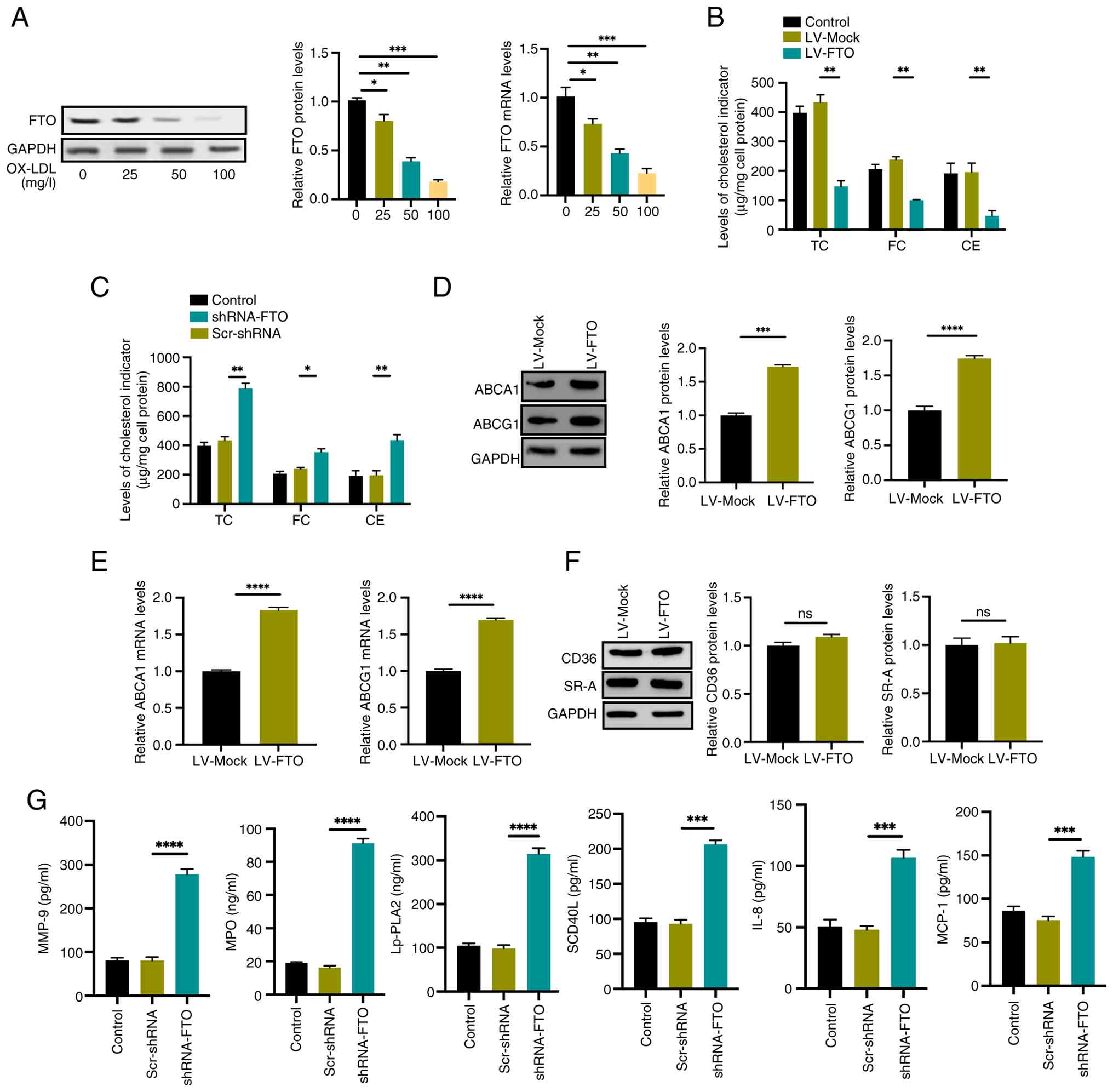 | Figure 1.FTO inhibits atherosclerotic plaque
stability in THP-1 macrophage-derived foam cells. (A) Analysis of
FTO protein and mRNA levels in human THP-1 monocytes treated with
increasing concentrations of ox-LDL (0, 25, 50, 100 mg/l) was
performed using western blotting and qPCR. (B) Quantification of
cellular TC, FC, and CE in THP-1 cells following FTO overexpression
was conducted via HPLC. (C) HPLC analysis of TC, FC, and CE levels
in THP-1 cells after FTO knockdown. (D and E) Western blotting and
qPCR analyses of ABCA1 and ABCG1 protein (D) and mRNA (E) levels
upon FTO overexpression or knockdown. (F) Protein levels of CD36
and SR-A were assessed by western blot in response to FTO
overexpression. (G) ELISA measurements of MMP-9, MPO, Lp-PLA2,
sCD40L, IL-8, and MCP-1 protein levels following FTO knockdown. For
all panels, data are derived from n=5 independent experiments;
error bars denote standard deviation. *P<0.05, **P<0.01,
***P<0.001, ****P<0.0001. ns, not significant; FTO, fat mass
and obesity-associated; qPCR, quantitative PCR; TC, total
cholesterol; FC, free cholesterol; CE, cholesteryl ester; HPLC,
high performance liquid chromatography; SR-A, scavenger receptor A;
ELISA, enzyme-linked immunosorbent assay; MPO, myeloperoxidase;
Lp-PLA2, lipoprotein-associated phospholipase A2; sCD40L, soluble
CD40 ligand; MCP-1, monocyte chemoattractant protein-1. |
Reduced cholesterol efflux via ABCA1 and ABCG1
markedly contributes to intracellular lipid accumulation (15). To elucidate the mechanism by which
FTO exerts its protective effects, the impact of FTO on the
expression of key cholesterol transporters and cholesterol efflux
in THP-1 macrophage-derived foam cells was analyzed. FTO
overexpression was found to upregulate both mRNA and protein levels
of ABCA1 and ABCG1 (Fig. 1D and
E). Given that enhanced cholesterol influx mediated by
scavenger receptors, such as CD36 and SR-A, also promotes lipid
accumulation (16,17), their expression levels were
concurrently assessed. Notably, no significant differences in CD36
or SR-A levels were observed following FTO overexpression (Fig. 1F). These results indicate that FTO
reduces lipid deposition by specifically promoting ABCA1- and
ABCG1-mediated cholesterol export.
In addition to their roles in promoting cholesterol
release, ABCA1 and ABCG1 are known to suppress the inflammatory
response (18,19). Considering that atherosclerotic
plaque stability is negatively associated with inflammatory cell
infiltration and necrotic core size, while positively associated
with fibrous cap thickness (20),
it was hypothesized that FTO might influence plaque stability by
modulating inflammation. Supporting this hypothesis, compounds such
as the contaminant derivative OXA17 have been shown to stabilize
plaques by upregulating ABCA1 (21). Consequently, the effect of FTO on
the secretion of pro-inflammatory cytokines and enzymes in THP-1
macrophage-derived foam cells was assessed. FTO knockdown markedly
increased the secretion of multiple pro-inflammatory mediators,
including MMP-9, MPO, sCD40L), lipoprotein-Associated Phospholipase
A2 (Lp-PLA2), IL-18, and MCP-1 (Fig.
1G). These data suggested that FTO mitigates the macrophage
inflammatory response, potentially contributing to improved
atherosclerotic plaque stability.
FTO promotes autophagy and
atherosclerotic plaque stability via upregulating ABCA1 and ABCG1
expression
Autophagy is a multi-step biological process
(22). To investigate its
regulatory role, functional studies involving both overexpression
and knockdown of FTO in THP-1 cells were conducted. Evaluation of
key autophagic markers revealed that FTO overexpression elevated
the LC3-II/I ratio while decreasing P62 levels (Fig. 2A and B). To directly monitor
autophagic flux, the mCherry-EGFP-LC3 reporter system was employed.
Consistent with these findings, FTO overexpression increased the
number of LC3 puncta. This system distinguishes autophagosomes
(yellow puncta, mCherry + EGFP+) from autolysosomes (red puncta,
mCherry + EGFP-). Notably, FTO overexpression markedly increased
the counts of both yellow and red puncta (Fig. 2C). Furthermore, the addition of
BafA1 exacerbated the accumulation of LC3-II induced by FTO
knockdown (shRNA-FTO; Fig. 2D).
These results indicated that FTO induces autophagy.
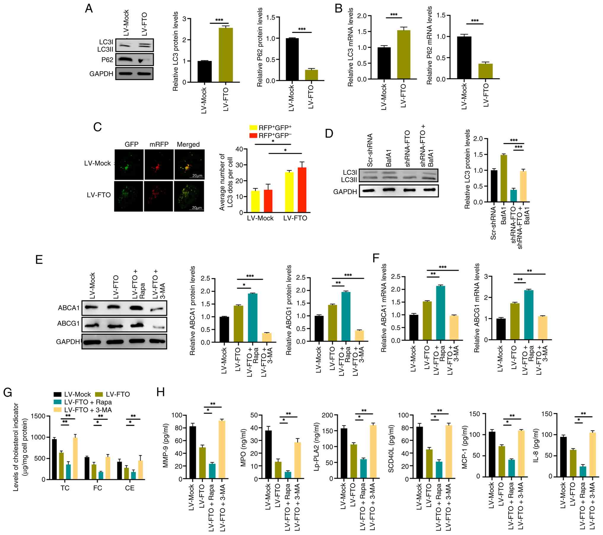 | Figure 2.FTO promotes autophagy and
atherosclerotic plaque stability via upregulation of ABCA1 and
ABCG1 expression. (A) Western blot analysis of protein levels and
(B) RT-qPCR analysis of mRNA levels for LC3-II/I and P62 in THP-1
cells following lentiviral FTO overexpression. (C) Autophagic flux
was assessed by transfection of the EGFP-mCherry-LC3 reporter into
THP-1 cells. Representative images show autophagosomes (yellow) and
autolysosomes (red) in control (LV-Mock) and FTO-overexpressing
cells, treated with or without Bafilomycin A1 (BafA1; 100 nM, 3 h).
Scale bar, 20 µm. (D) Western blot analysis of LC3-II/I protein
levels in cells subjected to FTO knockdown, with or without BafA1
co-treatment. Effects of pharmacological modulators
(Rapamycin/Rapa, 3-MA) and FTO overexpression on (E) ABCA1 and
ABCG1 protein and (F) mRNA expression levels. (G) Cellular
cholesterol profiles (TC, FC and CE) measured by HPLC under the
indicated conditions of autophagy modulation and FTO
overexpression. (H) Secreted levels of inflammatory mediators
(MMP-9, MPO, Lp-PLA2, sCD40L, IL-8, MCP-1) quantified by ELISA.
Data comprise results from n=5 independent experiments; error bars
represent standard deviation. *P<0.05, **P<0.01,
***P<0.001. ns, not significant; FTO, fat mass and
obesity-associated; RT-qPCR, reverse transcription-quantitative
PCR; ELISA, enzyme-linked immunosorbent assay; TC, total
cholesterol; FC, free cholesterol; CE, cholesteryl ester; HPLC,
high performance liquid chromatography; MPO, myeloperoxidase;
Lp-PLA2, lipoprotein-associated phospholipase A2; sCD40L, soluble
CD40 ligand; MCP-1, monocyte chemoattractant protein-1. |
To determine whether FTO regulates lipid metabolism
and plaque stability through autophagy, autophagic flux in THP-1
macrophage-derived foam cells was modulated using Rapamycin (an
agonist) and 3-MA (an inhibitor). Consistent with
autophagy-mediated regulation, the upregulation of ABCA1 and ABCG1
expression driven by FTO overexpression was further enhanced by
Rapamycin and suppressed by 3-MA (Fig.
2E and F). Similarly, HPLC analysis of intracellular
cholesterol revealed that the reduction in TC, FC and CE levels
induced by FTO overexpression was markedly amplified by Rapamycin.
Conversely, treatment with 3-MA attenuated this effect, leading to
increased intracellular accumulation of TC, FC, and CE (Fig. 2G).
These results indicated that FTO alleviates lipid
accumulation in THP-1 macrophage-derived foam cells by promoting
autophagic flux. Furthermore, the effect of autophagic modulation
on the secretion of pro-inflammatory mediators (MMP-9, MPO, sCD40L,
Lp-PLA2, IL-18, MCP-1) was assessed. Treatment with the autophagy
inhibitor 3-MA increased the levels of these factors, whereas the
autophagy inducer Rapamycin markedly reduced their secretion
(Fig. 2H). These findings
demonstrate that FTO enhances autophagy and promotes
atherosclerotic plaque stability by upregulating the expression of
ABCA1 and ABCG1.
FTO exerts its influence on autophagy
and atherosclerotic plaque stability via m6A-dependent
regulation of ATG5
To determine whether FTO regulates m6A
modification in THP-1 macrophage-derived foam cells, global
m6A levels were quantified using liquid
chromatography-tandem mass spectrometry (LC-MS/MS). FTO knockdown
resulted in a significant decrease in m6A modification
(Fig. 3A), confirming the role of
FTO as an m6A demethylase in these cells. To identify
the autophagy-related targets of FTO, several candidate genes were
screened via RT-qPCR. The mRNA level of ATG5 was markedly
downregulated following FTO knockdown and upregulated upon FTO
overexpression (Fig. 3B).
Consistent with these findings, FTO overexpression also increased
ATG5 protein levels (Fig. 3C). To
establish a functional link, a rescue experiment was conducted.
Silencing ATG5 effectively reversed the increase in the LC3-II/I
ratio and the decrease in SQSTM1/p62 levels induced by FTO
overexpression (Fig. 3D),
revealing that FTO promotes autophagy primarily by upregulating
ATG5 expression.
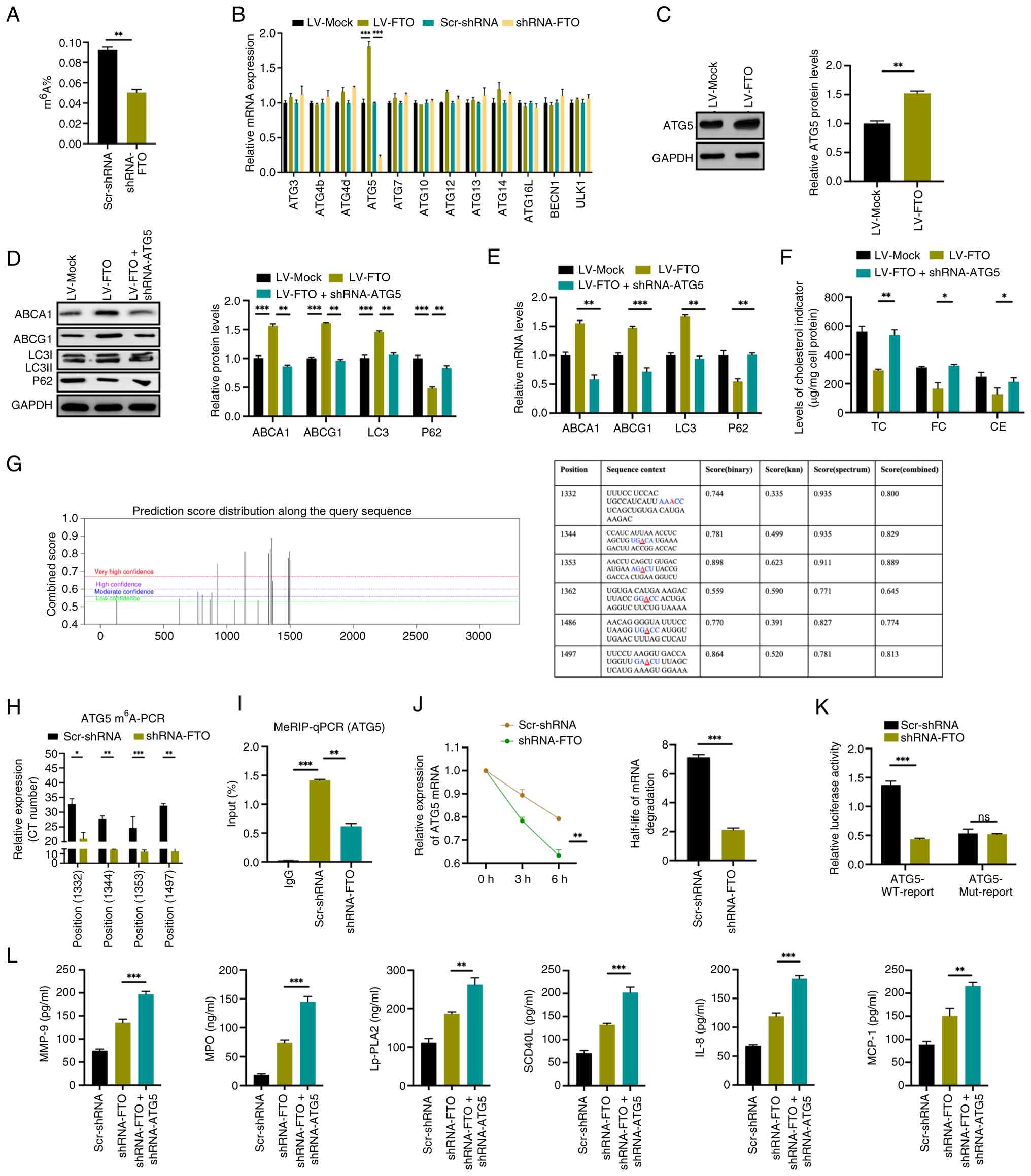 | Figure 3.FTO affects autophagy and
atherosclerotic plaque stability through targeting ATG5 in an
m6A-dependent manner. (A) Global m6A levels
in cellular mRNA from control vs. FTO-knockdown cells were
quantified using LC-MS/MS. (B) Transcript levels of key ATG genes
following FTO overexpression or knockdown were measured by qPCR.
(C) ATG5 protein expression in response to FTO overexpression was
analyzed by western blotting. (D) Protein and (E) mRNA expression
levels of ABCA1, ABCG1, LC3-II/I, and P62 under conditions of
combined FTO overexpression and ATG5 knockdown. (F) Cellular
cholesterol content (TC, FC and CE) was determined by HPLC in cells
subjected to ATG5 knockdown and FTO overexpression. (G) In
silico prediction of m6A modification sites within
ATG5 mRNA was conducted using SRAMP; the conserved RRACH motif is
highlighted, with the methylated adenosine underlined. (H)
Site-specific quantification of m6A on ATG5 mRNA in
control and FTO-deficient cells was performed using the SELECT
assay. (I) MeRIP-qPCR analysis demonstrated m6A
enrichment on ATG5 mRNA, indicating direct interaction with FTO.
(J) ATG5 mRNA stability assay following transcriptional inhibition
with Actinomycin D was conducted to calculate the degradation
half-life. (K) Luciferase reporter assay comparing the activity of
WT and MUT ATG5 3′ UTR constructs in control (Scr-shRNA) and
FTO-knockdown (shRNA-FTO) cells; firefly luciferase activity was
normalized to Renilla luciferase. (L) Secreted levels of
inflammatory factors (MMP-9, MPO, Lp-PLA2, sCD40L, IL-8, MCP-1)
were quantified by ELISA following ATG5 or FTO knockdown. Data
represent n=5 independent experiments. Error bars denote standard
deviation. *P<0.05; **P<0.01; ***P<0.001. FTO, fat mass
and obesity-associated; ATG, autophagy-related; LC-MS/MS, liquid
chromatography-tandem mass spectrometry; HPLC, high-performance
liquid chromatography; TC, total cholesterol; FC, free cholesterol;
CE, cholesteryl ester; MeRIP-qPCR, m6A RNA
immunoprecipitation-quantiative PCR; WT, wild-type; MUT, mutant;
sh, short hairpin; MPO, myeloperoxidase; Lp-PLA2,
lipoprotein-associated phospholipase A2; sCD40L, soluble CD40
ligand; MCP-1, monocyte chemoattractant protein-1; ELISA,
enzyme-linked immunosorbent assay. |
To assess whether FTO influences lipid accumulation
through ATG5, a rescue experiment was again performed. Silencing
ATG5 reversed the upregulation of ABCA1 and ABCG1 induced by FTO
overexpression (Fig. 3D and E),
indicating that FTO regulates lipid metabolism via ATG5. HPLC
analysis confirmed that ATG5 knockdown aggravated the reduction in
intracellular TC, FC, and CE levels associated with FTO
overexpression (Fig. 3F).
To investigate whether FTO regulates ATG5 through
m6A methylation, bioinformatics was initially employed
to predict potential m6A modification sites on ATG5 mRNA
(Fig. 3G). FTO knockdown decreased
the abundance of ATG5 mRNA containing m6A modifications
at these sites (Fig. 3H).
Methylated RIP-qPCR verified a direct interaction between FTO and
ATG5 mRNA in foam cells (Fig. 3I).
Furthermore, mRNA stability assays demonstrated that FTO depletion
accelerated the decay of ATG5 mRNA transcripts (Fig. 3J). A luciferase reporter assay
indicated that FTO knockdown suppressed the activity of a
wild-type, but not mutant, ATG5 3′ UTR construct (Fig. 3K). Additionally, the data showed
that shRNA-ATG5 further upregulated the secretion of
pro-inflammatory mediators, including MMP-9, MPO, sCD40L, Lp-PLA2,
IL-18, and MCP-1 (Fig. 3L). These
data demonstrated that FTO directly targets ATG5 mRNA in an
m6A-dependent manner to enhance its stability and
expression, thereby promoting autophagy and plaque stability.
YTHDF1 mediates mRNA expression of
ATG5 via m6A-dependent mechanism
A previous study established that reader proteins of
m6A modification can regulate various aspects of RNA
metabolism, including translation, splicing, stability, nuclear
export and degradation (23).
Among these proteins, the YTH domain-containing family (YTHDF1-3)
has been extensively studied. YTHDF2 has been shown to selectively
recognize m6A-modified mRNAs and facilitate their decay,
while YTHDF1 promotes the translation of m6A-marked
transcripts (24,25). YTHDF3 appears to interact with both
YTHDF1 and YTHDF2, potentially coordinating their functional roles
(26). To determine which YTHDF
protein primarily influences ATG5 transcript regulation, individual
knockdowns of YTHDF1, YTHDF2 and YTHDF3 were performed using small
interfering RNAs, with knockdown efficiency validated for each case
(Fig. S2). Knockdown of YTHDF1,
but not the other reader proteins, markedly increased ATG5 mRNA
levels (Fig. 4A). As depicted in
Fig. S3, the knockdown of YTHDF2
via shRNA did not result in a significant alteration of ATG5 or
ATG7 protein expression when compared to the Scr-shRNA control.
Conversely, shRNA-mediated knockdown of YTHDF1 led to a significant
reduction in ATG5 protein levels, while ATG7 levels remained
unaffected. Moreover, as illustrated in Fig. S4, the knockdown of FTO did not
affect ATG7 at the protein level. This led to the hypothesis that
YTHDF1 specifically recognizes FTO-mediated m6A
modification on the ATG5 transcript to promote its decay. A rescue
experiment supported this notion; YTHDF1 knockdown effectively
counteracted the effects of FTO overexpression, restoring the
LC3B-II/LC3B-I ratio and ABCA1/ABCG1 expression while reducing P62
accumulation (Fig. 4B and C), thus
demonstrating that YTHDF1 is a key mediator in the
m6A-dependent regulation of ATG5 by FTO. Furthermore,
immunofluorescence analysis using the mCherry-EGFP-LC3 reporter in
foam cells revealed that the increase in autophagic puncta (both
red and yellow) induced by LV-FTO was further augmented upon
treatment with shRNA-YTHDF1 (Fig.
4D).
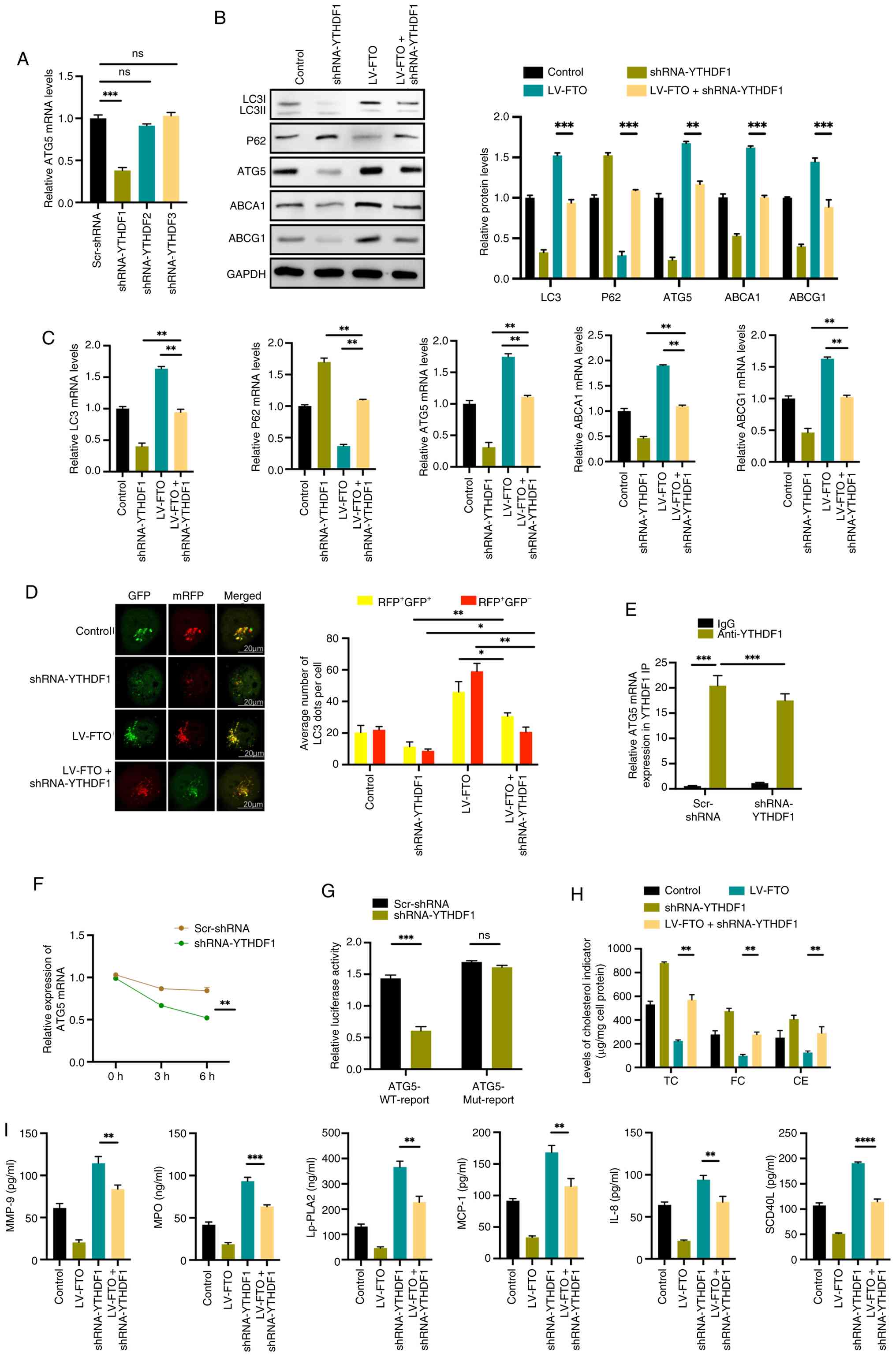 | Figure 4.YTHDF1 mediates mRNA expression of
ATG5 through an m6A-dependent mechanism. (A) ATG5 mRNA
levels in THP-1 cells following individual knockdown of YTHDF1,
YTHDF2, or YTHDF3 were analyzed by qPCR. (B) Protein and (C) mRNA
expression levels of LC3-II/I, P62, ATG5, ABCA1, and ABCG1 in cells
subjected to FTO overexpression or YTHDF1 knockdown. (D) Autophagic
flux was assessed by imaging of EGFP-mCherry-LC3 puncta
(autophagosomes, yellow; autolysosomes, red) in YTHDF1 knockdown
and FTO overexpressing cells. Scale bar, 20 µm. (E) RIP-qPCR
analysis of the interaction between YTHDF1 and ATG5 mRNA in control
vs. YTHDF1-knockdown cells, with enrichment normalized to input.
(F) ATG5 mRNA stability assay conducted following transcriptional
inhibition with Actinomycin D; degradation half-life is shown. (G)
Relative luciferase activity of WT vs. MUT ATG5 3′ UTR reporter
constructs in cells transfected with control (Scr-shRNA) or
YTHDF1-targeting (shRNA-YTHDF1) vectors; firefly luciferase
activity was normalized to Renilla luciferase activity. (H)
Cellular cholesterol content (TC, FC and CE) quantified by HPLC in
response to YTHDF1 knockdown and FTO overexpression. (I) Secreted
levels of inflammatory mediators (MMP-9, MPO, Lp-PLA2, sCD40L,
IL-8, MCP-1) were measured by ELISA following YTHDF1 knockdown or
FTO overexpression. Data are representative of n=5 independent
experiments. Error bars denote standard deviation. *P<0.05;
**P<0.01; ***P<0.001; ****P<0.001. ns, not significant.
YTHDF, YTH domain-containing family; qPCR, quantitative PCR; FTO,
fat mass and obesity-associated; RIP-qPCR, RNA
immunoprecipitation-quantitative PCR; WT, wild-type; MUT, mutant;
sh, short hairpin; TC, total cholesterol; FC, free cholesterol; CE,
cholesteryl ester; MPO, myeloperoxidase; Lp-PLA2,
lipoprotein-associated phospholipase A2; sCD40L, soluble CD40
ligand; MCP-1, monocyte chemoattractant protein-1. |
Additionally, RIP-qPCR experiments confirmed a
direct interaction between ATG5 mRNA and YTHDF1 (Fig. 4E). Subsequent mRNA stability assays
demonstrated that YTHDF1 knockdown delayed the decay of ATG5
transcripts and enhanced their stability (Fig. 4F). In luciferase reporter assays,
FTO knockdown suppressed the activity of wild-type constructs but
had no effect on mutant constructs (Fig. 4G). HPLC analysis indicated that
YTHDF1 knockdown reversed the cholesterol-lowering effect of FTO
overexpression, as evidenced by restored intracellular levels of
TC, FC and CE (Fig. 4H).
Furthermore, in a rescue experiment, YTHDF1 knockdown counteracted
the changes in the secretion of MMP-9, MPO, sCD40L, Lp-PLA2, IL-18
and MCP-1 induced by FTO overexpression (Fig. 4I). In summary, these results
revealed that YTHDF1 regulates ATG5 mRNA expression via an
m6A-dependent mechanism.
FTO regulates plaque stability through
ATG5-dependent autophagy in vivo
To further elucidate the relationship between FTO
and atherosclerotic plaque composition, plaque size in aortic
tissues was evaluated using hematoxylin-eosin staining, while
collagen content was assessed via Masson's trichrome staining.
Hematoxylin-eosin staining revealed that atherosclerotic lesions in
AAV-sh-FTO-treated mice were larger, exhibited increased lipid
accumulation, and demonstrated thicker fibrous caps compared to
controls. Masson's staining further indicated a significant
reduction in collagen fiber content within plaques from AAV-sh-FTO
mice relative to the AAV-Mock group (Fig. 5A). Immunofluorescence
co-localization analysis on arterial plaque sections was performed
to determine the cellular localization of FTO in endothelial cells
(CD31+), smooth muscle cells (αSMA+) and macrophages (CD68+). The
results demonstrated that FTO predominantly localizes within
macrophages (Fig. 5B).
Immunohistochemical analysis of key autophagy and cholesterol
efflux-related proteins showed that levels of LC3, ATG5, ABCA1 and
ABCG1 were downregulated, while P62 expression was upregulated in
the AAV-sh-FTO group (Fig. 5C).
Moreover, the production of plaque destabilization
markers-including MMP-9, MPO, sCD40L, Lp-PLA2, IL-18 and MCP-1 was
markedly upregulated in AAV-sh-FTO mice (Fig. 5D). These findings indicate that FTO
knockdown exacerbates atherosclerotic plaque development and
instability, likely due to impaired autophagic flux and cholesterol
homeostasis, accompanied by enhanced inflammation. In conclusion,
these findings support a protective role for FTO in mitigating the
progression of atherosclerosis.
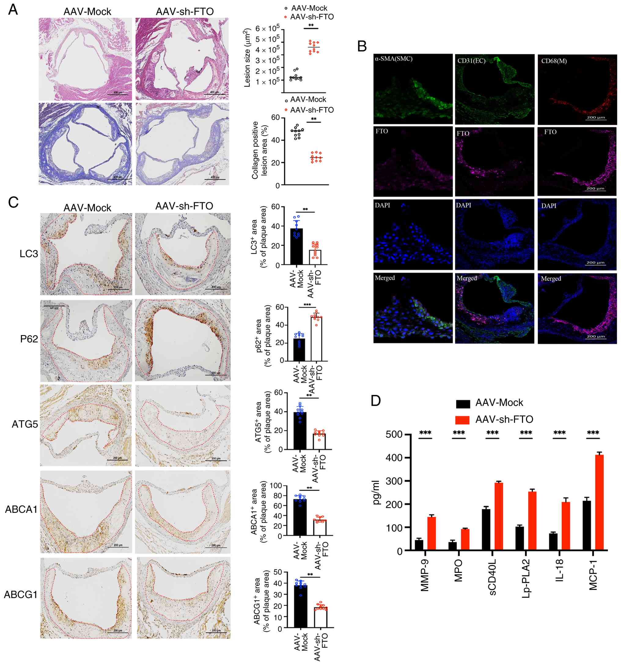 | Figure 5.FTO regulates plaque stability
through ATG5-dependent autophagy in vivo. (A) Representative
hematoxylin-eosin staining images and Masson's trichrome staining
of aortic valve sections, comparing atherosclerotic lesion areas
between AAV-Mock and AAV-sh-FTO groups fed with HFD. Data are
derived from n=10 independent experiments. **P<0.01.
Magnification, ×100. (B) FTO cellular localization in plaques was
demonstrated by co-staining with CD31 (endothelium), CD68
(macrophages) and α-SMA (smooth muscle cells). Magnification, ×200.
n=10 independent experiments. (C) Immunohistochemical detection of
LC3, P62, ATG5, ABCA1, and ABCG1 in aortic valves. Quantification
of LC3, P62, ATG5, ABCA1, and ABCG1 areas is shown. n=10
independent experiments. **P<0.01; ***P<0.001. Magnification,
×200. (D) ELISA quantification of MMP-9, MPO, SCD40L, Lp-PLA2,
IL-18, and MCP-1 chemokines normalized to tissue protein content.
n=10 independent experiments. ***P<0.001. FTO, fat mass and
obesity-associated; sh, short hairpin; HFD, high-fat diet; ELISA,
enzyme-linked immunosorbent assay; MPO, myeloperoxidase; Lp-PLA2,
lipoprotein-associated phospholipase A2; sCD40L, soluble CD40
ligand; MCP-1, monocyte chemoattractant protein-1; α-SMA, α-smooth
muscle actin. |
Discussion
Atherosclerotic cardiovascular disease (ASCVD)
continues to be a leading cause of mortality worldwide,
highlighting the inadequacies of existing therapeutic approaches in
effectively reducing its effects (27,28).
The progression to acute and often fatal coronary events is
commonly triggered by the rupture of unstable atherosclerotic
plaques (29). Consequently,
understanding the molecular pathways that regulate plaque stability
is a crucial research priority. Investigating these mechanisms is
vital for the development of complementary diagnostic strategies
and interventions designed to prevent disease progression and avert
acute cardiovascular events.
Autophagy is a conserved catabolic process crucial
for maintaining cellular homeostasis, characterized by the
degradation of damaged cytoplasmic components within lysosomes.
This dynamic process begins with the conversion of LC3-I to the
lipidated LC3-II form, a hallmark of autophagosome formation
(30), and concludes with
lysosomal degradation. In advanced atherosclerosis, this process is
often dysregulated (31,32), leading to exacerbated plaque
pathogenesis through the promotion of vascular aging and cellular
dysfunction. Notably, the genetic ablation of core autophagy
components, such as ATG5, increases apoptosis and oxidative stress,
thereby contributing to plaque necrosis (33). The present study identified FTO as
a significant factor within this critical pathway. It demonstrated
that FTO overexpression enhances autophagic flux, with experiments
involving Bafilomycin A1 suggesting that this effect is primarily
mediated through the promotion of autophagosome synthesis. This
significant connection between FTO-driven m6A demethylation and the
enhancement of autophagy highlights the therapeutic potential of
targeting this axis to stabilize atherosclerotic plaques.
Autophagy, a conserved intracellular degradative
mechanism, is pivotal in the renewal and recycling of cellular
components to maintain nutrient and energy homeostasis within the
cell. ATG5, a critical element of the autophagy process, is
instrumental in the formation of the autophagosome, a defining
feature of autophagy. ATG5 interacts with ATG12 and ATG16L1 to form
an E3-like ligase complex, which is essential for autophagosome
expansion. This process is meticulously regulated, with
autophagy-related (ATG) proteins playing key roles. Among the 41
identified ATG genes, ATG5 is indispensable for autophagosome
formation and positively influences autophagic activity (34). Moreover, m6A-modified USP13 has
been shown to induce autophagy and imatinib resistance in
gastrointestinal stromal tumor cells by modulating ATG5 stability.
The present study indicated that FTO regulates ATG5 mRNA expression
in an m6A-dependent manner, thereby affecting autophagy. A similar
mechanism was observed in a recent study, where FTO was found to
coordinate autophagy and energy metabolism by regulating ATG16L1 in
an m6A-dependent manner in osteoarthritis (35). FTO acts as a catalyst for diabetic
wound healing and influences autophagy by regulating TRIB3 in
keratinocytes (36). This
variation may be attributed to the specificity of m6A methylation
in autophagy across different tissue or cell types. mRNA
transcripts modified with m6A display distinct terminal structures
due to recognition by various m6A-binding proteins. YTHDF2 and
YTHDF3 work synergistically to promote the degradation of target
mRNA, thereby decreasing target gene expression. By contrast,
YTHDF1 and YTHDF3 collaborate to enhance the translation of target
mRNA, thus increasing target gene expression (37,38).
IGF2BPs contribute to mRNA stability, thereby augmenting
translational efficiency (39).
The findings suggested that YTHDF1 mediates the stability of ATG5
mRNA in an m6A-dependent manner, identifying ATG5 mRNA as a target
gene of YTHDF1.
In recent years, there has been an increasing
scholarly focus on the role of FTO-mediated m6A RNA methylation in
the context of cardiovascular disease. Genome-wide association
studies have definitively associated genetic variants within the
FTO gene with an elevated risk of several cardiovascular
conditions, such as hypertension, myocardial infarction, acute
coronary syndrome and graft rejection in heart transplant
recipients (40–44). Nonetheless, the direct functions
and specific molecular mechanisms by which FTO influences the
fundamental pathophysiology of atherosclerosis are not yet fully
elucidated.
Emerging evidence indicates that FTO plays complex,
cell-type-specific roles in atherosclerosis. For example, in
endothelial cells, FTO overexpression has been shown to diminish
the protective effects of Rb1 against atherosclerotic pathologies
and VCAM-1 expression in Apoe-/- mice (45). Conversely, in vascular smooth
muscle cells (VSMCs), FTO appears to mitigate ox-LDL-induced
senescence, thereby inhibiting VSMC aging within plaques (46). While these studies underscore
protective roles in specific cellular contexts, a fundamental
question persists: Does FTO regulate atherosclerosis through a
unifying, cross-cellular mechanism? Additionally, the potential
direct influence of FTO on critical plaque biology, such as
autophagy, via m6A demethylation remains entirely unexplored.
The present study endeavored to fill this
significant knowledge gap by elucidating the role of autophagy as a
pivotal downstream pathway through which FTO influences
atherosclerosis. It provided novel evidence demonstrating that FTO
directly targets the mRNA of ATG5, a critical gene involved in
autophagy, in a manner dependent on m6A modification. This
interaction enhances the stability of ATG5 mRNA and its protein
expression, thereby facilitating autophagic flux. This discovery
aligns with the broader emerging paradigm that recognizes m6A
modification as an essential upstream regulator of autophagy
(47,48). This study represents the first
documentation of the direct demethylation of ATG5 mRNA by the
enzyme FTO, commonly referred to as the ‘eraser’, specifically
within the context of atherosclerosis. This finding highlighted the
mechanistic specificity of the present study. Furthermore, the
present study demonstrated the pathophysiological significance of
this axis through a comprehensive series of in vitro and
in vivo experiments. Its results provided robust evidence
that the FTO-ATG5-autophagy signaling axis enhances plaque
stability. The present study introduced a novel paradigm for
understanding RNA epitranscriptomics in cardiovascular disease,
where m6A-dependent regulation of autophagy directly impacts
disease progression and plaque phenotype.
Traditionally, YTHDF1 has been acknowledged as a
promoter of translation; however, recent research indicates that it
can also facilitate mRNA degradation in a context-dependent manner
(49,50). Notably, the knockdown of YTHDF2
does not influence the expression of ATG5 or ATG7 in macrophages
stimulated with ox-LDL, whereas the depletion of YTHDF1 results in
decreased ATG5 levels. Furthermore, FTO does not regulate ATG7 in
our experimental model. Although Wang et al (51) reported that YTHDF2 mediates the
m6A-dependent degradation of Atg5 and Atg7 in adipocytes and
hepatocytes, the functional roles of m6A readers are increasingly
understood to be context-dependent (52). Studies have challenged the
traditional YTHDF model, revealing that all three paralogs
redundantly mediate mRNA degradation. YTHDF1 facilitates decay
through CCR4-NOT and AGO2-mediated phase separation, underscoring
its context-dependent role in mRNA stability (49,50,53).
Consequently, the identification of YTHDF1 as the principal m6A
reader regulating ATG5 in macrophages is consistent with
contemporary models of m6A regulation.
Several limitations warrant acknowledgment. First,
the findings were exclusively based on ApoE−/− mouse
models and in vitro macrophage lines, lacking validation in
human plaques. This limitation restricts the direct translational
relevance of the FTO-ATG5-YTHDF1 axis to patient scenarios. To
address this, future research will analyze human carotid
endarterectomy samples (comparing stable and symptomatic plaques)
for expression levels of FTO, ATG5 and YTHDF1, as well as their
correlation with plaque stability. Second, unbiased MeRIP-seq in
FTO-knockdown macrophages will help identify additional
autophagy-related targets of FTO. Third, techniques such as RNA
pull-down, CLIP-seq and macrophage-specific FTO knockout mice will
be employed to dissect the precise molecular mechanisms underlying
YTHDF1-mediated ATG5 mRNA degradation. Addressing these limitations
will enhance the translational potential of this epitranscriptomic
axis, although this potential requires validation through future
clinical studies.
Supplementary Material
Supporting Data
Acknowledgements
Not applicable.
Funding
The present study was supported by The Key Project of Medical
Science and Technology of Henan Province (grant no. LHGJ20230009),
and Science and Technology Tackling in Henan Province (grant no.
232102310084).
Availability of data and materials
The data generated in the present study may be
requested from the corresponding author.
Authors' contributions
FG and KZ was responsible for conceptualization,
data curation, formal analysis, methodology, project
administration, supervision, validation, visualization, writing the
original draft, reviewing and editing. BH was responsible for data
curation, formal analysis, methodology and validation. FG and KZ
were responsible for data curation and formal analysis. YL designed
the study. FG and BH confirm the authenticity of all the raw data.
MH was responsible for conducting the animal experiments, managing
the ethics approval process, and revising the sections on animal
experiment methods and ethical statements within the manuscript.
All authors read and approved the final manuscript.
Ethics approval and consent to
participate
All procedures adhered to the NIH guidelines
(Publication No. 85-23, 1996 revision) and received approval from
the Animal Ethics Committee of Zhengzhou No. 7 People's Hospital
(approval no. 2024-017).
Patient consent for publication
Not applicable.
Competing interests
The authors declare that they have no competing
interests.
References
|
1
|
Cagnina A, Chabot O, Davin L, Lempereur M,
Maréchal P, Oury C and Lancellotti P: Atherosclerosis, an
inflammatory disease. Rev Med Liege. 77:302–309. 2022.(In French).
PubMed/NCBI
|
|
2
|
Wolf D and Ley K: Immunity and
inflammation in atherosclerosis. Circ Res. 124:315–327. 2019.
View Article : Google Scholar : PubMed/NCBI
|
|
3
|
Libby P: Molecular bases of the acute
coronary syndromes. Circulation. 91:2844–2850. 1995. View Article : Google Scholar : PubMed/NCBI
|
|
4
|
Dong G, Yu J, Shan G, Su L, Yu N and Yang
S: N6-methyladenosine methyltransferase METTL3 promotes
angiogenesis and atherosclerosis by upregulating the JAK2/STAT3
pathway via m6A reader IGF2BP1. Front Cell Dev Biol. 9:7318102021.
View Article : Google Scholar : PubMed/NCBI
|
|
5
|
Wing MR, Ziegler J, Langefeld CD, Ng MC,
Haffner SM, Norris JM, Goodarzi MO and Bowden DW: Analysis of FTO
gene variants with measures of obesity and glucose homeostasis in
the IRAS family study. Hum Genet. 125:615–626. 2009. View Article : Google Scholar : PubMed/NCBI
|
|
6
|
Jalili V, Mokhtari Z, Rastgoo S, Hajipour
A, Bourbour F, Gholamalizadeh M, Mosavi Jarrahi A, JavadiKooshesh
S, Moslem A, Abdollahi M and Doaei S: The association between FTO
rs9939609 polymorphism and serum lipid profile in adult women.
Diabetol Metab Syndr. 13:1382021. View Article : Google Scholar : PubMed/NCBI
|
|
7
|
Levine B and Kroemer G: Biological
functions of autophagy genes: A disease perspective. Cell.
176:11–42. 2019. View Article : Google Scholar : PubMed/NCBI
|
|
8
|
Mizushima N and Levine B: Autophagy in
human diseases. N Engl J Med. 383:1564–1576. 2020. View Article : Google Scholar : PubMed/NCBI
|
|
9
|
Leidal AM, Levine B and Debnath J:
Autophagy and the cell biology of age-related disease. Nat Cell
Biol. 20:1338–1348. 2018. View Article : Google Scholar : PubMed/NCBI
|
|
10
|
Gulati P, Cheung MK, Antrobus R, Church
CD, Harding HP, Tung YC, Rimmington D, Ma M, Ron D, Lehner PJ, et
al: Role for the obesity-related FTO gene in the cellular sensing
of amino acids. Proc Natl Acad Sci USA. 110:2557–2562. 2013.
View Article : Google Scholar : PubMed/NCBI
|
|
11
|
Aas A, Isakson P, Bindesboll C, Alemu EA,
Klungland A and Simonsen A: Nucleocytoplasmic shuttling of FTO does
not affect starvation-induced autophagy. PLoS One. 12:e01681822017.
View Article : Google Scholar : PubMed/NCBI
|
|
12
|
Jin S, Zhang X, Miao Y, Liang P, Zhu K,
She Y, Wu Y, Liu DA, Huang J, Ren J and Cui J: m6A RNA
modification controls autophagy through upregulating ULK1 protein
abundance. Cell Res. 28:955–957. 2018. View Article : Google Scholar : PubMed/NCBI
|
|
13
|
Dong XH, Lu ZF, Kang CM, Li XH, Haworth
KE, Ma X, Lu JB, Liu XH, Fang FC, Wang CS, et al: The long
noncoding RNA RP11-728F11.4 promotes atherosclerosis. Arterioscler
Thromb Vasc Biol. 41:1191–1204. 2021. View Article : Google Scholar : PubMed/NCBI
|
|
14
|
Livak KJ and Schmittgen TD: Analysis of
relative gene expression data using real-time quantitative PCR and
the 2(−Delta Delta C(T)) method. Methods. 25:402–408. 2001.
View Article : Google Scholar : PubMed/NCBI
|
|
15
|
Rawlins SC: Biological control of insect
pests affecting man and animals in the tropics. Crit Rev Microbiol.
16:235–252. 1989. View Article : Google Scholar : PubMed/NCBI
|
|
16
|
Kunjathoor VV, Febbraio M, Podrez EA,
Moore KJ, Andersson L, Koehn S, Rhee JS, Silverstein R, Hoff HF and
Freeman MW: Scavenger receptors class A-I/II and CD36 are the
principal receptors responsible for the uptake of modified low
density lipoprotein leading to lipid loading in macrophages. J Biol
Chem. 277:49982–49988. 2002. View Article : Google Scholar : PubMed/NCBI
|
|
17
|
Bhargava P, Dinh D, Teramayi F, Silberg A,
Petler N, Anderson AM, Sadrerafi K, Clemens DM and O'Connor MS:
Selective removal of 7KC by a novel atherosclerosis therapeutic
candidate reverts foam cells to a macrophage-like phenotype.
bioRxiv [Preprint]. 2023.10.23.563623. 2023.
|
|
18
|
Yin K, Liao DF and Tang CK: ATP-binding
membrane cassette transporter A1 (ABCA1): A possible link between
inflammation and reverse cholesterol transport. Mol Med.
16:438–449. 2010. View Article : Google Scholar : PubMed/NCBI
|
|
19
|
Wojcik AJ, Skaflen MD, Srinivasan S and
Hedrick CC: A critical role for ABCG1 in macrophage inflammation
and lung homeostasis. J Immunol. 180:4273–4282. 2008. View Article : Google Scholar : PubMed/NCBI
|
|
20
|
Vakili H, Shirazi M, Charkhkar M, Khaheshi
I, Memaryan M and Naderian M: Correlation of platelet-to-lymphocyte
ratio and neutrophil-to-lymphocyte ratio with thrombolysis in
myocardial infarction frame count in ST-segment elevation
myocardial infarction. Eur J Clin Invest. 47:322–327. 2017.
View Article : Google Scholar : PubMed/NCBI
|
|
21
|
Tsui PF, Chern CY, Lien CF, Lin FY, Tsai
CS, Tsai MC and Lin CS: An octimibate derivative, Oxa17, enhances
cholesterol efflux and exerts anti-inflammatory and
atheroprotective effects in experimental atherosclerosis. Biochem
Pharmacol. 188:1145812021. View Article : Google Scholar : PubMed/NCBI
|
|
22
|
Li R, Wei X and Jiang DS: Protein
methylation functions as the posttranslational modification switch
to regulate autophagy. Cell Mol Life Sci. 76:3711–3722. 2019.
View Article : Google Scholar : PubMed/NCBI
|
|
23
|
Song D, Hou J, Wu J and Wang J: Role of
N6-methyladenosine RNA modification in cardiovascular
disease. Front Cardiovasc Med. 8:6596282021. View Article : Google Scholar : PubMed/NCBI
|
|
24
|
Wang X, Zhao BS, Roundtree IA, Lu Z, Han
D, Ma H, Weng X, Chen K, Shi H and He C: N(6)-methyladenosine
modulates messenger RNA translation efficiency. Cell.
161:1388–1399. 2015. View Article : Google Scholar : PubMed/NCBI
|
|
25
|
Du H, Zhao Y, He J, Zhang Y, Xi H, Liu M,
Ma J and Wu L: YTHDF2 destabilizes m(6)A-containing RNA through
direct recruitment of the CCR4-NOT deadenylase complex. Nat Commun.
7:126262016. View Article : Google Scholar : PubMed/NCBI
|
|
26
|
Roost C, Lynch SR, Batista PJ, Qu K, Chang
HY and Kool ET: Structure and thermodynamics of N6-methyladenosine
in RNA: A spring-loaded base modification. J Am Chem Soc.
137:2107–2115. 2015. View Article : Google Scholar : PubMed/NCBI
|
|
27
|
Yvan-Charvet L, Bonacina F, Guinamard RR
and Norata GD: Immunometabolic function of cholesterol in
cardiovascular disease and beyond. Cardiovasc Res. 115:1393–1407.
2019. View Article : Google Scholar : PubMed/NCBI
|
|
28
|
Yu XH, Zheng XL and Tang CK: Peroxisome
proliferator-activated receptor α in lipid metabolism and
atherosclerosis. Adv Clin Chem. 71:171–203. 2015. View Article : Google Scholar : PubMed/NCBI
|
|
29
|
Petkovic A, Erceg S, Munjas J, Ninic A,
Vladimirov S, Davidovic A, Vukmirovic L, Milanov M, Cvijanovic D,
Mitic T and Sopic M: LncRNAs as regulators of atherosclerotic
plaque stability. Cells. 12:18322023. View Article : Google Scholar : PubMed/NCBI
|
|
30
|
Kabeya Y, Mizushima N, Ueno T, Yamamoto A,
Kirisako T, Noda T, Kominami E, Ohsumi Y and Yoshimori T: LC3, a
mammalian homologue of yeast Apg8p, is localized in autophagosome
membranes after processing. EMBO J. 19:5720–5728. 2000. View Article : Google Scholar : PubMed/NCBI
|
|
31
|
Vion AC, Kheloufi M, Hammoutene A, Poisson
J, Lasselin J, Devue C, Pic I, Dupont N, Busse J, Stark K, et al:
Autophagy is required for endothelial cell alignment and
atheroprotection under physiological blood flow. Proc Natl Acad Sci
USA. 114:E8675–E8684. 2017. View Article : Google Scholar : PubMed/NCBI
|
|
32
|
LaRocca TJ, Henson GD, Thorburn A, Sindler
AL, Pierce GL and Seals DR: Translational evidence that impaired
autophagy contributes to arterial ageing. J Physiol. 590:3305–3316.
2012. View Article : Google Scholar : PubMed/NCBI
|
|
33
|
Liao X, Sluimer JC, Wang Y, Subramanian M,
Brown K, Pattison JS, Robbins J, Martinez J and Tabas I: Macrophage
autophagy plays a protective role in advanced atherosclerosis. Cell
Metab. 15:545–553. 2012. View Article : Google Scholar : PubMed/NCBI
|
|
34
|
Changotra H, Kaur S, Yadav SS, Gupta GL,
Parkash J and Duseja A: ATG5: A central autophagy regulator
implicated in various human diseases. Cell Biochem Funct.
40:650–667. 2022. View Article : Google Scholar : PubMed/NCBI
|
|
35
|
Jiang Z, Zhang C, Liu R, Zhu Z, Long D,
Wen X, Yang Z, Jiang D, Mao G, Liao W and Zhang Z: M6A
demethyltransferase FTO attenuates meniscus degeneration and
osteoarthritis via orchestrating autophagy and energetic
metabolism. Adv Sci (Weinh). 12:e24123792025. View Article : Google Scholar : PubMed/NCBI
|
|
36
|
Dong Z, Li S, Huang Y, Chen T, Ding Y and
Tan Q: RNA N6-methyladenosine demethylase FTO promotes
diabetic wound healing through TRIB3-mediated autophagy in an
m6A-YTHDF2-dependent manner. Cell Death Dis. 16:2222025.
View Article : Google Scholar : PubMed/NCBI
|
|
37
|
Liu T, Wei Q, Jin J, Luo Q, Liu Y, Yang Y,
Cheng C, Li L, Pi J, Si Y, et al: The m6A reader YTHDF1 promotes
ovarian cancer progression via augmenting EIF3C translation.
Nucleic Acids Res. 48:3816–3831. 2020. View Article : Google Scholar : PubMed/NCBI
|
|
38
|
Zhang Y, Wang X, Zhang X, Wang J, Ma Y,
Zhang L and Cao X: RNA-binding protein YTHDF3 suppresses
interferon-dependent antiviral responses by promoting FOXO3
translation. Proc Natl Acad Sci USA. 116:976–981. 2019. View Article : Google Scholar : PubMed/NCBI
|
|
39
|
Huang H, Weng H, Sun W, Qin X, Shi H, Wu
H, Zhao BS, Mesquita A, Liu C, Yuan CL, et al: Recognition of RNA
N6-methyladenosine by IGF2BP proteins enhances mRNA
stability and translation. Nat Cell Biol. 20:285–295. 2018.
View Article : Google Scholar : PubMed/NCBI
|
|
40
|
Ahmad T, Chasman DI, Mora S, Paré G, Cook
NR, Buring JE, Ridker PM and Lee IM: The fat-mass and
obesity-associated (FTO) gene, physical activity, and risk of
incident cardiovascular events in white women. Am Heart J.
160:1163–1169. 2010. View Article : Google Scholar : PubMed/NCBI
|
|
41
|
He D, Fu M, Miao S, Hotta K, Chandak GR
and Xi B: FTO gene variant and risk of hypertension: A
meta-analysis of 57,464 hypertensive cases and 41,256 controls.
Metabolism. 63:633–639. 2014. View Article : Google Scholar : PubMed/NCBI
|
|
42
|
Liu C, Mou S and Pan C: The FTO gene
rs9939609 polymorphism predicts risk of cardiovascular disease: A
systematic review and meta-analysis. PLoS One. 8:e719012013.
View Article : Google Scholar : PubMed/NCBI
|
|
43
|
Hubacek JA, Vrablik M, Dlouha D, Stanek V,
Gebauerova M, Adamkova V, Ceska R, Dostálová G, Linhart A, Vitek L
and Pitha J: Gene variants at FTO, 9p21, and 2q36.3 are
age-independently associated with myocardial infarction in Czech
men. Clin Chim Acta. 454:119–123. 2016. View Article : Google Scholar : PubMed/NCBI
|
|
44
|
Hubacek JA, Vymetalova J, Lanska V and
Dlouha D: The fat mass and obesity related gene polymorphism
influences the risk of rejection in heart transplant patients. Clin
Transplant. 32:e134432018. View Article : Google Scholar : PubMed/NCBI
|
|
45
|
Yang Z, Xiong M, Tang X, Wang P, Cui J,
Chen Y and Zhang T: Ginsenoside Rb1 mitigates atherosclerosis in
part through modulating FTO-mediated m6A RNA
modification in NETs-induced endothelial activation. Front
Pharmacol. 16:16310762025. View Article : Google Scholar : PubMed/NCBI
|
|
46
|
Sun J, Wang M, Jia F, Song J, Ren J and Hu
B: FTO stabilizes MIS12 to inhibit vascular smooth muscle cell
senescence in atherosclerotic plaque. J Inflamm Res. 17:1857–1871.
2024. View Article : Google Scholar : PubMed/NCBI
|
|
47
|
Yu H, Zhuang J, Zhou Z, Song Q, Lv J, Yang
X, Yang H and Lu Q: METTL16 suppressed the proliferation and
cisplatin-chemoresistance of bladder cancer by degrading PMEPA1
mRNA in a m6A manner through autophagy pathway. Int J Biol Sci.
20:1471–1491. 2024. View Article : Google Scholar : PubMed/NCBI
|
|
48
|
Zhang P, Feng K, Yu X, Yang Y, Luo S, Li
Q, Zhang H, Jia Y, Fei Q, Ren X, et al: The m6A methyltransferase
METTL14 promotes oncogenic Kras induced juvenile myelomonocytic
leukemia through dysregulating autophagy. Cell Death Differ.
33:156–170. 2026. View Article : Google Scholar : PubMed/NCBI
|
|
49
|
Zaccara S and Jaffrey SR: A unified model
for the function of YTHDF proteins in regulating
m6A-modified mRNA. Cell. 181:1582–1595.e18. 2020.
View Article : Google Scholar : PubMed/NCBI
|
|
50
|
Zaccara S and Jaffrey SR: Understanding
the redundant functions of the m6A-binding YTHDF
proteins. RNA. 30:468–481. 2024. View Article : Google Scholar : PubMed/NCBI
|
|
51
|
Wang X, Wu R, Liu Y, Zhao Y, Bi Z, Yao Y,
Liu Q, Shi H, Wang F and Wang Y: m6A mRNA methylation
controls autophagy and adipogenesis by targeting Atg5 and Atg7.
Autophagy. 16:1221–1235. 2020. View Article : Google Scholar : PubMed/NCBI
|
|
52
|
Shi H, Wei J and He C: Where, When, and
How: Context-dependent functions of RNA methylation writers,
readers, and erasers. Mol Cell. 74:640–650. 2019. View Article : Google Scholar : PubMed/NCBI
|
|
53
|
Li J, Chen K, Dong X, Xu Y, Sun Q, Wang H,
Chen Z, Liu C, Liu R, Yang Z, et al: YTHDF1 promotes mRNA
degradation via YTHDF1-AGO2 interaction and phase separation. Cell
Prolif. 55:e131572022. View Article : Google Scholar : PubMed/NCBI
|

















