Introduction
Metabolic syndrome (MetS) is defined by the World
Health Organization (WHO) as a cluster of disorders characterized
by insulin resistance accompanied by multiple abnormalities such as
abdominal obesity, dyslipidemia, hypertension and hyperglycemia
(1). This complex condition
notably increases the risk of type 2 diabetes mellitus (T2DM),
cardiovascular disease, non-alcoholic fatty liver disease (NAFLD)
and certain cancer types (2–4).
MetS represents a major global public-health challenge, affecting
~20% of the global population, with prevalence rates reaching up to
46% in high-burden geographical regions, such as the Middle-East
and Latin-America, and a rising prevalence observed among younger
age groups, namely adolescents and young adults aged 10–24 years
(5,6). Insulin resistance is the central
pathological mechanism of MetS, directly leading to impaired
glucose metabolism and compensatory hyperinsulinemia (7). Dyslipidemia promotes the release of
large amounts of pro-inflammatory factors from macrophages in
adipose tissue, which inhibit insulin signaling and promote
atherosclerosis (8). Oxidative
stress leads to the accumulation of reactive oxygen species (ROS),
damaging cellular structures and mutually amplifying inflammation
(9,10). Dysbiosis of the gut microbiota
disrupts the intestinal barrier, allowing endotoxins to enter the
bloodstream, thereby activating systemic inflammation and
disrupting glucose and lipid metabolism (11,12).
These mechanisms are interlinked, creating a vicious cycle that
ultimately leads to the coexistence of various abnormalities,
including disorders involving glucose and lipid metabolism, obesity
and hypertension.
Pharmacotherapies currently used for MetS management
are unable to target its full spectrum of pathological features and
have been associated with notable concerns regarding adverse
effects. For instance, although metformin can improve insulin
resistance, the effects of metformin on lipid metabolism disorders
and abdominal obesity are limited, and some patients discontinue
the medication after developing gastrointestinal side effects
(13). Sodium-glucose
cotransporter 2 (SGLT2) inhibitors promote weight loss and protect
renal function but increase the risk of urinary tract infections
(14). Therefore, a metabolic
modulator that acts on multiple disease-related pathways with a
favorable safety profile is urgently needed. Artemisinin is derived
from the traditional Chinese medicinal plant Artemisia
annua. Since Tu Youyou's group discovered the antimalarial
activity of artemisinin and was awarded the Nobel Prize in 2015,
artemisinin-based drugs have become the global first-line treatment
for malaria (15). Previous
research has revealed that the biological activities of artemisinin
and its derivatives may extend far beyond antimalarial therapy, and
have been reported to demonstrate antitumor, cardioprotective,
antifibrotic and antioxidant effects (16–19).
In the context of MetS, artemisinin and its
derivatives have been reported to improve metabolic disorders
through effects on obesity, insulin resistance, inflammation, the
gut microbiota and lipid metabolism (20–25).
Furthermore, these compounds are generally well-tolerated at
conventional therapeutic doses in short-term use, while long-term
safety data for chronic metabolic indications remain limited.
However, although artemisinin-based drugs have been shown to affect
multiple components of MetS, suggesting that these drugs may
address multiple pathological features of MetS, the specific
regulatory mechanisms of artemisinin-based drugs have not been
fully elucidated, and clinical translation requires long-term
evidence of safety and efficacy (16). In the present review, the
scientific basis for the potential transition of artemisinin from a
leading antimalarial agent to a metabolic modulator is summarized,
providing an in-depth assessment of artemisinin signaling networks
and molecular mechanisms to offer theoretical support and future
research directions for developing prevention and treatment
strategies for MetS.
Structural basis and pharmacological effects
of artemisinin and its derivatives
Artemisinin is a sesquiterpene lactone compound that
occurs naturally in the glandular trichomes of A. annua
(family: Asteraceae) and is an active ingredient in traditional
Chinese medicine (26,27). Artemisinin has the molecular
formula C15H22O5 and a molecular
weight of 282.33 g/mol. The core structure of artemisinin consists
of a 15-carbon sesquiterpene skeleton containing a lactone ring and
a 1,2,4-trioxane endoperoxide bridge, which serves as the key
pharmacophore responsible for the antimalarial and other biological
activities of artemisinin (28).
Structurally, the regulatory effects of artemisinin
and its derivatives on signaling pathways are rooted in two core
features: The endoperoxide bridge and the C-10 position, which
together define the bioactivity and target selectivity of
artemisinin and its derivatives. The endoperoxide bridge serves as
the principal pharmacophoric trigger. The reductive cleavage of the
endoperoxide bridge generates carbon-centered free radicals and
ROS, which elevate the AMP/ATP ratio and activate AMP-activated
protein kinase (AMPK), leading to mTOR suppression, which in turn
promotes autophagy, while also inhibiting NF-κB-driven
inflammation. In parallel, endoperoxide-derived reactive species
modulate upstream signaling components such as inhibitor of NF-κB
kinase, while direct myeloid differentiation protein 2 (MD2)
binding may disrupt toll-like receptor 4 (TLR4)/MD2 complex
formation, with both mechanisms converging on NF-κB repression
(29,30). The C-10 position, in turn, acts as
a key structural determinant of potency and selectivity.
Structure-activity relationship studies have demonstrated that C-10
substituents modulate TLR4/NF-κB inhibitory activity in a
chain-length-dependent bell-shaped manner, with optimal efficacy at
the 3,4 carbon atoms. Variations in C-10 substituents (hydroxyl,
methyl ether or hemisuccinate ester) alter the physicochemical
properties and target-binding affinity of the derivatives, thereby
fine-tuning their regulatory capacity on inflammatory signaling
pathways (31,32).
Given these unique structural features, artemisinin
exhibits broad biological activity. In addition to the role of
artemisinin as a first-line antimalarial agent, previous studies
have confirmed that artemisinin can target core components of MetS,
such as obesity, T2DM, insulin resistance and immune dysfunction,
demonstrating the potential of artemisinin for treating multiple
aspects of MetS (33,34). In terms of regulating glucose and
lipid metabolism, artemisinin modulates glucose transport and
utilization and markedly improves insulin sensitivity (21). In a model of non-alcoholic
steatohepatitis (NASH), artemisinin restores hepatic lipid
homeostasis by regulating transcription factors, such as peroxisome
proliferator-activated receptor α (PPARα), alleviates hepatic
steatosis and re-establishes lipid homeostasis (35,36).
In terms of anti-inflammatory and immunomodulatory effects,
artemisinin can suppress inflammatory pathways, reduce the
expression levels of pro-inflammatory factors and simultaneously
regulate macrophage polarization and regulatory T cell (Treg)
differentiation, thereby alleviating inflammatory responses in
models of atherosclerosis and ulcerative colitis (37–39).
Furthermore, artemisinin-based drugs exert beneficial regulatory
effects on the gut microbiota. In a dextran sulfate sodium-induced
murine model of ulcerative colitis, dihydroartemisinin (DHA)
restores gut microbiota diversity, promotes the enrichment of
butyrate-producing bacteria and enhances intestinal barrier
function while alleviating inflammation via the gut
microbiota-butyrate-Treg/T helper 17 cell (Th17) axis (40).
Owing to drug development bottlenecks, such as the
poor aqueous solubility and low oral bioavailability of natural
artemisinin, researchers have developed a series of derivatives via
structural modification at the C-10 key site. The primary
derivatives include: i) DHA, obtained by reducing the C-10 ketone
group of artemisinin to a hydroxyl group; ii) Artemether, the C-10
methyl ether derivative of DHA with enhanced lipophilicity; and
iii) Artesunate, the C-10 hemisuccinate ester of DHA, featuring
high water solubility (41).
According to the WHO guidelines for the treatment of malaria,
parenteral artesunate is the preferred treatment for severe malaria
(42), whereas artemether is
usually considered an alternative when artesunate is not available
(43). Beyond the licensed use of
artemether against uncomplicated malaria in the Food and Drug
Administration (FDA)-approved artemether/lumefantrine fixed-dose
combination, artemether also exerts independent metabolic
regulatory effects in a preclinical model, including ameliorating
insulin resistance and normalizing glucose and lipid homeostasis
(44,45). While mild gastrointestinal
suppression was occasionally observed at high doses, no severe
adverse effects were reported in these preclinical studies. It
should be emphasized that the long-term effects of artemether were
not investigated in these studies, and thus remain unknown
(24). DHA inhibits abnormal
aerobic glycolysis in tumor cells via a Myc
proto-oncogene-dependent pathway, demonstrating the capacity of
artemisinin derivatives to modulate pathological glucose metabolism
(46).
In summary, artemisinin and its derivatives affect
the core pathological pathways of MetS through the aforementioned
endoperoxide bridge-driven AMPK-mTOR-mediated autophagy induction
and NF-κB/TLR4/MD2 inflammatory repression, as well as C-10
substituent-modulated inflammatory-signaling mechanisms, offering
multiple metabolic benefits. These multifaceted biological
activities provide a strong rationale for further investigating the
therapeutic potential of artemisinin-based drugs in metabolic
diseases.
Mechanisms of artemisinin and its
derivatives in regulating MetS
Regulation of glucose metabolism and
insulin resistance
In MetS, reduced insulin sensitivity in organs, such
as the liver, notably decreases tissue glucose uptake and
utilization alongside abnormal increases in hepatic glucose output
and impaired peripheral glucose utilization, ultimately resulting
in abnormal blood glucose levels and impaired glucose tolerance
(47). Artemisinin and its
derivatives directly improve glycemic parameters and enhance
insulin signaling by activating cellular energy sensing.
Simultaneously, artemisinin and its derivatives inhibit
inflammatory and oxidative stress signaling cascades, thereby
blocking chronic inflammation-mediated damage to insulin signaling.
These effects contribute to comprehensively improving insulin
resistance and restoring glucose homeostasis (Fig. 1).
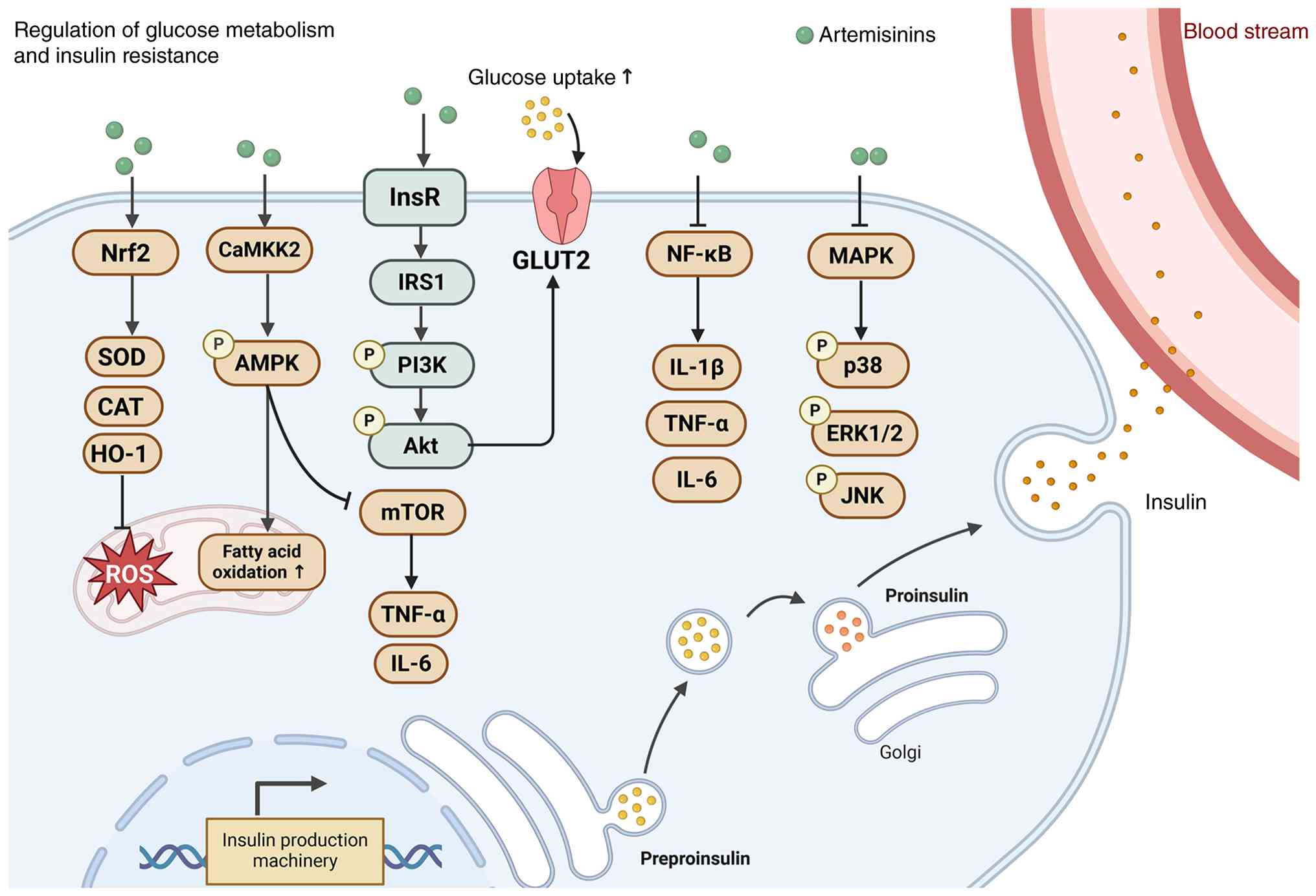 | Figure 1.Artemisinin and its derivatives
enhance glucose uptake and fatty acid oxidation via the CaMKK2/AMPK
and PI3K/Akt pathways, while suppressing MAPK-driven inflammation
to improve insulin sensitivity. Artemisinin and its derivatives
activate CaMKK2/AMPK to promote glucose uptake and fatty acid
oxidation and stimulate PI3K/Akt to facilitate glucose transport
and glycogen synthesis. Artemisinin and its derivatives also
inhibit MAPK signaling by reducing p38, ERK1/2 and JNK
phosphorylation, attenuating insulin resistance. Additionally,
artemisinin and its derivatives activate Nrf2 to upregulate
antioxidant enzymes and support insulin production. This figure
provides a general overview of the shared mechanisms of artemisinin
and its derivatives across multiple tissues. Created with
BioRender.com. AMPK, AMP-activated protein kinase; CaMKK2,
calcium/calmodulin-dependent protein kinase kinase 2; CAT,
catalase; GLUT2, glucose transporter type 2; HO-1, heme
oxygenase-1; InsR, insulin receptor; IRS1, insulin receptor
substrate 1; Nrf2, nuclear factor erythroid 2-related factor 2;
p38, p38 mitogen-activated protein kinase; ROS, reactive oxygen
species; SOD, superoxide dismutase. |
Liver
In db/db mice, artemisinin activates the hepatic
PI3K/Akt pathway, upregulating phosphorylated (p-)PI3K, p-Akt,
insulin receptor substrate 1 (IRS1) and glucose transporter type 2
(GLUT2) to enhance glucose uptake and suppress gluconeogenesis.
Additionally, artemisinin suppresses MAPK signaling by decreasing
phosphorylation of p38 mitogen-activated protein kinase (p38),
ERK1/2 and JNK, thus alleviating inflammation- and oxidative
stress-triggered insulin resistance (48). Artemisinin also inhibits aberrant
glycolysis through regulation of the PI3K/Akt/mTOR axis, thereby
preserving metabolic homeostasis. In the liver, artemisinin
improves insulin sensitivity and reduces ectopic lipid deposition
via the AMPK/nuclear factor erythroid 2-related factor 2 (Nrf2)
pathway. At the cellular level, in hepatocytes, artemisinin
potentiates IRS1 activity and PI3K/Akt signaling, promotes glucose
transport and glycogen synthesis and suppresses the NF-κB
inflammatory pathway, leading to reduced expression levels of
pro-inflammatory cytokines such as TNF-α, IL-1β and IL-6, which
otherwise disrupt insulin signaling (49,50).
Pancreas
Multiple animal studies have demonstrated that
artemisinin-based drugs can considerably reduce fasting and
postprandial blood glucose levels (48,51).
For instance, artemether and dihydroartemether notably reduced 2 h
blood glucose levels in intraperitoneal glucose tolerance and
intraperitoneal insulin tolerance tests in a T2DM mouse model.
Artemisinin-based drugs also improved serum insulin levels,
indicating that these drugs can systemically restore glucose
tolerance and insulin sensitivity rather than merely lowering blood
glucose levels (21). Artemisinin
is hypothesized to induce pancreatic β-cell regeneration to
alleviate hyperglycemia, offering a potential strategy to restore
pancreatic endocrine function. However, controversy remains
regarding this regenerative effect, as multiple studies have failed
to replicate α-to-β cell reprogramming in mammalian islet models,
highlighting the need for further mechanistic validation (52).
Adipose tissue
Consistent with the effect of artemether on glycemic
control, in a high-fat diet (HFD)-induced obese mouse model,
artemether effectively reduced body weight and enhanced systemic
insulin sensitivity and glucose tolerance, suggesting that the
hypoglycemic effect of artemether reflects a direct improvement in
insulin signaling rather than a mere glucose-lowering action
(20).
Systemic metabolic tissues
At the systemic level, artemisinin indirectly
improves the microenvironment associated with insulin resistance by
inhibiting the mTOR pathway, downregulating pro-inflammatory
factors (such as TNF-α and IL-6) and alleviating oxidative stress
(53,54). Artemisinin also activates the AMPK
pathway by upregulating calcium/calmodulin-dependent protein kinase
2, thereby promoting AMPK phosphorylation and consequently
enhancing glucose uptake and fatty acid oxidation. By interacting
with the Nrf2 antioxidant axis, artemisinin activates downstream
antioxidant enzymes, such as superoxide dismutase (SOD), catalase
and heme oxygenase-1, thereby mitigating the damage from oxidative
stress induced by high glucose or drug treatments, such as
amiodarone-triggered insults in human epithelial cell models.
Nrf2-knockdown experiments further confirmed the necessity of
artemisinin in maintaining insulin signaling (55,56).
Non-MetS disease models
Of note, in uveal melanoma cells, artemisinin exerts
an opposing effect on the PI3K/Akt/mTOR axis: Artemisinin inhibits
the phosphorylation of PI3K, Akt and mTOR, and Akt/mTOR activators
can counteract the inhibitory effect of artemisinin on cell
migration, demonstrating that artemisinin suppresses cancer cell
migration through inhibition of this pathway, highlighting that
artemisinin serves as a context-dependent modulator, exerting
differential regulatory effects across distinct tissues and
pathological states (57).
Given these findings, high-quality clinical studies
are needed to clarify the hypoglycemic and insulin-sensitizing
efficacy and safety of artemisinin-based drugs in patients with
MetS. Artemisinin-based combination regimens should also be
explored with metformin and statins to enhance therapeutic
efficacy, facilitating the translation of artemisinin derivatives
into potential therapeutic agents for MetS.
Regulation of lipid metabolism and
hepatic steatosis
Dyslipidemia and hepatic steatosis are interrelated
and together constitute a vicious cycle of lipid overload-insulin
resistance-chronic inflammation-liver damage (58,59).
Artemisinin and its derivatives inhibit hepatic lipid synthesis and
promote fatty acid oxidation through effects on several pathways,
correcting imbalances involving lipid absorption, synthesis and
transport. By modulating the gut-liver axis, artemisinin and its
derivatives improve lipid profiles while exerting anti-inflammatory
and antioxidant effects and restoring insulin signaling pathways,
comprehensively ameliorating lipid metabolism disorders and
reversing hepatic steatosis (Fig.
2).
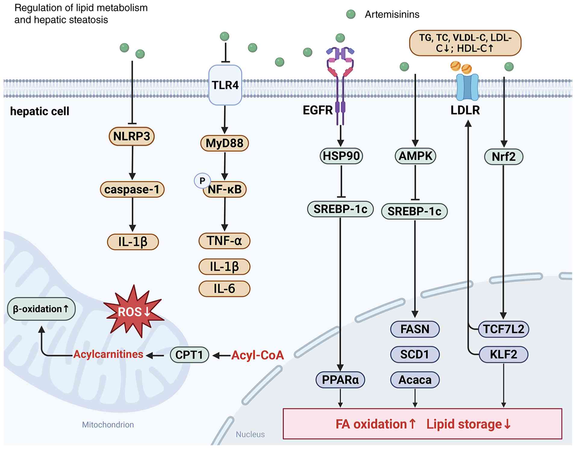 | Figure 2.Artemisinin and its derivatives
ameliorate hepatic steatosis by activating the AMPK/SREBP-1c and
PPARα pathways to suppress de novo lipogenesis while
concurrently enhancing fatty acid oxidation and reducing lipid
storage. Artemisinin and its derivatives also inhibit
TLR4/MyD88/NF-κB and NLRP3 inflammasome signaling to attenuate
hepatic inflammation. Collectively, these actions lower serum
lipids, restore lipid homeostasis and alleviate hepatic steatosis.
Created with BioRender.com. Acaca, acetyl-CoA carboxylase α; AMPK,
AMP-activated protein kinase; CPT1, carnitine palmitoyltransferase
1; FASN, fatty acid synthase; HDL-C, high-density lipoprotein
cholesterol; HSP90, heat shock protein 90; KLF2, Krüppel-like
factor 2; LDL-C, low-density lipoprotein cholesterol; LDLR,
low-density lipoprotein receptor; MyD88, myeloid differentiation
primary response 88; NLRP, nucleotide-binding oligomerization
domain, leucine-rich repeat and pyrin domain-containing protein;
Nrf2, nuclear factor erythroid 2-related factor 2; PPARα,
peroxisome proliferator-activated receptor α; ROS, reactive oxygen
species, SCD1, stearoyl-CoA desaturase 1; SREBP-1c, sterol
regulatory element-binding protein 1c; TC, total cholesterol;
TCF7L2, transcription factor 7-like 2; TG, triglyceride; TLR4,
Toll-like receptor 4; VLDL-C, very low-density lipoprotein
cholesterol. |
Liver
In the liver, artemether can reduce the endogenous
production of fatty acids and cholesterol by activating the AMPK
signaling pathway to inhibit sterol regulatory element-binding
protein-1c (SREBP-1c) and its downstream key lipogenic enzymes
(acetyl-CoA carboxylase α, fatty acid synthase and stearoyl-CoA
desaturase 1), suggesting that artemether can reduce hepatic lipid
accumulation by inhibiting excessive lipid synthesis at the source
(60). Meanwhile, artesunate
notably reduces serum triglyceride (TG) levels and increases
high-density lipoprotein cholesterol (HDL-C) in apolipoprotein
E-knockout (ApoE−/−) mice induced by a HFD
(61). Regarding the reparative
effects on hepatic steatosis, artemether targets the EGFR/heat
shock protein 90 (HSP90) complex and upregulates the
PPARα/carnitine palmitoyltransferase 1 (CPT1) axis to promote fatty
acid β-oxidation, thereby regulating hepatic lipid metabolism and
alleviating NASH (60,62,63).
Furthermore, artemether can modulate the gut-liver axis, reshape
the gut microbiota structure and reduce endotoxemia. By inhibiting
the TLR4/NF-κB pathway, artemether blocks activation signals in
Kupffer cells, reducing their secretion of pro-inflammatory factors
such as TNF-α, IL-1β and IL-6. This action effectively interrupts
the amplification of the hepatic inflammatory cascade, ultimately
alleviating inflammatory liver damage in NASH (64). Beyond the aforementioned
lipid-modulating effects, artemisinin derivatives regulate
peroxisome proliferator-activated receptor γ (PPARγ)-mediated lipid
metabolic pathways to rebuild the homeostasis of hepatic lipid
metabolism and inflammation. Such combined effects ameliorate
hepatic lipid deposition and metabolic inflammation synchronously
(65). Distinct from these
artemether-mediated mechanisms, DHA activates the Nrf2 pathway,
inhibits ferroptosis, improves mitochondrial function and
indirectly promotes lipid droplet clearance, alleviating hepatic
steatosis, inflammation and fibrosis (66).
Vascular tissue
In atherosclerosis models, artesunate reduces plasma
TG, total cholesterol (TC), low-density lipoprotein (LDL)
cholesterol (LDL-C), very-LDL-C and arterial lipid deposition
through activation of the Krüppel-like factor 2
(KLF2)/Nrf2/transcription factor 7-like 2 (TCF7L2) axis, which
enhances LDL receptor-related pathway activity (22). Artemisinin inhibits NOD-like
receptor family pyrin domain-containing 3 (NLRP3) inflammasome
activation in vascular tissue, reducing caspase-1 and IL-1β
expression and decreasing macrophage infiltration and CD68
deposition in arterial plaques. Artemisinin also suppresses NF-κB
and other pro-inflammatory signals by regulating the MAPK,
PI3K/Akt/mTOR and Nrf2/glutathione (GSH) peroxidase 4 pathways,
thereby alleviating vascular chronic inflammation and directly
attenuating atherosclerosis (54,61).
Systemic metabolic tissues
Notably, the lipid-regulating effects of artesunate
on TC and LDL appear to be model-dependent. In
ApoE−/− mice fed a HFD, artesunate markedly
reduced atherosclerotic plaque formation and serum TG levels, but
did not notably alter TC or LDL levels (61). By contrast, in a study using Wistar
rats with normal ApoE function, in which acute atherosclerosis was
induced by an atherogenic diet (1.25% cholesterol, 15% fat and 0.5%
cholic acid) combined with lipopolysaccharide (LPS), artesunate
markedly reduced TC and LDL levels, which was comparable to the
effect of rosuvastatin (67,68).
Similar lipid-lowering and anti-atherosclerotic effects of
artesunate have also been reported in HFD-fed ApoE−/−
mice and Western diet-fed rabbits (61,69).
This discrepancy may be attributed to the absence of functional
ApoE in ApoE−/− mice, as ApoE is a required
ligand for LDL receptor-mediated lipoprotein clearance, and this
genetic deficiency may limit the cholesterol-lowering efficacy of
artesunate (61,67). Artemisinin ester may also
participate in activating PPARα-related pathways and promoting
weight loss and energy expenditure, thereby indirectly improving
systemic lipid metabolism (20).
Regarding safety, while high doses of artemether may induce
gastrointestinal suppression in mice, overall preclinical data
indicate no severe adverse effects were observed in this study and
the long-term effects of artemether remain uncharacterized as they
were not explored in the aforementioned research (21).
Research on NASH models with different etiologies
(for example obesity or diabetes mellitus) has not been conducted
and clinical translation data are limited (48). Additionally, the regulatory effects
of artemether on lipid profiles yield inconsistent results across
different animal models and unified efficacy evaluation criteria
are lacking (20). Preclinical
studies and early-phase clinical trials are needed to test
artemisinin-based drugs at low doses over extended periods. These
studies should aim to refine the understanding of the
cross-regulatory network of the gut-liver axis, standardize
research models and evaluation criteria and explore the efficacy of
combination therapies with different types of artemisinin
derivatives to advance their clinical translation.
Anti-inflammatory and immune
regulatory effects
Chronic low-grade inflammation and immune
homeostasis imbalance are core pathological features of MetS that
are primarily manifested by excessive activation of innate immune
cells, dysregulation of adaptive immune responses and an imbalance
between anti- and pro-inflammatory reactions. These factors
continuously exacerbate insulin resistance and disturbances
involving glucose and lipid metabolism, forming a vicious cycle of
inflammation-metabolism (70–73).
Artemisinin and its derivatives correct the inflammation and immune
dysregulation associated with MetS by inhibiting pro-inflammatory
pathways, activating antioxidant signaling and regulating immune
cell homeostasis (Fig. 3).
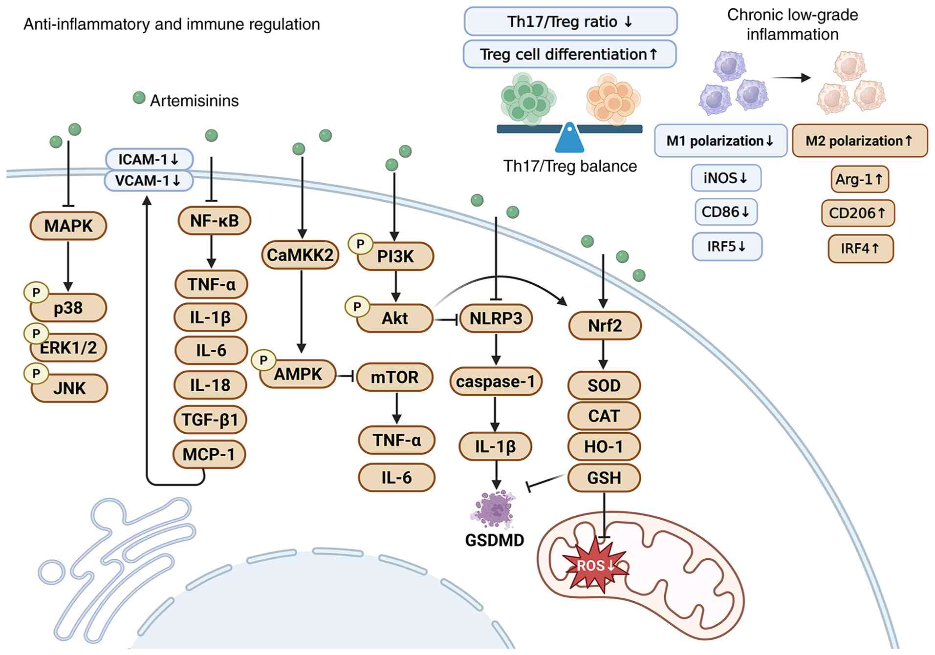 | Figure 3.Artemisinin and its derivatives
suppress chronic inflammation and restore immune homeostasis
through modulation of key signaling pathways and immune cell
differentiation. Artemisinin derivatives inhibit MAPK/NF-κB
signaling, reducing pro-inflammatory cytokine and adhesion molecule
production, while activating PI3K/Akt/mTOR and Nrf2 pathways to
reinforce antioxidant defenses. Artemisinin and its derivatives
also attenuate NLRP3 inflammasome activation, promote Treg
differentiation and drive macrophage polarization from the M1
toward the M2 phenotype. Collectively, these actions alleviate
chronic inflammation and help restore immune balance in MetS.
Created with BioRender.com. AMPK, AMP-activated protein kinase;
Arg-1, arginase 1; CaMKK2, calcium/calmodulin-dependent protein
kinase kinase 2; CAT, catalase; GSDMD, gasdermin D; GSH,
glutathione; HO-1, heme oxygenase-1; ICAM-1, intercellular adhesion
molecule-1; iNOS, inducible nitric oxide synthase; IRF4, interferon
regulatory factor 4; IRF5, interferon regulatory factor 5; MetS,
metabolic syndrome; MCP-1, monocyte chemoattractant protein-1;
NLRP, nucleotide-binding oligomerization domain, leucine-rich
repeat and pyrin domain-containing protein; Nrf2, nuclear factor
erythroid 2-related factor 2; ROS, reactive oxygen species; SOD,
superoxide dismutase; Treg, regulatory T cell; Th17, T helper 17
cell; VCAM-1, vascular cell adhesion molecule-1. |
Vascular tissue
At the molecular level, DHA suppresses NF-κB
activation by blocking upstream signaling and directly binding to
NF-κB. This dual inhibition downregulates pro-inflammatory
mediators, including TNF-α, IL-1β, IL-6, IL-18, monocyte
chemoattractant protein-1 and TGF-β1, as well as adhesion molecules
such as intercellular adhesion molecule-1 and vascular cell
adhesion molecule-1. Consequently, monocyte adhesion and migration
are attenuated and NLRP3 inflammasome activation is restrained.
These actions alleviate the chronic low-grade inflammatory immune
response in patients with MetS (22,54,74).
Furthermore, in ApoE−/− mice fed a HFD,
artemether inhibited signaling involving the ERK1/2/NF-κB/IL-1β
pathway, reduced the macrophage-like phenotypic transformation of
vascular smooth muscle cells and decreased NLRP3 and CD68
expression within plaques. Therefore, artemisinin-derived drugs
improve underlying metabolic disorders, protect blood vessels and
stabilize plaques, highlighting a potential interventional strategy
for cardiovascular complications (61).
Liver
In an NAFLD model, artemether notably alleviated
oxidative stress and inflammatory damage in hepatocytes by
activating the Nrf2 antioxidant pathway, thereby promoting Nrf2
nuclear translocation and the expression levels of downstream
antioxidant enzymes, including SOD and GSH (25).
Salivary glands
In a rat model of T2DM, artemether notably reduced
inflammatory markers such as caspase-1 and IL-1β in the salivary
glands by inhibiting the NF-κB/NLRP3 pathway, improving oxidative
stress and regulating oral dysbiosis. This indicates that the
anti-inflammatory effects of artemether are not limited to
traditional metabolic organs but can also improve systemic
multi-tissue microinflammation (74).
Systemic metabolic tissues
In db/db mice, artemisinin effectively alleviated
the inflammatory response associated with glucose and lipid
metabolism disorders by inhibiting the MAPK signaling pathway,
reducing the phosphorylation of p38, ERK1/2 and JNK and activating
the PI3K/Akt pathway (48).
Immune system
At the cellular immune level, artemisinin-based
drugs correct metabolic disorder-induced immune imbalances by
reshaping the phenotypic and functional balance of immune cells and
regulating the functions of key cells in innate and adaptive
immunity. DHA selectively regulates adaptive immune cells by
promoting CD4+ T cell proliferation and enhancing
IFN-γ+ CD8+ T cell activity, while inhibiting
germinal center B cell expansion and reducing the number of
circulating plasma cells and antibody production. Therefore, DHA
can coordinate immune activation and immune tolerance, thereby
preventing excessive immune responses from exacerbating metabolic
damage and maintaining immune homeostasis (75). Furthermore, the artemisinin-derived
compound β-aminoartemether maleate (SM934) can balance the
Th17/Treg cell ratio, promote Treg differentiation and inhibit
excessive Th17 cell activation, alleviating chronic low-grade
inflammation mediated by immune imbalance (41). Regarding the regulation of innate
immune cells, artemether upregulates interferon regulatory factor 4
(IRF4) expression in rodent and non-human primate models, promoting
the polarization of macrophages toward the anti-inflammatory M2
phenotype and inhibiting conversion to the pro-inflammatory M1
phenotype. This M2-biased polarization promotes IL-10 and TGF-β
secretion to exert its anti-inflammatory and tissue-repairing
effects while reducing the release of pro-inflammatory factors,
such as TNF-α and IL-1β, alleviating the inflammatory response in
vascular restenosis. These observations suggest that artemisinin is
not a fixed anti- or pro-inflammatory agent; artemisinin is an
immunomodulator capable of dynamically adapting to the tissue
microenvironment and pathological state (76).
Non-MetS disease models
By contrast, in patients with glioblastoma and a
number of tumor models (including models of breast, lung and
cervical cancer in vitro, as well as an in vivo
breast cancer model), artesunate binds to heme to generate ROS,
reversing tumor-associated macrophages from an M2-like,
tumor-promoting state toward an M1-like, pro-inflammatory and
antitumor state. This phenotypic shift reshapes the immune
microenvironment and potentiates antitumor immunity (77,78).
In the context of malaria infection, DHA promotes macrophage
polarization toward the M1 phenotype by activating NOD-like
receptor family pyrin domain-containing 12 (NLRP12), thereby
enhancing the host's ability to combat Plasmodium parasites
and facilitating parasite clearance (79). Collectively, these observations
demonstrate that the immunomodulatory effects of artemisinin
derivatives on macrophage polarization are context-dependent.
Rather than exerting a fixed anti- or pro-inflammatory effect,
artemisinin derivatives function as immunomodulators that
differentially regulate macrophage phenotypes depending on the
pathological setting, tissue microenvironment and disease
context.
However, the poor water solubility and short
half-lives of artemisinin derivatives make it difficult to maintain
sustained effects for chronic inflammation (80,81).
Furthermore, the immunomodulatory effects of artemisinin-based
drugs are dose- and microenvironment-dependent. In a malaria model,
DHA induced M1 macrophage polarization via an NLRP12-dependent
mechanism yet promoted M2 polarization in metabolic inflammation;
the critical threshold for this macrophage phenotypic switch
remains undefined (79,82). Future studies should focus on
developing long-acting sustained-release or targeted delivery
systems to meet the needs of chronic interventions. For instance,
apigenin-loaded nanoparticles nanocarriers may enhance the
macrophage targeting and M2 polarization regulation effects of
artemisinin-based drugs (83).
Regulation of the gut
microbiota-metabolism axis
Dysregulation of the gut microbiota-metabolism axis
is a key pathogenic mechanism underlying MetS. The
gut-microbiota-metabolism axis mediates critical processes such as
bile acid reprogramming and tryptophan metabolism through
bi-directional pathways, including gut-liver and gut-spleen
interactions. Disruption of the gut-microbiota-metabolism axis
leads to abnormal metabolites, including secondary bile acids,
hydrogen sulfide and dysregulated short-chain fatty acids, which
are transmitted via the gut-systemic metabolic axis, severely
affecting the host's metabolic and immune balance (84–88).
Artemisinin and its derivatives have been reported to modulate this
axis, with effects associated with microbiota-dependent pathways.
By reshaping the gut microbiota structure, artemisinin and its
derivatives optimize the profile of microbial metabolites, regulate
the bile acid signaling axis, strengthen the barrier function of
the gut-liver axis, alleviate intestinal inflammation and
ultimately contribute to the restoration of gut microbiota
homeostasis (Fig. 4).
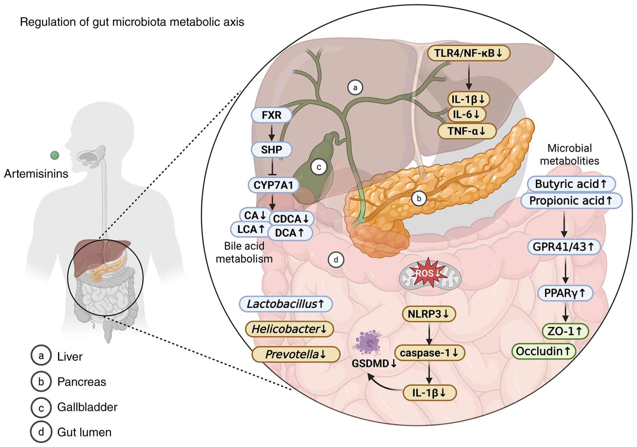 | Figure 4.Artemisinin and its derivatives
preserve intestinal barrier integrity by upregulating tight
junction proteins (ZO-1 and occludin) and boosting short-chain
fatty acids (butyrate and propionate) via GPR41/43 and PPARγ, while
regulating FXR/CYP7A1-mediated bile acid metabolism and restraining
endotoxin translocation to protect hepatic function through the
gut-liver axis. Notably, gut microbiota-related pathways in this
schematic are merely correlational, lacking rigorous causal
validation. Created with BioRender.com. CA, cholic acid; CDCA,
chenodeoxycholic acid; CYP7A1, cholesterol 7α-hydroxylase; DCA,
deoxycholic acid; FXR, farnesoid X receptor; GPR41/43, G
protein-coupled receptor 41/43; GSDMD, gasdermin D; LCA,
lithocholic acid; NLRP, nucleotide-binding oligomerization domain,
leucine-rich repeat and pyrin domain-containing protein; PPARγ,
peroxisome proliferator-activated receptor γ; ROS, reactive oxygen
species; SCFA, short-chain fatty acid; SHP, small heterodimer
partner; ZO-1, zonula occludens-1. |
Intestine
One animal study has shown that artemisinin
derivatives are associated with alterations in gut microbiota
composition. DHA enhances the α-diversity of the gut microbiota and
reshapes the microbial ecosystem structure in mice without causing
pathological damage to the liver, kidneys or intestines. DHA
increases the abundance of Firmicutes and Spirochaetes while
reducing the proportion of Bacteroidetes and Actinobacteria, and
these compositional shifts are accompanied by anti-inflammatory and
metabolic regulatory effects at the functional level. Metabolic
analysis has shown that DHA reduces serum TG levels, with TG
content exhibiting a positive correlation with the abundance of
Deferribacterota and a negative correlation with the abundance of
Spirochaeta, suggesting that the lipid-regulating effects of
DHA are partially dependent on gut microbiota remodeling (89). Conversely, artemether increases the
abundance of Lactobacillus while reducing the abundance of
pathogenic bacteria such as Helicobacter and
Prevotella. These changes are associated with AMPK
activation, inhibition of the mTOR/NF-κB pathway, reduced
pro-inflammatory factor levels and improved insulin resistance and
liver and pancreatic tissue damage, suggesting a potential
‘microbiota-metabolism-immunity’ axis that may contribute to the
effects of artemisinin-based drugs in the prevention and treatment
of T2DM (90). At the molecular
level, several pathways have been proposed to link
artemisinin-based drugs to the gut microbiota-metabolism axis, with
the tryptophan metabolic pathway being among the most studied.
Artemether can inhibit the tryptophan-kynurenine metabolic pathway
via a heme-dependent mechanism, blocking indoleamine
2,3-dioxygenase 1 (IDO1) activity and reducing kynurenine
production. By binding to the heme-binding pocket of IDO1 to
inhibit enzyme activity, artemether alleviates kynurenine-mediated
inflammation and improves the redox balance of intestinal cells
(91). DHA increases the abundance
of butyrate-producing bacteria and butyrate levels, whereas
artemether increases the content of short-chain fatty acids such as
propionate and valerate. One potential mechanism is that these
short-chain fatty acids activate G protein-coupled receptor 41/43
(GPR41/43) receptors and the PPARγ pathway, which in turn enhances
the expression levels of tight junction proteins, such as zonula
occludens-1 and occludin, in the colon and reduces intestinal
permeability. This could prevent endotoxins (for example LPS) from
entering the bloodstream via the gut and if so, might suppress
chronic low-grade inflammation mediated by the gut-metabolic axis
and thereby improve insulin resistance (40,90,92).
Liver
Within the framework of the gut
microbiota-metabolite axis, the gut-liver axis acts as an important
signaling conduit that transmits microbiota-derived metabolic
signals to the liver, and represents a plausible route through
which artemisinin and its derivatives exert hepatoprotective
effects. In terms of liver protection, DHA, as a farnesoid X
receptor (FXR) agonist, downregulates the expression level of
cholesterol 7α-hydroxylase (CYP7A1), which is a key rate-limiting
enzyme in the conversion of cholesterol to bile acids, via FXR
activation. Through this mechanism, DHA could mitigate the liver
damage caused by excessive bile acid accumulation, alleviate
hepatic steatosis and holistically protect liver function while
controlling obesity (93–97). Together, these pathways could
conceivably constitute a microbiota-metabolite-systemic immunity
tripartite regulatory network, enabling artemisinin-based drugs to
modulate the gut microbiota-metabolite axis.
Spleen
Circulating metabolites originating from gut
microbiota can mediate systemic immune regulation, affecting
peripheral immune organs such as the spleen. At the immune
regulatory level, artemether can inhibit the activation of
pro-inflammatory macrophages, modulate the splenic immune
microenvironment and immune cell function and restore host immune
homeostasis (98).
Notably, the microbiota-related mechanisms discussed
are predominantly correlational and the specific contributions of
gut microbiota remodeling to the metabolic effects of artemisinin
derivatives remain to be causally established. Causal evidence for
microbiota dependence remains tenuous, as direct validation using
germ-free animals, antibiotic depletion studies or fecal microbiota
transplantation have not yet been reported. Clinical translation is
still in its infancy, and the relative merits of distinct
artemisinin derivatives in modulating the gut-liver axis remain
unclear (99). Future research
should prioritize exploratory clinical trials built on the
microbiota-metabolite-immune axis to evaluate the long-term impacts
of treatment on gut microbiome stability.
Evidence grading and mechanism
validation summary
Evidence stratification criteria
Evidence is tiered according to the type of
experimental disease models employed. Direct metabolic disease
models, namely db/db mice, T2DM rodents and HFD-induced obese mice,
generate the most robust evidence to validate the metabolic
regulatory functions of artemisinin derivatives. Data obtained from
NAFLD/NASH and atherosclerosis models offer moderate supporting
evidence; these disorders share partial pathological features with
MetS yet come with intrinsic limitations due to incomplete
pathological overlap. Mechanistic clues derived from cancer and
malaria models remain indirect and preliminary, and further
validation in metabolic animal models is mandatory before
definitive conclusions related to MetS treatment can be
established.
Reliability grading of functional
pathways
Multiple pathways governing the metabolic effects of
artemisinin derivatives are fully validated in db/db mice,
HFD-induced obesity models and T2DM animals. The hepatic-skeletal
muscle PI3K/Akt-GLUT2/GLUT4 axis optimizes glucose metabolism and
suppresses gluconeogenesis, as demonstrated in db/db mice and
HFD-induced obesity models (48,100,101). AMPK/Nrf2 alleviates oxidative
stress-induced insulin resistance, the MAPK/NF-κB pathway breaks
the inflammation-insulin resistance cycle and IRF4 reduces systemic
inflammation by inducing M2 macrophage polarization, with these
mechanisms supported by studies in human retinal pigment epithelial
cells, bronchial epithelial cells, Nrf2-knockdown experiments, and
rodent and non-human primate models of vascular restenosis
(55,56,76).
Collectively, these findings constitute direct evidence, as they
are derived from both metabolic disease models and mechanistic
loss-of-function studies, thereby supporting the classification of
these pathways as well-supported.
A second set of pathways are supported by moderate
evidence, characterized only in NAFLD,
ApoE−/− atherosclerotic and other analogous
models without systematic verification in MetS, resulting in
inconsistent efficacy across species. Hepatic AMPK-SREBP-1c
inhibits lipogenesis with variable lipid-lowering effects, as
observed in methionine- and choline-deficient (MCD) diet-fed and
HFD-fed murine NASH models (60).
The gut-liver FXR/CYP7A1 pathway improves hepatic steatosis but
lacks long-term validation in obese MetS models, with evidence
derived primarily from studies using hepatocellular carcinoma cells
and conventional rodent models (93–97).
Vascular KLF2/Nrf2/TCF7L2 prevents atherosclerosis with unconfirmed
hypoglycemic activity, as demonstrated in
ApoE−/− mice fed a HFD (22). Hepatic TLR4/NF-κB suppresses
inflammation in Kupffer cells within NASH models, yet its link to
gut microbiota remains unclear, and PPARα/CPT1 combined with
EGFR/HSP90 alleviates fatty liver with poor experimental
reproducibility, based on findings in MCD diet-fed mice and oleic
acid/palmitic acid-treated HepG2 cells (60,62–64).
The remaining pathways are supported only by
indirect speculative evidence, among which certain mechanisms are
summarized from malaria and tumor research. Most intestinal-related
mechanisms are also grouped in the present review, including the
microbiota-metabolism-immune network, the
tryptophan-kynurenine-IDO1 cascade and the SCFA-GPR41/43-PPARγ
intestinal barrier axis, as these are backed solely by
correlational results without causal validation using germ-free
animals or fecal microbiota transplantation (89–92).
Moreover, the inverse regulation of PI3K/Akt/mTOR detected in
melanoma cells highlights the context-dependent pharmacological
properties of artemisinin derivatives, indicating that findings
from non-metabolic disease models require further validation before
extrapolation to metabolic tissue (57).
Clinical applications and emerging
therapeutic strategies for artemisinin and its derivatives
Artemisinin and its derivatives have a
well-established role in malaria treatment, but their translation
to MetS requires a distinct clinical framework focused on metabolic
and inflammatory endpoints.
Candidate derivatives and formulation
optimization
Among artemisinin derivatives, SM934 represents not
only a formulation-optimized derivative with improved aqueous
solubility but is also a candidate with preclinical evidence in
MetS. In HFD-induced obese mice, SM934 reduced body weight gain,
improved glucose tolerance and insulin sensitivity and decreased
ectopic lipid deposition in the liver. Mechanistically, SM934
inhibits α-enolase to lower phosphoenolpyruvate (PEP). PEP
stabilizes transforming growth factor-β-activated kinase 1 to drive
NF-κB-dependent inflammation, SM934 blocks this cascade, curbing
macrophage IL-1β secretion and lowering the adipose M1/M2 ratio to
relieve obesity-linked meta-inflammation (102). Meanwhile, SM934 activates Nrf2
and represses NF-κB/NLRP3 inflammasome signaling to restore overall
immune homeostasis (103). Phase
I trials have supported the favorable tolerability profile of SM934
in healthy participants (registered under CTR20160932 and
CTR20160933 in the Drug Clinical Trial Registration and Information
Disclosure Platform; available a www.chinadrugtrials.org.cn), and a Phase II trial in
patients with systemic lupus erythematosus is currently underway
(41). In addition, SM934 has
demonstrated notable efficacy in various autoimmune and
inflammatory disease models, including systemic lupus
erythematosus, rheumatoid arthritis, inflammatory bowel disease,
membranous nephropathy and dry eye syndrome (41).
Target patient phenotypes and clinical
endpoints
MetS encompasses a heterogeneous patient population
and clinical development should prioritize well-defined subgroups
with unmet needs. Polyendocrine metabolic ovarian syndrome (PMOS)
represents one of the most advanced clinical targets. A single-arm
pilot study in 19 patients with PMOS demonstrated that 40 mg DHA
three times daily for 12 weeks normalized menstrual cycles in
63.16% of participants, while also lowering total testosterone and
reducing antral follicle count (104). Metabolic dysfunction-associated
fatty liver disease (MAFLD) is another priority target. A
proof-of-concept trial (NCT07679542) is assessing whether DHA (20
mg three times daily for 12 weeks) reduces liver fat in adults with
MAFLD, using MRI-proton density fat fraction-assessed hepatic fat
content as the primary endpoint (105). These ongoing proof-of-concept
studies in defined patient cohorts represent the initial steps
toward a dedicated clinical development pathway for MetS. Candidate
clinical endpoints for MetS interventions should include enhanced
insulin sensitivity, as assessed by the homeostatic model
assessment of insulin resistance, improved glycemic control
quantified by glycated hemoglobin A1c, favorable lipid
modifications (TG, LDL-C and HDL-C), decreased inflammatory markers
(IL-6 and TNF-α) and ultimately, lowered cardiovascular event risk
or arrested T2DM progression.
Dosing, duration and long-term
safety
Chronic MetS management requires different dosing
strategies from acute malaria treatment. Evidence from a
preclinical study suggests that long-term drug exposure, rather
than short-term peak concentrations, is the primary driver of
artemisinin-related toxicity (106). Rapid oral elimination is
therefore associated with a more favorable safety profile compared
with delayed release from intramuscular formulations (107). Nevertheless, a 15-year
retrospective analysis of artemether-related adverse events from
the FDA Adverse Event Reporting System identified hemolytic anemia
and hemolysis as the most frequently reported events, with women
showing a higher incidence of reproductive-related events,
including spontaneous abortion and premature labor. Most adverse
events occurred within the first 30 days of artemether
administration, with the most affected systems being the
hematologic and hepatobiliary systems (108). To date, most clinical trials of
artemisinin derivatives have been limited to small-sample Phase I
malaria studies. Larger-scale Phase II/III trials are therefore
needed to comprehensively evaluate both efficacy and long-term
safety in metabolic populations. These findings underscore the need
for systematic long-term safety monitoring in future MetS trials,
including hematological, hepatic and reproductive assessments.
Drug-drug interactions with standard
MetS medications
Patients with MetS frequently receive multiple
concomitant medications, including metformin, statins, SGLT2
inhibitors, glucagon-like peptide-1 (GLP-1) receptor agonists and
antihypertensives. Artemisinin and its derivatives are primarily
metabolized by cytochrome P450 2B6 (CYP2B6) and cytochrome P450 3A
(CYP3A). Artemisinin can induce CYP2B6 and CYP3A4 protein
expression, raising the potential for pharmacokinetic interactions
with co-administered drugs metabolized by these pathways (109). Reduced enzyme function may lead
to subtherapeutic concentrations of the active metabolite DHA,
potentially compromising treatment efficacy. Interactions with
cardiovascular, antibiotic and antiparasitic agents have been
reported, and clinicians should remain vigilant for potential
drug-drug interactions when repurposing these agents for chronic
metabolic indications (110).
Systematic evaluation of interactions with standard MetS
medications (including metformin, statins, SGLT2 inhibitors and
GLP-1 receptor agonists) are therefore essential before clinical
adoption, with careful attention to patient stratification and
drug-drug interaction monitoring.
Antimalarial resistance risks
Expanding artemisinin use for non-malarial
indications raises legitimate concerns about accelerating
antimalarial resistance. Evidence suggests that prolonged or
widespread use in malaria-endemic regions could exert selection
pressure on Plasmodium parasites (33). To mitigate this risk,
non-antimalarial derivatives with altered metabolic profiles, such
as SM934, which exhibits distinct immunosuppressive mechanisms
independent of the antimalarial activity of SM934, or formulations
with shorter exposure windows should be prioritized for MetS
indications. Additionally, combination regimens analogous to triple
artemisinin combination therapies could be considered to reduce
resistance selection pressure in chronic use settings (111). These efforts, combined with
robust resistance surveillance, will determine whether artemisinin
derivatives can successfully transition from antimalarial agents to
viable therapeutics for MetS.
Conclusion and outlook
The global prevalence of MetS shows a sustained and
rapid increasing trend. Between 2000 and 2023, MetS prevalence
between women and men globally rose from 14.7 to 31.0% and 9.0 to
25.7%, respectively. As of 2023, ~1.54 billion adults worldwide
were affected by MetS (112).
This condition increases the risk of T2DM, cardiovascular disease,
NAFLD and various types of cancer and poses a severe challenge to
global public health (113,114). Given the complex nature of MetS,
artemisinin and its derivatives have attracted research attention
as potential preclinical candidates, largely due to their ability
to affect multiple disease-related pathways. Their mechanisms
include improving insulin resistance, regulating glucose and lipid
metabolism, alleviating chronic inflammation and restoring gut
microbiota homeostasis. These effects involve signaling pathways
including molecules, such as AMPK, PI3K/Akt, NF-κB and MAPK, and
gene regulation, such as SREBF1, PPARA, SLC2A2 and IRS1. Therefore,
downregulating the expression levels of proteins such as TNF-α,
IL-1β, IL-6 and caspase-1, upregulating IL-10 and TGF-β, and
regulating macrophage M1/M2 polarization, Th17/Treg
differentiation, tryptophan metabolism, short-chain fatty acid
production and the bile acid FXR/Takeda G protein-coupled receptor
5 signaling axis is key. Tables I
and II summarize the roles of
artemisinin and its derivatives in MetS-related models (Table I presents direct evidence derived
from MetS-related models, whereas Table II summarizes indirect mechanistic
evidence obtained from non-MetS models).
 | Table I.Roles of artemisinin and its
derivatives in MetS-related models: Direct evidence. |
Table I.
Roles of artemisinin and its
derivatives in MetS-related models: Direct evidence.
| First author/s,
year | Compound | Disease model | Species/cell
type | Dose, route and
duration | Endpoint
(metabolic/inflammatory) | Proposed
mechanism | Evidence level | Limitation | (Refs.) |
|---|
| Guo et al,
2024 | Artesunate | Obesity | Male C57BL/6J mice
(HFD-induced obese, DIO) | 5 or 20 mg/kg,
i.p., once every 2 days, 13–60 days | Metabolic: Body
weight ↓; food intake ↓; glucolipid metabolism ↑; insulin
sensitivity ↑. | GDF15 ↑; CHOP ↑;
c-Fos+ ↑ | Strong | Only male mice
tested; immune endpoints not evaluated | (20) |
|
|
|
|
|
| Inflammatory: Not
assessed |
|
|
|
|
| Wu et al,
2022 | Artemisinin | T2DM | SD rats; Wistar
rats; db/db mice | 25-300 mg/kg/day,
i.g. or i.p., 4–12 weeks | Metabolic: Glycemic
control ↑; insulin secretion ↑; oxidative stress ↓. | Arx ↓; Nrf2 ↑;
TNF-α/IL-6/MCP-1 ↓ | Strong | α-to-β cell
trans-differentiation remains | (21) |
|
|
|
|
|
| Inflammatory: |
|
| controversial |
|
|
|
|
|
|
| Pro-inflammatory
cytokines ↓ |
|
|
|
|
| Wu et al,
2022 | Artesunate | T2DM | SD rats; Wistar
rats; db/db mice | 10-100 mg/kg/day,
i.g. or i.p., 4–12 weeks | Metabolic: Blood
glucose ↓; pancreatic β-cell function ↑. Inflammatory: Inflammatory
signaling ↓ | SIRT1 ↑; NF-κB ↓;
BCL-2/BCL-X ↓, BAX ↑ | Strong | Broad dose range;
optimal dose not established | (21) |
| Wu et al,
2022 | Artemether | T2DM | db/db mice | 100-400 mg/kg/day,
i.g., 4 weeks | Metabolic: Fasting
blood glucose ↓; glucolipid metabolism ↑. | AMPK ↑; GLUT4/IRS1
↑ | Strong | Immune endpoints
not evaluated | (21) |
|
|
|
|
|
| Inflammatory: Not
assessed |
|
|
|
|
| Majidiani et
al, 2025 | Artemisinin |
Athero-sclerosis |
ApoE−/− mice,
HFD | 50 or 100 mg/kg,
i.g., 8 weeks | Metabolic: Serum
lipid profile improvement. Inflammatory: Macrophage inflammation
↓ | AMPK ↑; mTOR ↓;
ULK1 ↓; p62 ↓; LC3-II ↑; NF-κB ↓; NLRP3 ↓; IL-1β/IL-18/TGF-β1
↓ | Moderate |
ApoE−/− background
may limit translation to normal lipid metabolism | (22) |
| Chen et al,
2024 | Artesunate | T2DM | C57BL/KsJ-db/db
mice | 160 mg/kg/day,
i.g., 7 weeks | Metabolic: Fasting
glucose ↓; fasting insulin ↓; insulin resistance ↓; serum lipids ↑;
hepatic steatosis ↓. Inflammatory: Not directly assessed | PI3K/Akt ↑; MAPK
↓ | Strong | Single dose tested;
no inflammation-specific endpoint reported | (48) |
| Xu et al,
2022 | Artemether | NASH | In vivo:
C57BL/6J mice (MCD diet 4 week + HFD 28 week) In vitro:
HepG2 cells treated with oleic acid and palmitic acid | In vivo:
MCD: 100 or 200 mg/kg/day, i.g., 2 week; HFD: 20 mg/kg/week, i.p.,
7 week; In vitro: 6.25 or 25 µM, 24 h | Metabolic: Serum
transaminases ↓; hepatic steatosis ↓. Inflammatory: Hepatic
inflammation ↓; pro-inflammatory cytokines ↓; IL-10 ↑ |
SREBP-1c/ACC/FASN/SCD1 ↓;
ATGL/PGC-1α/CPT-1a ↑; NF-κB/TNF-α/IL-1β/M1 ↓; IL-10 ↑
TGF-β/α-SMA/Smad2 ↓ | Moderate | MCD diet model may
not fully recapitulate human NASH; no clinical validation | (60) |
| Liu et al,
2024 | Artesunate |
Athero-sclerosis | In vivo:
ApoE−/−mice, HFD; In vitro:
HVSMCs | In vivo: 10
mg/kg/day, i.p., 12 week; In vitro: 50–100 µM | Metabolic: Serum
lipids ↑ (TG, TC, VLDL-C, LDL-C ↓). Inflammatory: Plaque
inflammation ↓; inflammasome activation ↓ |
NLRP3/caspase-1/IL-1β ↓; MAPK/NF-κB/AP-1
↓; HPSE/MMP9/KLF4 ↓ | Moderate |
ApoE−/− background;
no systemic inflammatory markers assessed | (61) |
| Jiang et al,
2025 | Artemether | T2DM | C57BL/KsJ-db/db
mice | 80 or 160
mg/kg/day, i.g., 8 weeks | Metabolic: Blood
glucose ↑; insulin sensitivity ↑; serum lipids ↑; liver/pancreas
pathology ↑. Inflammatory: Inflammatory response ↓; NF-κB signaling
↓ | AMPK ↑; mTOR ↓; p38
MAPK ↓; NF-κB ↓; IκBα ↓; IRS1 ↓; TLR4 ↓; Occludin ↑; ZO-1/Claudin-1
↑ | Strong | Only one resistant
model; no human data | (90) |
| Table II.Roles of artemisinin and its
derivatives in non-MetS models: Indirect mechanistic evidence. |
Table II.
Roles of artemisinin and its
derivatives in non-MetS models: Indirect mechanistic evidence.
| First author/s,
year | Compound | Disease model | Species/cell
type | Dose, route and
duration | Endpoint
(metabolic/inflammatory) | Proposed
mechanism | Evidence level | Limitation | (Refs.) |
|---|
| Long et al,
2024 | DC32 (DHA
derivative) | Rheumatoid
arthritis | DBA/1 mice,
collagen-induced arthritis model | 25 mg/kg, oral, 30
days | Metabolic: Not
assessed. Inflammatory: Joint inflammation ↓, cartilage/bone
destruction ↓, Treg/Th17 balance restored | Nrf2/HO-1 ↑; p62 ↑;
Keap1 ↓; RORγt ↓; Foxp3 ↑; IL-6/IL-17 ↓ | Indirect | Mechanistically
suggestive but not in MetS context; no metabolic endpoints
evaluated | (54) |
| Li et al,
2022 | DHA | Malaria | BALB/c or C57BL/6
mice infected with P. berghei ANKA | 200 µl/day of 0.1
mg/ml DHA solution, i.g., intermittent preventive treatment | Metabolic: Not
assessed. Inflammatory: Anti-malarial cellular immunity ↑; peak
parasitemia delayed | MAPK ↑; CD25 ↑;
Tim-3 ↓ | Indirect | Malaria model not
relevant to MetS; no metabolic endpoints evaluated | (75) |
| Li et al,
2025 | DHA | Malaria | In vitro:
RAW264.7 macrophages + iRBC In vivo: C57BL/6 mice infected
with P. berghei ANKA | In vitro:
700 µM DHA (co-incubated with iRBC) In vivo: 2 mg/mouse oral
DHA, 2 days starting 12 h post-infection | Metabolic: Not
assessed. Inflammatory: M1 macrophage polarization ↑, parasite
clearance ↑ | NLRP12 ↑; M1 ↑
(CD86/i NOS/IRF5); M2 ↓ (Arg1/CD206/IRF4); IL-12/IL-6/MCP-1/TNF-α
↑; TSPO ↓; Lcn2 ↑ | Indirect | Opposite macrophage
polarization direction compared with metabolic contexts; MetS
relevance unclear | (79) |
| Guo et al,
2022 | DHA | Hepatocellular
carcinoma | In vitro:
HepG2, HepG2215 cells In vivo: C57BL/6 mice
(DEN/TCPOBOP-induced HCC model) | In vitro:
21.5 µM, 24 h; In vivo: 25 mg/kg, i.p. | Metabolic: Liver
histopathology ↑; HCC proliferation ↓ Inflammatory: Not
assessed | FXR ↑; YAP1 ↓;
CYP7A1 ↓; NR5A2 (LRH-1) ↓; CYP27A1 ↓; CA ↓; CDCA ↓ | Indirect | Cancer model not
MetS-relevant; FXR activation may be context-dependent | (93) |
Although artemisinin and its derivatives have
demonstrated potential in the treatment of MetS, several
limitations remain. First, regarding low bioavailability,
artemisinin-based drugs generally have poor water solubility and
variable oral absorption, leading to insufficient systemic exposure
and hindering their full therapeutic potential. Second, chronic
dosing regimens for MetS remain uncertain. Unlike the short-course,
high-dose regimens used for malaria treatment, MetS would require
long-term, sustained administration. The appropriate dose,
frequency and duration of treatment have not been established, and
the pharmacokinetic-pharmacodynamic relationships under chronic
dosing conditions remain poorly understood. Third, there is a lack
of high-quality, MetS-specific clinical trials, with most studies
focusing on the cellular and animal experimental stages, whereas
clinical research consists primarily of single-arm exploratory
trials with small samples, and large-scale, controlled clinical
trials are lacking. Such trials would be needed to validate the
long-term efficacy and safety of artemisinin-based drugs. Fourth,
long-term safety profiles remain unclear. Studies have largely
focused on short-term efficacy observations and systematic
evaluations of potential toxicity. Delayed adverse reactions
following repeated use or long-term exposure have not been
conducted. Furthermore, in various animal studies, the therapeutic
effects of artemisinin and its derivatives vary with dose.
Relatively low doses may not produce toxicity, which is crucial for
determining a safe and effective dosage range. Fifth, possible
drug-drug interactions represent an understudied concern.
Artemisinin derivatives are known to modulate cytochrome P450
enzymes, which could alter the metabolism of commonly prescribed
MetS medications, including statins, metformin and antihypertensive
agents. However, mechanistic and clinical studies on such
interactions remain scarce and the associated risks are unclear.
Sixth, evidence for specific populations is lacking, with a severe
shortage of pharmacokinetic, efficacy and safety data in vulnerable
populations, such as young children and pregnant women, limiting
the widespread clinical application of these drugs. Finally, the
public health issue of antimalarial resistance cannot be
overlooked. While artemisinin-based combination therapies remain
the mainstay of malaria treatment, partial resistance has emerged
across Africa and Southeast Asia (115). The long-term use of these drugs
for MetS could accelerate the spread of resistance, necessitating
careful risk assessment and consideration of non-antimalarial
derivatives or formulations with lower antimalarial selection
pressure. Notably, unlike conventional artemisinins that are
metabolized to DHA, a process linked to parasite quiescence
induction and resistance development, non-antimalarial analogues
such as 11-aza-artemisinins and the artemisone/artemiside series do
not generate DHA, yet remain potent against multidrug-resistant
strains, with no detectable artemisone resistance and even greater
efficacy for artemiside, supporting their prioritization for
metabolic indications (116,117).
In summary, non-clinical research surrounding
artemisinin and its derivatives has made progress; however, several
issues and limitations remain. Future efforts should focus on
addressing these challenges through in-depth research to elucidate
the unclear molecular mechanisms, establishing the basis for
rational drug design. This approach should be combined with
long-term clinical trials to establish safety parameters for
long-term use. Concurrently, efforts should be directed toward
developing safe and efficient drug delivery systems and exploring
novel formulations to enhance bioavailability and target
specificity. This approach will facilitate the clinical translation
of artemisinin and its derivatives to interventions for human
disease.
Acknowledgements
Not applicable.
Funding
The present study was supported by the Youth Natural Science
Foundation of Shandong Province (grant no. ZR2022QH340) and the
Youth Program of the Second Batch of Scientific Research Fund
Projects of Shandong University of Traditional Chinese Medicine
(grant no. KYZK2024Q05).
Availability of data and materials
Not applicable.
Authors' contributions
SC conceived the review and drafted the manuscript.
MY, SQ, JL and JW contributed to the literature analysis and
interpretation, and participated in manuscript writing and
revision. YZ revised the manuscript. All authors read and approved
the final version of the manuscript. Data authentication is not
applicable.
Ethics approval and consent to
participate
Not applicable.
Patient consent for publication
Not applicable.
Competing interests
The authors declare that they have no competing
interests.
Glossary
Abbreviations
Abbreviations:
|
Acaca
|
acetyl-CoA carboxylase α
|
|
AMPK
|
AMP-activated protein kinase
|
|
ApoE−/−
|
apolipoprotein E knockout
|
|
CPT1
|
carnitine palmitoyltransferase 1
|
|
CYP2B6
|
cytochrome P450 2B6
|
|
CYP3A
|
cytochrome P450 3A
|
|
CYP7A1
|
cholesterol 7α-hydroxylase
|
|
DHA
|
dihydroartemisinin
|
|
FASN
|
fatty acid synthase
|
|
FDA
|
Food and Drug Administration
|
|
FXR
|
farnesoid X receptor
|
|
GLP-1
|
glucagon-like peptide-1
|
|
GLUT
|
glucose transporter
|
|
GPR41/43
|
G protein-coupled receptor 41/43
|
|
GSH
|
glutathione
|
|
HDL-C
|
high density lipoprotein
cholesterol
|
|
HSP90
|
heat shock protein 90
|
|
MCD
|
methionine- and choline-deficient
|
|
IDO1
|
indoleamine 2,3-dioxygenase 1
|
|
IRF4
|
interferon regulatory factor 4
|
|
IRS1
|
insulin receptor substrate 1
|
|
KLF2
|
Krüppel-like factor 2
|
|
LDL
|
low-density lipoprotein
|
|
LDL-C
|
LDL-cholesterol
|
|
LPS
|
lipopolysaccharide
|
|
MD2
|
myeloid differentiation protein 2
|
|
MetS
|
metabolic syndrome
|
|
NAFLD
|
non-alcoholic fatty liver disease
|
|
NASH
|
non-alcoholic steatohepatitis
|
|
NLRP
|
nucleotide-binding oligomerization
domain, leucine-rich repeat and pyrin domain-containing protein
|
|
Nrf2
|
nuclear factor erythroid 2-related
factor 2
|
|
p38
|
p38 mitogen-activated protein
kinase
|
|
PEP
|
phosphoenolpyruvate
|
|
PPAR
|
peroxisome proliferator-activated
receptor
|
|
ROS
|
reactive oxygen species
|
|
SGLT2
|
sodium-glucose cotransporter 2
|
|
SM934
|
β-aminoartemether maleate
|
|
SOD
|
superoxide dismutase
|
|
SREBP-1c
|
sterol regulatory element-binding
protein 1c
|
|
T2DM
|
type 2 diabetes mellitus
|
|
TC
|
total cholesterol
|
|
TCF7L2
|
transcription factor 7-like 2
|
|
TG
|
triglyceride
|
|
Th17
|
T helper type 17 cell
|
|
TLR4
|
Toll-like receptor 4
|
|
Treg
|
regulatory T cell
|
|
WHO
|
World Health Organization
|
References
|
1
|
Alberti KG and Zimmet PZ: Definition,
diagnosis and classification of diabetes mellitus and its
complications. Part 1: Diagnosis and classification of diabetes
mellitus provisional report of a WHO consultation. Diabet Med.
15:539–553. 1998. View Article : Google Scholar : PubMed/NCBI
|
|
2
|
Neeland IJ, Lim S, Tchernof A, Gastaldelli
A, Rangaswami J, Ndumele CE, Powell-Wiley TM and Després JP:
Metabolic syndrome. Nat Rev Dis Primers. 10:772024. View Article : Google Scholar : PubMed/NCBI
|
|
3
|
Son DH, Ha HS, Park HM, Kim HY and Lee YJ:
New markers in metabolic syndrome. Adv Clin Chem. 110:37–71. 2022.
View Article : Google Scholar : PubMed/NCBI
|
|
4
|
Crecca E, Di Giuseppe G, Camplone C,
Vigiano Benedetti V, Melaiu O, Mezza T, Cencioni C and Spallotta F:
The multifaceted role of agents counteracting metabolic syndrome: A
new hope for gastrointestinal cancer therapy. Pharmacol Ther.
270:1088472025. View Article : Google Scholar : PubMed/NCBI
|
|
5
|
Li H, Ye Z, Zheng G and Su Z:
Polysaccharides targeting autophagy to alleviate metabolic
syndrome. Int J Biol Macromol. 283:1373932024. View Article : Google Scholar : PubMed/NCBI
|
|
6
|
Saklayen MG: The global epidemic of the
metabolic syndrome. Curr Hypertens Rep. 20:122018. View Article : Google Scholar : PubMed/NCBI
|
|
7
|
Hou L, Jiang F, Huang B, Zheng W, Jiang Y,
Cai G, Liu D, Hu CY and Wang C: Dihydromyricetin ameliorates
inflammation—induced insulin resistance via phospholipase
C-CaMKK-AMPK signal pathway. Oxid Med Cell Longev.
2021:85428092021. View Article : Google Scholar : PubMed/NCBI
|
|
8
|
Barbhuiya PA, Yoshitomi R and Pathak MP:
Understanding the link between sterol regulatory element binding
protein (SREBPs) and metabolic dysfunction associated steatotic
liver disease (MASLD). Curr Obes Rep. 14:362025. View Article : Google Scholar : PubMed/NCBI
|
|
9
|
Araújo MC, Soczek SHS, Pontes JP, Marques
LAC, Santos GS, Simão G, Bueno LR, Maria-Ferreira D, Muscará MN and
Fernandes ES: An overview of the TRP-oxidative stress axis in
metabolic syndrome: Insights for novel therapeutic approaches.
Cells. 11:12922022. View Article : Google Scholar : PubMed/NCBI
|
|
10
|
Zhang J, Lv W, Zhang G, Zeng M, Cao W, Su
J, Cao K and Liu J: Nuclear factor erythroid 2 related factor 2 and
mitochondria form a mutually regulating circuit in the prevention
and treatment of metabolic syndrome. Antioxid Redox Signal.
41:744–768. 2024. View Article : Google Scholar : PubMed/NCBI
|
|
11
|
Kalyan M, Tousif AH, Sonali S, Vichitra C,
Sunanda T, Praveenraj SS, Ray B, Gorantla VR, Rungratanawanich W,
Mahalakshmi AM, et al: Role of endogenous lipopolysaccharides in
neurological disorders. Cells. 11:40382022. View Article : Google Scholar : PubMed/NCBI
|
|
12
|
Mishra S, Jain S, Agadzi B and Yadav H: A
cascade of microbiota-leaky gut-inflammation: Is it a key player in
metabolic disorders? Curr Obes Rep. 14:322025. View Article : Google Scholar : PubMed/NCBI
|
|
13
|
Saadati S, Mason T, Godini R, Vanky E,
Teede H and Mousa A: Metformin use in women with polycystic ovary
syndrome (PCOS): Opportunities, benefits, and clinical challenges.
Diabetes Obes Metab. 27:31–47. 2025. View Article : Google Scholar : PubMed/NCBI
|
|
14
|
Shi Q, Nong K, Vandvik PO, Guyatt GH,
Schnell O, Rydén L, Marx N, Brosius FC III, Mustafa RA, Agarwal A,
et al: Benefits and harms of drug treatment for type 2 diabetes:
Systematic review and network meta-analysis of randomised
controlled trials. BMJ. 381:e0740682023. View Article : Google Scholar : PubMed/NCBI
|
|
15
|
Hu Y, Guo N, Yang T, Yan J, Wang W and Li
X: The potential mechanisms by which artemisinin and its
derivatives induce ferroptosis in the treatment of cancer. Oxid Med
Cell Longev. 2022:14581432022. View Article : Google Scholar : PubMed/NCBI
|
|
16
|
Meng Y, Ma N, Lyu H, Wong YK, Zhang X, Zhu
Y, Gao P, Sun P, Song Y, Lin L and Wang J: Recent pharmacological
advances in the repurposing of artemisinin drugs. Med Res Rev.
41:3156–3181. 2021. View Article : Google Scholar : PubMed/NCBI
|
|
17
|
Song C, Huang L, Li D and Zhao X:
Artemisinin alleviates alcohol-induced cardiotoxicity by inhibiting
ferroptosis via the Nrf2/NQO1 pathway in vivo and in vitro. J Agric
Food Chem. 73:12940–12948. 2025. View Article : Google Scholar : PubMed/NCBI
|
|
18
|
Zhang L, Yu L, Deng D, Liang Q, Du H, Mei
M, Deng C, Song J and Guo W: Artemisinin-hydroxychloroquine
combination from traditional antimalarial medicine attenuates
pulmonary fibrosis via PI3K/Akt/GSK3β-mediated inhibition of
epithelial-mesenchymal transition. J Ethnopharmacol.
355:1207172026. View Article : Google Scholar : PubMed/NCBI
|
|
19
|
Posadino AM, Giordo R, Pintus G, Mohammed
SA, Orhan IE, Fokou PVT, Sharopov F, Adetunji CO,
Gulsunoglu-Konuskan Z, Ydyrys A, et al: Medicinal and mechanistic
overview of artemisinin in the treatment of human diseases. Biomed
Pharmacother. 163:1148662023. View Article : Google Scholar : PubMed/NCBI
|
|
20
|
Guo X, Asthana P, Zhai L, Cheng KW, Gurung
S, Huang J, Wu J, Zhang Y, Mahato AK, Saarma M, et al: Artesunate
treats obesity in male mice and non-human primates through
GDF15/GFRAL signalling axis. Nat Commun. 15:10342024. View Article : Google Scholar : PubMed/NCBI
|
|
21
|
Wu T, Feng H, He M, Yue R and Wu S:
Efficacy of artemisinin and its derivatives in animal models of
type 2 diabetes mellitus: A systematic review and meta-analysis.
Pharmacol Res. 175:1059942022. View Article : Google Scholar : PubMed/NCBI
|
|
22
|
Majidiani H, Musavi M and Momtazi-Borojeni
AA: New roles of artemisinins in atherosclerosis progression.
Phytother Res. 39:1847–1857. 2025. View Article : Google Scholar : PubMed/NCBI
|
|
23
|
Zhang D, Wu J, Feng H, Tang P, Zhou Y,
Zhao C, Liu J, Feng W and Peng C: Gastrodin ameliorates ulcerative
colitis via modulating gut microbial tryptophan metabolism and
AhR/NLRP3 pathway. Phytomedicine. 147:1572172025. View Article : Google Scholar : PubMed/NCBI
|
|
24
|
Xu J, Huangfu B, Wang T, Ren X, Zhang F,
Huang K and He X: Multi-target regulation by artemether in MAFLD
through EGFR/HSP90 pathways. J Adv Res. 83:1047–1059. 2026.
View Article : Google Scholar : PubMed/NCBI
|
|
25
|
Meng Q, Zhong M, Zhao J, Xu H, Jiang W,
Lin C, Karishma G, Wang F, Xu F and Li Y: Artesunate reduces
oxidative stress and inflammation in NAFLD by activating the Nrf2
pathway. Biochem Pharmacol. 245:1176432026. View Article : Google Scholar : PubMed/NCBI
|
|
26
|
Li Y, Yang Y, Li L, Tang K, Hao X and Kai
G: Advanced metabolic engineering strategies for increasing
artemisinin yield in Artemisia annua L. Hortic Res.
11:uhad2922024. View Article : Google Scholar : PubMed/NCBI
|
|
27
|
Wani KI, Zehra A, Choudhary S, Naeem M,
Khan MMA, Khan R and Aftab T: Exogenous strigolactone (GR24)
positively regulates growth, photosynthesis, and improves glandular
trichome attributes for enhanced artemisinin production in
Artemisia annua. J Plant Growth Regul. 41:4606–4615.
2022.
|
|
28
|
Dong JX, Dan YM, Tan ZC, Zhao JN and Liu
Y: Low temperature molar heat capacities and thermal stability of
crystalline artemisinin. Thermochim Acta. 463:2–5. 2007. View Article : Google Scholar : PubMed/NCBI
|
|
29
|
Ho WE, Peh HY, Chan TK and Wong WS:
Artemisinins: Pharmacological actions beyond anti-malarial.
Pharmacol Ther. 142:126–139. 2014. View Article : Google Scholar : PubMed/NCBI
|
|
30
|
Cao Q, Du H, Fu X, Duan N, Liu C and Li X:
Artemisinin attenuated atherosclerosis in high-fat diet-fed
ApoE−/− mice by promoting macrophage
autophagy through the AMPK/mTOR/ULK1 Pathway. J Cardiovasc
Pharmacol. 75:321–332. 2020. View Article : Google Scholar : PubMed/NCBI
|
|
31
|
Farmanpour-Kalalagh K, Beyraghdar
Kashkooli A, Babaei A, Rezaei A and van der Krol AR: Artemisinins
in combating viral infections like SARS-CoV-2, inflammation and
cancers and options to meet increased global demand. Front Plant
Sci. 13:7802572022. View Article : Google Scholar : PubMed/NCBI
|
|
32
|
Wang S, Wang H, Lin C, Zhang T, Gao J, Wu
S, Wang Y, Li H, Min W, Liu C, et al: Structure-activity
relationship study of dihydroartemisinin C-10 hemiacetal
derivatives as Toll-like receptor 4 antagonists. Bioorg Chem.
114:1051072021. View Article : Google Scholar : PubMed/NCBI
|
|
33
|
Yuan DS, Chen YP, Tan LL, Huang SQ, Li CQ,
Wang Q and Zeng QP: Artemisinin: A panacea eligible for
unrestrictive use? Front Pharmacol. 8:7372017. View Article : Google Scholar : PubMed/NCBI
|
|
34
|
Shen S, Liao Q, Lyu M, Wong YK, Zhang X,
Zhou J, Ma N and Wang J: The potential of artemisinins as
anti-obesity agents via modulating the immune system. Pharmacol
Ther. 216:1076962020. View Article : Google Scholar : PubMed/NCBI
|
|
35
|
Jiang YY, Shui JC, Zhang BX, Chin JW and
Yue RS: The potential roles of artemisinin and its derivatives in
the treatment of type 2 diabetes mellitus. Front Pharmacol.
11:5854872020. View Article : Google Scholar : PubMed/NCBI
|
|
36
|
Yang J, Huang LJ, Ren TY, Zeng J, Shi YW
and Fan JG: Insight into the therapeutic effects of artesunate in
relieving metabolic-associated steatohepatitis from transcriptomic
and lipidomics analyses. J Dig Dis. 25:490–503. 2024. View Article : Google Scholar : PubMed/NCBI
|
|
37
|
Wang KS, Li J, Wang Z, Mi C, Ma J, Piao
LX, Xu GH, Li X and Jin X: Artemisinin inhibits inflammatory
response via regulating NF-κB and MAPK signaling pathways.
Immunopharmacol Immunotoxicol. 39:28–36. 2017. View Article : Google Scholar : PubMed/NCBI
|
|
38
|
Jiang Y, Du H, Liu X, Fu X, Li X and Cao
Q: Artemisinin alleviates atherosclerotic lesion by reducing
macrophage inflammation via regulation of AMPK/NF-κB/NLRP3
inflammasomes pathway. J Drug Target. 28:70–79. 2020. View Article : Google Scholar : PubMed/NCBI
|
|
39
|
Madan U and Awasthi A: Antimalarial drug
artemotil promotes induction of type 1 regulatory T cells.
Inflammation. 48:2431–2443. 2025. View Article : Google Scholar : PubMed/NCBI
|
|
40
|
Fan Q, Lu H, Chi X, Liu G, Dong Y, Zhang
P, Shen X and Fan G: Dihydroartemisinin alleviates ulcerative
colitis by modulating gut microbiota and butyrate to restore
Treg/Th17 balance and intestinal barrier function. Int
Immunopharmacol. 175:1164552026. View Article : Google Scholar : PubMed/NCBI
|
|
41
|
Tong X, Chen L, He SJ and Zuo JP:
Artemisinin derivative SM934 in the treatment of autoimmune and
inflammatory diseases: Therapeutic effects and molecular
mechanisms. Acta Pharmacol Sin. 43:3055–3061. 2022. View Article : Google Scholar : PubMed/NCBI
|
|
42
|
World Health Organization, . Guidelines
for the treatment of malaria. 3rd edition. World Health
Organization; Geneva: pp. 75–76. 2015
|
|
43
|
Esu EB, Effa EE, Opie ON and Meremikwu MM:
Artemether for severe malaria. Cochrane Database Syst Rev.
6:CD0106782019.PubMed/NCBI
|
|
44
|
Chang Y, Lyu T, Luan X, Yang Y, Cao Y, Qiu
Y and Feng H: Artesunate-multiple pharmacological effects beyond
treating malaria. Eur J Med Chem. 286:1172922025. View Article : Google Scholar : PubMed/NCBI
|
|
45
|
Fu W, Ma Y, Li L, Liu J, Fu L, Guo Y,
Zhang Z, Li J and Jiang H: Artemether regulates metaflammation to
improve glycolipid metabolism in db/db mice. Diabetes Metab Syndr
Obes. 13:1703–1713. 2020. View Article : Google Scholar : PubMed/NCBI
|
|
46
|
Zhang Y, Wang Y, Li Y, Huang C, Xiao X,
Zhong Z, Tang J, Lu H, Tang Y and Yang J: Dihydroartemisinin and
artesunate inhibit aerobic glycolysis via suppressing c-Myc
signaling in non-small cell lung cancer. Biochem Pharmacol.
198:1149412022. View Article : Google Scholar : PubMed/NCBI
|
|
47
|
James DE, Stöckli J and Birnbaum MJ: The
aetiology and molecular landscape of insulin resistance. Nat Rev
Mol Cell Biol. 22:751–771. 2021. View Article : Google Scholar : PubMed/NCBI
|
|
48
|
Chen L, Wang J, Ren Y, Ma Y, Liu J, Jiang
H and Liu C: Artesunate improves glucose and lipid metabolism in
db/db mice by regulating the metabolic profile and the
MAPK/PI3K/Akt signalling pathway. Phytomedicine. 126:1553822024.
View Article : Google Scholar : PubMed/NCBI
|
|
49
|
Jain S, Barella LF, Wess J, Reitman ML and
Jacobson KA: Adenosine A1 receptor is dispensable for hepatocyte
glucose metabolism and insulin sensitivity. Biochem Pharmacol.
192:1147392021. View Article : Google Scholar : PubMed/NCBI
|
|
50
|
Chen W, Tumanov S, Fazakerley DJ, Cantley
J, James DE, Dunn LL, Shaik T, Suarna C and Stocker R: Bilirubin
deficiency renders mice susceptible to hepatic steatosis in the
absence of insulin resistance. Redox Biol. 47:1021522021.
View Article : Google Scholar : PubMed/NCBI
|
|
51
|
Guo Y, Fu W, Xin Y, Bai J, Peng H, Fu L,
Liu J, Li L, Ma Y and Jiang H: Antidiabetic and antiobesity effects
of artemether in db/db mice. Biomed Res Int. 2018:86395232018.
View Article : Google Scholar : PubMed/NCBI
|
|
52
|
Zhang Y, Lin X and Li J: The controversy
about the effects of artemisinins on pancreatic α cell
reprogramming and diabetes. Trends Endocrinol Metab. 34:131–134.
2023. View Article : Google Scholar : PubMed/NCBI
|
|
53
|
Efferth T and Oesch F: The
immunosuppressive activity of artemisinin-type drugs towards
inflammatory and autoimmune diseases. Med Res Rev. 41:3023–3061.
2021. View Article : Google Scholar : PubMed/NCBI
|
|
54
|
Long Z, Xiang W, Xiao W, Min Y, Qu F,
Zhang B and Zeng L: Advances in the study of artemisinin and its
derivatives for the treatment of rheumatic skeletal disorders,
autoimmune inflammatory diseases, and autoimmune disorders: A
comprehensive review. Front Immunol. 15:14326252024. View Article : Google Scholar : PubMed/NCBI
|
|
55
|
Yang C, Zhao X, Zhou W, Li Q, Lazarovici P
and Zheng W: Artemisinin conferred cytoprotection to human retinal
pigment epithelial cells exposed to amiodarone-induced oxidative
insult by activating the CaMKK2/AMPK/Nrf2 pathway. J Transl Med.
22:8442024. View Article : Google Scholar : PubMed/NCBI
|
|
56
|
Yang C, Xiong W, Dong J, Zhao X, Liang G
and Zheng W: Artemisinin protected human bronchial epithelial cells
from amiodarone-induced oxidative damage via 5′-AMP-activated
protein kinase (AMPK) activation. Redox Rep. 30:24477212025.
View Article : Google Scholar : PubMed/NCBI
|
|
57
|
Farhan M, Silva M, Xingan X, Zhou Z and
Zheng W: Artemisinin inhibits the migration and invasion in uveal
melanoma via inhibition of the PI3K/AKT/mTOR signaling pathway.
Oxid Med Cell Longev. 2021:99115372021. View Article : Google Scholar : PubMed/NCBI
|
|
58
|
Shaaban HH, Alzaim I, El-Mallah A, Aly RG,
El-Yazbi AF and Wahid A: Metformin, pioglitazone, dapagliflozin and
their combinations ameliorate manifestations associated with NAFLD
in rats via anti-inflammatory, anti-fibrotic, anti-oxidant and
anti-apoptotic mechanisms. Life Sci. 308:1209562022. View Article : Google Scholar : PubMed/NCBI
|
|
59
|
Ren Z, Okyere SK, Xie L, Wen J, Wang J,
Chen Z, Ni X, Deng J and Hu Y: Oral administration of Bacillus
toyonensis strain SAU-20 improves insulin resistance and
ameliorates hepatic steatosis in type 2 diabetic mice. Front
Immunol. 13:8372372022. View Article : Google Scholar : PubMed/NCBI
|
|
60
|
Xu J, He X, Huang X, Zhang F, Ren X,
Asakiya C, Li Y and Huang K: Artemether ameliorates non-alcoholic
steatohepatitis by repressing lipogenesis, inflammation, and
fibrosis in mice. Front Pharmacol. 13:8513422022. View Article : Google Scholar : PubMed/NCBI
|
|
61
|
Liu P and Wang Y, Tian K, Bai X and Wang Y
and Wang Y: Artesunate inhibits macrophage-like phenotype switching
of vascular smooth muscle cells and attenuates vascular
inflammatory injury in atherosclerosis via NLRP3. Biomed
Pharmacother. 172:1162552024. View Article : Google Scholar : PubMed/NCBI
|
|
62
|
Xu J, Cheng X, Wang Q, Zhang F, Ren X,
Huang K, Hu Y, Gao R, Yang K, Yin J, et al: Artemether ameliorates
non-alcoholic steatohepatitis by restraining cross-talk between
lipotoxicity-induced hepatic hepatocytes and macrophages. Phytother
Res. 39:604–618. 2025. View Article : Google Scholar : PubMed/NCBI
|
|
63
|
Wang N, Zheng A, Yan Y, He T, Wu X, Xian
M, Luo J, Li C, Wei J, Wang Y, et al: Aging induces sarcopenia by
disrupting the crosstalk between the skeletal muscle
microenvironment and myofibers. J Adv Res. 82:785–802. 2026.
View Article : Google Scholar : PubMed/NCBI
|
|
64
|
Zhang Z, Zhang H, Guo H, Zhang X, Wang Y,
Wang B, Wang T, Zhao G, Zhang Q, Gao F, et al: Artesunate
ameliorates APAP-induced liver injury by promoting NEDD4L-mediated
ubiquitination and degradation of TXNIP. Adv Sci (Weinh).
13:e218182026. View Article : Google Scholar : PubMed/NCBI
|
|
65
|
Cui Y, Ling L, Huang Q and Xu H:
Artesunate reverses clozapine-induced lipid metabolism disorder in
BRL-3A cells by effecting AMPK pathway. Curr Chin Sci. 3:194–203.
2023. View Article : Google Scholar
|
|
66
|
Yin X, Liu Z, Li C and Wang J: Hinokitiol
ameliorates MASH in mice by therapeutic targeting of hepatic Nrf2
and inhibiting hepatocyte ferroptosis. Phytomedicine.
139:1564722025. View Article : Google Scholar : PubMed/NCBI
|
|
67
|
Cen Y, Xiong Y, Qin R, Tao H, Yang Q and
Pan X: Anti-malarial artesunate ameliorates atherosclerosis by
modulating arterial inflammatory responses via inhibiting the
NF-κB-NLRP3 inflammasome pathway. Front Pharmacol. 14:11237002023.
View Article : Google Scholar : PubMed/NCBI
|
|
68
|
Jiang W, Cen Y, Song Y, Li P, Qin R, Liu
C, Zhao Y, Zheng J and Zhou H: Artesunate attenuated progression of
atherosclerosis lesion formation alone or combined with
rosuvastatin through inhibition of pro-inflammatory cytokines and
pro-inflammatory chemokines. Phytomedicine. 23:1259–1266. 2016.
View Article : Google Scholar : PubMed/NCBI
|
|
69
|
Wang YL, Wang ZJ, Shen HL, Yin M and Tang
KX: Effects of artesunate and ursolic acid on hyperlipidemia and
its complications in rabbit. Eur J Pharm Sci. 50:366–371. 2013.
View Article : Google Scholar : PubMed/NCBI
|
|
70
|
Hinojosa Vera KF, Hemakumar C, Bilachi RS,
Ramirez DC and Gomez Mejiba SE: Macrophage phenotypic switch and
obesity-associated metabolic risk: Mechanisms and targets. Oxid Med
Cell Longev. 2025:67106412025. View Article : Google Scholar : PubMed/NCBI
|
|
71
|
Jiménez-Muro M, Soriano-Romaní L, Mora G,
Ricciardelli D and Nieto JA: The microbiota-metabolic syndrome axis
as a promoter of metabolic osteoarthritis. Life Sci.
329:1219442023. View Article : Google Scholar : PubMed/NCBI
|
|
72
|
Sahariah P, Saikia L, Bharali A, Law D,
Sen S, Sivasamugham LA and Dutta PP: Mechanistic insights into
natural product-driven modulation of NLRP3-inflammasome signalling
in metabolic syndrome. Inflamm Res. 75:1002026. View Article : Google Scholar : PubMed/NCBI
|
|
73
|
Wijngaarden LH, van der Harst E, Klaassen
RA, Dunkelgrun M, Kuijper TM, Klepper M, Ambagtsheer G, IJzermans
JNM, de Bruin RWF and Litjens NHR: Effects of morbid obesity and
metabolic syndrome on the composition of circulating immune
subsets. Front Immunol. 12:6750182021. View Article : Google Scholar : PubMed/NCBI
|
|
74
|
Zhang S, Zhao J, Zhan Y, Li J, Hang J,
Tang C and Nong X: Artesunate ameliorates diabetic xerostomia in
rats through regulating oral microbiota and metabolic profile in
salivary gland based on NF-κB/NLRP3 signaling pathway.
Phytomedicine. 142:1567462025. View Article : Google Scholar : PubMed/NCBI
|
|
75
|
Li Q, Yuan Q, Jiang N, Zhang Y, Su Z, Lv
L, Sang X, Chen R, Feng Y and Chen Q: Dihydroartemisinin regulates
immune cell heterogeneity by triggering a cascade reaction of CDK
and MAPK phosphorylation. Signal Transduct Target Ther. 7:2222022.
View Article : Google Scholar : PubMed/NCBI
|
|
76
|
Miao J, Yong Y, Zheng Z, Zhang K, Li W,
Liu J, Zhou S, Qin JJ, Sun H, Wang Y, et al: Artesunate inhibits
neointimal hyperplasia by promoting IRF4 associated macrophage
polarization. Adv Sci (Weinh). 12:e24089922025. View Article : Google Scholar : PubMed/NCBI
|
|
77
|
Wang DD, He L, Qi MH, Zhao HY, Yu AX and
Huang SW: Mitochondria-targeting artesunate-rhein conjugates:
Linker-modulated cell-permeability, heme-affinity and anticancer
activity. Eur J Med Chem. 282:1171002025. View Article : Google Scholar : PubMed/NCBI
|
|
78
|
Strik H, Efferth T and Kaina B: Artesunate
in glioblastoma therapy: Case reports and review of clinical
studies. Phytomedicine. 123:1552742024. View Article : Google Scholar : PubMed/NCBI
|
|
79
|
Li X, Li Q, Jiang N, Zheng K, Zhang Y,
Sang X, Feng Y, Chen R and Chen Q: Mechanism of dihydroartemisinin
in activating macrophages to enhance host resistance to malaria.
Phytomedicine. 143:1569132025. View Article : Google Scholar : PubMed/NCBI
|
|
80
|
Gao S, Chen J, Peng W, Yang Y, Yang Y, Hua
L, Guo Y, Wang Y and Zhang X: The preparation and relative
bioavailability of an artemisinin self-emulsifying drug delivery
system. Drug Deliv. 30:21687942023. View Article : Google Scholar : PubMed/NCBI
|
|
81
|
He W, Du Y, Li C, Wang J, Wang Y, Dogovski
C, Hu R, Tao Z, Yao C and Li X: Dimeric artesunate-choline
conjugate micelles coated with hyaluronic acid as a stable, safe
and potent alternative anti-malarial injection of artesunate. Int J
Pharm. 609:1211382021. View Article : Google Scholar : PubMed/NCBI
|
|
82
|
Du X, Liu L, Zhang Y, Zhang B, Yang L, Chu
M, Zhou L, Li Y, Zhu J, Dong K, et al: Dihydroartemisinin
facilitates intestinal mucosal healing in inflammatory bowel
disease by targeting 11βHSD-1 to reprogram macrophage metabolism.
Phytomedicine. 153:1577742026. View Article : Google Scholar : PubMed/NCBI
|
|
83
|
Mohaghegh N, Iyer A, Wang E, Balajam NZ,
Kang H, Akbari M, Barnhill MS, Khademhosseini A, Pearson RM and
Hassani Najafabadi A: Apigenin-loaded nanoparticles for obesity
intervention through immunomodulation and adipocyte browning. J
Control Release. 382:1136702025. View Article : Google Scholar : PubMed/NCBI
|
|
84
|
Li Z, Samui S, Liu J, Yang Y, Liu X, Chen
Q, Li J, Gopinath D, Luo P and Shan D: Gut microbiome and metabolic
health: Mechanisms and precision interventions. Gut Microbes.
18:26446772026. View Article : Google Scholar : PubMed/NCBI
|
|
85
|
Das TK and Ganesh BP: Interlink between
the gut microbiota and inflammation in the context of oxidative
stress in Alzheimer's disease progression. Gut Microbes.
15:22065042023. View Article : Google Scholar : PubMed/NCBI
|
|
86
|
Li Z, Zhou E, Liu C, Wicks H, Yildiz S,
Razack F, Ying Z, Kooijman S, Koonen DPY, Heijink M, et al: Dietary
butyrate ameliorates metabolic health associated with selective
proliferation of gut Lachnospiraceae bacterium 28-4. JCI Insight.
8:e1666552023. View Article : Google Scholar : PubMed/NCBI
|
|
87
|
Chen Z, Jia Y, Li H, Fan R, Cao Y, Ni L,
Yang L, Yuan Z, Zhu K, Gao Y and Lin Y: Effects of zacopride and
multidimensional impacts of cross-kingdom symbiosis: Gut microbiota
modulates coronary microvascular dysfunction via the
chlorophyll/heme-tryptophan metabolic axis. J Transl Med.
23:10972025. View Article : Google Scholar : PubMed/NCBI
|
|
88
|
Qi Q, Zhang H, Jin Z, Wang C, Xia M, Chen
B, Lv B, Peres Diaz L, Li X, Feng R, et al: Hydrogen sulfide
produced by the gut microbiota impairs host metabolism via reducing
GLP-1 levels in male mice. Nat Metab. 6:1601–1615. 2024. View Article : Google Scholar : PubMed/NCBI
|
|
89
|
Liu Y, Yang Y, Lei Y, Yang L, Zhang X,
Yuan J and Lei Z: Effects of dihydroartemisinin on the gut
microbiome of mice. Mol Med Rep. 22:707–714. 2020. View Article : Google Scholar : PubMed/NCBI
|
|
90
|
Jiang T, Du P, Liu D, Chen H, Ma Y, Hu B,
Li J, Jiang H and Li X: Exploring the glucose-lowering and
anti-inflammatory immune mechanism of artemether by AMPK/mTOR
pathway and microbiome based on multi-omics. Front Pharmacol.
16:15204392025. View Article : Google Scholar : PubMed/NCBI
|
|
91
|
Capatina AL, Czechowski T, Plunkett-Jones
C, Tonon T, Kourtzelis I, Lichman BR, Brackenbury WJ, Graham IA and
Lagos D: Artemether and Euphorbia factor L9 suppress kynurenine
production through distinct effects on tryptophan metabolism.
Biochem J. 483:541–564. 2026. View Article : Google Scholar : PubMed/NCBI
|
|
92
|
Pérez-Reytor D, Puebla C, Karahanian E and
García K: Use of short-chain fatty acids for the recovery of the
intestinal epithelial barrier affected by bacterial toxins. Front
Physiol. 12:6503132021. View Article : Google Scholar : PubMed/NCBI
|
|
93
|
Guo Y, Peng Q, Hao L, Ji J, Zhang Z, Xue
Y, Liu Y, Gao Y, Li C and Shi X: Dihydroartemisinin promoted FXR
expression independent of YAP1 in hepatocellular carcinoma. FASEB
J. 36:e223612022. View Article : Google Scholar : PubMed/NCBI
|
|
94
|
Shen H, Ding L, Baig M, Tian J, Wang Y and
Huang W: Improving glucose and lipids metabolism: Drug development
based on bile acid related targets. Cell Stress. 5:1–18. 2021.
View Article : Google Scholar : PubMed/NCBI
|
|
95
|
Zhou H, Huang Y, Chen C, Song M and
Hylemon PB: Gut microbiome and bile acid metabolism in liver
disease: Mechanisms, clinical implications, and therapeutic
opportunities. Pharmacol Rev. 78:1001202026. View Article : Google Scholar : PubMed/NCBI
|
|
96
|
Simbrunner B, Trauner M and Reiberger T:
Review article: Therapeutic aspects of bile acid signalling in the
gut-liver axis. Aliment Pharmacol Ther. 54:1243–1262. 2021.
View Article : Google Scholar : PubMed/NCBI
|
|
97
|
Guo YQ, Jia CX, Zhu TL, Sun Z, Xi YT, Liu
X, Li CC, Li P, Li CZ and Niu QQ: Mechanism and interventions of
the bile acid-gut-liver axis imbalance in the progression of
non-alcoholic fatty liver disease. Biochem Pharmacol.
249:1178972026. View Article : Google Scholar : PubMed/NCBI
|
|
98
|
Chen J, He X, Bai Y, Liu J, Wong YK, Xie
L, Zhang Q, Luo P, Gao P, Gu L, et al: Single-cell transcriptome
analysis reveals the regulatory effects of artesunate on splenic
immune cells in polymicrobial sepsis. J Pharm Anal. 13:817–829.
2023. View Article : Google Scholar : PubMed/NCBI
|
|
99
|
Xun Z, Yao X, Lin C, Yang X, Zhang Y, Yang
X, He Y, Jiang R, Lan Y, Ye Y, et al: A novel therapy targeting the
gut-liver axis for chronic hepatitis B: Ursodeoxycholic acid plus
Bifidobacterium. JHEP Rep. 7:1014562025. View Article : Google Scholar : PubMed/NCBI
|
|
100
|
Ahmad K, Shaikh S, Lim JH, Ahmad SS, Chun
HJ, Lee EJ and Choi I: Therapeutic application of natural compounds
for skeletal muscle-associated metabolic disorders: A review on
diabetes perspective. Biomed Pharmacother. 168:1156422023.
View Article : Google Scholar : PubMed/NCBI
|
|
101
|
Liu F, Chen J, Wu T, Xu H, Zhuang S, Ma X,
Yue H and Dai Y: Amelioration of diabetic myopathy by APMCG-1: A
mountain-cultivated ginseng-derived glycopeptide evaluated in
zebrafish and mouse models. J Ethnopharmacol. 355:1207472026.
View Article : Google Scholar : PubMed/NCBI
|
|
102
|
Pan J, Zhang Y, Du Z, Huang Y, Wang Y, Su
M, Cai Y, Xu W, Chen L, Huang C, et al: Artemisinin derivative
SM934 targets α-enolase to inhibit PEP-mediated TAK1 stabilization
and inflammation. Sci China Life Sci. 69:2328–2341. 2026.
View Article : Google Scholar : PubMed/NCBI
|
|
103
|
Yang FM, Fan D, Yang XQ, Zhu FH, Shao MJ,
Li Q, Liu YT, Lin ZM, Cao SQ, Tang W, et al: The artemisinin analog
SM934 alleviates dry eye disease in rodent models by regulating
TLR4/NF-κB/NLRP3 signaling. Acta Pharmacol Sin. 42:593–603. 2021.
View Article : Google Scholar : PubMed/NCBI
|
|
104
|
Mu L, Shen Y, Wang G, Liu C, Yang H, Li W,
Mol BW, Li X, Dong X and Jiang J: Dihydroartemisinin for the
treatment of polycystic ovary syndrome: Study protocol for a
multi-centre placebo-controlled randomised clinical trial. BMJ
Open. 16:e1089802026. View Article : Google Scholar : PubMed/NCBI
|
|
105
|
Hu Y, Peng X, Du G, Zhai Y, Xiong X and
Luo X: Dihydroartemisinin ameliorates the liver steatosis in
metabolic associated fatty liver disease mice by attenuating the
inflammation and oxidative stress and promoting autophagy. Acta Cir
Bras. 38:e3850232023. View Article : Google Scholar : PubMed/NCBI
|
|
106
|
Efferth T and Kaina B: Toxicity of the
antimalarial artemisinin and its derivatives. Crit Rev Toxicol.
40:405–421. 2010. View Article : Google Scholar : PubMed/NCBI
|
|
107
|
Gordi T and Lepist EI: Artemisinin
derivatives: Toxic for laboratory animals, safe for humans? Toxicol
Lett. 147:99–107. 2004. View Article : Google Scholar : PubMed/NCBI
|
|
108
|
Jiang B, Wei JX, Liu XC, He L, Hou T and
Niu B: Safety profile of artemether: Analysis based on 15-year data
retrieved from the FDA Adverse Event Reporting System (FAERS).
Asian Pac J Trop Med. 19:33–44. 2026. View Article : Google Scholar
|
|
109
|
Burk O, Arnold KA, Nussler AK, Schaeffeler
E, Efimova E, Avery BA, Avery MA, Fromm MF and Eichelbaum M:
Antimalarial artemisinin drugs induce cytochrome P450 and MDR1
expression by activation of xenosensors pregnane X receptor and
constitutive androstane receptor. Mol Pharmacol. 67:1954–1965.
2005. View Article : Google Scholar : PubMed/NCBI
|
|
110
|
Hernandez Maldonado J and Grundmann O:
Drug-drug interactions of artemisinin-based combination therapies
in malaria treatment: A narrative review of the literature. J Clin
Pharmacol. 62:1197–1205. 2022. View Article : Google Scholar : PubMed/NCBI
|
|
111
|
van der Pluijm RW, Amaratunga C, Dhorda M
and Dondorp AM: Triple artemisinin-based combination therapies for
malaria-a new paradigm? Trends Parasitol. 37:15–24. 2021.
View Article : Google Scholar : PubMed/NCBI
|
|
112
|
Noubiap JJ, Nansseu JR, Nyaga UF,
Ndoadoumgue AL, Ngouo AT, Tounouga DN, Tianyi FL, Foka AJ,
Lontchi-Yimagou E, Nkeck JR and Bigna JJ: Worldwide trends in
metabolic syndrome from 2000 to 2023: A systematic review and
modelling analysis. Nat Commun. 17:5732025. View Article : Google Scholar : PubMed/NCBI
|
|
113
|
Miyashita Y, Hitsumoto T, Fukuda H, Kim J,
Ito S, Kimoto N, Asakura K, Yata Y, Yabumoto M, Washio T and
Kitakaze M: Metabolic syndrome is linked to the incidence of
pancreatic cancer. EClinicalMedicine. 67:1023532023. View Article : Google Scholar : PubMed/NCBI
|
|
114
|
Zhou L, Gao H, Zhang J, Xu Q, Wang Q, Wang
L, Tan Y, Luo Z, Zhou J, Shuai H, et al: Metabolic syndrome and
cancer risk: A two-sample Mendelian randomization study of European
ancestry. Int J Surg. 111:311–321. 2025. View Article : Google Scholar : PubMed/NCBI
|
|
115
|
Rosenthal PJ, Asua V and Conrad MD:
Emergence, transmission dynamics and mechanisms of artemisinin
partial resistance in malaria parasites in Africa. Nat Rev
Microbiol. 22:373–384. 2024. View Article : Google Scholar : PubMed/NCBI
|
|
116
|
Watson DJ, Laing L, Gibhard L, Wong HN,
Haynes RK and Wiesner L: Toward new transmission-blocking
combination therapies: Pharmacokinetics of 10-amino-artemisinins
and 11-aza-artemisinin and comparison with dihydroartemisinin and
artemether. Antimicrob Agents Chemother. 65:e00990–21. 2021.
View Article : Google Scholar : PubMed/NCBI
|
|
117
|
Gibhard L, Coertzen D, Reader J, van der
Watt ME, Birkholtz LM, Wong HN, Batty KT, Haynes RK and Wiesner L:
The artemiside-artemisox-artemisone-M1 tetrad: Efficacies against
blood stage P. falciparum parasites, DMPK properties, and the case
for artemiside. Pharmaceutics. 13:20662021. View Article : Google Scholar : PubMed/NCBI
|

















