Introduction
Idiopathic pulmonary fibrosis (IPF) is a
progressive, chronic and fatal interstitial lung disease, yet the
cause of this disease remains ambiguous (1). The pathological features of IPF
include fibroblast proliferation, myofibroblast differentiation,
collagen synthesis, increased extracellular matrix (ECM) production
and interstitial thickening, which lead to gas-exchange impairment
and ventilation restriction (2).
The primary clinical features of IPF include decreased lung
function, aggravated pulmonary fibrosis shown by high-resolution
computed tomography, deterioration of symptoms and patient quality
of life, and premature patient mortality (3). However, the preferred anti-fibrotic
drugs for IPF treatment, nintedanib and pirfenidone, have been
shown to only slow the decline of lung function in a subset of
patients and cannot prolong patient survival (4–6).
Therefore, it is necessary to conduct in-depth research on the
pathogenesis of IPF in order to identify potential therapeutic
targets and improve the prognosis of this disease, thereby
extending the survival period of patients with IPF.
Myofibroblasts represent the typical pathological
fibroblast phenotype in the lungs of patients with IPF and several
mediators, including transforming growth factor-β (TGF-β)1, have
been shown to promote the differentiation of fibroblasts into
myofibroblasts (7). Myofibroblasts
secrete a large amount of ECM, including components such as
collagen and fibrin, which: i) Thickens the alveolar walls; ii)
leads to the formation of fibrous scars, the destruction of
alveolar structures and functional loss in the alveoli; and iii)
ultimately results in pulmonary fibrosis (8). The anti-fibrotic drugs pirfenidone
and nintedanib have been shown to exert anti-fibrotic effects in
IPF by inhibiting the transformation of pulmonary fibroblasts into
pulmonary myofibroblasts (9).
The solute carrier family 7 member 5
(SLC7A5) gene encodes L-type amino acid transporter 1, which
is one of the members of the system L amino acid transporter
protein family (10).
SLC7A5 is responsible for transporting various important
amino acids, such as histidine, isoleucine and leucine, as well as
a number of hormones and drugs, across cell membranes (11). The abnormal expression of
SLC7A5 has been shown to associate with a variety of
diseases, such as cancer, insulin resistance, immune disorders and
a number of neurological diseases (12). On the other hand, JPH203 is a novel
selective inhibitor targeting SLC7A5. Due to its therapeutic
efficacy in tumor models, as demonstrated in a preclinical
experiment, JPH203 represents a promising anti-tumor drug (13). However, there are no clear reports
on the roles of SLC7A5 and JPH203 in the treatment of
pulmonary-fibrotic diseases.
The present study demonstrated that SLC7A5
was upregulated in patients with IPF and animal and cell models of
lung fibrosis. By inhibiting the expression or function of
SLC7A5, the transformation of lung fibroblasts into lung
myofibroblasts can be inhibited by blocking the activation of the
PI3K/AKT/mTOR signaling pathway. Finally, the present study
demonstrated that JPH203 exerted anti-fibrotic effects in
lung-fibrosis model mice. Based on these findings, the present
study elucidated the potential impact of SLC7A5 on IPF,
providing a theoretical basis for identifying new targets for the
prevention and treatment of IPF.
Materials and methods
Analysis of differentially expressed
genes
The Gene Expression Omnibus (GEO) database was
searched for datasets on patients with IPF, and differential gene
analysis was subsequently conducted on datasets GSE49072, GSE40839,
GSE135065 and GSE150910 (14–17)
using the ‘limma’ package (version 3.66.0) in R (4.5.2). The
screening criterion for differentially expressed genes included
|log2fold-change|>1 and an adjusted P-value <0.05.
Venn diagrams were generated using the R package ggvenn (version
0.1.19).
Survival analysis
The GEO database was searched for datasets
containing survival information regarding patients with IPF.
Survival analysis was conducted using datasets GSE70866 and
GSE27957 (18,19). The Kaplan-Meier method was used to
analyze the overall survival period of patients with IPF, defining
death as the endpoint event. Patients in each dataset were divided
into high- and low-expression groups based on the optimal cutoff
value of SLC7A5 mRNA expression, which was determined
separately for each dataset: 6.78 for GSE70866 and 7.03 for
GSE27957, and log-rank tests were performed to compare the survival
of patients between groups. The survival curves were visualized
using the R package ‘survminer’ (version 0.4.9) and a risk table
was provided (Fig. 1).
Transcriptome sequencing analysis
RNA was extracted from HFL1 cells using the total
RNA extraction kit [Seven Innovation (Beijing) Biotechnology Co.,
Ltd.; cat. no. SM132] and sent to Novogene for sequencing. The
quality and integrity of the processed RNA samples were assessed
using an Agilent 2100 Bioanalyzer (Agilent Technologies, Inc.).
Sequencing libraries were prepared using the Fast RNA-seq Lib Prep
Kit V2 (cat. no. RK20306; ABclonal Biotech Co., Ltd.) according to
the manufacturer's instructions. The library concentration was
determined by quantitative PCR (qPCR), and the final loading
concentration of the library was 1.5 ng/µl. Sequencing was
performed on the NovaSeq X Plus platform (Illumina, Inc.) with
paired-end 150 bp (PE150) sequencing reads. The ‘limma’ package
(version 3.66.0) in R (4.5.2) was used to conduct differential
analysis on the expression data from transcriptome sequencing.
Gene Ontology (GO) functional enrichment analysis
and Kyoto Encyclopedia of Genes and Genomes (KEGG) pathway
enrichment analysis were both conducted using the ‘clusterProfiler’
package in R. Gene Set Enrichment Analysis (GSEA) was performed
using the GSEA_4.3.2 software. The hallmark gene sets, C2 curated
gene set collection and KEGG subset from the Molecular Signatures
Database (MSigDB; http://www.gsea-msigdb.org/gsea/msigdb/collections.jsp#C2)
was used for GSEA. GSEA was performed on the transcriptomic data of
datasets GSE101286 (20) and
GSE135065. Furthermore, the differentially expressed genes
identified via transcriptome sequencing were inputted into the
Search Tool for the Retrieval of Interacting Genes/Proteins
database platform (v12.0; cn.string-db.org) in order to conduct
protein-protein interaction (PPI) analyses. P<0.05 and false
discovery rate <0.25 were considered to indicate statistically
significant differences.
Animal breeding and in vivo
experiments
In the present study, healthy wild-type male
C57BL/6J mice aged 8–10 weeks were selected as the experimental
animals. Mice were purchased from Beijing Vital River Laboratory
Animal Technology Co., Ltd. (Charles River Laboratories, Inc.) and
each mouse weighed ~25 g. The mice used in the present study were
raised in a specific pathogen-free environment. The environmental
conditions were maintained at a constant indoor temperature of
20–26°C with a relative humidity of 40–70%, 12-h light/dark cycle,
and noise controlled <60 dB to provide a quiet environment. The
mice were given ad libitum access to sufficient drinking
water and food, which had been sterilized. The mouse cages and
bedding materials were sterilized by autoclaving (high-pressure
steam) and regularly replaced each week. All animal experiments
involved in the present study were approved by the Experimental
Animal Protection and Ethics Committee of Harbin Medical University
(Harbin, China; approval no. YJSDW2023-024).
A pulmonary fibrosis animal model was established in
C57BL/6J mice via intratracheal instillation of bleomycin (BLM;
cat. no. s1214; Selleck Chemicals). Mice were fasted and dehydrated
for 1–2 h before anesthesia. Subsequently, mice were anesthetized
by inhalation of isoflurane (induction concentration, 4%;
maintenance concentration, 2%) and 3.5 mg/kg of BLM was injected
into the trachea of model mice to establish a model of IPF; mice in
the normal saline (NS) control group were injected with the same
volume of NS.
JPH203 (cat. no. TQ0081; TargetMol Chemicals Inc.)
was used to treat mice with pulmonary fibrosis induced by BLM. From
days 1 to 13 following the establishment of the BLM model, mice
were administered JPH203 by intraperitoneal injection daily, with
mice divided into additional groups based on drug doses (12.5 and
25 mg/kg/d).
Immediately after the BLM infusion was completed,
the anesthesia recovery status of mice was observed, which included
monitoring the breathing, heart rate and body temperature of mice.
After modeling, mice in the NS, BLM group and BLM + JPH203 groups
were housed separately. The health status of mice was observed
daily for 14 consecutive days without interruption after BLM
induction, which involved monitoring for changes in body weight,
appearance of the mice, clinical symptoms, behavioral responses and
social behaviors. On the 14th day following BLM infusion, the mice
were euthanized and lung, heart, liver, spleen and kidney tissues
were collected. Mice were euthanized before reaching the
experimental endpoint if the following humane endpoints were
displayed: i) A decrease in body weight of 20–25%; ii) cachexia or
wasting symptoms; iii) breathing difficulties, specifically
regarding saliva flowing from the mouth and nose, cyanosis, arching
of the back or lying down; iv) a complete loss of appetite for 24 h
or a poor appetite (<50% of normal appetite) for 3 days; v)
inability to eat or drink without anesthesia; or vi) mice were only
able stand with great difficulty for 24 h. These humane endpoints
were similar to those reported in previous studies (21,22).
Euthanasia was performed by inducing deep anesthesia via inhalation
of 4% isoflurane followed by rapid dislocation of the head and
neck. After euthanasia, death was confirmed via the following
observations: i) No respiratory movement in the chest or nostrils;
ii) a lack of heartbeat observed through palpation of the femoral
artery; iii) using a flashlight to illuminate the pupils, no
contraction response was observed; and iv) no pain response or limb
withdrawal after pressing the toes of euthanized mice.
A total of 42 mice were used in the present study
(NS group, n=14; BLM group, n=14; BLM + 12.5 mg/kg group, n=7; BLM
+ 25 mg/kg group, n=7). A total of 2 mice were euthanized
prematurely, and 3 mice died prior to reaching humane endpoint
criteria; the deaths of these mice were observed between two daily
welfare checks, predominantly occurring between days 1 to 7
following model establishment, which is referred to as the acute
lung injury stage (23,24). Before these 3 mice died, they did
not exhibit any of the typical symptoms of respiratory distress,
such as saliva flowing from the mouth and nose, cyanosis, arching
of the back or lying down, nor did these mice meet the other
predetermined humane endpoint criteria of the present study. The
cause of the death in these mice was speculated to be BLM-related
lung injury and respiratory deficiency (21,25,26).
The mortality rate of BLM-induced model mice in the present study
was within the range of 10–80% as reported in previous literature
(27–29). The remaining 37 mice all reached
the 14-day set endpoint.
Cell culture
The human embryonic lung fibroblast cell lines HFL1
(iCell-h017) and MRC-5 (iCell-h146) were both purchased from iCell
Bioscience Inc. The authenticity of these cell lines was confirmed
through short tandem repeat analysis. MRC-5 cells were cultured in
DMEM (cat. no. C3103-0500; Shanghai VivaCell Biosciences, Ltd.),
whereas HFL1 cells were cultured in F12K medium (cat. no.
iCell-0007; iCell Bioscience Inc.). Cells were cultured in an
incubator at a constant temperature of 37°C with 5% CO2.
Before experiments using these cells, all cells were tested for
mycoplasma and those showing negative results for mycoplasma
contamination were used for subsequent formal experiments.
Recombinant TGF-β1 protein (cat. no. TMPY-02638;
TargetMol Chemicals Inc.) was added to the culture medium of both
cell lines at a concentration of 10 ng/ml. After incubation with
recombinant TGF-β1 for 48–72 h, the cells were collected for
subsequent experiments. For the JPH203-treatment group in each cell
line, 10 ng/ml of recombinant TGF-β1 protein and the corresponding
concentration of JPH203 (cat. no. TQ0081; TargetMol Chemicals Inc.)
were added simultaneously to the cell culture media. For control
cells, an equal volume of dimethyl sulfoxide (DMSO; cat. no.
KGL2329-100; Jiangsu KeyGen Biotech Co., Ltd.) was added to the
culture media, with the total concentration of DMSO not exceeding
1%.
Cell transfection with siRNA
HFL1 and MRC-5 were transfected with siRNAs using
Lipofectamine® 2000 (Invitrogen; Thermo Fisher
Scientific, Inc.) according to the manufacturer's instructions, the
final concentration of siRNAs is 30 nM. Cells were maintained at
37°C in a 5% CO2 incubator. The transfection medium was
removed after 8 h and replaced with fresh culture medium. For
verification of siRNA knockdown efficiency, cells were harvested at
24 h post-transfection for RNA extraction and at 48 h
post-transfection for protein extraction. For wound healing assays,
cells were harvested at 48 h post-transfection for both RNA and
protein analyses. For all other experiments, cells were harvested
at 96 h post-transfection for RNA and protein extraction. The siRNA
sequences used were synthesized by Seven Innovation (Beijing)
Biotechnology Co., Ltd. The specific sequences of siRNAs were as
follows: siRNA sequence targeting SLC7A5 (si-SLC7A5),
forward GGGUGAUGUGUCCAAUCUA, reverse UAGAUUGGACACAUCACCC; and
negative control siRNA (si-NC), forward UUCUCCGAACGUGUCACGU,
reverse ACGUGACACGUUCGGAGAA.
Transfection of overexpression
plasmids
The coding sequence for human SLC7A5 was
cloned into the pCDNA3.4 vector to create a pCDNA3.4-SLC7A5
overexpression construct. The unmodified pCDNA3.4 plasmid was used
throughout the present study as an empty vector control; both the
blank vector and SLC7A5 overexpression plasmid were
synthesized by Wuhan MiaoLing Biotech Science Co., Ltd. HFL1 cells
were transiently transfected with the overexpression plasmid using
Lipofectamine® 2000 (Invitrogen; Thermo Fisher
Scientific, Inc.) according to the manufacturer's instructions. A
total of 4 µg plasmid DNA and 10 µl Lipofectamine® 2000
were diluted separately in Opti-MEM® (cat. no. 31985062;
Thermo Fisher Scientific, Inc.), gently mixed and incubated at room
temperature for 5 min to form a complex. Then, this complex was
added to the cells. Cells were maintained at 37°C in a 5%
CO2 incubator. The transfection medium was removed after
8 h and replaced with fresh culture medium. Total RNA and proteins
were extracted from transfected HFL1 cells 48–72 h after
transfection and subjected to RT-qPCR and western blotting assays
respectively to verify the overexpression of the target protein.
For cells in the JPH203 co-treatment group, cells were incubated
with JPH203 (20 µM) 24 h after transfection.
Wound healing assay
A tightly packed monolayer of HFL1 cells was
scratched in a straight line (when cells were completely attached),
and siRNAs or JPH203 were added as appropriate. After the scratch,
the cells were cultured in 1% serum (341506; Sigma-Aldrich; Merck
KGaA) medium. Images were captured at the same position under an
optical microscope (TH4-200; Olympus Corporation) after incubation
at 37°C for 0, 24, and 48 h. The scratch area was calculated using
the ImageJ 1.54d image analysis software (National Institutes of
Health).
EdU cell proliferation assay
Cells were treated with the EdU-555 Cell
Proliferation Assay Kit (Beyotime Biotechnology) according to the
manufacturer's instructions. Cells were observed and images were
subsequently captured under a fluorescence microscope (TH4-200;
Olympus Corporation).
Cell Counting Kit-8 (CCK-8)
Cell viability in HFL1 cells was detected using a
Cell Counting Kit [Seven Innovation (Beijing) Biotechnology Co.,
Ltd.] according to the manufacturer's instructions. A blank well
containing culture medium and CCK-8 solution without cells was used
for background correction. The optical density value of each well
was measured using an enzyme detector (SpectraMax M5; Molecular
Devices, LLC) at a wavelength of 450 nm. The following formula was
used to calculate cell viability: Cell viability (%)=[A (with
drug)-A (blank))/(A (no drug)-A (blank)] ×100 (A=optical
density).
Apoptosis assay
HFL1 cells were divided into three groups: DMSO,
TGF-β1 + DMSO and TGF-β1 + 20 µM JPH203. After processing the
cells, the same number of cells (1×10⁴) were collected
from each treatment group. According to the manufacturer's
instructions (cat. no. C1062; Beyotime Biotechnology), annexin
V-fluorescein isothiocyanate and propidium iodide were used to
stain the cells. The apoptotic rate of the cells was detected using
a flow cytometer (BD FACSCelesta; BD Biosciences), and the results
were analyzed using FlowJo_v10.8.1 software (BD Biosciences).
Protein extraction and western
blotting
PMSF solution (cat. no. ST506; Beyotime
Biotechnology) and phosphatase inhibitors (cat. no. HY-K0021;
MedChemExpress) were added to RIPA lysis buffer (cat. no. P0013B;
Beyotime Biotechnology) to prepare the cell lysis solution. A total
of 1×10⁶ cells were lysed in approximately 100 µl of cell lysis
solution, and cells were harvested by gently scraping with a cell
scraper. For mouse tissues, 20–30 mg of lung tissue was added to
250 µl lysis solution and subsequently ground on ice using an
electric grinder. Cells or tissues were lysed on ice for 30 min and
subsequently centrifuged at 14,000 × g for 10 min at 4°C. Protein
concentration was measured using a BCA kit (cat. no. P0010;
Beyotime Biotechnology). The standard curve formula for protein
concentration measurements was as follows: y=0.397× + 0.191;
R2, 0.998. Proteins in each group were adjusted to reach
the same volume and mass (15 µl; 20 µg) using the cell lysis
solution and loading buffer (cat. no. LT101S; Shanghai Epizyme
Biopharmaceutical Technology Co., Ltd.; Ipsen Pharma) and were
subsequently heated at 100°C for 10 min.
PAGE gel (7.5%) was prepared according to the
instructions of the PAGE Gel Rapid Preparation Kit (cat. no. PG111;
Shanghai Epizyme Biopharmaceutical Technology Co., Ltd.; Ipsen
Pharma). Denatured protein samples were added to the wells of the
gel and pre-stained protein markers (cat. no. WJ103; Shanghai
Epizyme Biopharmaceutical Technology Co., Ltd.; Ipsen Pharma) were
added at both ends of the protein sample. Electrophoresis was
performed using fresh electrophoretic solution (cat. no. P0561;
Beyotime Biotechnology) at a constant voltage of 80 V until the
samples entered the stacking gel (~30 min), and then at 120 V until
the bromophenol blue dye front reached the bottom of the separating
gel (approximately 1–2 h). Proteins were transferred onto PVDF
membranes (cat. no. 03010040001; Roche Diagnostics (Shanghai) Co.,
Ltd.) at 300 mA for 90 min. The membranes were blocked with 5%
non-fat milk [SW128; Seven Innovation (Beijing) Biotechnology Co.,
Ltd.] in TBS with 0.05% Tween [TBST; SW142; Seven Innovation
(Beijing) Biotechnology Co., Ltd.] for 1 h at room temperature. The
membrane was washed with TBST and then incubated with primary
antibodies at 4°C overnight. The membranes were subsequently
incubated with the appropriate secondary antibodies. For mouse
primary antibodies, horseradish peroxidase (HRP)-conjugated goat
anti-mouse IgG (1:5,000; cat. no. RS0001; Immunoway) was used. For
rabbit primary antibodies, HRP-conjugated goat anti-rabbit IgG
(1:5,000; cat. no. RS0002; Immunoway) was used. Incubation was
performed at room temperature for 1 h with gentle shaking. After
washing with TBST, the membranes were exposed to the
MeilunBio® fg super sensitive ECL luminescence reagent
(cat. no. MA0186, MeilunBio) for protein band visualization. For
target proteins with similar or identical molecular weights, the
same membrane was used for sequential detection by stripping and
reprobing. After imaging the first target protein, the membrane was
incubated with a stripping buffer (cat. no. CW0056M; Jiangsu CoWin
Biotech Co., Ltd.) at room temperature for 15 min to remove the
primary and secondary antibodies, washed thoroughly with TBST,
blocked with 5% non-fat milk in TBST for 1 h at room temperature,
and then re-probed with the antibody against the second target
protein. Band intensity was detected using the Tanon 5200 (Tanon
Science and Technology Co., Ltd.) and the gray values of the bands
were analyzed using the ImageJ 1.54d software (National Institutes
of Health). Primary antibodies used in the present study included:
GAPDH (1:5,000, cat. no. 10494-1-AP; Proteintech Group, Inc.),
β-actin (1:5,000; cat. no. 66009-1-Ig; Proteintech Group, Inc.),
human SLC7A5 (1:1,000; cat. no. ab305251; Abcam), mouse SLC7A5
(1:1,000; cat. no. DF8065; Affinity Biosciences), fibronectin 1
(FN1; 1:2,000; cat. no. 66042-1-Ig; Proteintech Group, Inc.),
collagen type 1 α1 (COL1A1; 1:1,000; cat. no. 14695-1-AP;
Proteintech Group, Inc.), α-smooth muscle actin; (α-SMA; 1:1,000;
cat. no. ab5694; Abcam), Bcl-2 (1:1,000; cat. no. F0125; Selleck
Chemicals), Bax (1:1,000; cat. no. F0037; Selleck Chemicals),
phosphorylated-(p-)PI3K (1:1,000; cat. no. AP0427; ABclonal Biotech
Co., Ltd.), PI3K (1:1,000; cat. no. ab191606; Abcam), p-AKT
(1:1,000; cat. no. 4060; CST Biological Reagents Co., Ltd.), AKT
(1:1,000; cat. no. 4691; CST Biological Reagents Co., Ltd.), p-mTOR
(1:1,000; cat. no. 5536; CST Biological Reagents Co., Ltd.) and
mTOR (1:1,000; cat. no. 2983; CST Biological Reagents Co.,
Ltd.).
RT-qPCR
Total RNA was extracted from HFL1 cells, MRC-5 cells
and mouse lung tissues using the total RNA extraction kit [Seven
Innovation (Beijing) Biotechnology Co., Ltd.; cat. no. SM132]. cDNA
was synthesized according to the instructions of the Uni All-in-One
First-Strand cDNA Synthesis SuperMix for qPCR (TransGen Biotech
Co., Ltd.; cat. no. AU341-02-V2). RT-qPCR was performed using the
PerfectStart® Green qPCR SuperMix (TransGen Biotech Co.,
Ltd.). The thermocycling conditions were as follows: initial
denaturation at 94°C for 30 sec, followed by 40 cycles of
denaturation at 94°C for 5 sec and annealing/extension at 60°C for
30 sec. The qPCR was performed on the SLAN-96S using the SLAN
8.2.2.7z software (Shanghai Hongshi Medical Technology Co., Ltd.).
Gene expression was calculated using the following formulae:
ΔCq=Cq(target gene)-Cq(reference gene); ΔΔCq=ΔCq(study
sample)-ΔCq(control sample); total fold-change=2−ΔΔCq
(30). The sequences of the
primers used are detailed in Table
I.
 | Table I.Primer sequences. |
Table I.
Primer sequences.
| Gene | Forward sequence,
5′-3′ | Reverse sequence,
5′-3′ |
|---|
| Human SLC7A5 |
GGGGACCATTATCGGCTCG |
CCGCCCGATTTGGAGATGG |
| Human FN1 |
GCGAAGGGAAGCAAACTTGG |
ATTTGCTGAGCCTGCCTCTT |
| Human COL1A1 |
GAGGGCCAAGACGAAGACATC |
CAGATCACGTCATCGCACAAC |
| Human α-SMA |
AAAGCAAGTCCTCCAGCGTT |
TTCACAGGATTCTGGGAGCG |
| Human GAPDH |
GGAGCGAGATCCCTCCAAAAT |
GGCTGTTGTCATACTTCTCATGG |
| Mouse SLC7A5 |
GGTCTCTGTTCACGTCCTCAAG |
GAACACCAGTGATGGCACAGGT |
| Mouse FN1 |
ATGTGGACCCCTCCTGATAGT |
GCCCAGTGATTTCAGCAAAGG |
| Mouse COL1A1 |
TAAGGGTCCCCAATGGTGAGA |
GGGTCCCTCGACTCCTACAT |
| Mouse α-SMA |
TGTGACAGAGGCACCACTGAA |
CAGTTGTACGTCCAGAGGCATAG |
| Mouse GAPDH |
TGGCCTTCCGTGTTCCTAC |
GAGTTGCTGTTGAAGTCGCA |
Immunofluorescence
HFL1 cells were subjected to immunofluorescence
analysis in the following groups: si-NC group, si-NC + TGF-β1
group, si-SLC7A5 + TGF-β1 group, DMSO group, DMSO + TGF-β1 group,
and DMSO + 20 µM group. The tissue was embedded in paraffin and the
section thickness was 3 µm. Cells and tissue sections were fixed
with 4% paraformaldehyde at room temperature for 15 min. After
fixing the cells and tissue sections, 0.1% Triton permeabilization
solution was added. After washing, the samples were blocked with 5%
goat serum at room temperature for 1 h. The cells were then
incubated with primary antibodies overnight at 4°C. The samples
were incubated with CoraLite488-conjugated goat anti-rabbit IgG
(H+L) (1:250, cat. no. SA00013-2; Proteintech Group, Inc.) and
CoraLite594-conjugated goat anti-mouse IgG (H+L) (1:250, cat. no.
SA00013-3; Proteintech Group, Inc.) for rabbit and mouse primary
antibodies, respectively, at room temperature for 1 h in the dark.
Nuclei were counterstained with DAPI at room temperature for 10
min. After counterstaining with DAPI, images of samples were
captured under a fluorescence microscope (TH4-200; Olympus
Corporation). The primary antibodies used in the present study
included: COL1A1 (1:200; cat. no. 14695-1-AP; Proteintech Group,
Inc.) for analysis of HFL1 cells, SLC7A5 (1:100; cat. no. DF8065;
Affinity Biosciences) and α-SMA (1:100; cat. no. F2514; Selleck
Chemicals) for analysis of mouse lung tissues.
Immunohistochemistry
The tissue was embedded in paraffin and sectioned at
3 µm. After tissues were dewaxed and hydrated, an endogenous
peroxidase inhibitor (cat. no. PV6001; Beijing Zhongshan Jinqiao
Biotechnology Co., Ltd.) was added and the sections were rinsed
with PBS. Subsequently, sections were placed in immunohistochemical
antigen retrieval buffer (EDTA method; cat. no. MVS0099; Fuzhou
Maixin Biotech Co., Ltd.) and boiled for 10–20 min. After sections
were cooled to room temperature, 5% BSA (Wuhan Servicebio
Technology Co., Ltd.) was used for blocking for 30 min at room
temperature. After blocking, sections were incubated with the
primary SLC7A5 antibody (1:50; cat. no. DF8065; Affinity
Biosciences) at 4°C overnight. The sections were then washed with
PBS and incubated with the secondary antibody (cat. no. PV6001;
Beijing Zhongshan Jinqiao Biotechnology Co., Ltd.), which is
conjugated to horseradish peroxidase (HRP) via a polymer backbone,
for 30 min at room temperature. Color development was performed
according to the instructions of the DAB color development kit
(cat. no. DAB0031; Fuzhou Maixin Biotech Co., Ltd.) and sections
were counterstained with hematoxylin for 30 sec at room
temperature. After dehydration and clearing, the sections were
sealed, before sections were observed and imaged under a light
microscope (VS200; Olympus Corporation). Images were processed
using the Olympus OlyVIA 4.1 software. The percentage of the
positive area was calculated using ImageJ 1.54d software (National
Institutes of Health).
Detection of branched-chain amino acid
(BCAA) content within cells
The BCAA detection kit (Beyotime Biotechnology) was
used to detect BCAA content within HFL1 cell samples and was used
in accordance with the manufacturer's instructions. The optical
density value of each well was measured using an enzyme detector
(SpectraMax M5; Molecular Devices, LLC) at a wavelength of 450 nm.
A standard curve was established based on the standard sample:
y=0.0014× + 0.1765; R2, 0.9989. Subsequently, the
concentration of BCAA in each sample was calculated based on the
standard curve.
Hematoxylin and eosin (H&E) and
Masson's trichrome staining
The stained tissues included the lungs, heart,
liver, spleen and kidneys. Fresh tissues were fixed in 4%
paraformaldehyde at room temperature for 24 h. The tissue was
embedded in paraffin and sectioned at 3 µm. Samples were stained
using the improved H&E staining kit [Seven Innovation (Beijing)
Biotechnology Co., Ltd.; cat. no. SI107] at room temperature for 5
min in hematoxylin and 2 min in eosin. Masson's trichrome staining
was performed at room temperature according to the manufacturer's
protocol (Beijing Solarbio Science & Technology Co., Ltd.; cat.
no. G1346), with the following key steps: Hematoxylin staining for
2 min, Ponceau-acid fuchsin staining for 5 min, phosphomolybdic
acid treatment for 5 min and aniline blue staining for 5 min.
Samples were observed under a light microscope and images were
captured (VS200; Olympus Corporation). Images were processed using
the Olympus OlyVIA 4.1 software.
Statistical analysis
All experiments were repeated at least three times.
GraphPad Prism 10 software (Dotmatics) was used for the
visualization and statistical analysis of the experimental results
in the present study. For comparisons between two groups, unpaired
t-tests were used for analysis. Pearson's correlation analysis was
used to evaluate the linear correlation between two continuous
variables. Two-way ANOVA was used to analyze the influence of two
independent variables on the dependent variable, followed by
Bonferroni's post-hoc test. One-way ANOVA was used to analyze the
influence of one independent variable on the dependent variable,
for which Bonferroni's post-hoc test was selected. Data are shown
as mean ± SD. P<0.05 was considered to indicate a statistically
significant difference.
Results
Expression level of SLC7A5 is
correlated with IPF
In order to explore the relationship between the
expression level of SLC7A5 and IPF, differential gene
analysis was conducted on the GSE49072, GSE40839, GSE135065 and
GSE150910 datasets, which were retrieved from the GEO database and
contained data regarding the clinical samples of patients with IPF.
Differential genes identified in the four datasets were integrated
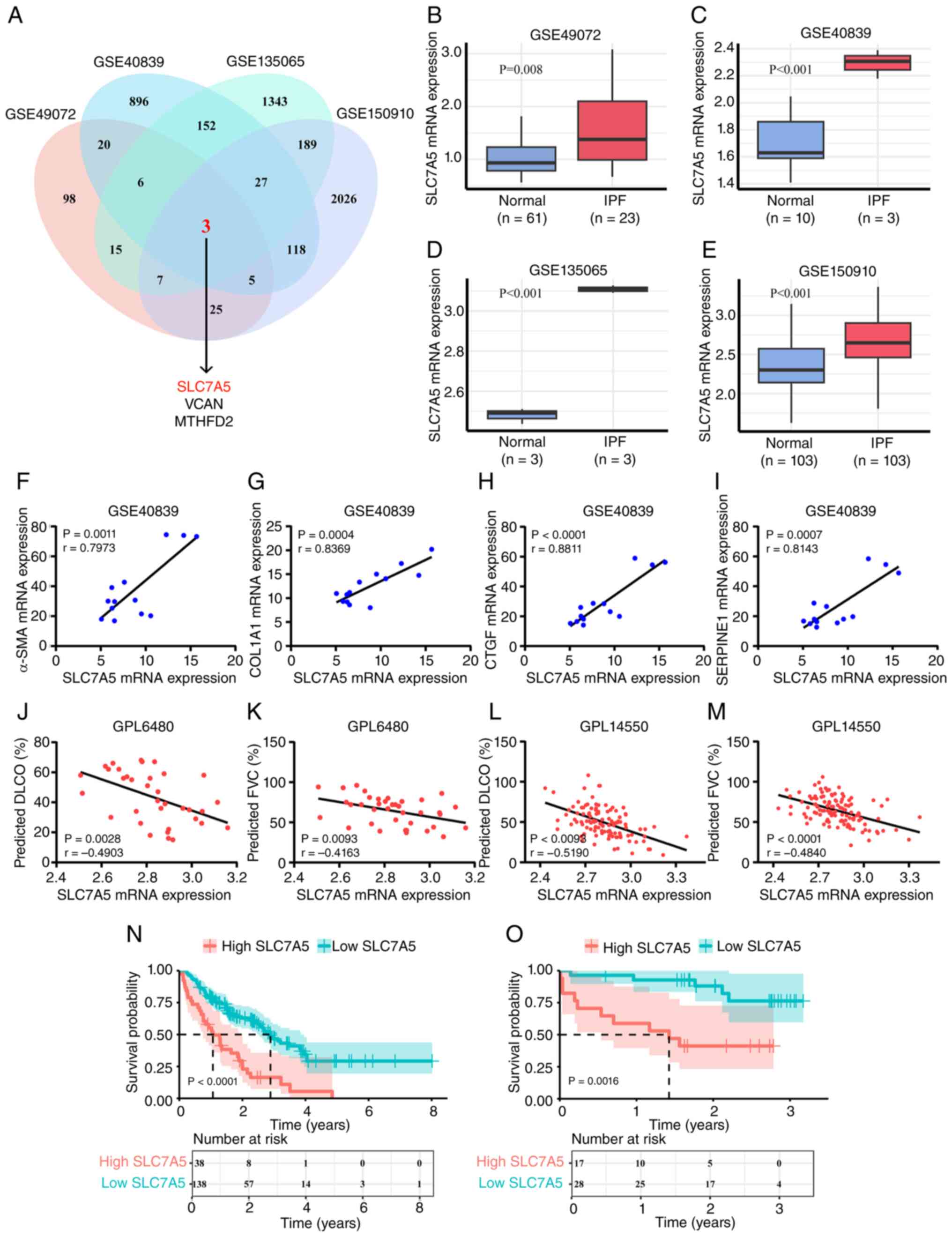
and visualized in a Venn diagram (Fig.
1A). The results showed that three genes, SLC7A5, versican
(VCAN) and methylenetetrahydrofolate dehydrogenase 2 (MTHFD2),
were significantly differentially expressed simultaneously in all
four datasets, and the expression levels of these genes were
increased in IPF. Notably, VCAN and MTHFD2 have been
reported to be associated with pulmonary fibrosis in several
studies (31–34). However, the relationship between
the high expression of SLC7A5 and the occurrence and
development of IPF remains to be fully elucidated. Therefore, a
deeper exploration of the correlation between SLC7A5 and
patients with IPF was conducted in the present study. Compared with
the healthy control group, SLC7A5 was significantly
upregulated in alveolar macrophages (Fig. 1B), lung fibroblasts (Fig. 1C and D) and lung tissues (Fig. 1E) in patients with IPF, suggesting
that the upregulation of SLC7A5 expression may have been
associated with IPF. To explore the correlation between the
expression level of SLC7A5 and related proteins in pulmonary
fibrosis, transcriptomic data from the GSE40839 dataset was
analyzed. Pearson's correlation analysis showed that the expression
level of SLC7A5 was significantly positively correlated with
that of α-SMA, COL1A1, connective tissue growth factor
(CTGF) and serpin E1 (SERPINE1) (Fig. 1F-I), further indicating the
correlation between SLC7A5 expression and pulmonary
fibrosis.
The results of pulmonary function tests in patients
with IPF frequently show a series of characteristic changes,
primarily including restrictive ventilation disorder, which
manifests as a decline of indicators such as forced vital capacity
(FVC) and total lung capacity, as well as reduced diffusion
function, which manifests as a decline in diffusing capacity for
carbon monoxide (DLCO) (35).
Pearson's correlation analysis was conducted on the lung function
test data of patients with IPF from two platforms (GPL6480 and
GPL14550) in the GSE47460 dataset. The results of this analysis
showed that in the GPL6480 platform, the expression level of
SLC7A5 was negatively correlated with DLCO %pred (percent of
predicted value) and FVC %pred in patients with IPF (Fig. 1J and K). In the GPL14550 platform,
the expression level of SLC7A5 also exhibited a negative
correlation with the DLCO %pred and FVC %pred values of patients
with IPF (Fig. 1L and M). This
indicated that the upregulation of SLC7A5 was correlated
with the decline of pulmonary function in patients with IPF.
To explore whether the expression level of
SLC7A5 affected the survival of patients with IPF, the
present study conducted survival analyses using the GSE70866 and
GSE27957 datasets. The results showed that in the GSE70866 dataset,
high expression levels of SLC7A5 in the bronchoalveolar
lavage fluid of patients with IPF was associated with a
significantly higher mortality rate than patients with low
SLC7A5 expression (Fig.
1N). In the GSE27957 dataset, the high expression of
SLC7A5 in the peripheral blood mononuclear cells of patients
with IPF was also associated with a significantly increased
mortality rate compared with patients with low SLC7A5
expression (Fig. 1O). These
results demonstrated that high SLC7A5 expression associated
with a poor prognosis in patients with IPF. Based on the results of
these bioinformatics analyses, it can be concluded that
SLC7A5 was highly expressed in patients with IPF, and that
the expression level of SLC7A5 was closely related to IPF
development.
SLC7A5 is highly expressed in the lung
tissue of mice with BLM-induced pulmonary fibrosis
To further investigate the effect of SLC7A5
on IPF, a mouse model of pulmonary fibrosis was established via
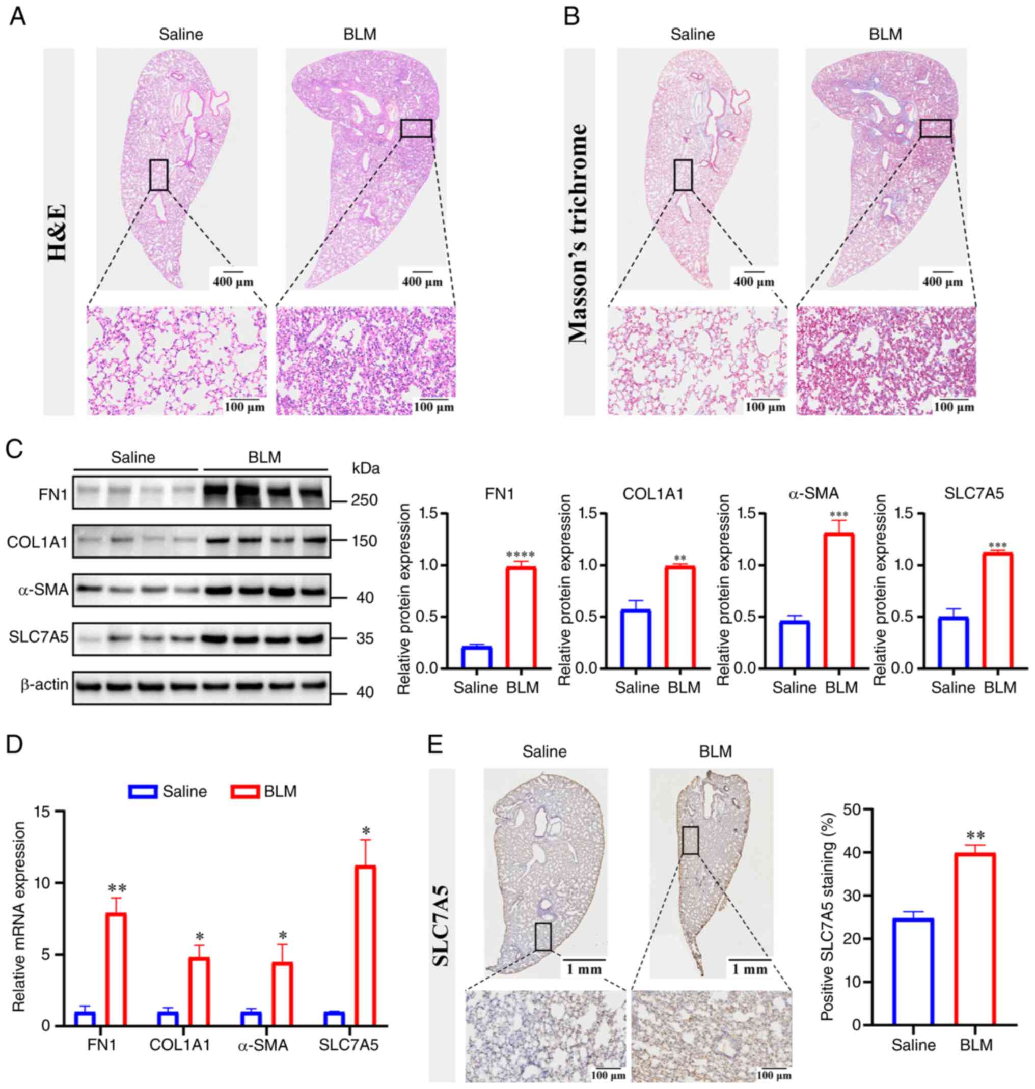
intratracheal BLM administration. Compared with the NS control
group, the lung tissues of mice in the BLM group showed notable
morphological changes, including thickening of the alveolar walls,
destruction of the alveolar septa and increased collagen content
(Fig. 2A and B). The results of
western blotting showed that compared with NS group mice, the
expression levels of the pulmonary fibrosis markers FN1,
COL1A1 and α-SMA, as well as those of the SLC7A5
protein, were significantly upregulated in the lung tissues of the
BLM group mice (Fig. 2C). RT-qPCR
also achieved consistent results (Fig.
2D). The results of immunohistochemical staining showed that
SLC7A5 staining, as shown by the brown staining of lung
tissues in the figure, was darker and covered a significantly
higher area proportion in BLM group mice compared with control mice
(Fig. 2E). To investigate the
localization of SLC7A5 in the lung tissues of mice, the
present study performed immunofluorescence co-staining for the lung
myofibroblast marker α-SMA together with SLC7A5. The
results showed that in the BLM group, the expression levels of both
α-SMA and SLC7A5 were markedly upregulated, and the
SLC7A5 protein was predominantly aggregated in proximity to
the α-SMA positive regions (Fig. S1). This indicated that
SLC7A5 was predominantly localized on lung myofibroblasts.
The aforementioned results indicated that the SLC7A5 was
upregulated in the lung tissues of the BLM group mice, and this
upregulation was demonstrated in vivo, supporting the
differential expression of SLC7A5 in the pulmonary fibrosis
model.
 | Figure 2.SLC7A5 expression in the lung
tissues of mice in the BLM group. (A) Hematoxylin and eosin and (B)
Masson's trichrome staining of lung tissue sections from
BLM-treated mice (n=4). (C) The expression of SLC7A5, FN1,
COL1A1 and α-SMA in the lung issue of BLM-treated mice
was detected by western blotting and semi-quantified (n=4; data are
shown as mean ± SD; **P<0.01, ***P<0.001 and ****P<0.0001
vs. saline group; unpaired t-test). (D) Detection of the expression
of SLC7A5, FN1, COL1A1 and α-SMA in the lung tissues
of BLM mice by reverse transcription-quantitative PCR (n=4; data
are shown as mean ± SD; *P<0.05 and **P<0.01; unpaired
t-test). (E) Immunohistochemical staining of SLC7A5 in
control and model mice (n=4), and statistical analysis of the
percentage of SLC7A5-positive staining (n=4; data are shown
as mean ± SD; **P<0.01 vs. saline group; unpaired t-test). BLM,
bleomycin; H&E, hematoxylin and eosin; FN1, fibronectin 1;
COL1A1, collagen type 1 α1; α-SMA, α-smooth muscle actin. |
SLC7A5 is highly expressed in
pulmonary myofibroblasts and is associated with the mTOR
pathway
In order to verify the expression of SLC7A5
in the in vitro experiments using human lung fibrosis cell
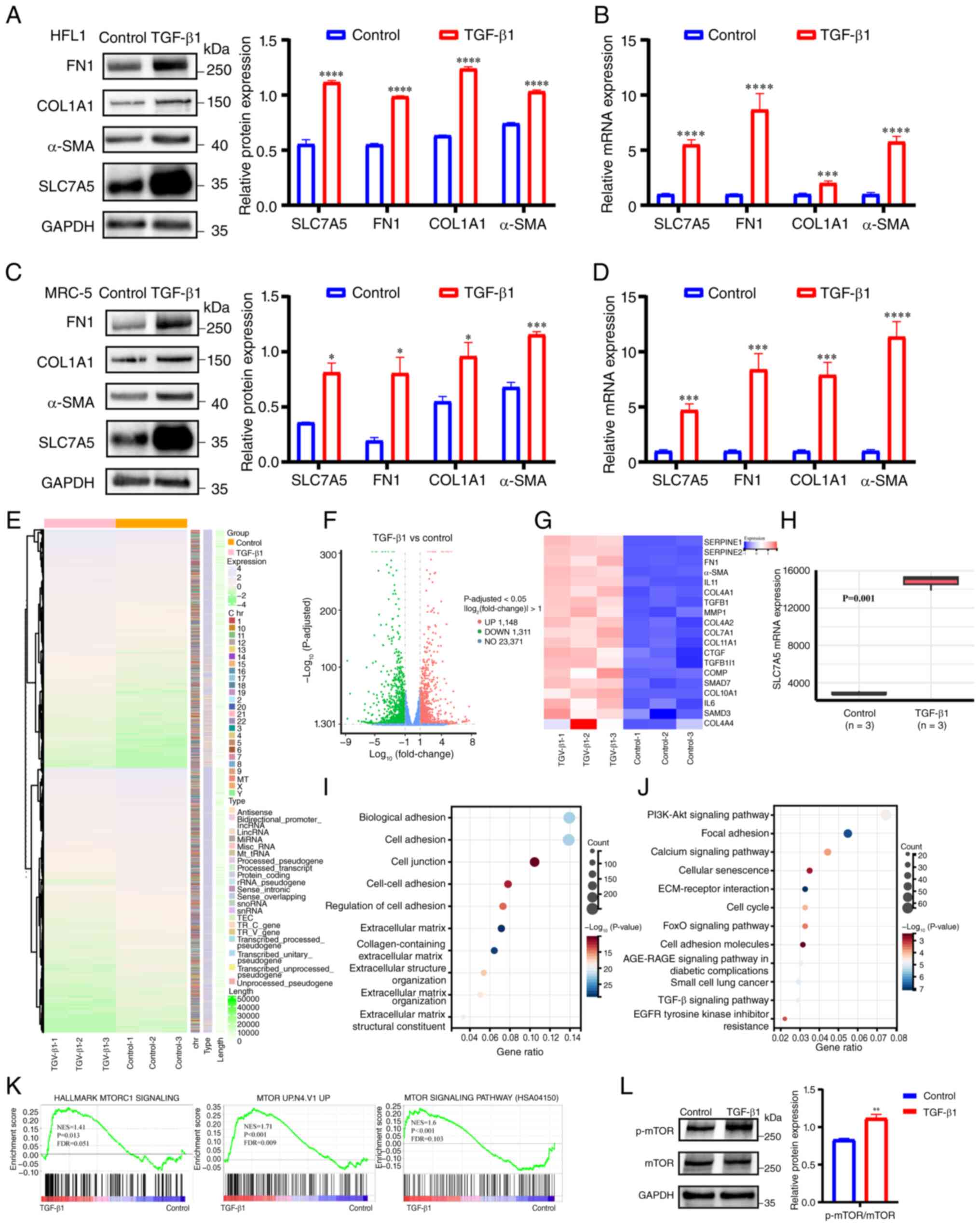
models, the TGF-β1 recombinant protein was used to induce HFL1 and
MRC-5 cell lines to transform into myofibroblasts. Western blotting
and RT-qPCR results showed that in HFL1 cells, after treatment with
the TGF-β1 recombinant protein, the protein and mRNA expressions of
FN1, COL1A1 and α-SMA were significantly upregulated
compared with untreated cells, indicating that fibroblasts
transformed into myofibroblasts. The expression of SLC7A5
was also significantly upregulated (Fig. 3A and B). The aforementioned results
were also demonstrated in the MRC-5 cell line (Fig. 3C and D), indicating that
SLC7A5 was highly expressed in the TGF-β1-induced lung
fibrosis cell model.
To explore the potential mechanisms by which
SLC7A5 played a role in pulmonary fibrosis, transcriptome
sequencing was performed on the mRNA of TGF-β1-induced HFL1 cells.
As shown in Fig. 3E and F,
different expression patterns of differentially expressed genes
were observed between the two treatment groups. Compared with the
control group, a total of 2,459 genes were differentially expressed
in the TGF-β1 group. Some fibrosis markers showed increased
expression levels in the TGF-β1 group, such as SERPINE1, FN1,
α-SMA, IL-11, collagen type IV alpha 1 (COL4A1), TGF-β1 and matrix
metallopeptidase 1 (MMP1) (Fig.
3G). Furthermore, the expression of SLC7A5 in cells in
the TGF-β1 group was significantly upregulated (Fig. 3H). Further exploration of the
relevant signaling pathways involved in the TGF-β1-mediated
induction of pulmonary fibroblasts involved GO functional
enrichment analysis and KEGG pathway enrichment analysis. These
analyses were conducted on the genes showing differential
expression between the two treatment groups. Several IPF-related
pathways were significantly enriched, including the Cell adhesion,
Extracellular matrix, and TGF-β signaling pathway (Fig. 3I and J). GSEA comparing the gene
expression profiles between the two groups revealed three
mTOR-related signaling pathways that were enriched in different
gene sets (HALLMARK, C2 and KEGG), and all of these pathways were
significantly upregulated in the TGF-β1 group (Fig. 3K). Notably, SLC7A5, as one
of the core genes contributing notably to the enrichment score, was
enriched in all three identified mTOR signaling pathways. To verify
the activation of mTOR signaling in TGF-β1-induced pulmonary
fibroblasts, western blotting was used to detect p-mTOR and mTOR
expression. The results showed that the p-mTOR/mTOR ratio was
significantly increased in the TGF-β1 group compared with the
control group, suggesting the activation of this pathway (Fig. 3L). These results indicated that
SLC7A5 may have regulated the mTOR signaling pathway in
order to affect the formation of pulmonary myofibroblasts and the
occurrence and development of IPF.
Knockdown of SLC7A5 inhibits the
formation of pulmonary myofibroblasts
To investigate whether SLC7A5 had any effect
on the occurrence and development of IPF, SLC7A5 was knocked
down using siRNA in HFL1 cells. Initially, the knockdown efficiency
of si-SLC7A5 was measured via RT-qPCR and western blotting.
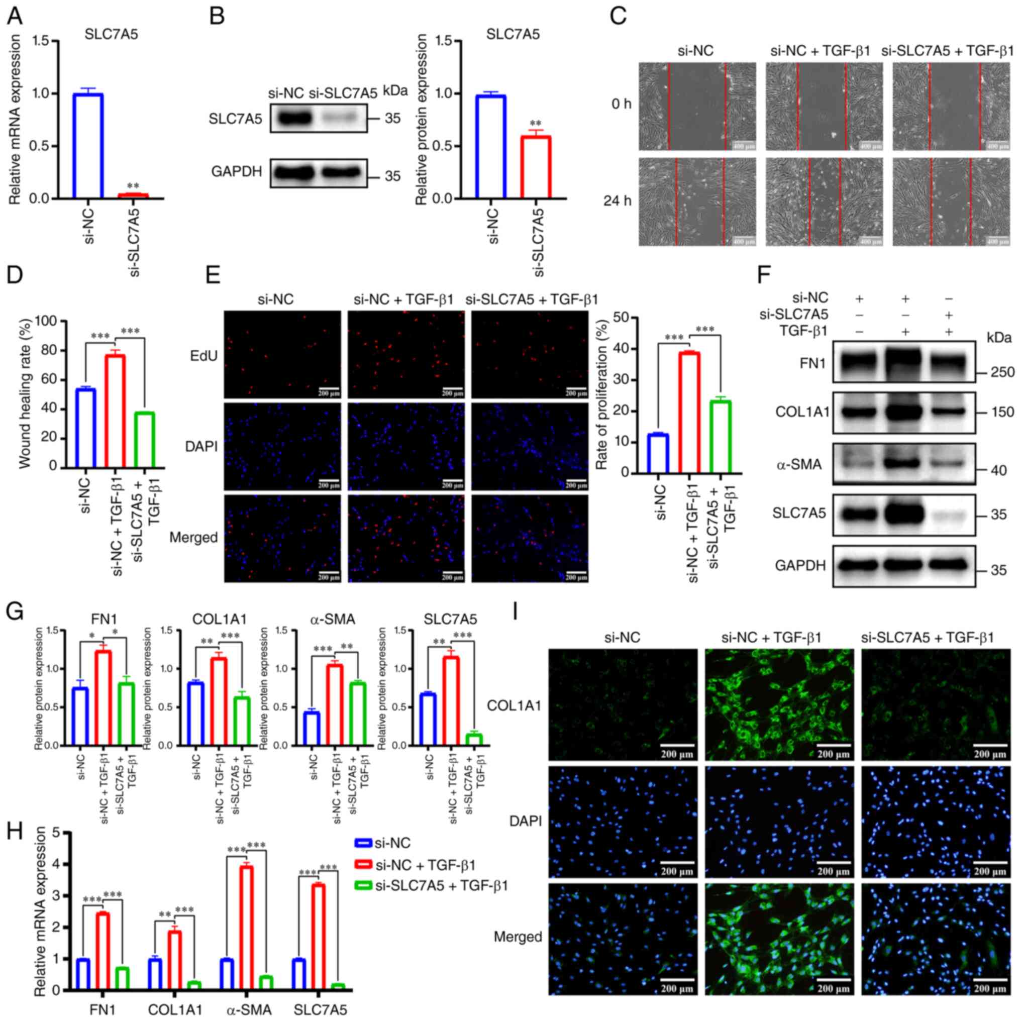
The results showed that, compared with the si-NC group, the mRNA
and protein expression levels of SLC7A5 in the
si-SLC7A5 group were significantly decreased (Fig. 4A and B). To investigate whether
silencing the expression of SLC7A5 would affect the cell
function of pulmonary myofibroblasts, the present study conducted
wound healing assays and EdU proliferation tests on transfected
HFL1 cells. The results showed that, compared with the si-NC group,
the cell migration and proliferation ability of si-NC-transfected
cells were significantly increased after the addition of TGF-β1.
Furthermore, compared with the si-NC + TGF-β1 group, the cell
migration and proliferation ability of the si-SLC7A5 +
TGF-β1 group were significantly decreased (Fig. 4C-E). Western blotting analysis
revealed that, compared with the si-NC + TGF-β1 group, the protein
expression of FN1, COL1A1 and α-SMA in the
si-SLC7A5 + TGF-β1 group was significantly reduced (Fig. 4F and G). These results were also
supported at the mRNA level (Fig.
4H). Immunofluorescence staining of HFL1 cells indicated that
the expression of COL1A1 in the si-SLC7A5 + TGF-β1
group was decreased compared with that in the si-NC + TGF-β1 group
(Fig. 4I). These results suggested
that silencing SLC7A5 expression affected the cellular
functions of pulmonary myofibroblasts.
 | Figure 4.Knockdown of SLC7A5 inhibits
the formation of pulmonary myofibroblasts. (A) The knockdown
efficiency of siRNA was verified by RT-qPCR (n=3; data are shown as
mean ± SD; **P<0.01 vs. si-NC group; unpaired t-test). (B) The
knockdown efficiency of siRNA was verified by western blotting
(n=3; data are shown as mean ± SD; **P<0.01 vs. si-NC; unpaired
t-test). (C) Wound healing assays were performed to detect the
migration of lung myofibroblasts. Scale bar, 200 µm (n=3). (D)
Statistical analysis of wound healing rate among the three
treatment groups (n=3; data are shown as mean ± SD; ***P<0.001;
one-way ANOVA). (E) The proliferation of lung myofibroblasts was
detected through EdU staining (scale bar, 100 µm) and subsequently
quantified (n=3; data are shown as mean ± SD; ***P<0.001;
one-way ANOVA. (F) Western blotting and (G) semi-quantification of
FN1, COL1A1, α-SMA and SLC7A5 expression in
TGF-β1-induced HFL1 cells after SLC7A5 knockdown (n=3; data
are shown as mean ± SD; *P<0.05, **P<0.01 and ***P<0.001;
one-way ANOVA). (H) Detection of relative FN1, COL1A1, α-SMA
and SLC7A5 expression in TGF-β1-induced HFL1 cells after
SLC7A5 knockdown using RT-qPCR (n=3; data are shown as mean
± SD; **P<0.01 and ***P<0.001; one-way ANOVA). (I) The
expression of COL1A1 across the three treatment groups was
observed via immunofluorescence staining. Scale bar, 100 µm (n=3).
FN1, fibronectin 1; COL1A1, collagen type 1 α1; α-SMA, α-smooth
muscle actin; TGF-β1, transforming growth factor-β1. |
In order to verify the reliability of siRNA-mediated
knockdown and the universality of subsequent changes in pulmonary
fibrosis markers in different human lung fibroblast cell lines, the
siRNA sequences in the present study were also used to knock down
SLC7A5 in MRC-5 cells. At both the mRNA and protein levels,
significant knockdown of SLC7A5 expression was observed
(Fig. S2A and B). Similarly,
western blotting and RT-qPCR results showed that after knocking
down SLC7A5, the expression levels of FN1, COL1A1 and
α-SMA, which were also induced by TGF-β1 treatment in MRC-5
cells, were also significantly reduced (Fig. S2C and D). These results suggested
that the upregulation of SLC7A5 may be the key factor
causing the formation and development of pulmonary fibrosis.
Selective inhibitor JPH203 of SLC7A5
inhibits the formation of pulmonary myofibroblasts
JPH203 is a selective inhibitor of SLC7A5
that predominantly exerts effects in cancers, such as gastric,
colon and bile duct cancer, by inhibiting the uptake of leucine
(36). However, to the best of our
knowledge, there is currently no literature reporting whether this
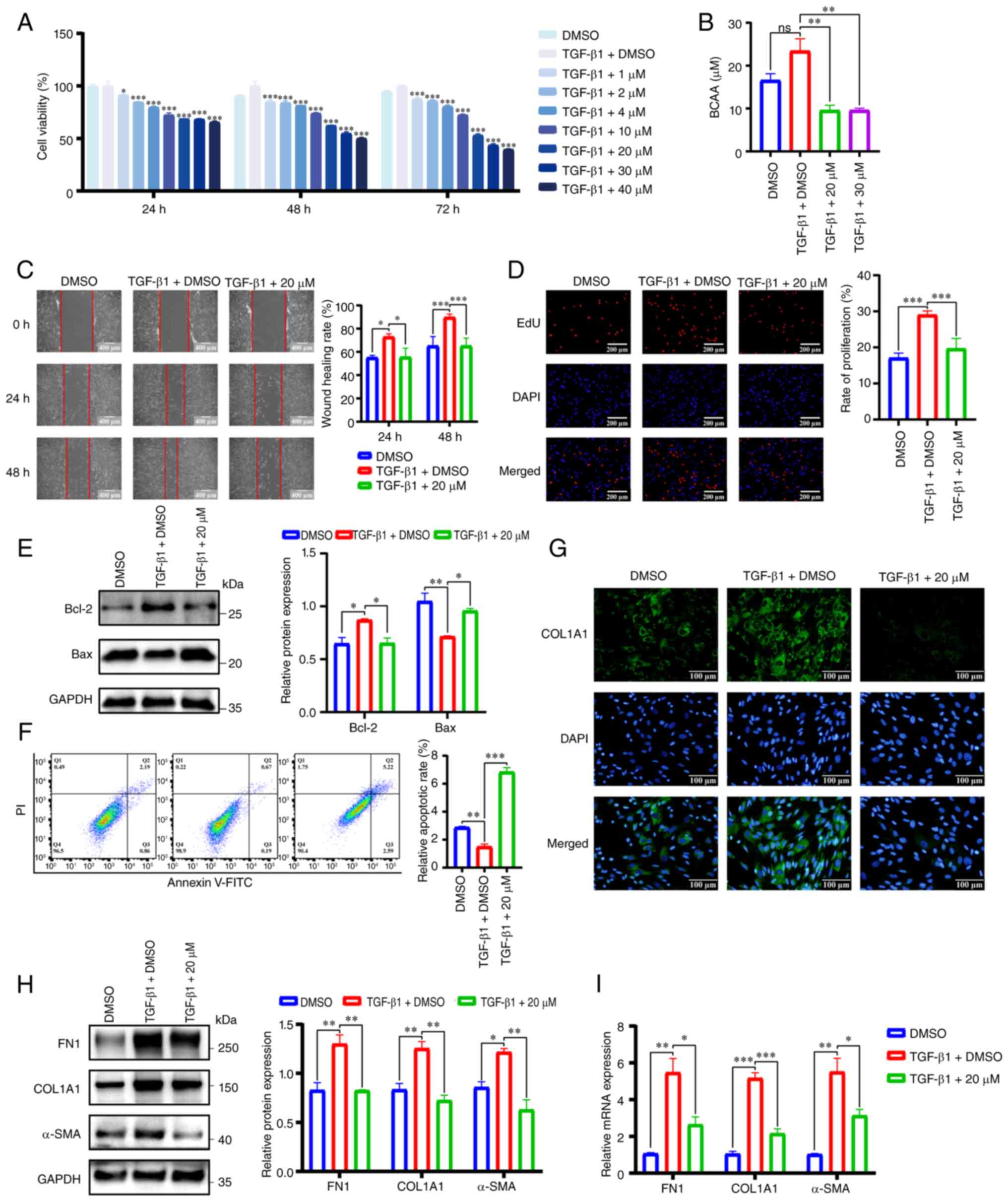
inhibitor can be used to treat pulmonary fibrosis. Primarily, CCK-8
assays were performed on HFL1 cells treated with various
concentrations of JPH203. The results of these assays indicated
that, compared with the control group, JPH203 significantly
inhibited the cell viability of pulmonary myofibroblasts and
exerted both concentration-dependent and time-dependent effects
(Fig. 5A). This inhibitory effect
was partially reversed via overexpression of SLC7A5 at 24
and 48 h (Fig. S3A-D). Based on
the results of the CCK-8 assay, the working concentrations of
JPH203 for subsequent experiments were set at 20 and 30 µM, as
these concentrations elicited moderate inhibition without excessive
cytotoxicity, while higher concentrations (40 µM) led to a marked
reduction that could confound the interpretation of
pathway-specific effects. Subsequently, BCAA content, including
leucine, isoleucine and valine levels, was detected in HFL1 cells
following treatment with JPH203. As shown in Fig. 5B, after incubating HFL1 cells with
JPH203 for 48 h, the content of BCAA in the TGF-β1 + 20 µM and
TGF-β1 + 30 µM groups were significantly reduced compared with the
TGF-β1 + DMSO group. These results indicated that in lung
fibroblasts, JPH203 specifically exerted its pharmacological
effects by inhibiting the translocation of BCAA from the
extracellular space to the intracellular space by targeting
SLC7A5.
 | Figure 5.Selective SLC7A5 inhibitor
JPH203 inhibits the formation of pulmonary myofibroblasts. (A) The
effect of JPH203 treatment on the cell viability of lung
myofibroblasts at 24, 48 and 72 h was detected via Cell Counting
Kit-8 assay (n=3; data are shown as mean ± SD; *P<0.05 and
***P<0.001 vs. TGF-β1 + DMSO group; two-way ANOVA). (B) BCAA
content in lung myofibroblasts after treatment with JPH203 (n=3;
data are shown as mean ± SD; **P<0.01; one-way ANOVA). (C) The
migration of lung myofibroblasts after treatment with JPH203 was
detected via wound healing assay (scale bar, 200 µm; n=3; data are
shown as mean ± SD; *P<0.05 and ***P<0.001; two-way ANOVA).
(D) The proliferation of lung fibroblasts was detected through EdU
staining (scale bar, 100 µm; n=3; data are shown as mean ± SD;
***P<0.001; one-way ANOVA). (E) The expression levels of
Bcl-2 and Bax in HFL1 cells treated with JPH203 were
detected by western blotting and semi-quantified (n=3; data are
shown as mean ± SD; *P<0.05 and **P<0.01; one-way ANOVA). (F)
The proportion of apoptotic HFL1 cells after JPH203 treatment was
detected using flow cytometry (n=3; data are shown as mean ± SD;
**P<0.01 and ***P<0.001; one-way ANOVA). (G) The expression
of COL1A1 in each treatment group was detected via
immunofluorescence staining (scale bar, 50 µm; n=3). (H) Western
blotting and semi-quantification of FN1, COL1A1 and
α-SMA expression in TGF-β1-induced HFL1 cells treated with
JPH203 (n=3; data are shown as mean ± SD; *P<0.05 and
**P<0.01; one-way ANOVA). (I) Detection of relative FN1,
COL1A1 and α-SMA expression in TGF-β1-induced HFL1 cells
after JPH203 co-treatment using reverse transcription-quantitative
PCR (n=3; data are shown as mean ± SD; *P<0.05, **P<0.01 and
***P<0.001; one-way ANOVA). DMSO, dimethyl sulfoxide; FN1,
fibronectin 1; COL1A1, collagen type 1 α1; α-SMA, α-smooth muscle
actin. |
Based on the aforementioned results, 20 µM was
selected as the working concentration of JPH203 for subsequent
experiments. This concentration was chosen because both 20 and 30
µM yielded similar levels of target inhibition in the preliminary
assays, and 20 µM was sufficient to produce a significant effect
while minimizing potential off-target cytotoxicity associated with
higher doses. In HFL1 cells, wound healing and EdU staining assays
were used to determine the effects of JPH203 on the migration and
proliferation abilities of human lung myofibroblasts. The results
demonstrated that, compared with the DMSO group, the migration and
proliferation of cells in the TGF-β1 + DMSO group were
significantly enhanced. Furthermore, JPH203 inhibited the
enhancement of cell migration and proliferation caused by TGF-β1
treatment (Fig. 5C and D). A study
has shown that TGF-β1, as a cytokine responsible for promoting
fibrosis, can enhance the resistance of lung fibroblasts to
apoptosis (37), thereby
inhibiting the apoptosis of lung myofibroblasts in the present
study. The results of western blotting (Fig. 5E) demonstrated that the expression
of Bcl-2 in the TGF-β1 + DMSO group was significantly
elevated compared with the DMSO group, whereas the expression of
Bax significantly decreased. Furthermore, compared with the
TGF-β1 + DMSO group, the expression of Bcl-2 in the TGF-β1 +
20 µM group significantly decreased, yet the expression of
Bax significantly increased.
In addition, the apoptotic rate of HFL1 cells
treated with JPH203 was detected using flow cytometry. Compared
with the DMSO group, the number of apoptotic cells in the TGF-β1 +
DMSO group significantly decreased and the proportions of early and
late apoptosis were reduced. However, compared with the TGF-β1 +
DMSO group, the apoptotic rate of cells in the TGF-β1 + 20 µM group
significantly increased, reversing the observed resistance to
apoptosis induced by TGF-β1 treatment (Fig. 5F); this indicated that JPH203
inhibited the resistance of lung myofibroblasts to cell apoptosis.
In order to investigate whether JPH203 affected the expression of
lung fibrosis markers, immunofluorescence staining was performed;
the results of this staining showed that JPH203 co-treatment
markedly inhibited the upregulation of COL1A1 caused by
TGF-β1 treatment (Fig. 5G).
Furthermore, western blotting was used to detect the protein
expression of FN1, COL1A1 and α-SMA in HFL1 lung
fibroblasts following JPH203 treatment. The results showed that the
protein expression levels of FN1, COL1A1 and α-SMA in
the TGF-β1 + 20 µM group were significantly decreased compared with
the TGF-β1 + DMSO group (Fig. 5H).
These results were also supported at the mRNA level (Fig. 5I). The aforementioned results
indicated that the SLC7A5 inhibitor JPH203 inhibited the
cellular functions of pulmonary myofibroblasts, and may therefore
have affected the formation and development process of pulmonary
fibrosis.
Modulation of SLC7A5 can inhibit the
activation of the PI3K/AKT/mTOR pathway
In order to explore the mechanism by which
SLC7A5 mediated the occurrence of IPF, transcriptome
sequencing analysis was conducted on the mRNA of HFL1 cells treated
with si-SLC7A5 + TGF-β1, with the si-NC + TGF-β1 group
representing the control group. The volcano plot (Fig. S4A) and heatmap (Fig. S4B) generated from these data
showed that, compared with the control group, 1,527 genes were
differentially expressed in the si-SLC7A5 + TGF-β1 group,
indicating that there were different expression patterns of genes
between the two treatment groups.
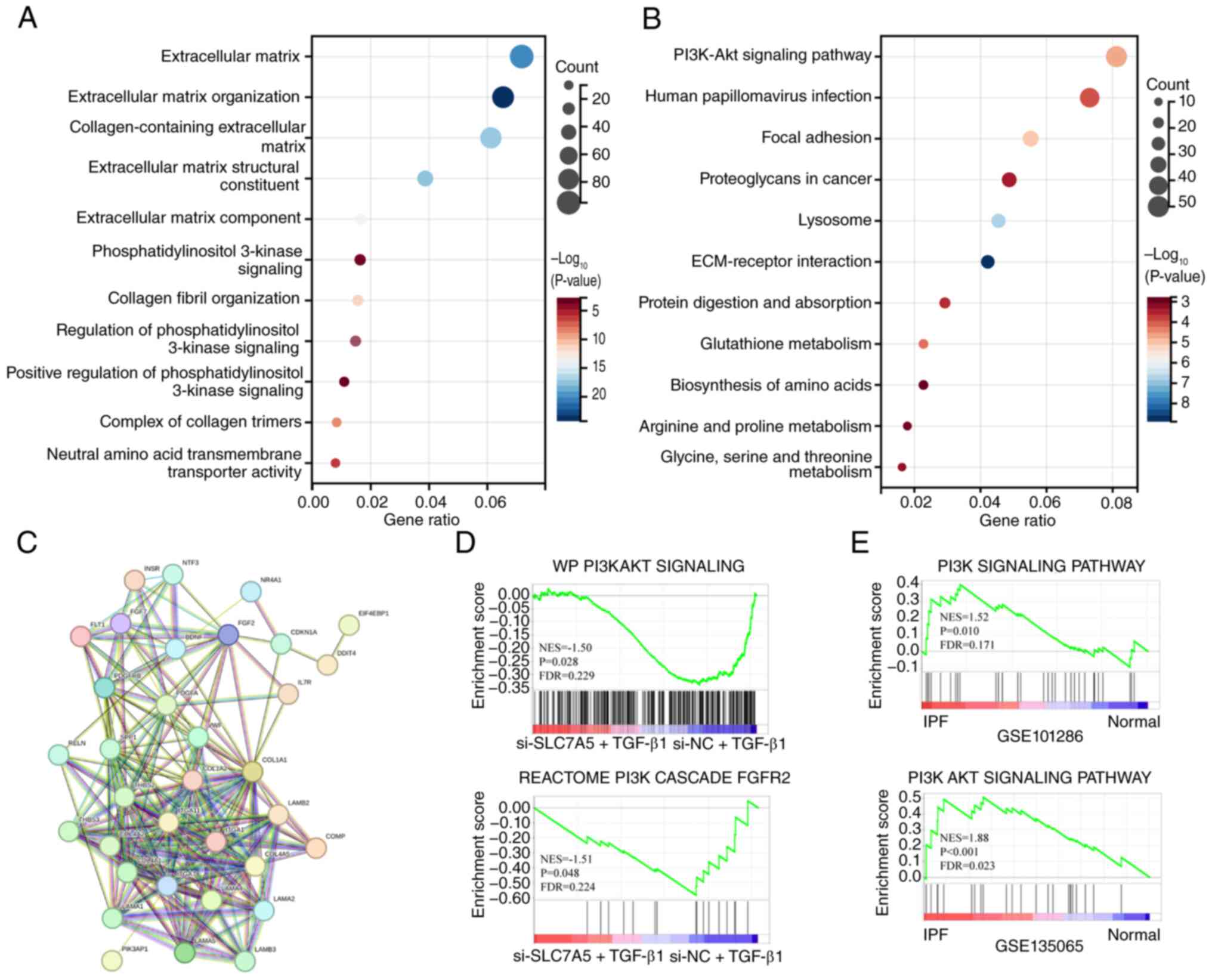
Subsequently, a GO functional enrichment analysis
was performed on the differentially expressed genes identified
between the two treatment groups (Fig.
6A). The results demonstrated that the pathways inhibited in
the si-SLC7A5 + TGF-β1 group included three PI3K signaling
pathways. The KEGG functional enrichment analysis also demonstrated
the significant downregulation of the ‘PI3K-Akt signaling pathway’,
which plays an important role in regulating the occurrence and
development of IPF (Fig. 6B). The
differentially expressed genes downregulated in the
si-SLC7A5 + TGF-β1 group were then subjected to PPI
analysis; it was found that these key nodes were also enriched in
the ‘PI3K-Akt signaling pathway’ in the KEGG dataset (Fig. 6C). Finally, GSEA was performed
between the gene expression profiles of the two groups. In the C2
gene set database, two PI3K/AKT-related signaling pathways were
found to be enriched, and both pathways were significantly
downregulated in the si-SLC7A5 + TGF-β1 group (Fig. 6D). By analyzing transcriptomic data
from the GEO datasets GSE101286 and GSE135065, the PI3K/AKT-related
pathway was shown to be significantly upregulated in patients with
IPF (Fig. 6E). This suggested that
the knockdown of SLC7A5 inhibited certain genes in the
PI3K/AKT pathway to affect the activation of the PI3K/AKT
pathway.
According to the aforementioned results,
SLC7A5 may have participated in the formation of
myofibroblasts by activating the mTOR signaling pathway; notably,
mTOR is a downstream effector of the PI3K/AKT signaling pathway
(38). Therefore, it can be
inferred that inhibiting the function of the SLC7A5 gene may
have affected the occurrence of pulmonary fibrosis by inhibiting
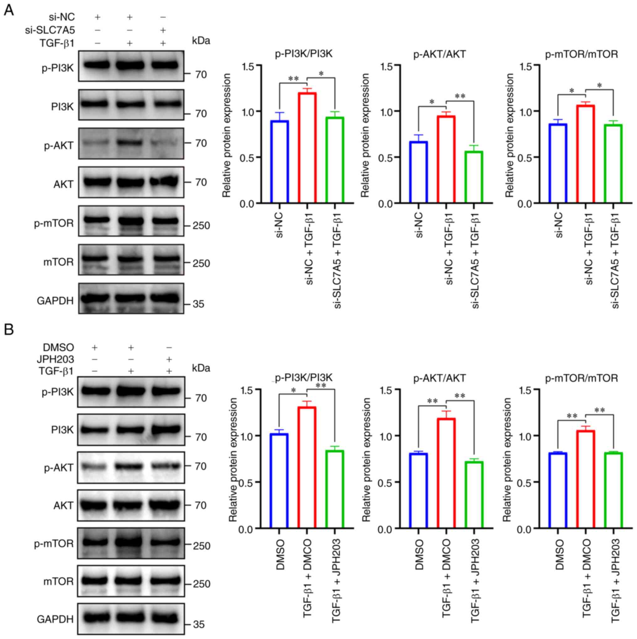
the PI3K/AKT/mTOR signaling pathway. The results of western
blotting showed that, compared with the si-NC group, the p-PI3K,
p-AKT and p-mTOR expression levels in the si-NC + TGF-β1 group were
significantly upregulated. Furthermore, compared with the si-NC +
TGF-β1 group, the p-PI3K, p-AKT and p-mTOR expression levels in the
si-SLC7A5 + TGF-β1 group were significantly downregulated
(Fig. 7A). On the other hand,
western blotting showed that, the p-PI3K, p-AKT and p-mTOR
expression levels in the TGF-β1 + DMSO group were significantly
upregulated compared with the DMSO group. Furthermore, by treating
TGF-β1-induced HFL1 cells with the JPH203 inhibitor (20 µM), the
p-PI3K, p-AKT and p-mTOR expression levels in the TGF-β1 + JPH203
group were downregulated compared with the TGF-β1 + DMSO group
(Fig. 7B). This indicated that
downregulating the expression of SLC7A5 by siRNA-mediated
knockdown or inhibiting the function of SLC7A5 via JPH203
treatment suppressed the activation of the PI3K/AKT/mTOR signaling
pathway, thereby inhibiting the formation of pulmonary
myofibroblasts and possibly suppressing the occurrence and
development of IPF.
 | Figure 7.Regulation of SLC7A5 on the
activity of the PI3K/AKT/mTOR signaling pathway. (A) The relative
expression of PI3K, p-PI3K, AKT, p-AKT, mTOR and p-mTOR in the
si-SLC7A5 + TGF-β1 group was detected by western blotting
(n=3; data are shown as mean ± SD; *P<0.05 and **P<0.01;
one-way ANOVA). (B) The relative expression of PI3K, p-PI3K, AKT,
p-AKT, mTOR and p-mTOR in the TGF-β1 + JPH203 group (20 µM) was
detected by western blotting (n=3; data are shown as mean ± SD;
*P<0.05 and **P<0.01; one-way ANOVA). SLC7A5, solute
carrier family 7 member 5; TGF-β1, transforming growth factor-β1;
p-. phosphorylated. |
JPH203 reduces the severity of
pulmonary fibrosis in BLM mice by inhibiting the activation of the
PI3K/AKT/mTOR pathway
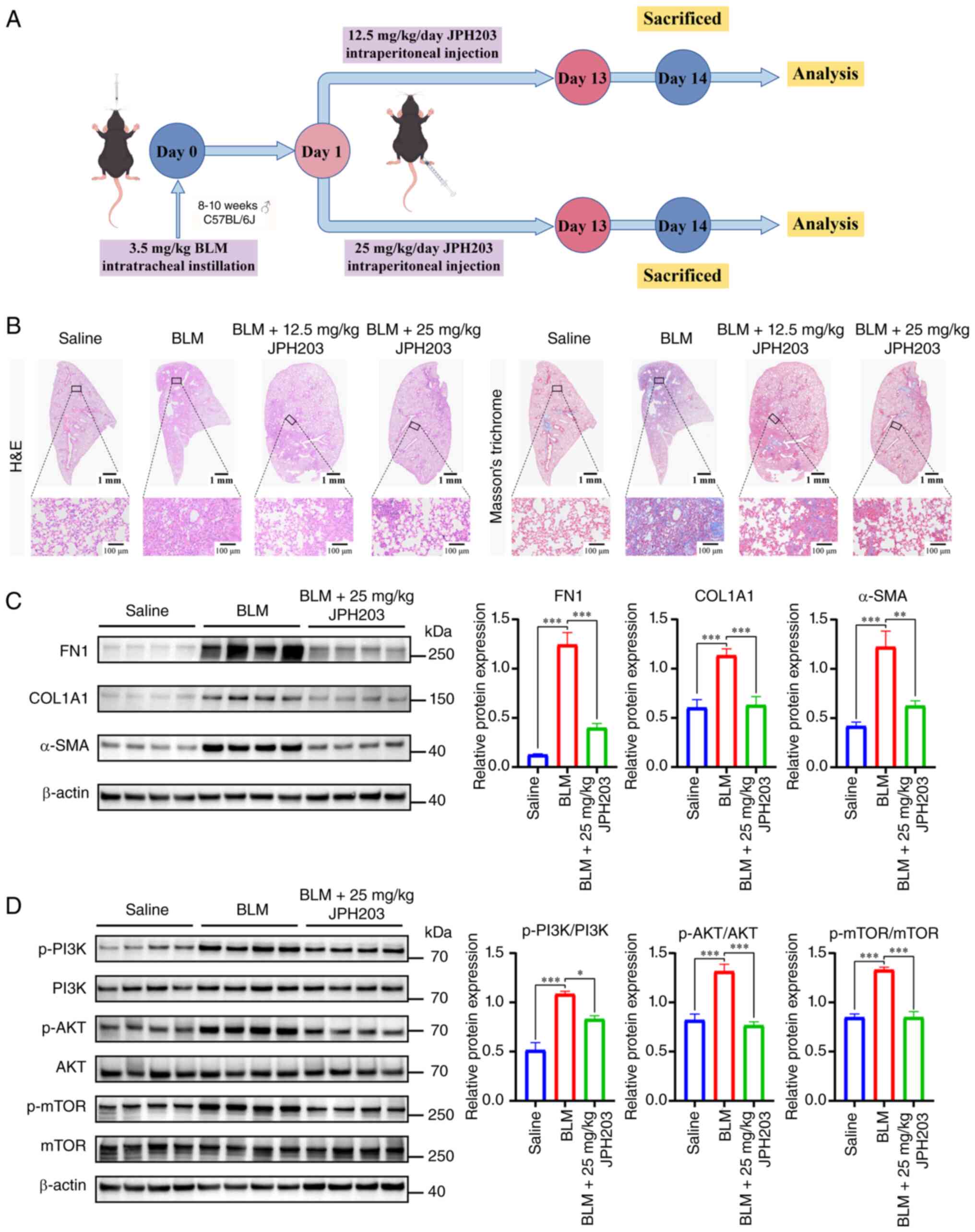
In order to verify the therapeutic effect of JPH203
on pulmonary fibrosis in vivo and to determine whether
SLC7A5 could represent a potential therapeutic target for
IPF, mice with BLM-induced pulmonary fibrosis were treated with
JPH203 by intraperitoneal injection for 13 consecutive days, as
shown in Fig. 8A. Intraperitoneal
injection was used as the administration method of JPH203 in
vivo, consistent with previous studies [e.g., JPH203 was
administered intraperitoneally in a thyroid carcinoma mouse model
(39)]. The present study selected
12.5 and 25 mg/kg as the experimental doses for JPH203 treatment.
These two doses have demonstrated notable antitumor effects in
multiple studies (40,41) and were tolerated well by mice in
the present study.
The lung tissue sections of experimental mice were
stained with H&E and Masson's trichrome, as shown in Fig. 8B. Compared with the BLM group, the
pathological changes in the lungs of the BLM + 12.5 mg/kg group
were notably alleviated, and the degree of pulmonary fibrosis in
the BLM + 25 mg/kg group was further improved. Based on the
tissue-staining results, the BLM + 25 mg/kg group was selected as
the treatment group for the subsequent experiments. The results of
western blotting showed that, compared with the BLM group, the
expression levels of FN1, COL1A1 and α-SMA in the
lung tissue of mice in the BLM + 25 mg/kg group were significantly
downregulated (Fig. 8C). RT-qPCR
also achieved consistent results (Fig. S5). Furthermore, the present study
used western blotting to verify that the PI3K/AKT/mTOR pathway was
activated in the lung tissues of mice with BLM-induced pulmonary
fibrosis, whereas the activity of this pathway was significantly
inhibited in the BLM + 25 mg/kg group (Fig. 8D). In addition, H&E staining
pathological analysis of heart, liver, spleen and kidney tissue
sections from BLM-induced mice was also conducted. The results
showed that, compared with the BLM group, the experimental groups
(BLM + 12.5 mg/kg and BLM + 25 mg/kg groups) demonstrated more
intact tissue structures in the heart, liver, spleen and kidneys,
without obvious inflammatory infiltration, cell necrosis, fibrosis
or other signs of pathological damage (Fig. S6). The aforementioned results
indicated that JPH203 improved BLM-induced pulmonary fibrosis in
mice by inhibiting the activation of the PI3K/AKT/mTOR pathway.
Furthermore, at the therapeutic dose used in the present study,
JPH203 demonstrated notable safety (Fig. S6), which provided important
implications for the clinical application of JPH203.
Discussion
IPF is a fatal progressive interstitial lung
disease. Current anti-fibrotic drugs have demonstrated limited
efficacy, so there is a notable requirement for new therapeutic
targets in IPF (1). The present
study utilized bioinformatics analysis to discover that
SLC7A5 was significantly upregulated in patients with IPF;
the present study also demonstrated that the expression level of
SLC7A5 was positively correlated with fibrosis markers and
negatively correlated with lung function, indicating that high
SLC7A5 expression associated with poor patient prognoses.
Experimental verification also showed that SLC7A5 was
upregulated in the mouse lung fibrosis model induced by BLM and in
lung fibroblasts stimulated by TGF-β1. SLC7A5 knockdown or
use of the selective SLC7A5 inhibitor JPH203 inhibited BCAA
transport, reduced the activity of pulmonary myofibroblasts and
improved lung fibrosis in mice. Further experiments demonstrated
that blocking or silencing SLC7A5 inhibited the
PI3K/AKT/mTOR pathway, prevented the formation of pulmonary
myofibroblasts and suggested that this pathway may have been
involved in the occurrence and development of IPF, providing a
theoretical basis for the search for new therapeutic targets in
IPF.
SLC7A5 is a non-sodium-dependent
high-affinity amino acid transporter (42). Previous studies have shown that
SLC7A5 is highly expressed in the cardiomyocytes of diabetic
mice (43) and during the
TGF-β1-induced mesenchymal-epithelial transition process (44). These results are consistent with
the findings of the present study, which found that SLC7A5
was significantly upregulated in patients with IPF. A continuous
decline of lung function represents the core pathophysiological
feature of IPF; patients with IPF usually exhibit a decrease in
DLCO (45). Consistent with this,
the present study found that the expression level of SLC7A5
in lung tissues was significantly negatively correlated with DLCO
and FVC in patients with IPF. Furthermore, high SLC7A5
expression was closely related to the poor prognosis of patients
with IPF. As such, the expression level of SLC7A5 was
closely related to the clinical indicators of patients with IPF and
may have been involved in the occurrence and development of
IPF.
The pathogenesis of IPF is related to genetic
factors, epigenetic factors and oxidative stress, and involves
multiple cellular signaling pathways (46,47).
Under the influence of genetic and environmental factors, alveolar
epithelial cells will develop metabolic disorders and exhibit
abnormal proliferation and repair. These cells also interact with
mesenchymal cells through various signaling pathways that promote
the transformation of fibroblasts into myofibroblasts, thereby
leading to IPF (48). In the
present study, RNA interference technology was used to knockdown
the SLC7A5 gene in pulmonary fibroblasts; it was found that
in TGF-β1-induced SLC7A5 knockdown cells, the proliferation
and migration of pulmonary fibroblasts were significantly inhibited
and the myofibroblast transformation was reversed, as shown in
Fig. 4. These results suggested
that SLC7A5 may have been involved in the development of
pulmonary fibrosis. A notable characteristic of IPF is that
fibroblasts are transformed into myofibroblasts under the influence
of TGF-β1 (49). As described in
the present study, the expression levels of FN1, COL1A1 and
α-SMA in TGF-β1-induced pulmonary fibroblasts were
upregulated, suggesting that TGF-β1 induced the differentiation
process of pulmonary fibroblasts. Silencing SLC7A5
expression significantly inhibited the upregulation of FN1,
COL1A1 and α-SMA induced by TGF-β1 and inhibited the
phenotypic transformation of fibroblasts.
SLC7A5 plays an important role in regulating
the mTOR signaling pathway. As an important amino acid transporter
with a high affinity for leucine and high transport capacity,
SLC7A5 is primarily responsible for the uptake of
extracellular leucine (50).
Leucine is not only necessary for protein synthesis but also a core
signaling molecule for the activation of mTORC1 (51). Although cell membranes have various
amino acid transporters, a study has shown that SLC7A5, due
to its efficient uptake characteristics, dominates the cellular
acquisition of leucine (50). In
the present study, the importance of the SLC7A5/mTOR axis
remains particularly prominent (52). By upregulating SLC7A5,
cancer cells can take in large amounts of leucine and continuously
activate the mTORC1 pathway, driving increased cell proliferation
(53). When SLC7A5 is
dysfunctional or knocked out genetically, the concentrations of
leucine and BCAA in the cells markedly decrease, thereby impairing
the activity of the mTORC1 signaling pathway (54). The association between
SLC7A5 and mTOR is insufficiently studied in fibrotic
diseases and, to the best of our knowledge, there is no report for
this association in pulmonary fibrosis. The present study used
transcriptome sequencing to determine that SLC7A5 was
enriched in mTOR-related pathways and support the activation of the
mTOR pathway in IPF (as shown in Fig.
3K), suggesting that, in the process of pulmonary fibroblasts
transforming into pulmonary myofibroblasts, SLC7A5 may have
affected this process through the mTOR pathway. Furthermore, the
present study observed, that after treatment with JPH203, the lung
fibroblast content of BCAA, which contained leucine, was
significantly reduced. This reduction was accompanied by a decrease
in the phosphorylation level of mTOR. Therefore, we hypothesize
that inhibiting SLC7A5 inhibited the mTOR pathway by
suppressing the transport of amino acids, thereby inhibiting
pulmonary fibrosis.
The PI3K/AKT/mTOR pathway has been shown to
regulate cell proliferation, differentiation and survival, and is
closely related to pulmonary fibrosis (55). A study reported by Li et al
(56) demonstrated that duvelisib
enhances autophagy and inhibits the activation of pulmonary
fibroblasts by blocking the PI3K/AKT/mTOR pathway, thereby
alleviating pulmonary fibrosis. Studies have also shown that the
PI3K/AKT pathway is involved in the occurrence and development of
myocardial, hepatic and renal interstitial fibrosis (57,58).
For example, a study reported by Wei et al (59) showed that the activity of the
PI3K/AKT/mTOR pathway is closely linked to the growth and
activation of hepatic stellate cells and can promote the
progression of fibrosis in the liver. A study has also shown that
the PI3K/AKT/mTOR pathway may directly participate in the
occurrence and development of fibrosis via an interaction that is
yet to be fully elucidated (60).
A previous study has shown that the overexpression of secreted
phosphoprotein 1 (SPP1) enhances activation of the
PI3K/AKT/mTOR pathway and thereby the proliferation of fibroblasts
in IPF; these effects were eliminated by treatment with PI3K
inhibitors (61). The
transcriptome sequencing data from the present study showed that in
the TGF-β1-induced model of IPF, activation of the PI3K/AKT pathway
was inhibited. It should be noted that, although the PI3K/AKT
pathway is typically regarded as an upstream pathway responsible
for activating mTOR, few studies have shown that the activity of
the PI3K/AKT may also be affected by SLC7A5. The
experimental results of the cell experiments and animal models in
the present study demonstrated that JPH203, by inhibiting the
activity of SLC7A5, reduced the intracellular levels of
essential amino acids, thereby blocking the activation of the
PI3K/AKT/mTOR signaling pathway and exerting potential therapeutic
effects in improving pulmonary fibrosis.
There are currently only two clinical anti-fibrotic
treatments available for patients with IPF: Pirfenidone and
nintedanib (4,62). However, treatments using these
drugs have demonstrated a number of adverse reactions, which forces
patients to reduce the dosage and in turn affects treatment
outcomes (5,63). By shifting the goal of
anti-fibrotic treatment for IPF from preventing disease progression
to improving quality of life and reducing drug toxicity, it is
expected that the therapies for IPF can reduce the occurrence of
adverse reactions. JPH203 inhibits the function of SLC7A5,
thereby preventing cells from absorbing essential amino acids and
leading to the depletion of amino acid reserves within the cells,
which subsequently inhibits cell proliferation (64). The first clinical trial
(UMIN000016546) using the selective SLC7A5 inhibitor JPH203
showed that this drug exhibits good tolerance in patients with
advanced solid tumors (13). A
study has shown that the expression of SLC7A5 is markedly
increased in peritoneal fibrosis, whereas JPH203 can markedly
reduce the fibrotic phenotype in affected cells both in vivo
and in vitro (44).
However, to the best of our knowledge, there are no studies
reporting the effect of JPH203 in pulmonary fibrosis.
The present study used JPH203 to treat
TGF-β1-induced pulmonary fibroblasts and found that JPH203
inhibited the proliferation, migration and anti-apoptotic ability
of pulmonary myofibroblasts. JPH203 treatment also suppressed the
expression levels of FN1, COL1A1 and α-SMA,
indicating that JPH203 blocked the transformation of pulmonary
fibroblasts by inhibiting the transport ability of SLC7A5
for BCAA. In a phase I clinical trial (UMIN000016546) of the
selective inhibitor JPH203, patients with advanced solid tumors
were treated with JPH203 daily via intravenous injection. Disease
control was achieved at dose levels of 12 and 25 mg/m2,
with a disease control rate of 60% for cholangiocarcinoma (13). In the present study, JPH203 was
able to improve BLM-induced pulmonary fibrosis in mice and this
improvement was more notable in the 25 mg/kg group. The tissue
structures of heart, liver, spleen and kidney tissues of
pulmonary-fibrosis model mice treated with 12.5 and 25 mg/kg doses
JPH203 were intact, without obvious inflammatory infiltration, cell
necrosis or pathological damage. From these results, the present
study observed that JPH203 exhibited good tolerance and efficacy
levels in vivo (as shown in Figs. 8 and S6). As such, the aforementioned results
also indicated that JPH203 may represent a new therapeutic agent
for IPF treatment, and a dose of 25 mg/kg JPH203 can be used for
investigation in future studies.
The predominant findings of the present study
involve the discovery of high SLC7A5 expression in patients
with IPF using a large sample dataset and the discovery that
SLC7A5 upregulation was associated with the decline in lung
function and poor prognosis of patients with IPF. Inhibiting the
function of SLC7A5 or silencing its expression through
inhibitors or siRNA sequences inhibited the function of pulmonary
myofibroblasts via suppression of the PI3K/AKT/mTOR signaling
pathway. Furthermore, the present study used the selective
SLC7A5 inhibitor JPH203 to treat BLM-induced pulmonary
fibrosis mice and supported the anti-IPF efficacy of JPH203 in
vivo.
However, the present study also exhibited certain
limitations. Primarily, although the use of BLM to induce pulmonary
fibrosis in mice is currently recognized as an effective method for
establishing an animal model of IPF (65,66),
pulmonary fibrosis in the BLM-induced pulmonary fibrosis mouse
model is temporary and self-limiting, whereas IPF is a chronic,
progressive and irreversible disease. This difference exemplifies
the limitations of the IPF model (67). In addition, a limitation of the
present study is that post-mortem analysis was not performed for
the three mice that died before the scheduled endpoint; therefore,
the exact cause of death could not be conclusively determined.
Furthermore, the present study used public datasets on patients
with IPF but did not include our own IPF patient cohort from
inpatient records. In subsequent research, patients with IPF should
be included to allow for in-depth explorations of SLC7A5
expression in clinical samples, as well as to elucidate the
association between SLC7A5 and clinical indicators, such as
disease severity. Finally, the present study did not explore
whether SLC7A5 activated the PI3K/AKT/mTOR signaling pathway
by affecting the transport of specific amino acids. Future studies
should explore the related pathways and downstream target genes of
SLC7A5, as well as the association between downstream amino
acids transported by SLC7A5 and IPF development.
In conclusion, SLC7A5 was significantly
upregulated in samples from patients with IPF, and the expression
of SLC7A5 was negatively correlated with the pulmonary
function of patients with IPF. The upregulation of SLC7A5
was also associated with the poor prognosis of patients with IPF.
SLC7A5 was significantly upregulated in the lung tissues of
mice with BLM-induced pulmonary fibrosis and in lung fibroblasts
induced by TGF-β1. SLC7A5 knockdown or selective inhibition
using JPH203 prevented the formation of pulmonary myofibroblasts by
inhibiting the activation of the PI3K/AKT/mTOR pathway. In
vivo, JPH203 improved the degree of fibrosis in the lungs of
mice with BLM-induced pulmonary fibrosis. Therefore, SLC7A5
may represent a new therapeutic target for IPF, and future research
should focus on regulating this target and translating
SLC7A5-targeted treatments into effective therapies for IPF
in clinical practice.
Supplementary Material
Supporting Data
Acknowledgements
Not applicable.
Funding
The present study was supported by the National Natural Science
Foundation of China Young Scientists Fund (grant no. 82400085), the
Project of the Department of Science and Technology of Heilongjiang
Province (grant no. JD25D004) and the Research Project of the
Health Commission of Heilongjiang Province (grant no.
20250303020028).
Availability of data and materials
The data generated in the present study may be
requested from the corresponding author. The transcriptome
sequencing data generated in the present study may be found in the
Science Data Bank repository at the following URLs: https://doi.org/10.57760/sciencedb.29996 (accession
number: CSTR: https://cstr.cn/31253.11.sciencedb.29996, Fig. 3) and
https://doi.org/10.57760/sciencedb.30161 (accession number: CSTR:
https://cstr.cn/31253.11.sciencedb.30161, Fig. 6).
Authors' contributions
CS and HC designed the study and supervised the
project. MW, YY and ZL wrote the manuscript, conducted experiments,
performed literature searches and analyzed the data. DX, YL, XZ, NS
and XL contributed towards data processing and interpretation,
statistical analysis, and editing the manuscript. HC and MW confirm
the authenticity of all the raw data. All authors read and approved
the final version of the manuscript.
Ethics approval and consent to
participate
All animal experiments involved in the present
study were approved by the Experimental Animal Protection and
Ethics Committee of Harbin Medical University (approval no.
YJSDW2023-024).
Patient consent for publication
Not applicable.
Competing interests
The authors declare that they have no competing
interests.
Glossary
Abbreviations
Abbreviations:
|
BCAA
|
branched-chain amino acid
|
|
BLM
|
bleomycin
|
|
DLCO
|
diffusing capacity for carbon
monoxide
|
|
ECM
|
extracellular matrix
|
|
FVC
|
forced vital capacity
|
|
GSEA
|
Gene Set Enrichment Analysis
|
|
H&E
|
hematoxylin and eosin
|
|
IPF
|
idiopathic pulmonary fibrosis
|
|
PPI
|
protein-protein interaction
|
|
TGF-β1
|
transforming growth factor-β1
|
References
|
1
|
Zhu J, Zhou D, Yu M and Li Y: Appraising
the causal role of smoking in idiopathic pulmonary fibrosis: A
Mendelian randomization study. Thorax. 79:179–181. 2024. View Article : Google Scholar : PubMed/NCBI
|
|
2
|
Zhu X, Mao S, Yang Y, Liu X, Liu Q, Zhang
N, Yang Y, Li Y, Gao M, Bao J, et al: Biomimetic topological
micropattern arrays regulate the heterogeneity of cellular fates in
lung fibroblasts between fibrosis and invasion. ACS Nano.
19:580–599. 2025. View Article : Google Scholar : PubMed/NCBI
|
|
3
|
Wijsenbeek M: Progress in the treatment of
pulmonary fibrosis. Lancet Respir Med. 8:424–425. 2020. View Article : Google Scholar : PubMed/NCBI
|
|
4
|
Lee WT, Lee H, Kim J, Jung Y, Choi E,
Jeong JH, Jeong JH, Lee JH and Youn YS: Alveolar macrophage
phagocytosis-evading inhaled microgels incorporating
nintedanib-PLGA nanoparticles and pirfenidone-liposomes for
improved treatment of pulmonary fibrosis. Bioact Mater. 33:262–278.
2023.PubMed/NCBI
|
|
5
|
Di Martino E, Provenzani A, Vitulo P and
Polidori P: Systematic review and Meta-analysis of pirfenidone,
nintedanib, and pamrevlumab for the treatment of idiopathic
pulmonary fibrosis. Ann Pharmacother. 55:723–731. 2021. View Article : Google Scholar : PubMed/NCBI
|
|
6
|
Maher TM, Ford P, Brown KK, Costabel U,
Cottin V, Danoff SK, Groenveld I, Helmer E, Jenkins RG, Milner J,
et al: Ziritaxestat, a novel autotaxin inhibitor, and lung function
in idiopathic pulmonary fibrosis: The ISABELA 1 and 2 randomized
clinical trials. JAMA. 329:1567–1578. 2023. View Article : Google Scholar : PubMed/NCBI
|
|
7
|
Jiang YH, Zhou M, Cheng MD, Chen S and Guo
YQ: CAR-engineered cytolytic Tregs reverse pulmonary fibrosis and
remodel the fibrotic niche with limited CRS. JCI Insight.
10:e1820502025. View Article : Google Scholar : PubMed/NCBI
|
|
8
|
Pei X, Zheng F, Li Y, Lin Z, Han X, Feng
Y, Tian Z, Ren D, Cao K and Li C: Niclosamide ethanolamine salt
alleviates idiopathic pulmonary fibrosis by modulating the
PI3K-mTORC1 pathway. Cells. 11:3462022. View Article : Google Scholar : PubMed/NCBI
|
|
9
|
Zhao T and Su Y: Mechanisms and
therapeutic potential of myofibroblast transformation in pulmonary
fibrosis. J Respir Biol Transl Med. 2:100012025.PubMed/NCBI
|
|
10
|
Yan R, Zhao X, Lei J and Zhou Q: Structure
of the human LAT1-4F2hc heteromeric amino acid transporter complex.
Nature. 568:127–130. 2019. View Article : Google Scholar : PubMed/NCBI
|
|
11
|
Morozova V, Pellegata D, Singer S, Charles
RP, Muller J, Altmann KH and Gertsch J: Pharmacodynamic analyses of
LAT1 inhibitors in vitro and in vivo by targeted metabolomics
reveal target-independent effects. Biomed Pharmacother.
190:1184022025. View Article : Google Scholar : PubMed/NCBI
|
|
12
|
Zhang J, Xu Y, Li D, Fu L, Zhang X, Bao Y
and Zheng L: Review of the correlation of LAT1 with diseases:
Mechanism and treatment. Front Chem. 8:5648092020. View Article : Google Scholar : PubMed/NCBI
|
|
13
|
Okano N, Naruge D, Kawai K, Kobayashi T,
Nagashima F, Endou H and Furuse J: First-in-human phase I study of
JPH203, an L-type amino acid transporter 1 inhibitor, in patients
with advanced solid tumors. Invest New Drugs. 38:1495–1506. 2020.
View Article : Google Scholar : PubMed/NCBI
|
|
14
|
Shi Y, Gochuico BR, Yu G, Tang X, Osorio
JC, Fernandez IE, Risquez CF, Patel AS, Shi Y, Wathelet MG, et al:
Syndecan-2 exerts antifibrotic effects by promoting
caveolin-1-mediated transforming growth factor-beta receptor I
internalization and inhibiting transforming growth factor-beta1
signaling. Am J Respir Crit Care Med. 188:831–841. 2013. View Article : Google Scholar : PubMed/NCBI
|
|
15
|
Lindahl GE, Stock CJ, Shi WX, Leoni P,
Sestini P, Howat SL, Bou Gharios G, Nicholson AG, Denton CP,
Grutters JC, et al: Microarray profiling reveals suppressed
interferon stimulated gene program in fibroblasts from
scleroderma-associated interstitial lung disease. Respir Res.
14:802013. View Article : Google Scholar : PubMed/NCBI
|
|
16
|
Negreros M, Hagood JS, Espinoza CR,
Balderas-Martinez YI, Selman M and Pardo A: Transforming growth
factor beta 1 induces methylation changes in lung fibroblasts. PLoS
One. 14:e02235122019. View Article : Google Scholar : PubMed/NCBI
|
|
17
|
Furusawa H, Cardwell JH, Okamoto T, Walts
AD, Konigsberg IR, Kurche JS, Bang TJ, Schwarz MI, Brown KK,
Kropski JA, et al: Chronic hypersensitivity pneumonitis, an
interstitial lung disease with distinct molecular signatures. Am J
Respir Crit Care Med. 202:1430–1444. 2020. View Article : Google Scholar : PubMed/NCBI
|
|
18
|
Prasse A, Binder H, Schupp JC, Kayser G,
Bargagli E, Jaeger B, Hess M, Rittinghausen S, Vuga L, Lynn H, et
al: BAL cell gene expression is indicative of outcome and airway
basal cell involvement in idiopathic pulmonary fibrosis. Am J
Respir Crit Care Med. 199:622–630. 2019. View Article : Google Scholar : PubMed/NCBI
|
|
19
|
Huang Y, Ma SF, Vij R, Oldham JM,
Herazo-Maya J, Broderick SM, Strek ME, White SR, Hogarth DK, Sandbo
NK, et al: A functional genomic model for predicting prognosis in
idiopathic pulmonary fibrosis. BMC Pulm Med. 15:1472015. View Article : Google Scholar : PubMed/NCBI
|
|
20
|
Horimasu Y, Ishikawa N, Taniwaki M,
Yamaguchi K, Hamai K, Iwamoto H, Ohshimo S, Hamada H, Hattori N,
Okada M, et al: Gene expression profiling of idiopathic
interstitial pneumonias (IIPs): Identification of potential
diagnostic markers and therapeutic targets. BMC Med Genet.
18:882017. View Article : Google Scholar : PubMed/NCBI
|
|
21
|
Cowley PM, Roberts CR and Baker AJ:
Monitoring the health status of mice with Bleomycin-induced lung
injury by using body condition scoring. Comp Med. 69:95–102. 2019.
View Article : Google Scholar : PubMed/NCBI
|
|
22
|
Bolourani S, Sari E, Brenner M and Wang P:
The role of eCIRP in bleomycin-induced pulmonary fibrosis in mice.
PLoS One. 17:e02661632022. View Article : Google Scholar : PubMed/NCBI
|
|
23
|
Peng R, Sridhar S, Tyagi G, Phillips JE,
Garrido R, Harris P, Burns L, Renteria L, Woods J, Chen L, et al:
Bleomycin induces molecular changes directly relevant to idiopathic
pulmonary fibrosis: A model for ‘active’ disease. PLoS One.
8:e593482013. View Article : Google Scholar : PubMed/NCBI
|
|
24
|
Ruscitti F, Ravanetti F, Donofrio G,
Ridwan Y, van Heijningen P, Essers J, Villetti G, Cacchioli A, Vos
W and Stellari FF: A multimodal imaging approach based on Micro-CT
and fluorescence molecular tomography for longitudinal assessment
of Bleomycin-induced lung fibrosis in mice. J Vis Exp.
564432018.doi: 10.3791/56443. PubMed/NCBI
|
|
25
|
Chow LN, Schreiner P, Ng BY, Lo B, Hughes
MR, Scott RW, Gusti V, Lecour S, Simonson E, Manisali I, et al:
Impact of a CXCL12/CXCR4 antagonist in bleomycin (BLM) induced
pulmonary fibrosis and carbon tetrachloride (CCl4) induced hepatic
fibrosis in mice. PLoS One. 11:e01517652016. View Article : Google Scholar : PubMed/NCBI
|
|
26
|
Gille T, Didier M, Rotenberg C, Delbrel E,
Marchant D, Sutton A, Dard N, Haine L, Voituron N, Bernaudin JF, et
al: Intermittent hypoxia increases the severity of
Bleomycin-induced lung injury in mice. Oxid Med Cell Longev.
2018:12401922018. View Article : Google Scholar : PubMed/NCBI
|
|
27
|
Cinar R, Gochuico BR, Iyer MR, Jourdan T,
Yokoyama T, Park JK, Coffey NJ, Pri-Chen H, Szanda G, Liu Z, et al:
Cannabinoid CB1 receptor overactivity contributes to the
pathogenesis of idiopathic pulmonary fibrosis. JCI Insight.
2:e922812017. View Article : Google Scholar : PubMed/NCBI
|
|
28
|
Swaney JS, Chapman C, Correa LD, Stebbins
KJ, Bundey RA, Prodanovich PC, Fagan P, Baccei CS, Santini AM,
Hutchinson JH, et al: A novel, orally active LPA(1) receptor
antagonist inhibits lung fibrosis in the mouse bleomycin model. Br
J Pharmacol. 160:1699–1713. 2010. View Article : Google Scholar : PubMed/NCBI
|
|
29
|
Xie Y, Jiang H, Zhang Q, Mehrotra S, Abel
PW, Toews ML, Wolff DW, Rennard S, Panettieri RA Jr, Casale TB and
Tu Y: Upregulation of RGS2: A new mechanism for pirfenidone
amelioration of pulmonary fibrosis. Respir Res. 17:1032016.
View Article : Google Scholar : PubMed/NCBI
|
|
30
|
Livak KJ and Schmittgen TD: Analysis of
relative gene expression data using real-time quantitative PCR and
the 2(−Delta Delta C(T)) method. Methods. 25:402–408. 2001.
View Article : Google Scholar : PubMed/NCBI
|
|
31
|
Gao H, Sun Z, Hu X, Song W, Liu Y, Zou M,
Zhu M and Cheng Z: Identification of glycolysis-related gene
signatures for prognosis and therapeutic targeting in idiopathic
pulmonary fibrosis. Front Pharmacol. 16:14863572025. View Article : Google Scholar : PubMed/NCBI
|
|
32
|
Qu Y, Zhai R, Wang D, Wang Z, Hou G, Wu C,
Tang M, Xiao X, Jiao J, Ba Y, et al: Mitochondrial folate pathway
regulates myofibroblast differentiation and silica-induced
pulmonary fibrosis. J Transl Med. 21:3652023. View Article : Google Scholar : PubMed/NCBI
|
|
33
|
Meliton AY, Cetin-Atalay R, Tian Y,
Szafran JCH, Shin KWD, Cho T, Sun KA, Woods PS, Shamaa OR, Chen B,
et al: Mitochondrial One-carbon metabolism is required for
TGF-β-Induced glycine synthesis and collagen protein production.
bioRxiv. Nov 7–2023.doi: 10.1101/2023.11.07.566074. PubMed/NCBI
|
|
34
|
Wang J, Lv G, Hou W, Chen G, Cai X, Cao Y,
Wang Y, Luo H and Wang D: STAT5/VCAN/PI3K signaling pathway
promotes fibroblast activation and lung fibrosis. Cell Signal.
134:1119702025. View Article : Google Scholar : PubMed/NCBI
|
|
35
|
Plantier L, Cazes A, Dinh-Xuan AT, Bancal
C, Marchand-Adam S and Crestani B: Physiology of the lung in
idiopathic pulmonary fibrosis. Eur Respir Rev. 27:1800762018.
View Article : Google Scholar : PubMed/NCBI
|
|
36
|
Muto Y, Furihata T, Kaneko M, Higuchi K,
Okunushi K, Morio H, Reien Y, Uesato M, Matsubara H and Anzai N:
Different response profiles of gastrointestinal cancer cells to an
L-Type amino acid transporter inhibitor, JPH203. Anticancer Res.
39:159–165. 2019. View Article : Google Scholar : PubMed/NCBI
|
|
37
|
Tanaka KI, Shimoda M, Sugizaki T, Ikeda M,
Takafuji A, Kawahara M, Yamakawa N and Mizushima T: Therapeutic
effects of eperisone on pulmonary fibrosis via preferential
suppression of fibroblast activity. Cell Death Discov. 8:522022.
View Article : Google Scholar : PubMed/NCBI
|
|
38
|
Wang J, Ge H, Yu Z and Wu L: Non-coding
RNAs as potential mediators of resistance to lung cancer
immunotherapy and chemotherapy. Oncol Res. 33:1033–1054. 2025.
View Article : Google Scholar : PubMed/NCBI
|
|
39
|
Hafliger P, Graff J, Rubin M, Stooss A,
Dettmer MS, Altmann KH, Gertsch J and Charles RP: The LAT1
inhibitor JPH203 reduces growth of thyroid carcinoma in a fully
immunocompetent mouse model. J Exp Clin Cancer Res. 37:2342018.
View Article : Google Scholar : PubMed/NCBI
|
|
40
|
Saito S, Ando K, Sakamoto S, Xu M, Yamada
Y, Rii J, Kanaoka S, Wei J, Zhao X, Pae S, et al: The LAT1
inhibitor JPH203 suppresses the growth of castration-resistant
prostate cancer through a CD24-mediated mechanism. Cancer Sci.
115:2461–2472. 2024. View Article : Google Scholar : PubMed/NCBI
|
|
41
|
Oda K, Hosoda N, Endo H, Saito K,
Tsujihara K, Yamamura M, Sakata T, Anzai N, Wempe MF, Kanai Y and
Endou H: L-type amino acid transporter 1 inhibitors inhibit tumor
cell growth. Cancer Sci. 101:173–179. 2010. View Article : Google Scholar : PubMed/NCBI
|
|
42
|
Bhakuni T, Norden PR, Ujiie N, Tan C, Lee
SK, Tedeschi T, Hsieh YW, Wang Y, Liu T, Fawzi AA and Kume T: FOXC1
regulates endothelial CD98 (LAT1/4F2hc) expression in retinal
angiogenesis and blood-retina barrier formation. Nat Commun.
15:40972024. View Article : Google Scholar : PubMed/NCBI
|
|
43
|
Zheng H, Zhang X, Li C, Wang D, Shen Y, Lu
J, Zhao L, Li X and Gao H: BCAA mediated microbiota-liver-heart
crosstalk regulates diabetic cardiomyopathy via FGF21. Microbiome.
12:1572024. View Article : Google Scholar : PubMed/NCBI
|
|
44
|
Wu T, Yu Z, Dai J, Li J, Ning F, Liu X,
Zhu N and Zhang X: JPH203 alleviates peritoneal fibrosis via
inhibition of amino acid-mediated mTORC1 signaling. Biochem Biophys
Res Commun. 734:1506562024. View Article : Google Scholar : PubMed/NCBI
|
|
45
|
Kang M, Veeraraghavan S, Martin GS and
Kempker JA: An updated approach to determine minimal clinically
important differences in idiopathic pulmonary fibrosis. ERJ Open
Res. 7:00142–2021. 2021. View Article : Google Scholar : PubMed/NCBI
|
|
46
|
Wang P, Xie D, Xiao T, Cheng C, Wang D,
Sun J, Wu M, Yang Y, Zhang A and Liu Q: H3K18 lactylation promotes
the progression of arsenite-related idiopathic pulmonary fibrosis
via YTHDF1/m6A/NREP. J Hazard Mater. 461:1325822024. View Article : Google Scholar : PubMed/NCBI
|
|
47
|
Lee JU, Song KS, Hong J, Shin H, Park E,
Baek J, Park S, Baek AR, Lee J, Jang AS, et al: Role of lung
ornithine aminotransferase in idiopathic pulmonary fibrosis:
Regulation of mitochondrial ROS generation and TGF-β1 activity. Exp
Mol Med. 56:478–490. 2024. View Article : Google Scholar : PubMed/NCBI
|
|
48
|
Reed EB, Orbeta S, Miao BA, Sitikov A,
Chen B, Levitan I, Solway J, Mutlu GM, Fang Y, Mongin AA and Dulin
NO: Anoctamin-1 is induced by TGF-beta and contributes to lung
myofibroblast differentiation. Am J Physiol Lung Cell Mol Physiol.
326:L111–L123. 2023. View Article : Google Scholar : PubMed/NCBI
|
|
49
|
Ye Z and Hu Y: TGF-β1: Gentlemanly
orchestrator in idiopathic pulmonary fibrosis (review). Int J Mol
Med. 48:1322021. View Article : Google Scholar : PubMed/NCBI
|
|
50
|
Nachef M, Ali AK, Almutairi SM and Lee SH:
Targeting SLC1A5 and SLC3A2/SLC7A5 as a potential strategy to
strengthen Anti-Tumor immunity in the tumor microenvironment. Front
Immunol. 12:6243242021. View Article : Google Scholar : PubMed/NCBI
|
|
51
|
Salisbury TB and Arthur S: The Regulation
and function of the L-Type amino acid transporter 1 (LAT1) in
cancer. Int J Mol Sci. 19:23732018. View Article : Google Scholar : PubMed/NCBI
|
|
52
|
Dunlap KN, Bender A, Bowles A, Bott AJ,
Tay J, Grossmann AH, Rutter J and Ducker GS: SLC7A5 is required for
cancer cell growth under arginine-limited conditions. Cell Rep.
44:1151302025. View Article : Google Scholar : PubMed/NCBI
|
|
53
|
Wu X, Fang Y, Gu Y, Shen H, Xu Y, Xu T,
Shi R, Xu D, Zhang J, Leng K, et al: Fat mass and
obesity-associated protein (FTO) mediated m6A modification of
circFAM192A promoted gastric cancer proliferation by suppressing
SLC7A5 decay. Mol Biomed. 5:112024. View Article : Google Scholar : PubMed/NCBI
|
|
54
|
Lin YJ, Wu YC, Liu YJ, Yeh WC, Huang YC,
Zhang SA, Chen A, Zheng MY, Liu HM, Chou FI, et al: Targeting
SLC7A5 in lung squamous cell carcinoma: Implications for cancer
metabolism shift and boron neutron capture therapy resistance.
Oncogenesis. 14:262025. View Article : Google Scholar : PubMed/NCBI
|
|
55
|
Hettiarachchi SU, Li YH, Roy J, Zhang F,
Puchulu-Campanella E, Lindeman SD, Srinivasarao M, Tsoyi K, Liang
X, Ayaub EA, et al: Targeted inhibition of PI3 kinase/mTOR
specifically in fibrotic lung f ibroblasts suppresses pulmonary
fibrosis in experimental models. Sci Transl Med. 12:eaay37242020.
View Article : Google Scholar : PubMed/NCBI
|
|
56
|
Li X, Ma X, Miao Y, Zhang J, Xi B, Li W,
Zhang Q, Chen L, Yang Y, Li H, et al: Duvelisib attenuates
bleomycin-induced pulmonary fibrosis via inhibiti ng the
PI3K/Akt/mTOR signalling pathway. J Cell Mol Med. 27:422–434. 2023.
View Article : Google Scholar : PubMed/NCBI
|
|
57
|
Qin W, Cao L and Massey IY: Role of
PI3K/Akt signaling pathway in cardiac fibrosis. Mol Cell Biochem.
476:4045–4059. 2021. View Article : Google Scholar : PubMed/NCBI
|
|
58
|
Lu Q, Liu J, Xiong Y, Jian J, Wang J, Chen
Z, Wan S, Liu X and Wang L: Cyanidin-3-glucoside upregulated NDRG2
through the PI3K/AKT pathway to alleviate EMT and ECM in renal
fibrosis. Sci Rep. 15:106952025. View Article : Google Scholar : PubMed/NCBI
|
|
59
|
Wei L, Chen Q, Guo A, Fan J, Wang R and
Zhang H: Asiatic acid attenuates CCl4-induced liver fibrosis in
rats by regulating the PI3K/AKT/mTOR and Bcl-2/Bax signaling
pathways. Int Immunopharmacol. 60:1–8. 2018. View Article : Google Scholar : PubMed/NCBI
|
|
60
|
Wang J, Hu K, Cai X, Yang B, He Q, Wang J
and Weng Q: Targeting PI3K/AKT signaling for treatment of
idiopathic pulmonary fibrosis. Acta Pharm Sin B. 12:18–32. 2022.
View Article : Google Scholar : PubMed/NCBI
|
|
61
|
Yue B, Xiong D and Chen J, Yang X, Zhao J,
Shao J, Wei D, Gao F, Huang M and Chen J: SPP1 induces idiopathic
pulmonary fibrosis and NSCLC progression via the PI3K/Akt/mTOR
pathway. Respir Res. 25:3622024. View Article : Google Scholar : PubMed/NCBI
|
|
62
|
Zhou BY, Wang WB, Wu XL, Zhang WJ, Zhou
GD, Gao Z and Liu W: Nintedanib inhibits keloid fibroblast
functions by blocking the phosphorylation of multiple kinases and
enhancing receptor internalization. Acta Pharmacol Sin.
41:1234–1245. 2020. View Article : Google Scholar : PubMed/NCBI
|
|
63
|
Liu GY, Budinger GRS and Dematte JE:
Advances in the management of idiopathic pulmonary fibrosis and
progressive pulmonary fibrosis. BMJ. 377:e0663542022. View Article : Google Scholar : PubMed/NCBI
|
|
64
|
Takegaki J, Saneyasu T and Honda K:
Treatment with L-type amino acid transporter 1 inhibitor JPH203
enhances protein synthesis in C2C12 myotubes. Sci Rep.
15:408052025. View Article : Google Scholar : PubMed/NCBI
|
|
65
|
Prele CM, Miles T, Pearce DR, O'Donoghue
RJ, Grainge C, Barrett L, Birnie K, Lucas AD, Baltic S, Ernst M, et
al: Plasma cell but not CD20-mediated B-cell depletion protects
from bleomycin-induced lung fibrosis. Eur Respir J. 60:21014692022.
View Article : Google Scholar : PubMed/NCBI
|
|
66
|
Weckerle J, Mayr CH, Fundel-Clemens K,
Lammle B, Boryn L, Thomas MJ, Bretschneider T, Luippold AH, Huber
HJ, Viollet C, et al: Transcriptomic and proteomic changes driving
pulmonary fibrosis resolution in young and old mice. Am J Respir
Cell Mol Biol. 69:422–440. 2023. View Article : Google Scholar : PubMed/NCBI
|
|
67
|
Song S, Fu Z, Guan R, Zhao J, Yang P, Li
Y, Yin H, Lai Y, Gong G, Zhao S, et al: Intracellular
hydroxyproline imprinting following resolution of bleomycin-induced
pulmonary fibrosis. Eur Respir J. 59:21008642022. View Article : Google Scholar : PubMed/NCBI
|