Introduction
Renal cell carcinoma (RCC) is a significant global
health burden, accounting for 2–3% of all malignancies worldwide
(1). According to GLOBOCAN 2024,
kidney and renal pelvis cancer accounted for ~442,570 new cases and
144,871 mortalities worldwide in 2024, ranking 15th in incidence
and 16th in mortality among all cancer types (2). Notably, there are marked geographical
variations in incidence rates, with higher rates observed in North
America and Europe compared with Asia and Africa. These differences
may influence the prevalence of specific molecular subtypes and
should be considered when designing region-specific treatment
strategies. In the United States, RCC incidence has risen steadily
over the past decades (3).
Similarly, in Europe, incidence rates have increased, particularly
among men. In East Asia, including China and Japan, RCC incidence
remains relatively lower but is gradually rising, likely due to
lifestyle changes and improved diagnostic techniques. These trends
underscore the growing need for effective therapeutic
strategies.
Despite advances in surgical techniques and systemic
therapies, the prognosis for patients with advanced or metastatic
RCC remains poor, with historical 5-year survival rates of <10%
in the pre-targeted therapy era (3). Given the limitations of traditional
cytokine-based therapies, the advent of targeted therapies and
immune checkpoint inhibitors (ICIs) has notably changed the RCC
treatment landscape. The majority of RCC cases are diagnosed at an
early stage, but a notable proportion progress to advanced stages
despite initial treatment. Current first-line systemic therapy for
advanced clear cell renal cell carcinoma (ccRCC) is dominated by
ICI-based combinations, including dual-ICI and ICI-tyrosine kinase
inhibitor (TKI) regimens; VEGF-TKI monotherapy remains appropriate
in selected patients, whereas mTOR inhibitors are generally
reserved for later-line or specific clinical contexts (4–8).
However, resistance to these therapies frequently develops, leading
to disease progression. Furthermore, the heterogeneity of RCC
tumors poses additional challenges, as genetic and molecular
variations influence treatment response. There is an urgent need
for novel therapeutic approaches, particularly those capable of
overcoming resistance mechanisms and improving patient
outcomes.
The landscape of cancer treatment has been
transformed by targeted therapies that specifically inhibit
molecular pathways implicated in carcinogenesis. In RCC, the
therapeutic landscape has similarly evolved, with multiple targeted
agents having established clinical roles across different treatment
settings, including sunitinib, pazopanib, axitinib, cabozantinib,
everolimus and temsirolimus (7,9–11).
Despite these therapies having improved progression-free survival
(PFS), their impact on overall survival (OS) remains variable,
prompting ongoing research into novel targets and combination
strategies.
The present review aimed to comprehensively evaluate
the current state of targeted therapies for RCC in the precision
oncology era. By synthesizing evidence from preclinical studies,
primary-source clinical trials and contemporary clinical guidance,
the present review offers clinicians and researchers a structured,
evidence-based analysis of the evolving RCC therapeutic landscape.
Furthermore, established and emerging approaches are examined,
including an assessment of controversies, failed strategies and
unresolved questions.
Genetic map of RCC
RCC has a complex genetic landscape that notably
influences its pathogenesis, progression and response to targeted
therapy. Von Hippel-Lindau (VHL) inactivation is a key molecular
event in ccRCC, while recurrent co-alterations in
chromatin-remodeling genes such as polybromo 1/BRG1-associated
factor 180 (PBRM1), BRCA1-associated protein 1 (BAP1) and SET
domain containing 2 (SETD2) further shape tumor biology and
therapeutic vulnerability across anti-angiogenic, immunotherapy and
hypoxia-inducible factor (HIF)-targeted approaches. This genetic
map informs precision medicine approaches. Table I summarizes the key genetic
alterations in RCC and their clinical implications.
 | Table I.Key genetic alterations in renal cell
carcinoma. |
Table I.
Key genetic alterations in renal cell
carcinoma.
| Gene | Chromosomal
location | Function |
Frequency/expression in ccRCC | Clinical
importance | (Refs.) |
|---|
| VHL | 3p25-26 | Tumor suppressor;
HIF regulation | 60-80% | Biologically
associated with HIF-VEGF axis activation; predictive value for
anti-angiogenic therapy remains investigational | (12,50) |
| EPAS1 (HIF-2α) | 2p21 | Transcription
factor; hypoxia response | Frequently
upregulated in VHL-deficient ccRCC | Therapeutic target
(belzutifan); prognostic associations of HIF-2α expression remain
context-dependent and are not clinically validated | (15,16,19,20) |
| PBRM1 | 3p21.1 | Chromatin
remodeling (SWI/SNF complex) | 30-40% | Association with
ICI response remains controversial; variable prognostic impact | (23,24,26,27) |
| BAP1 | 3p21.1 | Nuclear
deubiquitinase; DNA repair | 10-15% | Poor prognosis;
aggressive tumor features; potential DNA repair-associated
therapeutic vulnerability remains investigational | (31,33,35) |
| SETD2 | 3p21.31 | Histone
methyltransferase (H3K36me3) | ~15% | Poor prognosis;
potential therapeutic vulnerabilities in SETD2-deficient tumors;
clinical predictive value unvalidated | (36–39) |
| KDM5C | Xp11.22 | Histone demethylase
(H3K4) | Recurrently mutated
at relatively low frequency | Context-dependent
epigenetic effects; clinical prognostic and predictive importance
remains investigational | (40) |
| NF2 | 22q12 | Tumor suppressor;
Hippo pathway regulator | Infrequent but
recurrently altered in RCC | Uncommon in ccRCC;
associated with Hippo/YAP-TAZ pathway dysregulation; clinical
prognostic importance remains unvalidated | (42,43) |
| PTEN | 10q23.31 | Phosphatase;
PI3K/Akt negative regulator | 5-10% | Poor prognosis;
preclinical therapeutic vulnerability to PI3K/Akt/mTOR inhibition;
clinical predictive value remains unvalidated | (21) |
Genes regulating key signaling
pathways
VHL gene
The VHL gene, located on chromosome 3p25-26, is a
well-established tumor suppressor gene. VHL mutations are
predominantly found in ccRCC, accounting for 60–80% of cases. It
encodes a protein that has an important function in regulating
HIFs, primarily HIF-1α and HIF-2α. Under normoxic conditions, VHL
protein targets HIFs for ubiquitination and subsequent proteasomal
degradation. When VHL function is lost due to mutations or
deletions, HIFs accumulate and translocate to the nucleus,
initiating transcription of numerous genes involved in
angiogenesis, glucose metabolism, cell proliferation and survival
(12).
VHL inactivation underpins the biological rationale
for VEGF- and HIF-directed therapies in ccRCC. However, VHL status
has not been prospectively validated as a predictive biomarker for
selecting VEGF-targeted therapy and routine treatment selection
based on VHL alteration status is not established (7,12).
While VHL/HIF pathway activation represents the
central oncogenic driver in ccRCC, the clinical heterogeneity and
variable treatment responses observed across patients cannot be
fully explained by VHL status alone. Recent genomic studies have
revealed that co-occurring alterations in chromatin remodeling
genes, including PBRM1, BAP1 and SETD2, further modulate tumor
biology and may create additional therapeutic vulnerabilities
(13,14). Overall, the ccRCC genetic landscape
is characterized by VHL inactivation as a key molecular event, with
co-occurring alterations in chromatin remodeling (PBRM1, BAP1 and
SETD2) and epigenetic regulators creating distinct therapeutic
vulnerabilities that extend beyond the VEGF-HIF axis alone.
Endothelial PAS domain protein 1
(EPAS1; HIF-2α)
EPAS1 (encoding HIF-2α) is a transcription factor
involved in cellular hypoxia response. Under normoxic conditions,
HIF-2α is hydroxylated and targeted for proteasomal degradation by
the VHL tumor suppressor protein (15). Under hypoxic conditions or in
VHL-inactivated tumors, HIF-2α dimerizes with HIF-1β, translocates
to the nucleus and binds hypoxia-response elements (HREs) on target
genes (16). This activates
transcription of genes involved in angiogenesis, glucose metabolism
and cell survival, contributing to the aggressive RCC phenotype
(12).
In ccRCC, which constitutes 70–80% of all RCC cases,
HIF-2α is frequently upregulated due to VHL mutations (12). This contributes to expression of
downstream targets including VEGF, platelet-derived growth factor
(PDGF) and glucose transporter 1 (GLUT1), supporting tumor growth
and angiogenesis (16).
Preclinical studies have shown that HIF-2α inhibition can reduce
tumor growth and vascularization in ccRCC models (16–18).
Associations between HIF-2α expression and clinicopathological
prognosis have varied according to assay methodology and cellular
localization. Accordingly, HIF-2α is currently more well
established as a therapeutic target rather than a validated
prognostic biomarker in ccRCC. Belzutifan demonstrated
proof-of-concept efficacy in VHL disease-associated RCC (19), while in sporadic ccRCC after prior
systemic therapy, belzutifan showed PFS benefit vs. everolimus
(20). These data support HIF-2α
as a therapeutic target, though patient populations and evidence
levels differ.
PTEN and PI3K/Akt regulation
The PTEN gene, located at 10q23.31, encodes a
phosphatase that negatively regulates the PI3K/Akt signaling
pathway by dephosphorylating PIP3 (21). PTEN mutations or deletions in ccRCC
are associated with constitutive PI3K/Akt activation, which may
promote cell proliferation, survival and resistance to apoptosis
(21). PTEN alterations are
associated with aggressive tumor features, poor prognosis, shorter
OS and increased metastasis risk (21). Preclinical studies have suggested
PTEN-deficient tumors may be more susceptible to PI3K/Akt/mTOR
inhibitors (21,22) (Fig.
1).
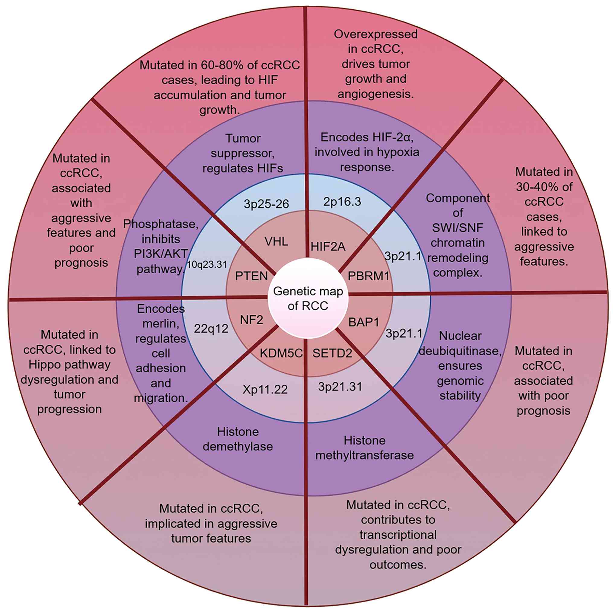 | Figure 1.Genetic landscape of renal cell
carcinoma. The associations between various genes and RCC are
shown. The visualization integrates various genetic alterations
such as histone modifiers and nuclear deubiquitinases, signifying
their role in genomic stability and the regulation of gene
expression, both of which have notable implications for developing
targeted therapies. These alterations contribute to RCC
pathogenesis and may influence prognosis, therapeutic vulnerability
or treatment response, although the strength and clinical maturity
of evidence vary among individual genes. Figure created with
Figdraw (www.figdraw.com). RCC, renal cell
carcinoma; ccRCC, clear cell renal cell carcinoma; HIF,
hypoxia-inducible factor; SWI/SNF, switch/sucrose non-fermentable;
NF2, neurofibromatosis type 2; VHL, von Hippel-Lindau; PBRM1,
polybromo 1; BAP1, BRCA1-associated protein 1; KDM5C,
lysine-specific demethylase 5C. |
Genes involved in chromatin and
epigenetic regulation
Molecular basis of PBRM1
The PBRM1 gene, located at 3p21.1, encodes
BRG1-associated factor 180, a key component of the switch/sucrose
non-fermentable chromatin remodeling complex. This complex
regulates gene expression by altering chromatin structure, thereby
influencing DNA transcription, replication and repair (23–25).
In RCC, particularly ccRCC, PBRM1 is frequently mutated or
inactivated, leading to impaired chromatin remodeling and aberrant
gene expression (26). Loss of
PBRM1 is associated with dysregulation of downstream targets
including genes involved in cell proliferation (such as cyclin D1),
angiogenesis (such as VEGFA) and metabolic pathways (such as solute
carrier family 2, which encodes GLUT1).
PBRM1 mutations are identified in 30–40% of ccRCC
cases, predominantly as inactivating mutations (26). Functionally, PBRM1 loss is
associated with increased expression of HIF target genes (such as
VEGFA and GLUT1), contributing to angiogenesis and glycolysis
(26). PBRM1-deficient cells
demonstrate enhanced migratory and invasive capabilities,
suggesting a role in metastatic progression (26,27).
Clinically, PBRM1-mutant tumors are associated with more advanced
disease stages and variable PFS outcomes (27). The association between PBRM1
alterations and immunotherapy response remains an active area of
investigation with conflicting data. Liu et al (28) reported that PBRM1 loss was
associated with reduced immune infiltration and potential
resistance to checkpoint inhibitors in the IMmotion150 and Memorial
Sloan Kettering-Integrated Mutation Profiling of Actionable Cancer
Targets cohorts. Conversely, Miao et al (29) reported that PBRM1 loss-of-function
was associated with clinical benefit from programmed cell death
protein 1 (PD-1)/programmed death-ligand 1 (PD-L1) blockade in
ccRCC and Braun et al (30)
further supported this association in an independent
randomized-trial cohort, potentially through modulation of the
tumor immune microenvironment. These discordant findings likely
reflect differences in patient populations, co-occurring mutations
(particularly BAP1 status) and the specific immunotherapy regimens
evaluated. Prospective biomarker-embedded trials are needed to
resolve this controversy. Integrated genomic analyses reveal that
PBRM1 mutations frequently co-occur with VHL inactivation and SETD2
mutations, indicating complex genetic interplay in ccRCC
development (23,24,26,27).
Clinical relevance of BAP1
The BAP1 gene, located at 3p21.1, encodes a nuclear
deubiquitinase involved in gene expression regulation, DNA repair,
cell cycle progression and apoptosis (31,32).
BAP1 interacts with BRCA1 and other proteins to maintain genomic
stability by removing ubiquitin modifications from histones and
proteins, thereby modulating chromatin structure (31).
BAP1 dysfunction in ccRCC is associated with tumor
development through increased genomic instability, chromatin
remodeling defects and impaired DNA damage response (31,33).
In genetically engineered mouse models, BAP1 loss has been shown to
drive ccRCC development and is associated with high-grade tumor
phenotypes and mTORC1 activation (34). Clinically, BAP1 mutations and
protein loss have been associated with aggressive tumor features
including high Fuhrman grade, advanced stage and poor prognosis
(33,35). Patients with BAP1-altered tumors
exhibit shorter OS and PFS compared with those with intact BAP1
(33,35). BAP1 loss disrupts DNA-damage
responses and has provided a mechanistic rationale for exploring
DNA-repair-directed strategies; however, whether BAP1 status
predicts sensitivity to poly(ADP-ribose) polymerase inhibition or
immune checkpoint blockade in RCC remains unproven (31,33).
Therapeutic targeting of SETD2
SETD2, located at 3p21.31, encodes the principal
histone methyltransferase responsible for H3K36 trimethylation and
contributes to transcriptional fidelity, DNA-damage responses and
genomic stability. Loss of SETD2 in RCC leads to broad epigenetic
and genomic dysregulation (36–38).
Preclinical RCC models further suggest that SETD2 loss may create
therapeutic vulnerabilities, including increased sensitivity to
PI3Kβ/Akt inhibition, despite no SETD2-based predictive strategy
having been clinically validated (39).
Lysine-specific demethylase 5C (KDM5C)
and chromatin remodeling
The KDM5C gene, located at Xp11.22, encodes a
histone lysine demethylase that specifically removes methyl groups
from lysine 4 of histone H3, serving a key role in transcriptional
regulation and chromatin remodeling. KDM5C contains numerous
functional domains including a catalytic JmjC domain, zinc
finger-C5HC2 domain, JmjN domain, Bright/ARID DNA-binding domain
and two plant homeodomain fingers for histone binding (40). In ccRCC, KDM5C mutations are
identified at relatively low frequency and are generally associated
with loss of function and altered histone methylation patterns
(40). Preclinical studies have
suggested that KDM5C loss may produce context-dependent epigenetic
effects and create therapeutic vulnerabilities in experimental
models; however, its clinical prognostic and predictive importance
remains investigational (40,41).
Genes regulating cell morphology and
the Hippo pathway
Neurofibromatosis type 2 (NF2) and the Hippo
pathway
The NF2 gene, located at 22q12, encodes the protein
merlin, which regulates cell adhesion, migration and proliferation
by interacting with cell membrane and cytoskeleton components
(42). Merlin functions upstream
of Hippo/yes-associated protein (YAP) signaling to restrain
YAP/transcriptional coactivator with PDZ-binding motif (TAZ)
activity and also interacts with focal-adhesion signaling (42). NF2 alterations are uncommon in
ccRCC; when present, NF2 loss disrupts merlin-mediated Hippo
pathway control, resulting in enhanced YAP/TAZ activity, increased
cell proliferation and invasiveness and resistance to apoptosis
(42,43).
Molecular pathways in RCC
RCC involves a complex interplay of molecular
pathways that drive tumor progression, metastasis and resistance to
therapy. The RCC therapeutic landscape involves three biologically
relevant axes: the VHL-HIF-angiogenesis axis, the immune checkpoint
axis and the chromatin-remodeling axis, each with distinct clinical
implications and evidence levels. Understanding these pathways
informs the development of targeted therapies and may ultimately
improve patient outcomes. Table
II summarizes the key molecular pathways, their key components
and therapeutic targets in RCC.
| Table II.Key molecular pathways in RCC. |
Table II.
Key molecular pathways in RCC.
| Pathway | Key components | Role in RCC |
Approved/investigational agents | (Refs.) |
|---|
| VEGF signaling | VEGF-A and
VEGFR-1/2/3 | Angiogenesis; tumor
vascularization | Sunitinib,
pazopanib, axitinib, cabozantinib and tivozanib | (7,44,45) |
| HIF signaling | HIF-1α, HIF-2α and
VHL | Hypoxia adaptation;
metabolic reprogramming | Belzutifan (HIF-2α
inhibitor) | (15,16,20,47) |
| mTOR signaling | mTORC1, mTORC2 and
TSC1/2 | Cell proliferation
and metabolism | Everolimus and
temsirolimus | (9,48,51) |
| PI3K/Akt | PI3K, Akt and
PTEN | Cell survival,
proliferation and metabolism | Investigational
(PI3K inhibitors and Akt inhibitors) | (21) |
| Ras/MAPK | KRAS, BRAF, MEK and
ERK | Cell proliferation
and differentiation | Investigational
(MEK inhibitors) | (54) |
| Immune
checkpoint | PD-1, PD-L1 and
CTLA-4 | Immune evasion | Nivolumab,
pembrolizumab, avelumab and ipilimumab | (4,5,56,57) |
| c-MET | HGF and c-MET | Cell migration,
invasion and angiogenesis | Cabozantinib | (73) |
| Metabolism | GLUT1, HK2, LDHA
and GLS | Warburg effect;
biosynthesis | Investigational
metabolism-directed approaches; no established standard RCC
therapy | (44,55) |
Angiogenesis-associated pathways
VEGF pathway
The VEGF pathway is involved in regulating
angiogenesis in RCC. VEGF upregulation is frequently driven by VHL
mutations, which prevent HIF degradation and contribute to
constitutive VEGF transcription (12,44).
VEGF-A, the most studied isoform, binds to VEGFR-1 and VEGFR-2 on
endothelial cells, activating downstream PI3K/Akt and MAPK pathways
that may promote endothelial cell proliferation, migration and
survival (45). VEGF
pathway-targeted TKIs have improved outcomes in ccRCC, but
intrinsic and acquired resistance remain key limitations of
VEGF-directed therapy (45).
Multi-target TKIs (such as cabozantinib) and combination strategies
pairing VEGF inhibition with ICIs represent approaches aimed at
addressing the adaptive capacity of RCC (46).
HIF-1 signaling and hypoxia
adaptation
The HIF-1 signaling pathway is involved in RCC
pathogenesis. HIF-1α accumulation drives transcription of
angiogenic and metabolic genes (44,47),
with PI3K/Akt/mTOR crosstalk enhancing oncogenic signaling
(48). Direct HIF-1α targeting has
not yet yielded an established therapeutic strategy in RCC, whereas
HIF-2α has proved more pharmacologically tractable; efforts have
therefore concentrated on downstream VEGF targets and HIF-2α
(49,50).
Cell proliferation and
metabolism-associated pathways
mTOR pathway
The mTOR pathway is involved in regulating cell
proliferation and metabolism. In RCC, mTOR activation occurs
through TSC1/2 mutations or upstream PI3K/Akt dysregulation, which
may contribute to uncontrolled cell proliferation (21,48).
Both everolimus and temsirolimus are rapamycin (sirolimus)
analogues that act as allosteric inhibitors of mTORC1, thereby
blocking downstream protein synthesis and proliferative signaling;
everolimus is an orally administered agent, whereas temsirolimus is
an intravenously administered ester prodrug of sirolimus that is
converted to sirolimus in vivo. Everolimus demonstrated PFS
benefit after prior VEGF-targeted therapy (9), whereas temsirolimus improved OS in
previously untreated poor-risk metastatic RCC (51). In the modern ICI/TKI era, mTOR
inhibition therefore occupies a more limited and context-dependent
role.
Preclinical genetically engineered mouse models have
implicated BAP1 loss in enhanced mTORC1 activation, although
whether BAP1 status confers differential sensitivity to mTOR
inhibitors remains unresolved (34,52).
In addition, VEGFR-TKIs and mTOR inhibitors may exert
immunomodulatory effects (such as a reduction of regulatory T cells
and myeloid-derived suppressor cells, enhanced dendritic-cell
antigen presentation and modulation of effector T-cell function)
within the RCC tumor microenvironment, although the clinical
predictive relevance of these immune effects remains uncertain
(53).
PI3K/Akt and cell survival
signaling
The PI3K/Akt pathway is initiated by growth factor
binding to receptor tyrosine kinases, activating PI3K to convert
PIP2 to PIP3, which recruits and activates Akt (21). Akt phosphorylates downstream
effectors including mTOR, GSK3β and FOXO transcription factors,
which may promote cell survival and proliferation. In ccRCC, PIK3CA
mutations and PTEN loss-of-function mutations are associated with
constitutive pathway activation (21). Although PI3K/Akt signaling remains
biologically relevant and selective PI3K or Akt inhibitors continue
to be investigated, neither class has established a clinically
validated single-agent role in RCC. By contrast, the downstream
mTOR inhibitors everolimus and temsirolimus have demonstrated
efficacy in defined clinical settings, although their roles have
become more limited in the contemporary ICI/TKI era (9,21,51).
Ras/MAPK and proliferation
control
Despite canonical KRAS mutations being uncommon in
ccRCC, MAPK pathway activation may occur through upstream receptor
tyrosine kinase signaling and pathway crosstalk (54). The Ras pathway intersects with the
VHL-HIF pathway, with hypoxia-induced HIFs enhancing Ras-mediated
oncogenic signaling (47). MEK
inhibitors such as trametinib have shown promise in preclinical
models (54), though clinical
application remains limited by pathway redundancy and compensatory
mechanisms. To the best of our knowledge, no MEK inhibitor has
demonstrated RCC-specific clinical activity sufficient to advance
to phase III development, illustrating the challenge of targeting
downstream signaling nodes in a pathway-driven tumor.
Metabolic reprogramming in RCC
RCC exhibits profound metabolic reprogramming,
including enhanced glycolysis (Warburg effect), glutaminolysis and
lipid synthesis, driven by VHL-HIF pathway activation (44,55).
While these metabolic alterations are well characterized
biologically, to the best of our knowledge, no metabolism-targeted
therapy has established clinical efficacy or a standard therapeutic
role in RCC and metabolic interventions remain investigational. The
primary therapeutic relevance of metabolic reprogramming currently
lies in its role as a resistance mechanism to VEGF-TKIs and ICIs,
rather than as a direct drug target.
Immune regulation-associated
pathways
Immune checkpoint regulation in RCC
The PD-1/PD-L1 and cytotoxic T-lymphocyte-associated
protein 4 (CTLA-4) axes represent important therapeutic targets in
RCC (56). PD-1 is an inhibitory
receptor expressed on T cells; its ligand PD-L1 is frequently
upregulated on RCC tumor cells. PD-1/PD-L1 interaction contributes
to immune evasion by inhibiting T-cell activation (56). PD-L1 expression has been explored
as a candidate biomarker in RCC trials; however, its predictive
utility has been inconsistent across regimens and is insufficient
for stand-alone treatment selection (4,56,57).
CTLA-4 inhibits T-cell activation at an earlier stage than PD-1;
CTLA-4 blockade enhances T-cell activation and tumor infiltration
(56). The phase III Nivolumab +
Ipilimumab vs. Sunitinib Trial (CheckMate 214) demonstrated durable
clinical benefit with dual CTLA-4/PD-1 blockade compared with
sunitinib in advanced RCC (4).
ICIs including nivolumab (anti-PD-1) and ipilimumab
(anti-CTLA-4) block these interactions, reactivating antitumor
immune responses (4,46). Combination approaches pairing ICIs
with anti-angiogenic agents have demonstrated improved antitumor
activity compared with VEGF-TKI monotherapy in phase III trials
(5,57), challenging the assumption that
immunotherapy benefit operates independently of VEGF signaling.
However, to the best of our knowledge, no direct ICI vs. ICI + TKI
head-to-head trials exist to conclusively establish comparative
efficacy.
IL-6/JAK/STAT3 signaling in RCC
The IL-6/JAK/STAT3 axis is a well-characterized
oncogenic and immunomodulatory pathway across cancers (58,59).
In metastatic ccRCC, elevated circulating IL-6 has been associated
with poorer clinical outcomes and reduced response to sunitinib
(60). Accordingly, the
IL-6/JAK/STAT3 pathway remains an investigational therapeutic axis
rather than an established treatment target in RCC.
Signal transduction-associated
pathways
NF-κB and TGF-β pathways
Both NF-κB and TGF-β signaling are involved in RCC
inflammatory microenvironment remodeling and immune evasion
(61,62). NF-κB activation is associated with
pro-survival and pro-inflammatory gene expression, while TGF-β is
implicated in immunosuppression through regulatory T-cell and
myeloid-derived suppressor cell (MDSC) recruitment (61,62).
Despite direct inhibitors of NF-κB and TGF-β signaling existing and
having been evaluated in preclinical and early-phase clinical
studies (63–65), to the best of our knowledge, no
study has demonstrated single-agent clinical activity in RCC. The
therapeutic relevance of these pathways in RCC lies primarily in
their role as mediators of resistance and microenvironmental
modulators. TGF-β inhibitors, for instance, are being explored
primarily as combination partners to enhance ICI efficacy by
reversing immunosuppression, rather than as standalone RCC
therapies. Similarly, NF-κB modulation remains investigational,
with its clinical utility currently limited to combination contexts
aimed at overcoming resistance to established therapies.
Cell adhesion and migration-associated
pathways
Epithelial-mesenchymal transition (EMT) and
metastatic potential
EMT contributes to RCC metastatic dissemination and
therapy resistance through transcriptional reprogramming (15,44).
However, EMT may represent a secondary consequence of oncogenic
signaling (VHL-HIF, TGF-β and receptor tyrosine kinase activation)
rather than a primary therapeutic target. No EMT-targeted agent has
demonstrated RCC-specific clinical activity and the concept of EMT
as a therapeutic target, despite extensive preclinical literature,
lacks sufficient prospective validation in RCC. EMT remains a
descriptive biological phenomenon rather than a druggable target,
with its clinical relevance limited to association with aggressive
disease phenotypes and as a potential biomarker of resistance to
VEGF-TKIs and ICIs (66).
Inflammatory response-associated
pathways
Inflammatory response pathway
The inflammatory response pathway in RCC involves a
number of cytokines, chemokines and immune cells. The IL-6
signaling axis is implicated in tumor growth, angiogenesis and
immune evasion through JAK/STAT pathway activation (58,59).
Tumor-associated macrophages adopt pro-tumorigenic phenotypes that
support tumor growth and metastasis (61,62).
Conceptually, inflammatory signaling bridges the angiogenic and
immune axes in RCC, explaining why anti-inflammatory strategies
alone have not achieved clinical utility while combination
approaches targeting both inflammation and angiogenesis or immune
checkpoints remain under active investigation.
Immune infiltration patterns differ significantly
among RCC molecular subtypes and may influence the response to
immunotherapy (67); however, no
inflammation-based signature has yet been sufficiently
prospectively validated for routine treatment selection. High
systemic inflammation, as measured by neutrophil-to-lymphocyte
ratio (NLR) or C-reactive protein, is associated with poor
prognosis (68).
Other pathways
Cell cycle dysregulation in RCC
Cell cycle dysregulation in RCC involves frequent
alterations in CDK signaling, cyclin expression and checkpoint
control, contributing to uncontrolled proliferation (67). Collectively, RCC molecular
pathogenesis is driven by coordinated dysregulation across
angiogenic, immune and epigenetic-metabolic axes rather than
isolated pathway activation, suggesting that therapeutic strategies
should address this multi-dimensional biology to achieve durable
clinical benefit (Fig. 2).
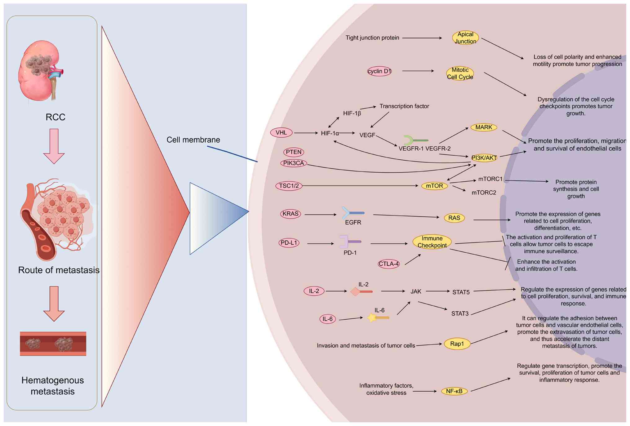 | Figure 2.Key molecular pathways driving RCC
progression and therapeutic targeting. Schematic diagram of the
molecular mechanisms underlying the key targeted pathways in renal
cell carcinoma. The figure presents a detailed schematic of the
signaling pathways and mechanisms involved in the progression and
metastasis of renal cell carcinoma, focusing on key targets for
potential therapies. This pathway map highlights key molecular
players and their interactions that drive tumorigenesis and
metastatic spread, offering insights into potential therapeutic
targets in the era of precision medicine. Figure created with
Figdraw (www.figdraw.com). RCC, renal cell
carcinoma; HIF, hypoxia-inducible factor; VHL, von Hippel-Lindau;
KRAS, Kirsten rat sarcoma viral oncogene homolog; RAS, rat sarcoma
virus; PD-1, programmed cell death protein 1; CTLA-4, cytotoxic
T-lymphocyte-associated protein 4; Rap1, Ras-proximate-1. |
Current clinical research and evidence
Targeted therapies have evolved notably in the
pursuit of precision medicine for RCC, offering diverse therapeutic
options. Phase III evidence establishes ICI combinations as active
first-line approaches, though trial diversity and the absence of
head-to-head comparisons limit definitive treatment hierarchies.
The following sections discuss clinical research and evidence
across various targeted approaches, from VEGF inhibition and
mTOR-targeted therapies to immunotherapies and emerging
combinations, highlighting advancements, comparative insights,
challenges and the role of biomarkers in guiding treatment
decisions. Efficacy data are primarily drawn from pivotal trial
publications, supplemented by mature or final follow-up analyses
where clinically relevant.
Targeted VEGF inhibition in RCC
The VEGF pathway is an important driver of
angiogenesis and tumor growth in RCC. First-generation VEGF
receptor TKIs established the foundation for targeted therapy. In a
pivotal phase III trial, sunitinib demonstrated a median PFS of 11
months compared with 5 months with interferon-α and an objective
response rate (ORR) of 31 vs. 6% (11,69).
Sorafenib improved median PFS to 5.5 months compared with 2.8
months with placebo in the TARGET trial (70). Axitinib, a second-generation
selective TKI, exhibited improved PFS (6.7 months) compared with
sorafenib (4.7 months) in previously treated advanced RCC (71).
Combination therapies are being explored to address
therapeutic resistance. A Phase Ib trial of axitinib +
pembrolizumab reported a 58% ORR and a median PFS of 20.9 months in
the dose-expansion cohort, illustrating the complementary
mechanisms of VEGF inhibition and immune checkpoint blockade
(72). Next-generation VEGF
inhibitors such as cabozantinib target mesenchymal-epithelial
transition (MET) and AXL in addition to VEGFR. The Cabozantinib vs.
Everolimus in Advanced Renal-Cell Carcinoma Trial (METEOR)
demonstrated that cabozantinib significantly improved PFS [7.4 vs.
3.9 months; hazard ratio (HR) 0.51; P<0.0001] and OS (21.4 vs.
17.1 months; HR 0.70; P=0.0002) compared with everolimus in
previously treated patients (73,74).
The Cabozantinib vs. Sunitinib in Metastatic Renal Cell Carcinoma
Trial (CABOSUN) demonstrated that cabozantinib improved median PFS
compared with sunitinib as first-line therapy in intermediate- and
poor-risk patients (8.6 vs. 5.3 months by independent review; HR
0.48), with updated median OS of 26.6 vs. 21.2 months (HR 0.80)
(75,76).
Tivozanib is a highly selective VEGFR-1/2/3
inhibitor with a narrower kinase inhibition profile compared with
broader-spectrum TKIs. Its clinical activity was initially
demonstrated in the phase III TIVO-1 trial (77), while the phase III TIVO-3 trial
further supported its role in previously treated advanced RCC by
showing improved PFS compared with sorafenib (78). Therefore, tivozanib is best
discussed as a VEGFR-selective option with particular relevance in
the later-line setting rather than as a broadly preferred
first-line standard (77,78). Despite these advancements,
challenges persist. Biomarkers to predict VEGF inhibitor response
remain limited and angiogenesis pathway redundancy contributes to
treatment failure (66). The
broader toxicity profile of cabozantinib (including palmar-plantar
erythrodysesthesia and gastrointestinal adverse events) requires
experience in management (79)
(Table III).
| Table III.Targeted therapeutic agents in
RCC. |
Table III.
Targeted therapeutic agents in
RCC.
| Target | Agent | Mechanism | Clinical
indication | Key trial | (Ref.) |
|---|
| VEGFR-1/2/3 | Sunitinib | Multi-targeted
TKI | Selected/historical
first-line option for advanced RCC | Phase III vs. IFN-α
(PFS 11 vs. 5 months) | (11) |
| VEGFR-1/2/3 | Pazopanib | Multi-targeted
TKI | Selected/historical
first-line option for advanced RCC | Phase III vs.
sunitinib (non-inferior) | (95) |
| VEGFR-1/2/3 | Axitinib | Selective VEGFR
TKI | Second-line
advanced RCC | AXIS (PFS 6.7 vs.
4.7 months vs. sorafenib) | (71) |
| VEGFR, MET and
AXL | Cabozantinib | Multi-targeted
TKI | Later-line therapy;
historical first-line evidence in intermediate/poor-risk
disease | METEOR and
CABOSUN | (73,75) |
| VEGFR-1/2/3 | Tivozanib | Selective VEGFR
TKI | Later-line advanced
RCC after prior systemic therapy; earlier-line activity evaluated
in TIVO-1 | TIVO-3: PFS 5.6 vs.
3.9 months vs. sorafenib; TIVO-1: PFS benefit vs. sorafenib | (77,78) |
| mTORC1 | Everolimus | mTOR inhibitor | Historical
post-TKI/later-line option in selected patients | RECORD-1 (PFS 4.9
vs. 1.9 months) | (9) |
| mTORC1 | Temsirolimus | mTOR inhibitor | Historical
first-line evidence in untreated poor-risk RCC; limited
contemporary role | ARCC (OS 10.9 vs.
7.3 months) | (51) |
| VEGFR, FGFR, PDGFR,
RET and KIT | Lenvatinib | Multi-targeted
TKI | First-line (combo);
second-line (combo) | CLEAR and Study
205 | (6,92) |
| HIF-2α | Belzutifan | HIF-2α
inhibitor | Post-anti-PD-L1 and
post-VEGF-TKI | LITESPARK-005 | (20) |
mTOR-targeted therapies
The mTOR pathway regulates cell proliferation and
angiogenesis in RCC. The Renal Cell Cancer Treatment with Oral
RAD001 Given Daily Trial (RECORD-1) trial established everolimus as
a commonly used second-line therapy after TKI failure,
demonstrating a median PFS of 4.9 vs. 1.9 months for placebo
(P<0.001) (9). Subsequent phase
II studies further evaluated single-agent everolimus in metastatic
RCC. The phase II trial by Amato et al (80) reported disease stabilization and
prolonged progression-free survival in patients with predominantly
ccRCC, supporting the antitumor activity of mTOR inhibition in this
setting. Temsirolimus demonstrated superior survival benefits in
patients with poor prognosis. The Advanced Renal Cell Carcinoma
Trial revealed a median OS of 10.9 months for temsirolimus vs. 7.3
months for interferon-α (P=0.008) in untreated high-risk patients
with metastatic RCC (51).
Biomarkers are key for personalizing mTOR-targeted
therapy. Combined subgroup analyses of the RECORD-1 and REACT
trials demonstrated that hyperglycemia and hypercholesterolemia
during everolimus treatment associated with improved PFS (7.4 vs.
5.3 months, P=0.01), suggesting metabolic toxicity as a potential
surrogate for drug activity (81).
Proactive management of everolimus-associated adverse events,
including stomatitis, noninfectious pneumonitis and metabolic
abnormalities, is important for maintaining dose intensity
(82).
Targeting PD-1/PD-L1 and CTLA-4 in
RCC
The PD-1/PD-L1 axis is key in tumor immune evasion.
Nivolumab, a monoclonal antibody targeting PD-1, demonstrated
notable efficacy in advanced RCC after prior VEGF-targeted therapy.
In the phase III CheckMate 025 trial, nivolumab showed superior OS
compared with everolimus (25.0 vs. 19.6 months; HR 0.73; P=0.002)
with an ORR of 25 vs. 5% (83).
Ipilimumab + nivolumab combination therapy has
reshaped first-line treatment. In an 8-year analysis of CheckMate
214, nivolumab + ipilimumab maintained an OS benefit vs. sunitinib
in both the intention-to-treat population (median OS, 52.7 vs. 37.8
months; HR 0.72, 95% CI 0.62–0.83) and the intermediate/poor-risk
population (median OS, 46.7 vs. 26.0 months; HR 0.69, 95% CI
0.59–0.81) (84). In the
intent-to-treat (ITT) population, median PFS was similar (12.4 vs.
12.3 months; HR 0.88), whereas greater PFS separation was observed
in the intermediate/poor-risk subgroup. At the final analysis with
9.3 years of median follow-up, the OS benefit remained durable in
the ITT population (HR 0.71; 95% CI 0.62–0.82), with estimated
108-month OS probabilities of 31.4 vs. 19.5% (85).
The Pembrolizumab + Axitinib vs. Sunitinib Trial
(KEYNOTE-426) demonstrated efficacy over sunitinib in the
first-line setting, with a median OS of 47.2 vs. 40.8 months (HR
0.84; 95% CI 0.71–0.99) and median PFS of 15.7 vs. 11.1 months (HR
0.69; 95% CI 0.59–0.81) (5). The
ORR was 60.6 vs. 39.6%, with complete responses in 11.6 vs. 4.0% of
patients. In addition, 5-year follow-up data demonstrated durable
benefit (5).
The Avelumab + Axitinib vs. Sunitinib Trial (JAVELIN
Renal 101) exhibited significant PFS improvement compared with
sunitinib in the primary analysis (13.8 vs. 8.4 months; HR 0.69;
95% CI 0.574–0.825; P<0.0001) (57). In the final analysis with a minimum
follow-up of 68 months, median PFS was 13.9 vs. 8.5 months (HR
0.66) and the median OS was 44.8 vs. 38.9 months in the overall
population (HR 0.88; 95% CI 0.749–1.039; P=0.0669) and 43.2 vs.
36.2 months in the PD-L1-positive population (HR 0.86; 95% CI
0.701–1.057; P=0.0755), favoring the avelumab + axitinib arm but
not reaching statistical significance (86). The ORR was 59.7 vs. 32.0% (86). Nivolumab + cabozantinib also
demonstrated durable first-line efficacy in CheckMate 9ER. In the
final analysis (median follow-up, 5.6 years), median PFS was 16.4
vs. 8.3 months (HR 0.58) and median OS was 46.5 vs. 35.5 months (HR
0.79) compared with sunitinib (8).
The Lenvatinib + Pembrolizumab or Everolimus for
Advanced Renal Cell Carcinoma Trial (CLEAR) demonstrated that
lenvatinib + pembrolizumab is an active first-line option. In the
final prespecified OS analysis, median OS was 53.7 months with
lenvatinib + pembrolizumab vs. 54.3 months with sunitinib (HR 0.79;
95% CI 0.63–0.99), while median PFS remained markedly longer with
the combination (23.9 vs. 9.2 months; HR 0.47), with an ORR of 71.3
vs. 36.7% (6,87). However, the toxicity profile of
lenvatinib + pembrolizumab (including hypertension, diarrhea and
proteinuria) requires careful patient selection and monitoring.
Targeting fibroblast growth factor
receptor (FGFR), cellular (c)-MET and HIF-2α
FGF/FGFR signaling has been implicated as a bypass
pathway contributing to resistance to VEGF-directed therapy in RCC
(66). However, FGFR-targeted
agents have not established a validated clinical role in RCC and
RCC-specific prospective evidence remains limited. c-MET signaling
is dysregulated in RCC, particularly in the papillary subtype
(88). Cabozantinib, a
multi-kinase inhibitor targeting c-MET, VEGFR and AXL, demonstrated
efficacy in the METEOR trial in previously treated patients
(73). Belzutifan, a HIF-2α
inhibitor, demonstrated proof-of-concept efficacy in VHL
disease-associated RCC (19) and
in post-treatment sporadic ccRCC (20).
Combination therapy
Combination therapies integrating ICIs with TKIs or
dual checkpoint blockade have emerged as clinically impactful
approaches. Table IV presents
verified efficacy data from pivotal phase II and III trials.
Furthermore, the heterogeneity and complexity of RCC necessitate
multifaceted treatment strategies. Combination therapies targeting
distinct pathways can synergistically inhibit tumor growth and
angiogenesis while mitigating resistance. Pairing ICIs with
VEGF-TKIs leverages immune recognition complemented by
anti-angiogenic effects, creating a more favorable tumor
microenvironment.
| Table IV.Key clinical trials of systemic
therapies in advanced renal cell carcinoma. |
Table IV.
Key clinical trials of systemic
therapies in advanced renal cell carcinoma.
| Trial (NCT) | Regimen | Phase | n (comparison
arms) | Median OS
(months) | Median PFS
(months) | ORR (%) | HR (OS) | HR (PFS) | (Refs.) |
|---|
| CheckMate 214 | Nivolumab +
ipilimumab | III | 1096 | 52.7 vs. 37.8 | 12.4 vs. 12.3 | 39.5 vs. 33.0 | 0.71 (ITT, | 0.88 (ITT) | (84,85) |
| (NCT02231749) | vs. sunitinib |
|
| (ITT) | (ITT) | (ITT) | final
analysis) |
|
|
| KEYNOTE-426 | Pembrolizumab +
axitinib | III | 861 | 47.2 vs. 40.8 | 15.7 vs. 11.1 | 60.6 vs. 39.6 | 0.84 | 0.69 | (5) |
| (NCT02853331) | vs. sunitinib |
|
|
|
|
|
|
|
|
| JAVELIN Renal
101 | Avelumab +
axitinib | III | 886 | 44.8 vs. 38.9 | 13.9 vs. 8.5 | 59.7 vs. 32.0 | 0.88 | 0.66 | (86) |
| (NCT02684006) | vs. sunitinib |
|
|
|
|
|
|
|
|
| CLEAR | Lenvatinib + | III | 712 (355 vs.
357) | 53.7 vs. 54.3 | 23.9 vs. 9.2 | 71.3 vs. 36.7 | 0.79 | 0.47 | (6,87) |
| (NCT02811861) | pembrolizumab vs.
sunitinib |
|
|
|
|
|
|
|
|
| CheckMate 9ER | Nivolumab + | III | 651 | 46.5 vs. 35.5 | 16.4 vs. 8.3 | 55.7 vs. 27.4 | 0.79 | 0.58 | (8) |
| (NCT03141177) | cabozantinib vs.
sunitinib |
|
|
|
|
|
|
|
|
| METEOR | Cabozantinib
vs. | III | 658 | 21.4 vs. 17.1 | 7.4 vs. 3.9 | 17.0 vs. 3.0 | 0.70 | 0.51 | (73,74) |
| (NCT01865747) | everolimus |
|
|
|
|
|
|
|
|
| CABOSUN | Cabozantinib vs.
sunitinib | II | 157 | 26.6 vs. 21.2 | 8.6 vs. 5.3 | 20.0 vs. 9.0 | 0.80 | 0.48 | (76) |
| (NCT01835158; |
|
|
|
| (IR) | (IR) |
|
|
|
| Alliance
A031203) |
|
|
|
|
|
|
|
|
|
| Study 205 | Lenvatinib +
everolimus | II | 101 (51 vs.
50) | 25.5 vs. 15.4 | 14.6 vs. 5.5 | 43,0 vs. 6.0 | 0.51 | 0.40 | (92) |
| (NCT01136733) | vs. everolimus |
|
|
|
|
|
|
|
|
| LITESPARK-005 | Belzutifan vs.
everolimus | III | 746 | 21.4 vs. 18.1 | 5.6 vs. 5.6 | 21.9 vs. 3.5 | 0.88 (NS) | 0.75 | (20) |
| (NCT04195750) |
|
|
| (P=0.200NS) | (P=0.002) |
|
|
|
|
| CheckMate 025 | Nivolumab vs.
everolimus | III | 821 | 25.0 vs. 19.6 | 4.6 vs. 4.4 | 25.0 vs. 5.0 | 0.73 | 0.88 | (83) |
| (NCT01668784) |
|
|
|
|
|
|
|
|
|
The CheckMate 214 trial established nivolumab +
ipilimumab as a commonly used option for intermediate- and
poor-risk patients, demonstrating improved long-term OS and ORR
compared with sunitinib (Table
IV) (84). However, nivolumab
+ ipilimumab has a distinct immune-mediated toxicity profile. In
long-term follow-up, overall grade 3–4 treatment-associated adverse
events were less frequent compared with sunitinib, although
immune-mediated adverse events may require corticosteroid treatment
and can lead to treatment discontinuation (85,89).
KEYNOTE-426, JAVELIN Renal 101 and CLEAR each demonstrated improved
PFS for their respective ICI-TKI combinations compared with
sunitinib (Table IV) (5,6,57).
A critical efficacy finding was demonstrated
through the Belzutifan vs. Everolimus for Advanced Renal-Cell
Carcinoma Trial (LITESPARK)-005, which evaluated belzutifan vs.
everolimus in heavily pretreated patients (post-ICI and
post-VEGF-TKI). While median PFS was identical between arms (5.6
vs. 5.6 months), PFS was significantly improved with belzutifan (HR
0.75, 95% CI 0.63–0.90; two-sided log-rank P=0.002) and ORR favored
belzutifan (21.9 vs. 3.5%), OS did not reach statistical
significance (HR 0.88; 95% CI 0.73–1.07; P=0.200), representing an
important limitation in this refractory population (20). The absence of a statistically
significant OS advantage at the reported analysis tempers
enthusiasm for belzutifan monotherapy in this population and
combination strategies may further define its optimal clinical
role. Other belzutifan-based combination strategies remain
investigational, whereas LITESPARK-011 has now reported phase III
efficacy results (90).
Building upon these findings, the phase III
LITESPARK-011 trial (NCT04586231) evaluated belzutifan + lenvatinib
vs. cabozantinib in patients with advanced clear cell RCC after
progression on anti-PD-L1 therapy; at the second interim analysis,
the combination significantly improved median PFS (14.8 vs. 10.7
months; HR 0.70; 95% CI 0.59–0.84; one-sided P<0.0001) and ORR
(52.6 vs. 40.2%), while median OS (34.9 vs. 27.6 months; HR 0.85)
did not reach statistical significance at the time of this interim
analysis (90). In addition, the
phase III LITESPARK-022 study (NCT05239728) evaluated adjuvant
pembrolizumab + belzutifan vs. pembrolizumab plus placebo in
patients with clear cell RCC at increased risk of recurrence after
nephrectomy, demonstrating significantly improved disease-free
survival (HR 0.72; 95% CI 0.59–0.87; two-sided P<0.001),
although OS data remain immature (91). These trials support an expanding
role for HIF-2α-directed therapy across sequential, combination and
adjuvant settings, although optimal patient selection, treatment
sequencing and mature OS benefit remain to be defined.
The combination of lenvatinib + everolimus
(Lenvatinib + Everolimus Combination Trial (Study 205)], a phase II
three-arm trial in previously treated patients, demonstrated a
median PFS of 14.6 vs. 5.5 months (HR 0.40; P=0.0005) and median OS
of 25.5 vs. 15.4 months (HR 0.51; P=0.024) compared with everolimus
alone (92). This supported
subsequent first-line combination development.
Ongoing trials are exploring novel combinations,
including the addition of C-X-C chemokine receptor type 4 (CXCR4)
inhibitors to immunotherapy regimens. A phase 1b trial of the CXCR4
inhibitor mavorixafor in combination with nivolumab in patients
with advanced ccRCC unresponsive to nivolumab monotherapy
demonstrated potential antitumor activity and a manageable safety
profile (93). In addition, the
search for reliable biomarkers to guide treatment selection and
predict responses remains a key area of investigation.
Current directions and challenges
Precision therapy approaches in
advanced RCC
The transition from empiric to precision therapy in
RCC requires integrating molecular profiling, validated biomarkers
and resistance-aware treatment sequencing, objectives that remain
incompletely realized. Precision medicine in RCC has progressed
beyond single-biomarker approaches toward integrated,
multi-dimensional decision models. Molecular profiling at baseline
establishes the biological rationale for subsequent therapeutic
decisions. VHL mutation status represents a central biological
axis: VHL inactivation activates the HIF-VEGF axis and provides a
strong biological rationale for sensitivity to anti-angiogenic
therapy (50); however, to the
best of our knowledge, VHL status has not been prospectively
validated as a predictive biomarker for treatment selection. Given
the important role of VHL-HIF axis dysregulation in ccRCC
pathogenesis, HIF-2α inhibition with belzutifan is a
mechanistically rational approach. However, translation from
VHL-disease biology to sporadic ccRCC efficacy requires careful
population-specific interpretation, as the clinical activity in VHL
disease does not fully predict outcomes in sporadic metastatic RCC
(20).
International Metastatic Renal Cell Carcinoma
Database Consortium (IMDC) risk stratification integrates clinical
and laboratory parameters (Karnofsky performance status, time from
diagnosis to treatment, hemoglobin, calcium, neutrophil count and
platelet count) to categorize patients into favorable, intermediate
and poor risk groups (94). IMDC
is an established prognostic model (94) and has subsequently been
incorporated into multiple phase III RCC trials (4–6).
However, whether IMDC risk independently predicts differential
benefit from specific therapeutic classes (ICI combinations vs. TKI
monotherapy) remains less certain, as cross-trial comparisons are
inherently limited by population differences.
Key chromatin remodeling gene mutations provide
additional prognostic information. BAP1 mutations associate with
aggressive disease features and shorter OS (33,35).
PBRM1 mutations show variable prognostic impact depending on
co-occurring alterations; the association between PBRM1 alterations
and immunotherapy response remains controversial, with conflicting
data across different cohorts that likely reflect co-occurring
mutations (especially BAP1), patient heterogeneity and assay
differences (28–30). SETD2 mutations are associated with
higher tumor grade and increased metastasis risk (37). PD-L1 expression has been evaluated
as an adjunctive biomarker in several RCC trials; however, its
predictive utility is inconsistent and it is not validated for
stand-alone treatment selection (57).
First-line treatment selection is guided primarily
by IMDC risk category and histological features. For patients of
IMDC favorable-risk, TKI monotherapy, including sunitinib (11) or pazopanib (95), remains an active treatment option
based on phase III evidence, particularly for patients with low
disease burden, indolent disease behavior or contraindications to
immunotherapy (7,11,95).
Alternatively, ICI + TKI combinations (such as pembrolizumab +
axitinib or nivolumab + cabozantinib) may be considered for
patients of favorable-risk with high disease burden or rapid
progression potential (5,8). The choice incorporates patient
comorbidities, performance status and toxicity preferences. For
patients of IMDC intermediate/poor-risk, dual ICI with nivolumab +
ipilimumab offers durable long-term benefit; at 8-year follow-up,
median OS was 46.7 months vs. 26.0 months with sunitinib (HR 0.69;
95% CI 0.59–0.81) (84). ICI + TKI
combinations such as pembrolizumab + axitinib, nivolumab +
cabozantinib and lenvatinib + pembrolizumab have produced high
objective response rates in their respective phase III trials and
may be considered when rapid tumor shrinkage is clinically
important (5–8,57).
However, to the best of our knowledge, no head-to-head trial
directly compares dual ICI with ICI + TKI regimens and indirect
response-rate comparisons across trials should therefore be
interpreted cautiously. For non-clear-cell RCC, treatment selection
should be histology-specific. In papillary RCC, cabozantinib has
randomized phase II evidence from the SWOG 1500/PAPMET trial
(96), whereas evidence for other
non-clear-cell histologies is more heterogeneous and enrollment in
clinical trials is encouraged.
Upon disease progression, treatment selection is
guided by the prior therapeutic context. After progression on
VEGF-TKI monotherapy, cabozantinib has shown activity in clinical
trials (multi-target TKI with MET/AXL inhibition) (73), nivolumab (ICI switch) (83), lenvatinib + everolimus (TKI + mTOR
combination) (92) or tivozanib
(selective VEGFR TKI) (77,78).
The choice depends on prior TKI exposure, residual toxicity, and
molecular features. After progression on ICI-based therapy,
VEGF-directed TKIs remain important treatment options. Cabozantinib
served as the active comparator in the phase III CONTACT-03 trial
after prior ICI therapy, whereas the addition of atezolizumab to
cabozantinib did not improve outcomes (97). Tivozanib represents another
later-line VEGFR-directed option supported by phase III TIVO-3 data
(78). Belzutifan (HIF-2α
inhibitor) is a mechanistically rational option after prior ICI and
VEGF-TKI therapy, based on LITESPARK-005 results showing PFS
benefit (20), though the absence
of a statistically significant OS advantage at the reported
analysis tempers enthusiasm for belzutifan monotherapy in this
population.
Continuous monitoring enables treatment
modification throughout the disease course. Among candidate
biomarkers, PBRM1 remains investigational because associations with
ICI response have been inconsistent across cohorts and may be
influenced by co-occurring alterations such as BAP1 (28–30).
BAP1 is more established as a prognostic marker associated with
aggressive tumor biology than as a validated predictive marker for
treatment selection (33,35). PD-L1 expression should not be used
as a stand-alone treatment-selection biomarker in RCC because its
predictive associations vary across therapeutic regimens, assay
platforms and cut-off definitions (4,57).
Circulating biomarkers, including circulating tumor (ctDNA) and
circulating tumor cells (CTCs), also remain investigational;
relatively low ctDNA shedding and the lack of prospectively
validated treatment-adaptation algorithms currently limit routine
clinical implementation. Systemic inflammatory markers such as NLR
and CRP provide prognostic information in ICI-treated RCC cohorts,
with elevated values generally associated with poorer outcomes
(68). Standard radiographic
assessment per RECIST remains the cornerstone of response
evaluation, performed at regular intervals (typically every 8–12
weeks during active therapy) (7,98).
Multi-omics integration and precision
medicine
RCC continues to pose challenges in oncology
despite key therapeutic advances. The integration of multi-omics
data, including genomics, transcriptomics, proteomics, metabolomics
and spatial profiling, holds promise for unraveling the complex
molecular landscape of RCC (67,99).
Recent studies have highlighted the importance of genomic
classification in RCC, emphasizing the need for personalized
treatment strategies (67,99). Notably, a multi-omic analysis of
100 ccRCC cases integrating genomic, transcriptomic, proteomic,
metabolomic and spatial data identified metabolic reprogramming
associated with disease progression (99). Metabolomic analyses have revealed
potential therapeutic targets and biomarkers. Single-cell omics
approaches are emerging but remain underexplored in RCC; their
application resolves intra-tumor heterogeneity and clarifies
differential therapeutic responses at the cellular level. By
combining multi-omics datasets, including prospective single-cell
profiling, researchers can develop more precise treatment plans
tailored to individual patients.
Artificial intelligence and radiomics are emerging
as complementary tools for precision oncology in RCC. Machine
learning algorithms applied to radiomic features extracted from
routine imaging may non-invasively predict molecular subtypes,
treatment response and resistance patterns, though these approaches
remain investigational and require prospective validation before
clinical implementation (100,101).
Drug resistance: Mechanisms and
clinical implications
Drug resistance in RCC follows distinct
evolutionary patterns depending on the therapeutic class.
Understanding these mechanisms informs rational treatment
sequencing and the design of clinical trials evaluating novel
combination approaches.
Resistance to VEGF-TKIs involves primary mechanisms
(alternative pro-angiogenic signaling through FGF/PDGF, MET pathway
activation and MDSC recruitment) (66) and acquired mechanisms (EMT,
alternative angiogenic factor upregulation and vessel co-option).
Collectively, VEGF resistance reflects adaptive vascular rewiring
rather than linear pathway escape, explaining why sequential
VEGF-directed strategies and combination approaches targeting
parallel axes (ICI, HIF-2α) have become necessary.
Resistance to ICIs reflects both tumor-intrinsic
and microenvironmental factors. An important challenge is that RCC
employs multiple redundant immune evasion mechanisms, antigen loss,
T-cell exhaustion and alternative checkpoint upregulation, meaning
no single intervention is likely to restore immune control in
resistant disease. Primary resistance involves an immunologically
‘cold’ phenotype with defective antigen presentation and an
immunosuppressive microenvironment (56). Acquired resistance develops through
T-cell exhaustion, antigen loss through immunoediting, and
compensatory checkpoint upregulation (56). Strategies intended to limit or
circumvent primary immune resistance include dual checkpoint
blockade and ICI-TKI combinations (4,6).
However, these first-line trials do not establish reversal of
acquired ICI resistance and to the best of our knowledge, no
strategy has shown consistent efficacy in reversing established
resistance. Novel targets such as lymphocyte activation gene 3
(LAG-3) and T-cell immunoreceptor with Ig and ITIM domains (TIGIT)
therefore remain under investigation.
Resistance to HIF-2α inhibition remains
incompletely characterized. Translational studies with the
first-generation HIF-2α inhibitor PT2385 identified an acquired
EPAS1/HIF-2α G323E gatekeeper mutation that interfered with drug
binding and preserved HIF-2 complex formation, providing evidence
for on-target acquired resistance in ccRCC (102). Additional HIF-independent or
alternative clonal escape mechanisms remain under investigation.
Belzutifan-based combinations are being explored clinically.
Notably, LITESPARK-011 has now reported phase III results
demonstrating improved PFS with belzutifan + lenvatinib vs.
cabozantinib after prior anti-PD-L1 therapy (90), whereas other belzutifan-based
combination strategies remain investigational.
Potential cross-class interactions merit
consideration. VEGF-pathway inhibition can exert immunomodulatory
effects and may alter the tumor microenvironment in ways that could
influence subsequent ICI activity (103,104). Conversely, VEGF-directed TKIs
retain clinical activity in selected patients after ICI
progression, as illustrated by the cabozantinib comparator arm in
CONTACT-03 (97). These
observations are hypothesis-generating and prospective evidence
defining optimal cross-class sequencing remains lacking (Fig. 3).
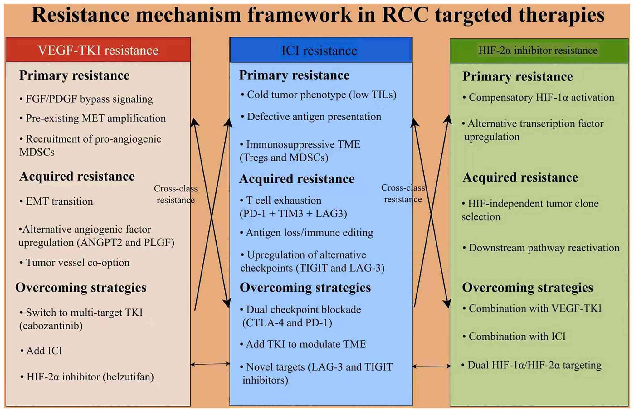 | Figure 3.Resistance mechanisms across systemic
therapies in RCC. Schematic overview of resistance mechanisms to
VEGF-TKIs, ICIs and HIF-2α inhibitors in RCC. VEGF-TKI resistance
arises from FGF/PDGF bypass signaling, MET pathway activation,
MDSC-driven angiogenesis and acquired EMT with compensatory
pro-angiogenic factors (such as ANGPT2 and PLGF) and vessel
co-option (66). ICI resistance is
mediated by a non-inflamed (‘cold’) tumor microenvironment,
defective antigen presentation and immunosuppressive infiltrates
(Tregs and MDSCs), while acquired escape involves T-cell exhaustion
(PD-1/TIM-3/LAG-3), immune editing and alternative checkpoint
upregulation (TIGIT and LAG-3) (56). HIF-2α inhibitor resistance includes
acquired EPAS1/HIF-2α gatekeeper mutations (such as G323E, reported
with the first-generation inhibitor PT2385) and potential
HIF-independent clonal escape; these mechanisms remain incompletely
defined and are based partly on translational and preclinical
evidence (102). Potential
cross-class adaptation and sequencing interactions reflect dynamic
therapeutic interdependence across angiogenic, immune and
hypoxia-driven axes (103,104).
Potential or investigational strategies include multi-target TKIs
(such as cabozantinib), ICI-based combinations (CTLA-4/PD-1
blockade), VEGF-ICI dual targeting and next-generation
HIF-2-directed approaches. Figure created with Figdraw (www.figdraw.com).RCC, renal cell carcinoma; TKI,
tyrosine kinase inhibitor; ICI, immune checkpoint inhibitor; HIF,
hypoxia-inducible factor; FGF, fibroblast growth factor; PDGF,
platelet-derived growth factor; MET, mesenchymal-epithelial
transition factor; MDSC, myeloid-derived suppressor cell; EMT,
epithelial-mesenchymal transition; ANGPT2, angiopoietin 2; PLGF,
placental growth factor; Treg, T regulatory cell; PD-1, programmed
cell death protein 1; TIM-3, T-cell immunoglobulin and mucin-domain
containing-3; LAG-3, lymphocyte-activation gene 3; TIGIT, T-cell
immunoreceptor with Ig and ITIM domains; EPAS1, endothelial PAS
domain protein 1. |
Controversies, unresolved questions
and evidence limitations
A number of therapeutic approaches that once
appeared promising have not yet established durable roles in RCC
clinical practice. Everolimus was commonly used as second-line
therapy after the RECORD-1 trial demonstrated PFS benefit vs.
placebo (9), but nivolumab
(83) and cabozantinib (73) have largely replaced it in this
setting based on improved OS data. Everolimus monotherapy now has a
limited role except in specific contexts where immunotherapy is
contraindicated. Although TIVO-1 did not establish tivozanib as a
broadly adopted first-line standard, subsequent TIVO-3 data
supported its role as a later-line option in previously treated
advanced RCC (77,78). Thus, tivozanib is better viewed as
a VEGFR-selective later-line therapy rather than as a failed
therapeutic strategy. FGFR inhibitors, including erdafitinib
(105), have demonstrated
activity in urothelial carcinoma but lack RCC-specific phase III
evidence; extrapolation from other tumor types is not supported by
current evidence without dedicated trials. Imatinib was evaluated
directly in metastatic RCC, but a small phase II study reported no
objective responses among 12 evaluable patients, illustrating the
limited clinical translation of KIT/PDGF receptor-directed therapy
in unselected RCC populations (106).
The biomarker landscape in RCC remains marked by
inconsistency and limited clinical utility. PBRM1 mutations
illustrate this challenge: A number of studies suggest enhanced ICI
benefit while others demonstrate resistance (28–30).
This controversy likely reflects co-occurring mutations (especially
BAP1), patient heterogeneity, differences in assay platforms and
varying analytical approaches across cohorts. No consensus exists
on the clinical utility of PBRM1 as a predictive biomarker and its
use in routine treatment selection is not supported by prospective
data. Overall, no single biomarker has achieved sufficient
consistency and prospective validation to guide routine clinical
decision-making in RCC. PD-L1 expression was incorporated into
prespecified analyses in trials such as JAVELIN Renal 101 (57), but its predictive value has not
been consistent across RCC trials. CheckMate 214 showed benefit
regardless of PD-L1 status (4) and
it is not validated as a stand-alone treatment-selection biomarker.
These discordant results limit routine clinical use and PD-L1
remains an adjunctive rather than definitive predictive marker. VHL
is a central pathway-defining alteration in ccRCC, but its clinical
predictive value for selecting among anti-angiogenic therapies
remains unvalidated; routine VHL testing to guide TKI selection has
not been adopted (12,50). ctDNA holds promise in other
malignancies for response monitoring and resistance detection, but
RCC-specific validation is lacking due to lower tumor shedding
rates and no prospective data support ctDNA-guided treatment
adaptation in RCC.
From a clinical implementation perspective and
consistent with the aforementioned biomarker-specific assessment,
none of these molecular biomarkers currently provide sufficient
prospective evidence for routine treatment selection in RCC. PBRM1,
BAP1, PD-L1 and circulating biomarkers such as ctDNA and CTCs all
remain investigational for treatment selection and their predictive
utility requires prospective validation in dedicated clinical
trials.
Numerous mechanistic assumptions underlying RCC
therapy have been challenged or tempered by clinical evidence. The
assumption that ‘VEGF inhibition alone is sufficient’ has been
challenged by the rapid emergence of resistance to single-agent
VEGF-TKIs and the subsequent need for multi-target TKIs and
combination approaches, though even these more intensive strategies
demonstrate limited long-term durability, highlighting the adaptive
capacity of RCC. The assumption that ‘immunotherapy benefit is
independent of VEGF’ has been challenged by ICI + TKI combination
data showing improved outcomes to historical ICI monotherapy data
(5,6,8),
though to the best of our knowledge, no direct ICI vs. ICI + TKI
head-to-head trials exist to conclusively address this question.
The assumption that ‘HIF-2α inhibition translates directly from
VHL-disease to sporadic RCC’ has been tempered by LITESPARK-005,
which demonstrated a PFS benefit, whereas OS did not meet the
prespecified statistical-significance criterion at the reported
analysis in a heavily pretreated population (20), suggesting that the mechanistic
rationale, while sound, may not fully capture the complexity of
treatment-resistant sporadic disease. Finally, the concept of ‘EMT
as a therapeutic target’, despite extensive preclinical literature
(66,107), lacks sufficient prospective
validation in RCC, EMT is a descriptive biological process rather
than a validated therapeutic target in RCC.
Evidence grading limitations should be acknowledged
when interpreting the RCC literature. Numerous biomarker claims
that are often cited as high-level evidence derive from post hoc
subgroup analyses of phase III trials rather than from
prospectively designed biomarker-stratified studies. This
distinction is key, as post-hoc analyses are hypothesis-generating
rather than hypothesis-confirming and their findings require
prospective validation before clinical implementation. The IMDC
prognostic model was established prospectively and validated across
multiple trials, but its predictive value for guiding treatment
selection (as opposed to prognosis) is less certain. Cross-trial
comparisons are inherently limited by differences in patient
populations, eligibility criteria, assessment methodologies and
follow-up durations, yet such comparisons frequently appear in the
literature and should be interpreted with caution. Real-world
patient populations are often excluded from pivotal trials,
including those with brain metastases, poor performance status and
non-clear cell histology, limiting the generalizability of trial
findings to the broader RCC population. These limitations
underscore the need for prospective biomarker-embedded trials and
real-world evidence studies to complement the existing randomized
controlled trial data.
Liquid biopsy and real-time
monitoring
Liquid biopsy technology (108), involving analysis of CTCs,
cell-free DNA and exosomes (109), offers a non-invasive alternative
for RCC diagnosis, prognosis and treatment monitoring. Cell-free
DNA analysis can detect actionable mutations, monitor treatment
response and identify emerging resistance mechanisms in real time.
However, RCC exhibits lower baseline ctDNA shedding rates compared
with malignancies such as lung or colorectal cancer, which may
limit the sensitivity of ctDNA-based monitoring in this disease.
Numerous studies have demonstrated the feasibility of liquid biopsy
in RCC (108–110), though prospective validation and
standardization in RCC remain ongoing challenges. Integration of
liquid biopsy into clinical practice could enable dynamic treatment
adaptation and early intervention at progression, pending
prospective RCC-specific validation.
Novel therapeutic targets and future
directions
The RCC therapeutic landscape continues to evolve
with investigation of novel targets. HIF-2α inhibition with
belzutifan may represent an emerging targeted approach directed at
the important VHL-HIF axis (19,111). Additional belzutifan-based
combination strategies remain investigational and belzutifan +
lenvatinib has now demonstrated a PFS benefit over cabozantinib in
the phase III LITESPARK-011 trial (90). In the adjuvant setting,
LITESPARK-022 further extends this strategy by combining HIF-2α
inhibition with PD-1 blockade; pembrolizumab + belzutifan notably
improved disease-free survival compared with pembrolizumab alone,
although OS data remain immature (91). Other investigational approaches
include novel immunotherapy combinations such as LAG-3, TIGIT and
T-cell immunoglobulin and mucin-domain containing-3 inhibitors,
tumor-infiltrating lymphocyte therapy and chimeric antigen-T cell
approaches; antibody-drug conjugates targeting RCC-specific
antigens; and metabolic pathway inhibitors targeting glycolysis,
glutaminolysis and lipid synthesis. The integration of these novel
approaches with established targeted and immunotherapy agents may
further improve outcomes for patients with advanced RCC.
Future directions in RCC research
The RCC therapeutic landscape continues to evolve
rapidly. Numerous areas warrant particular attention in future
research. First, the development of predictive biomarkers capable
of guiding treatment selection beyond IMDC risk stratification
remains a key unmet need. Prospective validation of circulating
tumor DNA, radiomic signatures and multi-omic integrative platforms
may enable dynamic treatment adaptation. Second, further phase III
validation, mature OS follow-up and prospective sequencing studies
are needed for HIF-2α-based combination strategies, while novel
checkpoint targets such as LAG-3 and TIGIT require dedicated
prospective clinical evaluation. Third, treatment approaches for
non-clear cell RCC histologies and patient populations
underrepresented in clinical trials demand dedicated investigation
and careful prospective trial design will be essential to validate
these emerging strategies (112).
Conclusion
RCC treatment has transitioned from cytokine-based
therapy to a multidimensional strategy integrating targeted therapy
and immunotherapy. Current evidence supports VEGF-TKIs and
ICI-based combinations as the backbone of first-line treatment,
particularly in advanced clear cell RCC. However, treatment
selection remains largely driven by clinical risk stratification
rather than validated predictive biomarkers. Although genomic and
transcriptomic alterations such as VHL, PBRM1 and BAP1 provide
mechanistic insight, their clinical utility in guiding therapy
remains limited. In addition, the absence of head-to-head trials
comparing these regimens requires treatment selection based on IMDC
risk, comorbidity profiles and toxicity considerations rather than
a universal first-line standard. Resistance to therapy arises
through interconnected angiogenic, immune and metabolic pathways,
highlighting the need for combination strategies and longitudinal
disease monitoring. Future progress will depend on prospective
biomarker-driven trials, integration of multi-omic datasets and
development of adaptive treatment strategies capable of addressing
tumor heterogeneity and evolutionary resistance. Resistance to
targeted and immunotherapy agents follows distinct class-specific
mechanisms with potential cross-class interactions, yet validated
sequencing strategies remain undefined. Understanding why some
biologically rational strategies show limited durability or
context-dependent benefit, such as single-pathway VEGF blockade,
EMT-directed approaches and HIF-2α monotherapy in heavily
pretreated disease, may inform rational trial design more
effectively than incremental additions to existing frameworks.
RCC is increasingly recognized as a complex disease
with multiple interacting biological processes, rather than a
purely VEGF-driven malignancy. Therapeutic strategies in RCC
address several biologically relevant pathways, including
angiogenesis (VHL/HIF/VEGF), immune modulation (checkpoint
response) and chromatin remodeling (PBRM1/BAP1/SETD2), though the
relative importance of each varies across individual tumors.
Progress in the field will depend on integrating multi-dimensional
molecular data across these axes to enable dynamic treatment
adaptation rather than static biomarker classification. Future
progress will depend on prospective biomarker-embedded trials,
standardized comparison methodologies and investigation of
underserved populations. The goal is to move toward more
individualized treatment approaches, with molecular monitoring
informing treatment selection where evidence supports such
integration. Advances in multi-omic profiling, liquid biopsy
technology and mechanism-informed combination design offer
potential for improving RCC treatment approaches.
Acknowledgements
Not applicable.
Funding
The present study was funded by the Shandong Province Medical
and Health Science and Technology Development Plan Project (grant
no. 202504050387) and the Science and Technology Program of Yantai
Affiliated Hospital of Binzhou Medical University (grant no.
YTFY2024KYQD01).
Availability of data and materials
Not applicable.
Authors' contributions
HC, YX and ZZ were responsible for the conception
of the present study. HC, YX, ZZ and HS were responsible for
literature retrieval, screening and evaluation. JS and PY were
responsible for the design and preparation of Fig. 1, Fig.
2, Fig. 3 and Table I, Table II, Table III, Table IV, and drafted the corresponding
sections of the manuscript. YuL and GZ contributed to literature
screening, data extraction and verification of the references list,
and participated in drafting the manuscript text. AT and JC
participated in the production of the figures and the collection of
the literature, and AT also participated in drafting the manuscript
text. YaL contributed to the language polishing and formatting of
the manuscript, and revised the manuscript for grammar and style.
HS critically revised the manuscript for important intellectual
content. All authors read and approved the final version of the
manuscript. Data authentication is not applicable.
Ethics approval and consent to
participate
Not applicable.
Patient consent for publication
Not applicable.
Competing interests
The authors declare that they have no competing
interests.
Glossary
Abbreviations
Abbreviations:
|
RCC
|
renal cell carcinoma
|
|
ccRCC
|
clear cell renal cell carcinoma
|
|
VHL
|
von Hippel-Lindau
|
|
HIF
|
hypoxia-inducible factor
|
|
EPAS1
|
endothelial PAS domain protein 1
|
|
PBRM1
|
polybromo 1/BRG1-associated factor
180
|
|
BAP1
|
BRCA1-associated protein 1
|
|
SETD2
|
SET domain containing 2
|
|
KDM5C
|
lysine-specific demethylase 5C
|
|
NF2
|
neurofibromatosis type 2
|
|
TKI
|
tyrosine kinase inhibitor
|
|
PD-1
|
programmed cell death protein 1
|
|
PD-L1
|
programmed death-ligand 1
|
|
CTLA-4
|
cytotoxic T-lymphocyte-associated
protein 4
|
|
ICI
|
immune checkpoint inhibitor
|
|
c-MET
|
cellular-mesenchymal-epithelial
transition factor
|
|
FGFR
|
fibroblast growth factor receptor
|
|
GLUT1
|
glucose transporter 1
|
|
JAK
|
janus kinase
|
|
EMT
|
epithelial-mesenchymal transition
|
|
MDSC
|
myeloid-derived suppressor cell
|
|
IMDC
|
International Metastatic Renal Cell
Carcinoma Database Consortium
|
|
PFS
|
progression-free survival
|
|
OS
|
overall survival
|
|
ORR
|
objective response rate
|
|
HR
|
hazard ratio
|
|
CTC
|
circulating tumor cell
|
|
ctDNA
|
circulating tumor DNA
|
|
NLR
|
neutrophil-to-lymphocyte ratio
|
|
LAG-3
|
lymphocyte-activation gene 3
|
|
TIGIT
|
T-cell immunoreceptor with Ig and
ITIM domains
|
References
|
1
|
Bray F, Laversanne M, Sung H, Ferlay J,
Siegel RL, Soerjomataram I and Jemal A: Global cancer statistics
2022: GLOBOCAN estimates of incidence and mortality worldwide for
36 cancers in 185 countries. CA Cancer J Clin. 74:229–263.
2024.PubMed/NCBI
|
|
2
|
Sung H, Filho AM, Laversanne M, Ferlay J,
Siegel RL, Soerjomataram I, Jemal A and Bray F: Global cancer
statistics 2024: GLOBOCAN estimates of incidence and mortality
worldwide for 34 cancers in 186 countries. CA Cancer J Clin.
76:e700902026.PubMed/NCBI
|
|
3
|
Bukavina L, Bensalah K, Bray F, Carlo M,
Challacombe B, Karam JA, Kassouf W, Mitchell T, Montironi R,
O'Brien T, et al: Epidemiology of renal cell carcinoma: 2022
update. Eur Urol. 82:529–542. 2022. View Article : Google Scholar : PubMed/NCBI
|
|
4
|
Motzer RJ, Tannir NM, McDermott DF, Arén
Frontera O, Melichar B, Choueiri TK, Plimack ER, Barthélémy P,
Porta C, George S, et al: Nivolumab plus ipilimumab versus
sunitinib in advanced Renal-cell carcinoma. N Engl J Med.
378:1277–1290. 2018. View Article : Google Scholar : PubMed/NCBI
|
|
5
|
Rini BI, Plimack ER, Stus V, Gafanov R,
Waddell T, Nosov D, Pouliot F, Alekseev B, Soulières D, Melichar B,
et al: Pembrolizumab plus axitinib versus sunitinib for advanced
clear cell renal cell carcinoma: 5-year survival and biomarker
analyses of the phase 3 KEYNOTE-426 trial. Nat Med. 31:3475–3484.
2025. View Article : Google Scholar : PubMed/NCBI
|
|
6
|
Motzer R, Alekseev B, Rha SY, Porta C, Eto
M, Powles T, Grünwald V, Hutson TE, Kopyltsov E, Méndez-Vidal MJ,
et al: Lenvatinib plus pembrolizumab or everolimus for advanced
renal cell carcinoma. N Engl J Med. 384:1289–1300. 2021. View Article : Google Scholar : PubMed/NCBI
|
|
7
|
Powles T, Albiges L, Bex A, Comperat E,
Grünwald V, Kanesvaran R, Kitamura H, McKay R, Porta C, Procopio G,
et al: Renal cell carcinoma: ESMO Clinical Practice Guideline for
diagnosis, treatment and follow-up. Ann Oncol. 35:692–706. 2024.
View Article : Google Scholar : PubMed/NCBI
|
|
8
|
Motzer RJ, Escudier B, Burotto M, Powles
T, Apolo AB, Bourlon MT, Shah AY, Porta C, Suárez C, Barrios CH, et
al: Final analysis of nivolumab plus cabozantinib for advanced
renal cell carcinoma from the randomized phase III CheckMate 9ER
trial. Ann Oncol. 37:33–43. 2026. View Article : Google Scholar : PubMed/NCBI
|
|
9
|
Motzer RJ, Escudier B, Oudard S, Hutson
TE, Porta C, Bracarda S, Grünwald V, Thompson JA, Figlin RA,
Hollaender N, et al: Efficacy of everolimus in advanced renal cell
carcinoma: A double-blind, randomised, placebo-controlled phase III
trial. Lancet. 372:449–456. 2008. View Article : Google Scholar : PubMed/NCBI
|
|
10
|
Ismail S, Meskawi M, Hansen J, Bianchi M,
Tian Z, Latour M, Graefen M, Montorsi F, Trinh QD, Perrotte P, et
al: A critical appraisal of systemic treatment options for
metastatic non-clear cell renal cell carcinoma. Crit Rev Oncol
Hematol. 90:49–57. 2014. View Article : Google Scholar : PubMed/NCBI
|
|
11
|
Motzer RJ, Hutson TE, Tomczak P,
Michaelson MD, Bukowski RM, Rixe O, Oudard S, Negrier S, Szczylik
C, Kim ST, et al: Sunitinib versus interferon alfa in metastatic
renal-cell carcinoma. N Engl J Med. 356:115–124. 2007. View Article : Google Scholar : PubMed/NCBI
|
|
12
|
Kaelin WG Jr: The von Hippel-Lindau tumour
suppressor protein: O2 sensing and cancer. Nat Rev Cancer.
8:865–873. 2008. View Article : Google Scholar : PubMed/NCBI
|
|
13
|
Jixiang B, Xin C, Xiaoxuan P, Mingli S,
Jieru H and Shuhui W: Emerging roles for the epigenetic modifiers
PBRM1, SETD2 and BAP1 in clear cell renal cell carcinoma
pathogenesis and prognosis beyond VHL. Discov Oncol. 17:5282026.
View Article : Google Scholar : PubMed/NCBI
|
|
14
|
He H, Liu XD and Jonasch E: Essential
genetic mutations impair DNA damage repair and modulate tumor
immune microenvironment in ccRCC. MedScience. 20:231–246. 2026.
View Article : Google Scholar : PubMed/NCBI
|
|
15
|
Esteban MA, Tran MG, Harten SK, Hill P,
Castellanos MC, Chandra A, Raval R, O'brien TS and Maxwell PH:
Regulation of E-cadherin expression by VHL and hypoxia-inducible
factor. Cancer Res. 66:3567–3575. 2006. View Article : Google Scholar : PubMed/NCBI
|
|
16
|
Cho H, Du X, Rizzi JP, Liberzon E,
Chakraborty AA, Gao W, Carvo I, Signoretti S, Bruick RK, Josey JA,
et al: On-target efficacy of a HIF-2α antagonist in preclinical
kidney cancer models. Nature. 539:107–111. 2016. View Article : Google Scholar : PubMed/NCBI
|
|
17
|
Chen W, Hill H, Christie A, Kim MS,
Holloman E, Pavia-Jimenez A, Homayoun F, Ma Y, Patel N, Yell P, et
al: Targeting renal cell carcinoma with a HIF-2α antagonist.
Nature. 539:112–117. 2016. View Article : Google Scholar : PubMed/NCBI
|
|
18
|
Wallace EM, Rizzi JP, Han G, When PM, Cao
Z, Du X, Cheng T, Czerwinski RM, Dixon DD, Goggin BS, et al: A
small-molecule antagonist of HIF2α is efficacious in preclinical
models of renal cell carcinoma. Cancer Res. 76:5491–5500. 2016.
View Article : Google Scholar : PubMed/NCBI
|
|
19
|
Jonasch E, Donskov F, Iliopoulos O,
Rathmell WK, Narayan VK, Maughan BL, Oudard S, Else T, Maranchie
JK, Welsh SJ, et al: Belzutifan for renal cell carcinoma in von
Hippel-lindau disease. N Engl J Med. 385:2036–2046. 2021.
View Article : Google Scholar : PubMed/NCBI
|
|
20
|
Choueiri TK, Powles T, Peltola K, de
Velasco G, Burotto M, Suarez C, Ghatalia P, Iacovelli R, Lam ET,
Verzoni E, et al: Belzutifan versus everolimus for advanced
Renal-cell carcinoma. N Engl J Med. 391:710–721. 2024. View Article : Google Scholar : PubMed/NCBI
|
|
21
|
Alves Â, Medeiros R, Teixeira AL and Dias
F: Decoding PTEN regulation in clear cell renal cell carcinoma:
Pathway for biomarker discovery and therapeutic insights. Biochim
Biophys Acta Rev Cancer. 1879:1891652024. View Article : Google Scholar : PubMed/NCBI
|
|
22
|
Liu XL, Zhang GM, Huang SS, Shi WH, Ye LX,
Ren ZL, Zhang JJ, Liu SW, Yu L and Li YL: PTEN loss confers
sensitivity to rapalogs in clear cell renal cell carcinoma. Acta
Pharmacol Sin. 43:2397–2409. 2022. View Article : Google Scholar : PubMed/NCBI
|
|
23
|
Porter EG and Dykhuizen EC: Individual
bromodomains of Polybromo-1 contribute to chromatin association and
tumor suppression in clear cell renal carcinoma. J Biol Chem.
292:2601–2610. 2017. View Article : Google Scholar : PubMed/NCBI
|
|
24
|
Bihr S, Ohashi R, Moore AL, Rüschoff JH,
Beisel C, Hermanns T, Mischo A, Corrò C, Beyer J, Beerenwinkel N,
et al: Expression and mutation patterns of PBRM1, BAP1 and SETD2
mirror specific evolutionary subtypes in clear cell renal cell
carcinoma. Neoplasia. 21:247–256. 2019. View Article : Google Scholar : PubMed/NCBI
|
|
25
|
De Silva SM, Dhiman A, Sood S, Mercedes
KF, Simmons WJ, Henen MA, Vögeli B, Dykhuizen EC and Musselman CA:
PBRM1 bromodomains associate with RNA to facilitate chromatin
association. Nucleic Acids Res. 51:3631–3649. 2023. View Article : Google Scholar : PubMed/NCBI
|
|
26
|
Aili A, Wen J, Xue L and Wang J:
Mutational analysis of PBRM1 and significance of PBRM1 mutation in
Anti-PD-1 immunotherapy of clear cell renal cell carcinoma. Front
Oncol. 11:7127652021. View Article : Google Scholar : PubMed/NCBI
|
|
27
|
Carril-Ajuria L, Santos M, Roldán-Romero
JM, Rodriguez-Antona C and de Velasco G: Prognostic and predictive
value of PBRM1 in clear cell renal cell carcinoma. Cancers (Basel).
12:162020. View Article : Google Scholar
|
|
28
|
Liu XD, Kong W, Peterson CB, McGrail DJ,
Hoang A, Zhang X, Lam T, Pilie PG, Zhu H, Beckermann KE, et al:
PBRM1 loss defines a nonimmunogenic tumor phenotype associated with
checkpoint inhibitor resistance in renal carcinoma. Nat Commun.
11:21352020. View Article : Google Scholar : PubMed/NCBI
|
|
29
|
Miao D, Margolis CA, Gao W, Voss MH, Li W,
Martini DJ, Norton C, Bossé D, Wankowicz SM, Cullen D, et al:
Genomic correlates of response to immune checkpoint therapies in
clear cell renal cell carcinoma. Science. 359:801–806. 2018.
View Article : Google Scholar : PubMed/NCBI
|
|
30
|
Braun DA, Ishii Y, Walsh AM, Van Allen EM,
Wu CJ, Shukla SA and Choueiri TK: Clinical validation of PBRM1
alterations as a marker of immune checkpoint inhibitor response in
renal cell carcinoma. JAMA Oncol. 5:1631–1633. 2019. View Article : Google Scholar : PubMed/NCBI
|
|
31
|
Lee SA, Lee D, Kang M, Kim S, Kwon SJ, Lee
HS, Seo HR, Kaushal P, Lee NS, Kim H, et al: BAP1 promotes the
repair of UV-induced DNA damage via PARP1-mediated recruitment to
damage sites and control of activity and stability. Cell Death
Differ. 29:2381–2398. 2022. View Article : Google Scholar : PubMed/NCBI
|
|
32
|
Wu L, Wang J, Chai L, Chen J and Jin X:
Roles of deubiquitinases in urologic cancers (review). Oncol Lett.
28:6092024. View Article : Google Scholar : PubMed/NCBI
|
|
33
|
Joseph RW, Kapur P, Serie DJ, Eckel-Passow
JE, Parasramka M, Ho T, Cheville JC, Frenkel E, Rakheja D,
Brugarolas J and Parker A: Loss of BAP1 protein expression is an
independent marker of poor prognosis in patients with low-risk
clear cell renal cell carcinoma. Cancer. 120:1059–1067. 2014.
View Article : Google Scholar : PubMed/NCBI
|
|
34
|
Gu YF, Cohn S, Christie A, McKenzie T,
Wolff N, Do QN, Madhuranthakam AJ, Pedrosa I, Wang T, Dey A, et al:
Modeling renal cell carcinoma in mice: Bap1 and Pbrm1 inactivation
drive tumor grade. Cancer Discov. 7:900–917. 2017. View Article : Google Scholar : PubMed/NCBI
|
|
35
|
Gallan AJ, Parilla M, Segal J, Ritterhouse
L and Antic T: BAP1-mutated clear cell renal cell carcinoma. Am J
Clin Pathol. 155:718–728. 2021. View Article : Google Scholar : PubMed/NCBI
|
|
36
|
Chiang YC, Park IY, Terzo EA, Tripathi DN,
Mason FM, Fahey CC, Karki M, Shuster CB, Sohn BH, Chowdhury P, et
al: SETD2 haploinsufficiency for microtubule methylation is an
early driver of genomic instability in renal cell carcinoma. Cancer
Res. 78:3135–3146. 2018. View Article : Google Scholar : PubMed/NCBI
|
|
37
|
Xie Y, Sahin M, Sinha S, Wang Y, Nargund
AM, Lyu Y, Han S, Dong Y, Hsieh JJ, Leslie CS, et al: SETD2 loss
perturbs the kidney cancer epigenetic landscape to promote
metastasis and engenders actionable dependencies on histone
chaperone complexes. Nat Cancer. 3:188–202. 2022. View Article : Google Scholar : PubMed/NCBI
|
|
38
|
Rao H, Liu C, Wang A, Ma C, Xu Y, Ye T, Su
W, Zhou P, Gao WQ, Li L and Ding X: SETD2 deficiency accelerates
sphingomyelin accumulation and promotes the development of renal
cancer. Nat Commun. 14:75722023. View Article : Google Scholar : PubMed/NCBI
|
|
39
|
Terzo EA, Lim AR, Chytil A, Chiang YC,
Farmer L, Gessner KH, Walker CL, Jansen VM and Rathmell WK: SETD2
loss sensitizes cells to PI3Kβ and AKT inhibition. Oncotarget.
10:647–659. 2019. View Article : Google Scholar : PubMed/NCBI
|
|
40
|
Müller M, Zodel K, Abhari BA, Cuomo F,
Nizamuddin S, Metzger P, Boerries M, Timmers HTM and Frew IJ: KDM5C
and KDM5D mutations have different consequences in clear cell renal
cell carcinoma cells. Commun Biol. 8:2442025. View Article : Google Scholar : PubMed/NCBI
|
|
41
|
Zheng Q, Li P, Zhou X, Qiang Y, Fan J, Lin
Y, Chen Y, Guo J, Wang F, Xue H, et al: Deficiency of the
X-inactivation escaping gene KDM5C in clear cell renal cell
carcinoma promotes tumorigenicity by reprogramming glycogen
metabolism and inhibiting ferroptosis. Theranostics. 11:8674–8691.
2021. View Article : Google Scholar : PubMed/NCBI
|
|
42
|
Xu D, Yin S and Shu Y: NF2: An
underestimated player in cancer metabolic reprogramming and tumor
immunity. NPJ Precis Oncol. 8:1332024. View Article : Google Scholar : PubMed/NCBI
|
|
43
|
Nouri SH, Nitturi V, Ledbetter E, English
CW, Lau S, Klisch TJ and Patel AJ: Role of NF2 mutation in the
development of eleven different cancers. Cancers (Basel).
17:642025. View Article : Google Scholar
|
|
44
|
Mazumder S, Higgins PJ and Samarakoon R:
Downstream targets of VHL/HIF-α signaling in renal clear cell
carcinoma progression: Mechanisms and therapeutic relevance.
Cancers (Basel). 15:13162023. View Article : Google Scholar : PubMed/NCBI
|
|
45
|
Carmeliet P and Jain RK: Molecular
mechanisms and clinical applications of angiogenesis. Nature.
473:298–307. 2011. View Article : Google Scholar : PubMed/NCBI
|
|
46
|
Buti S, Petrelli F, Ghidini A, Vavassori
I, Maestroni U and Bersanelli M: Immunotherapy-based combinations
versus standard first-line treatment for metastatic clear cell
renal cell carcinoma: A systematic review and meta-analysis. Clin
Transl Oncol. 22:1657–1663. 2020. View Article : Google Scholar : PubMed/NCBI
|
|
47
|
Semenza GL: Hypoxia-inducible factors:
Mediators of cancer progression and targets for cancer therapy.
Trends Pharmacol Sci. 33:207–214. 2012. View Article : Google Scholar : PubMed/NCBI
|
|
48
|
Sarbassov DD, Guertin DA, Ali SM and
Sabatini DM: Phosphorylation and regulation of Akt/PKB by the
rictor-mTOR complex. Science. 307:1098–1101. 2005. View Article : Google Scholar : PubMed/NCBI
|
|
49
|
Golijanin B, Malshy K, Khaleel S, Lagos G,
Amin A, Cheng L, Golijanin D and Mega A: Evolution of the HIF
targeted therapy in clear cell renal cell carcinoma. Cancer Treat
Rev. 121:1026452023. View Article : Google Scholar : PubMed/NCBI
|
|
50
|
Choueiri TK and Kaelin WG Jr: Targeting
the HIF2-VEGF axis in renal cell carcinoma. Nat Med. 26:1519–1530.
2020. View Article : Google Scholar : PubMed/NCBI
|
|
51
|
Hudes G, Carducci M, Tomczak P, Dutcher J,
Figlin R, Kapoor A, Staroslawska E, Sosman J, McDermott D, Bodrogi
I, et al: Temsirolimus, interferon alfa, or both for advanced
renal-cell carcinoma. N Engl J Med. 356:2271–2281. 2007. View Article : Google Scholar : PubMed/NCBI
|
|
52
|
Leung JY and Kim WY: Bap1 and Pbrm1:
Determinants of tumor grade and mTOR activation in VHL-Deficient
mouse models of renal cell carcinoma. Cancer Discov. 7:802–804.
2017. View Article : Google Scholar : PubMed/NCBI
|
|
53
|
Santoni M, Berardi R, Amantini C,
Burattini L, Santini D, Santoni G and Cascinu S: Role of natural
and adaptive immunity in renal cell carcinoma response to
VEGFR-TKIs and mTOR inhibitor. Int J Cancer. 134:2772–2777. 2014.
View Article : Google Scholar : PubMed/NCBI
|
|
54
|
Bridgeman VL, Wan E, Foo S, Nathan MR,
Welti JC, Frentzas S, Vermeulen PB, Preece N, Springer CJ, Powles
T, et al: Preclinical evidence that trametinib enhances the
response to antiangiogenic tyrosine kinase inhibitors in renal cell
carcinoma. Mol Cancer Ther. 15:172–183. 2016. View Article : Google Scholar : PubMed/NCBI
|
|
55
|
Heravi G, Yazdanpanah O, Podgorski I,
Matherly LH and Liu W: Lipid metabolism reprogramming in renal cell
carcinoma. Cancer Metastasis Rev. 41:17–31. 2022. View Article : Google Scholar : PubMed/NCBI
|
|
56
|
Pardoll DM: The blockade of immune
checkpoints in cancer immunotherapy. Nat Rev Cancer. 12:252–264.
2012. View Article : Google Scholar : PubMed/NCBI
|
|
57
|
Motzer RJ, Penkov K, Haanen J, Rini B,
Albiges L, Campbell MT, Venugopal B, Kollmannsberger C, Negrier S,
Uemura M, et al: Avelumab plus axitinib versus sunitinib for
advanced Renal-cell carcinoma. N Engl J Med. 380:1103–1115. 2019.
View Article : Google Scholar : PubMed/NCBI
|
|
58
|
Huang B, Lang X and Li X: The role of
IL-6/JAK2/STAT3 signaling pathway in cancers. Front Oncol.
12:10231772022. View Article : Google Scholar : PubMed/NCBI
|
|
59
|
Yao X, Huang J, Zhong H, Shen N, Faggioni
R, Fung M and Yao Y: Targeting interleukin-6 in inflammatory
autoimmune diseases and cancers. Pharmacol Ther. 141:125–139. 2014.
View Article : Google Scholar : PubMed/NCBI
|
|
60
|
Pilskog M, Nilsen GH, Beisland C and
Straume O: Elevated plasma interleukin 6 predicts poor response in
patients treated with sunitinib for metastatic clear cell renal
cell carcinoma. Cancer Treat Res Commun. 19:1001272019.PubMed/NCBI
|
|
61
|
Huang Y, Sun H and Guo P: Research
progress of tumor microenvironment targeted therapy for clear cell
renal cell carcinoma. Cancer Control. 30:107327482311557002023.
View Article : Google Scholar : PubMed/NCBI
|
|
62
|
Lin E, Liu X, Liu Y, Zhang Z, Xie L, Tian
K, Liu J and Yu Y: Roles of the dynamic tumor immune
microenvironment in the individualized treatment of advanced clear
cell renal cell carcinoma. Front Immunol. 12:6533582021. View Article : Google Scholar : PubMed/NCBI
|
|
63
|
Glueck M, Lucaciu A, Inukollu S, Kesavan
R, Janssen A, Pfeilschifter J, Subburayalu J, Krishnan RK and
Vutukuri R: Crosstalk between mTOR and NF-κB signaling pathways in
clear cell renal cell carcinoma. Int J Mol Sci. 27:76362026.
View Article : Google Scholar : PubMed/NCBI
|
|
64
|
Liu D, Su J, Chen F, Liu L, Qi X, Wang Q,
Wu G, Sun H, Zhu Z and Che X: MLN4924 suppresses kidney renal clear
cell carcinoma progression through coordinated inhibition of
Cullin1 neddylation and PSMB10-associated NF-κB inflammatory
signaling. Inflamm Res. 75:2282026. View Article : Google Scholar : PubMed/NCBI
|
|
65
|
Kim BG, Malek E, Choi SH, Ignatz-Hoover JJ
and Driscoll JJ: Novel therapies emerging in oncology to target the
TGF-β pathway. J Hematol Oncol. 14:552021. View Article : Google Scholar : PubMed/NCBI
|
|
66
|
Jin J, Xie Y, Zhang JS, Wang JQ, Dai SJ,
He WF, Li SY, Ashby CR Jr, Chen ZS and He Q: Sunitinib resistance
in renal cell carcinoma: From molecular mechanisms to predictive
biomarkers. Drug Resist Updat. 67:1009292023. View Article : Google Scholar : PubMed/NCBI
|
|
67
|
Hsieh JJ, Le V, Cao D, Cheng EH and
Creighton CJ: Genomic classifications of renal cell carcinoma: A
critical step towards the future application of personalized kidney
cancer care with pan-omics precision. J Pathol. 244:525–537. 2018.
View Article : Google Scholar : PubMed/NCBI
|
|
68
|
Suzuki K, Terakawa T, Furukawa J, Harada
K, Hinata N, Nakano Y and Fujisawa M: C-reactive protein and the
neutrophil-to-lymphocyte ratio are prognostic biomarkers in
metastatic renal cell carcinoma patients treated with nivolumab.
Int J Clin Oncol. 25:135–144. 2020. View Article : Google Scholar : PubMed/NCBI
|
|
69
|
Motzer RJ, Hutson TE, Tomczak P,
Michaelson MD, Bukowski RM, Oudard S, Negrier S, Szczylik C, Pili
R, Bjarnason GA, et al: Overall survival and updated results for
sunitinib compared with interferon alfa in patients with metastatic
renal cell carcinoma. J Clin Oncol. 41:1965–1971. 2023. View Article : Google Scholar : PubMed/NCBI
|
|
70
|
Escudier B, Eisen T, Stadler WM, Szczylik
C, Oudard S, Siebels M, Negrier S, Chevreau C, Solska E, Desai AA,
et al: Sorafenib in advanced clear-cell renal-cell carcinoma. N
Engl J Med. 356:125–134. 2007. View Article : Google Scholar : PubMed/NCBI
|
|
71
|
Rini BI, Escudier B, Tomczak P, Kaprin A,
Szczylik C, Hutson TE, Michaelson MD, Gorbunova VA, Gore ME,
Rusakov IG, et al: Comparative effectiveness of axitinib versus
sorafenib in advanced renal cell carcinoma (AXIS): A randomised
phase 3 trial. Lancet. 378:1931–1939. 2011. View Article : Google Scholar : PubMed/NCBI
|
|
72
|
Atkins MB, Plimack ER, Puzanov I, Fishman
MN, McDermott DF, Cho DC, Vaishampayan U, George S, Olencki TE,
Tarazi JC, et al: Axitinib in combination with pembrolizumab in
patients with advanced renal cell cancer: A non-randomised,
open-label, dose-finding, and dose-expansion phase 1b trial. Lancet
Oncol. 19:405–415. 2018. View Article : Google Scholar : PubMed/NCBI
|
|
73
|
Choueiri TK, Escudier B, Powles T,
Mainwaring PN, Rini BI, Donskov F, Hammers H, Hutson TE, Lee JL,
Peltola K, et al: Cabozantinib versus everolimus in advanced
Renal-cell carcinoma. N Engl J Med. 373:1814–1823. 2015. View Article : Google Scholar : PubMed/NCBI
|
|
74
|
Motzer RJ, Escudier B, Powles T, Scheffold
C and Choueiri TK: Long-term follow-up of overall survival for
cabozantinib versus everolimus in advanced renal cell carcinoma. Br
J Cancer. 118:1176–1178. 2018. View Article : Google Scholar : PubMed/NCBI
|
|
75
|
Choueiri TK, Halabi S, Sanford BL, Hahn O,
Michaelson MD, Walsh MK, Feldman DR, Olencki T, Picus J, Small EJ,
et al: Cabozantinib versus sunitinib as initial targeted therapy
for patients with metastatic renal cell carcinoma of poor or
intermediate risk: The alliance A031203 CABOSUN trial. J Clin
Oncol. 35:591–597. 2017. View Article : Google Scholar : PubMed/NCBI
|
|
76
|
Choueiri TK, Hessel C, Halabi S, Sanford
B, Michaelson MD, Hahn O, Walsh M, Olencki T, Picus J, Small EJ, et
al: Cabozantinib versus sunitinib as initial therapy for metastatic
renal cell carcinoma of intermediate or poor risk (Alliance A031203
CABOSUN randomised trial): Progression-free survival by independent
review and overall survival update. Eur J Cancer. 94:115–125. 2018.
View Article : Google Scholar : PubMed/NCBI
|
|
77
|
Motzer RJ, Nosov D, Eisen T, Bondarenko I,
Lesovoy V, Lipatov O, Tomczak P, Lyulko O, Alyasova A, Harza M, et
al: Tivozanib versus sorafenib as initial targeted therapy for
patients with metastatic renal cell carcinoma: Results from a phase
III trial. J Clin Oncol. 31:3791–3799. 2013. View Article : Google Scholar : PubMed/NCBI
|
|
78
|
Rini BI, Pal SK, Escudier BJ, Atkins MB,
Hutson TE, Porta C, Verzoni E, Needle MN and McDermott DF:
Tivozanib versus sorafenib in patients with advanced renal cell
carcinoma (TIVO-3): A phase 3, multicentre, randomised, controlled,
open-label study. Lancet Oncol. 21:95–104. 2020. View Article : Google Scholar : PubMed/NCBI
|
|
79
|
Gerendash BS and Creel PA: Practical
management of adverse events associated with cabozantinib treatment
in patients with renal-cell carcinoma. Onco Targets Ther.
10:5053–5064. 2017. View Article : Google Scholar : PubMed/NCBI
|
|
80
|
Amato RJ, Jac J, Giessinger S, Saxena S
and Willis JP: A phase 2 study with a daily regimen of the oral
mTOR inhibitor RAD001 (everolimus) in patients with metastatic
clear cell renal cell cancer. Cancer. 115:2438–2446. 2009.
View Article : Google Scholar : PubMed/NCBI
|
|
81
|
Bono P, Oudard S, Bodrogi I, Hutson TE,
Escudier B, Machiels JP, Thompson JA, Figlin RA, Ravaud A, Basaran
M, et al: Outcomes in patients with metastatic renal cell carcinoma
who develop Everolimus-Related hyperglycemia and
hypercholesterolemia: Combined subgroup analyses of the RECORD-1
and REACT trials. Clin Genitourin Cancer. 14:406–414. 2016.
View Article : Google Scholar : PubMed/NCBI
|
|
82
|
Porta C, Osanto S, Ravaud A, Climent MA,
Vaishampayan U, White DA, Creel P, Dickow B, Fischer P, Gornell SS,
et al: Management of adverse events associated with the use of
everolimus in patients with advanced renal cell carcinoma. Eur J
Cancer. 47:1287–1298. 2011. View Article : Google Scholar : PubMed/NCBI
|
|
83
|
Motzer RJ, Escudier B, McDermott DF,
George S, Hammers HJ, Srinivas S, Tykodi SS, Sosman JA, Procopio G,
Plimack ER, et al: Nivolumab versus everolimus in advanced
Renal-cell carcinoma. N Engl J Med. 373:1803–1813. 2015. View Article : Google Scholar : PubMed/NCBI
|
|
84
|
Tannir NM, Albigès L, McDermott DF,
Burotto M, Choueiri TK, Hammers HJ, Barthélémy P, Plimack ER, Porta
C, George S, et al: Nivolumab plus ipilimumab versus sunitinib for
first-line treatment of advanced renal cell carcinoma: Extended
8-year follow-up results of efficacy and safety from the phase III
CheckMate 214 trial. Ann Oncol. 35:1026–1038. 2024. View Article : Google Scholar : PubMed/NCBI
|
|
85
|
Choueiri TK, Albigès L, McDermott DF,
Hammers HJ, Escudier B, Burotto M, Plimack ER, Porta C, George S,
Powles T, et al: Nivolumab plus ipilimumab versus sunitinib for
first-line treatment of advanced renal cell carcinoma: Final
analysis of efficacy and safety from the phase III CheckMate 214
trial. Ann Oncol. 37:960–973. 2026. View Article : Google Scholar : PubMed/NCBI
|
|
86
|
Choueiri TK, Penkov K, Uemura H, Campbell
MT, Pal S, Kollmannsberger C, Lee JL, Venugopal B, van den Eertwegh
AJM, Negrier S, et al: Avelumab + axitinib versus sunitinib as
first-line treatment for patients with advanced renal cell
carcinoma: Final analysis of the phase III JAVELIN Renal 101 trial.
Ann Oncol. 36:387–392. 2025. View Article : Google Scholar : PubMed/NCBI
|
|
87
|
Motzer RJ, Porta C, Eto M, Powles T,
Grünwald V, Hutson TE, Alekseev B, Rha SY, Merchan J, Goh JC, et
al: Lenvatinib plus pembrolizumab versus sunitinib in first-line
treatment of advanced renal cell carcinoma: Final prespecified
overall survival analysis of CLEAR, a phase III study. J Clin
Oncol. 42:1222–1228. 2024. View Article : Google Scholar : PubMed/NCBI
|
|
88
|
Rhoades Smith KE and Bilen MA: A review of
papillary renal cell carcinoma and MET inhibitors. Kidney Cancer.
3:151–161. 2019. View Article : Google Scholar : PubMed/NCBI
|
|
89
|
Albiges L, Tannir NM, Burotto M, McDermott
D, Plimack ER, Barthélémy P, Porta C, Powles T, Donskov F, George
S, et al: Nivolumab plus ipilimumab versus sunitinib for first-line
treatment of advanced renal cell carcinoma: Extended 4-year
follow-up of the phase III CheckMate 214 trial. ESMO Open.
5:e0010792020. View Article : Google Scholar : PubMed/NCBI
|
|
90
|
Motzer RJ, McDermott R, Park SH, Burotto
M, Iacovelli R, Verzoni E, Suárez C, Eto M, Cutuli HJ, Porta C, et
al: Belzutifan plus lenvatinib versus cabozantinib in patients with
previously treated advanced renal cell carcinoma (LITESPARK-011):
An open-label, randomised, controlled, phase 3 trial. Lancet.
408:808–820. 2026. View Article : Google Scholar : PubMed/NCBI
|
|
91
|
Choueiri TK, Motzer RJ, Karam JA, Yip W,
Suárez C, Ye D, He Z, Caglevic C, Ferguson T, Chang YH, et al:
Adjuvant pembrolizumab plus belzutifan for renal-cell carcinoma. N
Engl J Med. 395:32–43. 2026. View Article : Google Scholar : PubMed/NCBI
|
|
92
|
Motzer RJ, Hutson TE, Glen H, Michaelson
MD, Molina A, Eisen T, Jassem J, Zolnierek J, Maroto JP, Mellado B,
et al: Lenvatinib, everolimus, and the combination in patients with
metastatic renal cell carcinoma: A randomised, phase 2, open-label,
multicentre trial. Lancet Oncol. 16:1473–1482. 2015. View Article : Google Scholar : PubMed/NCBI
|
|
93
|
Choueiri TK, Atkins MB, Rose TL, Alter RS,
Ju Y, Niland K, Wang Y, Arbeit R, Parasuraman S, Gan L and
McDermott DF: A phase 1b trial of the CXCR4 inhibitor mavorixafor
and nivolumab in advanced renal cell carcinoma patients with no
prior response to nivolumab monotherapy. Invest New Drugs.
39:1019–1027. 2021. View Article : Google Scholar : PubMed/NCBI
|
|
94
|
Heng DY, Xie W, Regan MM, Warren MA,
Golshayan AR, Sahi C, Eigl BJ, Ruether JD, Cheng T, North S, et al:
Prognostic factors for overall survival in patients with metastatic
renal cell carcinoma treated with vascular endothelial growth
factor-targeted agents: Results from a large, multicenter study. J
Clin Oncol. 27:5794–5799. 2009. View Article : Google Scholar : PubMed/NCBI
|
|
95
|
Motzer RJ, Hutson TE, Cella D, Reeves J,
Hawkins R, Guo J, Nathan P, Staehler M, de Souza P, Merchan JR, et
al: Pazopanib versus sunitinib in metastatic renal-cell carcinoma.
N Engl J Med. 369:722–731. 2013. View Article : Google Scholar : PubMed/NCBI
|
|
96
|
Pal SK, Tangen C, Thompson IM Jr,
Balzer-Haas N, George DJ, Heng DYC, Shuch B, Stein M, Tretiakova M,
Humphrey P, et al: A comparison of sunitinib with cabozantinib,
crizotinib, and savolitinib for treatment of advanced papillary
renal cell carcinoma: A randomised, open-label, phase 2 trial.
Lancet. 397:695–703. 2021. View Article : Google Scholar : PubMed/NCBI
|
|
97
|
Pal SK, Albiges L, Tomczak P, Suárez C,
Voss MH, de Velasco G, Chahoud J, Mochalova A, Procopio G,
Mahammedi H, et al: Atezolizumab plus cabozantinib versus
cabozantinib monotherapy for patients with renal cell carcinoma
after progression with previous immune checkpoint inhibitor
treatment (CONTACT-03): A multicentre, open-label, randomised,
phase 3 trial. Lancet. 402:185–195. 2023. View Article : Google Scholar : PubMed/NCBI
|
|
98
|
Eisenhauer EA, Therasse P, Bogaerts J,
Schwartz LH, Sargent D, Ford R, Dancey J, Arbuck S, Gwyther S,
Mooney M, et al: New response evaluation criteria in solid tumours:
Revised RECIST guideline (version 1.1). Eur J Cancer. 45:228–247.
2009. View Article : Google Scholar : PubMed/NCBI
|
|
99
|
Hu J, Wang SG, Hou Y, Chen Z, Liu L, Li R,
Li N, Zhou L, Yang Y, Wang L, et al: Multi-omic profiling of clear
cell renal cell carcinoma identifies metabolic reprogramming
associated with disease progression. Nat Genet. 56:442–457. 2024.
View Article : Google Scholar : PubMed/NCBI
|
|
100
|
He Z and Huang L: From radiomics to
radiogenomics: Decoding renal cell carcinoma biology for precision
medicine, a narrative review. Insights Imaging. 16:2782025.
View Article : Google Scholar : PubMed/NCBI
|
|
101
|
Sheikhy A, Dehghani Firouzabadi F, Lay N,
Jarrah N, Yazdian Anari P and Malayeri A: State of the art review
of AI in renal imaging. Abdom Radiol (NY). 50:5305–5323. 2025.
View Article : Google Scholar : PubMed/NCBI
|
|
102
|
Courtney KD, Ma Y, Diaz de Leon A,
Christie A, Xie Z, Woolford L, Singla N, Joyce A, Hill H,
Madhuranthakam AJ, et al: HIF-2 complex dissociation, target
inhibition, and acquired resistance with PT2385, a first-in-class
HIF-2 inhibitor, in patients with clear cell renal cell carcinoma.
Clin Cancer Res. 26:793–803. 2020. View Article : Google Scholar : PubMed/NCBI
|
|
103
|
Tannir N, Hammers H and Amin A: First-line
vascular endothelial growth factor targeted therapy in renal cell
carcinoma: Priming the tumor microenvironment for immunotherapy.
Curr Med Res Opin. 34:825–831. 2018. View Article : Google Scholar : PubMed/NCBI
|
|
104
|
Hirsch L, Flippot R, Escudier B and
Albiges L: Immunomodulatory roles of VEGF pathway inhibitors in
renal cell carcinoma. Drugs. 80:1169–1181. 2020. View Article : Google Scholar : PubMed/NCBI
|
|
105
|
Loriot Y, Necchi A, Park SH, Garcia-Donas
J, Huddart R, Burgess E, Fleming M, Rezazadeh A, Mellado B,
Varlamov S, et al: Erdafitinib in locally advanced or metastatic
urothelial carcinoma. N Engl J Med. 381:338–348. 2019. View Article : Google Scholar : PubMed/NCBI
|
|
106
|
Vuky J, Isacson C, Fotoohi M, dela Cruz J,
Otero H, Picozzi V, Malpass T, Aboulafia D and Jacobs A: Phase II
trial of imatinib (Gleevec) in patients with metastatic renal cell
carcinoma. Invest New Drugs. 24:85–88. 2006. View Article : Google Scholar : PubMed/NCBI
|
|
107
|
Zhang M, Zhang Y, Shen F, Yan M, Cheng P,
Teng J, Zou M, Yao W, Wang Z and Li W: Epithelial-mesenchymal
transition and sunitinib resistance in renal cell carcinoma:
Mechanisms and therapeutic strategies. Front Pharmacol.
17:17612802026. View Article : Google Scholar : PubMed/NCBI
|
|
108
|
Jani C, Abdallah N, Tan A and Mckay RR:
Liquid biopsy for renal cell carcinoma: A comprehensive review of
techniques, applications, and future prospects. Kidney Cancer.
8:205–225. 2024. View Article : Google Scholar : PubMed/NCBI
|
|
109
|
Li X, Xiao W, Yang H and Zhang X: Exosome
in renal cell carcinoma progression and implications for targeted
therapy. Front Oncol. 14:14586162024. View Article : Google Scholar : PubMed/NCBI
|
|
110
|
Basu A, Au C, Kommalapati A, Kandala H,
Sudhaman S, Mahmood T, Carson C, Pajak N, Dutta P, Calhoun M, et
al: Longitudinal testing of circulating tumor DNA in patients with
metastatic renal cell carcinoma. JCO Precis Oncol. 8:e24006672024.
View Article : Google Scholar : PubMed/NCBI
|
|
111
|
Cardenas LM, Deluce JE, Khan S, Gulam O,
Maleki Vareki S, Fernandes R and Lalani AA: Next wave of targets in
the treatment of advanced renal cell carcinoma. Curr Oncol.
29:5426–5441. 2022. View Article : Google Scholar : PubMed/NCBI
|
|
112
|
Escudier B, Heng DY, Smyth-Medina A and
Porta C: Considerations for the design of future clinical trials in
metastatic renal cell carcinoma. Clin Genitourin Cancer. 12:1–12.
2014. View Article : Google Scholar : PubMed/NCBI
|

















