Introduction
High-altitude cerebral edema (HACE) is a
life-threatening encephalopathy resulting from exposure to
high-altitude hypoxic conditions, where disruption of the
blood-brain barrier (BBB) serves as the primary pathological
factor, with altered mental status and ataxia as the main clinical
manifestations (1). Numerous
animal studies indicate that high-altitude hypoxia impairs tight
junctions in brain microvascular endothelial cells, which notably
contributes to BBB disruption and subsequent cerebral edema
(2,3). Recent investigation demonstrated that
restoring tight junction integrity and reducing the permeability of
brain microvascular endothelial cells represent promising
strategies to mitigate BBB disruption in HACE treatment (4). These findings highlight the
importance of a comprehensive understanding of the mechanisms
driving tight junction disruption in brain microvascular
endothelial cells under high-altitude hypoxic conditions.
HACE induces abnormal microvascular blood flow in
the brain, leading to abrupt changes in fluid shear stress (FSS)
experienced by brain microvascular endothelial cells. FSS is a
primary mechanical force exerted by blood flow on these cells. High
FSS (HFSS) reduces tight junction protein expression, compromising
barrier function and elevating endothelial permeability, a key
mechanism behind BBB disruption (5). Under hypoxic conditions, endothelial
metabolic dysfunction and oxidative stress markedly impair cell
viability and barrier function (6). Piezo1, a mechanosensitive cation
channel, possesses a unique mechanosensitive structure that enables
the detection of changes in cell membrane tension, converting
mechanical forces, such as shear stress and tensile stress, into
electrical signals or Ca2+ influx to regulate blood flow
(6). Up-regulation of Piezo1 has
been observed in rats with hypoxic-ischemic brain injury, while its
inhibition reduces brain damage in such models (7). In rats with vascular dementia induced
by chronic cerebral hypoperfusion, suppression of Piezo1 enhances
BBB integrity and maintains brain microvascular structure (8). Furthermore, in mice subjected to
cerebral ischemia/reperfusion injury, Piezo1 expression markedly
increases in cerebral vascular endothelial cells, while Piezo1
knockdown alleviates BBB disruption by preventing the degradation
of proteins critical to BBB integrity (9). Collectively, these findings suggest
that Piezo1 knockdown protects BBB integrity in brain injuries
caused by hypoxia and ischemia; however, its role in HACE has yet
to be reported. The present study represents the initial
investigation of the impact of Piezo1 on HACE, particularly
concerning tight junctions in brain microvascular endothelial
cells.
Calpain-1, a calcium-dependent protease functioning
as a downstream effector of Piezo1, is activated upon elevated
intracellular Ca2+ levels and modulates cellular
functions through the specific cleavage of target proteins,
including vascular endothelial cadherin (VE-cadherin) and occludin
(10,11). Excessive activation of Calpain-1 is
linked to increased BBB permeability and cerebral edema following
hyperlipidemia-induced focal cerebral ischemia (12). In mice with traumatic brain injury,
Calpain-1 inhibition preserves BBB integrity by enhancing tight
junction function in brain microvascular endothelial cells
(13). Based on this evidence, the
current study investigated, for the first time, whether Piezo1
contributes to HACE regulation by modulating calcium
influx-mediated Calpain-1 activity.
Materials and methods
Establishment of a mouse model of
HACE
C57BL/6 mice (male, 8 weeks old, 20–22 g; n=168)
were obtained from Changzhou Cavens Laboratory Animal Co., Ltd.
Piezo1 conditional knockout (CKO) mice (male, 8 weeks old, 20–22 g;
n=48) were generated by Cyagen Biosciences, Inc. Cas9 protein, two
guide RNAs (gRNA-1: 5′-GGACTGCCAGACACTGTTTAGGG-3′ and gRNA-2:
5′-CTGCTCTCTTGTCTCATGCGTGG-3′), and a donor vector containing two
loxP sequences flanking Exons 3–5 of the mouse Piezo1 gene were
co-injected into fertilized eggs. The gRNA-to-target sequence
alignments were shown in Fig. S1A and
B. The embryos were then transferred to recipient female mice
to obtain F0 founder mice. Genotyping of F0 founders and subsequent
generations was performed by PCR amplification using two pairs of
allele-specific primers: Pair 1 (F1: 5′-AGCAAGGCCAATGTAGTATCTGG-3′,
R1: 5′-CTATTGGTGCCTAGTTGGCAGAC-3′) spanning the 5′ loxP insertion
site, and Pair 2 (F2: 5′-CGGCTCCCAGTGACAGTTTTGAT-3′, R2:
5′-GTGGATTCGGACCAGTCTGA-3′) spanning the 3′ loxP insertion site.
These primer pairs were designed specifically against the floxed
allele and will not amplify the wild-type Piezo1 locus. Positive
PCR products were further confirmed by Sanger sequencing to verify
correct loxP insertion. F0 founder mice carrying the correctly
targeted allele were backcrossed with wild-type C57BL/6J mice to
generate heterozygous F1 offspring (Piezo1flox/+).
Subsequent intercrossing yielded homozygous
Piezo1flox/flox mice for experimental use.
Mice were housed in a constant temperature
environment at 25°C under a 12-h light-dark cycle. Standard feed
and water were provided ad libitum. Prior to
experimentation, a 7-day acclimation period was allowed. The Animal
Ethics Committee of Qinghai Provincial People's Hospital approved
all animal experiments [approval no. (2023)-246]. The mouse model
of HACE was established as follows: Mice were placed in a hypobaric
chamber and subjected to simulated altitude conditions of 7,600 m
(25,000 ft, 282 mmHg) at a descent velocity of 300 m/min for 12,
24, 48 and 72 h, respectively. After exposure, mice were returned
to sea level at a descent velocity of 300 m/min. Following the
experiment, mice were anesthetized via inhalation of 5% isoflurane
and maintained under anesthesia with 1.5% isoflurane. Deep
anesthesia was confirmed once the mice were unresponsive to
stimulation of the head, tail, limbs or body. Then they were
perfused with physiological saline (0.9%) to clear blood. After
perfusion, mice were already dead (the fluid flowing from the
incision in the right atrium became clear and free of blood; mice
became completely rigid). Then the whole brains were quickly
collected and stored in a cryogenic freezer at −80°C. Mice
maintained under normal conditions served as controls, with each
group comprising 24 mice. Animal grouping and detailed tests were
as follows:
Part I
A total of 120 C57BL/6 mice were randomly selected
and divided into the Control group, the H-12, H-24, H-48 and H-72 h
groups, with 24 mice per group. Within each group of 24, six mice
were designated for brain water content measurement, six for Evans
blue (EB) staining, six for the Fluorescein isothiocyanate
(FITC)-Dextran leakage assay and the remaining six for hematoxylin
and eosin (H&E) staining, immunofluorescence staining,
transmission electron microscopy, proteomics analysis, western blot
analysis and reverse transcription-quantitative polymerase chain
reaction (RT-qPCR).
Part II
The remaining 48 C57BL/6 mice and the 48 Piezo1
knockdown mice were utilized. The 48 C57BL/6 mice were randomly
divided into the Control and H-24 h groups, with 24 mice per group.
The 48 Piezo1 knockdown mice were randomly assigned to the
CKO-Piezo1 and CKO-Piezo1 + H-24 groups, with 24 mice per group.
For each group of 24 mice, six were used for brain water content
measurement, six for EB staining, six for FITC-Dextran leakage
assay and the remaining six for H&E staining, western blot
analysis and Calpain-1 activity detection.
In the animal experiments, randomization and
blinding were employed. After the 1-week acclimatization period, a
researcher not involved in subsequent experimental procedures or
data analysis randomly assigned animals to groups using a random
number table generated by Excel (Microsoft Corporation). Blinding
involved different researchers conducting experimental procedures
and evaluating results. All samples were labeled with numerical
codes, and results were unblinded only after statistical analysis
was completed.
Measurement of brain weight/body
weight ratio
Before euthanasia, the weight of each mouse was
recorded, followed by measurement of whole brain weight. The extent
of cerebral edema was quantified as the percentage of brain weight
relative to body weight.
Detection of brain water content
Subsequently, the whole brains of the mice were
placed in an 80°C drying oven for 72 h to determine dry weight.
Brain water content was calculated using the formula (wet
weight-dry weight)/wet weight ×100%.
BBB permeability detection
BBB permeability was assessed via tail vein
injection of EB dye. Following HACE modeling, mice received a tail
vein injection of a 2% EB saline solution (4 ml/kg) (Nanjing
Senbeiga Biotechnology Co., Ltd.) and were allowed to circulate for
1 h. Under anesthesia, mice were perfused with physiological saline
(0.9%) to clear the intravascular dye. After euthanasia, the brains
were collected and homogenized in a 50% trichloroacetic acid
solution. The resulting mixture was centrifuged (15,000 × g, 30
min), and the supernatant was diluted 1:3 in ethanol. The
absorbance of the diluted supernatant was measured using a
microplate reader (Thermo Fisher Scientific, Inc.) at a wavelength
of 630 nm to determine EB content, calculated using the formula (EB
concentration derived from the standard curve × dilution
factor)/brain tissue weight.
Magnetic resonance imaging (MRI)
examination and evaluation of apparent diffusion coefficient
(ADC)
After anesthesia with 1.5% isoflurane, mice
underwent MRI examination using a 7.0 T BioClinScan animal system
(Bruker Corporation). Multislice coronal spin-echo diffuse-weighted
images were acquired using an echo planar diffusion-trace MRI
sequence (matrix size=64×64, TE=23.82 msec, TR=2,500 msec, slice
thickness=1.0 mm, b value=1,030.5 sec/mm2).
Diffuse-weighted images were reconstructed using Para Vision
version 5.1 software (Bruker Corporation). Water diffusion was
evaluated by calculating the ADC values using the
formula-ln(S/S0)/b, where S and S0 represent signals acquired with
and without diffusion weighting, respectively.
H&E staining
Brains were fixed in 4% paraformaldehyde for 24 h at
4°C and embedded in paraffin, subsequently sectioned to a thickness
of 4 µm. Sections were baked for 1 h at 60°C in a drying oven.
After conventional dewaxing and rehydration, staining was performed
using H&E solutions (Shanghai Lianmai Biological Engineering
Co., Ltd.). Hematoxylin solution was utilized to stain the sections
for 5 min at room temperature, while eosin staining was performed
for 1 min at room temperature. Following dehydration and
transparency, sections were mounted in neutral resin and examined
for pathological changes in the hippocampus and cerebral cortex
using an optical microscope (Olympus Corporation).
FITC-Dextran leakage assay
Cerebral microvascular permeability was evaluated
using a FITC-Dextran leakage assay. Mice received an
intraperitoneal injection of 70 kDa FITC-dextran (Shanghai Maokang
Biotechnology Co., Ltd.) at a dosage of 125 µg/g. After 2 h, mice
were anesthetized with 1.5% isoflurane and perfused with
physiological saline (0.9%). After euthanasia, the brains were
harvested and fixed for 24 h at 4°C in 4% paraformaldehyde, then
sectioned to a thickness of 4 µm. Sections were probed overnight at
4°C with mouse anti-CD31 primary antibody (1:100; HY-P80599;
MedChemExpress) and subsequently reacted with Alexa Fluor
555-labeled goat anti-mouse secondary antibody (1:200; HY-P81006;
MedChemExpress) for 2 h at room temperature. Following
4′,6-diamidino-2-phenylindole (DAPI) (Nanjing Jiancheng
Bioengineering Institute) staining of cell nuclei for 5 min at room
temperature, brain sections were observed under a confocal
microscope (Olympus Corporation). The fluorescence intensity of
FITC-Dextran was quantified using ImageJ software (Version 1.54r;
National Institutes of Health).
Transmission electron microscope
(TEM)
Following fixation in 4% paraformaldehyde (for 24 h
at 4°C), mouse brains were embedded in epoxy resin (at 60°C for 48
h), and then dehydrated and sectioned into 60-nm ultrathin
sections. The sections were stained with 4% uranyl acetate (for 30
min at room temperature) and then 0.3% lead citrate (for 8 min at
room temperature), enabling examination of tight junctions in
cerebral microvessels using a TEM (HT7700; Hitachi, Ltd.).
Isolation of mice brain microvascular
endothelial cells
Brain microvascular endothelial cells were isolated
from mice as previously described (14). Fresh mouse brains were collected
and placed in a culture dish containing D-Hanks' medium (Shanghai
Yuanye Bio-Technology Co., Ltd.). Under a stereomicroscope (Leica
Microsystems GmbH), white matter, pia mater and large blood vessels
were carefully removed. The remaining cerebral cortex was rinsed
with D-Hanks' solution and sectioned into 1 mm3 pieces,
which were incubated with 0.1% type II collagenase (Beijing BioLab
Technology Co., Ltd.) for 40 min in a 37°C water bath. The mixture
was then centrifuged at 250 × g for 3 min, allowing collection of
microvascular endothelial cells from the pellet for downstream
analyses, including RT-qPCR, western blot analysis and Calpain-1
activity detection.
Proteomics analysis and Kyoto
Encyclopedia of Genes and Genomes (KEGG) enrichment analysis
Brain tissues from mice were lysed in
radio-immunoprecipitation assay (RIPA) lysis buffer (Jiangsu KeyGEN
BioTECH Co., Ltd.) to extract total proteins. Total protein
concentration was determined using a Bicinchoninic Acid Assay (BCA)
kit (Jiangsu KeyGEN BioTECH Co., Ltd.). Protein samples were loaded
onto a trap column (flow rate, 350 nl/min) and an analytical
column, followed by proteomic analysis using a Tims Time-of-Flight
(TOF) Pro mass spectrometer (Thermo Fisher Scientific, Inc.)
equipped with an EasySpray source (Thermo Fisher Scientific, Inc.).
Data were analyzed with Spectronaut Pulsar 18.4 (Biognosys AG),
with the uniprot-Mus musculus-10090-2024.2.1.fasta as the search
database (https://www.uniprot.org/).
Differentially expressed proteins were identified based on the
criteria of fold change ≥2 or ≤0.5 and P<0.05. A heatmap of the
differentially expressed proteins was generated using R software
(version 4.3.1; R Foundation for Statistical Computing), with
expression levels of Piezo1, VE-cadherin and occludin presented in
statistical graphs. KEGG enrichment analysis was performed using
the Database for Annotation, Visualization, and Integrated
Discovery (DAVID) functional annotation tool (https://davidbioinformatics.nih.gov/).
Cell culture, transfection and
treatment
Mouse brain microvascular endothelial cells (bEnd.3
cells) obtained from Shanghai Kelei Biopharmaceutical Technology
Co., Ltd., were cultured in standard medium consisting of 89%
Dulbecco's modified Eagle medium (DMEM), 10% fetal bovine serum
(FBS) and 1% penicillin-streptomycin at 37°C under normoxic
conditions (5% CO2 and 95% air). DMEM, FBS and
penicillin-streptomycin were sourced from Beyotime Biotechnology.
For hypoxia treatment, bEnd.3 cells were maintained in standard
medium at 37°C under hypoxic conditions (5% CO2, 94%
N2 and 1% O2).
Piezo1 small interfering RNA (siRNA; sense:
GCCCUGGUACUCAACACUATT; antisense: UAGUGUUGAGUACCAGGGCTT), siRNA
negative control (NC) (sense: UUCUCCGAACGUGUCACGUTT; antisense:
ACGUGACACGUUCGGAGAATT), NC vectors and Piezo1 overexpression
vectors were procured from Shanghai GeneChem Co., Ltd. bEnd.3
cells, seeded in serum-free medium, were transfected with siRNA (50
nM) or vectors (2 µg) using Lipofectamine 3000 (Thermo Fisher
Scientific, Inc.) for 8 h at 37°C. Post-transfection, cells were
cultured for 48 h in standard medium at 37°C under normoxic
conditions (5% CO2 and 95% air), and then collected for
subsequent experiments.
Reports indicate that high-altitude sickness can
lead to a marked increase in blood FSS, thereby causing vascular
endothelial dysfunction, the core pathological mechanism behind BBB
disruption. To simulate this key hemodynamic change, bEnd.3 cells
were subjected to HFSS (5,15,16).
According to previously reported protocols (17,18),
HFSS simulation was performed as follows: bEnd.3 cells plated on
flow chamber slides (Ibidi GmbH) were exposed to shear stress
levels of 10, 20, 30, or 40 dyn/cm2 for 24 h at 37°C.
The culture medium was circulated over the cell monolayer using an
IBIDI pump system (Ibidi GmbH), with a shear stress of 10
dyn/cm2 serving as the control condition.
Calpain-1 activity was inhibited using PD150606
(MedChemExpress) at a concentration of 50 µM for 24 h at 37°C.
Yoda1 (MedChemExpress), a specific Piezo1 agonist, was applied at 5
µM to treat cells for 24 h at 37°C. Additionally, PD151746
(MedChemExpress) was used to inhibit Calpain-1 at a concentration
of 10 µM by treating cells for 24 h at 37°C. Calcium chelator
1,2-bis-(2-aminophenoxy)ethane-N,N,N',N'-tetraacetic acid
acetoxymethyl ester (BAPTA-AM) (MedChemExpress) was utilized to
treat cells for 24 at 10 µM. The mechanism of action of BAPTA-AM is
to rapidly bind free Ca2+ within the cell with high
affinity, thereby buffering calcium signaling.
Cell grouping
Cell grouping and specific processing were
structured as follows.
Experiment I
Experiment I comprised the control group, si-NC
group and si-Piezo1 group. bEnd.3 cells in the control group were
cultured in normal medium and maintained under normoxic conditions
(5% CO2 and 95% air) for 24 h. Cells in the si-NC and
si-Piezo1 groups were separately transfected with siRNA NC and
Piezo1 siRNA, respectively, followed by culturing in normal medium
under normoxic conditions for 24 h.
Experiment II
Experiment II included the si-NC group, HFSS + si-NC
group, and HFSS + si-Piezo1 group. Cells from all three groups were
maintained under two distinct conditions for 24 h: Normoxia (5%
CO2 and 95% air) and hypoxia (5% CO2, 94%
N2 and 1% O2). Under these conditions, bEnd.3
cells in the si-NC group were transfected with siRNA NC and exposed
to a shear stress of 10 dyn/cm2. Cells in the HFSS +
si-NC and HFSS + si-Piezo1 groups were transfected with siRNA NC
and Piezo1 siRNA, respectively, and subjected to a shear stress of
30 dyn/cm2.
Experiment III
Experiment III included the control group, HFSS
group, and HFSS + PD150606 group. Under both normoxic (5%
CO2 and 95% air) and hypoxic conditions (5%
CO2, 94% N2, and 1% O2), bEnd.3
cells in the control group received no additional treatment. In the
HFSS group and HFSS + PD150606 group, cells were treated without or
with 50 µM PD150606, respectively, in the presence of 30
dyn/cm2 shear stress. All groups were maintained under
their respective conditions for 24 h.
Experiment IV
Experiment IV comprised the control group, model
group, Yoda1 group, and Model + Yoda1 group. Cells in the control
group were maintained under normoxic conditions (5% CO2
and 95% air) for 24 h. The Model group was exposed to hypoxia (5%
CO2, 94% N2, and 1% O2) for 24 h
at 37°C. The Yoda1 group was treated with 5 µM Yoda1 for 24 h under
normoxic conditions, while cells in the Model + Yoda1 group were
cultured in hypoxic conditions for 24 h at 37°C in the presence of
5 µM Yoda1.
Experiment V
Experiment V included the Vector group, Piezo1-OE
group, Vector + PD151746 group, and Piezo1-OE + PD151746 group.
Cells in the Vector and Piezo1-OE groups were separately
transfected with NC vectors and Piezo1 overexpression vectors and
subsequently cultured under normoxic conditions (5% CO2
and 95% air) for 24 h. The Vector + PD151746 group was transfected
with NC vectors and then exposed to 10 µM PD151746 for 24 h under
normoxic conditions. Cells in the Piezo1 + PD151746 group were
transfected with Piezo1 overexpression vectors and subsequently
treated with 10 µM PD151746 for 24 h under normoxic conditions.
Experiment VI
Experiment VI included the Vector group, Vector +
BAPTA-AM group, Piezo1-OE group, and Piezo1 + BAPTA-AM group. Cells
in the Vector and Piezo1-OE groups were separately transfected with
NC vectors and Piezo1 overexpression vectors. Those in the Vector +
BAPTA-AM group were transfected with NC vectors and then exposed to
BAPTA-AM at 10 µM for 24 h. Cells in the Piezo1 + BAPTA-AM group
were transfected with Piezo1 overexpression vectors and
subsequently treated with 10 µM BAPTA-AM for 24 h.
RT-qPCR
Total RNA was extracted from bEnd.3 cells and mouse
brain microvascular endothelial cells using TRIzol reagent
(Shanghai Yuanye Bio-Technology Co., Ltd.), followed by
complementary DNA synthesis employing a PrimeScript™ RT
reagent Kit (Shanghai Shanran Biotechnology Co., Ltd.).
Quantitative RT-qPCR was conducted using SYBR Green Master Mix
(Thermo Fisher Scientific, Inc.) on an ABI 7500 Real-Time PCR
System (Applied Biosystems; Thermo Fisher Scientific, Inc.). The
amplification program included an initial denaturation at 95°C for
3 min, followed by 40 cycles of 10 sec at 95°C and 30 sec at 60°C.
The primer sequences were as follows: Piezo1, forward:
CCTGTTACGCTTCAATGCTCT, reverse: GTGTAGGCATATCTGAAAGGCAA;
Calpain-1, forward: GGGGAGTTCTGGATGTCGTT, reverse:
GGTGCCCTCGTAAAATGTGGT; β-actin, forward:
GGCTGTATTCCCCTCCATCG, reverse: CCAGTTGGTAACAATGCCATGT (all
synthesized by Shanghai GeneChem Co., Ltd.). Relative Piezo1
mRNA expression was quantified using the 2−ΔΔCq method
(19), with β-actin serving
as the endogenous control. Alignment of genotyping primers to the
floxed Piezo1 allele and β-actin were shown in Fig. S1C.
Western blot analysis
Total proteins from bEnd.3 cells and mouse brain
microvascular endothelial cells were extracted using RIPA lysis
buffer (Jiangsu KeyGen Biotech Co., Ltd.). Isolation of mouse brain
microvascular endothelial cells was conducted as previously
described (20). Total protein
concentration was quantified using a BCA kit (Jiangsu KeyGen
Biotech Co., Ltd.). Proteins (30 µg per lane) were separated on 10%
gels using sodium dodecyl sulfate-polyacrylamide gel
electrophoresis and subsequently transferred to polyvinylidene
fluoride (PVDF) membranes. Membranes were blocked with 5% skimmed
milk (Shanghai Xinfan Biological Technology Co., Ltd.) for 1 h at
room temperature. Primary antibodies were applied for 12 h at 4°C,
including rabbit anti-Piezo1 (1:1,000; 28511-1-AP; Proteintech
Group, Inc.), rabbit anti-Calpain-1 (1:500; 23048R; Hangzhou Lianke
Meixun Biomedical Technology Co., Ltd.), rabbit anti-VE-cadherin
(1:1,000, HY-P86361; MedChemExpress), rabbit anti-occludin
(1:1,000; CY-10011R; Hangzhou Lianke Meixun Biomedical Technology
Co., Ltd.), rabbit anti-claudin-5 (1:1,000; ABP50990; AmyJet
Scientific, Inc.), rabbit anti-zonula occludens-1 (ZO-1; 1:500;
K27853-UOR; Beijing BioLab Technology Co., Ltd.), mouse
anti-Spectrin (1:1,000; sc-53444; Santa Cruz Biotechnology, Inc.),
rabbit anti-spectrin breakdown products (SBDP, also termed SNTF;
1:10,000; cleaved all-spectrin; ABN2264; MilliporeSigma) and rabbit
anti-β-actin (1:2,000; HY-P87821; MedChemExpress). Membranes were
subsequently incubated with horseradish peroxidase-conjugated goat
anti-rabbit (1:10,000; 111-035-003; AmyJet Scientific, Inc.) or
goat anti-mouse secondary antibodies (1:10,000; 1030-05; AmyJet
Scientific, Inc.) for 2 h at room temperature. Protein bands were
visualized using enhanced chemiluminescent reagent (Shanghai Yuanmu
Biotechnology Co., Ltd.) and semi-quantified with ImageJ software
(Version 1.54r; National Institutes of Health), with β-actin as the
internal reference.
Immunofluorescence staining
Following fixation with 4% paraformaldehyde for 24 h
at 4°C, mouse brain sections (4 µm) and bEnd.3 cells subjected to
various treatments were permeabilized with 0.5% Triton X-100
(Shanghai Xitang Biotechnology Co., Ltd.) for 20 min at room
temperature and blocked with normal goat serum (Wuhan Chundu
Biotechnology Co., Ltd.) for 1 h at room temperature. For detection
of Piezo1 in mouse brain microvascular endothelial cells, sections
were incubated overnight at 4°C with rabbit anti-Piezo1 primary
antibody (1:100; 28511-1-AP; Proteintech Group, Inc.) and mouse
anti-CD31 primary antibody (1:100; HY-P80599; MedChemExpress),
followed by incubation with Alexa Fluor 555-conjugated goat
anti-rabbit secondary antibody (1:200; GD-X0295G-AF555; Shanghai
Guduo Biotechnology Co., Ltd.) and Alexa Fluor 488-conjugated goat
anti-mouse secondary antibody (1:200, YT859-RJG, BioLab Technology,
Beijing, China) for 2 h at room temperature. To visualize Piezo1
and Calpain-1 in bEnd.3 cells, rabbit anti-Piezo1 primary antibody
(1:100; 28511-1-AP; Proteintech Group, Inc.) and rabbit
anti-Calpain-1 primary antibody (1:100; 23048R; Hangzhou Lianke
Meixun Biomedical Technology Co., Ltd.) were employed (overnight at
4°C), followed by Alexa Fluor 488-conjugated goat anti-rabbit
secondary antibody (1:200; yb0423; Shanghai Yubo Biotechnology Co.,
Ltd.) (for 2 h at room temperature). After nuclear staining with
DAPI for 5 min at room temperature, sections and cells were dried,
mounted in neutral resin and observed under a confocal microscope
(Olympus Corporation). Protein expression was quantified using
ImageJ software.
Fluo4-AM fluorescent probe
detection
Intracellular Ca2+ levels in bEnd.3 cells
were assessed using the Fluo4-AM fluorescent probe. Cells from each
experimental group were incubated with Fluo4-AM (Beijing BioLab
Technology Co., Ltd.) for 30 min at room temperature. Fluorescence
intensity was monitored with a fluorescence microscope (Olympus
Corporation) and quantified using ImageJ software.
Calpain-1 activity detection
Calpain-1 activity was measured with a commercial
assay kit (cat. no. KA0723; AmyJet Scientific, inc.). Cells and
mouse brain microvascular endothelial cells were collected and
suspended in a buffer containing 100 mM Tris-HCl, 1 mM
dithiothreitol, 0.5 mM phenylmethylsulfonyl fluoride, 5 mM
ethylenediaminetetraacetic acid, 5 mM benzamidine and 10 mM
β-mercaptoethanol. The mixture was ultrasonicated four times (10
sec each) at 4°C with 1-min intervals, followed by centrifugation
at 15,000 × g for 20 min. Calpain-1 activity was assessed with 40
µg of supernatant, 15 mM calcium (in 50 µl buffer) and 100 µM
fluorogenic calpain substrate (AmyJet Scientific, Inc.), with
excitation at 320 nm and emission at 480 nm. Calpain-1 activity was
quantified using ImageJ software.
Cell counting kit-8 (CCK-8) assay
Cell viability of bEnd.3 cells was evaluated using a
CCK-8 assay. Cells were seeded in 96-well plates and incubated with
10 µl CCK-8 solution (Jiangsu KeyGen Biotech Co., Ltd.) for 2 h at
37°C. Optical density (OD) values were measured on a microplate
reader (Huisong Pharmaceuticals) to estimate cell viability. Cell
viability was quantified using the formula: [(OD value of the
experimental group-OD value of the blank group)/(OD value of the
control group-OD value of the blank group)] ×100.
Trans-endothelial electrical
resistance (TEER) measurement
TEER of bEnd.3 cells was measured using an
epithelial voltmeter (Beijing Donggong Technology Co., Ltd.). Cells
were seeded on inserts at a density of 5×10⁴ cells per
insert and subjected to the respective treatment conditions.
Following the removal of the culture medium, inserts were
transferred to new wells and washed with phosphate-buffered saline.
Resistance across the cells was recorded and analyzed. TEER was
quantified using the formula: (Resistance value measured in the
chamber containing a monolayer of cells-the background resistance
value measured in the blank chamber without cells) × the area of
the polycarbonate membrane at the bottom of the inserts.
Statistical analysis
Data analysis was performed using a blinded method.
One researcher performed animal grouping and data collection, while
another independent researcher evaluated the results without
knowledge of group assignments. All samples were labeled with
numerical codes, with the blinded status revealed only after the
analysis of all data was completed.
Data are expressed as mean ± standard deviation.
Statistical analysis was conducted using GraphPad Prism 8.0
software (Dotmatics). Error bars represented standard deviation,
reflecting the dispersion of individual observations around the
group mean. Normality of data distribution was assessed using the
Shapiro-Wilk test. Intergroup statistical differences were compared
via Student's t-test (for two distinct groups) or one-way analysis
of variance (ANOVA) with post-hoc Tukey's test (for more than two
distinct groups). P<0.05 was considered to indicate a
statistically significant difference.
Results
Hypobaric hypoxia exposure resulted in
cerebral edema and increased BBB permeability in mice
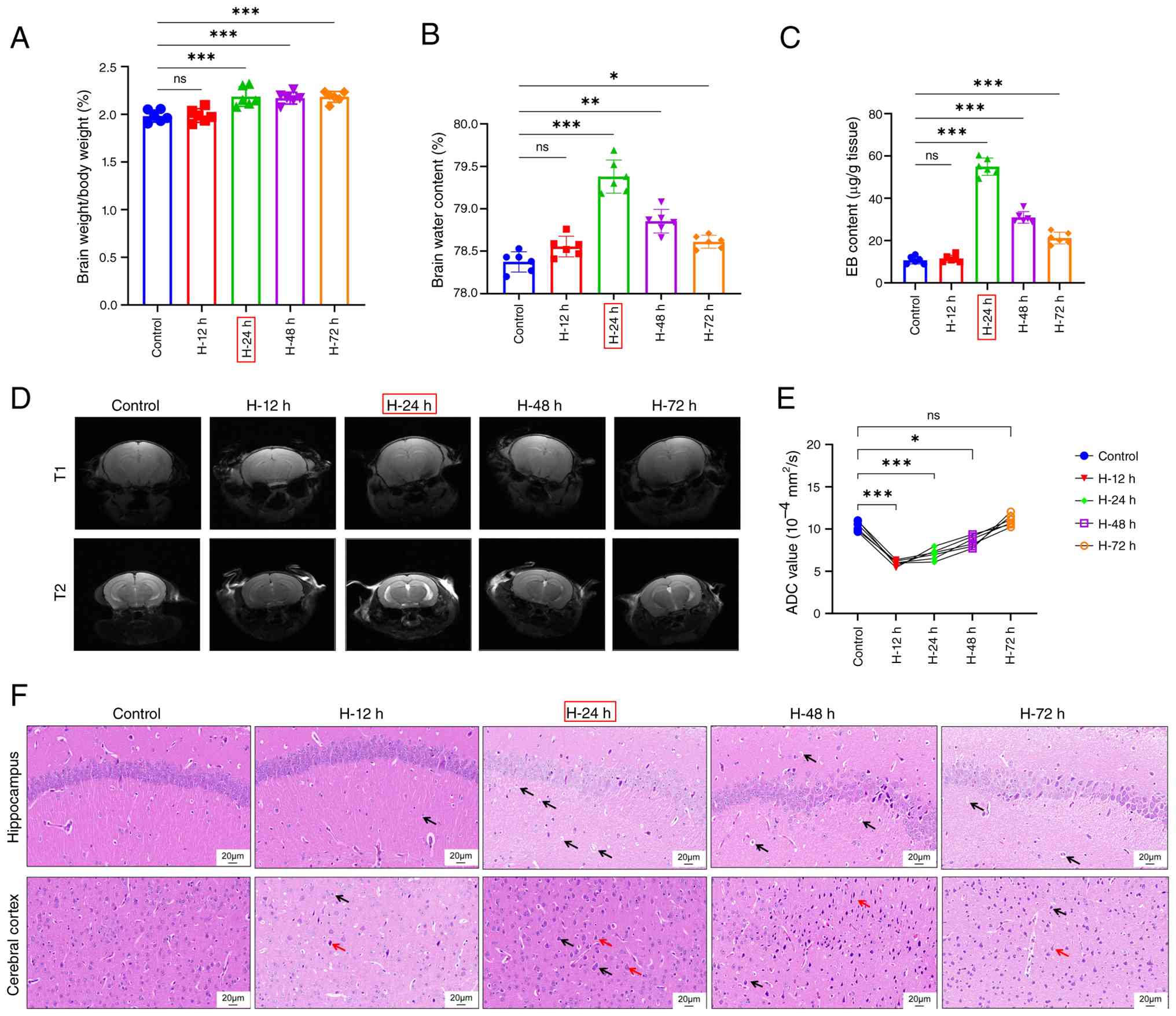
To establish a mouse model of HACE, C57BL/6 mice
were exposed to a hypobaric hypoxia environment for varying
durations. Cerebral edema was assessed by measuring the ratio of
brain weight to body weight (Fig.
1A) and brain water content (Fig.
1B). Mice subjected to hypobaric hypoxia for 24, 48 and 72 h
exhibited significantly increased brain weight/body weight ratios
and brain water content, with the most pronounced elevation
observed in the 24-h exposure group (H-24, H-48 and H-72 h vs.
control group; P<0.001 for the ratio of brain weight to body
weight at H-24, H-48 and H-72 h; P<0.001 for the brain water
content at H-24 h; P<0.01 for the brain water content at H-48 h;
P<0.05 for the brain water content at H-72 h). Additionally,
enhanced BBB permeability, indicated by significantly higher EB
content, was observed in mice treated with hypobaric hypoxia for
24, 48 and 72 h, especially in the 24-h group (H-24, H-48 and H-72
h vs. control group; P<0.001; Fig.
1C). Alterations in ADC values on brain MRI reflect changes in
brain tissue water content, thus facilitating assessment of brain
water content (21). The MRI
images are shown in Fig. 1D.
Significantly reduced ADC values were recorded in mice exposed to
hypobaric hypoxia for 12, 24 and 48 h (H-12, H-24 and H-48 h vs.
control group; P<0.05 at H-48 h, P<0.001 at H-12 h and at
H-24 h; Fig. 1E). H&E staining
revealed notable cellular edema (black arrows) and nuclear
condensation (red arrows) in the hippocampus and cerebral cortex of
mice, particularly in those exposed for 24 h (Fig. 1F). Thus, hypobaric hypoxia exposure
resulted in marked cerebral edema and increased BBB permeability in
mice, with 24-h exposure eliciting the most significant effects,
leading to its selection for subsequent studies.
Hypobaric hypoxia exposure caused
elevated microvascular permeability, impaired tight junctions and
increased Piezo1 expression in mice
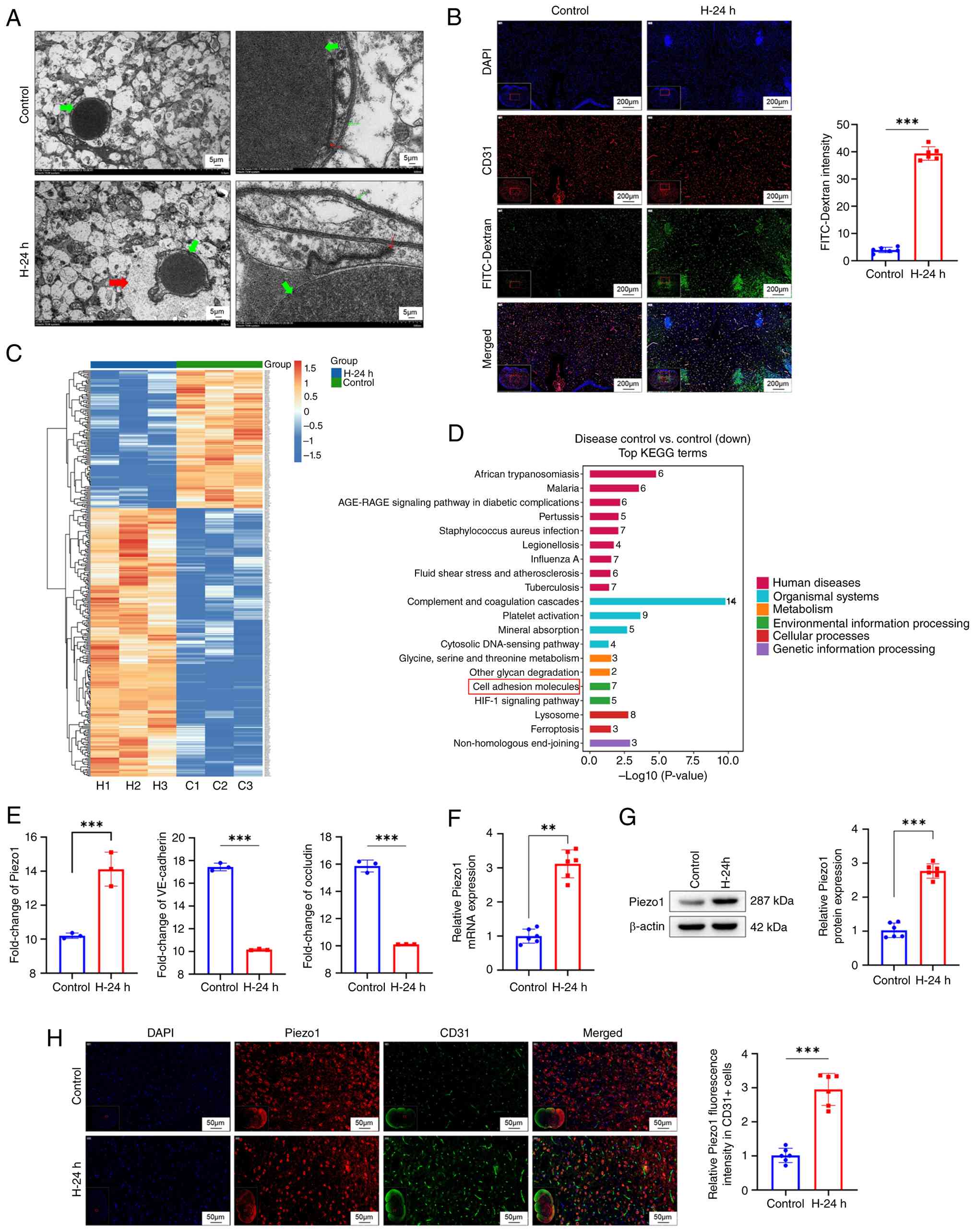
TEM images revealed impaired tight junctions in
brain microvessels of mice exposed to hypobaric hypoxia for 24 h
(H-24 h vs. control group; Fig.
2A). A FITC-Dextran leakage assay was conducted to assess brain
microvascular permeability, complemented by CD31 immunofluorescence
staining to localize brain microvascular endothelial cells.
Increased microvascular permeability was observed in the brains of
mice after 24 h of hypobaric hypoxia exposure (H-24 h vs. control
group; P<0.001; Fig. 2B). A
heatmap of differentially expressed proteins generated through
proteomics analysis showed differences between control mice and
those exposed to hypobaric hypoxia for 24 h (Fig. 2C). KEGG enrichment analysis
indicated an association between these differentially expressed
proteins and tight junctions, specifically the pathway ‘Cell
adhesion molecules’ was identified, with its associated proteins
significantly downregulated (Fig.
2D). Proteomics analysis demonstrated up-regulation of Piezo1
and down-regulation of VE-cadherin and occludin within the brain
microvascular endothelial cells of mice subjected to hypobaric
hypoxia for 24 h (H-24 h vs. control group; P<0.001; Fig. 2E).
To investigate Piezo1 expression in brain
microvascular endothelial cells, these cells were isolated and
subjected to RT-qPCR and western blot analyses. Increased mRNA
(P<0.01) and protein levels (P<0.001) of Piezo1 were observed
in brain microvascular endothelial cells of mice after 24 h of
hypobaric hypoxia exposure (H-24 h vs. control group; Fig. 2F and G). Immunofluorescence
staining of CD31/Piezo1 was performed to visualize Piezo1
expression within brain microvascular endothelial cells, revealing
enhanced expression after 24 h of hypobaric hypoxia exposure (H-24
h vs. control group; P<0.001; Fig.
2H). These results indicate that a hypobaric hypoxia
environment leads to increased microvascular permeability, impaired
tight junctions and enhanced Piezo1 expression in brain
microvascular endothelial cells.
Piezo1 silencing caused a decline in
intracellular Ca2+ levels and Calpain-1 activity within
the HFSS- and hypoxia-induced bEnd.3 cells
bEnd.3 cells were subjected to FSS at 10, 20, 30 and
40 dyn/cm2 for 24 h. A significant increase in Piezo1
protein expression was observed as FSS levels rose (P<0.01,
P<0.001). Notably, Piezo1 protein levels in cells exposed to 40
dyn/cm2 were comparable to those in cells exposed to 30
dyn/cm2. Consequently, HFSS at 30 dyn/cm2 was
designated as HFSS for subsequent experiments (Fig. S2A). Transfection of Piezo1 siRNA
into bEnd.3 cells resulted in a significant reduction in Piezo1
protein expression compared with the siRNA NC (si-Piezo1 vs. si-NC;
P<0.001; Fig. S2B).
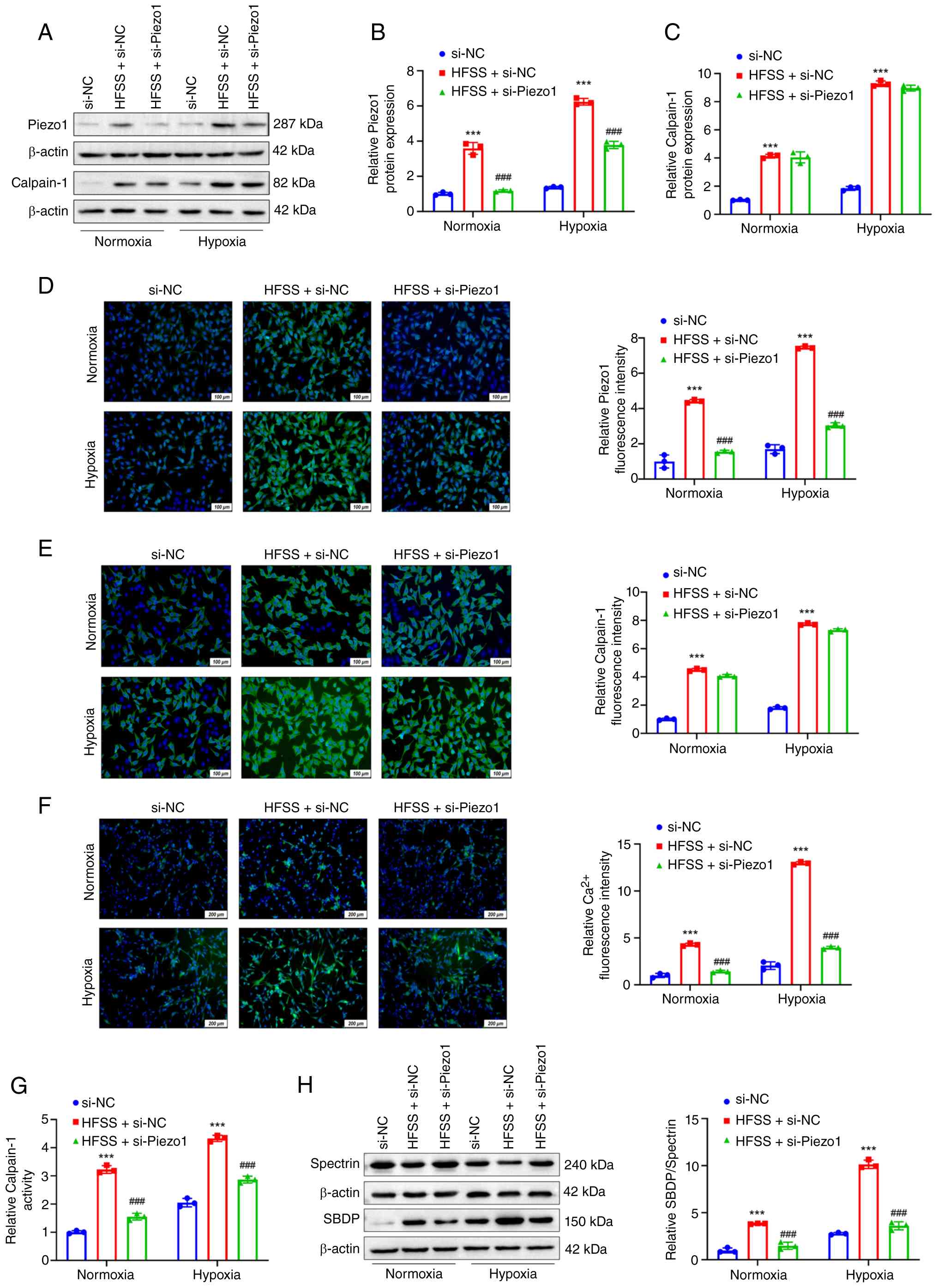
Under both normoxic and hypoxic conditions, HFSS
treatment significantly elevated Piezo1 mRNA and protein expression
in bEnd.3 cells (HFSS + si-NC vs. si-NC; P<0.001). Transfection
with Piezo1 siRNA led to decreased expression of Piezo1 mRNA and
protein in HFSS-treated cells under both conditions (HFSS +
si-Piezo1 vs. HFSS + si-NC; P<0.001; Fig. 3A and B). Western blot analysis
revealed that HFSS treatment increased Calpain-1 protein expression
in bEnd.3 cells under normoxia and hypoxia (HFSS + si-NC vs. si-NC;
P<0.001). Notably, Piezo1 silencing did not significantly affect
Calpain-1 protein expression in HFSS-treated cells under either
condition (HFSS + si-Piezo1 vs. HFSS + si-NC; Fig. 3A and C). Immunofluorescence
staining (Fig. 3D and E) and
detection with the Fluo4-AM fluorescent probe (Fig. 3F) were employed to monitor the
expression of Piezo1 and Calpain-1 proteins, as well as
intracellular Ca2+ levels in bEnd.3 cells. Following
exposure to normoxic and hypoxic conditions, HFSS-treated bEnd.3
cells exhibited elevated levels of Piezo1 and Calpain-1 proteins,
along with increased intracellular Ca2+ levels (HFSS +
si-NC vs. si-NC; P<0.001). By contrast, cells subjected to both
Piezo1 siRNA transfection and HFSS treatment demonstrated reduced
Piezo1 protein expression and intracellular Ca2+ levels
(HFSS + si-Piezo1 vs. HFSS + si-NC; P<0.001). However, under
HFSS conditions, Piezo1 silencing did not significantly affect
Calpain-1 protein expression (HFSS + si-Piezo1 vs. HFSS + si-NC).
Calpain-1 activity in bEnd.3 cells from different groups was
analyzed using a commercial kit (Fig.
3G). Regardless of normoxia or hypoxia, HFSS-induced cells
exhibited increased Calpain-1 activity (HFSS + si-NC vs. si-NC;
P<0.001), while this activity was reduced following Piezo1
silencing (HFSS + si-Piezo1 vs. HFSS + si-NC; P<0.001). Western
blot analysis indicated that HFSS treatment increased the
SBDP/Spectrin ratio in bEnd.3 cells under both normoxic and hypoxic
conditions (HFSS + si-NC vs. si-NC; P<0.001), an effect that was
abolished by Piezo1 silencing (HFSS + si-Piezo1 vs. HFSS + si-NC;
P<0.001). These results suggest that Piezo1 silencing reduces
intracellular Ca2+ levels and Calpain-1 activity in
HFSS-treated bEnd.3 cells under hypoxic conditions. Given that
Calpain-1 activity depends on calcium influx, the findings indicate
that Piezo1 silencing may attenuate Calpain-1 activity in
HFSS-treated bEnd.3 cells by reducing calcium influx.
Silencing Piezo1 alleviated the HFSS-
and hypoxia-induced tight junction dysfunction in microvascular
endothelial cells
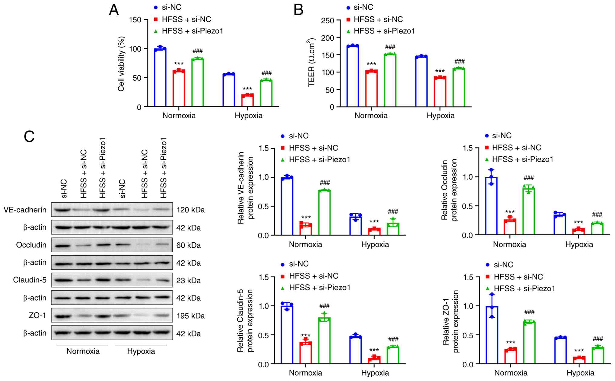
Cell viability of bEnd.3 cells was assessed using
the CCK-8 assay, revealing that HFSS treatment significantly
reduced cell viability under both normoxic and hypoxic conditions
(HFSS + si-NC vs. si-NC group; P<0.001). Following Piezo1
silencing, increased cell viability was observed in HFSS-treated
cells under both conditions (HFSS + si-Piezo1 vs. HFSS + si-NC
group; P<0.001) (Fig. 4A).
Subsequently, cell membrane permeability was evaluated through TEER
measurement (Fig. 4B), and the
expression of tight junction-associated proteins, including
VE-cadherin, occludin, claudin-5 and ZO-1, was examined by western
blot analysis (Fig. 4C). HFSS
treatment under normoxic and hypoxic conditions decreased TEER and
reduced the expression of VE-cadherin, occludin, claudin-5 and ZO-1
(HFSS + si-NC vs. si-NC group; P<0.001). Conversely, Piezo1
silencing reversed these effects, leading to increased TEER and
elevated expression of VE-cadherin, occludin, claudin-5 and ZO-1 in
HFSS-treated cells under both conditions (HFSS + si-Piezo1 vs. HFSS
+ si-NC group; P<0.001). These results indicate that Piezo1
silencing alleviates HFSS- and hypoxia-induced tight junction
dysfunction in microvascular endothelial cells.
Calpain-1 inhibition reduced Calpain-1
activity and enhanced tight junction expression within the HFSS-
and hypoxia-induced microvascular endothelial cells
Calpain-1 inhibitor PD150606 was applied to
HFSS-treated bEnd.3 cells. Under both normoxic and hypoxic
conditions, HFSS treatment alone significantly increased Calpain-1
activity (HFSS vs. control group; P<0.001), whereas PD150606
treatment reversed this increase (HFSS + PD150606 vs. HFSS group;
P<0.001) (Fig. 5A).
Furthermore, HFSS treatment decreased bEnd.3 cell viability (HFSS
vs. Control group; P<0.001), which was restored following
PD150606 treatment (HFSS + PD150606 vs. HFSS group; P<0.001)
(Fig. 5B). TEER measurements
(Fig. 5C) and western blot
analysis (Fig. 5D) demonstrated
that HFSS reduced TEER and decreased the expression of VE-cadherin,
occludin, claudin-5 and ZO-1 in bEnd.3 cells (HFSS vs. control
group; P<0.001). These reductions were reversed by PD150606
treatment (HFSS + PD150606 vs. HFSS group; P<0.001).
Collectively, these results indicate that inhibition of Calpain-1
decreases its activity and enhances tight junction expression in
HFSS- and hypoxia-induced microvascular endothelial cells. Overall,
the in vitro data suggest that Piezo1 silencing mitigates
HFSS- and hypoxia-induced tight junction dysfunction by reducing
Calpain-1 activity.
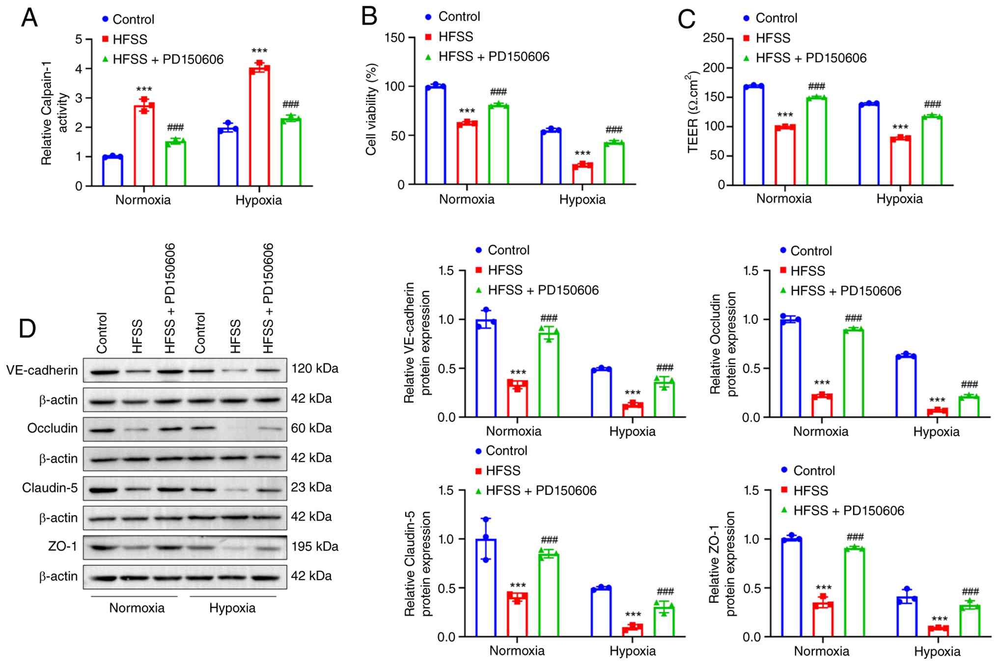 | Figure 5.Blockage of Calpain-1 suppressed
Calpain-1 activity and enhanced tight junctions in HFSS- and
hypoxia-induced microvascular endothelial cells. (A) Under normoxia
and hypoxia conditions, HFSS treatment enhanced Calpain-1 activity
in bEnd.3 cells, whereas PD150606 treatment abrogated this effect
(n=3). (B) Cell counting kit-8 assay showed that HFSS treatment
attenuated bEnd.3 cell viability under normoxia and hypoxia
conditions, which was enhanced by PD150606 treatment (n=3). (C)
TEER measurement indicated that HFSS treatment increased cell
membrane permeability of bEnd.3 cells under normoxia and hypoxia
conditions, and this effect was counteracted by PD150606 treatment
(n=3). (D) Based on western blot analysis, HFSS treatment reduced
the expression of tight junction-related proteins (including
VE-cadherin, occludin, claudin-5 and ZO-1) in bEnd.3 cells, which
was reversed by PD150606 treatment (n=3). ***P<0.001 vs. control
group. ###P<0.001 vs. HFSS group. Statistical tests
were performed using ANOVA with post-hoc Tukey's test. Error bars
represented SD, reflecting the dispersion of individual
observations around the group mean. HFSS, high fluid shear stress;
TEER, trans-endothelial electrical resistance; ZO-1, zonula
occludens; VE-cadherin, vascular endothelial cadherin. |
Piezo1-specific agonist might
exacerbate the disruption of tight junctions within microvascular
endothelial cells under hypoxic conditions by enhancing the calcium
influx-mediated Calpain-1 activity
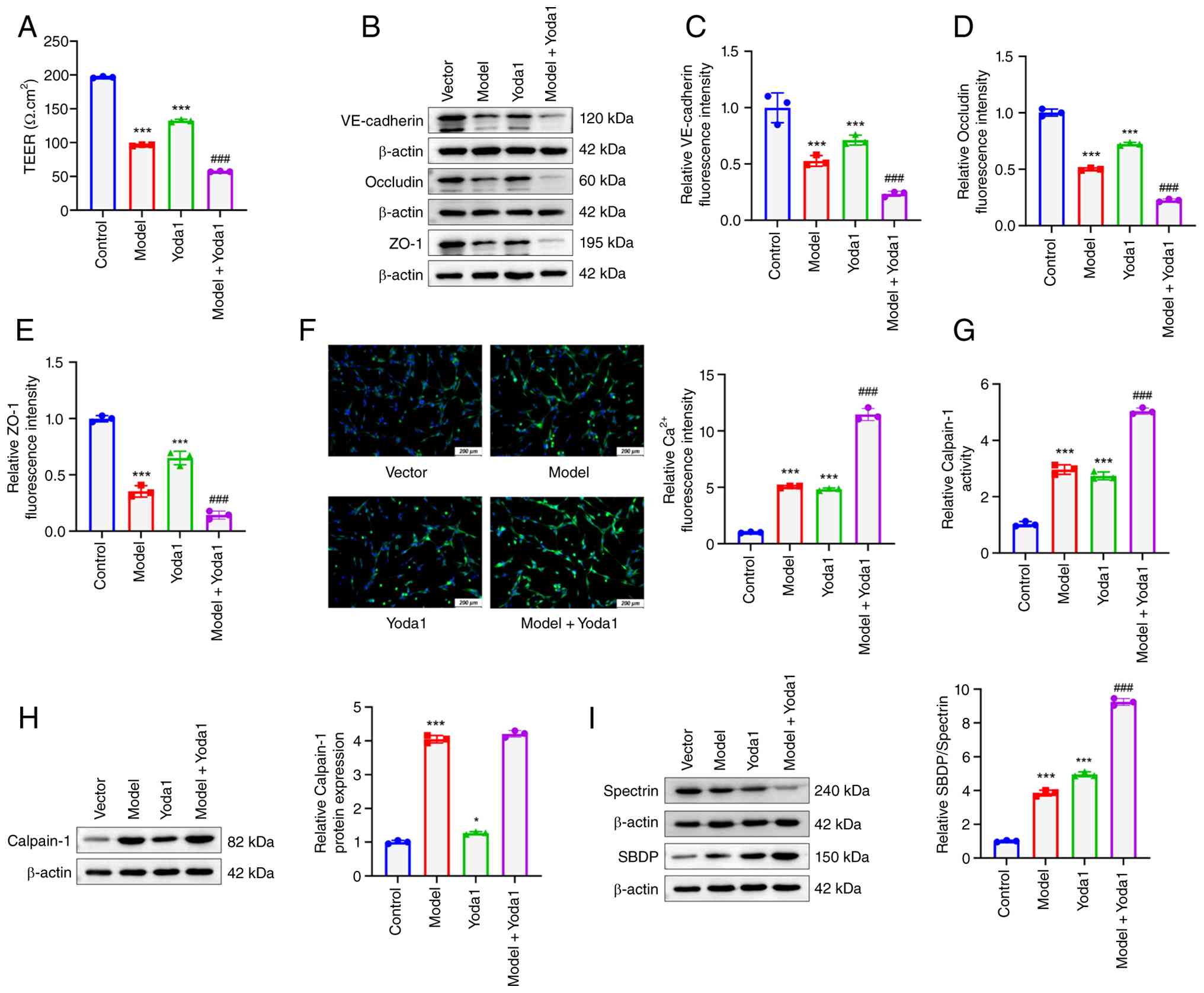
The classic HACE cell model was simulated by
exposing bEnd.3 cells to hypoxic conditions. Yoda1, a specific
agonist of Piezo1, was then applied to bEnd.3 cells. TEER
measurements indicated that both hypoxia exposure (model group) and
Yoda1 treatment (Yoda1 group) significantly decreased TEER in
bEnd.3 cells compared with the control group (P<0.001). Notably,
Yoda1 treatment further reduced TEER under hypoxic conditions
(model + Yoda1 group vs. model group; P<0.001) (Fig. 6A). Western blot analysis
demonstrated reductions in the protein expression of VE-cadherin,
occludin and ZO-1 in bEnd.3 cells exposed to hypoxia (model group
vs. control group) or treated with Yoda1 (Yoda1 group vs. control
group) (P<0.001). Yoda1 treatment under hypoxia resulted in
further decreases in the expression of these proteins (model +
Yoda1 group vs. model group; P<0.001) (Fig. 6B-E). Intracellular Ca2+
levels were measured using Fluo4-AM fluorescent probe detection,
revealing elevated Ca2+ levels in bEnd.3 cells subjected
to hypoxia (model group vs. control group) or Yoda1 treatment
(Yoda1 group vs. control group) (P<0.001). Under hypoxic
conditions, Yoda1 further increased intracellular Ca2+
levels in bEnd.3 cells (model + Yoda1 group vs. model group;
P<0.001) (Fig. 6F).
Calpain-1 activity was assessed using a commercial
kit (Fig. 6G), and its protein
expression was analyzed by western blot analysis (Fig. 6H). Both hypoxia exposure and Yoda1
treatment increased Calpain-1 activity (P<0.001) and protein
expression (P<0.05) in bEnd.3 cells (model group vs. control
group; Yoda1 group vs. control group). Yoda1 treatment under
hypoxia further enhanced Calpain-1 activity but did not
significantly alter Calpain-1 protein expression (model + Yoda1
group vs. model group) Furthermore, the SBDP/Spectrin protein ratio
was higher in bEnd.3 cells exposed to hypoxia and treated with
Yoda1 (model group vs. control group; Yoda1 group vs. control
group; P<0.001). After Yoda1 treatment under hypoxia, the
SBDP/Spectrin ratio increased further (model + Yoda1 group vs.
model group; P<0.001) (Fig.
6I). These results suggest that the Piezo1-specific agonist
Yoda1 exacerbates tight junction disruption in microvascular
endothelial cells under hypoxic conditions by enhancing calcium
influx-mediated Calpain-1 activity.
Calpain-1 inhibitor rescued the
disrupted tight junctions within microvascular endothelial cells
caused by Piezo1 overexpression
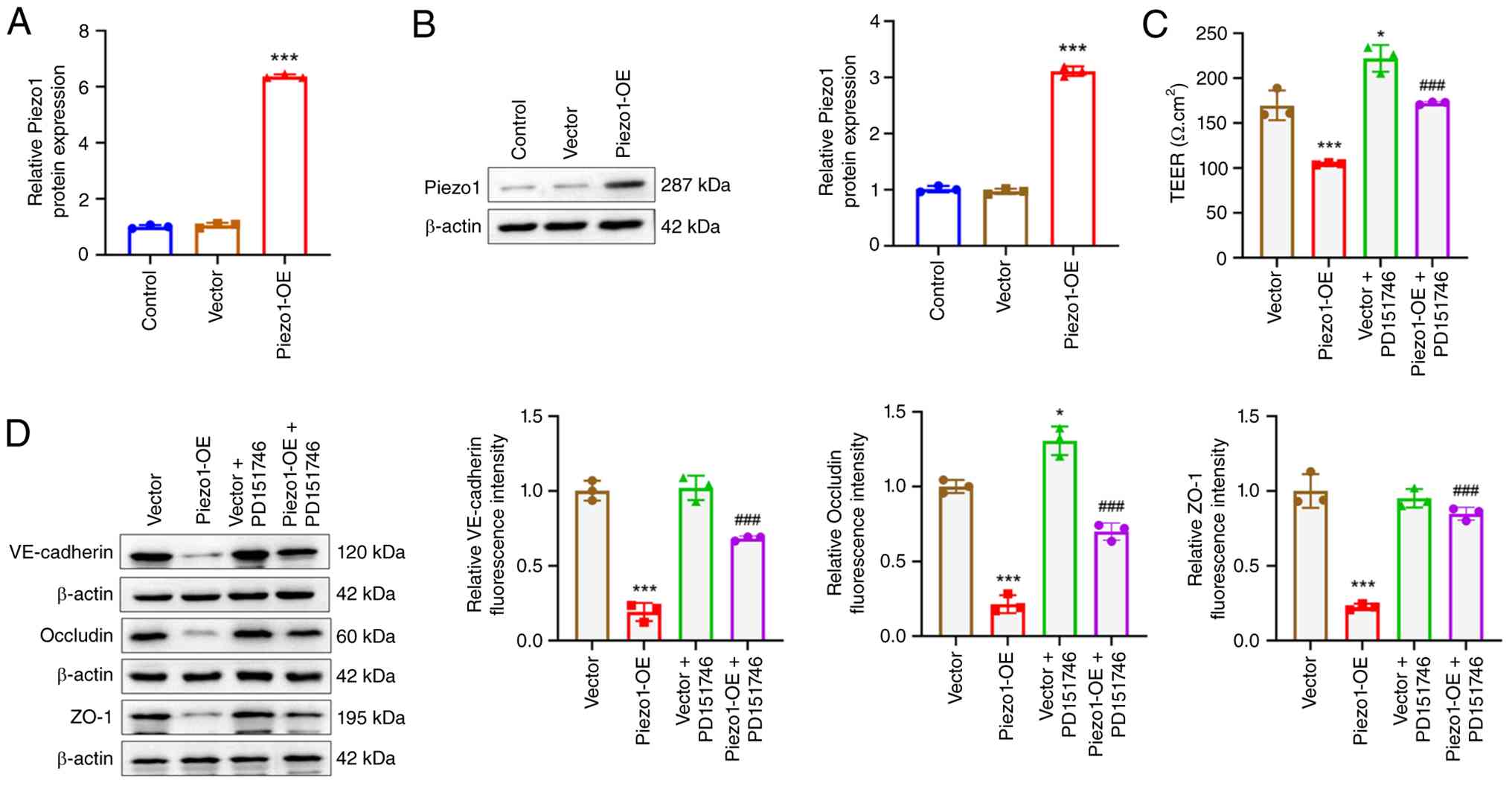
The experiments in this section were carried out
under normoxic conditions. Piezo1 overexpression vectors were
transfected into bEnd.3 cells, resulting in a significant increase
in Piezo1 mRNA and protein levels (Piezo1-OE group vs. Vector
group; P<0.001) (Fig. 7A and
B). TEER measurements (Fig.
7C) and western blot analysis (Fig. 7D) indicated that Piezo1
overexpression significantly decreased TEER and the expression of
VE-cadherin, occludin and ZO-1 proteins in bEnd.3 cells (Piezo1-OE
group vs. vector group; P<0.001). By contrast, treatment with
the Calpain-1 inhibitor PD151746 increased TEER and occludin
protein expression in bEnd.3 cells (vector + PD151746 group vs.
vector group; P<0.05), although PD151746 did not significantly
affect VE-cadherin or ZO-1 protein levels (vector + PD151746 vs.
vector group). Compared with the Piezo1-OE group, bEnd.3 cells in
the Piezo1 + PD151746 group exhibited higher TEER and increased
expression of VE-cadherin, occludin and ZO-1 proteins (P<0.001).
These findings indicate that Piezo1 overexpression induces
HACE-like phenotypes by reducing TEER and the expression of tight
junction proteins, while PD151746 reverses these effects,
suggesting that Piezo1 might disrupt tight junctions in
microvascular endothelial cells via Calpain-1 activation.
Piezo1 effect on Calpain-1 activity
was dependent on intracellular Ca2+ level
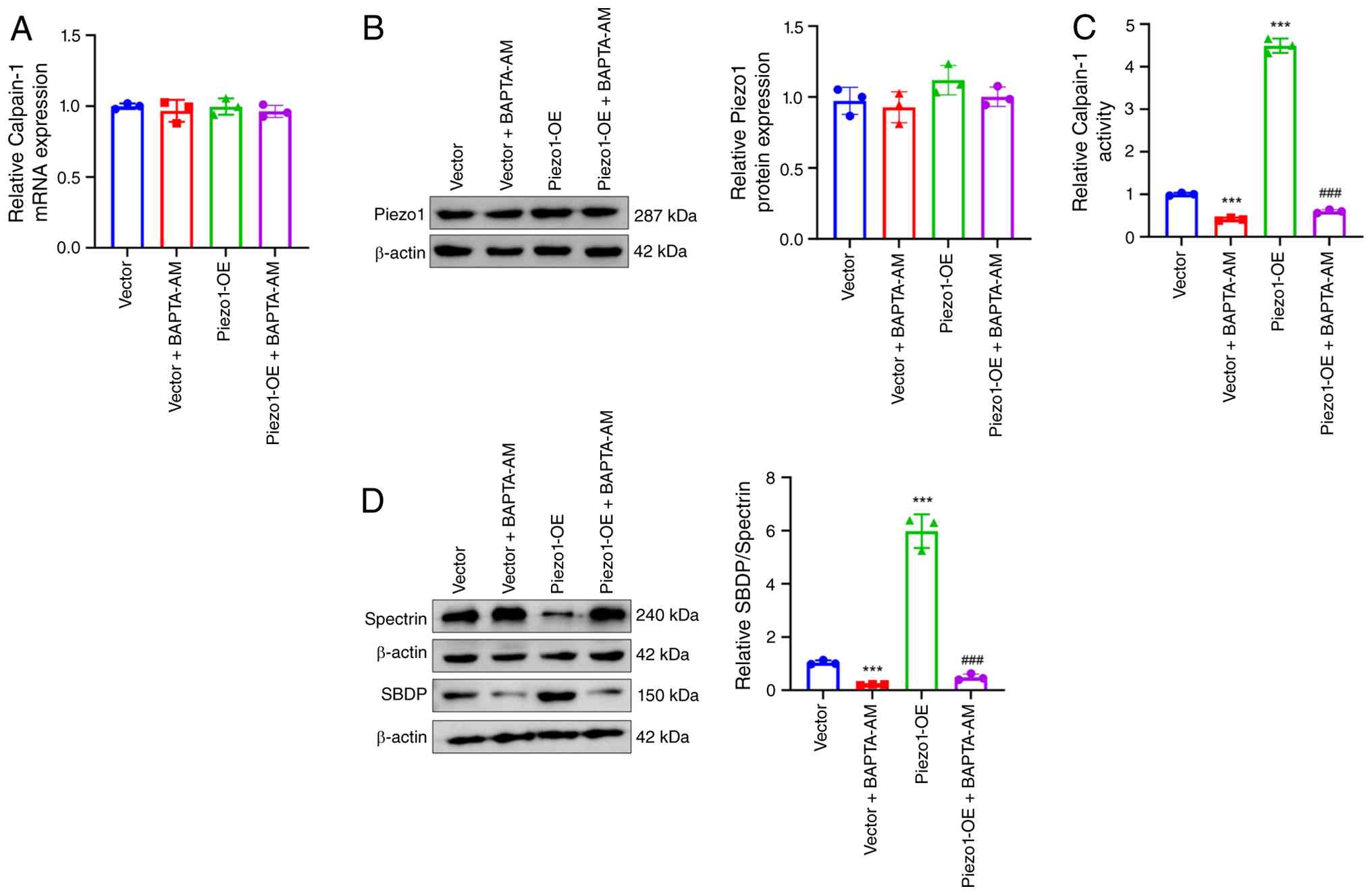
To investigate whether Piezo1 regulates Calpain-1
activity through Ca2+, bEnd.3 cells overexpressing
Piezo1 were treated with the Ca2+ chelator BAPTA-AM.
RT-qPCR and western blot analyses indicated that Piezo1
overexpression, BAPTA-AM treatment or their combination did not
significantly affect Calpain-1 mRNA or protein expression (Fig. 8A and B). However, BAPTA-AM
treatment resulted in a significant reduction in Calpain-1 activity
(vector + BAPTA-AM vs. vector group; P<0.001), while Piezo1
overexpression significantly elevated Calpain-1 activity (Piezo1
vs. vector group; P<0.001). Notably, the increased Calpain-1
activity induced by Piezo1 overexpression was significantly
decreased following BAPTA-AM treatment (Piezo1 + BAPTA-AM vs.
Piezo1-OE group; P<0.001) (Fig.
8C). Western blot analysis revealed that BAPTA-AM treatment
decreased the SBDP/Spectrin ratio in bEnd.3 cells (vector +
BAPTA-AM vs. vector group; P<0.001). Conversely, Piezo1
overexpression increased the SBDP/Spectrin ratio (Piezo1 vs. vector
group; P<0.001), which was reversed by BAPTA-AM treatment
(Piezo1 + BAPTA-AM vs. Piezo1-OE group) (P<0.001) (Fig. 8D). These data suggest that the role
of Piezo1 in enhancing Calpain-1 activity is dependent on
intracellular Ca2+ levels, indicating that increased
intracellular Ca2+ is necessary for Calpain-1
activation.
Piezo1 knockdown could attenuate
cerebral edema and microvascular permeability in mice exposed to
hypobaric hypoxia
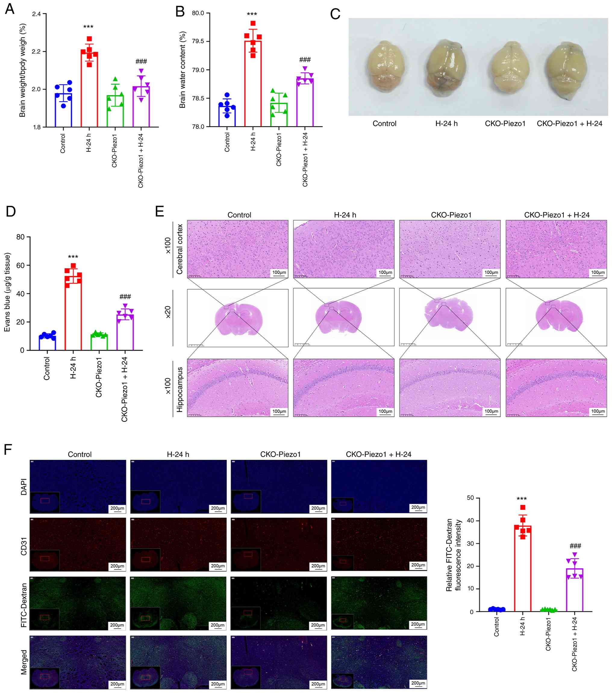
As shown in Fig.
S3, the expression of Piezo1 mRNA was markedly reduced in brain
microvascular endothelial cells of Piezo1 knockdown mice (the
CKO-Piezo1 group), as compared with the control group (P<0.001).
This suggested that Piezo1 expression was effectively
down-regulated in Piezo1 knockdown mice. To further confirm the
influence of Piezo1 on cerebral edema and microvascular
permeability in HACE, Piezo1 knockdown mice were exposed to a
hypobaric hypoxia environment. Mice subjected to hypobaric hypoxia
for 24 h exhibited higher brain weight/body weight ratios and brain
water content (H-24 h vs. control group; P<0.001). Piezo1
knockdown resulted in a marked decrease in brain weight/body weight
ratio and brain water content in mice exposed to hypobaric hypoxia
(CKO-Piezo1 + H-24 group vs. H-24 h group; P<0.001) (Fig. 9A and B).
EB staining indicated exacerbated BBB permeability
in mice after 24 h of hypobaric hypoxia exposure (H-24 h vs.
Control group; P<0.001), which was alleviated by Piezo1
knockdown (CKO-Piezo1 + H-24 group vs. H-24 h group; P<0.001)
(Fig. 9C and D). H&E staining
revealed marked cellular edema and nuclear condensation in the
hippocampus and cerebral cortex of mice exposed to hypobaric
hypoxia for 24 h (H-24 h vs. control group), with Piezo1 knockdown
mitigating this damage (CKO-Piezo1 + H-24 group vs. H-24 h group)
(Fig. 9E). The FITC-Dextran
leakage assay demonstrated intensified microvascular permeability
in the brains of mice exposed to hypobaric hypoxia for 24 h (H-24 h
vs. control group; P<0.001), which was reduced by Piezo1
knockdown (CKO-Piezo1 + H-24 group vs. H-24 h group; P<0.001)
(Fig. 9F). These results imply
that Piezo1 knockdown effectively alleviates cerebral edema and
microvascular permeability in the brains of mice exposed to
hypobaric hypoxia.
Piezo1 knockdown blocked Calpain-1
activity and increased tight junction expression within brain
microvascular endothelial cells of mice exposed to hypobaric
hypoxia
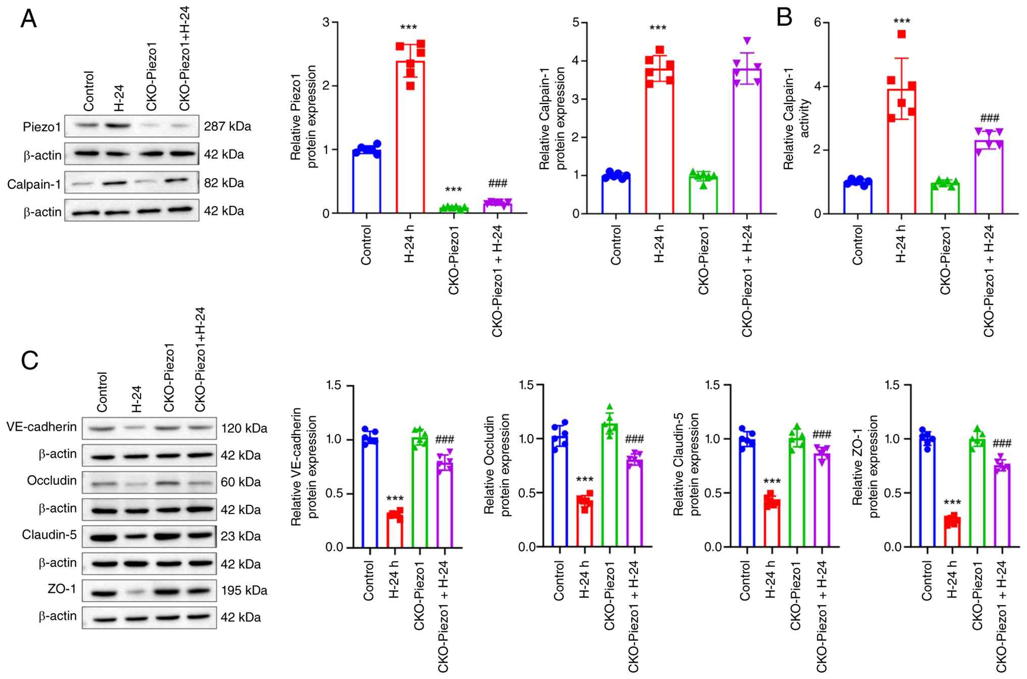
Western blot analysis demonstrated an increase in
Piezo1 and Calpain-1 protein expression in the brain microvascular
endothelial cells of mice subjected to 24 h of hypobaric hypoxia
treatment (H-24 h group vs. control group; P<0.001). Mice with
Piezo1 knockdown exhibited decreased Piezo1 protein levels in their
brain microvascular endothelial cells (CKO-Piezo1 group vs. control
group; P<0.001). Additionally, Piezo1 knockdown led to a
reduction in Piezo1 protein expression in the brain microvascular
endothelial cells of mice experiencing hypobaric hypoxia
(CKO-Piezo1 + H-24 group vs. H-24 h group) (P<0.001), while
having no significant effect on Calpain-1 protein levels
(CKO-Piezo1 + H-24 group vs. H-24 h group) (Fig. 10A).
Furthermore, mice exposed to hypobaric hypoxia for
24 h exhibited higher Calpain-1 activity in their brain
microvascular endothelial cells (H-24 h group vs. control group;
P<0.001), whereas Piezo1 knockdown significantly suppressed
Calpain-1 activity (CKO-Piezo1 + H-24 group vs. H-24 h group;
P<0.001) (Fig. 10B). Hypobaric
hypoxia exposure for 24 h resulted in a significant reduction in
the expression of VE-cadherin, occludin, claudin-5 and ZO-1
proteins in brain microvascular endothelial cells (H-24 h group vs.
control group; P<0.001). Notably, Piezo1 knockdown significantly
increased the expression of these proteins (CKO-Piezo1 + H-24 group
vs. H-24 h group; P<0.001) (Fig.
10C).
Therefore, Piezo1 knockdown effectively reduced
Calpain-1 activity and improved tight junction integrity within
brain microvascular endothelial cells of mice exposed to hypobaric
hypoxia. Collectively, these findings suggest that Piezo1 knockdown
may alleviate HACE by enhancing the integrity of tight junctions in
brain microvascular endothelial cells through the suppression of
calcium influx-mediated Calpain-1 activity.
Discussion
The present study demonstrated that Piezo1 is
notably upregulated in brain microvascular endothelial cells of
mice with HACE. Knockdown of Piezo1 exhibited an HACE-alleviating
effect, potentially by enhancing tight junction integrity in these
cells through the inhibition of calcium influx-mediated Calpain-1
activity. Although Piezo1 has received limited attention in the
context of HACE, it has previously been shown to enhance cerebral
edema under certain hypoxic-ischemic conditions (8). Current evidence suggests that Piezo1
knockdown may also contribute to the treatment of intracerebral
hemorrhage by restoring cerebral blood flow, alleviating cerebral
edema and reducing neurological deficits in mice (22,23).
Endothelial cells are critical components of the BBB. Following a
stroke, these cells are subjected to external pressure and tensile
strain, which activate Piezo1 and exacerbate BBB disruption.
Conversely, a deficiency of Piezo1 expression helps maintain BBB
integrity in both in vitro and in vivo settings
(9). Additionally, Piezo1
activation by mechanical forces related to vasogenic edema can
induce neuronal injury through Ca2+ signaling (24). In the context of vascular dementia
caused by chronic cerebral hypoperfusion, treatment with a Piezo1
antagonist reduced cerebral edema in rats and preserved BBB
integrity in relevant in vitro cell models (8). Evidence has also linked Piezo1 to BBB
dysfunction and neuroinflammation resulting from mechanical stress
(25). Furthermore, Piezo1 has
been associated with acute vascular damage following strokes or
trauma (26). Thus, Piezo1 emerges
as a promising therapeutic target for interventions in central
nervous system injuries (27). The
present study provided key insights, revealing that Piezo1 was
overexpressed in the setting of HACE and contributed to BBB damage.
Specifically, the established mouse model of HACE exhibited
hallmark features of cerebral edema, such as increased brain water
content, altered ADC values on MRI, hippocampal and cortical damage
and disrupted microvascular permeability and tight junctions
(2,28,29).
Notably, proteomics analysis performed in the present study,
identified the overexpression of Piezo1 in brain microvascular
endothelial cells of HACE-affected mice, which was further
validated by immunofluorescence staining. This observation aligns
with the patterns of Piezo1 expression seen in cerebral edema
induced by hypoxia-ischemia (8).
Notably, to the best of our knowledge, the present study represents
the first demonstration of Piezo1 activation in brain microvascular
endothelial cells following HACE. While Piezo1 has been implicated
in other cerebrovascular diseases such as ischemic stroke, its role
within the unique pathophysiological context of HACE had not been
explored until now. The findings from the present study open up new
avenues for HACE treatment, suggesting that targeted inhibition of
Piezo1 could be a promising strategy for preventing or treating
this condition.
In the present study, proteomics analysis indicated
a marked reduction in VE-cadherin and occludin expression in brain
microvascular endothelial cells of mice following HACE. Calpain-1,
a calcium-dependent protease, acts as a downstream effector of
Piezo1, being activated by elevated intracellular Ca2+
levels. Calpain-1 plays a role in regulating cellular functions by
specifically cleaving tight junction-associated proteins, such as
VE-cadherin and occluding (10,11).
Cerebral hypoxia-ischemia is known to induce vascular dysfunction,
and inhibition of Calpain-1 has been shown to alleviate tight
junction protein loss in in vitro models of
ischemia-reperfusion injury, as well as to mitigate BBB dysfunction
and promote tight junction protein expression in mild traumatic
brain injury models (30).
Therefore, inhibiting Calpain-1 is proposed as a potential strategy
to protect the cerebral vasculature following hypoxia-ischemia.
Accordingly, the present study aimed to investigate whether Piezo1
regulates tight junctions in brain microvascular endothelial cells
during HACE via Calpain-1, using in vitro experiments. A
cell model of HACE was established by exposing bEnd.3 cells to HFSS
and hypoxia. In these cells, increased intracellular
Ca2+ levels were observed, along with elevated Calpain-1
expression and activity. Silencing Piezo1 resulted in decreased
intracellular Ca2+ levels and Calpain-1 activity in
HFSS- and hypoxia-treated bEnd.3 cells; however, it did not
significantly alter Calpain-1 protein expression under these
conditions. Similarly, in the in vivo HACE mouse model,
Piezo1 knockdown inhibited Calpain-1 activity but did not
significantly affect Calpain-1 protein levels in the brain
microvascular endothelial cells. Although Piezo1 knockdown
displayed minor effects on Calpain-1 protein expression in both
in vitro and in vivo experiments, these changes were
not statistically significant. This suggests that Piezo1 silencing
may alleviate HACE primarily by inhibiting Calpain-1 activity
rather than modifying its protein expression levels.
Mechanistically, Piezo1 appears to regulate Calpain-1 activity
through the Ca2+/Calpain tight junction axis.
Specifically, Piezo1 is a mechanosensitive cation channel that
detects changes in membrane tension and mediates Ca2+
influx, while Calpain-1 is a Ca2+-dependent cysteine
protease that, when activated by increased intracellular
Ca2+, cleaves target proteins such as VE-cadherin and
occludin (11). In the present
study, Piezo1 silencing reduced intracellular Ca2+ and
inhibited Calpain-1 activity in the HACE cell model, indicating
that it may block Calpain-1 activity by decreasing intracellular
Ca2+. Spectrin is a known substrate of Calpain-1 and is
frequently used to assess Calpain-1 activity, with SBDP
representing a breakdown product of Spectrin (31). In the HACE cell model, Piezo1
silencing resulted in a reduced SBDP/Spectrin ratio, while
treatment with Yoda1, a Piezo1-specific agonist, increased this
ratio. These findings further support the conclusion that Piezo1
silencing inhibits Calpain-1 activity within the HACE cell model by
suppressing Spectrin cleavage.
Notably, the present study established a novel in
vitro model of HACE by exposing bEnd.3 cells to a combination
of HFSS and hypoxia, which represents an innovative aspect of the
research. High-altitude sickness induces a compensatory increase in
red blood cell count, leading to elevated blood viscosity and
consequently increased shear stress in blood flow (15). This abnormal shear stress
contributes to vascular endothelial dysfunction (16). Acute brain injury similarly results
in elevated shear stress (5).
Piezo1 is a mechanosensitive cation channel protein (32) with a unique mechanosensitive
structure that enables it to detect changes in cell membrane
tension, including shear stress. HFSS activates Piezo1, triggering
intracellular Ca2+ signaling, which increases vascular
permeability in endothelial cells (33–35).
Accordingly, the present study aimed to investigate the role of
Piezo1 in HACE using an in vitro model in which bEnd.3 cells
were subjected to HFSS and hypoxia. While the Piezo1/intracellular
Ca2+/Calpain-1/tight junction disruption pathway has
been studied in other neurological diseases (36,37),
the present study provides the first evidence that HFSS activates
this pathway in the context of HACE. Moreover, a classic HACE model
without HFSS stimulation was established in the present study to
assess the effects of a Piezo1 agonist, Piezo1 overexpression and a
Calpain-1-specific inhibitor on tight junctions in bEnd.3 cells.
The results indicated that Piezo1 may disrupt tight junctions in
bEnd.3 cells by enhancing Calpain-1 activity, which is consistent
with findings from the HFSS- and hypoxia-induced HACE cell model.
Notably, contrasting observations have been made in a mouse
atherosclerosis model where low shear stress down-regulates Piezo1
expression in arterial vessels, thereby promoting plaque formation.
By contrast, high shear stress up-regulates Piezo1 and exerts an
anti-atherosclerotic effect (38).
In the present study, however, high expression of Piezo1 was shown
to promote the progression of HACE in mice. This discrepancy may be
attributed to the differences between brain microvascular
endothelial cells and arterial endothelial cells. Consequently,
Piezo1 appears to exert distinct roles in various types of vascular
endothelial cells and in brain injury arising from different
etiologies.
The integrity of vascular endothelial cell barriers
within the central nervous system constitutes the BBB (39). As a critical measure for assessing
endothelial barrier function (40), TEER was employed to evaluate the
endothelial barrier integrity of bEnd.3 cells exposed to HFSS and
hypoxic conditions. Under these adverse conditions, silencing
Piezo1 markedly improved the impaired endothelial barrier
integrity. Tight junctions in brain microvascular endothelial cells
are maintained by proteins such as VE-cadherin, occludin, claudin-5
and ZO-1 (41). Piezo1 silencing
promotes VE-cadherin expression in endothelial cells (42) and improves cognitive function in
mice after cerebral infarction, likely by preserving ZO-1 and
occludin expression (43). In the
present study, Piezo1 silencing enhanced tight junction integrity
in bEnd.3 cells under HFSS and hypoxia, as evidenced by increased
expression of VE-cadherin, occludin, claudin-5 and ZO-1 proteins.
Similarly, treatment with the calpain-1 inhibitor PD150606 also
increased TEER and the expression of these tight junction proteins
in HFSS- and hypoxia-induced bEnd.3 cells. These findings suggest
that Piezo1 silencing protects BBB integrity in HACE by preserving
tight junctions in brain microvascular endothelial cells through
the inhibition of calpain-1 activity. Further in vivo
experiments demonstrated that Piezo1 knockdown alleviated cerebral
edema, protected against hippocampal and cortical damage,
maintained BBB integrity and tight junctions and suppressed
Calpain-1 activity without significantly affecting Calpain-1
protein expression in mice with HACE. Collectively, these results
indicate that Piezo1 knockdown mitigates HACE by maintaining tight
junctions in brain microvascular endothelial cells via the
inhibition of calcium influx-mediated Calpain-1 activity. Thus,
targeted inhibition of Piezo1 may represent a potential therapeutic
strategy for HACE.
Beyond alterations in endothelial cells, HACE
involves various cell types within the neurovascular unit,
including swelling of astrocytic endfeet and activation of
microglia, which are critical contributors to BBB damage (44,45).
Notably, Piezo1 has been implicated in microglial activation in
central nervous system disorders (46). Therefore, investigating the effects
of Piezo1 on astrocytic endfeet and microglia in the context of
HACE represents an important avenue for future research.
The present study has several limitations. First,
bEnd.3 cells were treated with HFSS to simulate blood FSS under
high-altitude conditions. However, high-altitude hypoxia may also
alter hemodynamics by increasing reverse shear stress (47). Developing relevant in vitro
models can facilitate a more comprehensive understanding of the
pathophysiological mechanisms underlying HACE. Second, to provide
stronger evidence for the direct regulation of BBB dysfunction
during HACE, it would be beneficial to perform in vivo
experiments utilizing Piezo1 overexpression or pharmacological
activation. Third, while various methods have been employed to
quantitatively assess the BBB, including EB staining, FITC-dextran
assay, brain tissue water content and ADC values from cranial MRI,
employing additional BBB assessment techniques and quantitative
analysis will provide a more thorough understanding of changes in
BBB permeability. Unfortunately, current laboratory constraints
precluded these experiments, which will be a priority for future
investigations.
In conclusion, the present study established a
mouse model of HACE and explored the impact of Piezo1 on HACE. The
findings indicated that Piezo1 knockdown alleviated HACE, resulting
in reduced brain water content, mitigated BBB disruption,
attenuated damage in the hippocampus and cerebral cortex and
maintained tight junctions in brain microvascular endothelial
cells. Through in vitro mechanistic research, Piezo1
silencing demonstrated a HACE-alleviating effect, likely by
enhancing tight junctions in brain microvascular endothelial cells
via blocking calcium influx-mediated Calpain-1 activity. These
discoveries illustrate that the targeted inhibition of Piezo1 may
serve as a promising approach for HACE therapy.
Supplementary Material
Supporting Data
Acknowledgements
Not applicable.
Funding
The present study was supported by the Guided Project of the
Health and Wellness Commission of Qinghai Province (grant no.
2025-wjzdx-70) and the Qinghai Scholar Program of the People's
Government of Qinghai Province [grant no. QZ (2024) 19].
Availability of data and materials
The mass spectrometry proteomics data generated in
the present study may be found in the China National Center for
Bioinformation database (https://ngdc.cncb.ac.cn/omix/release/OMIX020321;
data retrieval number: OMIX020321). All other data generated in the
present study may be requested from the corresponding author.
Authors' contributions
QZ conceptualized and designed the present study;
TC provided administrative support and wrote the manuscript; DC and
CY aided in the provision of study materials; DC performed the
animal experiments and acquired the data, and drafted and revised
the manuscript; CY contributed to the study design, performed the
molecular experiments and data acquisition, and critically revised
the manuscript for important intellectual content. DG, HM and SL
collected and assembled the data; TC, ZX and YZ performed the data
analysis and interpretation. All authors have read and approved the
final version of the manuscript. QZ and TC confirm the authenticity
of all the raw data.
Ethical approval and consent to
participate
Animal experiments have been approved by the Animal
Ethics Committee of Qinghai Provincial People's Hospital and are
reported in accordance with the ARRIVE guidelines [approval no.
(2023)-246].
Patient consent for publication
Not applicable.
Competing interests
The authors declare that they have no competing
interests.
References
|
1
|
Gatterer H, Villafuerte FC, Ulrich S,
Bhandari SS, Keyes LE and Burtscher M: Altitude illnesses. Nat Rev
Dis Primers. 10:432024. View Article : Google Scholar : PubMed/NCBI
|
|
2
|
Wang H, Zhang Q, Ma P, Huang N, Chen Y,
Cheng Y and Chen J: High-altitude-induced cerebral edema in mice is
alleviated by bestatin-mediated blood-brain barrier protection. J
Neurophysiol. 133:1902–1915. 2025. View Article : Google Scholar : PubMed/NCBI
|
|
3
|
Xue Y, Wang X, Wan B, Wang D, Li M, Cheng
K, Luo Q, Wang D, Lu Y and Zhu L: Caveolin-1 accelerates
hypoxia-induced endothelial dysfunction in high-altitude cerebral
edema. Cell Commun Signal. 20:1602022. View Article : Google Scholar : PubMed/NCBI
|
|
4
|
Xue Y, Wan B, Wang Z, Wang Z, Wang D, Yang
W, Wang X and Zhu L: Hydroxychloroquine prevents high-altitude
cerebral edema by inhibiting endothelial claudin-5 autophagic
degradation. Curr Neuropharmacol. 24:404–418. 2026. View Article : Google Scholar : PubMed/NCBI
|
|
5
|
Zhao N, Liu C, Tian X, Yang J and Wang T:
Acute brain injury increases pulmonary capillary permeability via
sympathetic activation-mediated high fluid shear stress and
destruction of the endothelial glycocalyx layer. Exp Cell Res.
434:1138732024. View Article : Google Scholar : PubMed/NCBI
|
|
6
|
Cheng X, Potenza DM, Brenna A, Ajalbert G,
Yang Z and Ming XF: Aging increases hypoxia-induced endothelial
permeability and blood-brain barrier dysfunction by upregulating
arginase-II. Aging Dis. 15:2710–5415. 2024.PubMed/NCBI
|
|
7
|
Jiang D, Zhao J, Zheng J, Zhao Y, Le M,
Qin D, Huang Q, Huang J, Zhao Q, Wang L and Dong X: LOX-mediated
ECM mechanical stress induces Piezo1 activation in hypoxic-ischemic
brain damage and identification of novel inhibitor of LOX. Redox
Biol. 76:1033462024. View Article : Google Scholar : PubMed/NCBI
|
|
8
|
Xu F, Xin Q, Ren M, Shi P and Wang B:
Inhibition of piezo1 prevents chronic cerebral
hypoperfusion-induced cognitive impairment and blood brain barrier
disruption. Neurochem Int. 175:1057022024. View Article : Google Scholar : PubMed/NCBI
|
|
9
|
Fu H, Yu Y, Wang S, Xu P, Sun Y, Li J, Ge
X and Pan S: Piezo1 disrupts blood-brain barrier via CaMKII/Nrf2 in
ischemic stroke. Cell Mol Life Sci. 82:2592025. View Article : Google Scholar : PubMed/NCBI
|
|
10
|
Wu HY and Lynch DR: Calpain and synaptic
function. Mol Neurobiol. 33:215–236. 2006. View Article : Google Scholar : PubMed/NCBI
|
|
11
|
Guo XW, Zhang H, Huang JQ, Wang SN, Lu Y,
Cheng B, Dong SH, Wang YY, Li FS and Li YW: PIEZO1 Ion channel
mediates ionizing radiation-induced pulmonary endothelial cell
ferroptosis via Ca2+/calpain/VE-cadherin signaling.
Front Mol Biosci. 8:7252742021. View Article : Google Scholar : PubMed/NCBI
|
|
12
|
ElAli A, Doeppner TR, Zechariah A and
Hermann DM: Increased blood-brain barrier permeability and brain
edema after focal cerebral ischemia induced by hyperlipidemia: Role
of lipid peroxidation and calpain-1/2, matrix
metalloproteinase-2/9, and RhoA overactivation. Stroke.
42:3238–3244. 2011. View Article : Google Scholar : PubMed/NCBI
|
|
13
|
Alluri H, Grimsley M, Anasooya Shaji C,
Varghese KP, Zhang SL, Peddaboina C, Robinson B, Beeram MR, Huang
JH and Tharakan B: Attenuation of blood-brain barrier breakdown and
hyperpermeability by calpain inhibition. J Biol Chem.
291:26958–26969. 2016. View Article : Google Scholar : PubMed/NCBI
|
|
14
|
Liu J, Wang YH, Li W, Liu L, Yang H, Meng
P and Han YS: Structural and functional damage to the hippocampal
neurovascular unit in diabetes-related depression. Neural Regen
Res. 14:289–297. 2019. View Article : Google Scholar : PubMed/NCBI
|
|
15
|
Tremblay JC, Ainslie PN, Turner R,
Gatterer H, Schlittler M, Woyke S, Regli IB, Strapazzon G, Rauch S
and Siebenmann C: Endothelial function and shear stress in
hypobaric hypoxia: Time course and impact of plasma volume
expansion in men. Am J Physiol Heart Circ Physiol. 319:H980–H994.
2020. View Article : Google Scholar : PubMed/NCBI
|
|
16
|
Tymko MM, Tremblay JC, Bailey DM, Green DJ
and Ainslie PN: The impact of hypoxaemia on vascular function in
lowlanders and high altitude indigenous populations. J Physiol.
597:5759–5776. 2019. View Article : Google Scholar : PubMed/NCBI
|
|
17
|
Karthika CL, Venugopal V, Sreelakshmi BJ,
Krithika S, Thomas JM, Abraham M, Kartha CC, Rajavelu A and Sumi S:
Oscillatory shear stress modulates Notch-mediated endothelial
mesenchymal plasticity in cerebral arteriovenous malformations.
Cell Mol Biol Lett. 28:222023. View Article : Google Scholar : PubMed/NCBI
|
|
18
|
Choublier N, Taghi M, Menet MC, Le Gall M,
Bruce J, Chafey P, Guillonneau F, Moreau A, Denizot C, Parmentier
Y, et al: Exposure of human cerebral microvascular endothelial
cells hCMEC/D3 to laminar shear stress induces vascular protective
responses. Fluids Barriers CNS. 19:412022. View Article : Google Scholar : PubMed/NCBI
|
|
19
|
Livak KJ and Schmittgen TD: Analysis of
relative gene expression data using real-time quantitative PCR and
the 2(−Delta Delta C(T)) method. Methods. 25:402–408. 2001.
View Article : Google Scholar : PubMed/NCBI
|
|
20
|
Nicolicht-Amorim P, Delgado-Garcia LM,
Nakamura TKE, Courbassier NR, Mosini AC and Porcionatto MA: Simple
and efficient protocol to isolate and culture brain microvascular
endothelial cells from newborn mice. Front Cell Neurosci.
16:9494122022. View Article : Google Scholar : PubMed/NCBI
|
|
21
|
Tejedor M, Cussó L, González-Nicolás MÁ,
San Felipe D, Calle D, Martín-Sánchez B, Uzun B, Martín-Palumbo G,
Alvarez-Sala Walther LA, Marco EM, et al: Recurrent intermittent
hyponatremia: A new experimental model. PLoS One. 21:e03417432026.
View Article : Google Scholar : PubMed/NCBI
|
|
22
|
Qi M, Liu R, Zhang F, Yao Z, Zhou ML,
Jiang X and Ling S: Roles of mechanosensitive ion channel PIEZO1 in
the pathogenesis of brain injury after experimental intracerebral
hemorrhage. Neuropharmacology. 251:1098962024. View Article : Google Scholar : PubMed/NCBI
|
|
23
|
Qu J, Zong HF, Shan Y, Zhang SC, Guan WP,
Yang Y and Zhao HL: Piezo1 suppression reduces demyelination after
intracerebral hemorrhage. Neural Regen Res. 18:1750–1756.
2023.PubMed/NCBI
|
|
24
|
Guo XW, Lu Y, Zhang H, Huang JQ and Li YW:
PIEZO1 might be involved in cerebral ischemia-reperfusion injury
through ferroptosis regulation: A hypothesis. Med Hypotheses.
146:1103272021. View Article : Google Scholar : PubMed/NCBI
|
|
25
|
Pirri C: PIEZO channels in
mechano-inflammation: Gatekeepers of neuroimmune crosstalk.
Diseases. 13:2632025. View Article : Google Scholar : PubMed/NCBI
|
|
26
|
Shi G, Tan R, Cao Y, Zheng X and Zhan R:
Piezo1 in the central nervous system: Decoding the mechanical
signature of neuroinflammation. J Neuroinflammation. 23:2372026.
View Article : Google Scholar : PubMed/NCBI
|
|
27
|
Zhang M, Ge Y, Wang Z, Li H, Wang G, Zhang
Y, Wang X, Hou H, Meng L, Wei L, et al: Insights into the role of
the mechanosensitive Piezo1 channel and signaling mechanisms in CNS
functions and diseases. Neurosci Biobehav Rev. 183:1065622026.
View Article : Google Scholar : PubMed/NCBI
|
|
28
|
Shi H, Li P, Zhou H, Nie Z, Zhang J, Sui
X, Guo J, Wang Y and Wang L: Dynamic cerebral blood flow changes
with FOXOs stimulation are involved in neuronal damage associated
with high-altitude cerebral edema in mice. Brain Res.
1790:1479872022. View Article : Google Scholar : PubMed/NCBI
|
|
29
|
Schoonman GG, Sándor PS, Nirkko AC, Lange
T, Jaermann T, Dydak U, Kremer C, Ferrari MD, Boesiger P and
Baumgartner RW: Hypoxia-induced acute mountain sickness is
associated with intracellular cerebral edema: A 3 T magnetic
resonance imaging study. J Cereb Blood Flow Metab. 28:198–206.
2008. View Article : Google Scholar : PubMed/NCBI
|
|
30
|
Knopp RC, Jastaniah A, Dubrovskyi O,
Gaisina I, Tai L and Thatcher GRJ: Extending the calpain-cathepsin
hypothesis to the neurovasculature: Protection of brain endothelial
cells and mice from neurotrauma. ACS Pharmacol Transl Sci.
4:372–385. 2021. View Article : Google Scholar : PubMed/NCBI
|
|
31
|
Li J, Yang S and Zhu G: Postnatal calpain
inhibition elicits cerebellar cell death and motor dysfunction.
Oncotarget. 8:87997–88007. 2017. View Article : Google Scholar : PubMed/NCBI
|
|
32
|
Lei M, Wang W, Zhang H, Gong J, Cai H,
Wang Z, Zhu L, Yang X, Wang S and Ma C: Piezo1 regulates
stiffness-dependent DRG axon regeneration via modifying
cytoskeletal dynamics. Adv Sci (Weinh). 11:e24057052024. View Article : Google Scholar : PubMed/NCBI
|
|
33
|
Maneshi MM, Ziegler L, Sachs F, Hua SZ and
Gottlieb PA: Enantiomeric Aβ peptides inhibit the fluid shear
stress response of PIEZO1. Sci Rep. 8:142672018. View Article : Google Scholar : PubMed/NCBI
|
|
34
|
Maneshi MM, Gottlieb PA and Hua SZ: A
microfluidic approach for studying piezo channels. Curr Top Membr.
79:309–334. 2017. View Article : Google Scholar : PubMed/NCBI
|
|
35
|
Swain SM and Liddle RA: Piezo1 acts
upstream of TRPV4 to induce pathological changes in endothelial
cells due to shear stress. J Biol Chem. 296:1001712021. View Article : Google Scholar : PubMed/NCBI
|
|
36
|
Wang YY, Zhang H, Ma T, Lu Y, Xie HY, Wang
W, Ma YH, Li GH and Li YW: Piezo1 mediates neuron oxygen-glucose
deprivation/reoxygenation injury via Ca2+/calpain
signaling. Biochem Biophys Res Commun. 513:147–153. 2019.
View Article : Google Scholar : PubMed/NCBI
|
|
37
|
Ma T, Wang YY, Lu Y, Feng L, Yang YT, Li
GH, Li C, Chu Y, Wang W and Zhang H: Inhibition of
Piezo1/Ca2+/calpain signaling in the rat basal forebrain
reverses sleep deprivation-induced fear memory impairments. Behav
Brain Res. 417:1135942022. View Article : Google Scholar : PubMed/NCBI
|
|
38
|
Zhu Y, Wang T, Yang Y, Wang Z, Chen X,
Wang L, Niu R, Sun Z, Zhang C, Luo Y, et al: Low shear stress
exacerbates atherosclerosis by inducing the generation of
neutrophil extracellular traps via Piezo1-mediated
mechanosensation. Atherosclerosis. 391:1174732024. View Article : Google Scholar : PubMed/NCBI
|
|
39
|
Ayloo S, Lazo CG, Sun S, Zhang W, Cui B
and Gu C: Pericyte-to-endothelial cell signaling via
vitronectin-integrin regulates blood-CNS barrier. Neuron.
110:1641–1655.e6. 2022. View Article : Google Scholar : PubMed/NCBI
|
|
40
|
Ugodnikov A, Chebotarev O, Persson H and
Simmons CA: Sensitivity and validation of porous membrane
electrical cell substrate impedance spectroscopy (PM-ECIS) for
measuring endothelial barrier properties. ACS Biomater Sci Eng.
10:5327–5335. 2024. View Article : Google Scholar : PubMed/NCBI
|
|
41
|
Katt ME, Xu ZS, Gerecht S and Searson PC:
Human brain microvascular endothelial cells derived from the BC1
iPS cell line exhibit a blood-brain barrier phenotype. PLoS One.
11:e01521052016. View Article : Google Scholar : PubMed/NCBI
|
|
42
|
Pabst A, Brock N, Hardt S, Grannemann CCF,
Kubiza H, Thiebes AL, Ludwig A and Babendreyer A: Mechanoresponsive
ion channels Piezo1 and TRPV4 stimulate ADAM10 and ADAM17 with
differential impact on endothelial migration and proliferation.
Cell Commun Signal. 24:522026. View Article : Google Scholar : PubMed/NCBI
|
|
43
|
Tang L, Xie D, Wang S, Gao C and Pan S:
Piezo1 knockout improves post-stroke cognitive dysfunction by
inhibiting the interleukin-6 (IL-6)/glutathione peroxidase 4 (GPX4)
pathway. J Inflamm Res. 17:2257–2270. 2024. View Article : Google Scholar : PubMed/NCBI
|
|
44
|
Lafuente JV, Bermudez G, Camargo-Arce L
and Bulnes S: Blood-brain barrier changes in high altitude. CNS
Neurol Disord Drug Targets. 15:1188–1197. 2016. View Article : Google Scholar : PubMed/NCBI
|
|
45
|
Wang X, Chen G, Wan B, Dong Z, Xue Y, Luo
Q, Wang D, Lu Y and Zhu L: NRF1-mediated microglial activation
triggers high-altitude cerebral edema. J Mol Cell Biol.
14:mjac0362022. View Article : Google Scholar : PubMed/NCBI
|
|
46
|
Zong B, Yu F, Zhang X, Pang Y, Zhao W, Sun
P and Li L: Mechanosensitive Piezo1 channel in physiology and
pathophysiology of the central nervous system. Ageing Res Rev.
90:1020262023. View Article : Google Scholar : PubMed/NCBI
|
|
47
|
Tremblay JC: Shear stress and
flow-mediated dilation in the normoxic and hypoxic human. Appl
Physiol Nutr Metab. 44:9022019. View Article : Google Scholar : PubMed/NCBI
|