Introduction
Lung cancer is the leading cause of
cancer-associated deaths worldwide, with non-small cell lung cancer
(NSCLC) representing ~85% of all cases (1,2).
Although improvements have been made in terms of surgery,
chemotherapy and targeted therapies, the prognosis for patients
with advanced NSCLC remains poor, primarily due to the development
of drug resistance associated with current treatment modalities
(3). Therefore, there is an urgent
clinical need to develop novel therapeutic approaches that
demonstrate both high efficacy and minimal adverse effects.
Hyperthermia, employed as an adjuvant anticancer
treatment, works by disrupting intracellular proteostasis through
the elevation of temperature within tumor tissues (4). This thermal stress consequently leads
to the accumulation of unfolded proteins, thereby inducing
endoplasmic reticulum (ER) stress (4). When ER stress is severe or sustained,
it can initiate apoptotic pathways via various mechanisms,
including the disturbance of intracellular calcium ion
(Ca2+) homeostasis and the excessive generation of
reactive oxygen species (ROS) (4,5).
However, conventional hyperthermia techniques are often limited by
the risk of thermal damage to adjacent normal tissues. To overcome
this issue, our research group has developed a novel hyperthermic
approach that has been termed ‘thermal cycling-stimulation’ (TCS).
Unlike continuous heating, TCS involves alternating cycles of high
and low temperatures, which has been demonstrated to minimize
damage to normal cells, while preserving substantial inhibitory
effects on cancer cell viability (6–10).
Moreover, our previous studies (6–10)
have shown that TCS works synergistically with various anticancer
drugs, including herbal compounds and targeted therapies. This
synergy both facilitates a reduction in the required drug dosages
and enhances selective anticancer efficacy.
Although hyperthermia holds therapeutic potential,
its clinical effectiveness is constrained by the activation of
cellular protective mechanisms in response to heat stress. Exposure
to heat stress has been shown to quickly trigger the upregulation
of heat shock proteins (HSPs) within cancer cells (11). These molecular chaperones assist in
the refolding of denatured proteins, thereby preserving
proteostasis, which may lead to thermotolerance, also diminishing
the susceptibility of cancer cells to hyperthermia-induced
cytotoxicity (11,12). In addition to protein folding, the
ubiquitin-proteasome system (UPS) contributes to proteostasis
through degrading abnormal proteins, thereby helping cells to
withstand stressful conditions (13). Hence, molecular inhibitors that
target HSPs or the UPS have been proposed as promising approaches
to potentiate the anticancer efficacy of hyperthermia (11,14,15).
On the other hand, epigallocatechin gallate (EGCG),
the most prevalent and biologically active polyphenol found in
green tea, has been recognized for its therapeutic potential
against NSCLC (16). Importantly,
previous studies have indicated that EGCG can suppress the
expression of HSP70 and inhibit proteasome activity (17,18).
Additionally, EGCG has been shown to induce the production of ROS,
thereby leading to mitochondrial dysfunction in cancer cells
(19). Despite these promising
effects, however, the clinical application of EGCG is limited due
to its poor chemical stability and low bioavailability, which
restricts its accumulation in tumor tissues at therapeutically
effective levels (20).
Consequently, strategies to enhance the efficacy of EGCG are
essential for its successful translation into clinical use.
In the present study, the synergistic anticancer
effects of EGCG in combination with TCS on A549 NSCLC cells have
been investigated. Our previous study demonstrated synergistic
anticancer effects of EGCG combined with TCS in PANC-1 pancreatic
cancer cells (8). However, the
molecular basis underlying this interaction was not fully
investigated. Consistent with published studies reporting that EGCG
inhibits HSPs and the UPS (17,18),
the findings of the present study demonstrated that the combination
of EGCG and TCS causes a substantial accumulation of ubiquitinated
proteins, also exacerbating ER stress. Further mechanistic analysis
suggested that this combined treatment may be associated with a
reciprocal crosstalk between Ca2+ release from the ER
via inositol 1,4,5-trisphosphate receptor (IP3R)
channels and ROS generation, ultimately inducing mitochondrial
apoptosis. Importantly, the combination of EGCG and TCS was shown
to exhibit no cytotoxic effects on IMR-90 normal lung fibroblast
cells. Overall, the present study aimed to elucidate the
synergistic anticancer effects of EGCG and TCS in combination, and
the associated underlying molecular mechanisms, in an attempt to
offer a promising in vitro proof-of-concept strategy to
attenuate heat-induced cytoprotective responses, while lowering the
required dosage of EGCG. Collectively, the findings of the present
study have provided a valuable foundation for the potential
development of combination therapies against NSCLC.
Materials and methods
Cell culture
The human NSCLC cell lines A549 (cat. no. 60074) and
NCI-H460 (cat. no. 60373), as well as the human normal lung
fibroblast cell line IMR-90 (cat. no. 60204), were obtained from
the Bioresource Collection and Research Center (BCRC) of the Food
Industry Research and Development Institute. All cell lines were
authenticated using short tandem repeat profiling and certified as
mycoplasma-free by the BCRC prior to distribution. To maintain cell
integrity, all experiments were performed using cells within 10
passages after resuscitation. Both A549 and NCI-H460 cells were
cultured in Dulbecco's Modified Eagle Medium (HyClone™; Cytiva),
whereas IMR-90 cells were maintained in Eagle's Minimum Essential
Medium (Corning, Inc.). All culture media were supplemented with
10% fetal bovine serum (HyClone™; Cytiva) and 1% (v/v)
penicillin-streptomycin (Gibco®; Thermo Fisher
Scientific, Inc.). All cell lines were maintained at 37°C in a
humidified incubator containing 5% CO2. Cells were
subcultured or harvested for subsequent experiments in 0.05%
trypsin-0.5 mM EDTA solution (Gibco®; Thermo Fisher
Scientific, Inc.).
TCS exposure
TCS was performed using a modified PCR system
equipped with waterproof capabilities in the heating area, as
described in previous studies (6–8). A
minimal volume of water was employed as a thermal conduction
medium, enabling the direct placement of cell culture dishes onto
the heating area. The modified PCR apparatus controlled both
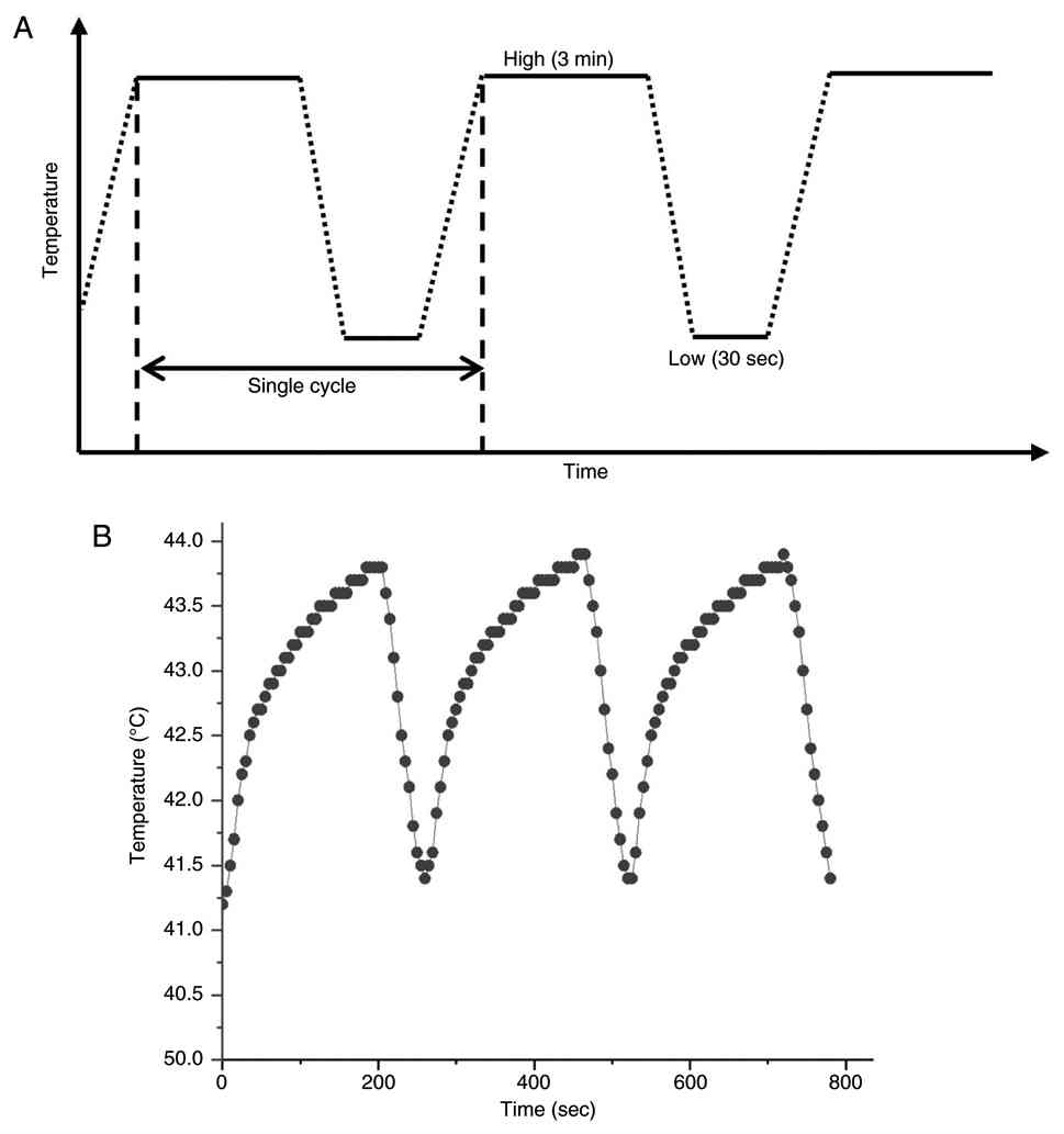
heating and cooling phases. The TCS exposure comprised 15 cycles,
each consisting of a 3-min high-temperature phase followed by a
30-sec passive cooling interval (Fig.
1A). This heating-cooling period was established based on prior
investigations involving various cancer cell lines (6,7). The
low-temperature setting was maintained at 37°C to simulate
physiological human body temperature. Actual temperatures
experienced by cells at the well bottom were monitored using a
needle thermocouple. Temperature cycling reached a steady state
after the third cycle, with the recorded temperature cycling
between 41.4 and 43.8°C, as shown in Fig. 1B.
Cell treatments
EGCG (MedChemExpress; cat. no. HY-13653),
2-aminoethoxydiphenyl borate (2-APB) (MedChemExpress; cat. no.
HY-W009724) and N-acetylcysteine (NAC; Sigma-Aldrich; Merck KGaA;
cat. no. A9165) were dissolved in sterile distilled water to
prepare stock solutions at concentrations of 20, 5 and 500 mM,
respectively. By contrast, GSK2606414 [MedChemExpress; cat. no.
HY-18072; an inhibitor of the protein kinase R-like ER kinase
(PERK)], JNJ-28583113 [MedChemExpress; cat. no. HY-149143; an
antagonist of transient receptor potential cation channel subfamily
M member 2 (TRPM2)], and SB-705498 [MedChemExpress; cat. no.
HY-10633; an antagonist of transient receptor potential cation
channel subfamily V member 1 (TRPV1)] were solubilized in dimethyl
sulfoxide (DMSO; Sigma-Aldrich; Merck KGaA) at concentrations of 1,
5 and 5 mM, respectively, to make stock solutions. All stock
solutions were stored at −20°C until further use. Cells were seeded
in 96-well plates at 3,000 cells/well, or in 6-well plates at
100,000 cells/well, and incubated overnight at 37°C to facilitate
cell adherence. For the combined treatment of EGCG and TCS, EGCG
was administered immediately prior to TCS exposure. Cells were
pretreated with NAC or the molecular inhibitors 1 h prior to the
administration of EGCG. Unless specifically indicated, cells were
maintained at 37°C in a humidified incubator for an additional 24 h
after treatment for further analysis.
Cell viability assay
Cell viability was assessed using the
3-(4,5-dimethylthiazol-2-yl)-2,5-diphenyltetrazolium bromide (MTT)
(Sigma-Aldrich; Merck KGaA) assay. MTT powder was dissolved in
distilled water to make a 5 mg/ml stock solution, which was
subsequently stored at 4°C. Immediately before use, the MTT stock
solution was diluted with culture medium to a final concentration
of 0.5 mg/ml. At 24 h after the treatment, the culture medium in
the 96-well plates was replaced with 100 µl of the MTT working
solution. The cells were then incubated at 37°C in the dark for 4
h, during which time mitochondrial dehydrogenases in viable cells
reduced MTT to insoluble formazan crystals. To dissolve the
formazan crystals, 100 µl 10% sodium dodecyl sulfate (SDS) (Bioshop
Canada, Inc.) solution in 0.01 M hydrochloric acid (HCl) (Echo
Chemical Co., Ltd.) was added to each well. After an overnight
incubation, the absorbance was measured at 570 nm with background
subtraction at 690 nm using a Multiskan GO spectrophotometer
(Thermo Fisher Scientific, Inc.). Finally, cell viability was
expressed as a ratio based on the relative quantity of formazan
compared with the untreated control.
Assessment of synergy using the Bliss
independence model
To evaluate the synergistic interaction between EGCG
and TCS, the Bliss independence model was applied. The expected
fractional inhibition on cell viability for the combination
treatment (Eexp) was calculated using the
formula: Eexp=EEGCG +
ETCS-(EEGCG ×
ETCS), where EEGCG and
ETCS represent the fractional inhibitions induced
by individual treatments of EGCG and TCS, respectively. The Bliss
excess value (ΔE) was defined as the difference between the
observed fractional inhibition (Eobs) and
Eexp (namely,
ΔE=Eobs-Eexp). A
positive Bliss excess value (ΔE >0) was indicative of
synergy (21).
Colony formation assay
To assess their colony-forming capabilities, A549
cells were seeded into 35-mm culture dishes at a density of 300
cells per dish and allowed to adhere overnight. On the following
day, cells were treated with EGCG, TCS, or their combination. The
culture medium was replaced after 24 h, followed by an additional
10-day incubation period. Subsequently, colonies were fixed using
4% paraformaldehyde (Sigma-Aldrich; Merck KGaA) for 10 min and
stained with 0.1% crystal violet (Sigma-Aldrich; Merck KGaA) for 15
min at room temperature. Aggregates containing 50 or more cells
were identified as viable colonies. Colony numbers were quantified
manually using an optical microscope, and the results were
expressed relative to the untreated control group.
Western blot analysis
Protein levels in A549 cells were assessed using
western blotting. After treatment, cells were washed with
phosphate-buffered saline (PBS) and subsequently lysed on ice for 1
h using RIPA lysis buffer (50 mM Tris-HCl, pH 7.4, 0.15 M NaCl,
0.25% deoxycholic acid, 1% NP-40, 1% Triton X-100, 0.1% SDS, 1 mM
EDTA) (MilliporeSigma) supplemented with protease and phosphatase
inhibitor cocktails (MilliporeSigma). Lysates were centrifuged at
10,000 × g for 30 min at 4°C, and the supernatants were then
collected. Protein concentrations were determined using the
Bradford protein assay (Bioshop Canada Inc.). For the detection of
total ubiquitinated proteins, cells were directly harvested and
lysed in pre-heated sample loading buffer at 95°C for 10 min to
rapidly deactivate endogenous deubiquitinating enzymes and preserve
polyubiquitinated species. Equal amounts of protein samples (20 µg)
were separated by SDS-polyacrylamide gel electrophoresis (SDS-PAGE;
12% acrylamide gels), and then transferred onto polyvinylidene
fluoride (PVDF) membranes (MilliporeSigma). To facilitate the
detection of total ubiquitinated proteins, PVDF membranes were
soaked in PBS and subsequently subjected to autoclaving at 121°C
for 30 min prior to the blocking step, thereby improving epitope
accessibility. Membranes were blocked with 5% bovine serum albumin
(BioShop Canada Inc.) in Tris-buffered saline with Tween-20 (TBST;
20 mM Tris, 150 mM NaCl and 0.1% Tween 20) for 1 h at room
temperature, followed by an overnight incubation at 4°C with
primary antibodies targeting proteasome subunit alpha type-3
(PSMA3; cat. no. 12446; 1:1,000), 26S proteasome regulatory subunit
6A (PSMC3; cat. no. 13923; 1:1,000), HSP70 (cat. no. 4872; 1:1,000)
and phosphorylated c-Jun N-terminal kinase (p-JNK; Thr183/Tyr185;
cat. no. 4668; 1:1,000) (all from Cell Signaling Technology, Inc.),
and HSP105 (cat. no. GTX03248; 1:1,000), ubiquitin (cat. no.
GTX128826; 1:5,000), phosphorylated eukaryotic initiation factor 2
subunit α (p-eIF2α; Ser51; cat. no. GTX130006; 1:1,000),
total-eIF2α (t-eIF2α; cat. no. GTX101241; 1:2,000), total-JNK
(t-JNK; cat. no. GTX133806; 1:1,000), poly(ADP-ribose) polymerase
(PARP; cat. no. GTX132329; 1:1,000) and GAPDH (cat. no. GTX100118;
1:10,000) (all from GeneTex, Inc.). After washing with TBST, the
membranes were incubated overnight at 4°C with horseradish
peroxidase-conjugated secondary antibodies (Jackson ImmunoResearch
Laboratories, Inc.; 1:10,000). Finally, the immunoreactive signals
were visualized using an enhanced chemiluminescence substrate
(Advansta Inc.) and detected in an Amersham Imager 600 imaging
system [Cytiva (formerly GE Healthcare Life Sciences)]. Finally,
protein bands were quantified using Image Lab software (Bio-Rad
Laboratories, Inc.).
RNA extraction, reverse transcription,
and X-box binding protein 1 (XBP1) mRNA splicing analysis
Total RNA was extracted from A549 cells using the
miTotal RNA Extraction Miniprep System (Viogene), following the
manufacturer's instructions. Reverse transcription (RT) and PCR
amplification were performed using a HiScript II One-Step RT-PCR
Kit (Vazyme Biotech Co., Ltd.) with gene-specific primers. The
primer sequences for human XBP1 were as follows: Forward,
5′-TTACGAGAGAAAACTCATGGCC-3′ and reverse,
5′-GGGTCCAAGTTGTCCAGAATGC-3′; note that the primers flanked the
26-nucleotide intron that was excised by inositol-requiring enzyme
1α (IRE1α)-mediated splicing. GAPDH was amplified as an internal
control using the following primers: Forward,
5′-GGAGCGAGATCCCTCCAAAAT-3′, and reverse
5′-GGCTGTTGTCATACTTCTCATGG-3′. To achieve optimal resolution among
the unspliced (XBP1u), heteroduplex (XBP1h) and spliced (XBP1s)
isoforms, PCR products were separated on a 2.5% agarose gel
prepared in TBE buffer (89 mM Tris-borate and 2 mM EDTA, pH 8.3)
and electrophoresed at 70 V for 1.5 h. Subsequently, the gel was
stained for 45 min using GelRed Nucleic Acid Stain (Biotium, Inc.)
diluted at a ratio of 3:10,000 in a 0.1 M NaCl aqueous solution.
DNA bands were visualized using the Keta S Standalone Gel
Documentation System (Wealtec Bioscience Co., Ltd.). Quantification
of band intensities was performed using ImageJ software (version
1.53k; National Institutes of Health), and the ratio of XBP1 mRNA
splicing was calculated according to the formula: Splicing
ratio=(Intensity of XBP1s + 0.5× Intensity of XBP1h)/(Intensity of
XBP1s + Intensity of XBP1h + Intensity of XBP1u) (22).
Measurement of cytosolic
Ca2+ levels
Cytosolic Ca2+ levels were measured using
the fluorescent dye Fluo-4 AM (MedChemExpress) to quantify
cytosolic Ca2+ levels. A 1 mM stock solution of Fluo-4
AM was prepared by dissolving the dye in DMSO, after which it was
stored at −20°C until further use. At 24 h after treatments, cells
were harvested and washed with Hanks' Balanced Salt Solution (HBSS)
and subsequently incubated with 1 µM Fluo-4 AM in HBSS for 1 h at
37°C in the dark to load the dye. Following the incubation, cells
were washed with HBSS and finally resuspended in fresh HBSS.
Fluorescence intensity of Fluo-4 AM was measured using a FACSLyric
flow cytometer (BD Biosciences) in the FITC channel. For each
sample, at minimum of 9,000 events were acquired. Cellular debris
was excluded based on the forward scatter (FSC) and side scatter
(SSC) characteristics. Finally, cytosolic Ca2+ levels
were quantified by calculating the mean fluorescence intensity and
expressed as fold changes relative to the untreated control
group.
Measurement of intracellular ROS
levels
The dihydroethidium (DHE) (Sigma-Aldrich; Merck
KGaA) fluorescent dye was employed to measure ROS levels in A549
cells. At 24 h after treatments, cells were harvested and washed
with PBS prior to staining. Cells were subsequently incubated with
a 5 µM DHE working solution for 30 min at 37°C in the dark.
Fluorescence intensity was measured using a FACSLyric flow
cytometer (BD Biosciences) in the PE channel. For each sample, at
minimum of 9,000 events were acquired. Cellular debris was excluded
based on FSC and SSC characteristics. Finally, ROS levels were
determined by calculating the mean fluorescence intensity and
expressed as fold changes compared with the control group.
Mitochondrial membrane potential (MMP)
analysis
The MMP integrity was assessed using the JC-1
(MedChemExpress) fluorescent dye. At 24 h after treatments, cells
were harvested and rinsed with PBS. Subsequently, cells were
incubated with 5 µg/ml JC-1 in PBS for 30 min at 37°C in the dark.
Fluorescence intensities of JC-1 were measured using the FACSLyric
system (BD Biosciences) in both the PE (red fluorescence) and FITC
(green fluorescence) channels. For each sample, at minimum of 9,000
events were acquired. Cellular debris was excluded based on the FSC
and SSC characteristics, and MMP disruption was quantified through
assessing the proportion of cells exhibiting a fluorescence shift
from red (JC-1 aggregates) to green (JC-1 monomers).
Apoptosis analysis
Apoptotic rates were quantified using an annexin
V-fluorescein isothiocyanate (FITC) and propidium iodide (PI)
double staining kit (BD Biosciences). At 24 h after treatments,
cells were collected, washed with PBS, and subsequently incubated
in a binding buffer containing annexin V-FITC and PI for 30 min at
37°C in the dark, following the manufacturer's instructions.
Fluorescence intensities were measured using the FACSLyric system
(BD Biosciences) in both the PE and FITC channels. Early and late
apoptotic cells were quantified as the percentages of cells
identified in the (PE−/FITC+) and
(PE+/FITC+) quadrants, respectively. For each
sample, a minimum of 9,000 events were acquired. Cellular debris
was excluded based on the FSC and SSC characteristics, and the
percentages of these populations were calculated accordingly.
Statistical analysis
Three independent biological experiments were
performed for each experimental condition using separately prepared
cell cultures. Data are presented as the mean ± standard deviation.
Statistical analyses were performed using one-way analysis of
variance (ANOVA), followed by Tukey's post hoc test, using the
OriginPro 2015 software (version 92E; OriginLab Corporation). A
P-value <0.05 was considered to indicate a statistically
significant difference. For clarity, only selected pairwise
comparisons are shown in the figures.
Results
Synergistic inhibitory effects of EGCG
and TCS on NSCLC cell viability
To assess the effect of EGCG and TCS on the
viability of A549 cells, MTT assays were performed 24 h after
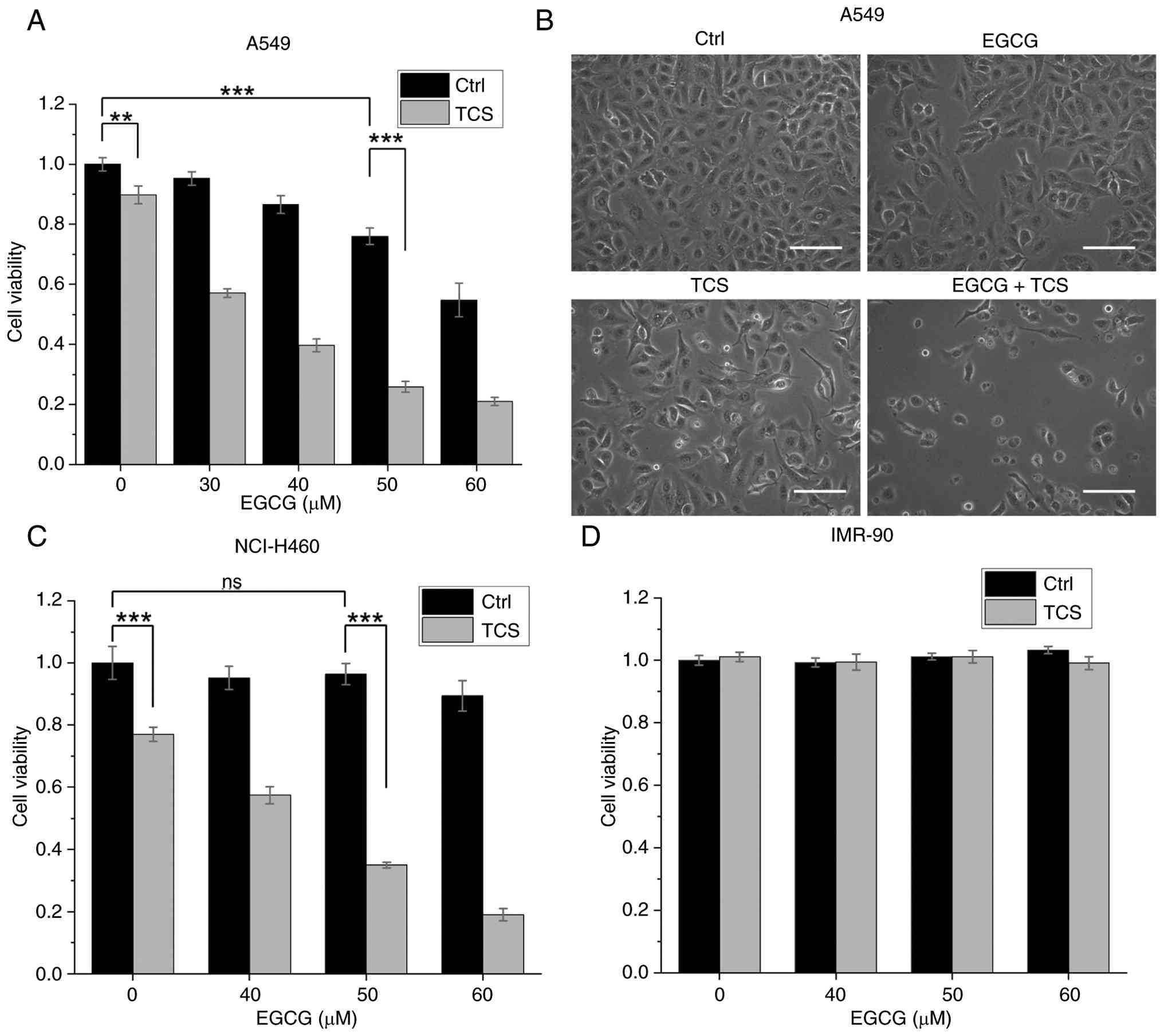
treatment. As shown in Fig. 2A,
EGCG induced a significant and dose-dependent reduction in A549
cell viability. By contrast, TCS alone caused only a slight
decrease in A549 cell viability, as ~90% of the control levels were
maintained. Notably, however, the combined administration of EGCG
and TCS produced a more substantial inhibition effect on A549 cell
viability. The Bliss excess value (ΔE), as calculated in
Table IA, confirmed a synergistic
cytotoxic effect of EGCG and TCS, with ΔE >0 at EGCG
concentrations ranging from 30 to 60 µM. Consistent with the MTT
results, morphological examination of the cells under a light
microscope at 24 h post-treatment (Fig. 2B) demonstrated that the combined
treatment of EGCG and TCS induced pronounced morphological
alterations in A549 cells, including reduced cell density, cellular
shrinkage, and an increased proportion of non-adherent cells. To
further validate that these effects were more widely applicable,
the MTT assay was subsequently performed on a different NSCLC cell
line, NCI-H460. The results obtained (as shown in Fig. 2C) confirmed that a synergistic
cytotoxic effect also occurred upon treatment with EGCG and TCS in
NCI H460 cells, with the corresponding Bliss excess values
presented in Table IB. According
to the data from Table IA, the
combination of 50 µM EGCG and TCS produced the highest Bliss excess
value in A549 cells, thereby justifying the selection of 50 µM EGCG
for subsequent experiments in the present study. Notably, as shown
in Fig. 2D, the combination of
EGCG and TCS did not lead to a reduction in the viability of the
IMR-90 normal lung fibroblasts, demonstrating that this combined
treatment is well tolerated by non-malignant lung fibroblasts. To
further evaluate whether this combined treatment could impair
long-term reproductive viability, a colony formation assay was
performed using A549 cells. As shown in Fig. S1A and B, the combination of EGCG
and TCS significantly suppressed the clonogenic capability of the
A549 cells, resulting in a significantly lower colony count
compared with single treatments. Taken together, these results
showed that EGCG and TCS work synergistically to inhibit both the
short-term metabolic viability, and the long-term clonogenic
survival, of NSCLC cells, while exhibiting minimal cytotoxicity on
IMR-90 normal lung fibroblasts in vitro.
 | Table I.Bliss excess for EGCG in combination
with TCS in A549 and NCI-H460 cells. |
Table I.
Bliss excess for EGCG in combination
with TCS in A549 and NCI-H460 cells.
| A, A549 cells |
|---|
|
|---|
| EGCG (µM) | 0 | 30 | 40 | 50 | 60 |
|---|
| Bliss excess
(ΔE) | 0.00 | 0.28 | 0.38 | 0.42 | 0.28 |
|
| B, NCI-H460
cells |
|
| EGCG (µM) | 0 | 40 | 50 | 60 |
|
|
| Bliss excess
(ΔE) | 0.00 | 0.16 | 0.39 | 0.50 |
|
|
| TCS, thermal
cycling-stimulation; EGCG, epigallocatechin gallate. |
Effects of EGCG and TCS on
proteostatic impairment in A549 cells
To elucidate the mechanism underlying the
synergistic effect of EGCG and TCS, it was first investigated
whether these treatments could influence the regulation of HSPs in
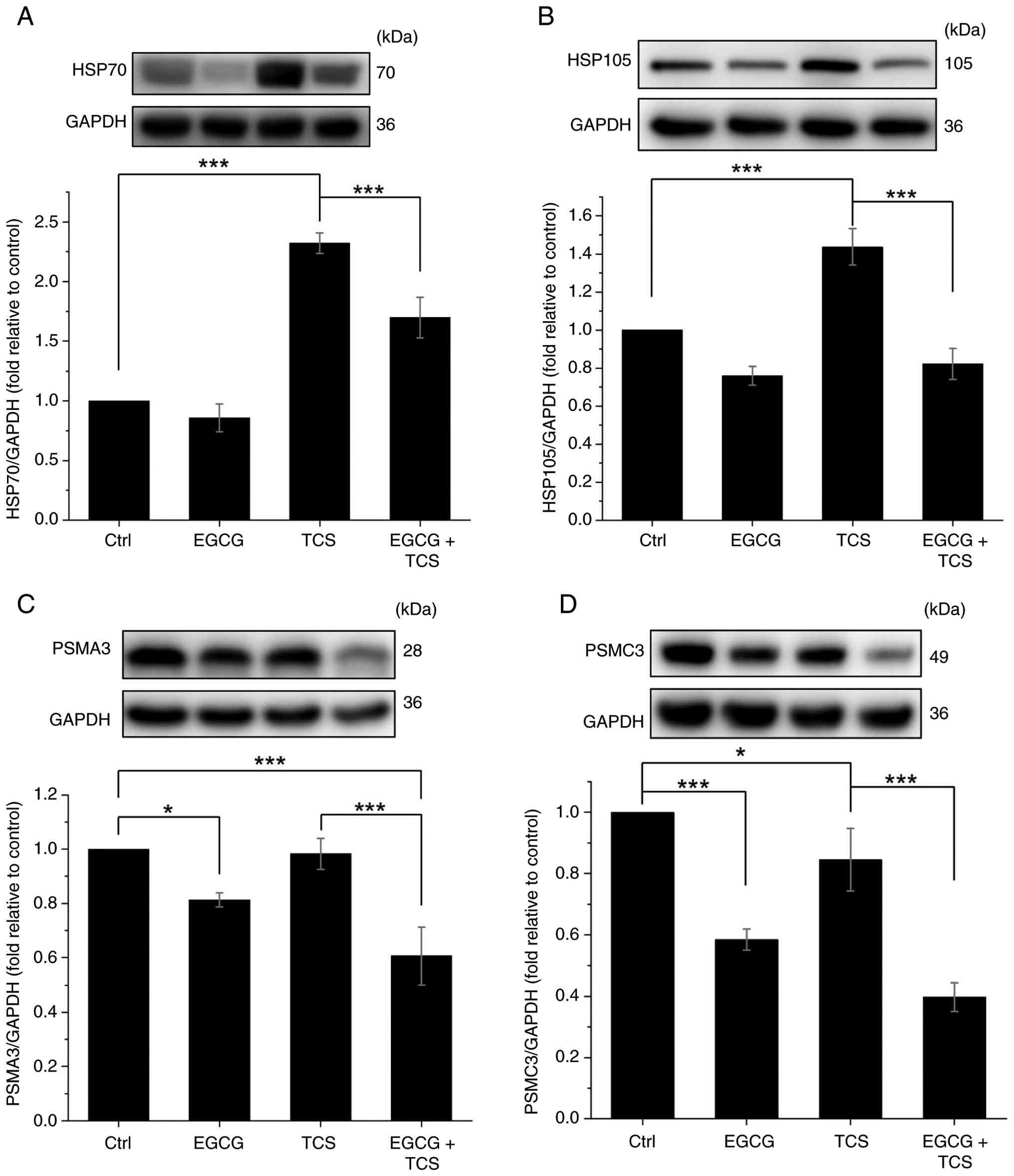
A549 cells. Western blot analysis was employed to quantify the
expression levels of HSP70 and HSP105. As shown in Fig. 3A and B, administering TCS increased
the expression levels of both HSP70 and HSP105 in A549 cells,
whereas combined treatment with EGCG significantly reduced this
induction of protein expression. In addition to HSPs, the
expression levels of the proteasome subunits, PSMA3 and PSMC3, were
also evaluated via western blot analysis. It was revealed that TCS
alone exerted a negligible effect on PSMA3 expression, and induced
only a slight reduction in PSMC3 levels (Fig. 3C and D). By contrast, EGCG was
found to significantly decrease the expression of both PSMA3 and
PSMC3 in A549 cells treated with TCS. To evaluate the overall
status of cellular proteostasis under these conditions, the levels
of total ubiquitinated proteins were subsequently measured. As
shown in Fig. S2A and B,
co-treatment with EGCG and TCS resulted in a significant increase
in the accumulation of ubiquitinated proteins compared with either
treatment in isolation. Taken together, these findings suggested
that EGCG suppresses the expression of critical components of the
proteostatic machinery, including HSP70, HSP105, and the proteasome
subunits. Moreover, this inhibition is associated with an increased
accumulation of ubiquitinated proteins in the combination treatment
group, reflecting heightened proteostatic dysfunction in A549
cells.
EGCG and TCS induce ER stress in A549
cells
The intracellular accumulation of unfolded proteins
is known to provoke ER stress, which, if sustained or severe, may
culminate in cellular damage (23). In the present study, the
involvement of the PERK-eIF2α signaling axis, a key ER stress
pathway, was therefore explored in mediating cell death induced by
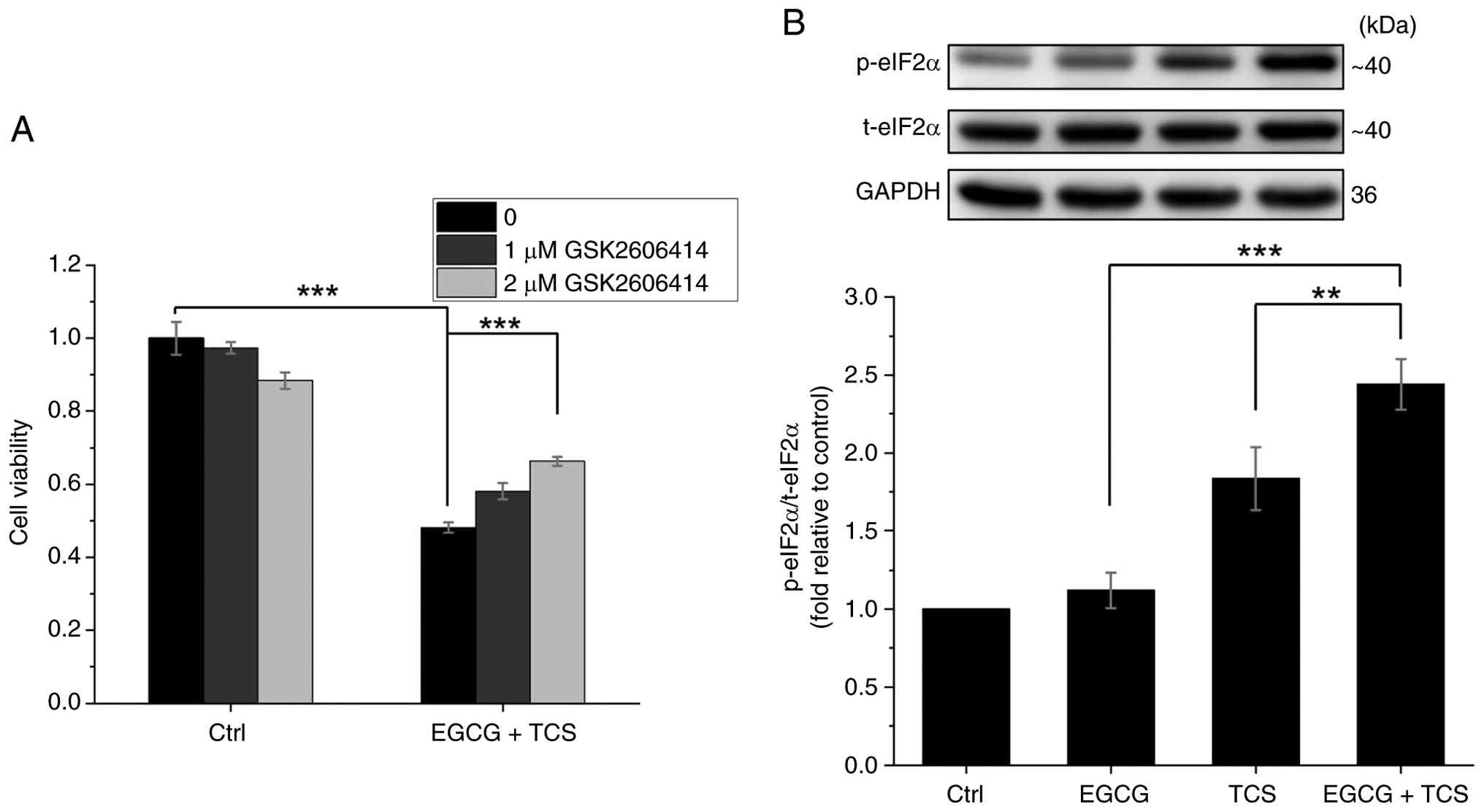
EGCG and TCS. As shown in Fig. 4A,
administration of the PERK inhibitor GSK2606414 partially mitigated
the reduction in A549 cell viability caused by combined EGCG and
TCS treatment in a dose-dependent manner. Additionally, western
blot analysis of the downstream signaling components revealed that
TCS significantly enhanced the phosphorylation of eIF2α, with the
combination of EGCG and TCS producing an even more pronounced
effect (Fig. 4B). Concurrently,
TCS treatment alone induced substantial XBP1 mRNA splicing
(Fig. S3A), thereby confirming
IRE1α activation. Notably, co-treatment with EGCG and TCS led to a
significant enhancement in JNK phosphorylation (Fig. S3B), a pro-apoptotic signaling
event that can be triggered either downstream of IRE1α or via
ROS-apoptosis signal-regulating kinase 1 (ASK1)-dependent pathways.
These findings suggest that the combination of EGCG and TCS
activates both the PERK-eIF2α and the IRE1α-JNK pathways of ER
stress signaling, which may partially contribute to the observed
reduction in viability of A549 cells.
Induction of IP3R-mediated
ER Ca2+ release by the combined administration of EGCG
and TCS in A549 cells
ER stress is frequently associated with
intracellular Ca2+ dysregulation, in which
Ca2+ stored in the ER can be released via
IP3R channels (24).
Therefore, cytosolic Ca2+ levels were further
quantitatively measured using the Fluo-4 AM fluorescent dye via
flow cytometric analysis. As shown in Fig. 5A and B, treatment with EGCG alone
did not significantly affect cytosolic Ca2+ levels in
A549 cells, whereas TCS alone led to only a slight increase. By
contrast, the combination of EGCG and TCS produced a substantial
elevation in cytosolic Ca2+ levels. Notably, this
increase was found to be greatly attenuated through administering
the IP3R inhibitor 2-APB at a concentration of 50 µM,
suggesting that the elevation in Ca2+ level might be
mediated via the activation of IP3R channels. To further
elucidate the role of ER Ca2+ release in the synergistic
cytotoxicity induced by EGCG and TCS in A549 cells, a rescue
experiment employing 2-APB was performed using the MTT viability
assay. As revealed in Fig. 5C,
2-APB significantly ameliorated the combined treatment's reduction
of A549 cell viability in a dose-dependent manner. In addition to
its effect on IP3R channels, 2-APB is also known to
affect thermosensitive transient receptor potential (TRP) channels,
such as TRPV1 and TRPM2 (25,26).
However, in the experiments of the present study, it was found that
selective inhibitors for TRPM2 (JNJ-28583113) and TRPV1 (SB-705498)
did not reverse the synergistic cytotoxic effects of EGCG and TCS
(Fig. 5D), suggesting that these
TRP channels are unlikely to mediate a significant role in this
process. Taken together, these pharmacological rescue experiments
suggested the potential involvement of IP3R channels and
associated ER Ca2+ release in contributing to the
synergistic anticancer effects induced by the combined EGCG and TCS
treatment.
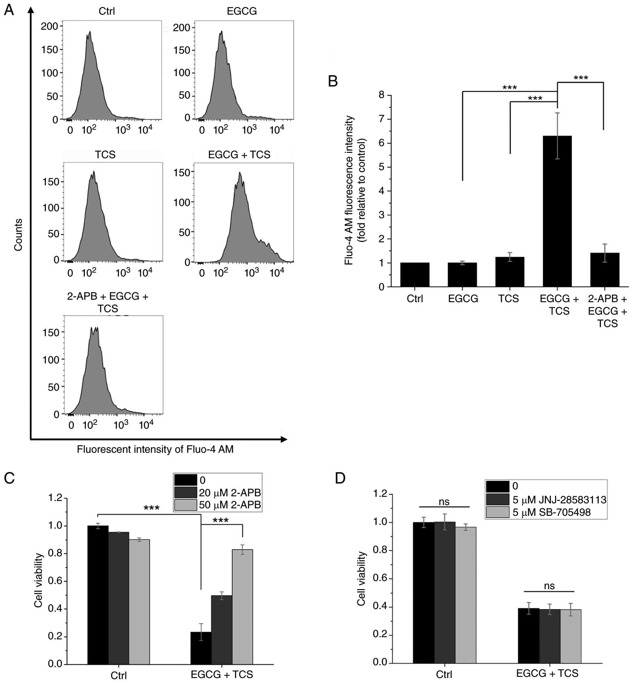 | Figure 5.Investigation of the effects of TCS
and EGCG on Ca2+ homeostasis in A549 cells. (A)
Cytosolic Ca2+ levels in A549 cells were measured
utilizing the fluorescent dye Fluo-4 AM via flow cytometry. (B)
Quantitative analysis of the mean fluorescence intensities of
Fluo-4 AM, as shown in panel (A). (C) Cell viability of A549 cells
was evaluated using the MTT assay 24 h following combined EGCG and
TCS treatment, in the presence or absence of the IP3R
inhibitor 2-APB. The 2-APB inhibitor was administered 1 h prior to
the combined EGCG and TCS treatment. (D) Assessment of A549 cell
viability was also performed using the MTT assay 24 h after
combined EGCG and TCS treatment, with or without the TRPM2
inhibitor JNJ-28583113 and the TRPV1 inhibitor SB-705498.
Similarly, these inhibitors were applied 1 h before the combined
treatment with EGCG and TCS. ***P<0.001. TCS, thermal
cycling-stimulation; EGCG, epigallocatechin gallate; 2-APB,
2-aminoethoxydiphenyl borate; TRPM2, transient receptor potential
cation channel, subfamily M, member 2; TRPV1, transient receptor
potential cation channel subfamily V member 1; ns,
non-significant. |
The combination of EGCG and TCS
induces ROS production in A549 cells
ER stress and intracellular Ca2+
dysregulation have previously been shown to coincide with excessive
ROS generation (5). In the present
study, intracellular ROS levels in A549 cells were measured using
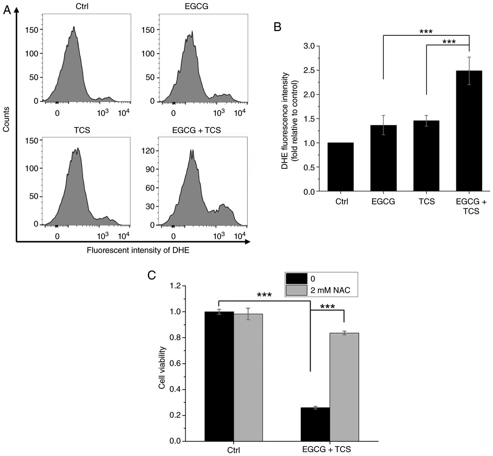
the fluorescent dye DHE via flow cytometric analysis. As
demonstrated in Fig. 6A and the
resultant quantification of the data in Fig. 6B, treatment with either EGCG or TCS
alone resulted in only a slight, and statistically non-significant,
increase in ROS levels. On the other hand, the combined
administration of EGCG and TCS produced a pronounced and
statistically significant elevation of ROS levels in A549 cells. To
explore the role of ROS in the reduction of cell viability caused
by this combination, a rescue experiment employing the antioxidant
NAC was performed using the MTT assay. The results shown in
Fig. 6C demonstrate that NAC could
effectively mitigate the synergistic cytotoxicity induced by the
combination of EGCG and TCS in A549 cells, suggesting that the
excessive ROS production may contribute to the reduced viability in
A549 cells.
Potential reciprocal interplay of ROS
production and ER Ca2+ release under combined EGCG and
TCS treatment in A549 cells
To explore the potential association between ER
Ca2+ release and ROS generation, ROS levels in A549
cells were first assessed by DHE staining in the presence of 50 µM
2-APB, an inhibitor of IP3R-mediated Ca2+
release. As shown in Fig. 7A and
B, 2-APB treatment led to a significant attenuation of the ROS
elevation triggered by the combined EGCG and TCS treatment,
suggesting that ER Ca2+ release through IP3R
channels may contribute to the substantial increase in ROS levels.
On the other hand, the cytosolic Ca2+ levels were
measured using Fluo-4 AM staining in the presence of 2 mM NAC
(Fig. 7C and D). These experiments
revealed that NAC treatment effectively suppressed the rise in
cytosolic Ca2+ levels that was induced by the combined
EGCG and TCS treatment, suggesting that elevated ROS levels may
facilitate ER Ca2+ release. Collectively, these
convergent pharmacological findings support a working model of
reciprocal interplay between the ER Ca2+ release and ROS
generation events, wherein mutual amplification of these signals
culminates in enhanced cell death.
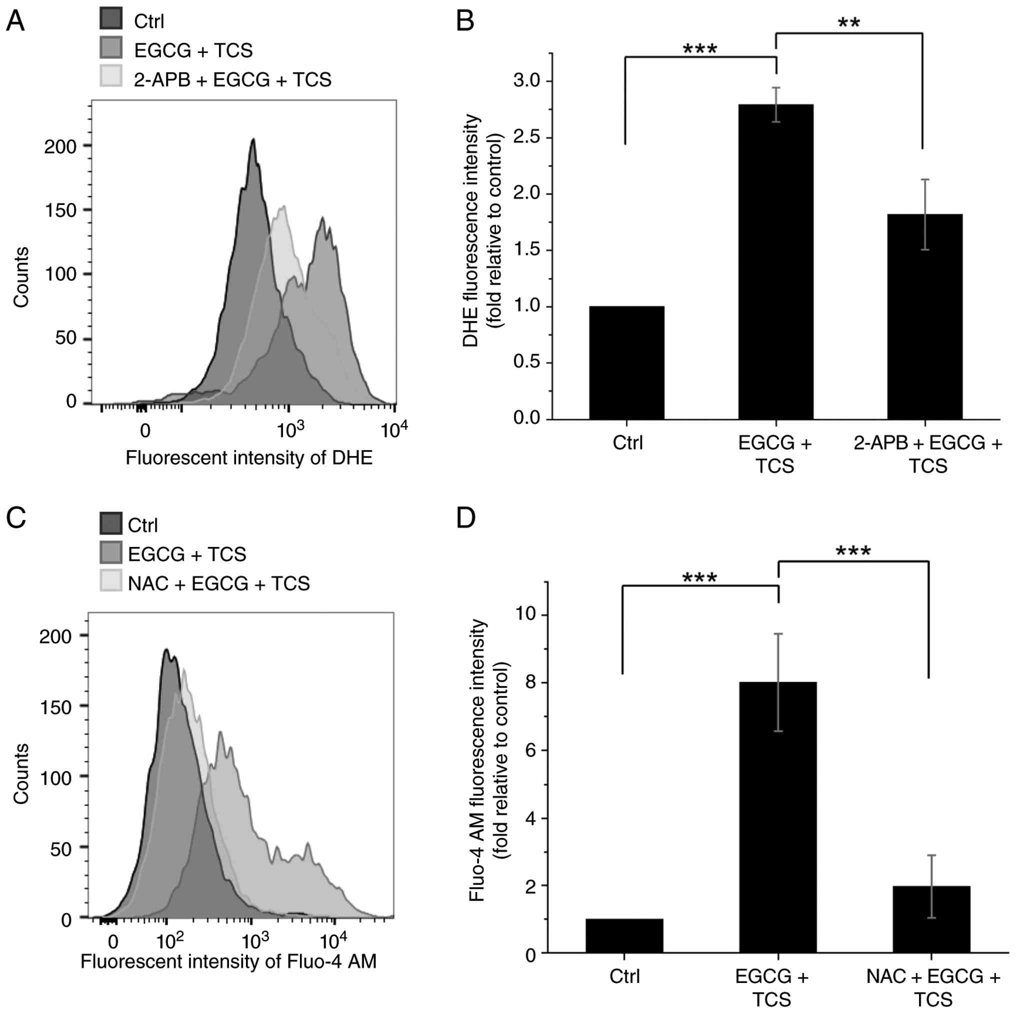 | Figure 7.Potential effects of TCS and EGCG on
the ROS-Ca2+ reciprocal interplay in A549 cells. (A)
Intracellular ROS levels in A549 cells were measured using the
fluorescent dye DHE via flow cytometry, following combined
treatment with EGCG and TCS in the absence or presence of 2-APB.
(B) Quantitative analysis of the mean fluorescent intensities of
DHE, as shown in panel (A). (C) Cytosolic Ca2+ levels in
A549 cells were assessed using the fluorescent dye Fluo-4 AM via
flow cytometry following combined EGCG and TCS treatment, in the
absence or presence of NAC. (D) Quantification of the mean
fluorescent intensities of Fluo-4 AM, as shown in panel (C).
**P<0.01 and ***P<0.001. TCS, thermal cycling-stimulation;
EGCG, epigallocatechin gallate; ROS, reactive oxygen species; DHE,
dihydroethidium; 2-APB, 2-aminoethoxydiphenyl borate; NAC,
N-acetylcysteine. |
The combination of EGCG and TCS
induces mitochondrial apoptosis in A549 cells
Excessive ROS accumulation and mitochondrial
Ca2+ overload are known to cause mitochondrial damage
(27,28). Given that, in these experiments,
the combined EGCG and TCS treatment effectively triggered ER
Ca2+ release and an increase in ROS levels, it was
sought to further evaluate changes in the MMP using the fluorescent
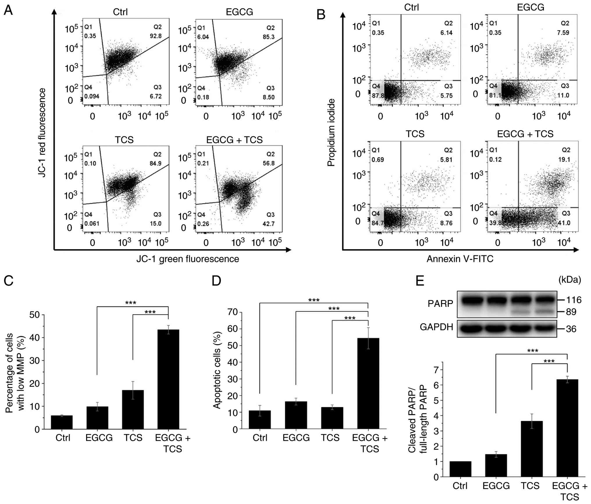
dye JC-1 via flow cytometric analysis. As revealed in Fig. 8A, the combined EGCG and TCS
treatment caused a significant shift in JC-1 fluorescence from red
to green, indicative of MMP disruption. The quantitative analysis
shown in Fig. 8C demonstrates that
the combined treatment caused a more substantial loss of MMP
compared with either treatment alone. Mitochondrial damage may
eventually activate the intrinsic apoptotic pathway (27,28).
Accordingly, the evaluation of apoptosis was performed using
annexin V-FITC/PI double staining by flow cytometric analysis. As
demonstrated in Fig. 8B and D, the
combination of EGCG and TCS caused a significant increase in the
proportion of apoptotic cells compared with single treatments. To
further confirm the occurrence of apoptosis at the molecular level,
the cleavage of PARP, a well-established indicator of caspase
activation, was examined. As shown in Fig. 8E, western blot analysis
demonstrated that the combined treatment with EGCG and TCS led to a
significant enhancement in the cleavage of full-length PARP (116
kDa) into its characteristic 89-kDa inactive fragment.
Collectively, these results confirmed that the combined treatment
of EGCG with TCS effectively induced mitochondrial apoptosis in
A549 cells.
Discussion
The considerable challenges associated with the
treatment of NSCLC have prompted extensive investigation into more
effective combination therapies (2,6).
EGCG, a natural compound, exhibits strong anticancer properties,
although its clinical application has been limited due to poor
bioavailability, low stability and potential hepatotoxicity at
elevated doses (20,29). The data presented in the present
study, as shown in Fig. 2,
demonstrate that the combined administration of 50 µM EGCG and TCS
synergistically suppresses the viability of the NSCLC cell lines
A549 and NCI-H460, while exhibiting minimal cytotoxic effects on
IMR-90 normal lung fibroblasts. These findings suggest that TCS is
able to enhance the cytotoxic effect of EGCG in vitro,
implying that combination treatment may provide a potential
strategy for partially improving the therapeutic efficacy of EGCG
by lowering its required dosage.
To understand the mechanisms underlying this
synergistic effect, the influence of EGCG and TCS was first studied
on HSPs and the UPS, two critical regulators of intracellular
proteostasis. HSPs, especially HSP70, are often overexpressed in
various types of malignancies, including NSCLC, where they
facilitate cell proliferation, inhibit apoptosis, and contribute to
drug resistance (30–32). Consequently, several molecular
inhibitors targeting HSPs have been developed as anticancer agents
(11,12). Nevertheless, hyperthermia-induced
upregulation of HSP70 and HSP105 has been shown to be associated
with the development of thermotolerance, which diminishes the
clinical efficacy of thermal therapies (11,33).
Notably, EGCG has been shown to suppress the expression of HSP70
and to competitively bind to its ATPase domain, thereby inhibiting
its chaperone function (17).
Consistent with these findings, the results obtained in the present
study have demonstrated that EGCG effectively attenuates the
TCS-induced elevation of HSP70 and HSP105 levels (Fig. 3A and B), suggesting that EGCG may
both disrupt adaptive thermotolerance and sensitize A549 cells to
TCS-induced proteotoxic stress. In addition to HSPs, the UPS has a
crucial role in oncogenesis and progression, rendering it a viable
therapeutic target in cancer treatments (15,34).
A study by Nam et al (35)
demonstrated that EGCG acts as a blocker to the chymotrypsin-like
function of the 20S proteasome. The findings of the present study
have further revealed that the combination of EGCG and TCS caused a
marked decrease in the protein expression of the proteasome
subunits, PSMA3 and PSMC3 (Fig. 3C and
D). Importantly, these alterations in the proteostatic
machinery were accompanied by a pronounced accumulation of total
ubiquitinated proteins in the combination treatment group (Fig. S2). Collectively, these results
suggested that the combined administration of EGCG and TCS
suppresses both chaperones and proteasome components, and that this
was associated with disrupted proteostasis, therefore having a
possible role in the induction of ER stress in NSCLC cells.
ER stress serves as a crucial cellular response to
stress and has a dual effect on cancer cells. When cancer cells
experience mild proteotoxic stress in the tumor microenvironment,
they activate the unfolded protein response (UPR) to enhance
survival. However, if ER stress becomes persistent or severe, it
overwhelms this adaptive capacity, eventually leading to apoptosis
(23,36). Consequently, manipulating this
balance to shift the UPR signaling from a pro-survival to a
pro-apoptotic mode represents a promising therapeutic strategy for
treating NSCLC (36–38). In the present study, it has been
demonstrated that pretreatment with the PERK inhibitor GSK2606414
partially restored the reduction in viability of A549 cells that
was caused by the combined treatment of EGCG and TCS (Fig. 4A). Additionally, phosphorylation of
the downstream target eIF2α was found to have been enhanced in the
combination treatment group (Fig.
4B). In addition to the PERK pathway, ER stress induces
activation of the IRE1α sensor, which facilitates the splicing of
XBP1 mRNA (39). The results shown
in Fig. S3A demonstrated that
administering TCS led to a significant increase in XBP1 mRNA
splicing. In addition to XBP1 splicing, IRE1α activation and stress
signals such as ROS-ASK1 converge on the pro-apoptotic JNK
signaling cascade (39,40). In the present study, it was shown
that co-treatment with EGCG and TCS significantly increased JNK
phosphorylation compared with either treatment in isolation
(Fig. S3B). Collectively, these
findings suggested that activation of both the PERK-eIF2α signaling
axis and pro-apoptotic JNK signaling may contribute to the reduced
viability of NSCLC cells following combined EGCG and TCS
treatment.
Severe ER stress is frequently associated with ER
Ca2+ release, which can cause further cellular damage
(5,24,41).
Notably, activation of the PERK pathway has been shown to promote
Ca2+ release from ER via the IP3R (24,41).
Consistent with these findings, the data shown in Fig. 5A and B revealed that the
combination of EGCG and TCS significantly elevates cytosolic
Ca2+ levels. Furthermore, treatment with the
IP3R inhibitor 2-APB led to a marked reduction in this
Ca2+ increase, suggesting that the effect might be
dependent on IP3R activity. Concurrently, ER stress is
often accompanied by excessive ROS production (5,42).
The results shown in Fig. 6A and B
confirmed that the combined treatment of EGCG and TCS effectively
increased intracellular ROS levels in A549 cells. Importantly, both
the release of Ca2+ from the ER and the production of
ROS are widely recognized as interrelated processes. Previous
studies have shown that oxidation of key thiol groups in the
IP3R protein enhances its channel activity (43,44).
At the same time, increased cytosolic or mitochondrial
Ca2+ levels activate ROS-generating enzymes, thereby
facilitating free radical formation (45–47).
In the present study, it was found that the increase in cytosolic
Ca2+ caused by the combination treatment of EGCG and TCS
was reduced by administering the antioxidant NAC, whereas ROS
levels were lowered by 2-APB (Fig.
7). Although pharmacological agents inherently possess
potential off-target effects, these reciprocal observations point
toward a plausible bidirectional crosstalk between Ca2+
dysregulation and excessive ROS production, a well-documented
pathophysiological amplification loop governing cellular stress
responses (48). Moreover,
treatments with either NAC or 2-APB alone significantly lessened
the decrease in cell viability caused by the combined
administration of EGCG and TCS in A549 cells (Figs. 5C and 6C), supporting a candidate working model
wherein this crosstalk mediates the synergistic cytotoxic effects
observed in A549 cells. Future investigations utilizing genetic
silencing (for example, IP3R knockdown) and
organelle-targeted ROS probes are essential to definitively
establish causality, to identify specific ROS species, and to
delineate their primary sources.
Mitochondrial ROS and Ca2+ overload are
potential triggers for the opening of the mitochondrial
permeability transition pore (mPTP), which subsequently leads to
the loss of MMP (24,27,28).
Building upon the present findings that the combination of EGCG and
TCS causes intracellular Ca2+ dysregulation and ROS
generation, in subsequent experiments it was further demonstrated
that this combination could effectively reduce the levels of MMP in
A549 cells, as shown by JC-1 staining (Fig. 8A and C). Opening of the mPTP
results in mitochondrial matrix swelling and rupture of the outer
mitochondrial membrane, facilitating the release of cytochrome
c from the intermembrane space into the cytosol, thereby
initiating the intrinsic apoptotic pathway (49,50).
Consistent with this mechanism, combined EGCG and TCS treatment was
shown to cause a significant increase in the proportion of
apoptotic cells (Fig. 8B and D),
also significantly enhancing the cleavage of PARP (Fig. 8E), a signature downstream event in
the caspase cascade. Considered altogether, these findings
suggested that the combination of EGCG and TCS induces intrinsic
apoptosis through promoting mitochondrial ROS accumulation and
Ca2+ overload in A549 cells.
Although the present study has provided valuable
mechanistic insights derived from in vitro experiments, the
successful clinical translation of this combination therapy
necessitates overcoming substantial practical challenges,
especially in the areas of precise drug delivery and targeted
hyperthermia techniques. The 50 µM concentration of EGCG employed
herein surpassed plasma levels attainable through oral
administration; however, strategies such as liposomal encapsulation
combined with surface ligand modifications may enhance EGCG
stability (51,52). Furthermore, TCS could be
implemented using clinically applicable heating modalities, such as
radiofrequency (53), focused
ultrasound (54) or modulated
electro-hyperthermia (55).
Delivering heat in cycles may exploit tissue-specific hemodynamics,
as heterogeneous tumor perfusion hinders heat dissipation during
the passive cooling phase and promotes intratumoral heat
accumulation (55). Therefore, TCS
has the potential to augment tumor-specific heating, while
minimizing thermal exposure to surrounding healthy tissues.
Nevertheless, further in vivo studies are essential to
optimize the kinetics of localized EGCG delivery, and also to
refine TCS dosing protocols.
In conclusion, the present study has demonstrated
the synergistic effects of EGCG and TCS in reducing the viability
of NSCLC cells; furthermore, a potential underlying mechanism has
been proposed. Specifically, TCS causes protein unfolding, whereas
EGCG suppresses the activity and expression of HSPs (for example,
HSP70 and HSP105) and proteasomes, accompanied by the accumulation
of ubiquitinated proteins and the activation of ER
stress-associated pathways. Furthermore, the results obtained in
the present study have also suggested a mutual amplification
between IP3R-mediated ER Ca2+ release and
excessive ROS production, which ultimately induces mitochondrial
apoptosis. Notably, this combination treatment selectively damages
NSCLC cells while sparing IMR-90 normal lung fibroblasts in
vitro, and this effect is potentially attributable to the
elevated basal ROS levels, and more vulnerable proteostasis
regulation, in cancer cells (56,57).
This combined approach potentially reduces the necessary dose of
EGCG and also attenuates TCS-induced heat-shock responses, thereby
offering a promising in vitro proof-of-concept strategy.
Although the A549 cell line has served as the primary
representative model for the present study, however, further
validation using other NSCLC cell lines, and subsequent in
vivo evaluations (such as assessing normal tissue toxicity,
pharmacokinetics and therapeutic efficacy), are essential to both
confirm these mechanistic insights and advance the clinical
translation of this combination therapy.
Supplementary Material
Supporting Data
Acknowledgements
The authors would like to acknowledge the service
provided by the Research Core Facilities 3 Laboratory of the
Department of Medical Research at National Taiwan University
hospital for the use of the flow cytometry system.
Funding
The present study was supported by research grants from the
National Science and Technology Council (grant nos. NSTC
114-2112-M-002-025, NSTC 113-2112-M-002-024 and NSTC
112-2112-M-002-033) and the Ministry of Science and Technology
(grant no. MOST 110-2112-M-002-004) of Taiwan, R.O.C.
Availability of data and materials
The data generated in the present study may be
requested from the corresponding author.
Authors' contributions
CYC conceived and supervised the study. FTH and CYC
designed the study. FTH, HHL, YK and CJL participated in the
acquisition of data. FTH, HHL, YK, CJL and CYC confirm the
authenticity of all the raw data. FTH and CYC contributed to data
analysis and wrote the manuscript. All authors performed the
experiments. All authors read and approved the final version of the
manuscript.
Ethics approval and consent to
participate
Not applicable.
Patient consent for publication
Not applicable.
Competing interests
The authors declare that they have no competing
interests.
Glossary
Abbreviations
Abbreviations:
|
NSCLC
|
non-small cell lung cancer
|
|
ER
|
endoplasmic reticulum
|
|
ROS
|
reactive oxygen species
|
|
TCS
|
thermal cycling-stimulation
|
|
HSP
|
heat shock protein
|
|
UPS
|
ubiquitin-proteasome system
|
|
EGCG
|
epigallocatechin gallate
|
|
IP3R
|
inositol 1,4,5-trisphosphate
receptor
|
|
BCRC
|
Bioresource Collection and Research
Center
|
|
2-APB
|
2-aminoethoxydiphenyl borate
|
|
NAC
|
N-acetylcysteine
|
|
PERK
|
protein kinase R-like endoplasmic
reticulum kinase
|
|
TRPM2
|
transient receptor potential cation
channel subfamily M member 2
|
|
TRPV1
|
transient receptor potential cation
channel subfamily V member 1
|
|
DMSO
|
dimethyl sulfoxide
|
|
MTT
|
3-(4,5-dimethylthiazol-2-yl)-2,5-diphenyltetrazolium bromide
|
|
SDS
|
sodium dodecyl sulfate
|
|
PBS
|
phosphate-buffered saline
|
|
SDS-PAGE
|
SDS-polyacrylamide gel
electrophoresis
|
|
PVDF
|
polyvinylidene fluoride
|
|
TBST
|
Tris-buffered saline with
Tween-20
|
|
PSMA3
|
proteasome subunit alpha type-3
|
|
PSMC3
|
26S proteasome regulatory subunit
6A
|
|
p-JNK
|
phosphorylated c-Jun N-terminal
kinase
|
|
p-eIF2α
|
phosphorylated eukaryotic initiation
factor 2 subunit α
|
|
t-eIF2α
|
total-eIF2α
|
|
t-JNK
|
total-JNK
|
|
PARP
|
poly(ADP-ribose) polymerase
|
|
XBP1
|
X-box binding protein 1
|
|
RT
|
reverse transcription
|
|
IRE1α
|
inositol-requiring enzyme 1α
|
|
XBP1u
|
unspliced XBP1
|
|
XBP1h
|
heteroduplex XBP1
|
|
XBP1s
|
spliced XBP1
|
|
HBSS
|
Hanks' balanced salt solution
|
|
FSC
|
forward scatter
|
|
SSC
|
side scatter
|
|
DHE
|
dihydroethidium
|
|
MMP
|
mitochondrial membrane potential
|
|
FITC
|
fluorescein isothiocyanate
|
|
PI
|
propidium iodide
|
|
ANOVA
|
analysis of variance
|
|
UPR
|
unfolded protein response
|
|
mPTP
|
mitochondrial permeability transition
pore
|
References
|
1
|
Sung H, Ferlay J, Siegel RL, Laversanne M,
Soerjomataram I, Jemal A and Bray F: Global cancer statistics 2020:
GLOBOCAN estimates of incidence and mortality worldwide for 36
cancers in 185 countries. CA Cancer J Clin. 71:209–249.
2021.PubMed/NCBI
|
|
2
|
Belani CP, Goss G and Blumenschein G Jr:
Recent clinical developments and rationale for combining targeted
agents in non-small cell lung cancer (NSCLC). Cancer Treat Rev.
38:173–184. 2012. View Article : Google Scholar : PubMed/NCBI
|
|
3
|
Xiang Y, Liu X, Wang Y, Zheng D, Meng Q,
Jiang L, Yang S, Zhang S, Zhang X, Liu Y and Wang B: Mechanisms of
resistance to targeted therapy and immunotherapy in non-small cell
lung cancer: Promising strategies to overcoming challenges. Front
Immunol. 15:13662602024. View Article : Google Scholar : PubMed/NCBI
|
|
4
|
Oršolić N, Kučan D and Jazvinšćak Jembrek
M: Enhancing cancer therapy with hyperthermia: Synergistic effects
with natural compounds and conventional treatments. Int J Mol Sci.
27:16502026. View Article : Google Scholar : PubMed/NCBI
|
|
5
|
Hempel N and Trebak M: Crosstalk between
calcium and reactive oxygen species signaling in cancer. Cell
Calcium. 63:70–96. 2017. View Article : Google Scholar : PubMed/NCBI
|
|
6
|
Lin GB, Chen WT, Kuo YY, Liu HH, Chen YM,
Leu SJ and Chao CY: Thermal cycling-hyperthermia sensitizes
non-small cell lung cancer A549 cells to EGFR tyrosine kinase
inhibitor erlotinib. Oncol Rep. 53:582025. View Article : Google Scholar : PubMed/NCBI
|
|
7
|
Chen WT, Chen YM, Lin GB, Kuo YY, Liu HH
and Chao CY: Combination of cycling hyperthermia and echinacoside
creates a synergistic curing effect on pancreatic cancer PANC-1
cells. Ann Med Surg (Lond). 87:7111–7121. 2025. View Article : Google Scholar : PubMed/NCBI
|
|
8
|
Lu CH, Chen WT, Hsieh CH, Kuo YY and Chao
CY: Thermal cycling-hyperthermia in combination with polyphenols,
epigallocatechin gallate and chlorogenic acid, exerts synergistic
anticancer effect against human pancreatic cancer PANC-1 cells.
PLoS One. 14:e02176762019. View Article : Google Scholar : PubMed/NCBI
|
|
9
|
Lu CH, Kuo YY, Lin GB, Chen WT and Chao
CY: Application of non-invasive low-intensity pulsed electric field
with thermal cycling-hyperthermia for synergistically enhanced
anticancer effect of chlorogenic acid on PANC-1 cells. PLoS One.
15:e02221262020. View Article : Google Scholar : PubMed/NCBI
|
|
10
|
Chen WT, Sun YK, Lu CH and Chao CY:
Thermal cycling as a novel thermal therapy to synergistically
enhance the anticancer effect of propolis on PANC-1 cells. Int J
Oncol. 55:617–628. 2019.PubMed/NCBI
|
|
11
|
Yi GY, Kim MJ, Kim HI, Park J and Baek SH:
Hyperthermia treatment as a promising anti-cancer strategy:
Therapeutic targets, perspective mechanisms and synergistic
combinations in experimental approaches. Antioxidants (Basel).
11:6252022. View Article : Google Scholar : PubMed/NCBI
|
|
12
|
Zhao K, Zhou G, Liu Y, Zhang J, Chen Y,
Liu L and Zhang G: HSP70 family in cancer: Signaling mechanisms and
therapeutic advances. Biomolecules. 13:6012023. View Article : Google Scholar : PubMed/NCBI
|
|
13
|
Kandel R, Jung J and Neal S: Proteotoxic
stress and the ubiquitin proteasome system. Semin Cell Dev Biol.
156:107–120. 2024. View Article : Google Scholar : PubMed/NCBI
|
|
14
|
Sha Z and Goldberg AL: Multiple myeloma
cells are exceptionally sensitive to heat shock, which overwhelms
their proteostasis network and induces apoptosis. Proc Natl Acad
Sci USA. 117:21588–21597. 2020. View Article : Google Scholar : PubMed/NCBI
|
|
15
|
Chen F, Rezavi R, Wang CC and Harrison LE:
Proteasome inhibition potentiates the cytotoxic effects of
hyperthermia in HT-29 colon cancer cells through inhibition of heat
shock protein 27. Oncology. 73:98–103. 2007. View Article : Google Scholar : PubMed/NCBI
|
|
16
|
Yang Q, Li G, Lu T, Mi S, Li T, Zhang J,
Yun Z, Xue C, Si Y, Zhang Y, et al: Integrating network
pharmacology, bioinformatics and molecular docking to explore the
anti-NSCLC mechanisms of EGCG in green tea. Medicine (Baltimore).
104:e440702025. View Article : Google Scholar : PubMed/NCBI
|
|
17
|
Tran PL, Kim SA, Choi HS, Yoon JH and Ahn
SG: Epigallocatechin-3-gallate suppresses the expression of HSP70
and HSP90 and exhibits anti-tumor activity in vitro and in vivo.
BMC Cancer. 10:2762010. View Article : Google Scholar : PubMed/NCBI
|
|
18
|
Yang H, Landis-Piwowar K, Chan TH and Dou
QP: Green tea polyphenols as proteasome inhibitors: Implication in
chemoprevention. Curr Cancer Drug Targets. 11:296–306. 2011.
View Article : Google Scholar : PubMed/NCBI
|
|
19
|
Ferrari E, Bettuzzi S and Naponelli V: The
potential of epigallocatechin gallate (EGCG) in targeting autophagy
for cancer treatment: A narrative review. Int J Mol Sci.
23:60752022. View Article : Google Scholar : PubMed/NCBI
|
|
20
|
Dai W, Ruan C, Zhang Y, Wang J, Han J,
Shao Z, Sun Y and Liang J: Bioavailability enhancement of EGCG by
structural modification and nano-delivery: A review. J Funct Foods.
65:1037322020. View Article : Google Scholar
|
|
21
|
Zhao W, Sachsenmeier K, Zhang L, Sult E,
Hollingsworth RE and Yang H: A new Bliss independence model to
analyze drug combination data. J Biomol Screen. 19:817–821. 2014.
View Article : Google Scholar : PubMed/NCBI
|
|
22
|
Cho H, Stanzione F, Oak A, Kim GH, Yerneni
S, Qi L, Sum AK and Chan C: Intrinsic structural features of the
human IRE1α transmembrane domain sense membrane lipid saturation.
Cell Rep. 27:307–320.e5. 2019. View Article : Google Scholar : PubMed/NCBI
|
|
23
|
Siwecka N, Rozpędek W, Pytel D,
Wawrzynkiewicz A, Dziki A, Dziki Ł, Diehl JA and Majsterek I: Dual
role of endoplasmic reticulum stress-mediated unfolded protein
response signaling pathway in carcinogenesis. Int J Mol Sci.
20:43542019. View Article : Google Scholar : PubMed/NCBI
|
|
24
|
Ladin DA, Nelson MM, Cota E, Colonna C,
Burns C, Robidoux J, Fisher-Wellman KH and Van Dross-Anderson R:
Calcium signaling induced by 15-deoxy-prostamide-J2 promotes cell
death by activating PERK, IP3R, and the mitochondrial permeability
transition pore. Oncotarget. 13:1380–1396. 2022. View Article : Google Scholar : PubMed/NCBI
|
|
25
|
Keleş ÖF, Bayir MH, Çiçek HA, Ahlatcı A
and Yıldızhan K: The effect of selenium against cadmium-induced
nephrotoxicity in rats: The role of the TRPM2 channel. Toxics.
13:872025. View Article : Google Scholar : PubMed/NCBI
|
|
26
|
Togashi K, Inada H and Tominaga M:
Inhibition of the transient receptor potential cation channel TRPM2
by 2-aminoethoxydiphenyl borate (2-APB). Br J Pharmacol.
153:1324–1330. 2008. View Article : Google Scholar : PubMed/NCBI
|
|
27
|
Shao F, Han J, Tian Z, Wang Z, Liu S and
Wu Y: Synergistic ROS generation and directional overloading of
endogenous calcium induce mitochondrial dysfunction in living
cells. Biomaterials. 301:1222842023. View Article : Google Scholar : PubMed/NCBI
|
|
28
|
Zhou Y, Jing S, Liu S, Shen X, Cai L, Zhu
C, Zhao Y and Pang M: Double-activation of mitochondrial
permeability transition pore opening via calcium overload and
reactive oxygen species for cancer therapy. J Nanobiotechnology.
20:1882022. View Article : Google Scholar : PubMed/NCBI
|
|
29
|
Acosta L, Byham-Gray L, Kurzer M and
Samavat H: Hepatotoxicity with high-dose green tea extract: Effect
of catechol-o-methyltransferase and uridine
5′-diphospho-glucuronosyltransferase 1A4 genotypes. J Diet Suppl.
20:850–869. 2023. View Article : Google Scholar : PubMed/NCBI
|
|
30
|
Hu B, Liu G, Zhao K and Zhang G: Diversity
of extracellular HSP70 in cancer: Advancing from a molecular
biomarker to a novel therapeutic target. Front Oncol.
14:13889992024. View Article : Google Scholar : PubMed/NCBI
|
|
31
|
Cai RD, Lin MJ and Ye QM: Overcoming
cancer drug resistance through small-molecule targeting of HSP90
and HSP70. Cancer Drug Resist. 8:602025.PubMed/NCBI
|
|
32
|
Zhuang H, Jiang W, Zhang X, Qiu F, Gan Z,
Cheng W, Zhang J, Guan S, Tang B, Huang Q, et al: Suppression of
HSP70 expression sensitizes NSCLC cell lines to TRAIL-induced
apoptosis by upregulating DR4 and DR5 and downregulating c-FLIP-L
expressions. J Mol Med (Berl). 91:219–235. 2013. View Article : Google Scholar : PubMed/NCBI
|
|
33
|
Matozaki M, Saito Y, Yasutake R, Munira S,
Kaibori Y, Yukawa A, Tada M and Nakayama Y: Involvement of Stat3
phosphorylation in mild heat shock-induced thermotolerance. Exp
Cell Res. 377:67–74. 2019. View Article : Google Scholar : PubMed/NCBI
|
|
34
|
Chua ADW, Thaarun T, Yang H and Lee A:
Proteasome inhibitors in the treatment of nonsmall cell lung
cancer: A systematic review of clinical evidence. Health Sci Rep.
6:e14432023. View Article : Google Scholar : PubMed/NCBI
|
|
35
|
Nam S, Smith DM and Dou QP: Ester
bond-containing tea polyphenols potently inhibit proteasome
activity in vitro and in vivo. J Biol Chem. 276:13322–13330. 2001.
View Article : Google Scholar : PubMed/NCBI
|
|
36
|
Agalakova NI: Modulation of endoplasmic
reticulum stress in experimental anti-cancer therapy. Int J Mol
Sci. 26:64072025. View Article : Google Scholar : PubMed/NCBI
|
|
37
|
Zhang F, Qi Y, Li J, Liu B, Liu Z and Cui
X: Activin A induces apoptosis of human lung adenocarcinoma A549
cells through endoplasmic reticulum stress pathway. Oncol Rep.
51:292024. View Article : Google Scholar : PubMed/NCBI
|
|
38
|
Tang Z, Du W, Xu F, Sun X, Chen W, Cui J,
Tang W, Yang F, Teng F, Lin J, et al: Icariside II enhances
cisplatin-induced apoptosis by promoting endoplasmic reticulum
stress signalling in non-small cell lung cancer cells. Int J Biol
Sci. 18:2060–2074. 2022. View Article : Google Scholar : PubMed/NCBI
|
|
39
|
Jiang D, Niwa M and Koong AC: Targeting
the IRE1α-XBP1 branch of the unfolded protein response in human
diseases. Semin. Cancer Biol. 33:48–56. 2015. View Article : Google Scholar
|
|
40
|
Hayakawa R, Hayakawa T, Takeda K and
Ichijo H: Therapeutic targets in the ASK1-dependent stress
signaling pathways. Proc Jpn Acad Ser B Phys Biol Sci. 88:434–453.
2012. View Article : Google Scholar : PubMed/NCBI
|
|
41
|
Verfaillie T, Rubio N, Garg AD, Bultynck
G, Rizzuto R, Decuypere JP, Piette J, Linehan C, Gupta S, Samali A
and Agostinis P: PERK is required at the ER-mitochondrial contact
sites to convey apoptosis after ROS-based ER stress. Cell Death
Differ. 19:1880–1891. 2012. View Article : Google Scholar : PubMed/NCBI
|
|
42
|
Chen X, Shi C, He M, Xiong S and Xia X:
Endoplasmic reticulum stress: Molecular mechanism and therapeutic
targets. Signal Transduct Target Ther. 8:3522023. View Article : Google Scholar : PubMed/NCBI
|
|
43
|
Joseph SK, Booth DM, Young MP and
Hajnóczky G: Redox regulation of ER and mitochondrial Ca2+
signaling in cell survival and death. Cell Calcium. 79:89–97. 2019.
View Article : Google Scholar : PubMed/NCBI
|
|
44
|
Csordás G and Hajnóczky G:
SR/ER-mitochondrial local communication: Calcium and ROS. Biochim
Biophys Acta. 1787:1352–1362. 2009. View Article : Google Scholar : PubMed/NCBI
|
|
45
|
Delierneux C, Kouba S, Shanmughapriya S,
Potier-Cartereau M, Trebak M and Hempel N: Mitochondrial calcium
regulation of redox signaling in cancer. Cells. 9:4322020.
View Article : Google Scholar : PubMed/NCBI
|
|
46
|
Wang Y, Qi H, Liu Y, Duan C, Liu X, Xia T,
Chen D, Piao HL and Liu HX: The double-edged roles of ROS in cancer
prevention and therapy. Theranostics. 11:4839–4857. 2021.
View Article : Google Scholar : PubMed/NCBI
|
|
47
|
Rosenblatt RB, Frank JA and Burks SR:
Cytosolic Ca2+ transients during pulsed focused ultrasound generate
reactive oxygen species and cause DNA damage in tumor cells.
Theranostics. 11:602–613. 2021. View Article : Google Scholar : PubMed/NCBI
|
|
48
|
Görlach A, Bertram K, Hudecova S and
Krizanova O: Calcium and ROS: A mutual interplay. Redox Biol.
6:260–271. 2015. View Article : Google Scholar : PubMed/NCBI
|
|
49
|
Su L, Xu J, Lu C, Gao K, Hu Y, Xue C and
Yan X: Nano-flow cytometry unveils mitochondrial permeability
transition process and multi-pathway cell death induction for
cancer therapy. Cell Death Discov. 10:1762024. View Article : Google Scholar : PubMed/NCBI
|
|
50
|
Bernardi P, Gerle C, Halestrap AP, Jonas
EA, Karch J, Mnatsakanyan N, Pavlov E, Sheu SS and Soukas AA:
Identity, structure, and function of the mitochondrial permeability
transition pore: Controversies, consensus, recent advances, and
future directions. Cell Death Differ. 30:1869–1885. 2023.
View Article : Google Scholar : PubMed/NCBI
|
|
51
|
Lv J, Ma Z, Zhang S, Gao S, Wang F, Li D,
Zhang X and Sun B: Hyaluronic acid-modified epigallocatechin
gallate nanoliposomes: Preparation and evaluation of the stability
and anticancer activity. Carbohydr Polym Technol Appl.
11:1008922025.
|
|
52
|
Farabegoli F and Pinheiro M:
Epigallocatechin-3-gallate delivery in lipid-based nanoparticles:
Potentiality and perspectives for future applications in cancer
chemoprevention and therapy. Front Pharmacol. 13:8097062022.
View Article : Google Scholar : PubMed/NCBI
|
|
53
|
Paulides MM, Dobsicek Trefna H, Curto S
and Rodrigues DB: Recent technological advancements in
radiofrequency-andmicrowave-mediated hyperthermia for enhancing
drug delivery. Adv Drug Deliv Rev. 163-164:3–18. 2020. View Article : Google Scholar : PubMed/NCBI
|
|
54
|
Lyon PC, Gray MD, Mannaris C, Folkes LK,
Stratford M, Campo L, Chung DYF, Scott S, Anderson M, Goldin R, et
al: Safety and feasibility of ultrasound-triggered targeted drug
delivery of doxorubicin from thermosensitive liposomes in liver
tumours (TARDOX): A single-centre, open-label, phase 1 trial.
Lancet Oncol. 19:1027–1039. 2018. View Article : Google Scholar : PubMed/NCBI
|
|
55
|
Szasz A: Pulsing addition to modulated
electro-hyperthermia. Bioengineering (Basel). 11:7252024.
View Article : Google Scholar : PubMed/NCBI
|
|
56
|
Ju S, Singh MK, Han S, Ranbhise J, Ha J,
Choe W, Yoon KS, Yeo SG, Kim SS and Kang I: Oxidative stress and
cancer therapy: Controlling cancer cells using reactive oxygen
species. Int J Mol Sci. 25:123872024. View Article : Google Scholar : PubMed/NCBI
|
|
57
|
Deshaies RJ: Proteotoxic crisis, the
ubiquitin-proteasome system, and cancer therapy. BMC Biol.
12:942014. View Article : Google Scholar : PubMed/NCBI
|