Introduction
End-stage kidney disease (ESKD) represents the final
stage of chronic kidney disease (CKD), which affects millions of
individuals worldwide and is associated with substantial morbidity
and mortality despite advances in renal replacement therapy
(1). Patients receiving dialysis
constitute a particularly vulnerable population, experiencing
cardiovascular event rates that exceed those of the general
population and 5-year survival rates below those of numerous
malignancies (2). Accumulating
evidence has shifted the paradigm from viewing the kidney as an
isolated filter organ toward recognizing the gut-kidney axis both
as a critical determinant of systemic health in ESKD and as a
bidirectional communication network wherein intestinal microbial
communities and host physiology constantly interact (3,4).
This bidirectional communication network involves complex
interactions between intestinal microbial communities and host
physiology, wherein kidney dysfunction alters gut microbial
ecology, and reciprocally, microbial metabolites influence systemic
inflammation and cardiovascular risk. The conceptual foundation of
the gut-kidney axis was established by a series of original studies
over the past two decades. Evenepoel et al (5) provided a comprehensive overview
establishing that protein-bound solutes such as indoxyl sulfate
(IS) and p-cresyl sulfate (PCS) originate from colonic microbial
fermentation of aromatic amino acids. This colonic origin was
definitively confirmed by Aronov et al (6), who demonstrated that these solutes
were nearly absent in anephric patients without a colon, supporting
their gut microbial provenance. Concurrently, Vaziri et al
(7) performed the first systematic
characterization of gut microbiota in chronic kidney disease,
revealing notable compositional alterations and overgrowth of
pathogenic bacteria. The clinical relevance of these metabolites
was underscored by Barreto et al (8), who showed that serum IS levels
independently predicted vascular disease and mortality, while
Meijers et al (9)
established that PCS levels were associated with cardiovascular
risk. Mechanistically, Lekawanvijit et al (10) demonstrated that IS exerted direct
toxic effects on cardiac fibroblasts and myocytes, providing a
biological basis for the observed clinical associations. These
foundational observations were formally synthesized into the
‘gut-kidney axis’ paradigm by Meijers and Evenepoel (11), which now serves as the conceptual
framework for understanding microbiome-driven complications in
patients receiving dialysis. This work forms the basis for the
current understanding of microbiome-driven complications in
dialysis patients.
Among the factors associated with poor outcomes in
patients receiving dialysis, gut dysbiosis has emerged as a
potential contributor. Lin et al (12) demonstrated in a prospective cohort
of patients receiving hemodialysis (HD) that gut dysbiosis
independently predicted all-cause mortality, with specific
taxonomic shifts associating with inflammatory markers and adverse
clinical events. This observation suggested that microbial
alterations may influence prognosis. Furthermore, circulating
microbial signatures have been linked to cardiovascular death in
ESKD populations. Sumida et al (13) reported that elevated levels of
gut-derived circulating lipopolysaccharide (LPS)-binding protein
were associated with increased cardiovascular mortality, supporting
an association between bacterial product translocation and vascular
pathology in patients receiving dialysis.
A critical methodological consideration in
interpreting the gut microbiome literature is the distinction
between correlation and causation. Most studies in dialysis
populations are cross-sectional or prospective observational in
design, which can establish associations but cannot establish
causality (12,14). The evidence hierarchy for
microbiome-disease relationships typically progresses from
descriptive taxonomic associations to mechanistic in vitro
and animal model studies, followed by human intervention trials
that test whether modulating the microbiome alters clinical
outcomes (14). In the present
review, the strength of evidence for each proposed mechanism is
critically appraised, explicitly distinguishing findings supported
by experimental validation (such as animal models or human
intervention studies) from those that remain associative
(cross-sectional or observational). This evidence-based approach
allows identification of knowledge gaps where causal inference is
uncertain and highlights priority areas for future research.
Cross-sectional studies, which constitute the majority of the human
data reviewed, can identify associations but cannot establish
temporal or causal relationships. Therefore, causal claims in the
present review are limited to findings supported by experimental
models (in vitro or animal studies) or human intervention
trials; all cross-sectional and observational associations are
presented as correlational and require prospective longitudinal
validation.
The uremic environment fundamentally disrupts
intestinal homeostasis through multiple converging mechanisms.
Popkov et al (3)
comprehensively reviewed how accumulating uremic toxins reshape gut
microbial composition, favoring the expansion of bacteria harboring
urease, uricase and indole-producing enzymes while suppressing
beneficial commensals. Shi et al (15) subsequently performed metagenomic
analysis in patients receiving HD, revealing marked enrichment of
LPS biosynthesis pathways and bacterial secretion systems that may
potentiate systemic inflammation. Conventional dialysis modalities
inadequately address protein-bound uremic toxins derived from gut
microbial metabolism, as these solutes bind to albumin and escape
efficient diffusive removal (3,16).
This fundamental limitation of current renal replacement strategies
fails to interrupt the self-perpetuating cycle of dysbiosis-driven
toxin accumulation.
Dialysis modality exerts independent and distinct
effects on gut microbial ecology. Luo et al (17) directly compared patients receiving
HD with those receiving peritoneal dialysis (PD), demonstrating
that PD was associated with greater preservation of beneficial
commensals, although both modalities exhibited marked dysbiosis
relative to healthy controls. However, the mechanistic basis for
these differences remains incompletely understood, and whether
modality selection could be guided by baseline microbial profiles
to optimize outcomes represents an unanswered question. Beyond
uremia itself, the dialysis population faces unique dysbiosis
drivers including dietary restrictions, polypharmacy and
procedure-related factors. Martín-del-Campo et al (18) reviewed how protein-energy wasting
(PEW), which affects a substantial proportion of patients receiving
dialysis, interacts bidirectionally with gut microbial
disturbances, creating a vicious cycle of nutritional decline and
microbial dysfunction.
The clinical consequences of dysbiosis extend across
multiple organ systems. Bao et al (19) established a relationship between
gut microbiota composition and vascular calcification severity in
patients receiving HD, identifying specific bacterial taxa
associated with calcification risk. He et al (20) further demonstrated that higher
serum trimethylamine-N-oxide levels were independently associated
with increased abdominal aortic calcification, extending these
findings to clinically relevant outcomes. The protein-bound uremic
toxin PCS has been mechanistically implicated in cardiovascular
injury. Chaves et al (21)
showed that unconjugated p-cresol activates macrophage
macropinocytosis leading to increased low-density lipoprotein
uptake, providing cellular evidence for how gut-derived metabolites
promote atherosclerotic pathology. Collectively, these findings
suggest that dysbiosis-driven metabolite imbalance may contribute
to the accelerated cardiovascular disease observed in dialysis
populations.
Despite observational evidence, translating these
insights into clinical practice has proven challenging. Voroneanu
et al (14) systematically
reviewed gut microbiota modulation strategies in CKD, concluding
that while probiotic, prebiotic and synbiotic interventions show
promise, response heterogeneity across trials precludes definitive
recommendations. Koshida et al (22) comprehensively characterized
microbiome and metabolome profiles in patients with ESKD,
emphasizing that interindividual variability in microbial
composition and metabolic capacity likely underlies differential
treatment responses. This heterogeneity supports the need for
personalized microbiome-based care. Emerging evidence further
implicates the gut-brain axis in dialysis-related cognitive
dysfunction. Yu et al (23)
recently reviewed research progress on the kidney-gut-brain axis in
patients receiving maintenance HD, proposing that microbial
metabolites including IS cross the blood-brain barrier and promote
neuroinflammation, adding another layer of complexity to the
clinical implications of dysbiosis.
Given these accumulating insights, the present
review synthesizes current evidence on gut dysbiosis in patients
receiving dialysis, critically evaluates mechanistic pathways
linking microbial alterations to systemic complications and
proposes a roadmap toward personalized microbiome-based care.
Unlike previous narrative summaries, the present review
specifically focuses on the unique context of dialysis, distinct
from non-dialysis CKD, and integrates multi-omics discoveries with
emerging therapeutic strategies. By identifying knowledge gaps and
highlighting opportunities for precision interventions, the present
review aimed to accelerate translation of microbiome research into
improved outcomes for the dialysis population.
Drivers and landscape of gut dysbiosis in
dialysis
Patients receiving dialysis are exposed to a unique
constellation of dysbiosis drivers that extend beyond the uremic
state and collectively reshape gut microbial ecology. These factors
include uremic toxin accumulation, dietary restrictions,
polypharmacy, dialysis modality-specific effects and
procedure-related exposures. Understanding this multifactorial
landscape is essential for identifying modifiable targets and
interpreting the heterogeneous microbial alterations reported
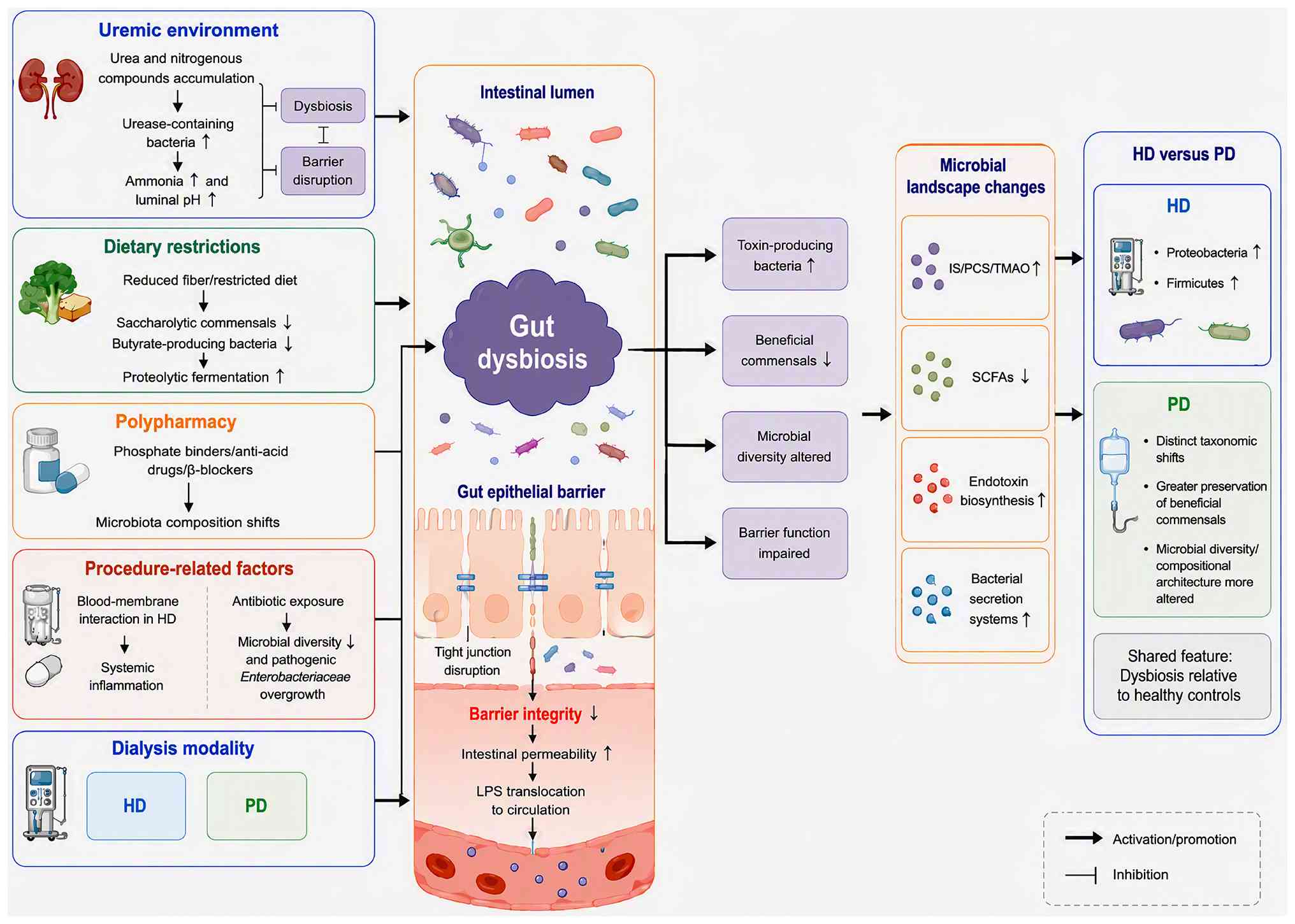
across studies. Fig. 1 provides an
overview of the key drivers and their differential impacts on gut
microbiota composition in HD vs. PD populations.
Uremic environment and intestinal
barrier disruption
The uremic milieu fundamentally alters host-microbe
interactions by promoting the overgrowth of toxin-producing
bacteria while suppressing commensal populations. Davenport
(24) demonstrated that more
frequent HD does not effectively clear protein-bound azotemic
solutes derived from gut microbial metabolism, indicating that
conventional dialysis inadequately addresses the circulating toxin
burden originating from the intestinal compartment. The
accumulation of urea and other nitrogenous compounds in the
intestinal lumen favors the expansion of urease-containing
bacteria, which convert urea into ammonia, raising luminal pH and
further disrupting microbial homeostasis. Shi et al
(15) identified notable
alterations in gut microbial pathways and virulence factors in
patients receiving HD, with enrichment of endotoxin biosynthesis
and bacterial secretion systems that may potentiate systemic
inflammation. The integrity of the intestinal epithelial barrier is
progressively compromised in the uremic state, allowing luminal
bacterial products such as LPS to translocate into the systemic
circulation (25). This
bidirectional relationship, whereby uremia drives dysbiosis and
dysbiosis exacerbates toxin accumulation, establishes a
self-perpetuating cycle that remains largely uninterrupted by
standard dialysis care.
HD vs. PD: Distinct microbial
signatures
Despite similar overall microbial diversity indices,
evidence indicates that dialysis modality exerts independent and
distinct effects on gut microbiota composition. In a comparative
study, Stadlbauer et al (26) performed structural and functional
analyses of gut microbiomes of patients receiving HD and those
receiving PD and identified that PD was associated with greater
alterations in both microbial diversity and compositional
architecture compared with HD. Conversely, Chen et al
(27) reported that patients
receiving HD exhibited enrichment of Proteobacteria and Firmicutes,
whereas patients receiving PD showed distinct taxonomic shifts that
associated with dialysis vintage. Teixeira et al (28) compared patients receiving PD with
their household contacts to control for environmental and dietary
confounders, suggesting that PD-specific factors, rather than
shared lifestyle elements, may contribute to unique microbial
signatures. More recently, Voroneanu et al (29) characterized distinct gut microbiome
signatures between HD and kidney transplant populations,
emphasizing that the dialysis environment creates microbial
alterations that are not simply reversed by transplantation. The
superior removal of middle molecules in PD compared with HD may
differentially impact circulating mediators that modulate gut
microbial ecology, although the precise mechanisms remain
incompletely elucidated (30).
Dietary restrictions, nutritional
status and PEW
Dietary management in patients receiving dialysis
involves stringent restrictions on phosphorus, potassium and fluid
intake, often coupled with reduced protein consumption, all of
which influence gut microbial substrate availability. Lin and Hung
(31) conducted a case-control
study examining the association between subjective global
assessment of nutritional status and gut microbiota in patients
receiving HD, demonstrating that malnourished individuals harbored
reduced abundances of beneficial butyrate-producing genera. PEW,
which affects a substantial proportion of the dialysis population,
has been mechanistically linked to gut microbial alterations. Hu
et al (32) identified a
notable association between intestinal flora disruption and PEW in
patients with ESKD, with depletion of short-chain fatty acid (SCFA)
producers representing a consistent finding. In a longitudinal
observational study, Bi et al (33) tracked gut microbiota changes in
patients receiving HD with PEW over time and reported that
microbial instability predicted worsening nutritional parameters,
suggesting that dysbiosis may both result from and contribute to
PEW pathogenesis. The relationship between nutritional status and
the microbiome is further complicated by reduced dietary fiber
intake, which reduces substrate availability for saccharolytic
commensals and shifts microbial metabolism toward proteolytic
fermentation pathways that generate nephrotoxic metabolites
(18).
Medications as unrecognized microbiome
modulators
The pharmacologic burden in patients receiving
dialysis is substantial, and accumulating evidence indicates that
commonly prescribed medications exert previously unrecognized
effects on gut microbial communities. Phosphate binders, used to
control hyperphosphatemia, have emerged as modulators. Wu et
al (34) compared gut
microbiome differences between ferric citrate and calcium carbonate
phosphate binders in patients with ESKD, demonstrating that
iron-based binders produced distinct compositional shifts,
including increased abundances of commensal Lactobacillaceae.
Abdelbary et al (35)
further characterized the impact of sucroferric oxyhydroxide on
both oral and intestinal microbiomes in patients receiving HD,
observing that binder-induced changes in gut microbiota associated
with reductions in serum phosphorus and inflammatory markers.
Conversely, the non-calcium-based binder sevelamer has been
associated with unfavorable effects on gut-derived uremic toxins.
Dai et al (36) reported
that sevelamer use in patients with ESKD was paradoxically
associated with higher levels of gut-derived uremic toxins, raising
the possibility of drug-microbiome interactions that warrant
further investigation. Beyond phosphate binders, anti-acid
medications and β-blockers also modulate gut microbial composition.
Lin et al (37)
demonstrated that anti-acid drug treatment induces notable changes
in the gut microbiome of patients receiving HD, while the same
group (38) identified distinct
microbial differences between patients receiving HD who were
treated with or without β-blockers, highlighting the influence of
polypharmacy.
Procedure-related factors:
Blood-membrane interactions and antibiotic exposures
The dialysis procedure itself introduces unique
stressors that may influence gut microbial ecology. In HD, contact
between blood and dialysis membranes triggers systemic inflammatory
responses that can indirectly affect the intestinal
microenvironment (39). He and Xie
(40) compared the effects of
different hemodialysis methods on gut microbiota in patients with
uremia, finding that high-flux and hemofiltration modalities
differentially modulated microbial composition compared with
conventional HD. In PD, the introduction of dialysate into the
peritoneal cavity alters intra-abdominal pressure and may influence
intestinal transit and permeability (30). Frequent antibiotic exposures (for
vascular access infections in HD or peritonitis in PD) represent a
major iatrogenic driver of dysbiosis. Zhou et al (41,42)
explored the association between intestinal flora and PD-related
peritonitis, identifying an association between prior antibiotic
courses and dysbiosis characterized by reduced microbial diversity
and overgrowth of pathogenic Enterobacteriaceae. More recently,
Zhang et al (43) performed
risk factor assessment and microbiome analysis in PD-related
peritonitis, reporting that cumulative antibiotic exposure was
independently associated with loss of commensal
Bifidobacterium species. The repeated disruption and
incomplete recovery of gut microbial communities following
antibiotic courses likely contribute to the progressive dysbiosis
observed with longer dialysis vintage (2). Collectively, these diverse drivers
operate in combination rather than isolation, necessitating
integrated approaches to characterize and ultimately modify the gut
microbiome in patients receiving dialysis.
Pathophysiological mechanisms linking gut
dysbiosis to systemic complications
Gut dysbiosis in patients receiving dialysis is
linked to systemic complications through interconnected mechanistic
pathways centered on microbial metabolite imbalance, intestinal
barrier disruption and host inflammatory signaling. The
overproduction of microbiota-derived uremic toxins including IS,
PCS and trimethylamine-N-oxide (TMAO) promotes vascular
inflammation and fibrosis, whereas depletion of protective SCFAs
compromises immune regulation. Concomitant gut barrier dysfunction
facilitates bacterial product translocation, activating NOD-like
receptor family pyrin domain containing 3 (NLRP3) inflammasome and
systemic inflammatory cascades that link dysbiosis to
cardiovascular disease, immune dysfunction and other systemic
complications. Fig. 2 summarizes
the central pathophysiological mechanisms connecting gut dysbiosis
to systemic complications in patients receiving dialysis.
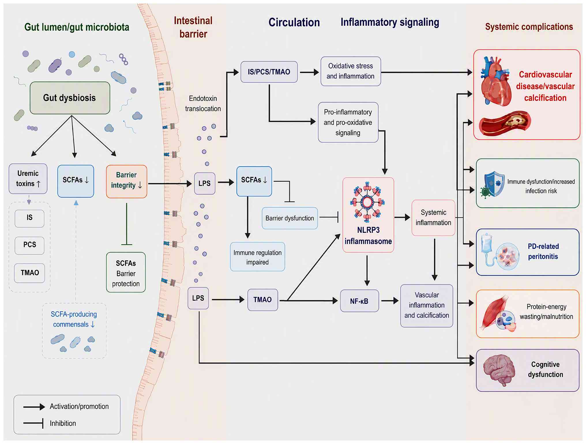 | Figure 2.Schematic illustrating
pathophysiological cascades linking gut dysbiosis to systemic
complications in patients receiving dialysis. Uremia induces gut
microbial shifts, such as the overproduction of protein-bound
uremic toxins (for example, IS, PCS and TMAO) and depletion of
protective SCFAs. Diminished SCFA-mediated mucosal protection and
uremic damage disrupts intestinal tight-junctions, increasing gut
permeability and enabling LPS translocation into circulation.
Circulating microbial metabolites and endotoxins trigger oxidative
stress and activate the NLRP3 inflammasome-NF-κB inflammatory axis.
Persistent systemic inflammation drives major dialysis-associated
adverse outcomes, including cardiovascular disease and vascular
calcification, immune dysfunction, PD-related peritonitis,
protein-energy wasting and cognitive dysfunction via the
kidney-gut-brain axis. Solid arrows indicate process
activation/promotion; bar-ended lines represent inhibition. IS,
indoxyl sulfate; PCS, p-cresyl sulfate; TMAO,
trimethylamine-N-oxide; SCFAs, short-chain fatty acids; LPS,
lipopolysaccharide; NLRP3, NOD-like receptor family pyrin domain
containing 3; PD, peritoneal dialysis. |
Microbiota-derived uremic toxins
Protein-bound uremic toxins originating from gut
microbial metabolism accumulate in patients receiving dialysis due
to inadequate clearance by conventional HD. Brito et al
(44) demonstrated that dietary
tryptophan intake associates with plasma IS levels in patients
receiving HD. Stockler-Pinto et al (45) further showed that IS and PCS
modulate NF-κB/Nrf2 expression, providing mechanistic evidence for
how gut-derived toxins propagate oxidative stress and inflammation.
The cardiovascular impact of TMAO was elucidated by Zhang et
al (46) demonstrated that
this metabolite promotes vascular calcification through NLRP3
inflammasome and NF-κB pathway activation. More recently, He et
al (20) reported that higher
serum TMAO levels are independently associated with increased
abdominal aortic calcification in patients receiving HD, extending
these findings to clinical outcomes. Bao et al (19) further confirmed a relationship
between gut microbiota composition and vascular calcification
severity, identifying specific bacterial taxa associated with
calcification risk.
Collectively, these studies suggest that gut-derived
uremic toxins may contribute to the accelerated cardiovascular
pathology observed in dialysis populations. However, it is
important to distinguish between experimental and clinical
evidence. The mechanistic pathways linking TMAO to vascular
calcification via NLRP3 inflammasome and NF-κB activation have been
validated in animal models and cell-based systems (46), whereas the association between gut
microbiota composition and calcification severity in humans remains
observational and subject to confounding by dietary intake and
medication use (19,20). The protein-bound toxins IS and PCS
have demonstrated pro-inflammatory and pro-oxidative effects in
vitro, but the extent to which these mechanisms operate in the
complex uremic milieu of patients receiving dialysis requires
further investigation through prospective intervention studies
(45,47).
SCFA depletion and barrier
integrity
The depletion of SCFA-producing commensals
represents a consistent finding in patients receiving dialysis with
consequences for intestinal barrier function and immune
homeostasis. Lin and Hung (31)
demonstrated that malnourished patients receiving HD exhibit
reduced abundances of butyrate-producing genera, including
Faecalibacterium, Roseburia and Coprococcus, linking
nutritional status to SCFA availability. Hu et al (48) employed high-throughput sequencing
to characterize intestinal flora changes in patients with ESKD,
identifying marked reductions in SCFA producers compared with
healthy controls. The functional implications of SCFA depletion
were examined by Ficek et al (49), who demonstrated associations among
plasma zonulin (a marker of intestinal permeability), bacterial LPS
and inflammatory markers in patients receiving HD, suggesting that
loss of SCFA-mediated barrier protection promotes endotoxin
translocation. Engen et al (50) similarly reported that gut-derived
inflammation associates with increased intestinal permeability in
neurodegenerative disease models, a mechanism that may also be
relevant to dialysis-related systemic inflammation. The study by de
Andrade et al (51) found
that dietary interventions in patients receiving PD modulated
gut-derived uremic toxins but did not fully restore SCFA profiles,
indicating persistent dysbiosis despite nutritional manipulation.
These findings suggest that SCFA depletion may be both a
consequence and a driver of gut barrier dysfunction.
Endotoxemia and systemic
inflammation
The translocation of bacterial products across a
compromised intestinal barrier generates persistent low-grade
endotoxemia that fuels systemic inflammation in patients receiving
dialysis. Ficek et al (49)
established associations between circulating LPS levels and
proinflammatory cytokines in patients receiving HD, indicating an
association between gut barrier dysfunction and systemic
inflammatory burden. Borges et al (52) demonstrated in a randomized trial
that probiotic supplementation modulates inflammatory markers in
patients with CKD, providing proof-of-concept that targeting gut
microbiota can attenuate systemic inflammation. Longitudinal
evidence from Lin et al (12) revealed that gut dysbiosis predicts
mortality in patients receiving HD, with inflammatory pathways
mediating this association. The mechanistic underpinnings were
further elucidated by Holle et al (53), demonstrated that in pediatric
patients with CKD gut dysbiosis and metabolite imbalance drive
inflammation through integrin-mediated signaling. More recently,
Mafra et al (54)
specifically examined the link between gut microbiota and NLRP3
inflammasome activation in patients receiving HD, identifying that
microbial alterations associate with increased caspase-1 activity
and IL-1β production. Samaan et al (55) reported that Ramadan fasting
modulates inflammatory markers and gut bacterial endotoxins in
patients receiving HD, suggesting that dietary patterns influence
endotoxin burden. Collectively, these studies establish endotoxemia
as a mechanistic bridge between gut dysbiosis and the systemic
inflammatory state observed in dialysis populations.
NLRP3 inflammasome as an integrative
hub
Emerging evidence positions the NLRP3 inflammasome
as a central molecular integrator linking gut microbial alterations
to sterile inflammation in patients receiving dialysis. Zhang et
al (46) first demonstrated
that TMAO promotes vascular calcification through NLRP3 activation,
establishing a direct pathway from gut microbial metabolite to
inflammatory cascade. The relevance to dialysis populations was
confirmed by Mafra et al (54), who showed that NLRP3 inflammasome
components are upregulated in patients receiving HD with pronounced
gut dysbiosis. Guo et al (56) demonstrated that sodium butyrate
exerts neuroprotective effects in Parkinson's disease models by
restoring gut microbiota and inhibiting Toll-like receptor 4
signaling, suggesting that SCFAs may counterbalance inflammasome
activation. Qu et al (57)
further established that SCFAs modulate α-synuclein aggregation and
neuroinflammation via the G protein-coupled receptor 43
(GPR43)-NLRP3 signaling pathway, wherein SCFA binding to GPR43
inhibits NLRP3 inflammasome activation and downstream inflammatory
cascades; this mechanism is potentially relevant to the
kidney-gut-brain axis in patients undergoing dialysis. Liu et
al (58) recently reported
that Bacteroides coprocola protects dopaminergic neurons
through inhibition of the NLRP3 signaling pathway, highlighting
that specific commensals may modulate inflammasome activity.
Together, these findings suggest that NLRP3 may represent a
potential molecular target for interventions aimed at breaking the
dysbiosis-inflammation cycle in patients receiving dialysis.
Interplay with host signaling
systems
Gut dysbiosis interacts with host signaling systems
beyond canonical inflammatory pathways, including the
renin-angiotensin system (RAS) and immune checkpoint regulators. Wu
et al (34) demonstrated
that phosphate binders differentially modulate gut microbiota
composition, suggesting that medications targeting mineral
metabolism indirectly influence host-microbe signaling. The RAS
connection was explored by Li et al (59), who showed that uremic clearance
granules affect p38 mitogen-activated protein kinase/NF-κB
signaling pathways in rats receiving PD, with concurrent
alterations in microbial and metabolic profiles. Zhang et al
(60) demonstrated that TMAO
increases peritoneal inflammation and peritonitis risk in patients
receiving PD, establishing a link between gut microbial metabolism
and local host responses in the peritoneal cavity. The programmed
cell death protein 1/programmed death-ligand 1 (PD-1/PD-L1) immune
checkpoint axis has also been implicated, with Chen et al
(61) showing that Prevotellaceae
produces butyrate to alleviate PD-1/PD-L1 inhibitor-related
toxicity via the peroxisome proliferator-activated receptor
α-cytochrome P450 family 4 subfamily X member 1 axis in colonic
macrophages. Wang et al (62) further demonstrated that Fushen
Granule modulates gut microbiota to prevent chronic renal failure,
with effects partly mediated through host metabolic pathways. These
observations indicate that gut dysbiosis exerts pleiotropic effects
on host signaling networks, with therapeutic implications for
personalized microbiome-based care.
Collectively, the pathophysiological evidence
suggests that gut dysbiosis may contribute to systemic
complications through a convergent triad of mechanisms: Microbial
metabolite imbalance (excess uremic toxins, deficient SCFAs),
barrier disruption with endotoxin translocation and NLRP3
inflammasome-mediated sterile inflammation. These pathways are not
mutually exclusive but operate synergistically, with microbial
metabolites directly activating inflammatory cascades while
simultaneously compromising barrier integrity, creating a
self-reinforcing cycle of dysbiosis, inflammation and tissue
injury.
Major complications associated with gut
dysbiosis in dialysis
Gut dysbiosis in patients receiving dialysis is
associated with systemic complications through interconnected
pathways involving microbial metabolite imbalance, intestinal
barrier disruption and sustained inflammation. The clinical impact
extends beyond traditional cardiovascular disease to encompass
immune dysfunction, PD-related peritonitis, protein-energy wasting
and cognitive impairment. These complications collectively
contribute to poor clinical outcomes and increased mortality,
supporting a potential role for the gut microbiome in the prognosis
of patients receiving dialysis. Table
I illustrates the major complications linked to gut dysbiosis
and their underlying mechanistic connections.
 | Table I.Major complications associated with
gut microbiota dysbiosis in patients receiving dialysis: Clinical
manifestations and mechanistic pathways. |
Table I.
Major complications associated with
gut microbiota dysbiosis in patients receiving dialysis: Clinical
manifestations and mechanistic pathways.
| Complication | Key clinical
manifestations | Gut
microbiota-related mechanisms | Primary microbial
metabolites/pathways involved | (Refs.) |
|---|
| Cardiovascular
disease and vascular calcification | Accelerated
atherosclerosis, arterial stiffness, abdominal aortic calcification
and increased cardiovascular mortality | Endotoxemia, NLRP3
inflammasome activation, NF-κB signaling and oxidative stress | TMAO, IS, PCS and
LPS | (46,20,19,63) |
| Immune dysfunction
and infection risk | Susceptibility to
bacterial infections, reduced vaccine response, T cell
dysregulation, high sepsis-related mortality | Increased
intestinal permeability, bacterial translocation, chronic low-grade
inflammation, altered Treg/Th17 balance | LPS, zonulin, IS,
PCS | (53,65,49,66) |
| Peritoneal
dialysis-related peritonitis | Recurrent or
refractory peritonitis, increased catheter removal,
technique\failure, hospitalization | Gut-peritoneal
translocation of pathogenic bacteria, antibiotic-induced dysbiosis,
loss of commensal Bifidobacterium | TMAO,
Escherichia coli enrichment, reduced SCFAs | (41–43,60,68) |
| Protein-energy
wasting and malnutrition | Loss of muscle
mass, low serum albumin, poor nutritional status, reduced quality
of life | Reduced SCFA
production, increased proteolytic fermentation, chronic
inflammation, altered appetite regulation | Depletion of
butyrate-producing genera (such as Faecalibacterium and
Roseburia) and increased uremic toxins | (72) |
| Cognitive
dysfunction and kidney-gut-brain axis | Mild cognitive
impairment, executive dysfunction, memory decline, potential
progression to dementia | Neuroinflammation,
blood-brain barrier disruption, microglial activation, altered
neurotransmitter metabolism | IS, PCS, TMAO,
reduced SCFAs, tryptophan/tyrosine pathway metabolites | (23,74–77) |
Cardiovascular disease and vascular
calcification
Cardiovascular disease remains the leading cause of
mortality in patients receiving dialysis, and accumulating evidence
implicates gut-derived uremic toxins in vascular pathology. Zhang
et al (46) demonstrated
that TMAO promotes vascular calcification through activation of
NLRP3 inflammasome and NF-κB signaling pathways, providing
mechanistic evidence linking gut microbial metabolism to arterial
pathology. Extending these findings to clinical outcomes, He et
al (20) reported that higher
serum TMAO levels are independently associated with increased
abdominal aortic calcification in patients receiving HD. Bao et
al (19) further reported a
notable relationship between gut microbiota composition and
vascular calcification severity, identifying specific bacterial
taxa associated with calcification risk. The protein-bound toxins
IS and PCS also contribute to cardiovascular injury. In the
aforementioned study, higher abundances of bacteria in the
Escherichia-Shigella and Ruminococcus groups
were positively associated with vascular calcification, whereas
lower abundances of Ruminococcus, Adlercreutzia, Alistipes
and Ruminococcaceae were negatively associated with calcification
severity. In terms of relative contribution to calcification risk
as determined by importance ranking analysis,
Escherichia-Shigella exerted the greatest influence,
followed by Ruminococcus and Butyricimonas. Velasquez
et al (63) reviewed the
role of gut microbiota in cardiovascular uremic toxicities,
emphasizing that these metabolites promote endothelial dysfunction
and oxidative stress. Kanitsoraphan et al (64) conducted a systematic evaluation
demonstrating that elevated TMAO levels are associated with
increased cardiovascular risk and mortality across populations with
CKD. Mechanistically, the persistence of these toxins despite
conventional dialysis reflects inadequate clearance of
protein-bound solutes, as highlighted by Gryp et al
(47), who characterized PCS as a
prototypical gut-derived cardiovascular toxin. Collectively, these
findings support a potential role for gut dysbiosis in the
accelerated cardiovascular pathology observed in patients receiving
dialysis, although prospective interventional studies targeting the
microbiome to reduce cardiovascular events remain limited.
However, despite these mechanistic and observational
data, establishing causality in humans remains challenging. Most
evidence for direct vascular injury derives from in vitro
and animal models, whereas human studies are predominantly
cross-sectional or prospective observational, which are susceptible
to residual confounding by dietary intake and comorbid conditions
(19,20). Furthermore, whether reducing these
metabolites through gut modulation translates into decreased
cardiovascular events has not been established in adequately
powered randomized trials. In the evidence hierarchy, the current
data support strong associations and plausible mechanistic
pathways, but definitive causal proof would require demonstration
that microbiome-targeted interventions specifically reduce
cardiovascular events through toxin-dependent mechanisms (14). This distinction is not merely
semantic; it has direct implications for clinical trial design and
the interpretation of intervention studies that show metabolite
reduction without corresponding improvements in hard clinical
outcomes.
Immune dysfunction and increased
infection risk
Patients receiving HD exhibit heightened
susceptibility to infections and experience disproportionately high
mortality following septic events, with gut microbiota
dysregulation increasingly recognized as a contributing mechanism.
Holle et al (53)
demonstrated in pediatric patients with CKD that gut dysbiosis and
metabolite imbalance drive inflammation through integrin-mediated
signaling, establishing a mechanistic link between microbial
alterations and immune dysfunction. Lv et al (65) recently performed 16S ribosomal RNA
(16S rRNA) sequencing-based analysis of gut flora and T cell
profiles in patients receiving maintenance HD, revealing that
immune-microbiota dysregulation characterized by reduced regulatory
T cell populations associates with specific taxonomic shifts. The
translocation of bacterial products across a compromised intestinal
barrier perpetuates systemic inflammation that impairs host
defense. Ficek et al (49)
established associations among plasma zonulin (a marker of
intestinal permeability), bacterial LPS and inflammatory markers in
patients receiving HD, indicating an association between gut
barrier dysfunction and immune activation. Glorieux et al
(66) reviewed the role of
gut-derived metabolites in immune dysfunction in CKD, emphasizing
that protein-bound uremic toxins suppress phagocytic function and
alter lymphocyte responses. Furthermore, Al Khodor and Shatat
(67) described the bidirectional
relationship between gut microbiome and kidney disease, noting that
dysbiosis-induced inflammation creates a vicious cycle that further
compromises immune competence. While observational associations
support a relationship, causality remains to be definitively
established, and whether microbiome-targeted interventions can
reduce infection risk in patients receiving dialysis requires
prospective validation.
PD-related peritonitis
Gut microbiome dysbiosis has been associated with
peritonitis in patients receiving PD, creating a clinically
actionable connection between intestinal microbial health and
infectious outcomes of the peritoneal cavity. Zhang et al
(43) performed risk factor
assessment and microbiome analysis in PD-related peritonitis,
demonstrating that cumulative antibiotic exposure was independently
associated with loss of commensal Bifidobacterium species
and that gut microbiota composition predicted peritonitis risk.
Zhou et al (41,42) explored the association between
intestinal flora and PD-related peritonitis in two complementary
studies, identifying an association between prior antibiotic
courses and dysbiosis characterized by reduced microbial diversity
and overgrowth of pathogenic Enterobacteriaceae, with specific
enrichment of Escherichia coli in culture-positive cases.
The gut microbial metabolite TMAO also contributes to peritoneal
inflammation. Zhang et al (60) demonstrated that TMAO increases
peritoneal inflammation and peritonitis risk in patients receiving
PD, establishing a link between gut microbial metabolism and local
host responses in the peritoneal cavity. Xu et al (68) recently provided a new perspective
on gut microbiome and peritoneal inflammation, proposing that
microbial metabolite profiling could identify patients at elevated
peritonitis risk before clinical infection manifests. Conversely,
traditional risk factors such as hydration status also modulate
peritonitis susceptibility, as Santhakumaran et al (69) demonstrated that fluid overload
measured by bioimpedance spectroscopy represents a modifiable risk
factor for peritonitis. These observations suggest that integrated
approaches combining microbiome assessment with conventional risk
stratification may optimize peritonitis prevention strategies.
Malnutrition and PEW
PEW affects a substantial proportion of patients
receiving dialysis and has been associated with gut microbial
alterations through bidirectional interactions. Lin and Hung
(31) demonstrated that
malnourished patients receiving HD harbored reduced abundances of
beneficial butyrate-producing genera, establishing an association
between nutritional status and gut microbiota composition.
Bakinowska et al (70)
comprehensively reviewed the pathogenesis of sarcopenia in CKD,
identifying gut dysbiosis as a contributing factor through
inflammation-mediated muscle catabolism and metabolic
dysregulation. The relationship between nutritional status and the
microbiome is further complicated by reduced dietary fiber intake,
which reduces substrate availability for saccharolytic commensals
and shifts microbial metabolism toward proteolytic fermentation
pathways that generate nephrotoxic metabolites. March et al
(71) conducted a systematic
review and meta-analysis of randomized controlled trials (RCTs)
examining prebiotic, probiotic and synbiotic supplementation in
patients receiving dialysis, finding that these interventions
significantly reduced circulating levels of gut-derived
metabolites, including endotoxin [standardized mean difference
(SMD), −0.61; 95% confidence interval (CI), −1.03 to −0.20],
indoxyl sulfate (SMD, −0.34; 95% CI, −0.64 to −0.04) and p-cresyl
sulfate (SMD, −0.34; 95% CI, −0.61 to −0.07), but noting
inconsistent effects on nutritional parameters. Pan et al
(72) performed a RCT
demonstrating that probiotic supplementation improved malnutrition
and health-related quality of life in patients receiving PD,
suggesting that targeting gut microbiota may improve nutritional
outcomes. Nevertheless, Azamian et al (73) reported that synbiotic
supplementation reduced advanced glycation end products and
cardiovascular risk factors in patients receiving HD without
notable improvements in nutritional indices, suggesting that the
effects on PEW may be indirect and require longer intervention
periods. The heterogeneity of outcomes across studies highlights
the need for larger, adequately powered trials with standardized
nutritional endpoints.
Nevertheless, it remains unresolved whether gut
dysbiosis is a primary driver of PEW or a consequence of the
malnourished state itself. A short-term RCT showed improvement in
nutritional parameters (72),
whereas another reported no significant improvement in nutritional
indices (73), and the
heterogeneity in definitions of PEW and intervention protocols
prevents definitive conclusions regarding a causal
relationship.
Cognitive dysfunction and the
kidney-gut-brain axis
Brain dysfunction in patients receiving maintenance
HD has been increasingly linked to the kidney-gut-brain axis, with
gut microbiota disorders and intestinal mucosal damage constituting
key components of a systemic communication network extending beyond
the traditional gut-kidney framework. Zhu et al (74) performed a single-center
observational study identifying abnormalities in gut microbiota and
serum metabolites in patients receiving HD with mild cognitive
decline, demonstrating that reduced abundances of SCFA-producing
genera associated with poorer cognitive performance. Wang et
al (75) extended these
findings to patients receiving PD, showing that gut microbiome
composition associates with cognitive impairment and that specific
taxa including Lactobacillus and Bifidobacterium were
depleted in patients with worse cognitive scores. Yu et al
(23) comprehensively reviewed
research progress on the kidney-gut-brain axis in brain dysfunction
in patients receiving maintenance HD, proposing that microbial
metabolites including IS and PCS cross the blood-brain barrier and
promote neuroinflammation. The mechanistic underpinnings were
elucidated by Shah et al (76), who demonstrated that PCS causes
cerebrovascular damage through EGF receptor activation, providing
direct experimental evidence linking gut-derived metabolites to
neurological injury. Martín-del-Campo et al (77) recently reported that gut microbiome
composition is related to cognitive impairment in patients
receiving PD, identifying that reduced microbial diversity and
depletion of Faecalibacterium prausnitzii were independently
associated with lower Mini-Mental State Examination scores.
Furthermore, Gao et al (78) employed combined 16S ribosome DNA
and shotgun metagenome sequencing to analyze intestinal flora and
cognitive function in patients receiving maintenance HD, revealing
that specific metabolic pathways including tryptophan and tyrosine
metabolism were enriched in patients with cognitive decline.
Collectively, these findings position the gut microbiome as a
potential therapeutic target for preserving cognitive function in
patients receiving dialysis, although longitudinal studies tracking
microbiota changes before and after the onset of cognitive decline
are needed to establish temporality and causality.
Across these five complication domains, common
mechanistic threads emerge, including microbial metabolite excess
(IS, PCS and TMAO), SCFA depletion or endotoxin-mediated
inflammation. The strength of causal evidence varies considerably,
with cardiovascular disease supported by extensive mechanistic
studies, whereas cognitive impairment and PEW remain predominantly
associative. This pattern highlights the need for
mechanism-targeted interventions and suggests that microbiome
modulation may offer a unified strategy for mitigating multiple
complications simultaneously.
Therapeutic modulation of the gut microbiome
in dialysis: Current evidence
A growing armamentarium of gut-targeted
interventions offers potential avenues for mitigating dysbiosis in
patients receiving dialysis, although the evidence base remains at
an early stage. These strategies include probiotics, prebiotics,
synbiotics and dietary modifications, phosphate binders and fecal
microbiota transplantation. Table
II provides a comparative overview of these therapeutic
approaches and key clinical findings in dialysis populations.
| Table II.Summary of microbiome-targeted
therapeutic strategies in patients receiving dialysis. |
Table II.
Summary of microbiome-targeted
therapeutic strategies in patients receiving dialysis.
| First author,
year | Intervention | Specific
strategy | Dialysis
modality | Key findings in
dialysis populations | Key
limitation/note | (Refs.) |
|---|
| Dai et al,
2020 | Phosphate
binders | Sevelamer | Mixed HD/PD | Associated with
higher levels of gut-derived uremic toxins | Potential
drug-microbiome interaction; observational | (36) |
| Borges et
al, 2018 | Probiotics | Probiotic
supplementation | HD | Modulated
inflammatory markers; reduced PCS | TMAO unchanged;
small sample size | (52) |
| Pan et al,
2021 | Probiotics | Probiotic
supplementation | PD | Improved
malnutrition and health-related quality of life | Long-term clinical
outcomes not assessed | (72) |
| Soleimani et
al, 2017 | Probiotics | Multi-strain
probiotics | HD | Improved metabolic
profiles (fasting glucose, lipids) in patients with diabetes
receiving HD | Uremic toxin levels
not reported | (79) |
| Borges et
al, 2019 | Probiotics | Probiotic
supplementation | HD | No significant
reduction in TMAO levels | Pilot study | (80) |
| Lopes et al,
2019 | Synbiotics | Synbiotic meal | HD | Decreased uremic
toxins | Single-meal design;
not long-term | (81) |
| Mirzaeian et
al, 2020 | Synbiotics | Synbiotic
supplementation | HD | Reduced serum IS,
PCS and systemic inflammation | Double-blind but
moderate sample size | (82) |
| Zhou et al,
2024 | Probiotics | Probiotic
treatment | PD | Induced intestinal
microbiota changes | No effect on SCFA
levels despite compositional shifts | (83) |
| Wang et al,
2025 | Probiotics | Probiotic
supplementation | HD | Enhanced sleep
quality and reduced indoxyl sulfate linked to gut microbiota
modulation | Observational
component; causality not proven | (84) |
| Abdelnasier et
al, 2026 | Probiotics | Probiotic use | HD | Reduced gut leakage
(zonulin), PCS and inflammation | Prospective
controlled but not randomized | (85) |
| Stanford et
al, 2021 | Dietary
intervention | Plant-based diet
quality | HD | Higher adherence
associated with lower gut-derived uremic toxins | Cross-sectional;
causality unclear | (86) |
| Li et al,
2020 | Prebiotics | Inulin-type
fructans | PD | Restricted increase
in gut-generated indole | Crossover design;
short intervention | (87) |
| He et al,
2022 | Prebiotics | Inulin-type
prebiotics | PD | Reduced serum uric
acid levels via gut microbiota modulation | Effects on
protein-bound toxins not evaluated | (88) |
| Li et al,
2024 | Prebiotics | Inulin-type
prebiotics | PD | Exerted
arsenic-lowering effect | Specific to
arsenic; generalizability limited | (89) |
| Esgalhado et
al, 2018 | Prebiotics | Resistant
starch | HD | Reduced IS and PCS
levels; attenuated inflammation | Small pilot; needs
larger confirmation | (90) |
| Azevedo et
al, 2020 | Prebiotics | Resistant
starch | HD | Reduced plasma
indole-3-acetic acid and AhR mRNA expression | Secondary analysis
of pilot trial | (91) |
| de Paiva et
al, 2020 | Prebiotics | Resistant
starch | HD | Attenuated
inflammation | Pilot study | (92) |
| Rocchetti et
al, 2020 | Emerging
approach | Divinylbenzenic
resin | HD | Removed IS and PCS
in vitro and in a pilot in vivo study | Pilot in
vivo; limited number of patients | (101) |
Probiotics, prebiotics and
synbiotics
Probiotic, prebiotic and synbiotic supplementation
represents the most extensively investigated class of
microbiome-targeted interventions in patients receiving dialysis,
although trial results have been heterogeneous. Soleimani et
al (79) conducted a
randomized trial in patients with diabetes receiving HD,
demonstrating that probiotic supplementation produced beneficial
metabolic effects including reduced fasting blood glucose and
improved lipid profiles. Borges et al (52) further showed that probiotic
supplementation in patients with CKD modulated inflammatory
markers, providing early proof-of-concept for anti-inflammatory
effects. However, subsequent studies have yielded inconsistent
findings regarding uremic toxin reduction. In a pilot study, Borges
et al (80) reported that
probiotic supplementation did not significantly lower TMAO plasma
levels in patients receiving HD, suggesting that single-strain or
multi-strain formulations may have limited impact on this specific
metabolite.
The combination of probiotics with prebiotics as
synbiotics has shown more promising results in certain contexts.
Lopes et al (81)
demonstrated that a synbiotic meal decreased uremic toxin levels in
individuals receiving HD compared with placebo. Similarly,
Mirzaeian et al (82)
conducted a double-blind, randomized clinical trial showing that
synbiotic supplementation reduced protein-bound uremic toxins and
systemic inflammation in patients receiving HD. Nevertheless, Zhou
et al (83) recently
reported that probiotic treatment induced changes in intestinal
microbiota but did not alter SCFA levels in patients receiving PD,
highlighting that microbial composition shifts do not necessarily
translate into functional metabolic changes. Pan et al
(72), who demonstrated in a RCT
that probiotic supplementation improved malnutrition and
health-related quality of life in patients receiving PD.
Additionally, Wang et al (84) recently showed that enhanced sleep
quality and reduced IS levels following probiotic supplementation
were linked to gut microbiota modulation in patients receiving HD.
Abdelnasier et al (85)
prospectively demonstrated that probiotic use reduced gut leakage
measured by zonulin and decreased PCS levels and inflammation in
patients receiving HD, providing mechanistic evidence for
barrier-stabilizing effects. Collectively, these studies indicate
that while probiotics and synbiotics hold promise, response
heterogeneity across trials likely reflects differences in strains,
doses, dialysis modalities and patient populations, necessitating
larger, adequately powered trials with standardized endpoints.
The discrepancy in outcomes across these RCTs
highlights issues, including variations in bacterial strains,
treatment duration and the lack of baseline microbiome
stratification (52,79,80,83).
Furthermore, the consistent observation that changes in microbial
composition do not necessarily translate into altered functional
metabolites underscores a gap in understanding the
taxonomy-function relationship, indicating that current evidence is
insufficient to support broad clinical recommendations (83).
Dietary interventions
Dietary modulation, particularly through
plant-based diets and resistant starch supplementation, has emerged
as a physiologically rational approach to reshape gut microbial
metabolism in patients receiving dialysis. Stanford et al
(86) examined associations among
plant-based diet quality, uremic toxins and gut microbiota profile
in adults undergoing HD, demonstrating that higher adherence to
plant-based diets was associated with lower circulating levels of
gut-derived toxins. The prebiotic effects of inulin-type fructans
have been rigorously evaluated in PD populations. Li et al
(87) conducted a randomized
crossover study demonstrating that inulin-type fructan intervention
restricted the increase in gut microbiome-generated indole in
patients receiving PD. Extending these findings, He et al
(88) showed that inulin-type
prebiotics reduced serum uric acid levels via gut microbiota
modulation in a randomized controlled crossover trial in patients
receiving PD. Furthermore, Li et al (89) recently reported that inulin-type
prebiotics exerted an arsenic-lowering effect in patients with
end-stage renal disease (ESRD), suggesting additional
detoxification benefits.
Resistant starch represents another promising
prebiotic approach. Esgalhado et al (90) conducted a pilot RCT showing that
resistant starch supplementation improved inflammatory and
oxidative stress biomarkers in patients receiving HD, although
uremic toxin levels were not notably altered. Azevedo et al
(91) further demonstrated that
resistant starch supplementation reduced plasma indole-3-acetic
acid and aryl hydrocarbon receptor mRNA expression in patients
receiving HD. De Paiva et al (92) reported that resistant starch
supplementation attenuated inflammation in patients receiving HD,
corroborating the anti-inflammatory effects observed in earlier
studies. However, the optimal formulation and duration of dietary
interventions remain unclear, and head-to-head comparisons between
different prebiotic agents are lacking. Nevertheless, these dietary
approaches offer the advantage of targeting fundamental substrate
availability rather than merely modifying microbial composition,
positioning them as potential components of integrated
microbiome-directed care in patients receiving dialysis.
Phosphate binders as microbiome
modulators
Phosphate binders, prescribed to control
hyperphosphatemia in patients receiving dialysis, have been
increasingly recognized for their unintended but potentially
beneficial effects on gut microbial ecology; however, not all
binders exert equivalent effects. Dai et al (36) reported that sevelamer use in
patients with ESKD was associated with higher levels of gut-derived
uremic toxins, raising the possibility of detrimental
drug-microbiome interactions. This finding contrasts with the
expected benefit of phosphate control and suggests that sevelamer
may alter gut microbial composition or function in ways that
promote toxin generation. Conversely, iron-based binders have shown
more favorable microbiome profiles (34), although comparative trials directly
evaluating binder effects on clinical outcomes remain limited.
These observations highlight that medication selection in patients
receiving dialysis may have previously unrecognized consequences
for gut microbial homeostasis, warranting prospective comparative
effectiveness studies.
Fecal microbiota transplantation
(FMT)
FMT is used in the treatment of recurrent
Clostridioides difficile infection and is being explored
across various disease states, although its application in dialysis
populations remains limited. Studies in other fields have
established the feasibility and safety of FMT. Baruch et al
(93) demonstrated that FMT
promoted response in immunotherapy-refractory patients with
melanoma, while Davar et al (94) showed that FMT overcame resistance
to anti-PD-1 therapy in patients with melanoma. In Parkinson's
disease, several randomized trials have evaluated FMT with mixed
results. Scheperjans et al (95) conducted a RCT of FMT for
Parkinson's disease, and Zhang et al (96) recently reported that gut microbiota
modulation via repeated donor FMT improved motor and
gastrointestinal symptoms in drug-naïve patients with Parkinson's
disease. However, no published RCT has specifically evaluated FMT
in patients receiving dialysis. Challenges include safety concerns
related to immune dysfunction, infection risk from donor material
and the absence of standardized protocols for this vulnerable
population. While FMT holds theoretical promise for restoring a
healthy gut ecosystem in patients receiving dialysis, rigorous
phase I and II trials are essential before clinical adoption can be
considered. For phase I trials in patients receiving dialysis,
proposed safety endpoints should include monitoring for bacteremia,
peritonitis in patients receiving PD and febrile reactions within
72 h post-FMT, alongside standard adverse event reporting. Donor
stool should be rigorously screened for multidrug-resistant
organisms and potential pathogens, with particular attention to
cytomegalovirus and Epstein-Barr virus status given the immune
dysfunction in patients receiving dialysis (97,98).
Patient selection for initial trials should prioritize clinically
stable individuals without active infections or recent antibiotic
exposure, with gradual dose escalation to assess tolerability
(99).
Emerging approaches
Novel strategies beyond traditional probiotics and
prebiotics are being investigated. Colon-targeted adsorbents
represent an approach that removes gut-derived toxins without
altering living microbial communities. Messaoudene et al
(100) demonstrated in a phase I
trial in healthy volunteers that the DAV132 colon-targeted
adsorbent did not interfere with plasma concentrations of
antibiotics but prevented antibiotic-related dysbiosis, offering a
paradigm for toxin sequestration rather than microbial
modification. Rocchetti et al (101) evaluated the efficacy of
divinylbenzenic resin in removing IS and PCS in patients receiving
HD through in vitro and pilot in vivo studies,
showing proof-of-concept for adsorption-based strategies.
Additionally, engineered microbiota approaches and postbiotic
formulations targeting specific metabolite pathways are emerging
from preclinical models. Collectively, these approaches may enable
more precise, mechanism-based interventions compared with
traditional probiotic supplementation, although clinical
translation in dialysis populations remains distant.
In summary, the evidence for microbiome-targeted
therapies in dialysis remains preliminary, with probiotics and
synbiotics showing the most promise but also the greatest
heterogeneity. Several consistent themes emerge: Responses vary
substantially across individuals, microbial composition changes do
not reliably predict functional metabolic improvements and no
single intervention has demonstrated consistent efficacy across
studies. These observations collectively indicate that successful
therapeutic modulation may require personalized approaches guided
by baseline microbial profiling and metabolomic phenotyping rather
than uniform supplementation protocols.
Multi-omics approaches to characterizing
dysbiosis in dialysis
Multi-omics technologies have transformed the
investigation of gut microbiota alterations in patients receiving
dialysis, enabling taxonomic, functional and metabolic
characterization of dysbiosis beyond traditional culture-based
methods. However, the heterogeneity of sequencing platforms,
analytical pipelines and population characteristics has generated
divergent findings that complicate cross-study comparisons.
Table III provides an integrated
overview of key multi-omics discoveries in dialysis-associated
dysbiosis, highlighting methodological considerations and major
biological insights.
| Table III.Multi-omics characterizations of gut
microbiota dysbiosis in patients receiving dialysis: Key findings
from 16S rRNA sequencing, metagenomics, metabolomics and
integrative analyses. |
Table III.
Multi-omics characterizations of gut
microbiota dysbiosis in patients receiving dialysis: Key findings
from 16S rRNA sequencing, metagenomics, metabolomics and
integrative analyses.
| First author,
year | Population | Dialysis
modality | Omics
technology/platform | Key microbial and
metabolite findings | (Refs.) |
|---|
| Shi et al,
2022 | 24 patients
receiving HD, 16 controls | HD | Shotgun
metagenomics | Enrichment of LPS
biosynthesis pathways and bacterial secretion systems | (15) |
| Luo et al,
2021 | 25 patients
receiving HD, 25 patients receiving PD, 25 controls | HD vs. PD | 16S rRNA sequencing
(V3-V4) | PD associated with
greater preservation of beneficial commensals; both modalities
showed marked dysbiosis relative to controls | (17) |
| Hu et al,
2020 | 16 patients with
ESRD, 16 patients with CKD, 20 controls | Not specified | 16S rRNA
high-throughput sequencing (V3-V4) | Reduced abundance
of SCFA producers (such as Lachnospiraceae and Ruminococcaceae) in
patients with ESRD compared with patients with CKD and
controls | (48) |
| Lv et al,
2025 | 45 patients
receiving MHD, 30 controls (HD-LI vs. HD-NLI) | MHD | 16S rRNA sequencing
(V3-V4) + T cell profiling | Reduced
SCFA-producing bacteria in immunodeficient state; Prevotella
copri, Bacteroides vulgatus and Agathobacter depleted;
Escherichia-Shigella, Blautia and Citrobacter
enriched | (65) |
| Zhu et al,
2020 | 77 patients
receiving MHD (classified as NCF or MCD) and 30 healthy
controls | MHD | 16S rRNA sequencing
+ serum metabolomics | Reduced
SCFA-producing genera (Lachnospira, Roseburia,
Faecalibacterium) in cognitively impaired patients; altered
tryptophan and tyrosine metabolites associated with cognitive
decline | (74) |
| Gao et al,
2024 | 36 patients
receiving MHD, 18 controls | MHD | 16S rDNA + shotgun
metagenome sequencing | Roseburia
reduced by both methods; Faecalibacterium, Megamonas and
Bifidobacterium positively associated with cognitive
function; enriched KEGG pathways (oxidative phosphorylation,
photosynthesis and riboflavin metabolism) in cognitively impaired
patients | (78) |
| Li et al,
2019 | 26 patients with
CKD stage 5, 20 controls | Maintenance HD | 16S rRNA sequencing
(V3-V4) | Enrichment of
pathogenic Enterobacteriaceae, depletion of butyrate-producing
Roseburia and Faecalibacterium; positive association
between toxin-producing taxa and serum indoxyl sulfate levels | (103) |
| Zhang et al,
2023 | 378 patients with
ESRD receiving HD, 290 controls | HD | Deep metagenomic
sequencing + MAG reconstruction | 348 differentially
abundant species, including ESRD-elevated Blautia, Dorea,
Eggerthellaceae, and depleted Prevotella, Roseburia; gut
microbial composition predicted serum uremic toxin (IS, PCS, PAG,
TMAO) concentrations | (104) |
| Qin et al,
2025 | 20 patients
receiving MHD, 20 controls | MHD | 16S rRNA sequencing
(ASV-based) | Decrease in
Escherichia-Shigella abundance; 19 differentially abundant
genera identified in patients receiving MHD compared with
controls | (105) |
| Bao et al,
2022 | Patients receiving
PD (number not specified in the original study) | PD | 16S rRNA sequencing
(V3-V4) + serum metabolomics | Reduced
Lactobacillus and Bifidobacterium abundances
associated with higher serum PCS | (108) |
| James et al,
2025 | Adults initiating
PD | PD | Metagenomic
sequencing + serum LC-MS/MS | Baseline microbial
composition predicted subsequent uremic toxin elevations; serum IS,
PCS, TMAO, PAG levels | (109) |
| Manokasemsan et
al, 2025 | Patients receiving
PD | PD | LC-MS/MS
metabolomics + 16S rRNA sequencing | Microbial
compositional shifts associated with histidine metabolism; elevated
imidazole propionate and other gut-derived metabolite alterations;
histidine metabolism identified as previously underappreciated
pathway | (110) |
| Kuo et al,
2025 | 85 patients
receiving HD | HD | 16S rRNA sequencing
+ targeted serum metabolomics | Distinct gut
microbiota signatures associated with serotonin pathway
metabolites; serum serotonin pathway metabolites (tryptophan,
5-HIAA, 5-HTP) associated with specific taxa (Eubacterium
eligens, Roseburia hominis) | (111) |
| Li et al,
2024 | 73 patients
receiving CAPD (different dialysis vintages) | CAPD | 16S rRNA sequencing
+ untargeted metabolomics | Fungal signatures
discriminated patients with different dialysis vintages;
microbiome-derived metabolites associated with dialysis
vintage | (112) |
| Krukowski et
al, 2026 | 130 (metagenomics)
+ 4,420 (cross-study) | Mixed (dialysis and
non-dialysis) | Quantitative fecal
metagenomics | Host factors (diet,
medications and socioeconomic status) explain more variance in gut
microbiome composition than eGFR | (106) |
| Wang et al,
2024 | Patients with ESRD
receiving PD (sample size not specified in the available data) | PD | Metagenomic
sequencing (shotgun) | PD promoted
microbial-driven biosynthesis pathways of sesquiterpenes and
triterpenes; sesquiterpene and triterpene metabolites with
immunomodulatory properties | (117) |
16S rRNA and metagenomic
sequencing
Taxonomic profiling using 16S rRNA sequencing has
consistently identified reduced microbial diversity and
compositional shifts in patients receiving dialysis. Crespo-Salgado
et al (102) performed a
pediatric study demonstrating that patients with ESRD exhibit
decreased abundances of Lactobacillaceae and Bifidobacteriaceae
compared with healthy controls. Similarly, Li et al
(103) reported that patients
with CKD stage 5 receiving maintenance HD showed enrichment of
pathogenic Enterobacteriaceae and depletion of butyrate-producing
Roseburia and Faecalibacterium, with these changes
being positively associated with serum IS levels. Hu et al
(48) extended these findings
using high-throughput sequencing, revealing that patients with ESRD
had markedly lower abundances of SCFA producers compared with those
in patients with CKD and healthy controls. Notably, Luo et
al (17) directly compared
patients receiving HD with those receiving PD, demonstrating that
PD was associated with greater preservation of beneficial
commensals, although both modalities exhibited marked dysbiosis
relative to healthy controls. Chen et al (27) later reported that patients
receiving HD showed enrichment of Proteobacteria and Firmicutes,
whereas transplant recipients displayed more diverse and eubiotic
profiles. Collectively, these 16S-based studies support a
reproducible core dysbiosis signature, yet the substantial
variability in reported taxa across cohorts underscores the
influence of geographic, dietary and medication-related
confounders.
Metagenomic shotgun sequencing provides functional
insights beyond taxonomic assignment. Shi et al (15) identified notable alterations in gut
microbial pathways and virulence factors in patients receiving HD,
with enrichment of LPS biosynthesis and bacterial secretion systems
that may exacerbate systemic inflammation. Zhang et al
(104) performed metagenome-wide
analysis in patients with ESRD and uncovered taxon-specific
functional signatures, including enhanced urease and
indole-producing pathways in Escherichia coli and
Klebsiella species. More recently, Lv et al (65) integrated 16S sequencing with T cell
profiling, revealing that immune-microbiota dysregulation
characterized by reduced regulatory T cell populations associates
with specific taxonomic shifts in patients receiving maintenance
HD. Qin et al (105)
reported a paradoxical decrease in Escherichia-Shigella in
patients with ESKD undergoing HD, contrasting with earlier reports,
suggesting that disease severity and dialysis vintage may
differentially impact specific taxa. These functional metagenomic
approaches suggest that the enzymatic capacity for toxin synthesis,
rather than taxonomic composition per se, may be an
important determinant of circulating uremic toxin burden in
patients receiving dialysis.
Despite these consistent findings, methodological
heterogeneity, including differences in DNA extraction, primer
selection and bioinformatic pipelines, complicates direct
cross-study comparisons (17,27,48).
Additionally, as demonstrated by Krukowski et al (106), host factors such as diet and
socioeconomic status account for substantial variance in gut
microbiome composition, often exceeding the effect of kidney
function itself, suggesting that simple disease-centered
interpretations of dysbiosis may be oversimplified.
Metabolomics and microbial
metabolites
Metabolomic profiling directly captures the
functional output of gut microbial communities and has identified
specific metabolite signatures associated with clinical
complications in dialysis populations. Li et al (107) performed targeted metabolomics of
serum bile acids in patients receiving HD, demonstrating distinct
profiles of primary and secondary bile acids that associated with
inflammatory markers. Zhu et al (74) combined 16S sequencing with serum
metabolomics in patients receiving HD with mild cognitive decline,
identifying that reduced SCFA-producing genera associated with
altered tryptophan and tyrosine metabolites. Bao et al
(108) specifically examined
gut-derived PCS in patients receiving PD, showing that serum levels
were associated with reduced abundances of Lactobacillus and
Bifidobacterium. James et al (109) recently characterized associations
between uremic toxins and gut microbiota in adults initiating PD,
finding that baseline microbial composition predicted subsequent
toxin elevations. Manokasemsan et al (110) employed liquid
chromatography-tandem mass spectrometry to identify elevated
imidazole propionate and other gut-derived metabolite alterations
in patients receiving PD, highlighting histidine metabolism as a
previously underappreciated pathway. Kuo et al (111) identified gut microbiome
signatures associated with the serotonin pathway in tryptophan
metabolism of patients receiving HD, linking microbial metabolites
to neurobehavioral complications. A critical observation from these
metabolomic studies is that circulating toxin levels do not always
associate with the abundance of their cognate biosynthetic taxa,
indicating that pathway activity, substrate availability and host
clearance mechanisms collectively determine metabolite
concentrations.
Emerging dimensions: Mycobiome, virome
and transkingdom interactions
While most studies have focused on bacterial
communities, emerging evidence suggests that non-bacterial
components of the gut ecosystem contribute to dysbiosis in patients
receiving dialysis. Li et al (112) performed integrated microbiome and
metabolome analysis in patients receiving continuous ambulatory PD,
noting that fungal signatures discriminated patients with different
dialysis vintages, although the mycobiome remains largely
unexplored. Regarding host factors that shape the microbiome,
Krukowski et al (106)
recently demonstrated that host genetics and lifestyle factors
dictate gut microbiome alterations in CKD more strongly than kidney
function itself, a finding that challenges simplistic
disease-centered interpretations of dysbiosis. Additionally, the
urogenital microbiome, which may interact with the gut reservoir,
was characterized by Araujo et al (113) in patients receiving PD, revealing
that specific taxa enriched in the urinary tract associate with
peritonitis risk. These transkingdom and multi-compartment
investigations remain in their infancy, yet they point to a more
complex microbial ecology than bacterial-focused analyses can
capture. A standardized framework for integrating fungal, viral and
bacterial datasets in dialysis populations is currently lacking,
representing a priority for future research.
Integrative multi-omics for
mechanistic discovery
Integrating metagenomic and metabolomic datasets
has enabled the investigation of potential mechanistic links
between specific microbial functions and clinical phenotypes. Wu
et al (114) developed a
gut microbiota-based diagnostic tool that mirrored disease
progression and circulating nephrotoxin levels in CKD, achieving
high predictive accuracy for distinguishing mild from advanced
disease. Zhou et al (115)
characterized gut microbiota in patients receiving HD with
sarcopenia and subsequently demonstrated that FMT from these
patients induced muscle dysfunction in mice, providing causal
evidence for the microbiota-sarcopenia axis. Similarly, Tang et
al (116) showed that the gut
microbiota from patients with sarcopenia receiving HD influenced
muscle function in germ-free mice, providing experimental support
for this association. Gao et al (78) combined 16S and shotgun metagenome
sequencing to analyze intestinal flora and cognitive function in
patients receiving HD, revealing that specific metabolic pathways
including tryptophan and tyrosine metabolism were enriched in
cognitively impaired individuals. Wang et al (117) demonstrated that PD promoted
microbial-driven biosynthesis pathways of sesquiterpenes and
triterpenes, metabolites with previously unrecognized
immunomodulatory properties. These integrative approaches have
moved beyond descriptive cataloging toward hypothesis-driven
mechanistic interrogation, yet the majority of studies remain
cross-sectional, limiting causal inference. Prospective
longitudinal multi-omics studies tracking patients from dialysis
initiation through complication development are urgently
needed.
Methodological challenges and
standardization needs
Despite the power of multi-omics approaches,
substantial technical and interpretative challenges hinder progress
in dialysis microbiome research. Mortelé et al (118) optimized an in vitro gut
microbiome biotransformation platform, demonstrating that fecal
sample processing and storage conditions affect measured microbial
activity, a finding with direct implications for clinical study
design. Lockwood et al (119) assessed the acceptability and
feasibility of fecal microbiome and serum metabolite sample
collection in patients receiving HD, reporting high patient
compliance but noting logistical barriers that may introduce bias.
Methodological heterogeneity across studies includes differences in
DNA extraction kits, primer selection for 16S amplification,
sequencing platforms (Illumina vs. nanopore), bioinformatic
pipelines (QIIME2 vs. mothur) and metabolite extraction protocols.
Furthermore, Krukowski et al (106) demonstrated that host factors such
as diet, medication adherence and socioeconomic status explain more
variance in gut microbiome composition than estimated glomerular
filtration rate, emphasizing that observational studies without
rigorous covariate adjustment may yield spurious associations. The
absence of standardized positive and negative controls across
dialysis microbiome studies limits meta-analytic synthesis. Future
investigations should adopt minimum reporting guidelines including
shotgun metagenomic depth, metabolite quantification limits and
comprehensive covariate documentation to enable meaningful
cross-study comparisons.
While integrative multi-omics analyses have
identified associations between microbial functions and clinical
phenotypes (114,116), these findings remain largely
correlational. Establishing causality requires prospective
longitudinal sampling to determine temporality, coupled with
functional validation in animal models, which still have limited
translatability to the human uremic condition. Standardized
reporting protocols for multi-omics studies should include shotgun
metagenomic sequencing depth (minimum 5 million reads per sample),
metabolomic quantification limits with internal standards and
comprehensive documentation of demographic, dietary, medication and
dialysis vintage data (106,118). Harmonized bioinformatic pipelines
and reference databases are also essential to enable cross-study
comparisons (13).
Integrative multi-omics frameworks and
systems biology approaches
Moving beyond descriptive cataloging toward
mechanistic inference requires integrative frameworks that
synthesize metagenomic, metabolomic and clinical data into coherent
biological models (106,120). Multi-omics integration can be
conceptualized at three levels: Parallel analysis of separate
datasets, correlation-based integration identifying statistical
associations between omic layers and mechanistic integration
inferring causal relationships through network modeling,
longitudinal tracking or intervention studies (106,114). Most dialysis microbiome studies
currently operate at the first two levels. Network-based approaches
provide a powerful integration framework; microbial co-occurrence
networks reveal ecological relationships disrupted in dysbiosis
(107), while host-microbe
correlation networks link specific microbial features to clinical
parameters and circulating metabolites (114). Wu et al (114) demonstrated that gut microbiota
correlation networks mirrored disease progression and nephrotoxin
levels in CKD, achieving high predictive accuracy, while Mansour
et al (121) constructed
association networks between gut-derived uremic toxins and bile
acid dysregulation.
Causal inference represents a critical frontier.
While cross-sectional multi-omics studies generate hypotheses,
establishing causality requires integration with longitudinal data
or experimental manipulation. Zhou et al (115) characterized gut microbiota in
patients with sarcopenia receiving HD, and Tang et al
(116) subsequently showed that
fecal transplantation from these patients induced muscle
dysfunction in mice. Machine learning enables data-driven
integration for patient stratification and biomarker discovery;
Zhang et al (104)
employed random forest classification to identify gut microbiota
biomarkers of CKD progression, while Liu et al (122) proposed a diet-microbiota-kidney
axis framework leveraging machine learning to integrate dietary,
microbial and metabolic data for precision nutrition. Pathway-level
integration aggregates multiple omic features into functional
units. Clinical translation of integrative findings requires
rigorous covariate adjustment for host factors (diet, medications
and socioeconomic status) that substantially influence gut
microbiome composition (107),
standardization of analytical protocols (118) and prospective validation in
independent cohorts (120).
Despite these challenges, integrative multi-omics frameworks hold
promise for transforming dialysis microbiome research from
descriptive association studies toward mechanistic, predictive and
prescriptive approaches that can inform personalized care.
Taken together, multi-omics approaches have
transformed gut microbiota research in dialysis from descriptive
taxonomic cataloging toward functional, mechanistic and
systems-level understanding. However, the translation of
multi-omics discoveries into clinical practice faces persistent
challenges, including methodological heterogeneity, lack of
standardized protocols and the substantial influence of host
factors on microbial composition. A potential path forward involves
integrative frameworks that combine metagenomic, metabolomic and
clinical data with network-based modeling and machine learning to
generate testable hypotheses and ultimately guide personalized
interventions.
Addressing inconsistencies across
studies
Despite a reproducible core dysbiosis signature in
patients receiving dialysis, several notable inconsistencies emerge
across studies. For example, some reports found enrichment of
Escherichia-Shigella in patients receiving HD (65), whereas others reported a
paradoxical decrease (105).
Bifidobacterium depletion has been associated with both
cognitive impairment (75) and
peritonitis risk (43), yet the
specific species affected and the magnitude of depletion vary
considerably. Regarding dialysis modality, Stadlbauer et al
(26) reported that PD was
associated with greater alterations in microbial diversity than HD,
whereas Luo et al (17)
found greater preservation of beneficial commensals in patients
receiving PD. These discrepancies are not necessarily contradictory
but likely reflect true biological variation across populations and
methodological differences.
Multiple factors contribute to inter-study
heterogeneity. Geographic location and dietary habits shape
baseline gut microbiota composition (106). Medication use, including
phosphate binders, proton pump inhibitors and antibiotics, exerts
substantial and differential effects on microbial communities
(35–37). Dialysis vintage may also influence
microbial profiles, as Qin et al (105) suggested that disease severity and
treatment duration differentially impact specific taxa.
Furthermore, methodological heterogeneity across sequencing
platforms (16S vs. shotgun metagenomics), DNA extraction kits,
primer selection, bioinformatic pipelines and metabolite
quantification protocols complicates cross-study comparisons
(118). Studies also vary in
sample size, inclusion criteria and covariate adjustment, with
numerous lacking rigorous control for confounders that were
demonstrated by Krukowski et al (106) to explain more variance in gut
microbiome composition than kidney function itself.
Acknowledging these sources of heterogeneity is
essential for interpreting the literature and designing future
studies. The field would benefit from standardized reporting
guidelines that include detailed documentation of demographic,
dietary and medication data, along with harmonized analytical
pipelines to enable meaningful meta-analyses (118). Rather than viewing discrepant
findings as problematic, they should be recognized as opportunities
to identify patient subgroups, geographical or environmental
influences and intervention-specific responses that may ultimately
inform personalized approaches (106).
Toward personalized microbiome-based care in
dialysis
Personalizing microbiome-based interventions in
patients receiving dialysis requires moving beyond
one-size-fits-all strategies. Accumulating evidence from randomized
trials on prebiotics, probiotics, synbiotics and dietary fibers
reveals marked heterogeneity in therapeutic responses. This
variability may reflect differences in baseline microbiota
composition, dialysis modality, concomitant medications and host
metabolic profiles. A roadmap integrating clinical phenotyping,
microbial profiling and metabolomic biomarkers is therefore
essential to guide individualized selection of microbiome-targeted
therapies (Fig. 3).
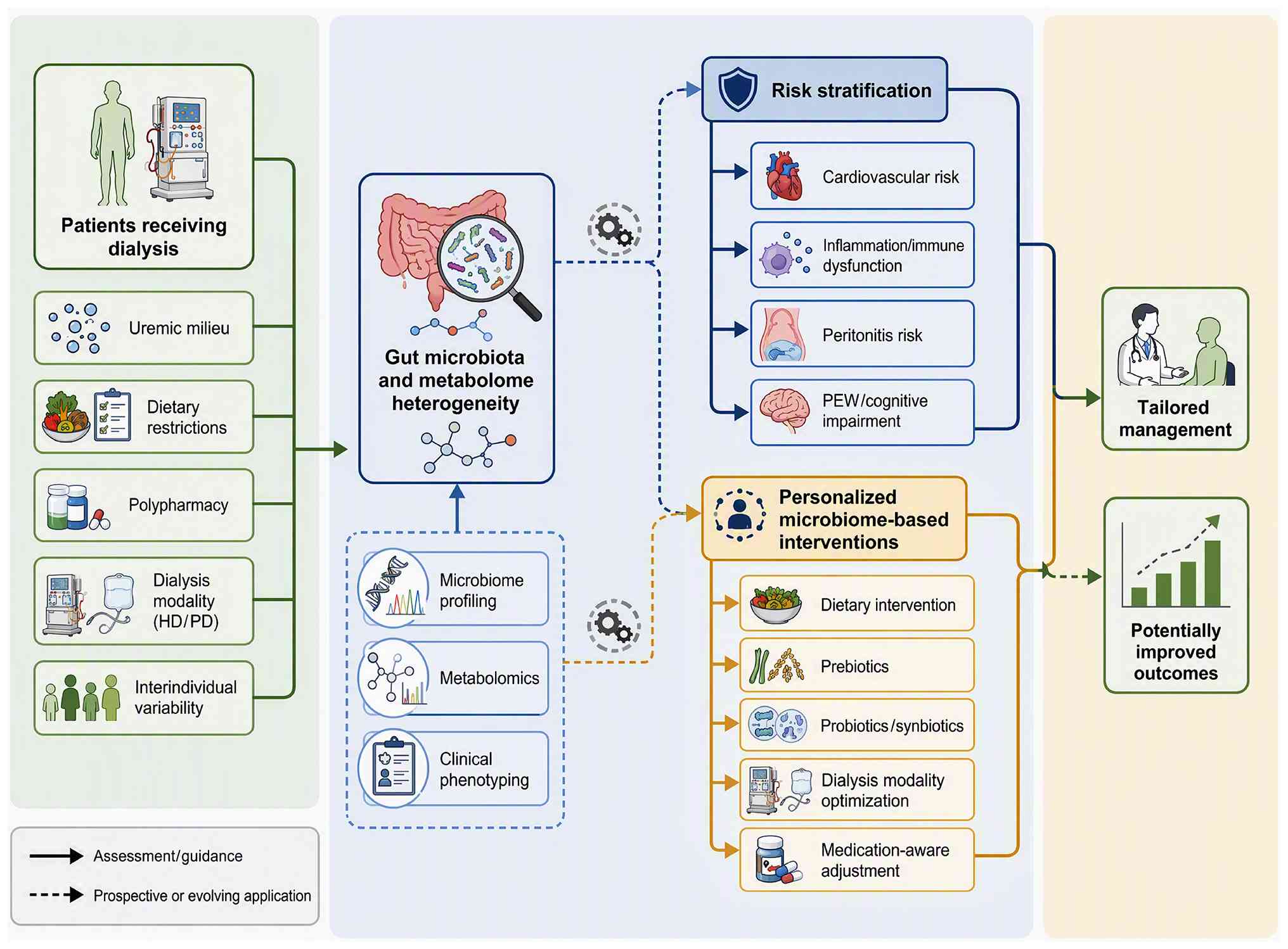 | Figure 3.Translational roadmap for
personalized microbiome-based care among patients receiving
dialysis. Baseline factors including uremic milieu, dietary
restrictions, polypharmacy, dialysis modality (HD/PD) and
inter-individual variation drive gut microbiome-metabolome
heterogeneity. Integrated assessments comprising microbiome
profiling, metabolomics and clinical phenotyping feed into
machine-learning-assisted risk stratification, which stratifies
patients by their individual risk of cardiovascular events,
inflammation-immune defects, peritonitis, PEW and cognitive
impairment. Stratified subgroups receive tailored interventions,
including dietary adjustment, prebiotics, probiotics/synbiotics,
dialysis modality optimization and medication adjustments
considering drug-microbiome interactions. Longitudinal follow-up
enables adaptive management to improve clinical outcomes. Solid
arrows represent assessment and clinical guidance; dashed arrows
denote prospective, evolving translational applications. HD,
hemodialysis; PD, peritoneal dialysis; PEW, protein-energy
wasting. |
Dietary and prebiotic strategies
Dietary modulation represents a foundational
approach to reshape gut microbiota in patients receiving dialysis.
Increased intake of dietary fiber has been associated with lower
levels of gut-derived uremic toxins, although adherence remains
suboptimal (123,124). Among prebiotics, inulin-type
fructans have been investigated. Li et al (87) conducted a randomized crossover
trial in patients receiving PD and found that inulin-type fructan
supplementation notably restricted the increase in serum indole, a
microbiota-generated uremic toxin. Similarly, in a controlled
crossover trial in patients receiving PD, inulin-type prebiotics
reduced serum uric acid levels via gut microbiota modulation
(88); however, the effects on
protein-bound solutes such as IS and PCS have been less consistent.
A pilot study of inulin supplementation in patients receiving HD
reported altered microbiota composition but no significant changes
in circulating IS or PCS (125),
suggesting that baseline microbial composition or dialysis modality
may influence responses.
Resistant starch (RS) has emerged as another
prebiotic. In patients receiving HD, RS type-2 enriched cookies
markedly reduced plasma IS and PCS levels while attenuating
inflammation (92,126). A subsequent RCT further
demonstrated that RS supplementation increased the proportion of
Faecalibacterium, a butyrate-producing genus with
anti-inflammatory properties (127). However, a recent double-blind
controlled trial reported that pea hull fiber supplementation did
not modulate uremic metabolites in adults receiving HD (128), highlighting that not all fibers
confer equivalent benefits. Collectively, these data indicate that
prebiotic efficacy is fiber-type specific and likely depends on
baseline dysbiosis severity, necessitating personalized selection
based on individual microbial profiles.
Probiotic and synbiotic
interventions
Probiotic supplementation has been extensively
evaluated in dialysis populations, albeit with heterogeneous
outcomes. Soleimani et al (79) demonstrated that a multi-strain
probiotic improved metabolic profiles in patients with diabetes
receiving HD. Subsequently, Borges et al (52,80)
reported that probiotic supplementation reduced plasma PCS and
inflammatory markers but did not notably lower TMAO levels. In a
larger randomized trial, Liu et al (129) showed that probiotics altered gut
microbiota composition in patients receiving HD, yet changes in
uremic toxins did not reach statistical significance. More
recently, Zhou et al (83)
found that probiotic treatment induced intestinal microbiota
changes but did not alter SCFA levels in patients receiving PD.
These discrepancies may be explained by differences in probiotic
strains, dosing regimens, treatment duration and patient
characteristics.
Synbiotics, which combine probiotics with
prebiotics, have shown benefits. A randomized trial by Mirzaeian
et al (82) reported that
synbiotic supplementation markedly reduced serum IS and PCS along
with systemic inflammation in patients receiving HD. Similarly,
Lopes et al (81)
demonstrated that a synbiotic meal decreased uremic toxins in
individuals receiving HD. In patients receiving PD, synbiotics
alleviated gut indole load and dysbiosis (130). A recent 2025 randomized trial by
Climent et al (131)
revealed that psychobiotic-containing nutritional supplements
improved mood and gastrointestinal symptoms in patients receiving
HD, extending benefits beyond metabolic endpoints. Despite these
results, the lack of head-to-head comparisons and standardized
outcome measures precludes definitive recommendations, and future
trials should incorporate baseline microbiome profiling to identify
responders.
Novel and adjunctive approaches
Beyond classical prebiotics and probiotics,
emerging strategies offer additional avenues for personalization.
SCFA supplementation, particularly sodium propionate, improved
inflammatory parameters and reduced gut-derived uremic toxins in a
pilot study in patients receiving HD (132). Oral vancomycin-induced gut
microbiome perturbation lowered plasma IS and PCS concentrations in
patients with ESRD (133),
providing proof-of-concept that targeted antimicrobial modulation
can be effective, albeit with safety concerns regarding resistance
and dysbiosis. Another approach involves oral administration of
engineered microbial cocktails. Zheng et al (134) developed an orally delivered
microbial formulation that effectively removed nitrogenous waste in
animal models of kidney failure, representing a potential synthetic
biology strategy for future clinical translation.
Phosphate binders, routinely used in dialysis, may
exert unintended microbiome-modulating effects. Sevelamer use was
associated with poor vitamin K status and elevated gut-derived
uremic toxins, suggesting a drug-microbiome interaction (36). Conversely, sucroferric oxyhydroxide
showed differential impacts on the oral and intestinal microbiome
compared with calcium acetate (35,135). These observations raise the
possibility of selecting phosphate binders based on their
collateral microbiome effects. Furthermore, FMT has been explored
in small studies. A pilot trial in children on kidney replacement
therapy demonstrated the feasibility of FMT for recurrent
Clostridium difficile infection (97), but its application for uremic toxin
reduction remains investigational and requires rigorous safety
evaluation.
Microbiome-based patient
stratification and multi-omics integration
A step toward personalization is systematic
stratification of patients receiving dialysis into distinct
microbial subtypes, or microbiotypes, based on compositional and
functional profiles. Enterotype-based stratification has been
explored in CKD populations, with patients with CKD stages 3–5
categorized into distinct enterotypes whose formation showed weak
dependence on environmental factors, suggesting that microbial
stratification may offer insights beyond traditional clinical
classification (106). In
dialysis populations, distinct microbial communities distinguish
patients receiving PD from those receiving HD despite similar
overall diversity, and specific taxonomic shifts, such as depletion
of Bifidobacterium adolescentis, have been linked to
nutritional risk (17).
Beyond enterotyping, integrative clustering
approaches that combine fecal microbiome data with urinary
peptidome profiles have been applied to explore patient clustering
in CKD (120). Extending such
multi-omics clustering to dialysis populations could enable
identification of patient subgroups with shared microbial dysbiosis
patterns, thereby guiding selection of targeted interventions. For
instance, patients with predominant depletion of SCFA producers may
benefit more from prebiotic fiber supplementation, whereas those
with overrepresentation of proteolytic toxin-generating taxa might
require different approaches such as adsorbents or dietary protein
modification.
Metabolite-guided therapeutic selection represents
another dimension of personalization. Serum metabolomics has
identified gut-derived uremic toxins and bile acid dysregulation
associated with CKD severity (121), and baseline microbial composition
has been shown to predict subsequent uremic toxin elevations in
patients initiating PD (110).
Such predictive capacity could inform preemptive selection of
interventions targeting specific metabolite pathways. For example,
patients with elevated baseline TMAO may be candidates for
strategies specifically aimed at reducing TMAO production, while
those with high IS or PCS might benefit from adsorbent-based
approaches or dietary tryptophan restriction (101).
The integration of multi-omics data, including
metagenomics, metabolomics and metatranscriptomics, is essential
for moving beyond descriptive taxonomy toward functional
personalization (120).
Metagenome-wide analyses have uncovered taxon-specific functional
signatures in ESRD (104), and
combined 16S and shotgun metagenome sequencing has revealed
metabolic pathway enrichment associated with specific clinical
phenotypes such as cognitive impairment (78). However, integrating these disparate
data types into clinically actionable frameworks remains
challenging. Krukowski et al (106) demonstrated that host factors
including diet, medications and socioeconomic status explain more
variance in gut microbiome composition than estimated glomerular
filtration rate, emphasizing that patient-level covariates must be
incorporated into any personalization algorithm.
Predictive modeling and artificial
intelligence (AI)
Machine learning and AI are emerging as tools for
translating multi-omics data into personalized recommendations. In
HD populations, machine learning has been applied to investigate
how patient characteristics, medications and clinical parameters
affect gut microbiome diversity over time (122). Random forest classification and
other machine learning approaches have validated key microbial
alterations associated with disease, and gut microbiota biomarkers
of CKD progression have been identified using 16S ribosomal DNA
sequencing combined with machine learning (114).
The application of AI to the gut-kidney axis has
been comprehensively reviewed, with AI utilized for multi-omics
analysis to better understand CKD pathogenesis (122). In PD, multi-omics and AI have
been proposed to enhance personalized nutritional management by
processing datasets from continuous glucose monitoring, genetic,
metabolic and microbiome profiles. AI can identify patterns and
predict outcomes that may be difficult for humans to detect,
enabling personalized recommendations for diet, medication and
dialysis management. More recently, the
diet-microbiota-metabolite-kidney axis framework has been proposed,
identifying AI-assisted dietary planning as a potential direction
for precision nutrition (122).
Predictive modeling of clinical outcomes using
microbial signatures is also advancing. Specific taxa such as
Bifidobacteriaceae have been identified as potential markers for
predicting skeletal muscle mass decline and PEW (70). Ongoing clinical trials are actively
developing AI models for early cardiovascular risk prediction in
patients receiving HD using gut microbiome multi-omics data
(122). These developments
underscore the potential for AI-driven predictive models to inform
clinical decision-making, although prospective validation in
real-world dialysis settings remains limited.
Roadmap to personalization
Accumulating evidence indicates that a
one-size-fits-all approach is unlikely to succeed in
microbiome-based care for patients receiving dialysis. Host
factors, including diet, medications and genetic background, shape
gut microbiota notably more than kidney function itself (106). Longitudinal multi-omics profiling
integrating metagenomics, metabolomics and clinical parameters will
be essential to stratify patients into distinct microbiotypes. For
instance, serum metabolomics has identified gut-derived uremic
toxins and bile acid dysregulation associated with CKD severity
(121), and guild-level
signatures of gut microbiome have been linked to diabetic kidney
disease (136). Machine learning
models incorporating baseline microbiota and metabolome data could
predict individual responses to specific interventions, enabling
algorithm-driven prescription of prebiotics, probiotics or dietary
regimens (122). Additionally,
dynamic monitoring during follow up would allow adaptive
modifications. The practical implementation of such a framework
will require standardized protocols for sample collection and
processing, development of clinically validated algorithms and
integration with electronic health records to enable real-time
decision support. Realizing this vision requires large-scale,
well-phenotyped cohort studies with multi-omics characterization
and standardized interventional protocols. Until then, pragmatic
attempts at personalization should consider dialysis modality,
residual kidney function, dietary habits, concomitant medications
(such as proton pump inhibitors and phosphate binders) and baseline
toxin levels as initial stratification variables.
Clinical translation: Feasibility,
barriers and future opportunities
Translating microbiome research into dialysis
clinical practice requires addressing several practical
considerations. The feasibility of microbiome profiling in patients
receiving dialysis has been assessed in a pilot study, with
Lockwood et al (119)
reporting high patient compliance in fecal sample collection,
although logistical barriers including sample processing, storage
and shipping remain challenging (118). Potential biomarkers for risk
stratification include gut-derived uremic toxins (IS, PCS and TMAO)
measured by serum metabolomics (121), specific taxa such as
Bifidobacteriaceae for predicting muscle mass decline (70) and microbial diversity indices
associated with cognitive impairment (78) and mortality risk (12). However, none of these biomarkers
have been validated in prospective interventional trials for
clinical decision-making.
Barriers to clinical implementation include a lack
of standardized analytical protocols across institutions, which
limits cross-study comparability (106,118); the substantial influence of host
factors (diet, medications and socioeconomic status) on microbiome
composition, complicating interpretation of individual patient
profiles (106); the absence of
clinically validated thresholds for microbiome-based risk
stratification; and limited evidence that microbiome-targeted
interventions improve hard clinical outcomes. Despite these
barriers, future opportunities include integration of microbiome
profiling with electronic health records to enable real-time
decision support, development of point-of-care metagenomic testing
and AI-assisted algorithms for predicting treatment responses based
on baseline microbial profiles (122). Pragmatic initial steps may
include using baseline toxin levels and dialysis modality as
stratification variables while standardized protocols are being
developed (14).
Future perspectives
Despite substantial progress in characterizing gut
dysbiosis in patients receiving dialysis, the field remains at a
stage where observational associations must be translated into
mechanism-based interventions. Several knowledge gaps warrant
attention. First, the causal relationship between specific
microbial taxa and clinical outcomes remains incompletely
established. Krukowski et al (106) demonstrated that host factors
including diet, medications and socioeconomic status explain more
variance in gut microbiome composition than estimated glomerular
filtration rate, challenging simplistic disease-centered
interpretations of dysbiosis. Similarly, Holle et al
(53) showed in pediatric patients
with CKD that gut dysbiosis and metabolite imbalance drive
inflammation through integrin-mediated signaling, yet whether
similar mechanisms operate in dialysis populations requires direct
validation. These findings collectively suggest that future studies
must incorporate rigorous covariate adjustment and preferably
employ longitudinal designs to disentangle cause from consequence.
To move from association to causation in dialysis microbiome
research, the field requires a stepwise approach: Prospective
longitudinal cohorts that document microbial changes before and
after complication development to establish temporality;
mechanistic validation in animal models with human-derived
microbial transplants to test candidate causal pathways (115,116); and RCTs of microbiome-targeted
interventions that demonstrate both modulation of specific
microbial functions and corresponding improvement in clinical
outcomes (14). Currently, most
evidence in patients receiving dialysis remains at the first level
of this hierarchy, with only limited progress toward the second and
third levels. The substantial heterogeneity in treatment responses
observed in probiotic and prebiotic trials may partly reflect
unrecognized differences in baseline microbial composition and
metabolic capacity, underscoring the need for baseline microbiome
stratification in future RCTs (83).
Emerging multi-omics technologies offer
opportunities for mechanistic discovery. Wu et al (120) employed integrated metagenomic,
metabolomic and deep immune profiling to uncover complex
host-microbe interactions in chronic kidney disease, demonstrating
the power of multi-omics approaches for patient stratification.
Extending this approach, Mansour et al (121) identified gut-derived uremic
toxins and bile acid dysregulation associated with CKD severity
using serum metabolomics, suggesting that metabolite signatures may
outperform taxonomic markers for risk stratification. Notably, Liu
et al (122) proposed a
diet-microbiota-kidney axis reprogramming framework emphasizing
that microbial metabolite-driven precision nutrition represents a
potential avenue for personalized care. Nevertheless, Lockwood
et al (119) reported high
patient compliance but noted logistical barriers in fecal sample
collection from patients receiving HD, highlighting that
methodological standardization remains a prerequisite for clinical
implementation.
FMT has emerged as a potential approach, although
evidence in patients receiving dialysis remains sparse. Samaey
et al (97) conducted a
pilot study demonstrating the feasibility of FMT for recurrent
Clostridioides difficile infection in children on kidney
replacement therapy, but no trial has specifically evaluated FMT
for uremic toxin reduction in dialysis populations. Conversely,
Arteaga-Muller et al (137) reported changes in CKD progression
following FMT, while Zhong et al (138) showed that washed microbiota
transplantation improved renal function in patients with renal
dysfunction. However, Liu et al (98) cautioned that gut dysbiosis in
kidney injury requires careful patient selection for FMT, given
potential safety concerns related to immune dysfunction in patients
receiving dialysis. A recent case report by Marasco et al
(99) described FMT for
Clostridioides. difficile infection in a patient receiving
PD, but larger controlled trials are needed. These mixed findings
underscore that FMT protocols must be optimized specifically for
dialysis populations before clinical adoption.
Beyond FMT, novel therapeutic paradigms are rapidly
evolving. Xie et al (139)
reported that alteration of gut microbiota contributes to
peritoneal fibrosis through increased production of TMAO,
identifying a previously unrecognized mechanism linking dysbiosis
to peritoneal membrane injury. Furthermore, Ban et al
(140) employed multi-omics to
demonstrate that Akkermansia muciniphila confers renal
protection in CKD, suggesting that single-organism supplementation
may represent a more targeted approach than whole-community
transplantation. The application of AI to predict individual
responses represents another area of investigation, with Mahdavi
(141) advocating for precision
nutritional genomics integrating gut microbiota data with machine
learning algorithms to guide personalized interventions.
Advancing the field from observational associations
to microbiome-guided dialysis care will therefore require several
coordinated efforts. Prospective longitudinal multi-omics studies
tracking patients from dialysis initiation through complication
development are needed, as are adequately powered RCTs of FMT and
next-generation probiotics with standardized endpoints. Moreover,
integration of host genetic, dietary and medication data into
predictive models of treatment response will be essential for
realizing personalized approaches, alongside the development of
regulatory frameworks for microbiome-based therapeutics
specifically tailored to the dialysis population. Until these goals
are achieved, pragmatic personalization should incorporate dialysis
modality, residual kidney function and baseline toxin profiles as
stratification variables for interventional studies.
Conclusions
Gut dysbiosis in patients receiving dialysis may
contribute to systemic complications through metabolite imbalance,
barrier disruption and inflammation. Conventional therapies
inadequately address this cycle. Personalized microbiome-based care
that integrates multi-omics profiling, dietary modulation and
targeted interventions holds promise. Practical translation will
require overcoming barriers in analytical standardization,
biomarker validation and clinical integration, with pragmatic
initial steps focusing on baseline toxin stratification and
dialysis modality-specific approaches. Future research must
establish causality and optimize strategies to improve clinical
outcomes in this population.
Acknowledgements
Not applicable.
Funding
Funding: No funding was received.
Availability of data and materials
Not applicable.
Authors' contributions
CJ conducted the literature search, extracted and
synthesized the published literature, and drafted the manuscript.
YW conceived the review and critically revised the manuscript. Both
authors read and approved the final version of the manuscript. Data
authentication is not applicable.
Ethics approval and consent to
participate
Not applicable.
Patient consent for publication
Not applicable.
Competing interests
The authors declare that they have no competing
interests.
Glossary
Abbreviations
Abbreviations:
|
16S rRNA
|
16S ribosomal RNA
|
|
CKD
|
chronic kidney disease
|
|
ESKD
|
end-stage kidney disease
|
|
ESRD
|
end-stage renal disease
|
|
FMT
|
fecal microbiota transplantation
|
|
HD
|
hemodialysis
|
|
IS
|
indoxyl sulfate
|
|
LPS
|
lipopolysaccharide
|
|
NLRP3
|
NOD-like receptor family pyrin domain
containing 3
|
|
PCS
|
p-cresyl sulfate
|
|
PD
|
peritoneal dialysis
|
|
PEW
|
protein-energy wasting
|
|
RAS
|
renin-angiotensin system
|
|
RS
|
resistant starch
|
|
SCFAs
|
short-chain fatty acids
|
|
TMAO
|
trimethylamine-N-oxide
|
|
AI
|
artificial intelligence
|
|
PD-1
|
programmed cell death protein 1
|
|
PD-L1
|
programmed death-ligand 1
|
|
RCT
|
randomized controlled trial
|
References
|
1
|
Kalantar-Zadeh K, Jafar TH, Nitsch D,
Neuen BL and Perkovic V: Chronic kidney disease. Lancet.
398:786–802. 2021. View Article : Google Scholar : PubMed/NCBI
|
|
2
|
Lim X, Ooi L, Ding U, Wu HHL and
Chinnadurai R: Gut Microbiota in patients receiving dialysis: A
review. Pathogens. 13:8012024. View Article : Google Scholar : PubMed/NCBI
|
|
3
|
Popkov VA, Zharikova AA, Demchenko EA,
Andrianova NV, Zorov DB and Plotnikov EY: Gut Microbiota as a
source of uremic toxins. Int J Mol Sci. 23:4832022. View Article : Google Scholar : PubMed/NCBI
|
|
4
|
Tourountzis T, Lioulios G, Fylaktou A,
Moysidou E, Papagianni A and Stangou M: Microbiome in chronic
kidney disease. Life (Basel). 12:15132022.PubMed/NCBI
|
|
5
|
Evenepoel P, Meijers BK, Bammens BR and
Verbeke K: Uremic toxins originating from colonic microbial
metabolism. Kidney Int Suppl. 114:S12–S19. 2009. View Article : Google Scholar : PubMed/NCBI
|
|
6
|
Aronov PA, Luo FJ, Plummer NS, Quan Z,
Holmes S, Hostetter TH and Meyer TW: Colonic contribution to uremic
solutes. J Am Soc Nephrol. 22:1769–1776. 2011. View Article : Google Scholar : PubMed/NCBI
|
|
7
|
Vaziri ND, Wong J, Pahl M, Piceno YM, Yuan
J, DeSantis TZ, Ni Z, Nguyen TH and Andersen GL: Chronic kidney
disease alters intestinal microbial flora. Kidney Int. 83:308–315.
2013. View Article : Google Scholar : PubMed/NCBI
|
|
8
|
Barreto FC, Barreto DV, Liabeuf S, Meert
N, Glorieux G, Temmar M, Choukroun G, Vanholder R and Massy ZA;
European Uremic Toxin Work Group (EUTox), : Serum indoxyl sulfate
is associated with vascular disease and mortality in chronic kidney
disease patients. Clin J Am Soc Nephrol. 4:1551–1558. 2009.
View Article : Google Scholar : PubMed/NCBI
|
|
9
|
Meijers BK, Claes K, Bammens B, de Loor H,
Viaene L, Verbeke K, Kuypers D, Vanrenterghem Y and Evenepoel P:
p-Cresol and cardiovascular risk in mild-to-moderate kidney
disease. Clin J Am Soc Nephrol. 5:1182–1189. 2010. View Article : Google Scholar : PubMed/NCBI
|
|
10
|
Lekawanvijit S, Adrahtas A, Kelly DJ,
Kompa AR, Wang BH and Krum H: Does indoxyl sulfate, a uraemic
toxin, have direct effects on cardiac fibroblasts and myocytes? Eur
Heart J. 31:1771–1779. 2010. View Article : Google Scholar : PubMed/NCBI
|
|
11
|
Meijers BK and Evenepoel P: The gut-kidney
axis: Indoxyl sulfate, p-cresyl sulfate and CKD progression.
Nephrol Dial Transplant. 26:759–761. 2011. View Article : Google Scholar : PubMed/NCBI
|
|
12
|
Lin TY, Wu PH, Lin YT and Hung SC: Gut
dysbiosis and mortality in hemodialysis patients. NPJ Biofilms
Microbiomes. 7:202021. View Article : Google Scholar : PubMed/NCBI
|
|
13
|
Sumida K, Pierre JF, Han Z, Mims TS,
Potukuchi PK, Yuzefpolskaya M, Colombo PC, Demmer RT, Datta S and
Kovesdy CP: Circulating microbial signatures and cardiovascular
death in patients with ESRD. Kidney Int Rep. 6:2617–2628. 2021.
View Article : Google Scholar : PubMed/NCBI
|
|
14
|
Voroneanu L, Burlacu A, Brinza C and Covic
A, Balan GG, Nistor I, Popa C, Hogas S and Covic A: Gut Microbiota
in chronic kidney disease: From composition to modulation towards
better outcomes-a systematic review. J Clin Med. 12:19482023.
View Article : Google Scholar : PubMed/NCBI
|
|
15
|
Shi X, Gao B, Srivastava A, Izzi Z,
Abdalla Y, Shen W and Raj D: Alterations of gut microbial pathways
and virulence factors in hemodialysis patients. Front Cell Infect
Microbiol. 12:9042842022. View Article : Google Scholar : PubMed/NCBI
|
|
16
|
Kashani K, Cozzolino MG, Massy ZA,
Blankestijn PJ, Stenvinkel P, Rosner MH and Ronco C: Proposal for a
new classification of solutes of interest in uremia and
hemodialysis. Blood Purif. 52:233–241. 2023. View Article : Google Scholar : PubMed/NCBI
|
|
17
|
Luo D, Zhao W, Lin Z, Wu J, Lin H, Li Y,
Song J, Zhang J and Peng H: The effects of hemodialysis and
peritoneal dialysis on the gut microbiota of end-stage renal
disease patients, and the relationship between gut microbiota and
patient prognoses. Front Cell Infect Microbiol. 11:5793862021.
View Article : Google Scholar : PubMed/NCBI
|
|
18
|
Martín-Del-Campo F, Avesani CM, Stenvinkel
P, Lindholm B, Cueto-Manzano AM and Cortés-Sanabria L: Gut
microbiota disturbances and protein-energy wasting in chronic
kidney disease: A narrative review. J Nephrol. 36:873–883. 2023.
View Article : Google Scholar : PubMed/NCBI
|
|
19
|
Bao WH, Yang WL, Su CY, Lu XH, He L and
Zhang AH: Relationship between gut microbiota and vascular
calcification in hemodialysis patients. Ren Fail. 45:21485382023.
View Article : Google Scholar : PubMed/NCBI
|
|
20
|
He L, Yang W, Yang P, Zhang X and Zhang A:
Higher serum trimethylamine-N-oxide levels are associated with
increased abdominal aortic calcification in hemodialysis patients.
Ren Fail. 44:2019–2027. 2022. View Article : Google Scholar : PubMed/NCBI
|
|
21
|
Chaves LD, Abyad S, Honan AM, Bryniarski
MA, McSkimming DI, Stahura CM, Wells SC, Ruszaj DM, Morris ME,
Quigg RJ and Yacoub R: Unconjugated p-cresol activates macrophage
macropinocytosis leading to increased LDL uptake. JCI Insight.
6:e1444102021. View Article : Google Scholar : PubMed/NCBI
|
|
22
|
Koshida T, Gohda T, Sugimoto T, Asahara T,
Asao R, Ohsawa I, Gotoh H, Murakoshi M, Suzuki Y and Yamashiro Y:
Gut Microbiome and Microbiome-derived metabolites in patients with
end-stage kidney disease. Int J Mol Sci. 24:114562023. View Article : Google Scholar : PubMed/NCBI
|
|
23
|
Yu J, Li Y, Zhu B, Shen J and Miao L:
Research progress on the kidney-gut-brain axis in brain dysfunction
in maintenance hemodialysis patients. Front Med (Lausanne).
12:15380482025. View Article : Google Scholar : PubMed/NCBI
|
|
24
|
Davenport A: More frequent hemodialysis
does not effectively clear protein-bound azotemic solutes derived
from gut microbiome metabolism. Kidney Int. 91:1008–1010. 2017.
View Article : Google Scholar : PubMed/NCBI
|
|
25
|
Rysz J, Franczyk B, Ławiński J, Olszewski
R, Ciałkowska-Rysz A and Gluba-Brzózka A: The Impact of CKD on
uremic toxins and gut microbiota. Toxins (Basel). 13:2522021.
View Article : Google Scholar : PubMed/NCBI
|
|
26
|
Stadlbauer V, Horvath A, Ribitsch W,
Schmerböck B, Schilcher G, Lemesch S, Stiegler P, Rosenkranz AR,
Fickert P and Leber B: Structural and functional differences in gut
microbiome composition in patients undergoing haemodialysis or
peritoneal dialysis. Sci Rep. 7:156012017. View Article : Google Scholar : PubMed/NCBI
|
|
27
|
Chen G, Zhang X and Chen F: A
cross-sectional study on gut microbiota in patients with chronic
kidney disease undergoing kidney transplant or hemodialysis. Am J
Transl Res. 15:1756–1765. 2023.PubMed/NCBI
|
|
28
|
Teixeira RR, de Andrade LS, Pereira NBF,
Montenegro H, Hoffmann C and Cuppari L: Gut microbiota profile of
patients on peritoneal dialysis: Comparison with household
contacts. Eur J Clin Nutr. 77:90–97. 2023. View Article : Google Scholar : PubMed/NCBI
|
|
29
|
Voroneanu L and Covic A, Iliescu S, Baluta
CV, Agavriloaei BD, Stefan AE, Amărandi RM, Văcărean-Trandafir IC,
Ivanov IC and Covic A: Distinct gut microbiome signatures in
hemodialysis and kidney transplant populations. J Clin Med.
14:80322025. View Article : Google Scholar : PubMed/NCBI
|
|
30
|
Fülöp T, Zsom L, Tapolyai MB, Molnar MZ,
Abdul Salim S, Arany I, Hamrahian M and Rosivall L: Peritoneal
dialysis: The unique features by compartmental delivery of renal
replacement therapy. Med Hypotheses. 108:128–132. 2017. View Article : Google Scholar : PubMed/NCBI
|
|
31
|
Lin TY and Hung SC: Association of
subjective global assessment of nutritional status with gut
microbiota in hemodialysis patients: A case-control study. Nephrol
Dial Transplant. 36:1104–1111. 2021. View Article : Google Scholar : PubMed/NCBI
|
|
32
|
Hu J, Zhong X, Liu Y, Yan J, Zhou D, Qin
D, Xiao X, Zheng Y, Wen L, Tan R, et al: Correlation between
intestinal flora disruption and protein-energy wasting in patients
with end-stage renal disease. BMC Nephrol. 23:1302022. View Article : Google Scholar : PubMed/NCBI
|
|
33
|
Bi X, Liu Y, Yao L, Ling L, Lu J, Hu C and
Ding W: Gut microbiota dysbiosis and protein energy wasting in
patients on hemodialysis: An observational longitudinal study.
Front Nutr. 10:12706902023. View Article : Google Scholar : PubMed/NCBI
|
|
34
|
Wu PH, Liu PY, Chiu YW, Hung WC, Lin YT,
Lin TY, Hung SC, Delicano RA, Kuo MC and Wu CY: Comparative gut
microbiome differences between ferric citrate and calcium carbonate
phosphate binders in patients with End-Stage kidney disease.
Microorganisms. 8:20402020. View Article : Google Scholar : PubMed/NCBI
|
|
35
|
Abdelbary MMH, Kuppe C, Michael SS, Krüger
T, Floege J and Conrads G: Impact of sucroferric oxyhydroxide on
the oral and intestinal microbiome in hemodialysis patients. Sci
Rep. 12:96142022. View Article : Google Scholar : PubMed/NCBI
|
|
36
|
Dai L, Meijers BK, Bammens B, de Loor H,
Schurgers LJ, Qureshi AR, Stenvinkel P and Evenepoel P: Sevelamer
use in End-Stage kidney disease (ESKD) patients associates with
poor vitamin k status and high levels of Gut-derived uremic toxins:
A Drug-bug interaction? Toxins (Basel). 12:3512020. View Article : Google Scholar : PubMed/NCBI
|
|
37
|
Lin YT, Lin TY, Hung SC, Liu PY, Wu PH,
Chuang YS, Hung WC, Chiu YW, Kuo MC and Wu CY: Anti-acid drug
treatment induces changes in the gut microbiome composition of
hemodialysis patients. Microorganisms. 9:2862021. View Article : Google Scholar : PubMed/NCBI
|
|
38
|
Lin YT, Lin TY, Hung SC, Liu PY, Hung WC,
Tsai WC, Tsai YC, Delicano RA, Chuang YS, Kuo MC, et al:
Differences in the microbial composition of hemodialysis patients
treated with and without β-Blockers. J Pers Med. 11:1982021.
View Article : Google Scholar : PubMed/NCBI
|
|
39
|
Ahmadmehrabi S and Tang WHW:
Hemodialysis-induced cardiovascular disease. Semin Dial.
31:258–267. 2018. View Article : Google Scholar : PubMed/NCBI
|
|
40
|
He H and Xie Y: Effect of different
hemodialysis methods on microbiota in uremic patients. Biomed Res
Int. 2020:67397622020. View Article : Google Scholar : PubMed/NCBI
|
|
41
|
Zhou J, Yang C, Lei W, Yang Z, Chen J and
Lin H: Exploration of the correlation between intestinal flora and
peritoneal dialysis-related peritonitis. Clin Exp Nephrol.
26:1030–1038. 2022. View Article : Google Scholar : PubMed/NCBI
|
|
42
|
Zhou J, Yang C, Lei W, Yang Z, Chen J, Lin
H, Li Q and Yuan W: Exploration of the correlation between
intestinal flora and Escherichia coli peritoneal
dialysis-related peritonitis. BMC Nephrol. 23:762022. View Article : Google Scholar : PubMed/NCBI
|
|
43
|
Zhang L, Zhang H, Su S, Jia Y, Liang C,
Fang Y, Hong D, Li T and Ma F: Risk factor assessment and
microbiome analysis in peritoneal dialysis-related peritonitis
reveal etiological characteristics. Front Immunol. 15:14434682024.
View Article : Google Scholar : PubMed/NCBI
|
|
44
|
Brito JS, Borges NA, Dolenga CJ,
Carraro-Eduardo JC, Nakao LS and Mafra D: Is there a relationship
between tryptophan dietary intake and plasma levels of indoxyl
sulfate in chronic kidney disease patients on hemodialysis? J Bras
Nefrol. 38:396–402. 2016. View Article : Google Scholar : PubMed/NCBI
|
|
45
|
Stockler-Pinto MB, Soulage CO, Borges NA,
Cardozo LFMF, Dolenga CJ, Nakao LS, Pecoits-Filho R, Fouque D and
Mafra D: From bench to the hemodialysis clinic: Protein-bound
uremic toxins modulate NF-κB/Nrf2 expression. Int Urol Nephrol.
50:347–354. 2018. View Article : Google Scholar : PubMed/NCBI
|
|
46
|
Zhang X, Li Y, Yang P, Liu X, Lu L, Chen
Y, Zhong X, Li Z, Liu H, Ou C, et al: Trimethylamine-N-Oxide
promotes vascular calcification through activation of NLRP3
(Nucleotide-Binding domain, leucine-rich-containing family, pyrin
Domain-Containing-3) inflammasome and NF-κB (Nuclear Factor κB)
signals. Arterioscler Thromb Vasc Biol. 40:751–765. 2020.
View Article : Google Scholar : PubMed/NCBI
|
|
47
|
Gryp T, Vanholder R, Vaneechoutte M and
Glorieux G: p-Cresyl Sulfate. Toxins (Basel). 9:522017. View Article : Google Scholar : PubMed/NCBI
|
|
48
|
Hu J, Zhong X, Yan J, Zhou D, Qin D, Xiao
X, Zheng Y and Liu Y: High-throughput sequencing analysis of
intestinal flora changes in ESRD and CKD patients. BMC Nephrol.
21:122020. View Article : Google Scholar : PubMed/NCBI
|
|
49
|
Ficek J, Wyskida K, Ficek R, Wajda J,
Klein D, Witkowicz J, Rotkegel S, Spiechowicz-Zatoń U,
Kocemba-Dyczek J, Ciepał J, et al: Relationship between plasma
levels of zonulin, bacterial lipopolysaccharides, D-lactate and
markers of inflammation in haemodialysis patients. Int Urol
Nephrol. 49:717–725. 2017. View Article : Google Scholar : PubMed/NCBI
|
|
50
|
Engen PA, Dodiya HB, Naqib A, Forsyth CB,
Green SJ, Voigt RM, Kordower JH, Mutlu EA, Shannon KM and
Keshavarzian A: The potential role of Gut-derived inflammation in
multiple system atrophy. J Parkinsons Dis. 7:331–346. 2017.
View Article : Google Scholar : PubMed/NCBI
|
|
51
|
de Andrade LS, Sardá FAH, Pereira NBF,
Teixeira RR, Rodrigues SD, de Lima JD, Dalboni MA, Aoike DT, Nakao
LS and Cuppari L: Effect of unripe banana flour on Gut-derived
uremic toxins in individuals undergoing peritoneal dialysis: A
randomized, Double-Blind, Placebo-controlled, crossover trial.
Nutrients. 13:6462021. View Article : Google Scholar : PubMed/NCBI
|
|
52
|
Borges NA, Carmo FL, Stockler-Pinto MB, de
Brito JS, Dolenga CJ, Ferreira DC, Nakao LS, Rosado A, Fouque D and
Mafra D: Probiotic supplementation in chronic kidney disease: A
Double-blind, randomized, placebo-controlled trial. J Ren Nutr.
28:28–36. 2018. View Article : Google Scholar : PubMed/NCBI
|
|
53
|
Holle J, Bartolomaeus H, Löber U, Behrens
F, Bartolomaeus TUP, Anandakumar H, Wimmer MI, Vu DL, Kuhring M,
Brüning U, et al: Inflammation in children with CKD linked to gut
dysbiosis and metabolite imbalance. J Am Soc Nephrol. 33:2259–2275.
2022. View Article : Google Scholar : PubMed/NCBI
|
|
54
|
Mafra D, Alvarenga L, F M F Cardozo L,
Schultz J, Rosado AS and Borges NA: Gut microbiota and NLRP3
inflammasome activation in hemodialysis patients: Exploring the
link with systemic inflammation. Mol Biol Rep. 52:4652025.
View Article : Google Scholar : PubMed/NCBI
|
|
55
|
Samaan E, Eldeeb AA, Ibrahim AB, Erman M,
Sabry AA and Mahmoud MA: Effect of Ramadan fasting on chronic
inflammation markers and gut bacterial endotoxins among Egyptian
hemodialysis patients. Ther Apher Dial. 27:655–660. 2023.
View Article : Google Scholar : PubMed/NCBI
|
|
56
|
Guo TT, Zhang Z, Sun Y, Zhu RY, Wang FX,
Ma LJ, Jiang L and Liu HD: Neuroprotective effects of sodium
butyrate by restoring gut microbiota and inhibiting TLR4 signaling
in mice with MPTP-Induced Parkinson's disease. Nutrients.
15:9302023. View Article : Google Scholar : PubMed/NCBI
|
|
57
|
Qu Y, An K, Wang D, Yu H, Li J, Min Z,
Xiong Y, Xue Z and Mao Z: Short-Chain fatty acid aggregates
alpha-Synuclein accumulation and neuroinflammation via GPR43-NLRP3
signaling pathway in a model Parkinson's disease. Mol Neurobiol.
62:6612–6625. 2025. View Article : Google Scholar : PubMed/NCBI
|
|
58
|
Liu Z, Nie J, Li Y, Huang M, Chen Z, Yu S,
Zheng J, Tan Y and Chen S: Bacteroides coprocola protects
dopaminergic neurons in rotenone-induced Parkinson's disease mouse
model by modulating gut microbiota dysbiosis and inhibiting the
NLRP3 signaling pathway. Transl Neurodegener. 15:72026. View Article : Google Scholar : PubMed/NCBI
|
|
59
|
Li X, Zheng J, Wang J, Tang X, Zhang F,
Liu S, Liao Y, Chen X, Xie W and Tang Y: Effects of uremic
clearance granules on p38 MAPK/NF-κB signaling pathway, microbial
and metabolic profiles in End-stage renal disease rats receiving
peritoneal dialysis. Drug Des Devel Ther. 16:2529–2544. 2022.
View Article : Google Scholar : PubMed/NCBI
|
|
60
|
Zhang L, Xie F, Tang H, Zhang X, Hu J,
Zhong X, Gong N, Lai Y, Zhou M, Tian J, et al: Gut microbial
metabolite TMAO increases peritoneal inflammation and peritonitis
risk in peritoneal dialysis patients. Transl Res. 240:50–63. 2022.
View Article : Google Scholar : PubMed/NCBI
|
|
61
|
Chen Y, Liu Y, Wang Y, Chen X, Wang C,
Chen X, Yuan X, Liu L, Yang J and Zhou X: Prevotellaceae produces
butyrate to alleviate PD-1/PD-L1 inhibitor-related cardiotoxicity
via PPARα-CYP4X1 axis in colonic macrophages. J Exp Clin Cancer
Res. 41:12022. View Article : Google Scholar : PubMed/NCBI
|
|
62
|
Wang L, Xu A, Wang J, Fan G, Liu R, Wei L
and Pei M: The effect and mechanism of Fushen Granule on gut
microbiome in the prevention and treatment of chronic renal
failure. Front Cell Infect Microbiol. 13:13342132024. View Article : Google Scholar : PubMed/NCBI
|
|
63
|
Velasquez MT, Centron P, Barrows I,
Dwivedi R and Raj DS: Gut Microbiota and cardiovascular uremic
toxicities. Toxins (Basel). 10:2872018. View Article : Google Scholar : PubMed/NCBI
|
|
64
|
Kanitsoraphan C, Rattanawong P, Charoensri
S and Senthong V: Trimethylamine N-oxide and risk of cardiovascular
disease and mortality. Curr Nutr Rep. 7:207–213. 2018. View Article : Google Scholar : PubMed/NCBI
|
|
65
|
Lv Y, Yang X, Sun X and Ren X:
Immune-microbiota dysregulation in maintenance hemodialysis: A 16S
rRNA sequencing-based analysis of gut flora and T cell profiles.
Ren Fail. 47:24986302025. View Article : Google Scholar : PubMed/NCBI
|
|
66
|
Glorieux G, Gryp T and Perna A:
Gut-derived metabolites and their role in immune dysfunction in
chronic kidney disease. Toxins (Basel). 12:2452020. View Article : Google Scholar : PubMed/NCBI
|
|
67
|
Al Khodor S and Shatat IF: Gut microbiome
and kidney disease: A bidirectional relationship. Pediatr Nephrol.
32:921–931. 2017. View Article : Google Scholar : PubMed/NCBI
|
|
68
|
Xu P, Zhang M, Cui S, Wang L, Li Y, Li Y
and Jiang C: Gut microbiome and peritoneal inflammation: A new
perspective on peritoneal dialysis-associated peritonitis. Ren
Fail. 47:25425322025. View Article : Google Scholar : PubMed/NCBI
|
|
69
|
Santhakumaran T, Samad N and Fan SL:
Hydration status measured by BCM: A potential modifiable risk
factor for peritonitis in patients on peritoneal dialysis.
Nephrology (Carlton). 21:404–409. 2016. View Article : Google Scholar : PubMed/NCBI
|
|
70
|
Bakinowska E, Olejnik-Wojciechowska J,
Kiełbowski K, Skoryk A and Pawlik A: Pathogenesis of sarcopenia in
chronic kidney Disease-The role of inflammation, metabolic
dysregulation, gut dysbiosis, and microRNA. Int J Mol Sci.
25:84742024. View Article : Google Scholar : PubMed/NCBI
|
|
71
|
March DS, Jones AW, Bishop NC and Burton
JO: The efficacy of prebiotic, probiotic, and synbiotic
supplementation in modulating Gut-derived circulatory particles
associated with cardiovascular disease in individuals receiving
dialysis: A systematic review and Meta-analysis of randomized
controlled trials. J Ren Nutr. 30:347–359. 2020. View Article : Google Scholar : PubMed/NCBI
|
|
72
|
Pan Y, Yang L, Dai B, Lin B, Lin S and Lin
E: Effects of probiotics on malnutrition and Health-related quality
of life in patients undergoing peritoneal dialysis: A randomized
controlled trial. J Ren Nutr. 31:199–205. 2021. View Article : Google Scholar : PubMed/NCBI
|
|
73
|
Azamian Y, Abdollahzad H, Rezaeian S,
Rouhani MH and Fatehi MH: The effect of synbiotic supplementation
on plasma levels of advanced glycation end products and
cardiovascular risk factors in hemodialysis patients: A
double-blind clinical trial. Food Sci Nutr. 12:6864–6872. 2024.
View Article : Google Scholar : PubMed/NCBI
|
|
74
|
Zhu B, Shen J, Jiang R, Jin L, Zhan G, Liu
J, Sha Q, Xu R, Miao L and Yang C: Abnormalities in gut microbiota
and serum metabolites in hemodialysis patients with mild cognitive
decline: A single-center observational study. Psychopharmacology
(Berl). 237:2739–2752. 2020. View Article : Google Scholar : PubMed/NCBI
|
|
75
|
Wang J, Wu S, Zhang J, Li Y, Wu Y and Qi
X: Correlation between gut microbiome and cognitive impairment in
patients undergoing peritoneal dialysis. BMC Nephrol. 24:3602023.
View Article : Google Scholar : PubMed/NCBI
|
|
76
|
Shah SN, Knausenberger TB, Pontifex MG,
Connell E, Le Gall G, Hardy TAJ, Randall DW, McCafferty K, Yaqoob
MM, Solito E, et al: Cerebrovascular damage caused by the gut
microbe/host co-metabolite p-cresol sulfate is prevented by
blockade of the EGF receptor. Gut Microbes. 16:24316512024.
View Article : Google Scholar : PubMed/NCBI
|
|
77
|
Martín-Del-Campo F, Vega-Magaña N,
Salazar-Félix NA, Cueto-Manzano AM, Peña-Rodríguez M,
Cortés-Sanabria L, Romo-Flores ML and Rojas-Campos E: Gut
microbiome is related to cognitive impairment in peritoneal
dialysis patients. Nutrients. 16:26592024. View Article : Google Scholar : PubMed/NCBI
|
|
78
|
Gao Q, Li D, Wang Y, Zhao C, Li M, Xiao J,
Kang Y, Lin H and Wang N: Analysis of intestinal flora and
cognitive function in maintenance hemodialysis patients using
combined 16S ribosome DNA and shotgun metagenome sequencing. Aging
Clin Exp Res. 36:282024. View Article : Google Scholar : PubMed/NCBI
|
|
79
|
Soleimani A, Zarrati Mojarrad M, Bahmani
F, Taghizadeh M, Ramezani M, Tajabadi-Ebrahimi M, Jafari P,
Esmaillzadeh A and Asemi Z: Probiotic supplementation in diabetic
hemodialysis patients has beneficial metabolic effects. Kidney Int.
91:435–442. 2017. View Article : Google Scholar : PubMed/NCBI
|
|
80
|
Borges NA, Stenvinkel P, Bergman P,
Qureshi AR, Lindholm B, Moraes C, Stockler-Pinto MB and Mafra D:
Effects of probiotic supplementation on Trimethylamine-N-Oxide
plasma levels in hemodialysis patients: A pilot study. Probiotics
Antimicrob Proteins. 11:648–654. 2019. View Article : Google Scholar : PubMed/NCBI
|
|
81
|
Lopes RCSO, Theodoro JMV, da Silva BP,
Queiroz VAV, de Castro Moreira ME, Mantovani HC, Hermsdorff HH and
Martino HSD: Synbiotic meal decreases uremic toxins in hemodialysis
individuals: A placebo-controlled trial. Food Res Int. 116:241–248.
2019. View Article : Google Scholar : PubMed/NCBI
|
|
82
|
Mirzaeian S, Saraf-Bank S, Entezari MH,
Hekmatdoost A, Feizi A and Atapour A: Effects of synbiotic
supplementation on microbiota-derived protein-bound uremic toxins,
systemic inflammation, and biochemical parameters in patients on
hemodialysis: A double-blind, placebo-controlled, randomized
clinical trial. Nutrition. 73:1107132020. View Article : Google Scholar : PubMed/NCBI
|
|
83
|
Zhou S, Yan Y, Chu R, Chen N, Wang L,
Zhang H, Wang Y, Wang M, Na L, Ren H, et al: Probiotic treatment
induces changes in intestinal microbiota but does not alter SCFA
levels in peritoneal dialysis patients-a randomized,
placebo-controlled trial. Sci Rep. 14:314132024. View Article : Google Scholar : PubMed/NCBI
|
|
84
|
Wang YC, Tsai IJ, Hsieh TH, Wu CC, Lu KC
and Liao MT: Enhanced sleep quality and reduced indoxyl sulfate
levels following probiotic supplementation were linked to gut
microbiota modulation in hemodialysis patients. J Formos Med Assoc.
Aug 27–2025.(Epub ahead of print). doi: 10.1016/j.jfma.2025.08.036.
View Article : Google Scholar
|
|
85
|
Abdelnasier M, Elshinnawy H, Abdelmohsen
WA, Daniel D, Mehanna N and Tohami N: The effect of probiotic use
on gut leakage measured by Zonulin and its relation to P Cresyl
sulfate and inflammation in hemodialysis patients: A prospective
controlled clinical study. Clin Ter. 177:405–413. 2026.PubMed/NCBI
|
|
86
|
Stanford J, Charlton K, Stefoska-Needham
A, Zheng H, Bird L, Borst A, Fuller A and Lambert K: Associations
among plant-Based diet quality, uremic toxins, and gut microbiota
profile in adults undergoing hemodialysis therapy. J Ren Nutr.
31:177–188. 2021. View Article : Google Scholar : PubMed/NCBI
|
|
87
|
Li L, Xiong Q, Zhao J, Lin X, He S, Wu N,
Yao Y, Liang W, Zuo X and Ying C: Inulin-type fructan intervention
restricts the increase in gut microbiome-generated indole in
patients with peritoneal dialysis: A randomized crossover study. Am
J Clin Nutr. 111:1087–1099. 2020. View Article : Google Scholar : PubMed/NCBI
|
|
88
|
He S, Xiong Q, Tian C, Li L, Zhao J, Lin
X, Guo X, He Y, Liang W, Zuo X and Ying C: Inulin-type prebiotics
reduce serum uric acid levels via gut microbiota modulation: A
randomized, controlled crossover trial in peritoneal dialysis
patients. Eur J Nutr. 61:665–677. 2022. View Article : Google Scholar : PubMed/NCBI
|
|
89
|
Li L, Zhao J, Wang J, Xiong Q, Lin X, Guo
X, Peng F, Liang W, Zuo X and Ying C: The arsenic-lowering effect
of inulin-type prebiotics in end-stage renal disease: A randomized
crossover trial. Food Funct. 15:355–371. 2024. View Article : Google Scholar : PubMed/NCBI
|
|
90
|
Esgalhado M, Kemp JA, Azevedo R, Paiva BR,
Stockler-Pinto MB, Dolenga CJ, Borges NA, Nakao LS and Mafra D:
Could resistant starch supplementation improve inflammatory and
oxidative stress biomarkers and uremic toxins levels in
hemodialysis patients? A pilot randomized controlled trial. Food
Funct. 9:6508–6516. 2018. View Article : Google Scholar : PubMed/NCBI
|
|
91
|
Azevedo R, Esgalhado M, Kemp JA, Regis B,
Cardozo LF, Nakao LS, Brito JS and Mafra D: Resistant starch
supplementation effects on plasma indole 3-acetic acid and aryl
hydrocarbon receptor mRNA expression in hemodialysis patients:
Randomized, double blind and controlled clinical trial. J Bras
Nefrol. 42:273–279. 2020.(In English, Portuguese). View Article : Google Scholar : PubMed/NCBI
|
|
92
|
de Paiva BR, Esgalhado M, Borges NA, Kemp
JA, Alves G, Leite PEC, Macedo R, Cardozo LFMF, de Brito JS and
Mafra D: Resistant starch supplementation attenuates inflammation
in hemodialysis patients: A pilot study. Int Urol Nephrol.
52:549–555. 2020. View Article : Google Scholar : PubMed/NCBI
|
|
93
|
Baruch EN, Youngster I, Ben-Betzalel G,
Ortenberg R, Lahat A, Katz L, Adler K, Dick-Necula D, Raskin S,
Bloch N, et al: Fecal microbiota transplant promotes response in
immunotherapy-refractory melanoma patients. Science. 371:602–609.
2021. View Article : Google Scholar : PubMed/NCBI
|
|
94
|
Davar D, Dzutsev AK, McCulloch JA,
Rodrigues RR, Chauvin JM, Morrison RM, Deblasio RN, Menna C, Ding
Q, Pagliano O, et al: Fecal microbiota transplant overcomes
resistance to anti-PD-1 therapy in melanoma patients. Science.
371:595–602. 2021. View Article : Google Scholar : PubMed/NCBI
|
|
95
|
Scheperjans F, Levo R, Bosch B, Lääperi M,
Pereira PAB, Smolander OP, Aho VTE, Vetkas N, Toivio L, Kainulainen
V, et al: Fecal Microbiota transplantation for treatment of
Parkinson disease: A randomized clinical trial. JAMA Neurol.
81:925–938. 2024. View Article : Google Scholar : PubMed/NCBI
|
|
96
|
Zhang R, Feng R, Wang J, Chen Y, Liu H,
Zhu Q, Tian H, Qin C, Teng J, Tang B, et al: Gut microbiota
modulation via repeated donor fecal transplantation improves motor
and gastrointestinal symptoms in drug-naïve Parkinson's disease: A
randomized phase 2 trial. Signal Transduct Target Ther. 11:942026.
View Article : Google Scholar : PubMed/NCBI
|
|
97
|
Samaey A, Vázquez-Castellanos JF,
Caenepeel C, Evenepoel P, Vermeire S, Raes J and Knops N: Effects
of fecal microbiota transplantation for recurrent Clostridium
difficile infection in children on kidney replacement therapy: A
pilot study. Pediatr Nephrol. 39:1201–1212. 2024. View Article : Google Scholar : PubMed/NCBI
|
|
98
|
Liu J, Ning X, Yuan J, Yu Z, Qin Y, Xing
Y, Zhao J and Sun S: Gut dysbiosis in kidney injury: Therapeutic
potential of fecal microbiota transplantation. Eur J Med Res. Mar
9–2026.(Epub ahead of print). doi: 10.1186/s40001-026-04100-w.
|
|
99
|
Marasco G, Cannarile DC, Cremon C, Papalia
G, Marangoni A, Zucchelli A, Barone M, Lazzarotto T, Brigidi P,
Stanghellini V and Barbara G: Fecal microbiota transplantation for
Clostridioides difficile infection in a peritoneal dialysis
patient: A case report. Perit Dial Int. 45:247–250. 2025.
View Article : Google Scholar : PubMed/NCBI
|
|
100
|
Messaoudene M, Ferreira S, Saint-Lu N,
Ponce M, Truntzer C, Boidot R, Le Bescop C, Loppinet T, Corbel T,
Féger C, et al: The DAV132 colon-targeted adsorbent does not
interfere with plasma concentrations of antibiotics but prevents
antibiotic-related dysbiosis: A randomized phase I trial in healthy
volunteers. Nat Commun. 15:80832024. View Article : Google Scholar : PubMed/NCBI
|
|
101
|
Rocchetti MT, Cosola C, di Bari I, Magnani
S, Galleggiante V, Scandiffio L, Dalfino G, Netti GS, Atti M,
Corciulo R and Gesualdo L: Efficacy of Divinylbenzenic resin in
removing indoxyl sulfate and P-cresol sulfate in hemodialysis
patients: Results from an in vitro study and an in vivo pilot trial
(xuanro4-Nature 3.2). Toxins (Basel). 12:1702020. View Article : Google Scholar : PubMed/NCBI
|
|
102
|
Crespo-Salgado J, Vehaskari VM, Stewart T,
Ferris M, Zhang Q, Wang G, Blanchard EE, Taylor CM, Kallash M,
Greenbaum LA and Aviles DH: Intestinal microbiota in pediatric
patients with end stage renal disease: A Midwest Pediatric
Nephrology Consortium study. Microbiome. 4:502016. View Article : Google Scholar : PubMed/NCBI
|
|
103
|
Li Y, Su X, Zhang L, Liu Y, Shi M, Lv C,
Gao Y, Xu D and Wang Z: Dysbiosis of the gut microbiome is
associated with CKD5 and correlated with clinical indices of the
disease: A case-controlled study. J Transl Med. 17:2282019.
View Article : Google Scholar : PubMed/NCBI
|
|
104
|
Zhang P, Wang X, Li S, Cao X, Zou J, Fang
Y, Shi Y, Xiang F, Shen B, Li Y, et al: Metagenome-wide analysis
uncovers gut microbial signatures and implicates taxon-specific
functions in end-stage renal disease. Genome Biol. 24:2262023.
View Article : Google Scholar : PubMed/NCBI
|
|
105
|
Qin Y, Zhao J, Wang L, Yang X, Wang J, Li
S, Chen Y, Guo J, Wang F and Luo K: Decrease in
Escherichia-Shigella in the gut microbiota of ESKD patients
undergoing maintenance hemodialysis. BMC Nephrol. 26:982025.
View Article : Google Scholar : PubMed/NCBI
|
|
106
|
Krukowski H, Valkenburg S, Vich Vila A,
Maciel LF, Vázquez-Castellanos JF, Gryp T, Joossens M, Van Biesen
W, Verbeke F, Derrien M, et al: Host factors dictate gut microbiome
alterations in chronic kidney disease more strongly than kidney
function. Nat Microbiol. 11:664–677. 2026. View Article : Google Scholar : PubMed/NCBI
|
|
107
|
Li R, Zeng L, Xie S, Chen J, Yu Y and
Zhong L: Targeted metabolomics study of serum bile acid profile in
patients with end-stage renal disease undergoing hemodialysis.
PeerJ. 7:e71452019. View Article : Google Scholar : PubMed/NCBI
|
|
108
|
Bao M, Zhang P, Guo S, Zou J, Ji J, Ding X
and Yu X: Altered gut microbiota and gut-derived p-cresyl sulfate
serum levels in peritoneal dialysis patients. Front Cell Infect
Microbiol. 12:6396242022. View Article : Google Scholar : PubMed/NCBI
|
|
109
|
James P, Stanford J, Dixit OVA, Nicdao MA,
McWhinney B, Sud K, Ryan M, Read S, Ahlenstiel G, Lambert K, et al:
Associations between uraemic toxins and gut microbiota in adults
initiating peritoneal dialysis. Toxins (Basel). 17:3342025.
View Article : Google Scholar : PubMed/NCBI
|
|
110
|
Manokasemsan W, Jariyasopit N,
Wanichthanarak K, Poungsombat P, Kurilung A, Limjiasahapong S,
Thapa K, Sirivatanauksorn Y, Raksasuk S, Srithongkul T, et al:
LC-MS/MS identifies elevated imidazole propionate and gut-derived
metabolite alterations in peritoneal dialysis patients. Comput
Struct Biotechnol J. 27:5271–5280. 2025. View Article : Google Scholar : PubMed/NCBI
|
|
111
|
Kuo TH, Wu PH, Liu PY, Chuang YS, Tai CJ,
Kuo MC, Chiu YW and Lin YT: Identification of gut microbiome
signatures associated with serotonin pathway in tryptophan
metabolism of patients undergoing hemodialysis. Int J Mol Sci.
26:104632025. View Article : Google Scholar : PubMed/NCBI
|
|
112
|
Li J, Xing H, Lin W, Yu H, Yang B, Jiang
C, Zhang J, Wu R, Ding F, Pei M and Yang H: Specific gut microbiome
and metabolome changes in patients with continuous ambulatory
peritoneal dialysis and comparison between patients with different
dialysis vintages. Front Med (Lausanne). 10:13023522024. View Article : Google Scholar : PubMed/NCBI
|
|
113
|
Araujo R, Merino-Ribas A, Pereira L,
Campos J, Silva N, Alencastre IS, Pestana M and Sampaio-Maia B: The
urogenital microbiome in chronic kidney disease patients on
peritoneal dialysis. Nefrologia (Engl Ed). 44:194–203. 2024.
View Article : Google Scholar : PubMed/NCBI
|
|
114
|
Wu IW, Lin CY, Chang LC, Lee CC, Chiu CY,
Hsu HJ, Sun CY, Chen YC, Kuo YL, Yang CW, et al: Gut microbiota as
diagnostic tools for mirroring disease progression and circulating
nephrotoxin levels in chronic kidney disease: Discovery and
validation study. Int J Biol Sci. 16:420–434. 2020. View Article : Google Scholar : PubMed/NCBI
|
|
115
|
Zhou Q, Zhang H, Yin L, Li G, Liang W and
Chen G: Characterization of the gut microbiota in hemodialysis
patients with sarcopenia. Int Urol Nephrol. 54:1899–1906. 2022.
View Article : Google Scholar : PubMed/NCBI
|
|
116
|
Tang J and Zhang H, Yin L, Zhou Q and
Zhang H: The gut microbiota from maintenance hemodialysis patients
with sarcopenia influences muscle function in mice. Front Cell
Infect Microbiol. 13:12259912023. View Article : Google Scholar : PubMed/NCBI
|
|
117
|
Wang X, Yao S, Yang X, Li Y, Yu Z, Huang J
and Wang J: Peritoneal dialysis promotes microbial-driven
biosynthesis pathways of sesquiterpenes and triterpenes compounds
in end-stage renal disease patients. BMC Microbiol. 24:3772024.
View Article : Google Scholar : PubMed/NCBI
|
|
118
|
Mortelé O, Iturrospe E, Breynaert A,
Verdickt E, Xavier BB, Lammens C, Malhotra-Kumar S, Jorens PG,
Pieters L, van Nuijs ALN and Hermans N: Optimization of an in vitro
gut microbiome biotransformation platform with chlorogenic acid as
model compound: From fecal sample to biotransformation product
identification. J Pharm Biomed Anal. 175:1127682019. View Article : Google Scholar : PubMed/NCBI
|
|
119
|
Lockwood MB, Fischer MJ, Silva K,
Contreras BN, Zamora G, Goldstein A, Meinel M, Holden C, Lash J,
Steffens A and Doorenbos A: Acceptability and feasibility of fecal
microBIOME and serum metabolite sample collection in people with
end-stage kidney disease and pain being treated with HemoDialysis:
A pilot study (BIOME-HDp). Contemp Clin Trials Commun.
29:1009952022. View Article : Google Scholar : PubMed/NCBI
|
|
120
|
Wu IW, Chang LC, Wu YL, Yang HY, Twu YC,
Tsai PY, Paulus S, Resnick R, Chung WH, Yang CW, et al: Gut flora
metagenomic analysis coupled with metabolic and deep immune
profiling in chronic kidney disease. Nephrol Dial Transplant.
39:1333–1343. 2024. View Article : Google Scholar : PubMed/NCBI
|
|
121
|
Mansour NY, Ismail MF, Sayed NH, El-Ansary
AR and Mohanad M: Serum metabolomics identifies gut-derived uremic
toxins and bile acid dysregulation associated with chronic kidney
disease severity. Sci Rep. 16:123752026. View Article : Google Scholar : PubMed/NCBI
|
|
122
|
Liu X, Li Z, Liu H, Xiao M, Zhang X, Deng
Y, Tian B, Sun Y and Xiao X: Diet-microbiota-kidney axis
reprogramming: Mechanistic insights into microbial
metabolite-driven precision nutrition for chronic kidney disease.
Kidney Res Clin Pract. Mar 25–2026.(Epub ahead of print). doi:
10.23876/j.krcp.25.312.
|
|
123
|
Cupisti A, D'Alessandro C, Gesualdo L,
Cosola C, Gallieni M, Egidi MF and Fusaro M: Non-traditional
aspects of renal diets: Focus on fiber, alkali and vitamin K1
intake. Nutrients. 9:4442017. View Article : Google Scholar : PubMed/NCBI
|
|
124
|
El Amouri A, Snauwaert E, Foulon A, Vande
Moortel C, Van Dyck M, Van Hoeck K, Godefroid N, Glorieux G, Van
Biesen W, Vande Walle J, et al: Dietary fibre intake is low in
paediatric chronic kidney disease patients but its impact on levels
of gut-derived uraemic toxins remains uncertain. Pediatr Nephrol.
36:1589–1595. 2021. View Article : Google Scholar : PubMed/NCBI
|
|
125
|
Biruete A, Cross TL, Allen JM, Kistler BM,
de Loor H, Evenepoel P, Fahey GC Jr, Bauer L, Swanson KS and Wilund
KR: Effect of dietary inulin supplementation on the gut microbiota
composition and derived metabolites of individuals undergoing
hemodialysis: A pilot study. J Ren Nutr. 31:512–522. 2021.
View Article : Google Scholar : PubMed/NCBI
|
|
126
|
Esgalhado M, Kemp JA, Paiva BR, Brito JS,
Cardozo LFMF, Azevedo R, Cunha DB, Nakao LS and Mafra D: Resistant
starch type-2 enriched cookies modulate uremic toxins and
inflammation in hemodialysis patients: A randomized, double-blind,
crossover and placebo-controlled trial. Food Funct. 11:2617–2625.
2020. View Article : Google Scholar : PubMed/NCBI
|
|
127
|
Laffin MR, Tayebi Khosroshahi H, Park H,
Laffin LJ, Madsen K, Kafil HS, Abedi B, Shiralizadeh S and Vaziri
ND: Amylose resistant starch (HAM-RS2) supplementation increases
the proportion of Faecalibacterium bacteria in end-stage renal
disease patients: Microbial analysis from a randomized
placebo-controlled trial. Hemodial Int. 23:343–347. 2019.
View Article : Google Scholar : PubMed/NCBI
|
|
128
|
Fatani AMN, Suh JH, Auger J, Alabasi KM,
Wang Y, Segal MS and Dahl WJ: Pea hull fiber supplementation does
not modulate uremic metabolites in adults receiving hemodialysis: A
randomized, double-blind, controlled trial. Front Nutr.
10:11792952023. View Article : Google Scholar : PubMed/NCBI
|
|
129
|
Liu S, Liu H, Chen L, Liang SS, Shi K,
Meng W, Xue J, He Q and Jiang H: Effect of probiotics on the
intestinal microbiota of hemodialysis patients: A randomized trial.
Eur J Nutr. 59:3755–3766. 2020. View Article : Google Scholar : PubMed/NCBI
|
|
130
|
Yang CY, Chen TW, Lu WL, Liang SS, Huang
HD, Tseng CP and Tarng DC: Synbiotics alleviate the gut indole load
and dysbiosis in chronic kidney disease. Cells. 10:1142021.
View Article : Google Scholar : PubMed/NCBI
|
|
131
|
Climent E, Hevilla F, Padial M,
Barril-Cuadrado G, Blanca M, Jiménez-Salcedo T, López-Picasso M,
Nogueira-Pérez Á and Olveira G: Psychobiotic protection of
nutritional supplements and probiotics in patients undergoing
hemodialysis: A randomized trial. Nutrients. 17:6522025. View Article : Google Scholar : PubMed/NCBI
|
|
132
|
Marzocco S, Fazeli G, Di Micco L, Autore
G, Adesso S, Dal Piaz F, Heidland A and Di Iorio B: Supplementation
of short-chain fatty acid, sodium propionate, in patients on
maintenance hemodialysis: Beneficial effects on inflammatory
parameters and gut-derived uremic toxins, a pilot study (PLAN
study). J Clin Med. 7:3152018. View Article : Google Scholar : PubMed/NCBI
|
|
133
|
Nazzal L, Roberts J, Singh P, Jhawar S,
Matalon A, Gao Z, Holzman R, Liebes L, Blaser MJ and Lowenstein J:
Microbiome perturbation by oral vancomycin reduces plasma
concentration of two gut-derived uremic solutes, indoxyl sulfate
and p-cresyl sulfate, in end-stage renal disease. Nephrol Dial
Transplant. 32:1809–1817. 2017. View Article : Google Scholar : PubMed/NCBI
|
|
134
|
Zheng DW, Pan P, Chen KW, Fan JX, Li CX,
Cheng H and Zhang XZ: An orally delivered microbial cocktail for
the removal of nitrogenous metabolic waste in animal models of
kidney failure. Nat Biomed Eng. 4:853–862. 2020. View Article : Google Scholar : PubMed/NCBI
|
|
135
|
Merino-Ribas A, Araujo R, Bancu I,
Graterol F, Vergara A, Noguera-Julian M, Paredes R, Bonal J and
Sampaio-Maia B: Gut microbiome in hemodialysis patients treated
with calcium acetate or treated with sucroferric oxyhydroxide: A
pilot study. Int Urol Nephrol. 54:2015–2023. 2022. View Article : Google Scholar : PubMed/NCBI
|
|
136
|
Tang S, Wu G, Liu Y, Xue B, Zhang S, Zhang
W, Jia Y, Xie Q, Liang C, Wang L, et al: Guild-level signature of
gut microbiome for diabetic kidney disease. mBio. 15:e00735242024.
View Article : Google Scholar : PubMed/NCBI
|
|
137
|
Arteaga-Muller GY, Flores-Treviño S,
Bocanegra-Ibarias P, Robles-Espino D, Garza-González E,
Fabela-Valdez GC and Camacho-Ortiz A: Changes in the progression of
chronic kidney disease in patients undergoing fecal microbiota
transplantation. Nutrients. 16:11092024. View Article : Google Scholar : PubMed/NCBI
|
|
138
|
Zhong HJ, Xie X, Chen WJ, Zhuang YP, Hu X,
Cai YL, Zeng HL, Xiao C, Li Y, Ding Y, et al: Washed microbiota
transplantation improves renal function in patients with renal
dysfunction: A retrospective cohort study. J Transl Med.
21:7402023. View Article : Google Scholar : PubMed/NCBI
|
|
139
|
Xie W, Yuan J, Feng C, Shi H, Zhang C, Xi
T, Huang J, Fang W, Ni Z, Gu L and Jiang N: Alteration of gut
microbiota contributes to peritoneal fibrosis through increased
production of trimethylamine N-oxide. Gut Microbes. 18:26570482026.
View Article : Google Scholar : PubMed/NCBI
|
|
140
|
Ban Y, Zhang H, Xu Y, Chen F, Wei Q, Wen
X, Yin L, Dong Z, Zhou Q and Ge W: Akkermansia muciniphila
confers renal protection in chronic kidney disease: A multi-omics
mechanistic investigation. BMC Microbiol. 26:3872026. View Article : Google Scholar : PubMed/NCBI
|
|
141
|
Mahdavi S: Precision nutritional genomics,
gut microbiota and artificial intelligence in chronic kidney
disease. J Am Nutr Assoc. 45:165–178. 2026.PubMed/NCBI
|