Introduction
Sepsis is a life-threatening condition caused by a
dysregulated host response to infection that triggers systemic
inflammation and frequently culminates in multiorgan dysfunction
and death (1,2). In this pathophysiological context,
the liver serves as both a major metabolic center and an immune
organ, which is essential for maintaining metabolic-immune
homeostasis (3–5). Clinical evidence increasingly
identifies sepsis-induced liver injury (SLI) as a notable
determinant of prognosis, as the extent of hepatic damage is
associated with disease severity and clinical outcomes (6,7). SLI
arises from complex interactions among multiple hepatic cell
populations, including hepatocytes and hepatic macrophages
(8). Hepatic macrophages comprise
tissue-resident Kupffer cells and recruited monocyte-derived
macrophages, which have distinct functions during liver injury
(9,10). Emerging evidence indicates that
monocyte-derived macrophages shape the inflammatory
microenvironment in SLI by acquiring a pro-inflammatory phenotype
characterized by increased cytokine production and inflammasome
activation, thereby amplifying inflammation and tissue injury
(11). Elucidating the molecular
mechanisms that regulate macrophage activation and inflammatory
responses is therefore essential for developing effective
treatments for SLI.
Excessive reactive oxygen species (ROS) production
contributes substantially to sepsis-induced multiorgan dysfunction
and is closely associated with NLRP3 inflammasome activation and
subsequent pyroptotic cell death (12–14).
Pyroptosis is characterized by inflammasome assembly, proteolytic
activation of inflammatory caspases, particularly caspase-1 and
cleavage of gasdermin family proteins, culminating in membrane pore
formation and the release of cellular contents and pro-inflammatory
mediators, including IL-1β and IL-18 (15–17).
The NOD-like receptor family pyrin domain containing 3 (NLRP3)
inflammasome is a cytoplasmic multiprotein complex comprising
NLRP3, the adaptor apoptosis-associated speck-like protein
containing a CARD (ASC) and pro-caspase-1 (18). Chen et al (19) reported that administration of the
caspase-1 inhibitor Ac-YVAD-CMK inhibited hepatocyte pyroptosis and
alleviated CLP-induced SLI.
ErbB2 interacting protein (Erbin) is a PDZ
domain-containing adaptor protein that interacts with ERBB2/HER2
receptors and contains 16 N-terminal leucine-rich repeats and a
C-terminal PDZ domain (20).
Beyond its role in epithelial signaling, Erbin regulates
inflammation by binding to NOD2 and modulating NOD2-dependent NF-κB
activation and cytokine production (21). Consistently, Erbin-deficient mice
exhibit increased susceptibility to experimental colitis and more
severe intestinal barrier impairment (22). Xie et al (23) further demonstrated that Erbin
degradation activates the SAG-dependent RAS-RAF pathway, leading to
ROS accumulation, autophagy and cellular senescence. NLRP3
inflammasome activators can also increase ROS production, whereas
ROS inhibition suppresses caspase-1 activation and IL-1β release in
alveolar macrophages (24). These
findings support a broader role for Erbin in oxidative and
inflammatory signaling. Nevertheless, its contribution to
sepsis-induced hepatic dysfunction remains unclear.
The aim of the present study was to determine the
role of Erbin in SLI and to investigate whether Erbin regulates
mitochondrial ROS (mtROS) accumulation and NLRP3 inflammasome
activation, with a specific focus on macrophage-intrinsic
inflammatory mechanisms. Given the complexity of hepatic cellular
interactions, bone marrow-derived macrophages (BMDMs) were employed
to examine macrophage-intrinsic inflammatory mechanisms. The
findings indicate that Erbin deficiency exacerbates mtROS-dependent
NLRP3 activation and inflammatory responses, thereby contributing
to SLI progression.
Materials and methods
Ethics approval
All animal experiments were approved by the Animal
Experimentation Committee of Wuhan University (Wuhan, China;
approval no. WQ20210298). All procedures were performed in
accordance with the National Institutes of Health Guide for the
Care and Use of Laboratory Animals (25) and the ARRIVE 2.0 guidelines
(26).
Animal experiments
Male C57BL/6 mice aged 6–8 weeks were housed under
specific pathogen-free conditions with a 12-h light/dark cycle and
ad libitum access to food and water. Experimental and control mice
underwent the same fasting protocol before cecal ligation and
puncture (CLP) surgery. A total of two genetically modified mouse
models were used for distinct experimental purposes. Global
Erbin-knockout (Erbin−/−) mice, obtained from Wuhan
Xianran Biological Technology Co., Ltd., were used exclusively in
the in vivo CLP model to evaluate the overall effect of
Erbin deficiency on SLI. Macrophage-specific Erbin
conditional-knockout mice (Erbinfl/fl/Lyz2-Cre) were
generated by crossing Erbinfl/fl mice with Lyz2-Cre mice
(both Cyagen Biosciences, Inc.). BMDMs isolated from
Erbinfl/fl/Lyz2-Cre mice and Erbinfl/fl
littermate controls were used exclusively for in vitro
mechanistic experiments examining the macrophage-intrinsic role of
Erbin in inflammatory signaling. Primers used to confirm the
genotype can be found in Table I.
A schematic of how the mice were used across experiments is
presented in Fig. S1.
 | Table I.Primers used for mouse
genotyping. |
Table I.
Primers used for mouse
genotyping.
| Mouse model | Primer | Sequence
(5′-3′) | Expected
amplicon |
|---|
| Erbin floxed
allele | Erbin-F1 |
ACACTGATGCTGTGATTGTAGTGT | Floxed allele: 226
bp; WT allele: 158 bp |
|
| Erbin-R1 |
CATCTCTCTTTTACAGGCCTCCAG |
|
| Lyz2-Cre
transgene | Cre-F |
CATATTGGCAGAACGAAAACGC | Cre-positive
allele: 400 bp |
|
| Cre-R |
CCTGTTTCACTATCCAGGTTACGG |
|
Sepsis model
General anesthesia was induced by intraperitoneal
administration of pentobarbital sodium (50 mg/kg). The mice were
placed in the supine position, and a 1-cm midline abdominal
incision was made to expose the cecum. The cecum was ligated with
3–0 silk and punctured with a 21-gauge needle. It was then returned
to the abdominal cavity, and the incision was closed. Subsequently,
1 ml of prewarmed 0.9% saline was administered subcutaneously for
fluid resuscitation. Sham-operated mice underwent the same surgical
procedure without cecal ligation or puncture (27). Following sepsis induction, the mice
were monitored at least twice daily. Humane endpoints included
>20% loss of body weight, inability to eat or drink, persistent
recumbency or markedly reduced responsiveness, severe respiratory
distress or cyanosis, severe dehydration, seizures, paralysis, coma
or other signs of a moribund condition. Animals reaching any of
these endpoints were immediately euthanized.
Histological analysis
Mice were euthanized by CO2 inhalation in
accordance with GB/T 39760-2021. CO2 was delivered at a
flow rate of 9 l/min, which corresponds to a volume displacement
rate of 30% per minute for the 30-L euthanasia chamber, until
respiration ceased, followed by cervical dislocation as a secondary
method. Death was confirmed by the absence of respiration,
heartbeat and pedal withdrawal reflex before tissue collection.
Hepatic tissues were collected 24 h after CLP for histological
evaluation. The right liver lobe was rinsed with PBS, fixed in 4%
paraformaldehyde at 4°C for 24 h, embedded in paraffin, sectioned
at 4 µm. The sections were then stained with hematoxylin at room
temperature for 4 min and eosin at room temperature for 1 min.
Histopathological changes were examined by light microscopy and
digitally imaged. Liver injury was scored semi-quantitatively
according to six parameters: Cytoplasmic pallor, vacuolization,
nuclear condensation, nuclear fragmentation, chromatin dissolution
and sinusoidal erythrocyte accumulation. Each parameter was graded
from 0 to 3, yielding a total score of 0–18 per microscopic field
(28).
Survival analysis
After CLP surgery, mice were monitored every 12 h
for 7 days, and survival was recorded throughout the observation
period. Mice that failed to respond to external stimuli were
considered dead. The sample size was comparable to those commonly
used in survival analyses of CLP-induced sepsis models (29,30).
Survival curves were generated using the Kaplan-Meier method and
compared using the log-rank (Mantel-Cox) test in GraphPad Prism
(version 10.6.1; Dotmatics).
Serum biochemical analysis
Commercial assay kits were used to measure serum
alanine aminotransferase (ALT; cat. no. C009-2-1; Nanjing Jiancheng
Bioengineering Institute), aspartate aminotransferase (AST; cat.
no. C010-2-1; Nanjing Jiancheng Bioengineering Institute) and
lactate dehydrogenase (LDH; cat. no. A020-2-2; Nanjing Jiancheng
Bioengineering Institute) levels according to the manufacturer's
instructions.
Immunohistochemistry
Liver tissues were fixed in 10% neutral buffered
formalin (NBF) at 4°C for 24 h, embedded in paraffin, sectioned at
4 µm thickness and then incubated overnight at 4°C with an
anti-NLRP3 antibody (1:200; cat. no. GB114320-50; Wuhan Servicebio
Technology Co., Ltd.). After three 5-min washes with PBS, the
sections were incubated with the appropriate secondary antibody
(1:200; cat. no. G1312; Wuhan Servicebio Technology Co., Ltd.) at
37°C for 30 min and developed using 3,3′-diaminobenzidine. The
sections were counterstained with hematoxylin at room temperature
for 4 min, dehydrated, mounted and examined by light microscopy.
NLRP3-positive areas were quantified using ImageJ 1.54j software
(National Institutes of Health).
Transmission electron microscopy
Liver samples were fixed with 2.5% glutaraldehyde at
4°C for 1 h and then with 1% osmium tetroxide at room temperature
for 2 h. After dehydration, the samples were infiltrated with epoxy
resin at 37°C for 8 h and embedded in epoxy resin at 60°C for 48 h.
Ultrathin sections (80 nm) were cut and stained with 2% uranyl
acetate at room temperature for 10 min and then with lead citrate
at room temperature for 5 min, followed by examination under a
Hitachi H-600 transmission electron microscope (Hitachi, Ltd.).
Abnormal mitochondria were identified as previously described
(31).
Cell culture and treatments
Consistent with previous studies investigating
macrophage-mediated inflammatory mechanisms in liver disease
(32–34), BMDMs from macrophage-specific Erbin
conditional-knockout mice
(Erbinfl/fl/Lyz2-Cre) and
Erbinfl/fl littermate controls were used to examine the
macrophage-intrinsic effects of Erbin deficiency. Following
euthanasia, the femurs and tibiae were dissected, cleared of soft
tissue and immersed in 75% ethanol for 5 min. Bone marrow was
flushed with PBS, passed through a 70-µm cell strainer, and
centrifuged at 300 × g for 10 min. Erythrocytes were lysed using
ACK buffer (cat. no. R1013; Beijing Solarbio Science &
Technology Co., Ltd.). The remaining cells were cultured in
high-glucose DMEM (cat. no. 11995; Beijing Solarbio Science &
Technology Co., Ltd.) supplemented with 10% FBS (Procell Life
Science & Technology Co., Ltd.) and 25 ng/ml macrophage
colony-stimulating factor (M-CSF; MedChemExpress) at 37°C in 5%
CO2 for 7 days to generate mature adherent BMDMs. To
activate the inflammasome, BMDMs were primed with
lipopolysaccharide (LPS; 1 µg/ml; MilliporeSigma) for 4 h and
subsequently stimulated with ATP (5 mM; MedChemExpress) for 30 min
at 37°C (35). For MitoQ
experiments, BMDMs were pretreated with 1 µM MitoQ (HY-100116A;
MedChemExpress; dissolved in 0.02% DMSO) for 2 h at 37°C before
LPS/ATP stimulation. A concentration of 0.02% DMSO was used as the
vehicle control in the corresponding groups.
Cell death assays
For the cell counting kit (CCK)-8 assay, BMDMs were
seeded in 96-well plates at 1×10⁴ cells/well and subjected to the
indicated treatments. CCK-8 reagent (10 µl; Biosharp Life Sciences)
was then added to each well, followed by incubation at 37°C for 30
min, and cell viability was measured using a microplate reader.
For the lactate dehydrogenase (LDH) assay, culture
supernatants were collected and centrifuged at 500 × g for 10 min
at 4°C. LDH release was measured using a commercial kit (cat. no.
A020-2-2; Beyotime Biotechnology) according to the manufacturer's
instructions.
For Calcein-AM/propidium iodide (PI) staining, BMDMs
were seeded in 24-well plates at 1×10⁵ cells/well and subjected to
LPS/ATP stimulation with or without MitoQ. Cells were stained using
a Calcein-AM/PI kit (cat. no. BB-4126; Bebo Biotech). Briefly,
cells were incubated with Calcein-AM working solution at 37°C in
the dark for 30 min, washed twice with PBS, and then incubated with
PI working solution under the same conditions for an additional 30
min. Following PBS washes, fluorescence images were acquired by
microscopy, and cell death was quantified using ImageJ
software.
ROS measurement
At 24 h after CLP, liver sections were prepared for
dihydroethidium (DHE) staining as previously described (36). Intracellular ROS in
LPS/ATP-stimulated BMDMs was assessed using a commercial ROS assay
kit (cat. no. BL3157A; Biosharp Life Sciences) according to the
manufacturer's instructions. Briefly, cells were incubated with 10
µM DCFH-DA at 37°C for 30 min in the dark. Fluorescence images were
acquired by microscopy, and fluorescence intensity was quantified
using ImageJ software.
Commercial biochemical assay kits were used to
measure malondialdehyde (MDA; cat. no. A003-1-2; Nanjing Jiancheng
Bioengineering Institute) levels and superoxide dismutase (SOD;
cat. no. A001-1-2; Nanjing Jiancheng Bioengineering Institute)
activity in liver and BMDM protein homogenates according to the
manufacturer's instructions.
mtROS measurement
mtROS production was assessed using MitoSOX Red
(40778ES50; Shanghai Yeasen Biotechnology Co., Ltd.). BMDMs were
seeded in 12-well plates, subjected to the indicated treatments,
and incubated with 500 nmol/l MitoSOX Red in HBSS containing
calcium and magnesium at 37°C for 30 min in the dark. After two
washes with prewarmed PBS, fluorescence images were acquired by
microscopy and analyzed using ImageJ software.
Mitochondrial membrane potential
measurement
Mitochondrial membrane potential was assessed using
a JC-1 Mitochondrial Membrane Potential Assay Kit (C2006; Beyotime
Biotechnology) according to the manufacturer's instructions. BMDMs
were seeded in six-well plates and subjected to the indicated
treatments, followed by incubation with JC-1 working solution for
20 min at 37°C. The cells were then collected and analyzed by flow
cytometry (Beckman Coulter, Inc.).
Cytokine quantification
Liver tissue (100 mg) was rinsed with ice-cold PBS
and homogenized on ice in 900 µl PBS (pH 7.4) containing 1×
protease inhibitor cocktail (Roche Applied Science). Homogenates
were centrifuged at 12,000 × g for 15 min at 4°C, and the
supernatants were aliquoted and stored at −80°C until analysis.
Cytokine concentrations were normalized to total protein
concentrations determined using a BCA assay. TNF-α (cat. no.
RK00027), IL-18 (cat. no. RK00104), IL-1β (cat. no. RK04878) and
IL-6 (cat. no. RK00008) levels in liver homogenate supernatants
(37) and BMDM culture
supernatants were measured using ELISA kits (all from ABclonal
Biotech Co., Ltd.) according to the manufacturer's instructions.
Absorbance was measured at 450 nm using a microplate reader
(BioTek; Agilent Technologies, Inc.).
Reverse transcription-quantitative PCR
(RT-qPCR)
Total RNA was extracted using TRIzol reagent
(Beijing Solarbio Science & Technology Co., Ltd.), and reverse
transcription was performed using ABScript III RT Mix (ABclonal
Biotech Co., Ltd.) under the following conditions: 37°C for 2 min,
55°C for 15 min, 85°C for 5 min and then held at 4°C. qPCR was
performed using 2X Universal SYBR Green Fast qPCR Mix (ABclonal
Biotech Co., Ltd.) with the following thermocycling conditions:
Initial denaturation at 95°C for 3 min, followed by 42 cycles of
95°C for 5 sec and 60°C for 30 sec. Relative gene expression was
calculated using the 2−ΔΔCq method (38), with β-actin as the internal
control. Primer sequences are listed in Table II.
| Table II.Primer sequences for PCR
analyses. |
Table II.
Primer sequences for PCR
analyses.
| Primer name | Primer
sequences |
|---|
| Mouse-TNF-α forward
primer |
GGCATGGATCTCAAAGACAAC |
| Mouse-TNF-α reverse
primer |
CAAATCGGCTGACGGTGTG |
| Mouse-IL-1β forward
primer |
GAAATGCCACCTTTTGACAGTG |
| Mouse-IL-1β reverse
primer |
TGGATGCTCTCATCAGGACAG |
| Mouse-Il-6 forward
primer |
CATGTTCTCTGGGAAAT.CGTGG |
| Mouse-Il-6 reverse
primer |
GTACTCCAGGTAGCTATGGTAC |
| Mouse-Il-18 forward
primer |
GACTCTTGCGTCAACTTCAAGG |
| Mouse-Il-18 reverse
primer |
CAGGCTGTCTTTTGTCAACGA |
| β-actin forward
primer |
AGACCTTCAACACCCCAG |
| β-actin reverse
primer |
CACGATTTCCCTCTCAGC |
Co-immunoprecipitation (Co-IP)
Whole-cell lysates were prepared in NP-40 lysis
buffer (Beijing Solarbio Science & Technology Co., Ltd.)
supplemented with freshly prepared protease inhibitor cocktail and
centrifuged at 14,000 × g for 10 min at 4°C to remove insoluble
material. For each sample 1 ml supernatant was collected, of which
100 µl was retained as the input and the remaining 900 µl was used
for co-immunoprecipitation. For each co-immunoprecipitation
reaction, 900 µl of lysate containing ~3 mg total protein was
incubated overnight at 4°C with 4.5 µg anti-NLRP3 antibody (1.5
µg/mg protein; cat. no. A5652; ABclonal Biotech Co., Ltd.) or 4.5
µg species-matched normal IgG (1.5 µg/mg protein; cat. no.
30000-0-AP; Proteintech Group, Inc.) under gentle agitation. A
volume of 30 µl Protein A/G agarose beads (MedChemExpress) were
then added and incubated for an additional 2–4 h with rotation at
4°C. The beads were extensively washed three times with ice-cold
immunoprecipitation buffer, and the immunoprecipitated proteins
were eluted by heating in SDS-PAGE loading buffer at 95–100°C for
5–10 min. Eluted proteins were separated by SDS-PAGE and analyzed
by western blotting. Normal IgG was used as the negative
immunoprecipitation control.
Western blotting
Total proteins were extracted from liver tissues and
BMDMs using RIPA lysis buffer (Beyotime Biotechnology) supplemented
with protease inhibitors (BioSharp Life Sciences). Protein
concentrations were determined using a BCA assay kit (Beyotime
Biotechnology). A total of 20 µg protein per lane was separated on
8–12% SDS-PAGE gels and transferred to polyvinylidene difluoride
membranes (MilliporeSigma). Membranes were blocked with 5% non-fat
milk and incubated overnight at 4°C with primary antibodies against
Erbin (1:1,000; cat. no. NBP2-56104; Novus Biologicals, LLC),
thioredoxin-interacting protein (TXNIP; 1:1,000; 14715; Cell
Signaling Technology, Inc.), NLRP3 (1:2,000; A5652; ABclonal
Biotech Co., Ltd.), GSDMD (1:1,000; A10164; ABclonal Biotech Co.,
Ltd.), caspase-1 (1:500; AG-20B-0042; AdipoGen Life Sciences) and
β-actin (1:10,000; AC006; ABclonal Biotech Co., Ltd.). Membranes
were subsequently incubated with species-matched horseradish
peroxidase-conjugated secondary antibodies (1:10,000; cat. nos.
SA00001-1 and SA00001-2; Proteintech Group, Inc.). Protein bands
were visualized using an enhanced chemiluminescence detection
system (Tanon Science and Technology Co., Ltd.) and quantified
using ImageJ software.
Bioinformatics analysis
Publicly available transcriptomic datasets GSE294059
(39), GSE228541 (40) and GSE28750 (41) were obtained from the Gene
Expression Omnibus (GEO; http://www.ncbi.nlm.nih.gov/geo/) database and
analyzed using R software (version 4.5.2; RStudio, Inc.). Samples
were classified according to the phenotype information provided in
GEO. For GSE294059, sepsis and non-septic control samples were
included; for GSE228541, sepsis and healthy control samples were
included; and for GSE28750, only sepsis and healthy control samples
were retained, whereas post-surgical samples were excluded. Each
dataset was analyzed independently without cross-dataset
normalization or batch correction.
For the RNA-sequencing datasets GSE294059 and
GSE228541, gene-level count matrices were imported into R and
matched with the corresponding sample information using GEO
accession identifiers. Gene identifiers were converted to official
gene symbols using the AnnotationDbi (https://bioconductor.org/packages/AnnotationDbi/)
and org.Hs.eg.db (https://bioconductor.org/packages/org.Hs.eg.db/)
packages. ERBIN was identified using the official symbol ERBIN, its
previous symbol ERBB2IP, and Entrez Gene ID 55914, whereas NLRP3
was identified using its official symbol and Entrez Gene ID 114548.
Library sizes were normalized using the trimmed mean of M-values
method implemented in the edgeR package (https://bioconductor.org/packages/edgeR/), and
normalized expression was expressed as log2 counts per million
(log2-CPM; prior.count=1). For the microarray dataset GSE28750, the
GEO-provided RMA-normalized expression matrix was used. Probe sets
were annotated using the hgu133plus2.db package (https://bioconductor.org/packages/hgu133plus2.db/.),
and multiple probes mapping to the same target gene were averaged
to obtain gene-level expression values. ERBIN expression was
compared between sepsis and corresponding control groups using a
two-sided Wilcoxon rank-sum test, with Welch's t-test additionally
performed as a sensitivity analysis. Associations between ERBIN and
NLRP3 expression were evaluated within the sepsis group using
Spearman's rank correlation as the primary analysis, with Pearson's
correlation additionally reported. A two-sided P<0.05 was
considered to indicate a statistically significant difference.
Statistical analysis
Statistical analyses were performed using GraphPad
Prism version 8.0.1 (Dotmatics). Data are presented as the mean ±
SD unless otherwise indicated. Normality was assessed using the
Shapiro-Wilk test before analysis. Comparisons between two unpaired
groups with normally distributed data were performed using an
unpaired Student's t-test, whereas multiple-group comparisons were
performed using one-way ANOVA followed by Tukey's post hoc test.
For experiments involving genotype and treatment as two independent
factors, statistical differences were analyzed using ordinary
two-way ANOVA, followed by Šídák's multiple-comparisons test for
prespecified biologically relevant comparisons. Non-normally
distributed data were analyzed using the Mann-Whitney U test.
Survival curves were generated using the Kaplan-Meier method and
compared using the log-rank (Mantel-Cox) test. P<0.05 was
considered to indicate a statistically significant difference.
Results
Erbin deficiency aggravates
sepsis-induced liver injury
To investigate the role of Erbin in SLI, liver
tissue and serum samples were collected 24 h after CLP.
Histological analysis showed that CLP induced hepatocyte death,
inflammatory cell infiltration, disruption of the hepatic
vasculature and disorganization of hepatic cords. These
pathological changes were more notable in Erbin−/−+CLP
mice than in WT+CLP mice (Fig. 1A and
B). Consistently, serum AST, ALT and LDH levels were markedly
higher in Erbin-deficient mice after CLP (Fig. 1C and D). The 7-day survival was
lower in Erbin−/−+CLP mice than in WT+CLP mice [20.0%
(4/20) vs. 55.6% (10/18)], respectively; Fig. 1E). In addition, liver homogenates
from Erbin−/−+CLP mice contained significantly higher
levels of the pro-inflammatory cytokines TNF-α, IL-1β, IL-18, and
IL-6 than those from WT+CLP mice (Fig.
1F). Collectively, these findings indicate that Erbin
deficiency exacerbates sepsis-induced liver injury.
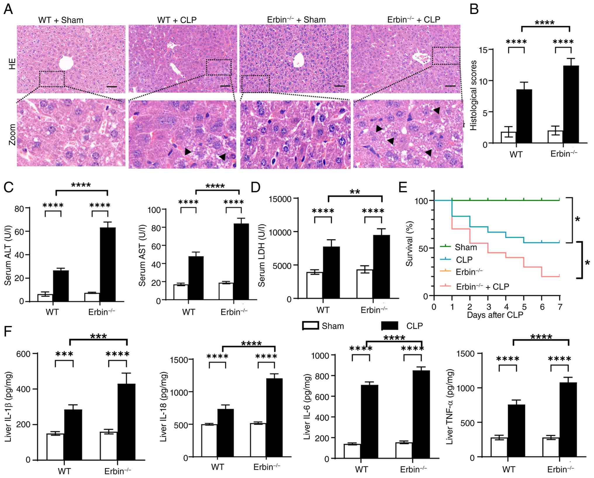 | Figure 1.Erbin deficiency aggravates
sepsis-induced liver injury. (A) Representative H&E-stained
liver sections from WT and Erbin-/-mice subjected to sham or CLP
surgery. Black arrowheads indicate swollen hepatocytes. Scale bar,
50 µm. (B) Liver injury scores (n=5). (C and D) Serum AST, ALT and
LDH levels (n=6). (E) 7-day survival after CLP (WT+Sham, n=10;
WT+CLP, n=18; Erbin−/−+Sham, n=10;
Erbin−/−+CLP, n=20). (F) TNF-α, IL-6, IL-1β and IL-18
levels in liver homogenate supernatants (n=4). Data are presented
as the mean ± SD. **P<0.01, ***P<0.001 and ****P<0.001.
WT, wild type; ALT, alanine aminotransferase; AST, aspartate
aminotransferase; LDH, lactate dehydrogenase; CLP, cecal ligation
and puncture. |
Erbin deficiency enhances NLRP3
inflammasome activation during sepsis-induced liver injury
To determine whether Erbin regulates
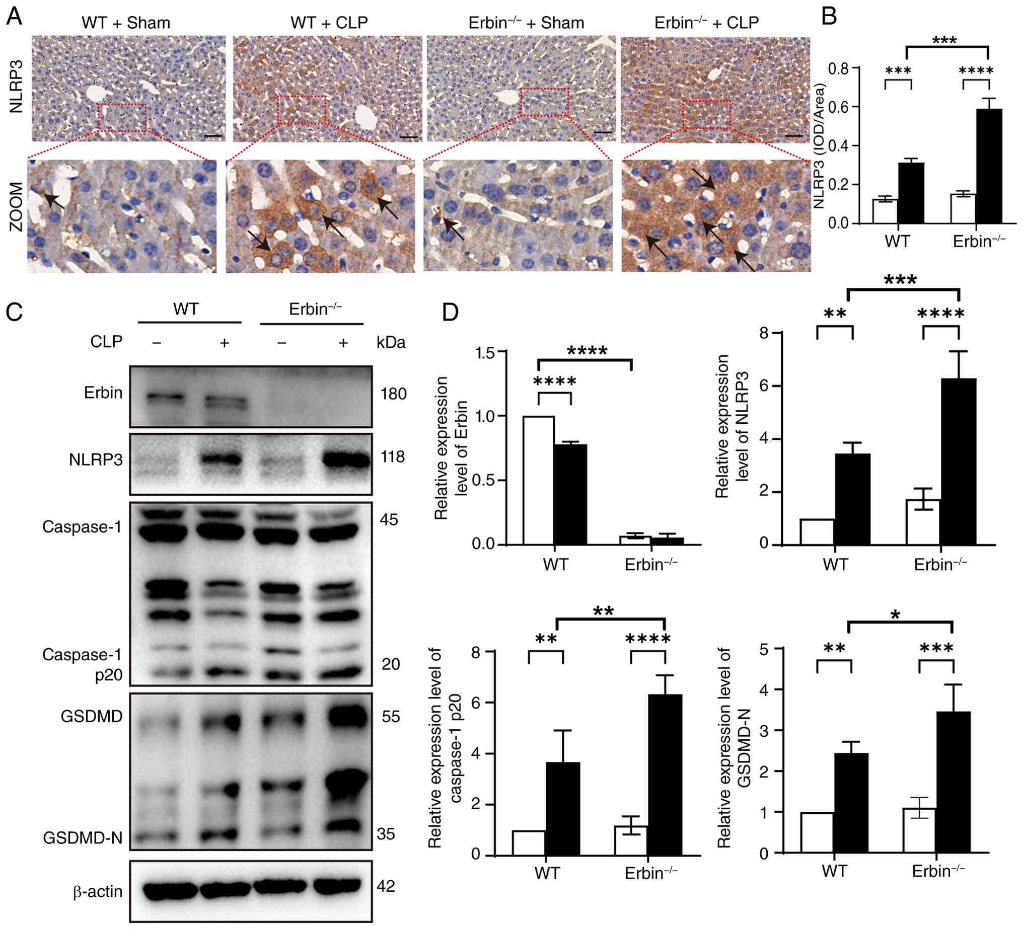
pyroptosis-associated signaling during SLI, NLRP3, caspase-1 p20
and GSDMD-N expression was examined by western blotting. CLP
markedly activated the NLRP3 inflammasome pathway, and this
activation was further enhanced in Erbin−/− mice
(Fig. 2C and D). Consistently,
immunohistochemical analysis showed increased hepatic NLRP3
expression after CLP, with a greater increase in Erbin-deficient
mice (Fig. 2A and B). These
findings indicate that Erbin deficiency promotes NLRP3 inflammasome
activation during SLI.
Erbin deficiency enhances pyroptosis
and inflammation in BMDMs
Macrophage-hepatocyte crosstalk is increasingly
recognized as a notable contributor to SLI progression (42). To investigate the
macrophage-intrinsic role of Erbin, BMDMs were stimulated with LPS
and ATP to activate the NLRP3 inflammasome and induce pyroptosis
(43,44). Erbin deficiency markedly enhanced
LPS/ATP-induced expression of NLRP3, caspase-1 p20, and GSDMD-N
(Fig. 3A and B). CCK-8 assays, LDH
release measurements and Calcein-AM/PI staining further showed
reduced cell viability and increased cell death in Erbin-deficient
BMDMs after LPS/ATP stimulation (Fig.
3C-F). In addition, LPS/ATP increased IL-6, IL-18, TNF-α and
IL-1β mRNA expression, and these increases were further amplified
by Erbin deficiency (Fig. 3G).
Collectively, these findings indicate that Erbin negatively
regulates pyroptotic and inflammatory responses in macrophages.
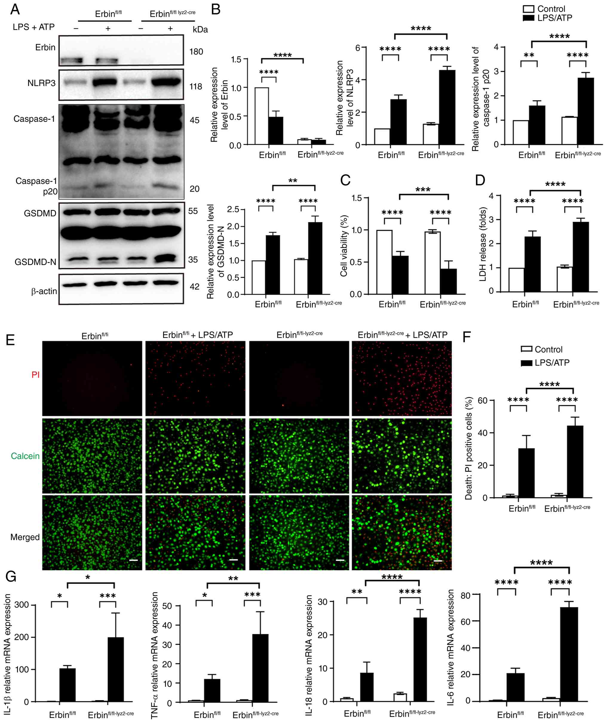 | Figure 3.Erbin deficiency enhances pyroptosis
and inflammation in BMDMs. (A and B) Western blot analysis and
quantification of Erbin, NLRP3, caspase-1 and GSDMD in BMDMs (n=3).
(C) Cell viability measured using the cell counting kit-8 assay
(n=6). (D) Relative LDH release (n=5). (E and F) Representative
Calcein-AM/PI staining and quantification of cell death (n=5).
Calcein-AM and PI indicate live and dead cells, respectively. Scale
bar, 100 µm. (G) Relative mRNA levels of IL-6, IL-18, TNF-α and
IL-1β measured by RT-qPCR (n=3). Data are presented as the mean ±
SD. *P<0.05, **P<0.01, ***P<0.001 and ****P<0.001.
BMDMs, bone marrow-derived macrophages; LDH, lactate
dehydrogenase. |
Erbin deficiency aggravates oxidative
stress in vivo and in vitro
To evaluate the role of Erbin in mitochondrial
injury and oxidative stress during SLI, mitochondrial
ultrastructure and ROS production were examined in vivo and
in vitro. Transmission electron microscopy revealed that
CLP-induced mitochondrial abnormalities, including swelling, loss
of cristae and matrix clearing, were more severe in
Erbin−/− mice than in WT mice (Fig. 4A and C). This mitochondrial damage
was accompanied by increased hepatic ROS production, as indicated
by DHE staining (Fig. 4B and D).
Biochemical assays further showed higher MDA levels and lower SOD
activity in Erbin−/−+CLP mice than in WT+CLP mice
(Fig. 4E and F). Consistent
results were obtained in vitro, where Erbin-deficient BMDMs
exhibited greater intracellular ROS accumulation after LPS/ATP
stimulation (Fig. 4G and H),
together with higher MDA levels and lower SOD activity (Fig. 4I and J).
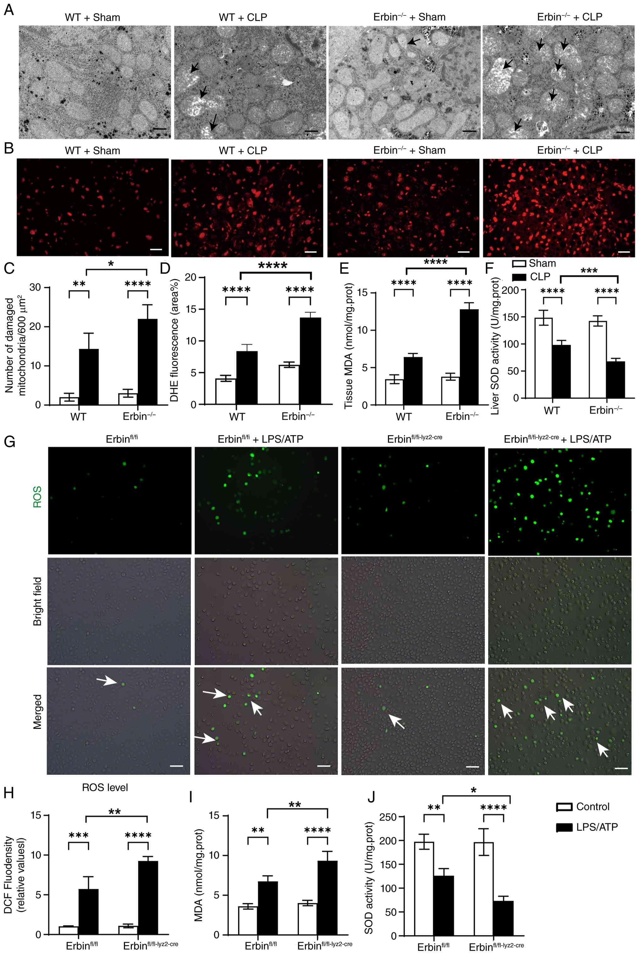 | Figure 4.Erbin deficiency aggravates oxidative
injury in vivo and in vitro. (A and C) Representative
transmission electron microscopy images of liver mitochondria and
quantification of damaged mitochondria (n=3). Arrows indicate
damaged mitochondria. Scale bar, 1 µm. (B and D) Representative DHE
staining of liver sections and fluorescence quantification (n=5).
Scale bar, 50 µm. (E) Hepatic MDA levels and (F) SOD activity
(n=5). (G and H) Representative DCFH-DA staining of BMDMs and
fluorescence quantification (n=5). White arrows indicate
ROS-positive cells. Scale bar, 50 µm. (I and J) MDA levels and SOD
activity in BMDMs (n=5). Data are presented as the mean ± SD.
*P<0.05, **P<0.01, ***P<0.001 and ****P<0.001. MDA,
malondialdehyde; SOD, superoxide dismutase; WT, wild type; CLP,
cecal ligation and puncture; ROS, reactive oxygen species; BMDMs,
bone marrow-derived macrophages; LPS, lipopolysaccharide. |
Erbin deficiency promotes TXNIP/NLRP3
pathway activation in vivo and in vitro
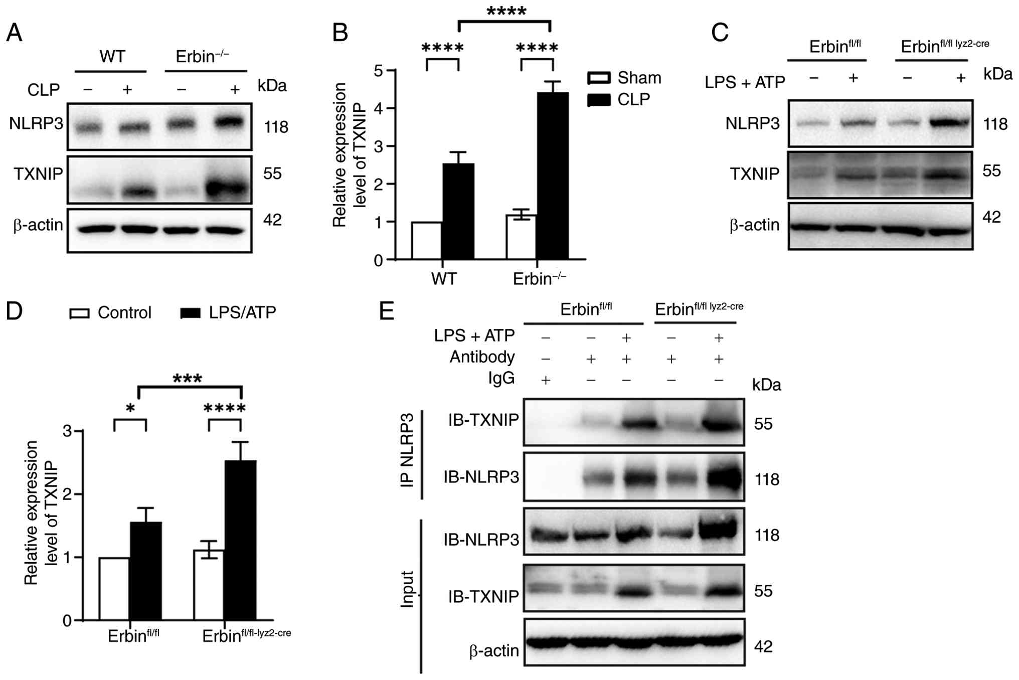
Excessive ROS promotes the interaction of TXNIP with
NLRP3 and thereby activates the inflammasome (45). Because Erbin deficiency exacerbated
sepsis-induced oxidative stress, TXNIP expression and its
interaction with NLRP3 were examined. TXNIP and NLRP3 protein
levels were increased after CLP and were further elevated in
Erbin−/− mice (Fig. 5A and
B). Similarly, LPS/ATP increased TXNIP and NLRP3 expression in
BMDMs, with greater increases observed in Erbin-deficient cells
(Fig. 5C and D).
Co-immunoprecipitation further showed that LPS/ATP enhanced the
TXNIP-NLRP3 interaction and that Erbin deficiency further
strengthened this association (Fig.
5E).
Erbin regulates pyroptosis through the
mitochondrial ROS/TXNIP axis
To determine whether mtROS mediates the enhanced
pyroptotic response associated with Erbin deficiency, MitoQ, a
mitochondria-targeted antioxidant, was used to reduce mtROS.
LPS/ATP stimulation increased mtROS levels in BMDMs, and this
increase was further amplified by Erbin deficiency; MitoQ
pretreatment markedly reduced mtROS accumulation in Erbin-deficient
BMDMs (Fig. 6A and B).
Mitochondrial membrane potential was subsequently assessed using
JC-1 staining. LPS/ATP induced mitochondrial depolarization, which
was exacerbated by Erbin deficiency, whereas MitoQ partially
restored mitochondrial membrane potential in Erbin-deficient BMDMs
(Fig. 6C and D). MitoQ did not
alter Erbin expression (Fig. 6E and
G), indicating that it acted downstream of Erbin. However,
MitoQ partially reduced TXNIP, NLRP3 and GSDMD-N expression in
Erbin-deficient BMDMs after LPS/ATP stimulation (Fig. 6F and H). Consistently,
Calcein-AM/PI staining and cytokine assays showed that MitoQ
attenuated LPS/ATP-induced cell death and inflammatory cytokine
production (IL-1β, TNF-α, and IL-18) (Fig. 6I-K). Collectively, these results
support a role for mtROS in Erbin deficiency-associated TXNIP/NLRP3
inflammasome activation and pyroptosis.
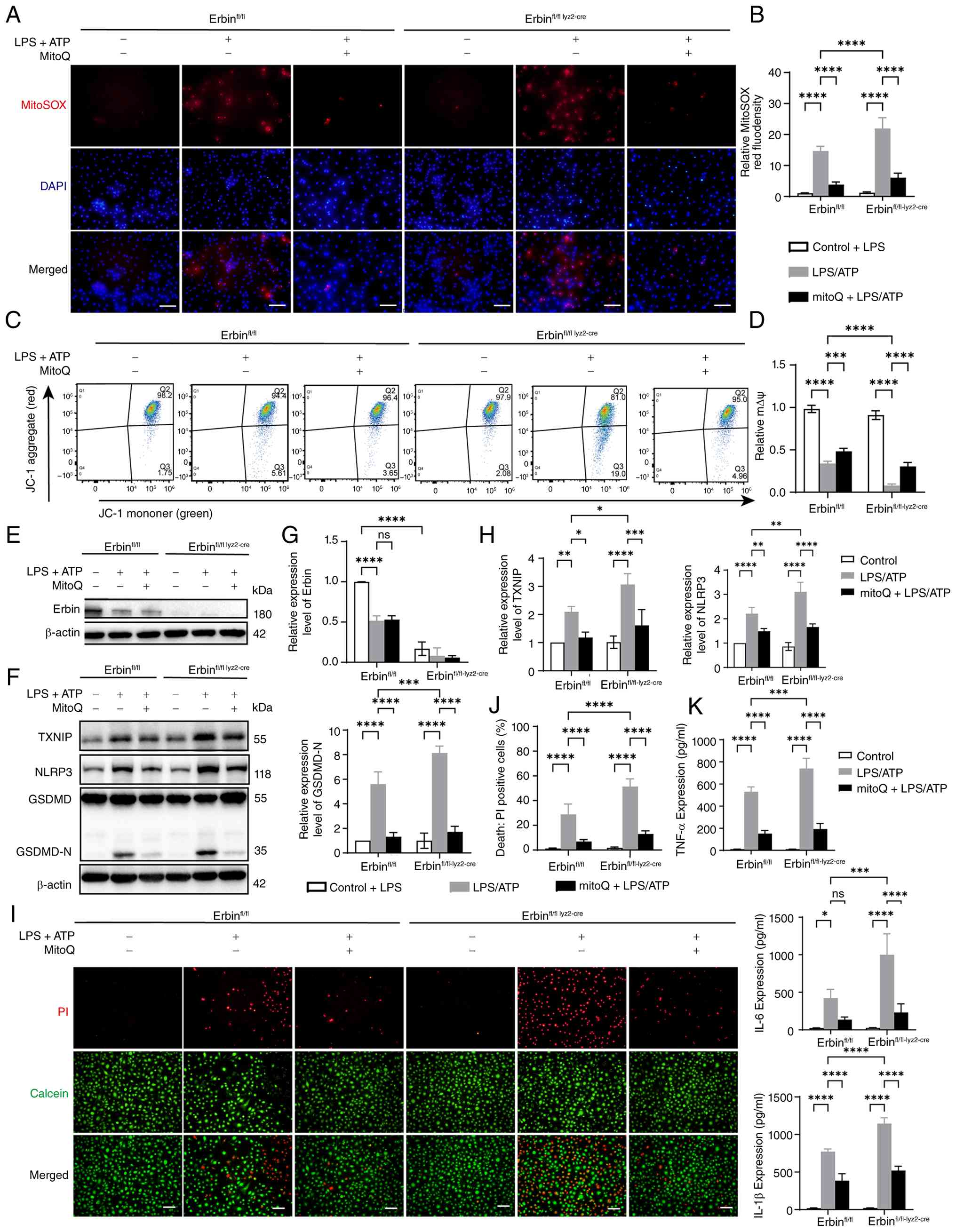 | Figure 6.Erbin regulates pyroptosis through
the mitochondrial ROS/TXNIP axis. (A and B) Representative MitoSOX
Red staining and quantification of mitochondrial reactive oxygen
species in BMDMs (n=4). Scale bar, 50 µm. (C) Representative flow
cytometry plots of JC-1 staining. (D) Quantification of the JC-1
red/green fluorescence ratio (n=4). (E and G) Western blot analysis
and quantification of Erbin in BMDMs (n=3). (F and H) Western blot
analysis and quantification of TXNIP, NLRP3 and GSDMD in BMDMs
(n=3). (I and J) Representative Calcein-AM/PI staining and
quantification of cell death (n=5). Calcein-AM and PI indicate live
and dead cells, respectively. Scale bar, 100 µm. (K) TNF-α, IL-6
and IL-1β levels in BMDM culture supernatants (n=4). Data are
presented as the mean ± SD. *P<0.05, **P<0.01, ***P<0.001
and ****P<0.001. BMDMs, bone marrow-derived macrophages; LPS,
lipopolysaccharide; ns, not significant. |
Clinical relevance of Erbin expression
in human sepsis
To further examine the clinical relevance of Erbin,
three independent GEO datasets containing samples from patients
with sepsis and healthy controls were analyzed. Erbin expression
was significantly decreased in patients with sepsis in GSE294059,
whereas no significant differences were observed in GSE228541 or
GSE28750 (Fig. S2A, C and E).
Next, the correlation between Erbin and NLRP3 expression in sepsis
samples was examined. No significant correlation was detected in
GSE294059 (Pearson r=0.499, P=0.392; Spearman ρ=0.400, P=0.505),
GSE228541 (Spearman ρ=0.332, P=0.246) or GSE28750 (Pearson
r=−0.431, P=0.213; Spearman ρ=−0.358, P=0.310) (Fig. S2B, D and F). These data indicate
that Erbin expression is altered in at least one human sepsis
cohort, although this change and its association with NLRP3
expression were not consistent across datasets.
Discussion
Sepsis is a life-threatening syndrome characterized
by systemic inflammation and organ dysfunction. Liver dysfunction
is notably associated with poor outcomes in patients with sepsis
(46), yet effective therapeutic
strategies remain limited. In the present study, a murine CLP model
was used to investigate the role of Erbin in SLI (19). Our previous work identified Erbin
as a regulator of pyroptosis in sepsis-induced acute kidney injury
and LPS-stimulated HK-2 cells, although the underlying mechanism
remained unclear (47). Extending
these observations, the present findings demonstrate that Erbin
deficiency aggravates CLP-induced liver injury and enhances NLRP3
inflammasome activation. Notably, the present study extends
previous observations by examining the contribution of Erbin to
macrophage inflammatory responses during SLI. Specifically, the
present findings show that Erbin deficiency aggravates
sepsis-induced liver injury and enhances mitochondrial
ROS/NLRP3-associated inflammatory responses, while the BMDM
experiments further support a macrophage-intrinsic contribution to
this process.
During sepsis, pattern-recognition receptors detect
pathogen-associated and damage-associated molecular patterns,
promoting NLRP3 inflammasome assembly. Activated NLRP3 recruits ASC
and pro-caspase-1, leading to caspase-1 activation and cleavage of
pro-IL-1β, pro-IL-18 and GSDMD, thereby inducing cytokine release
and pyroptosis (48). NLRP3
inflammasome activation has a notable role in SLI. BATF-mediated
transcriptional activation of GBP5 aggravates SLI by promoting
NLRP3 inflammasome activation (49), whereas maresin 1 alleviates SLI by
suppressing NLRP3-mediated macrophage pyroptosis and inflammation
(50). Consistent with these
studies, CLP activated hepatic pyroptosis in the present study, and
Erbin deficiency further enhanced inflammasome activation and
inflammatory responses. Excessive pro-inflammatory cytokine
production by activated hepatic macrophages, including Kupffer
cells and infiltrating monocyte-derived macrophages, contributes to
sepsis-induced hepatic injury (51). Therefore, BMDMs were used in the
present study as an in vitro model to investigate the
macrophage-intrinsic role of Erbin under inflammatory stimulation.
The in vitro studies with BMDMs consistently showed that
Erbin depletion enhanced LPS/ATP-induced inflammation and
pyroptosis activation. These findings support a negative regulatory
role of Erbin in macrophage pyroptotic responses.
Oxidative stress is a major contributor to
sepsis-induced organ dysfunction (52,53).
Accumulated ROS can activate the NLRP3 inflammasome and thereby
amplify inflammatory injury (54).
Erbin, an endogenous inhibitor of RAS-RAF signaling, has previously
been implicated in ROS regulation (23). Consistent with the present study,
Erbin deficiency increased ROS production and promoted TXNIP/NLRP3
inflammasome activation in both CLP-challenged mice and
LPS/ATP-stimulated BMDMs. The ROS/TXNIP/NLRP3 axis is an
established mechanism of inflammatory injury, and inhibition of ROS
accumulation can suppress this pathway activation and reduce tissue
damage (55,56). In line with this concept, loss of
Erbin disrupted redox homeostasis and enhanced TXNIP/NLRP3
inflammasome activation and pyroptotic responses. Furthermore,
Co-IP analysis showed that Erbin deficiency increased the
interaction between TXNIP and NLRP3, a key event that promotes
caspase-1 activation and IL-1β maturation (57).
To further examine the causal contribution of mtROS,
BMDMs were treated with MitoQ, a mitochondria-targeted antioxidant
previously shown to suppress mtROS accumulation and NLRP3
inflammasome activation (58).
MitoQ reduced mtROS accumulation and partially restored
mitochondrial membrane potential in LPS/ATP-stimulated
Erbin-deficient BMDMs. These changes were accompanied by reduced
TXNIP expression and decreased NLRP3 and GSDMD-N levels. Because
TXNIP links oxidative stress to NLRP3 inflammasome activation
(56,59), these findings support mtROS as an
upstream mediator of TXNIP/NLRP3 signaling in Erbin-deficient
macrophages. MitoQ also attenuated cell death and inflammatory
cytokine production, consistent with its reported anti-inflammatory
effects (60). Notably, MitoQ did
not alter Erbin expression, indicating that its protective effects
occurred downstream of Erbin; however, the absence of a MitoQ-only
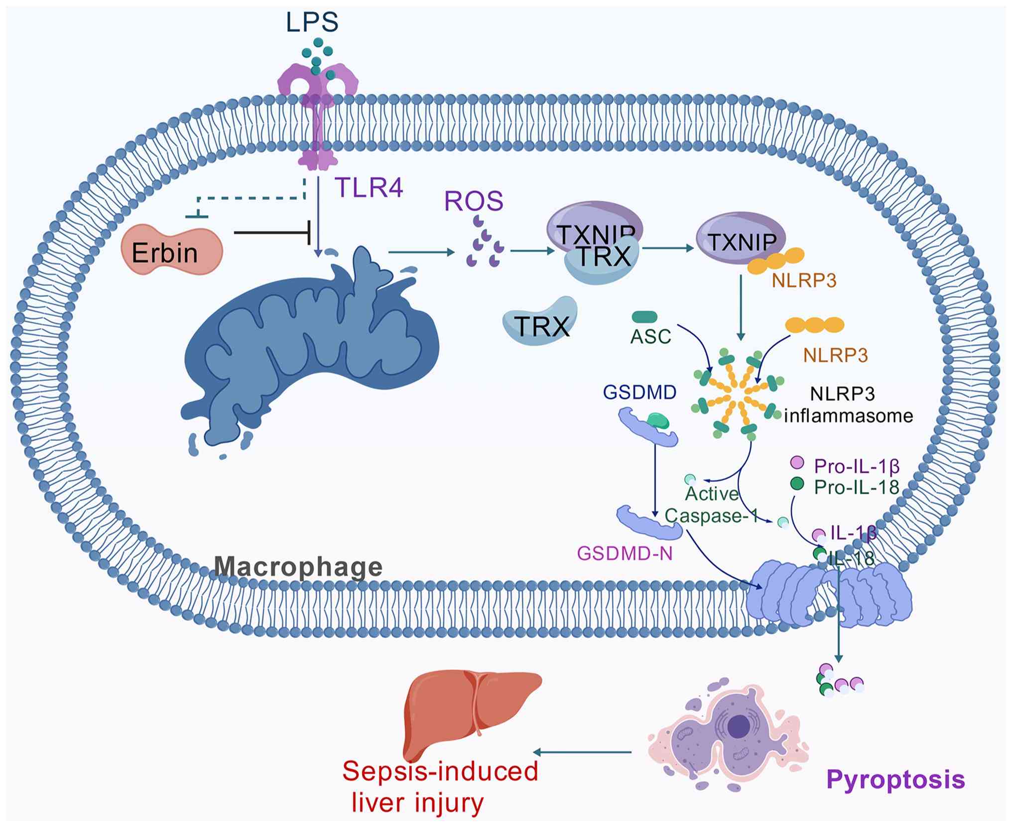
control group limits assessment of its basal effects. Collectively,
the present findings support a model in which Erbin deficiency
aggravates SLI, at least in part, by enhancing mitochondrial
ROS/TXNIP/NLRP3 signaling, while MitoQ attenuates this inflammatory
response (Fig. 7).
Analysis of three independent human sepsis datasets
showed significantly reduced Erbin expression in GSE294059, whereas
no significant difference was observed in GSE228541 or GSE28750. In
addition, Erbin expression was not significantly correlated with
NLRP3 expression in the analyzed sepsis samples. The variability
among clinical cohorts may be related to differences in disease
stage, patient characteristics, treatment and sample composition.
Moreover, NLRP3 inflammasome activation is regulated at both
transcriptional and post-transcriptional levels and therefore may
not be directly reflected by the correlation between Erbin and
NLRP3 mRNA abundance. Further validation in well-characterized
clinical cohorts, particularly measurements of circulating Erbin
together with IL-1β and IL-18, will be necessary to determine the
clinical relevance of this pathway.
Several limitations should be considered. First, the
use of global Erbin-knockout mice in vivo and BMDMs derived
from macrophage-specific conditional-knockout mice in vitro
limits cell type-specific interpretation. Although the in
vivo findings demonstrate that Erbin deficiency aggravates SLI,
the relative contributions of hepatocytes, Kupffer cells, recruited
macrophages and other hepatic cell populations cannot be
distinguished. Furthermore, BMDMs do not fully reproduce the
heterogeneity and tissue-specific functions of hepatic macrophages,
particularly resident Kupffer cells. Cell type-specific
Erbin-knockout models and primary hepatic macrophages will
therefore be required to define the contributions of individual
hepatic cell populations. Second, although the survival difference
was statistically significant, the relatively small sample size
warrants validation in larger cohorts. Third, analysis of three
independent human sepsis datasets showed significantly decreased
Erbin expression in GSE294059, whereas no significant difference
was observed in GSE228541 or GSE28750. In addition, Erbin
expression was not significantly correlated with NLRP3 expression
in the analyzed sepsis samples. These findings suggest that
alterations in Erbin expression may occur in human sepsis, but they
are not consistent across different clinical cohorts. The present
study also lacks direct validation using clinical samples. Serum
Erbin levels and their associations with inflammatory markers and
disease severity remain to be investigated in future clinical
cohorts.
In summary, the present findings support a
protective role of Erbin in experimental SLI, associated with
suppression of mitochondrial ROS accumulation and TXNIP/NLRP3
inflammasome activation. Further studies using cell type-specific
models and clinical samples are needed to establish the
translational relevance of Erbin and to determine whether it may
represent a therapeutic target for SLI.
Supplementary Material
Supporting Data
Acknowledgements
Not applicable.
Funding
The present study was supported by the National Natural Science
Foundation of China (grant nos. 82371370, 82172144 and
82372156).
Availability of data and materials
The data generated in the present study may be
requested from the corresponding author.
Authors' contributions
JZ wrote the original draft, conceived the study,
performed the animal and cell experiments, including establishment
of the CLP model, isolation and culture of BMDMs, and biochemical
and molecular experiments and organized the experimental data and
performed the statistical analyses. XWa reviewed and edited the
manuscript and participated in the design and implementation of the
animal, cell and molecular biology experiments. GJ supervised the
study and contributed to the study design and conceptualization.
XWu reviewed and edited the manuscript, supervised the study,
acquired funding and contributed to the conceptualization of the
study. XS wrote the original draft, participated in the study
design and acquired funding. JZ and XS confirm the authenticity of
all the raw data. All authors read and approved the final version
of the manuscript.
Ethics approval and consent to
participate
All animal experiments were approved by the Animal
Experimentation Committee of Wuhan University (Wuhan, China;
approval no. WQ20210298) and were conducted in accordance with the
ARRIVE guidelines.
Patient consent for publication
Not applicable.
Competing interests
The authors declare that they have no competing
interests.
Glossary
Abbreviations
Abbreviations:
|
Erbin
|
ErbB2 interacting protein
|
|
SLI
|
sepsis-induced liver injury
|
|
CLP
|
cecal ligation and puncture
|
|
LPS
|
lipopolysaccharide
|
|
ATP
|
adenosine triphosphate
|
|
BMDMs
|
bone marrow-derived macrophages
|
|
ROS
|
reactive oxygen species
|
|
mtROS
|
mitochondrial ROS
|
|
NLRP3
|
NOD-like receptor family pyrin domain
containing 3
|
|
TXNIP
|
thioredoxin-interacting protein
|
|
IL-1β
|
interleukin-1 β
|
|
IL-18
|
interleukin-18
|
|
IL-6
|
interleukin-6
|
|
TNF-α
|
tumor necrosis factor-alpha
|
|
GSDMD
|
gasdermin D
|
|
qPCR
|
quantitative PCR
|
|
WT
|
wild type
|
References
|
1
|
Selim SA, El-Baset SAA, Kattaia AAA, Askar
EM and Elkader EA: Bone marrow-derived mesenchymal stem cells
ameliorate liver injury in a rat model of sepsis by activating Nrf2
signaling. Histochem Cell Biol. 151:249–262. 2019. View Article : Google Scholar : PubMed/NCBI
|
|
2
|
Singer M, Deutschman CS, Seymour CW,
Shankar-Hari M, Annane D, Bauer M, Bellomo R, Bernard GR, Chiche
JD, Coopersmith CM, et al: The third international consensus
definitions for sepsis and septic shock (sepsis-3). JAMA.
315:801–810. 2016. View Article : Google Scholar : PubMed/NCBI
|
|
3
|
Yan J and Li S and Li S: The role of the
liver in sepsis. Int Rev Immunol. 33:498–510. 2014. View Article : Google Scholar : PubMed/NCBI
|
|
4
|
Kikuchi S, Piraino G, O'Connor M, Wolfe V,
Ridings K, Lahni P and Zingarelli B: Hepatocyte-specific deletion
of AMPKα1 results in worse outcomes in mice subjected to sepsis in
a sex-specific manner. Front Immunol. 11:2102020. View Article : Google Scholar : PubMed/NCBI
|
|
5
|
Kim TS and Choi DH: Liver dysfunction in
sepsis. Korean J Gastroenterol. 75:182–187. 2020. View Article : Google Scholar : PubMed/NCBI
|
|
6
|
Liang H, Song H, Zhang X, Song G, Wang Y,
Ding X, Duan X, Li L, Sun T and Kan Q: Metformin attenuated
sepsis-related liver injury by modulating gut microbiota. Emerg
Microbes Infect. 11:815–828. 2022. View Article : Google Scholar : PubMed/NCBI
|
|
7
|
Savio LEB, de Andrade Mello P, Figliuolo
VR, de Avelar Almeida TF, Santana PT, Oliveira SDS, Silva CLM,
Feldbrügge L, Csizmadia E, Minshall RD, et al: CD39 limits P2X7
receptor inflammatory signaling and attenuates sepsis-induced liver
injury. J Hepatol. 67:716–726. 2017. View Article : Google Scholar : PubMed/NCBI
|
|
8
|
Guo Y, Guo W, Chen H, Sun J and Yin Y:
Mechanisms of sepsis-induced acute liver injury: A comprehensive
review. Front Cell Infect Microbiol. 15:15042232025. View Article : Google Scholar : PubMed/NCBI
|
|
9
|
Heymann F, Hammerich L, Storch D, Bartneck
M, Huss S, Rüsseler V, Gassler N, Lira SA, Luedde T, Trautwein C
and Tacke F: Hepatic macrophage migration and differentiation
critical for liver fibrosis is mediated by the chemokine receptor
C-C motif chemokine receptor 8 in mice. Hepatology. 55:898–909.
2012. View Article : Google Scholar : PubMed/NCBI
|
|
10
|
Nakamoto N, Ebinuma H, Kanai T, Chu PS,
Ono Y, Mikami Y, Ojiro K, Lipp M, Love PE, Saito H and Hibi T:
CCR9+ macrophages are required for acute liver inflammation in
mouse models of hepatitis. Gastroenterology. 142:366–376. 2012.
View Article : Google Scholar : PubMed/NCBI
|
|
11
|
Xu W, Hou H, Yang W, Tang W and Sun L:
Immunologic role of macrophages in sepsis-induced acute liver
injury. Int Immunopharmacol. 143:1134922024. View Article : Google Scholar : PubMed/NCBI
|
|
12
|
Jia Y, Li Z, Feng Y, Cui R, Dong Y, Zhang
X, Xiang X, Qu K, Liu C and Zhang J: Methane-rich saline
ameliorates sepsis-induced acute kidney injury through
anti-inflammation, antioxidative, and antiapoptosis effects by
regulating endoplasmic reticulum stress. Oxid Med Cell Longev.
2018:47568462018. View Article : Google Scholar : PubMed/NCBI
|
|
13
|
Zou Z, Liu B, Zeng L, Yang X, Huang R, Wu
C, Zhu H, Gao Y, Yuan D and Yu J: Cx43 inhibition attenuates
sepsis-induced intestinal injury via downregulating ROS transfer
and the activation of the JNK1/Sirt1/FoxO3a signaling pathway.
Mediators Inflamm. 2019:78543892019. View Article : Google Scholar : PubMed/NCBI
|
|
14
|
Zhang X, Zhang JH, Chen XY, Hu QH, Wang
MX, Jin R, Zhang QY, Wang W, Wang R, Kang LL, et al: Reactive
oxygen species-induced TXNIP drives fructose-mediated hepatic
inflammation and lipid accumulation through NLRP3 inflammasome
activation. Antioxid Redox Signal. 22:848–870. 2015. View Article : Google Scholar : PubMed/NCBI
|
|
15
|
Kovacs SB and Miao EA: Gasdermins:
Effectors of pyroptosis. Trends Cell Biol. 27:673–684. 2017.
View Article : Google Scholar : PubMed/NCBI
|
|
16
|
Coll RC, Schroder K and Pelegrín P: NLRP3
and pyroptosis blockers for treating inflammatory diseases. Trends
Pharmacol Sci. 43:653–668. 2022. View Article : Google Scholar : PubMed/NCBI
|
|
17
|
Vasudevan SO, Behl B and Rathinam VA:
Pyroptosis-induced inflammation and tissue damage. Semin Immunol.
69:1017812023. View Article : Google Scholar : PubMed/NCBI
|
|
18
|
He H, Jiang H, Chen Y, Ye J, Wang A, Wang
C, Liu Q, Liang G, Deng X, Jiang W and Zhou R: Oridonin is a
covalent NLRP3 inhibitor with strong anti-inflammasome activity.
Nat Commun. 9:25502018. View Article : Google Scholar : PubMed/NCBI
|
|
19
|
Chen YL, Xu G, Liang X, Wei J, Luo J, Chen
GN, Yan XD, Wen XP, Zhong M and Lv X: Inhibition of hepatic cells
pyroptosis attenuates CLP-induced acute liver injury. Am J Transl
Res. 8:5685–5695. 2016.PubMed/NCBI
|
|
20
|
Birrane G, Chung J and Ladias JAA: Novel
mode of ligand recognition by the Erbin PDZ domain. J Biol Chem.
278:1399–1402. 2003. View Article : Google Scholar : PubMed/NCBI
|
|
21
|
McDonald C, Chen FF, Ollendorff V, Ogura
Y, Marchetto S, Lécine P, Borg JP and Nuñez G: A role for Erbin in
the regulation of Nod2-dependent NF-kappaB signaling. J Biol Chem.
280:40301–40309. 2005. View Article : Google Scholar : PubMed/NCBI
|
|
22
|
Shen T, Li S, Cai LD, Liu JL, Wang CY, Gan
WJ, Li XM, Wang JR, Sun LN, Deng M, et al: Erbin exerts a
protective effect against inflammatory bowel disease by suppressing
autophagic cell death. Oncotarget. 9:12035–12049. 2018. View Article : Google Scholar : PubMed/NCBI
|
|
23
|
Xie CM, Wei D, Zhao L, Marchetto S, Mei L,
Borg JP and Sun Y: Erbin is a novel substrate of the Sag-βTrCP E3
ligase that regulates KrasG12D-induced skin tumorigenesis. J Cell
Biol. 209:721–737. 2015. View Article : Google Scholar : PubMed/NCBI
|
|
24
|
Abais JM, Xia M, Zhang Y, Boini KM and Li
PL: Redox regulation of NLRP3 inflammasomes: ROS as trigger or
effector? Antioxid Redox Signal. 22:1111–1129. 2015. View Article : Google Scholar : PubMed/NCBI
|
|
25
|
Hawkins P, Morton DB, Burman O, Dennison
N, Honess P, Jennings M, Lane S, Middleton V, Roughan JV, Wells S,
et al: A guide to defining and implementing protocols for the
welfare assessment of laboratory animals: Eleventh report of the
BVAAWF/FRAME/RSPCA/UFAW Joint working group on refinement. Lab
Anim. 45:1–13. 2011. View Article : Google Scholar : PubMed/NCBI
|
|
26
|
Percie du Sert N, Hurst V, Ahluwalia A,
Alam S, Avey MT, Baker M, Browne WJ, Clark A, Cuthill IC, Dirnagl
U, et al: The ARRIVE guidelines 2.0: Updated guidelines for
reporting animal research. PLoS Biol. 18:e30004102020. View Article : Google Scholar : PubMed/NCBI
|
|
27
|
Rittirsch D, Huber-Lang MS, Flierl MA and
Ward PA: Immunodesign of experimental sepsis by cecal ligation and
puncture. Nat Protoc. 4:31–36. 2009. View Article : Google Scholar : PubMed/NCBI
|
|
28
|
Bi J, Zhang J, Ren Y, Du Z, Li Q, Wang Y,
Wei S, Yang L, Zhang J, Liu C, et al: Irisin alleviates liver
ischemia-reperfusion injury by inhibiting excessive mitochondrial
fission, promoting mitochondrial biogenesis and decreasing
oxidative stress. Redox Biol. 20:296–306. 2019. View Article : Google Scholar : PubMed/NCBI
|
|
29
|
Tsuji N, Tsuji T, Yamashita T, Hayase N,
Hu X, Yuen PS and Star RA: BAM15 treats mouse sepsis and kidney
injury, linking mortality, mitochondrial DNA, tubule damage, and
neutrophils. J Clin Invest. 133:e1524012023. View Article : Google Scholar : PubMed/NCBI
|
|
30
|
Yin XY, Tang XH, Wang SX, Zhao YC, Jia M,
Yang JJ, Ji MH and Shen JC: HMGB1 mediates synaptic loss and
cognitive impairment in an animal model of sepsis-associated
encephalopathy. J Neuroinflammation. 20:692023. View Article : Google Scholar : PubMed/NCBI
|
|
31
|
Cho HI, Kim SJ, Choi JW and Lee SM:
Genipin alleviates sepsis-induced liver injury by restoring
autophagy. Br J Pharmacol. 173:980–991. 2016. View Article : Google Scholar : PubMed/NCBI
|
|
32
|
Zhu J, Jin Z, Wang J, Wu Z, Xu T, Tong G,
Shen E, Fan J, Jiang C, Wang J, et al: FGF21 ameliorates septic
liver injury by restraining proinflammatory macrophages activation
through the autophagy/HIF-1α axis. J Adv Res. 69:477–494. 2025.
View Article : Google Scholar : PubMed/NCBI
|
|
33
|
Zhan Y, Xu D, Tian Y, Qu X, Sheng M, Lin
Y, Ke M, Jiang L, Xia Q, Kaldas FM, et al: Novel role of macrophage
TXNIP-mediated CYLD-NRF2-OASL1 axis in stress-induced liver
inflammation and cell death. JHEP Rep. 4:1005322022. View Article : Google Scholar : PubMed/NCBI
|
|
34
|
Wang Z, Chen X, Li J, Chen M, Zhu H, Li X,
Yu W, Xia N, Zhang Y, Sun L, et al: Macrophages Atp6v0d2 regulates
XBP1-mediated cholesterol metabolism to suppress metabolic
dysfunction-associated steatohepatitis progression. Int
Immunopharmacol. 161:1150882025. View Article : Google Scholar : PubMed/NCBI
|
|
35
|
Chiu LY, Huang DY and Lin WW: PARP-1
regulates inflammasome activity by poly-ADP-ribosylation of NLRP3
and interaction with TXNIP in primary macrophages. Cell Mol Life
Sci. 79:1082022. View Article : Google Scholar : PubMed/NCBI
|
|
36
|
Feng Y, Cui R, Li Z, Zhang X, Jia Y, Zhang
X, Shi J, Qu K, Liu C and Zhang J: Methane alleviates
acetaminophen-induced liver injury by inhibiting inflammation,
oxidative stress, endoplasmic reticulum stress, and apoptosis
through the Nrf2/HO-1/NQO1 signaling pathway. Oxid Med Cell Longev.
2019:70676192019. View Article : Google Scholar : PubMed/NCBI
|
|
37
|
Liao J, Cao Y, Zhao J, Yu B, Wang Y, Li W,
Li H, Lv S, Wen W, Cui H and Chen Y: Aqueous extract of Polygala
japonica Houtt. ameliorated nonalcoholic steatohepatitis in mice
through restoring the gut microbiota disorders and affecting the
metabolites in feces and liver. Phytomedicine. 118:1549372023.
View Article : Google Scholar : PubMed/NCBI
|
|
38
|
Livak KJ and Schmittgen TD: Analysis of
relative gene expression data using real-time quantitative PCR and
the 2(−Delta Delta C(T)) method. Methods. 25:402–408. 2001.
View Article : Google Scholar : PubMed/NCBI
|
|
39
|
Luo W, Xu W, Yin Q, Song J, Wang J, Guan
Y, Huang J, Yu J, Zou K, Jin D, et al: DPEP2 suppresses
hyperinflammation via metabolic reprogramming of macrophages in
sepsis. Nat Commun. 17:37102026. View Article : Google Scholar : PubMed/NCBI
|
|
40
|
Oda S, Matsumoto H, Togami Y, Yoshimura J,
Ito H, Onishi S, Muratsu A, Mitsuyama Y, Okuzaki D, Ogura H, et al:
mRNA-miRNA integration analysis of T-cell exhaustion in sepsis from
community-acquired pneumonia. Acute Med Surg. 12:e700542025.
View Article : Google Scholar : PubMed/NCBI
|
|
41
|
Sutherland A, Thomas M, Brandon RA,
Brandon RB, Lipman J, Tang B, McLean A, Pascoe R, Price G, Nguyen
T, et al: Development and validation of a novel molecular biomarker
diagnostic test for the early detection of sepsis. Crit Care.
15:R1492011. View Article : Google Scholar : PubMed/NCBI
|
|
42
|
Possamai LA, Thursz MR, Wendon JA and
Antoniades CG: Modulation of monocyte/macrophage function: A
therapeutic strategy in the treatment of acute liver failure. J
Hepatol. 61:439–445. 2014. View Article : Google Scholar : PubMed/NCBI
|
|
43
|
Biasizzo M, Trstenjak-Prebanda M, Dolinar
K, Pirkmajer S, Završnik J, Turk B and Kopitar-Jerala N: Cystatin C
deficiency increases LPS-induced sepsis and NLRP3 inflammasome
activation in mice. Cells. 10:20712021. View Article : Google Scholar : PubMed/NCBI
|
|
44
|
Liu YG, Chen JK, Zhang ZT, Ma XJ, Chen YC,
Du XM, Liu H, Zong Y and Lu GC: NLRP3 inflammasome activation
mediates radiation-induced pyroptosis in bone marrow-derived
macrophages. Cell Death Dis. 8:e25792017. View Article : Google Scholar : PubMed/NCBI
|
|
45
|
Tschopp J and Schroder K: NLRP3
inflammasome activation: The convergence of multiple signalling
pathways on ROS production? Nat Rev Immunol. 10:210–215. 2010.
View Article : Google Scholar : PubMed/NCBI
|
|
46
|
Piano S, Bartoletti M, Tonon M,
Baldassarre M, Chies G, Romano A, Viale P, Vettore E, Domenicali M,
Stanco M, et al: Assessment of Sepsis-3 criteria and quick SOFA in
patients with cirrhosis and bacterial infections. Gut.
67:1892–1899. 2018. View Article : Google Scholar : PubMed/NCBI
|
|
47
|
Liu Y, Fang Q, Ming T, Zuo J, Jing G and
Song X: Knockout of Erbin promotes pyroptosis via regulating
NLRP3/caspase-1/Gasdermin D pathway in sepsis-induced acute kidney
injury. Clin Exp Nephrol. 27:781–790. 2023. View Article : Google Scholar : PubMed/NCBI
|
|
48
|
He W, Dong H, Wu C, Zhong Y and Li J: The
role of NLRP3 inflammasome in sepsis: A potential therapeutic
target. Int Immunopharmacol. 115:1096972023. View Article : Google Scholar : PubMed/NCBI
|
|
49
|
Guo H, Ni M, Xu J, Chen F, Yao Z, Yao Y,
Liu C and Du Q: Transcriptional enhancement of GBP-5 by BATF
aggravates sepsis-associated liver injury via NLRP3 inflammasome
activation. FASEB J. 35:e216722021. View Article : Google Scholar : PubMed/NCBI
|
|
50
|
Yang W, Tao K, Zhang P, Chen X, Sun X and
Li R: Maresin 1 protects against
lipopolysaccharide/d-galactosamine-induced acute liver injury by
inhibiting macrophage pyroptosis and inflammatory response. Biochem
Pharmacol. 195:1148632022. View Article : Google Scholar : PubMed/NCBI
|
|
51
|
Gong S, Yan Z, Liu Z, Niu M, Fang H, Li N,
Huang C, Li L, Chen G, Luo H, et al: Intestinal microbiota mediates
the susceptibility to polymicrobial sepsis-induced liver injury by
granisetron generation in mice. Hepatology. 69:1751–1767. 2019.
View Article : Google Scholar : PubMed/NCBI
|
|
52
|
Li Z, Liu T, Feng Y, Tong Y, Jia Y, Wang
C, Cui R, Qu K, Liu C and Zhang J: PPARγ alleviates sepsis-induced
liver injury by inhibiting hepatocyte pyroptosis via inhibition of
the ROS/TXNIP/NLRP3 signaling pathway. Oxid Med Cell Longev.
2022:12697472022. View Article : Google Scholar : PubMed/NCBI
|
|
53
|
Galley HF: Oxidative stress and
mitochondrial dysfunction in sepsis. Br J Anaesth. 107:57–64. 2011.
View Article : Google Scholar : PubMed/NCBI
|
|
54
|
Latz E, Xiao TS and Stutz A: Activation
and regulation of the inflammasomes. Nat Rev Immunol. 13:397–411.
2013. View Article : Google Scholar : PubMed/NCBI
|
|
55
|
Liu T, Wang W, Liu M, Ma Y, Mu F, Feng X,
Zhang Y, Guo C, Ding Y and Wen A: Z-Guggulsterone alleviated
oxidative stress and inflammation through inhibiting the
TXNIP/NLRP3 axis in ischemic stroke. Int Immunopharmacol.
89:1070942020. View Article : Google Scholar : PubMed/NCBI
|
|
56
|
Han Y, Xu X, Tang C, Gao P, Chen X, Xiong
X, Yang M, Yang S, Zhu X, Yuan S, et al: Reactive oxygen species
promote tubular injury in diabetic nephropathy: The role of the
mitochondrial ros-txnip-nlrp3 biological axis. Redox Biol.
16:32–46. 2018. View Article : Google Scholar : PubMed/NCBI
|
|
57
|
Chen D, Dixon BJ, Doycheva DM, Li B, Zhang
Y, Hu Q, He Y, Guo Z, Nowrangi D, Flores J, et al: IRE1α inhibition
decreased TXNIP/NLRP3 inflammasome activation through miR-17-5p
after neonatal hypoxic-ischemic brain injury in rats. J
Neuroinflammation. 15:322018. View Article : Google Scholar : PubMed/NCBI
|
|
58
|
Chen W, Guo C, Huang S, Jia Z, Wang J,
Zhong J, Ge H, Yuan J, Chen T, Liu X, et al: MitoQ attenuates brain
damage by polarizing microglia towards the M2 phenotype through
inhibition of the NLRP3 inflammasome after ICH. Pharmacol Res.
161:1051222020. View Article : Google Scholar : PubMed/NCBI
|
|
59
|
Zhou R, Tardivel A, Thorens B, Choi I and
Tschopp J: Thioredoxin-interacting protein links oxidative stress
to inflammasome activation. Nat Immunol. 11:136–140. 2010.
View Article : Google Scholar : PubMed/NCBI
|
|
60
|
Cen M, Ouyang W, Zhang W, Yang L, Lin X,
Dai M, Hu H, Tang H, Liu H, Xia J and Xu F: MitoQ protects against
hyperpermeability of endothelium barrier in acute lung injury via a
Nrf2-dependent mechanism. Redox Biol. 41:1019362021. View Article : Google Scholar : PubMed/NCBI
|