Introduction
Corneal blindness is a major contributor to global
vision loss (1). Among its
etiologies, corneal opacity and corneal neovascularization (CNV)
secondary to inflammation are the major pathological features
responsible for visual impairment. Corneal inflammation and
neovascularization often occur secondary to various diseases,
including severe dry eye, chemical burns, infectious keratitis,
corneal graft rejection and other conditions (2–5).
Oxidative stress, inflammation and CNV are the
primary contributors to corneal structural disruption and visual
deterioration, although the underlying mechanisms remain poorly
understood. Excessive inflammation is a key determinant of poor
prognosis. After injury, neutrophils, monocytes and lymphocytes are
recruited to the lesion site where they release pro-inflammatory
cytokines, such as IL-1β, IL-6, tumor necrosis factor-α (TNF-α),
nuclear factor-κB (NF-κB) and vascular endothelial growth factor A
(VEGF-A), which further promote corneal ulceration and neovascular
formation. Therefore, suppressing inflammatory progression is
crucial for the management of alkali burn-induced corneal injury
(6). Currently, topical
corticosteroids and nonsteroidal anti-inflammatory drugs combined
with epithelial healing-promoting eye drops remain the standard
therapeutic approach (7,8).
Glycyrrhizic acid (GA), a triterpenoid saponin
derived from Glycyrrhiza uralensis (licorice root),
possesses diverse pharmacological properties, such as
anti-inflammatory, antiviral, antitumor and immunomodulatory
activities (9). Pharmacological
studies have demonstrated that GA exerts glucocorticoid-like
activities by specifically inhibiting 11β-hydroxysteroid
dehydrogenase, an enzyme responsible for regulating systemic
cortisol levels (10). Due to its
limited water solubility, GA is clinically applied in salt form,
such as dipotassium glycyrrhizinate (DG) or monoammonium
glycyrrhizinate. GA derivative-containing eye drops are widely used
in ophthalmology to manage various ocular surface disorders, such
as dry eye, itching, foreign-body sensation and allergic
conjunctivitis (11). A previous
study showed that GA alleviates inflammation and oxidative stress
in corneal epithelial cells and diabetic corneal models (12). DG-baicalin nanomicelle eye drops
promote corneal epithelial and nerve regeneration in diabetic mice.
This effect is mediated through the downregulation of high-mobility
group box 1 (HMGB1), its receptors, receptor for advanced glycation
and products and Toll-like receptor 4 and the inflammatory
mediators interleukin (IL)-1β and IL-6 (13). Moreover, a previous clinical pilot
study demonstrated that topical administration of 25 mg/ml
glycyrrhizin eye drops for 28 days effectively improved symptoms in
patients with moderate-stage dry eye, showing good tolerability
(14). Nevertheless, the precise
mechanisms underlying the protective effects of GA on corneal
injury have not yet been fully elucidated.
The corneal alkali-burn model is a well-established
experimental system for investigating ocular surface inflammation
and neovascularization, characterized by marked inflammatory
infiltration, CNV and stromal fibrosis (15). Topical corticosteroids, such as
dexamethasone (Dex), are used during the acute phase to suppress
inflammation and promote tissue recovery (16). However, prolonged or repeated
corticosteroid use can cause adverse effects, including elevated
intraocular pressure and corneal melting (17). Given its glucocorticoid-like
anti-inflammatory properties and reduced side effects, GA may serve
as a natural alternative to corticosteroids for the treatment of
corneal injury.
Therefore, the present study aimed to investigate
the protective effects of GA in an NaOH-induced corneal alkali-burn
model, focusing on its roles in promoting corneal epithelial
repair, inhibiting CNV and reducing inflammatory responses, as well
as to elucidate the potential involvement of the focal adhesion
kinase (FAK)/PI3K/AKT pathway in these processes.
Materials and methods
Cell culture
Human umbilical vein endothelial cells (HUVECs;
CTCC-003-0025) and human corneal epithelial cells (HCECs; cat. no.
NCL-10051) were purchased from Mesien CTCC (Zhejiang Meisen Cell
Technology Co., Ltd.). HUVECs were cultured in endothelial cell
medium (ECM; cat. no. 1001; ScienCell Research Laboratories, Inc.)
supplemented with 10% fetal bovine serum (FBS; cat. no. A52556701;
Gibco; Thermo Fisher Scientific, Inc.) and 1%
penicillin-streptomycin (cat. no. P1400; Beijing Solarbio Science
& Technology Co., Ltd.). HCECs were maintained in keratinocyte
serum-free medium (Keratinocyte-SFM; Invitrogen; Thermo Fisher
Scientific, Inc.) supplemented with 10% FBS, 5 ng/ml epidermal
growth factor (cat. no. HY-P7109; MedChemExpress), 0.005 mg/ml
insulin (cat. no. 11070-73-8; Merck KGaA), 0.05 mg/ml bovine
pituitary extract, 500 ng/ml hydrocortisone and 1%
penicillin-streptomycin. All cells were incubated at 37°C in a
humidified 5% CO2 atmosphere. The PI3K inhibitor,
LY294002 (cat. no. HY-10108; MedChemExpress), was applied to
pretreat cells at a final concentration of 10 µM for 2 h before
subsequent NaOH and GA treatment to block PI3K/AKT signaling
(18).
Cell viability assay
Cell viability/proliferation was assessed using a
Cell Counting Kit-8 (CCK-8) (cat. no. CK-04; Dojindo Laboratories,
Inc.). Briefly, HUVECs were seeded into 96-well plates at
5×10³ cells/well and treated with VEGF-A or VEGF-A plus
GA for 48 h, followed by the addition of 10 µl CCK-8 reagent/well
and incubation for 1–2 h. Optical density (OD) was measured at 450
nm. HCECs were exposed to 6 mM NaOH for 50 sec, washed with
phosphate-buffered saline (PBS; cat. no. BL302A; Biosharp Life
Sciences), cultured for 24 h and analyzed with a CCK-8, as
aforementioned for HUVECs.
Western blotting
Cells and tissues were lysed using RIPA buffer (cat.
no. P0013B; Beyotime Biotechnology) supplemented with protease and
phosphatase inhibitors. Protein concentrations were determined
using a bicinchoninic acid assay kit (cat. no. 23225; Thermo Fisher
Scientific, Inc.). After protein quantification, 10–20 µg total
protein/lane was loaded, mixed with 5X loading buffer, heated at
95°C for 5 min and subsequently separated via 10% SDS-PAGE).
Proteins were then transferred to polyvinylidene fluoride membranes
(cat. no. IPVH00010; MilliporeSigma). After blocking with 5% nonfat
milk (1 h, 23–25°C), the membranes were incubated overnight at 4°C
with primary antibodies (Table I).
After washing with PBS containing 0.1% Tween 20, the membranes were
incubated with horseradish peroxidase (HRP)-conjugated secondary
antibodies (Bioss, detailed catalogue number and dilution ratio
listed in Table I) for 2 h at room
temperature. β-actin was used as the reference protein and loading
control for normalization. The signals were visualized using an
enhanced chemiluminescence system (cat. no. 34096; Thermo Fisher
Scientific, Inc.) and imaged with an Amersham imaging platform (GE
Healthcare). Band intensities were quantified using ImageLab
software (v.6.1; Bio-Rad Laboratories, Inc.).
 | Table I.Antibodies. |
Table I.
Antibodies.
| Antibody | Supplier | Dilution | Catalogue
number |
|---|
| PI3K P85 alpha | Affinity
Biosciences | 1:1,000 | AF6241 |
| Phosphorylated PI3K
P85 alpha (Tyr607) | Affinity
Biosciences | 1:1,000 | AF3241 |
| AKT | Affinity
Biosciences | 1:1,000 | AF6261 |
| Phosphorylated
pan-AKT1/2/3 (Ser473) | Affinity
Biosciences | 1:1,000 | AF3263 |
| FAK | Proteintech Group,
Inc. | 1:5,000 | 12636-1-AP |
| Phosphorylated FAK
(Tyr397) | Proteintech Group,
Inc. | 1:1,000 | 83933-1-RR |
| VEGF-A (for
WB) | Proteintech Group,
Inc. | 1:5,000 | 81323-2-RR |
| VEGF-A (for
IHC) | Affinity
Biosciences | 1:200 | AF5131 |
| Bax | Affinity
Biosciences | 1:1,000 | AF0120 |
| Bcl-2 | Affinity
Biosciences | 1:1,000 | AF6139 |
| Caspase-3 | Affinity
Biosciences | 1:1,000 | AF6311 |
| Cyclin D1 | Affinity
Biosciences | 1:1,000 | AF0931 |
| Beta-actin | Affinity
Biosciences | 1:5,000 | AF7018 |
| HRP-conjugated Goat
Anti Rabbit IgG (H+L) | BIOSS | 1:5,000 | bs-0295G |
| HRP-conjugated Goat
Anti Mouse IgG (H+L) | BIOSS | 1:5,000 | bs-0296G |
Cell migration assay
Cell migration was assessed using Transwell chambers
(8-µm pore size). HUVECs pretreated for 48 h with VEGF-A alone or
in combination with GA were resuspended in serum-free DMEM and
seeded into the upper chambers at 2×10⁴ cells/well. The
lower chambers received 800 µl complete medium (containing 10% FBS)
as the chemoattractant. After a 48-h incubation, the migrated cells
were fixed with 1% paraformaldehyde (PFA) at room temperature (for
20 min, stained with Giemsa at room temperature for 15 min and
quantified via light microscopy.
EdU proliferation assay
HCECs seeded on coverslips in 24-well plates at a
density of 2×10⁴ cells/well. Cells were incubated with
10 µM EdU at 37°C for 2 h, fixed in 4% paraformaldehyde 15 min at
room temperature, permeabilized with 0.3% Triton X-100 in PBS for
10 min at room temperature, then reacted with Click-iT reaction
solution for 30 min at room temperature in the dark. Nuclei were
counterstained with 1X Hoechst 33342 for 10 min at room
temperature. Fluorescence images were acquired using a fluorescence
microscope. For quantitative analysis, at least five random fields
of view were captured for each group, and EdU-positive cell ratio
was quantified with ImageJ software (v1.53; National Institutes of
Health). The assay was performed using the BeyoClick EdU-594 Cell
Proliferation Detection Kit (C0078S, Beyotime, China) following the
manufacturer's instructions.
Tube formation assay
HUVECs (2×10³ cells/well) were seeded
into 96-well plates precoated with Matrigel (cat. no. 354234;
Corning Inc.) and incubated in a medium containing VEGF-A or VEGF-A
plus GA at 37°C for 12 h to induce capillary-like structures.
Images were captured with a Nikon TS2 inverted microscope (Nikon
Corporation). Tube formation was analyzed by measuring length and
branch points using ImageJ software (v.1.53; National Institutes of
Health).
Flow cytometric analysis of
apoptosis
Apoptosis was evaluated using the FITC Annexin V
Apoptosis Detection Kit I (cat. no. 556547; BD Biosciences). Cells
were washed twice with PBS, resuspended in 100 µl of 1X binding
buffer and stained with 5 µl each of FITC Annexin V and propidium
iodide at room temperature for 15 min in the dark, following the
manufacturer's instructions. Subsequently, 400 µl binding buffer
was added and the samples immediately subjected to flow cytometric
analysis using BD FACSCanto II flow cytometer (BD Biosciences). The
data were analyzed using FlowJo software (v.10.8; BD Biosciences).
The apoptotic rate was calculated as the sum of the percentage of
early (Annexin V+/PI−, Q1-LR) and late
apoptotic cells (Annexin V+/PI+, Q1-UR).
Mouse corneal alkali-burn model
All animal procedures were reviewed and approved by
the Animal Ethics Committee of the Wuxi Affiliated Hospital of
Nanjing University of Chinese Medicine (Wuxi, China; approval no.
2024-171-01) and complied with Chinese regulations on animal
welfare and the ARVO Statement for the Use of Animals in Ophthalmic
and Vision Research. C57BL/6J mice (male, 8 weeks old; weight 20–22
g) were purchased from GemPharmatech Co., Ltd. A total of 120 mice
were randomly assigned to six groups (n=20 per group) for this
in vivo study. All animals were housed and maintained under
standard conditions (20–26°C; 30–70% humidity; 12-h light/dark
cycle) with food and water provided ad libitum. Following
acclimation, an alkali burn-induced corneal injury model was
established as previously described (19,20).
The mice were anesthetized by intraperitoneal injections of
ketamine (80 mg/kg) and xylazine (16 mg/kg), followed by a topical
application of 0.5% proparacaine to the right eye. Subsequently, a
2-mm-diameter circular filter paper soaked in 1 N NaOH (cat. no.
484024; Merck KGaA) was placed on the central cornea of the right
eye for 30 sec. The ocular surface was then thoroughly irrigated
with PBS for 1 min to remove residual alkali. On day 14 after
injury, the mice were anesthetized by intraperitoneal
administration of ketamine (80 mg/kg) and xylazine (16 mg/kg) and
then sacrificed by gradual-fill CO2 inhalation at a
displacement rate of 20% of the chamber volume per minute.
CO2 exposure was continued for 5 min. Following
CO2 exposure, the mice were continuously observed for at
least 3 min, during which the absence of spontaneous respiration
and heartbeat was confirmed. The pedal withdrawal reflex and the
corneal reflex of the unaffected eye were subsequently assessed.
Cervical dislocation was then performed as a secondary physical
method to ensure death before eyeball enucleation.
Clinical evaluation
A total of 120 mice were randomly assigned to six
groups (n=20/group): normal, PBS, low-dose GA (0.5 mg/ml),
medium-dose GA (1 mg/ml), high-dose GA (2 mg/ml) and positive
control (1 mg/ml Dex) groups. Mice were divided into two batches.
The first batch (n=8/group) was used for in vivo clinical
evaluation and immunohistochemistry (IHC) and the second batch
(n=12/group) sacrificed for other histological and molecular
analyses, with a total of 20 mice/group. The normal group was not
subjected to alkali burn-induced corneal injury and only treated
with PBS. All other groups were subjected to alkali burn-induced
corneal injury and treatments. Each eye received 10 µl eye drops 4
times daily for 14 consecutive days. Corneal lesions were examined
under a slit-lamp microscope on days 3, 7 and 14 after injury (day
0=model induction) and scored for corneal opacity, CNV extent and
neovascular density, as shown in Table II (7).
| Table II.Clinical scoring criteria for corneal
opacity and neovascularization following alkali burn. |
Table II.
Clinical scoring criteria for corneal
opacity and neovascularization following alkali burn.
| Criteria | Corneal lesions as
examined under a slit-lamp microscope | Score |
|---|
| Corneal
opacity | Clear | 0 |
|
| Slight haze, iris
clearly visible | 1 |
|
| Moderate haze, iris
blurred but visible | 2 |
|
| Severe haze, iris
barely discernible | 3 |
|
| Total opacity, iris
invisible | 4 |
| Corneal
neovascularization extent | None | 0 |
|
| Confined to
limbus | 1 |
|
| Extending toward
central cornea | 2 |
|
| Reaching the
central cornea | 3 |
| Neovascular
density | None | 0 |
|
| Only
microscopically visible | 1 |
|
| Clearly visible
microscopically | 2 |
|
| Visible to the
naked eye | 3 |
Histology and IHC
On day 14, mice were humanely sacrificed and their
right eyeballs enucleated and fixed in 4% PFA at 4°C overnight.
Tissues were washed with PBS, dehydrated through graded ethanol,
cleared, paraffin-embedded and sectioned at a thickness of 4 µm.
Tissue sections were stained with hematoxylin-eosin (H&E) at
room temperature for 5 (hematoxylin) and 2 min (eosin) for
histological evaluation and IHC for protein expression analysis.
For IHC, the paraffin-embedded sections were deparaffinized,
rehydrated and permeabilized with 0.1% Triton X-100 for 15 min.
Nonspecific binding was blocked using 10% goat serum (Wuhan
Servicebio Technology Co., Ltd.) for 2 h at 25°C. To quench
endogenous peroxidase activity, the sections were treated with 3%
H2O2 in methanol for 15 min at room
temperature and subsequently incubated with a primary antibody
against VEGF-A (1:200; cat. no. AF5131; Affinity Biosciences) at
4°C for 12 h. After washing with PBS, the sections were incubated
with an HRP-conjugated secondary antibody (1:200; cat. no. GB23301;
Wuhan Servicebio Technology Co., Ltd.) for 60 min at 37°C.
Immunoreactive signals were visualized using DAB. Sections were
counterstained with hematoxylin at room temperature for 2 min.
Microscopy was performed using a light microscope (Nikon
Corporation). The central corneal thickness and average OD of the
IHC signals were assessed and quantified using ImageJ (v.1.53;
National Institutes of Health).
Corneal whole-mount
immunofluorescence
Mice were sacrificed on day 14 post-alkali burn and
their eyes enucleated via blunt dissection. Briefly, the eyelids
were retracted using the thumb and index finger, followed by the
placement of forceps beneath the globe. The forceps were then
closed and used to gently extract the eyeball from the orbit. The
enucleated eyes were fixed in 4% PFA at 4°C overnight. After PBS
washes, the corneas were transferred to a primary antibody solution
and incubated at 4°C for three days. The solution was prepared in
1X PBS containing 1% bovine serum albumin, 0.1% Triton X-100 and
the following antibodies: Dylight 550-conjugated anti-PECAM-1/CD31
(1:100, cat. no. AF6191; Affinity Biosciences) and Alexa Fluor
488-conjugated anti-LYVE-1 (1:100, cat. no. AF4202; Affinity
Biosciences). Following incubation, the corneas were washed three
times with 1× PBS for 20 min each. Whole-mount images were captured
using a laser confocal microscope (LSM700; Carl Zeiss AG) and the
fluorescence intensities of CD31 and LYVE-1 quantified using ImageJ
(v.1.53; National Institutes of Health). CD31 is highly expressed
on the vascular endothelial cells of nascent and mature blood
vessels, whereas LYVE-1 labels lymphatic endothelial cells and is
used to assess lymphangiogenesis (15).
Western blotting analysis
For protein extraction from corneal tissues, the
aforementioned western blotting workflow was adopted. Briefly,
harvested corneal tissues were fully homogenized in RIPA lysis
buffer supplemented with protease and phosphatase inhibitors prior
to protein quantification. GAPDH was selected as the loading
control for corneal tissue lysates, whereas β-actin served as the
internal reference for cell samples. The procedures of SDS-PAGE,
membrane transfer, antibody incubation, ECL detection and band
intensity quantification were consistent between cells and tissues.
Detailed information of all primary antibodies is summarized in
Table I.
RNA extraction and reverse
transcription-quantitative (RT-q) PCR
Following extraction with TRIzol® reagent
(cat. no. 15596018; Thermo Fisher Scientific, Inc.), RNA quality
was verified using a NanoDrop spectrophotometer (A260/A280;
NanoDrop Technologies; Thermo Fisher Scientific, Inc.). A reverse
transcription kit (cat. no. R433; Vazyme Biotech Co., Ltd.) was
used to synthesize cDNA from total RNA with incubation at 50°C for
5 min and inactivation at 85°C for 5 sec. For qPCR, reactions
containing SYBR Green Master Mix (cat. no. Q711; Vazyme Biotech
Co., Ltd.) were run on a CFX96 real-time PCR system (Bio-Rad
Laboratories, Inc.) under the following conditions: 95°C for 30
sec, then 40 cycles of 95°C for 10 sec and 60°C for 30 sec,
followed by melting curve analysis. Relative expression
(2−ΔΔCq) was calculated using GAPDH as the
internal control. The primer sequences used for RT-qPCR analysis
were as follows: mouse BAX (NM-007527), F:
5′-AGGATGCGTCCACCAAGAAGCT-3′ and R: 5′-TCCGTGTCCACGTCAGCAATCA-3′;
mouse BCL2 (NM-009741), F: 5′-CCTGTGGATGACTGAGTACCTG-3′ and R:
5′-AGCCAGGAGAAATCAAACAGAGG-3′; mouse caspase-3 (NM-009810), F:
5′-GGAGTCTGACTGGAAAGCCGAA-3′ and R: 5′-CTTCTGGCAAGCCATCTCCTCA-3′;
mouse-GAPDH (NM-008084), F: 5′-CATCACTGCCACCCAGAAGACTG-3′ and R:
5′-ATGCCAGTGAGCTTCCCGTTCAG-3′.
Data-independent acquisition
(DIA)-based quantitative proteomics
DIA-based quantitative proteomics was conducted to
compare the protein expression profiles of corneal tissues before
and after GA treatment. Tissue samples were lysed and digested
using an iST sample preparation kit (PreOmics), followed by high-pH
reversed-phase fractionation to generate the spectral library.
Liquid chromatography-tandem mass spectrometry was performed using
an EASY-nLC 1200 system coupled to an Orbitrap Lumos mass
spectrometer (Thermo Fisher Scientific, Inc.) equipped with an
Acclaim PepMap C18 analytical column (75 µm ×25 cm). Peptides were
separated over a 120 min linear gradient of 5–35% solvent B (0.1%
formic acid in acetonitrile) at a flow rate of 200 nl/min. Data
were acquired in positive ion mode via nano-electrospray ionization
(nano-ESI), with an electrospray voltage of 2 kV. The ion transfer
capillary temperature was set to 320°C. Raw data were processed
against the UniProt database using Spectronaut X (Biognosys AG).
Identification thresholds were set at a 1% false discovery rate at
both the peptide and protein levels. Differentially expressed
proteins (DEPs) were defined by a fold change >1.5 and P-value
<0.05. Kyoto Encyclopedia of Genes and Genomes (KEGG) and Gene
Ontology (GO) enrichment analyses were conducted to identify key
pathways. This high-throughput DIA-based proteomic analysis was
performed as an exploratory approach to identify DEPs and pathways
associated with GA treatment.
Enzyme-linked immunosorbent assay
(ELISA)
Levels of TNF-α (cat. no. BMS607-3TEN), IL-6 (cat.
no. BMS603-2) and IL-17 (cat. no. BMS6001) were quantified using
commercial ELISA kits (Thermo Fisher Scientific, Inc.), according
to the manufacturer's instructions. Samples were diluted 1:5 and
added to antibody-precoated 96-well plates (100 µl/well) in
duplicate, followed by 2 h incubation at room temperature. After
washing, the plates were sequentially incubated with
Biotin-conjugated detection antibodies and streptavidin-HRP
supplied with the kits, each for 2 and 1 h at room temperature,
respectively. Color development was achieved using
tetramethylbenzidine substrate for 30 min in the dark; the reaction
was terminated with stop solution (1 M phosphoric acid) and
absorbance measured at 450 nm. Cytokine concentrations were
interpolated by comparison with a standard curve generated using
the supplied standards.
Network pharmacology and molecular
docking
The Simplified Molecular Input Line Entry System and
Structure Data Format structures of GA were retrieved from the
PubChem database (https://pubchem.ncbi.nlm.nih.gov/). Potential targets
were screened using SwissTargetPrediction (https://swisstargetprediction.ch/) and disease-related
targets for ‘corneal alkali burn’ obtained from the GeneCards
database (https://www.genecards.org/). The
intersection of GA and disease targets was computed in R (v.4.2.1;
https://www.r-project.org; The R Foundation,
Vienna, Austria). GO and KEGG enrichment analyses were conducted
using Bioconductor packages (http://bioconductor.org/packages/limma) to construct a
compound-target-pathway-disease network. PubChem and the Protein
Data Bank (rcsb.org/) were used to download three-dimensional
structures of the compounds and target proteins. Protein structures
were preprocessed (such as removal of solvent molecules) with PyMOL
(v.2.6.0; https://www.pymol.org; DeLano
Scientific), followed by hydrogenation and charge assignment in
AutoDock Tools (v.1.5.7; Scripps Research). Docking was performed
using AutoDock Vina and visualization achieved with PyMOL and
Discovery Studio (BIOVIA).
Statistical analysis
All experiments were repeated thrice. Data are
presented as mean ± SD. Statistical analyses were conducted using
GraphPad Prism 10 (Dotmatics). The Shapiro-Wilk test was used to
assess data normality and Levene's test applied to evaluate the
homogeneity of variances. If these assumptions were met, one-way
analysis of variance followed by Tukey's post hoc test was
performed. P<0.05 was considered to indicate a statistically
significant difference.
Results
GA attenuates corneal inflammation and
neovascularization in alkali burn-induced mice
To evaluate the in vivo effects of GA on
corneal inflammation and neovascularization, a mouse corneal
alkali-burn model was established and administered different
concentrations of GA, Dex, or PBS. Slit-lamp microscopy was
performed on days 3, 7 and 14 after injury to monitor corneal
morphological changes (Fig. 1A).
In the PBS group, extensive CNV was observed by day 14, covering
nearly the entire cornea, accompanied by severe opacity and
vascular congestion in the central pupillary area (Fig. 1B). By contrast, both Dex and GA
treatments markedly improved corneal pathology (Fig. 1C). Notably, the high-dose GA group
(2 mg/ml) showed a therapeutic efficacy comparable to that of Dex
on day 14 (scores, 4.125 vs. 3.125). Gross observations further
confirmed these findings: corneal transparency was substantially
restored in GA-treated mice, with background details clearly
discernible (Fig. 1D).
Histological examination supported these observations. H&E
staining revealed a significant reduction in inflammatory-cell
infiltration within the corneal stroma and restoration of
epithelial thickness in GA-treated mice compared with those in the
model group, suggesting GA exhibits both anti-inflammatory and
wound-healing properties.
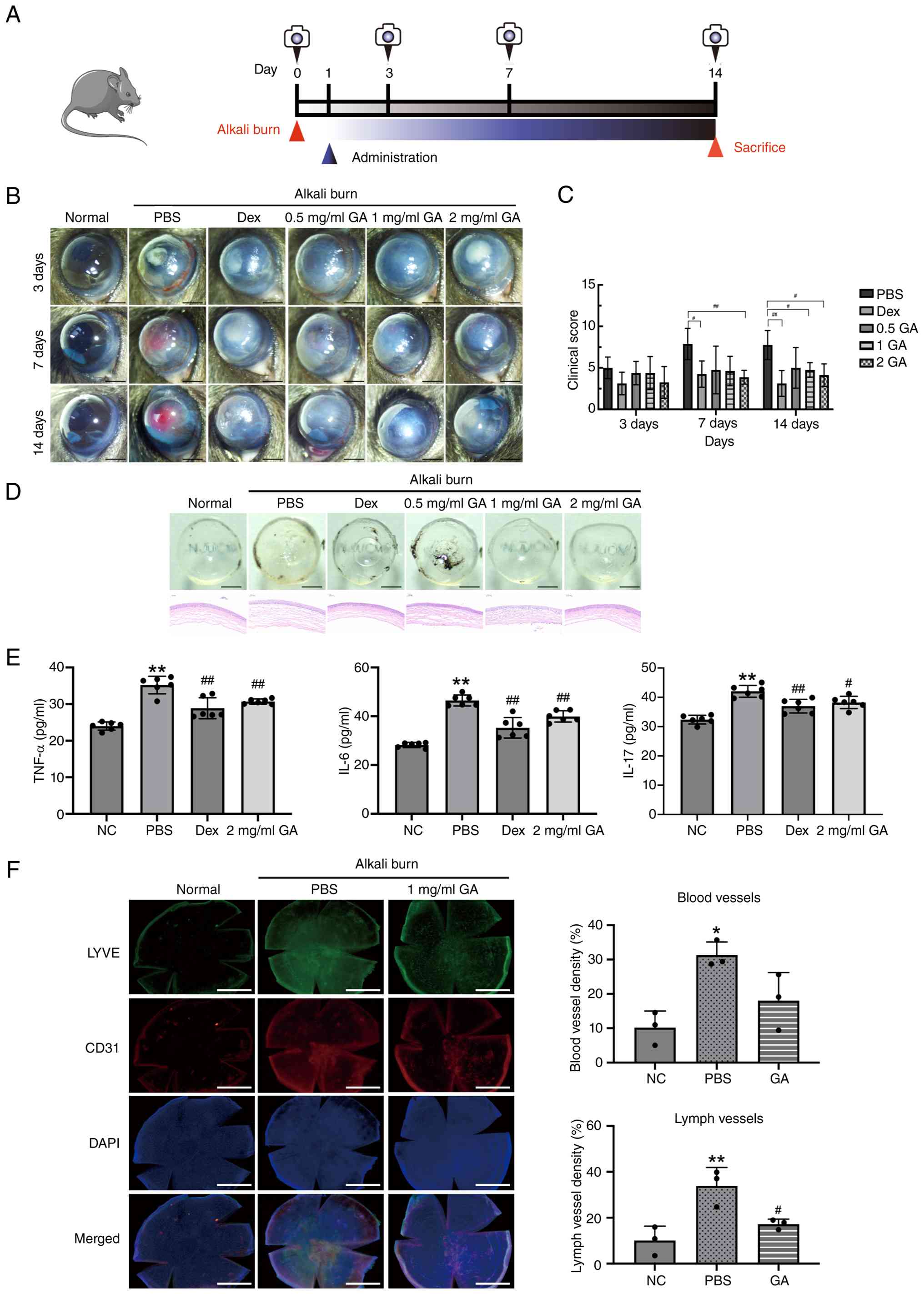 | Figure 1.GA ameliorates alkali burn-induced
corneal injury. (A) Schematic diagram of the modeling,
administration and photographing procedures performed in mice. (B)
Images of the anterior ocular segment in the normal, PBS-treated, 1
mg/ml dexamethasone-treated, 0.5 mg/ml GA-treated, 1 mg/ml
GA-treated and 2 mg/ml GA-treated groups. (C) Quantitative analysis
of the clinical assessment score on days 3, 7 and 14 (n=8). (D)
Cornea images on day 14, including hematoxylin-eosin staining
images (left; scale bars, 0.05 mm). (E) ELISA results showing the
corneal levels of TNF-α IL-6 and IL-17. (F) Immunohistochemistry
revealing blood and lymph vessels using the anti-CD31 and
anti-LYVE-1 mAbs, respectively. Quantification of the blood and
lymph vessel density, as indicated. Data are presented as means ±
SD (n=3). ns, not significant; *P<0.05 and **P<0.01 vs. NC
group; #P<0.05 and ##P<0.01 vs. PBS
group. GA, glycyrrhizic acid; TNF-α, tumor necrosis factor-α; IL,
interleukin. |
To further elucidate the anti-inflammatory mechanism
of GA, inflammatory cytokine levels in corneal tissues were
analyzed. ELISA demonstrated that GA markedly suppressed IL-6,
IL-17 and TNF-α expression compared with that in the PBS-treated
group (Fig. 1E), indicating that
GA alleviated corneal inflammation by inhibiting pro-inflammatory
cytokine production. Alkali injury induces pathological corneal
angiogenesis and lymphangiogenesis, which are closely associated
with elevated inflammatory cytokines (21). Immunofluorescence staining showed
that vascular (18.05%) and lymphatic vessel (17.20%) densities in
GA-treated corneas were markedly reduced compared with those in the
PBS group (31.27 and 33.89%, respectively; Fig. 1F), further confirming that GA
effectively inhibits pathological-blood and lymphatic-vessel
formation. The present findings highlighted that GA ameliorates
alkali burn-induced corneal injury by suppressing inflammatory
cytokine expression and pathological angiogenesis, thereby
alleviating corneal inflammation and promoting tissue recovery.
GA inhibits VEGF-A-induced
proliferation, migration and tube formation in HUVECs
To elucidate the role of GA in angiogenesis, the
present study systematically evaluated its effects on
VEGF-A-induced proliferation, migration and tube formation in
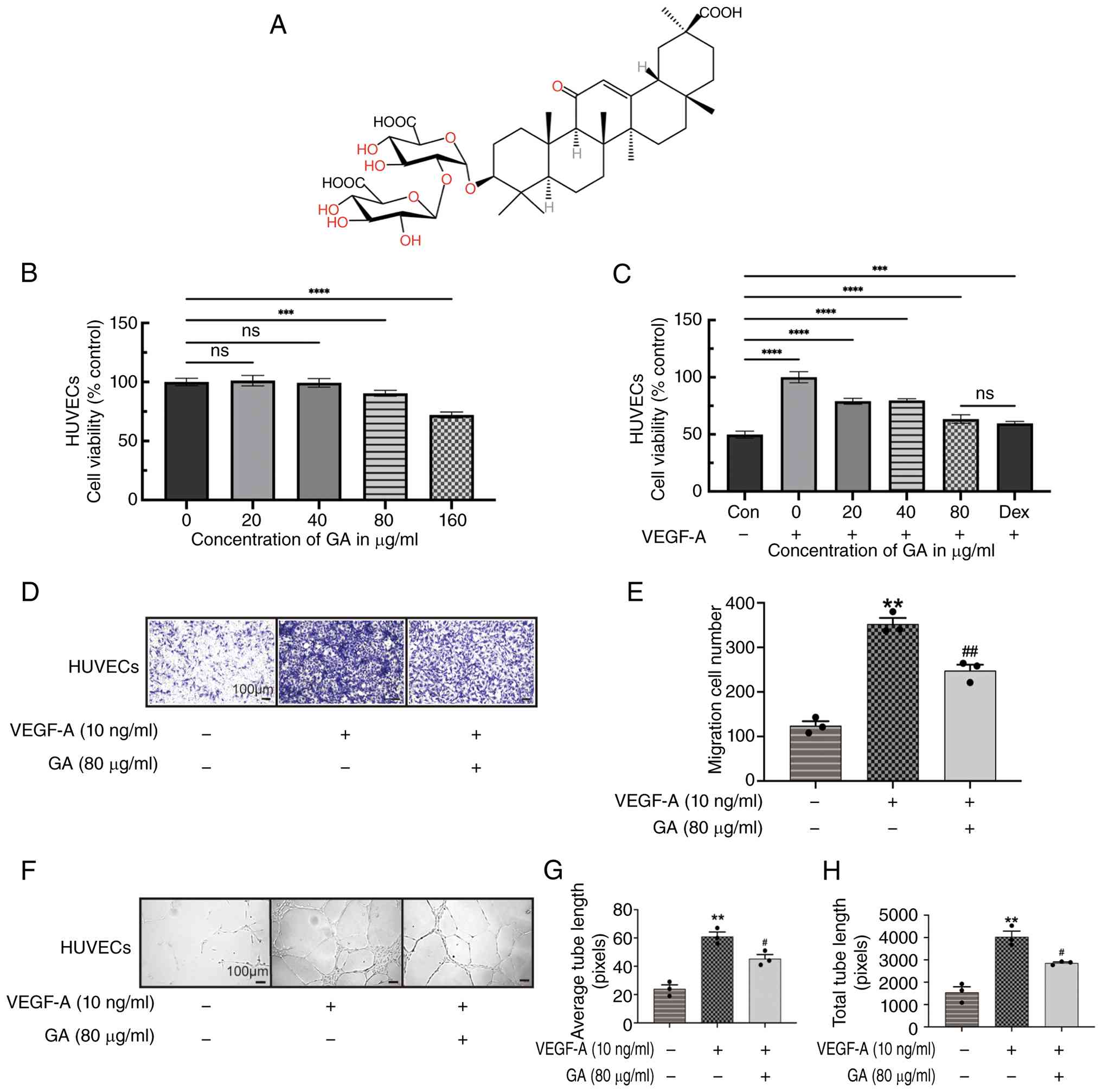
HUVECs. Initially, GA cytotoxicity toward HUVECs was assessed using
the CCK-8 assay (Fig. 2B). GA
showed no significant effect on cell viability within the
concentration range of 0–40 µg/ml. However, a slight inhibitory
effect was observed at 80 µg/ml and marked cytotoxicity occurred at
160 µg/ml (P<0.05). Accordingly, 80 µg/ml was selected as the
working concentration for subsequent experiments. Further results
demonstrated that 80 µg/ml GA markedly suppressed VEGF-A-induced
HUVEC proliferation, exhibiting an inhibitory effect comparable to
that of Dex (Fig. 2C).
Endothelial cell migration is a critical step in
angiogenesis (22). Transwell
migration assays were employed to evaluate the effect of GA on
VEGF-A-stimulated HUVEC migration (Fig. 2D-E). VEGF-A treatment markedly
enhanced endothelial cell migration, whereas GA treatment markedly
reduced the number of migrated cells. Quantitative analysis
confirmed that GA intervention substantially attenuated
VEGF-A-induced migratory capacity. The formation of capillary-like
tubular networks by endothelial cells is another key phase of
angiogenesis (23,24). To further examine the
anti-angiogenic potential of GA, Matrigel tube-formation assays
were performed (Fig. 2F-H). VEGF-A
markedly promoted the formation of complex, highly branched tubular
structures, whereas GA treatment markedly reduced tube length,
branch points and total lumen numbers, indicating a potent
inhibition of angiogenesis. These results indicate that GA
effectively suppresses VEGF-A-mediated HUVEC proliferation,
migration and tube formation, suggesting that it exerts
considerable anti-angiogenic activity in vitro.
GA suppresses VEGF-A expression in
vivo and in vitro
To investigate the regulatory effect of GA on VEGF-A
expression, experiments were performed in HUVECs and a mouse
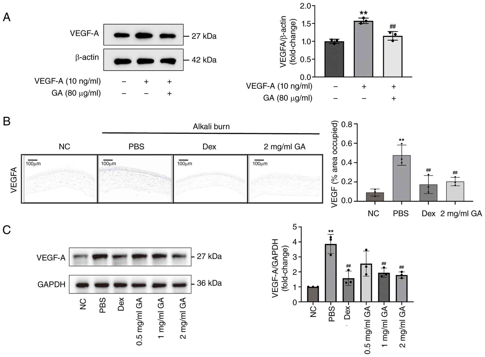
corneal alkali-burn model. Western blotting revealed that GA
treatment markedly decreased VEGF-A protein levels in HUVECs
compared with that in the control group (Fig. 3A), indicating that GA directly
suppresses VEGF-A upregulation in endothelial cells.
Furthermore, IHC and western blotting were performed
to examine VEGF-A expression in alkali-burned corneal tissues. IHC
results showed that corneas in the PBS group exhibited marked
stromal edema and thickening, accompanied by strong VEGF-A
immunoreactivity. By contrast, treatment with 2 mg/ml GA markedly
reduced VEGF-A expression, with the corneal structure largely
preserved (Fig. 3B). Consistently,
western blotting analysis confirmed that GA effectively reversed
the alkali burn-induced upregulation of VEGF-A expression in
corneal tissues (Fig. 3C).
Collectively, these findings demonstrated that GA
exerted anti-angiogenic effects in vivo and in vitro
by downregulating VEGF-A expression, thereby inhibiting CNV.
Identification of potential targets of
GA and GO/KEGG pathway analyses
To further elucidate the potential molecular
mechanisms by which GA exerts its therapeutic effects in
alkali-burned corneal tissues, DIA-based quantitative proteomics
was performed on corneal samples collected on day 7 after injury.
Differential protein expression profiles were compared between the
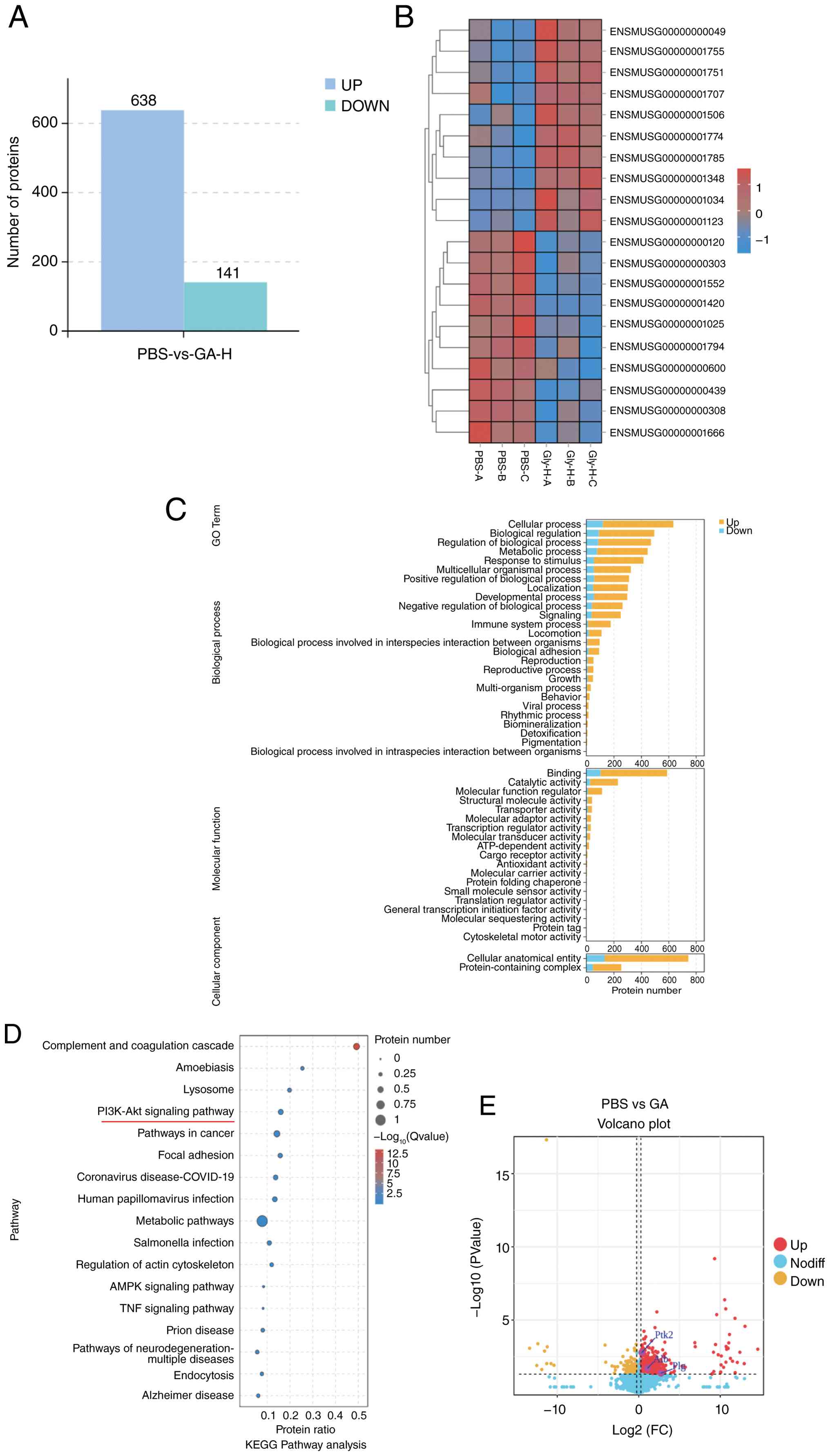
PBS and high-dose GA groups (2 mg/ml). In total, 779 DEPs were
identified, including 638 upregulated and 141 downregulated
proteins (Fig. 4A). Distribution
and expression patterns of the top 20 DEPs are presented in
Fig. 4B. GO functional enrichment
analysis (Fig. 4C) indicated that
these DEPs were mainly enriched in 26 biological process, 19
molecular function and 2 cellular component categories. KEGG
pathway enrichment analysis (Fig.
4D) revealed that the top five markedly enriched pathways
included ‘metabolic pathways’, ‘complement and coagulation
cascades’, ‘pathways in cancer’, the ‘PI3K/AKT signaling pathway’
and ‘human papillomavirus infection’.
Given that corneal alkali burn is typically
accompanied by severe inflammation, pathological
neovascularization, corneal ulceration and loss of transparency and
based on previous studies (25–28),
the PI3K/AKT pathway serves a key role in regulating inflammatory
responses and tissue repair. Proteomic analysis showed increased
abundance of FAK-and PI3K-related proteins in GA-treated corneas,
indicating that the FAK/PI3K/AKT pathway may contribute to
GA-mediated corneal repair.
Therefore, both the corneal alkali-burn model and
NaOH-induced HCECs were employed in subsequent experiments to
further validate the regulatory effects of GA on the PI3K/AKT
signaling pathway in vivo and in vitro, aiming to
clarify the molecular mechanisms underlying corneal repair and
anti-angiogenic activity.
Network pharmacology analysis of
potential targets and mechanisms of GA in improving corneal alkali
burn
Network pharmacology has emerged as a powerful
approach for identifying potential targets and elucidating the
signaling pathways of bioactive compounds in traditional Chinese
medicine (29,30). To further investigate the molecular
mechanisms by which GA regulates CNV following alkali burn injury,
a network pharmacology-based strategy was applied to screen and
analyze potential targets and signaling pathways associated with
GA.
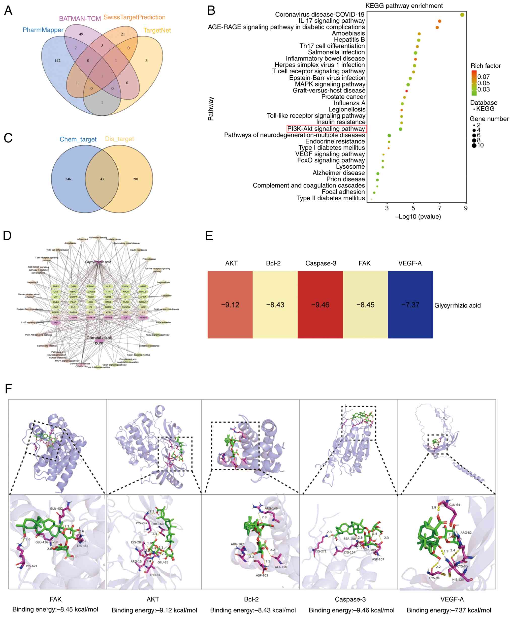
Potential targets of GA were predicted using
multiple databases, including BATMAN (probability ≥50%),
PharmMapper (score ≥0.5), TargetNet (score ≥0.5) and
SwissTargetPrediction (probability >0). After removing duplicate
entries, 389 candidate targets were identified (Fig. 5A). Disease-related targets
associated with ‘corneal alkali burn’ were retrieved from the
GeneCards database (score >1), yielding 244 disease targets. The
intersection of the two datasets identified 43 potential
overlapping targets that may mediate the therapeutic effects of GA
on corneal alkali burn (Fig. 5C).
KEGG pathway enrichment analysis of these 43 targets revealed that
the top 30 markedly enriched pathways included the PI3K/AKT
signaling pathway, consistent with previous proteomic findings
(Fig. 5B). Based on these results,
a compound-target-pathway-disease network was constructed using the
core pathways, key targets and bioactive compound (Fig. 5D). The network comprised 75 nodes
and 241 edges, representing 43 targets, 30 signaling pathways and
one compound. The color intensity of the nodes corresponded to
their degree values, where higher degree values indicate greater
importance within the network.
Finally, molecular docking analysis was performed to
evaluate the binding affinity between GA and the core targets
associated with corneal alkali burn (Fig. 5E). The binding energy was used to
estimate the stability and strength of the ligand-protein
interactions. Generally, a binding energy ≤-5.0 kcal/mol indicates
good binding activity, whereas a binding energy ≤-7.5 kcal/mol
reflects strong binding affinity. The visualized docking
interactions are presented in Fig.
5F.
The present study intersected the DEPs identified
through quantitative proteomics with candidate targets predicted
via network pharmacology and the two shared core positive proteins,
albumin (ALB) and plasminogen (PLG), were screened for cellular
validation using western blotting assays. The western blotting
results confirmed that protein levels of ALB and PLG in HCECs were
markedly upregulated in the GA-treated group compared with those in
the alkali-injured model group, which validated the reliability of
the DEPs screened through proteomics and the target prediction
results of network pharmacology. Mechanistically, ALB neutralizes
toxic oxidized lipids, eliminates reactive oxygen species and
stabilizes epithelial junctions to suppress inflammatory
infiltration. PLG participates in extracellular matrix remodeling
and facilitates epithelial cell migration, thereby accelerating the
healing of alkali-induced corneal wounds. Moderate upregulation of
PLG expression by GA maintains physiological fibrinolytic balance
and avoids excessive degradation of the corneal stroma.
Collectively, ALB and PLG synergistically restore the activity of
the protective FAK/PI3K/AKT signaling cascade, thereby alleviating
alkali-triggered corneal injury and restraining pathological CNV.
The western blotting verification data for ALB and PLG are
presented in Fig. S1.
GA inhibits apoptosis in corneal
epithelial cells
To clarify the mechanism underlying how GA
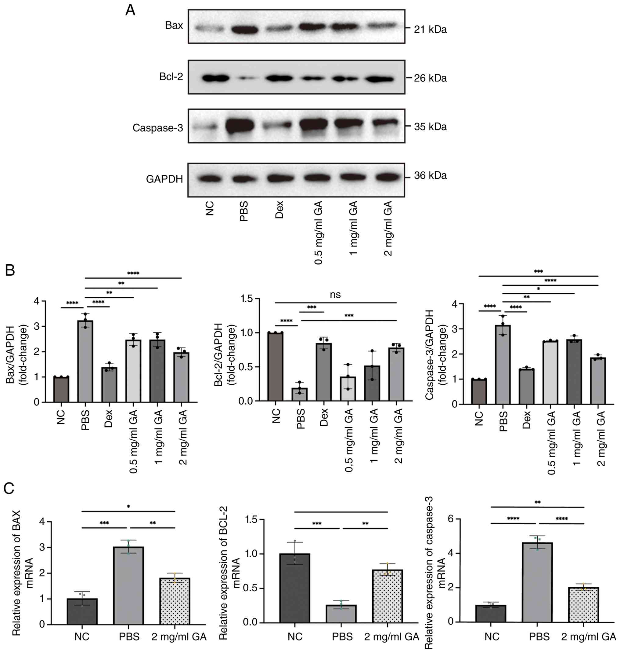
attenuates corneal epithelial-cell apoptosis, we assessed the
expression of key apoptosis regulators (Bax, Bcl-2 and caspase-3).
Alkali burn markedly increased pro-apoptotic Bax and caspase-3
expression levels while decreasing those of anti-apoptotic Bcl-2
compared with the expression levels measured in the normal control
(Fig. 6A and B). GA treatment
effectively reversed these changes, with the most potent
anti-apoptotic effect observed at 2 mg/ml.
Collectively, these results suggested that GA exerts
a protective effect against alkali burn-induced corneal epithelial
injury by modulating apoptosis-related signaling pathways and
inhibiting excessive cell apoptosis.
GA promotes proliferation and
migration while inhibiting HCEC apoptosis by activating the
FAK/PI3K/AKT signaling pathway
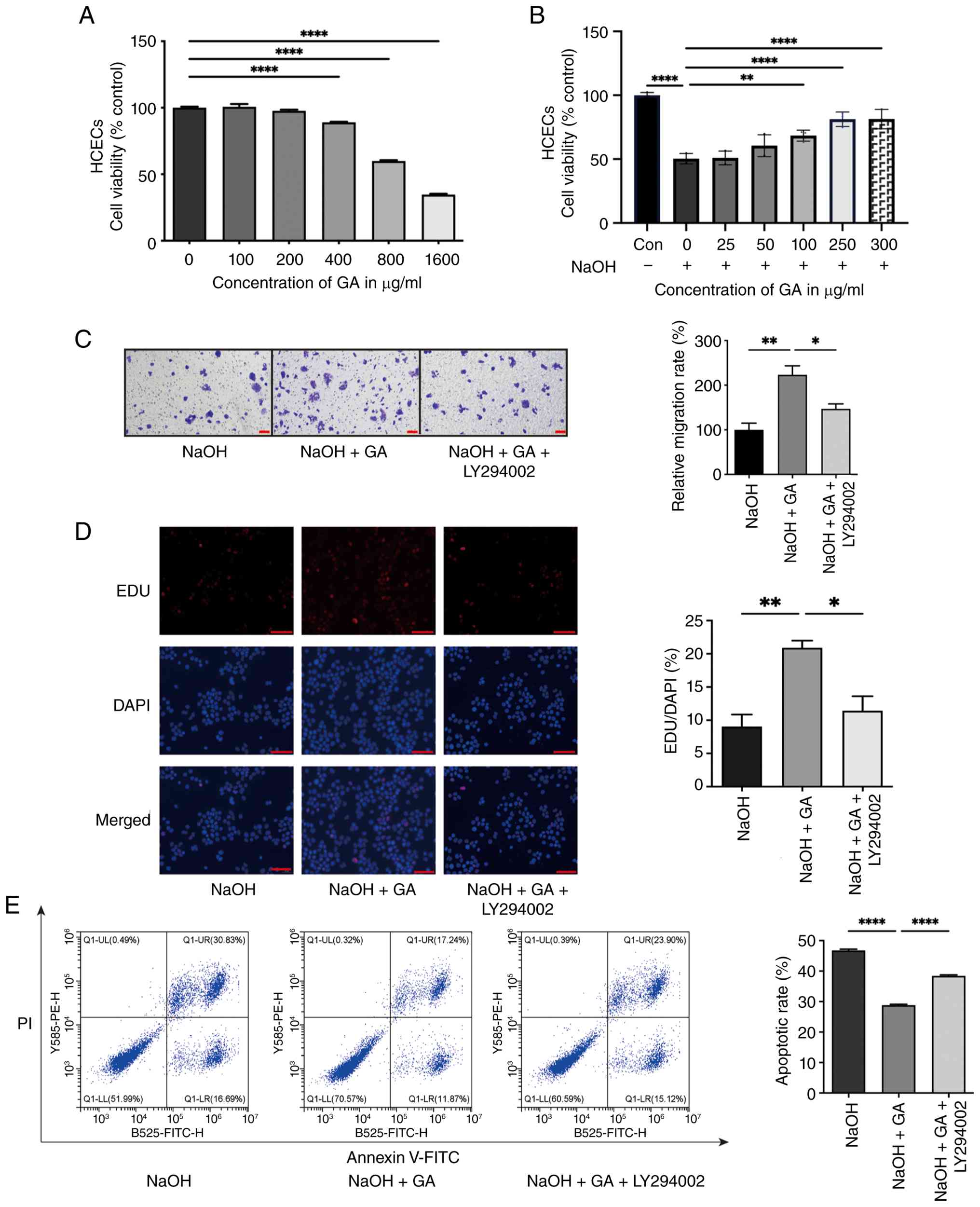
To evaluate the effects of GA on HCEC viability, its
cytotoxicity at different concentrations was initially determined
using the CCK-8 assay. The results showed that GA had no
significant effect on cell viability within the concentration range
of 0–200 µg/ml; however, a notable decrease in cell viability was
observed at 400 µg/ml (P<0.05; Fig.
7A). Subsequently, HCECs were exposed to 6 mM NaOH for 50 sec
to establish a corneal injury model and then co-incubated with GA
(0–300 µg/ml) to determine the optimal treatment concentration. The
results indicate that 250 µg/ml GA provided the most pronounced
protective and proliferative effects on NaOH-damaged cells; hence,
this concentration was chosen for subsequent experiments (Fig. 7B). Transwell migration and EdU
proliferation assays revealed that GA markedly enhanced the
migratory and proliferative ability of HCECs, whereas these effects
were markedly attenuated by co-treatment with a PI3K inhibitor
(Fig. 7C and D). In addition, flow
cytometric analysis demonstrated that GA effectively reduced
NaOH-induced apoptosis in HCECs, whereas the inhibition of PI3K
reversed this effect and increased the apoptotic rate (Fig. 7E).
To clarify whether the anti-neovascularization
effect of GA relies on the FAK/PI3K/AKT signaling pathway and
identify the upstream-downstream order of VEGF-A and PI3K, the
present study performed western blotting-based PI3K inhibitor
(LY294002) rescue assays in alkali-stimulated HCECs. Consistent
with previous data, GA intervention elevated phosphorylated FAK,
PI3K and AKT levels, whereas co-treatment with LY294002 markedly
abolished the activation of PI3K/AKT without altering VEGF-A
expression. These results indicated that LY294002 blocks downstream
PI3K/AKT signaling but does not reverse GA-mediated VEGF-A
suppression, suggesting that the inhibitory effect of GA on VEGF-A
is largely independent of PI3K/AKT activation.
Combined with the functional data showing that GA
inhibits VEGF-A-induced angiogenesis in HUVECs, the present study
confirmed that GA exerts anti-CNV effects by primarily repressing
VEGF-A production and that the FAK/PI3K/AKT pathway acts as a
downstream effector mediating epithelial-protective functions
rather than as an upstream regulator of VEGF-A (Fig. 8A-B). Moreover, GA treatment
markedly increased FAK, PI3K and AKT phosphorylation, with a
concomitant upregulation of the downstream targets, Bcl-2 and
cyclin D1. By contrast, co-treatment with the PI3K inhibitor
markedly attenuated these effects. (Fig. 8C-I).
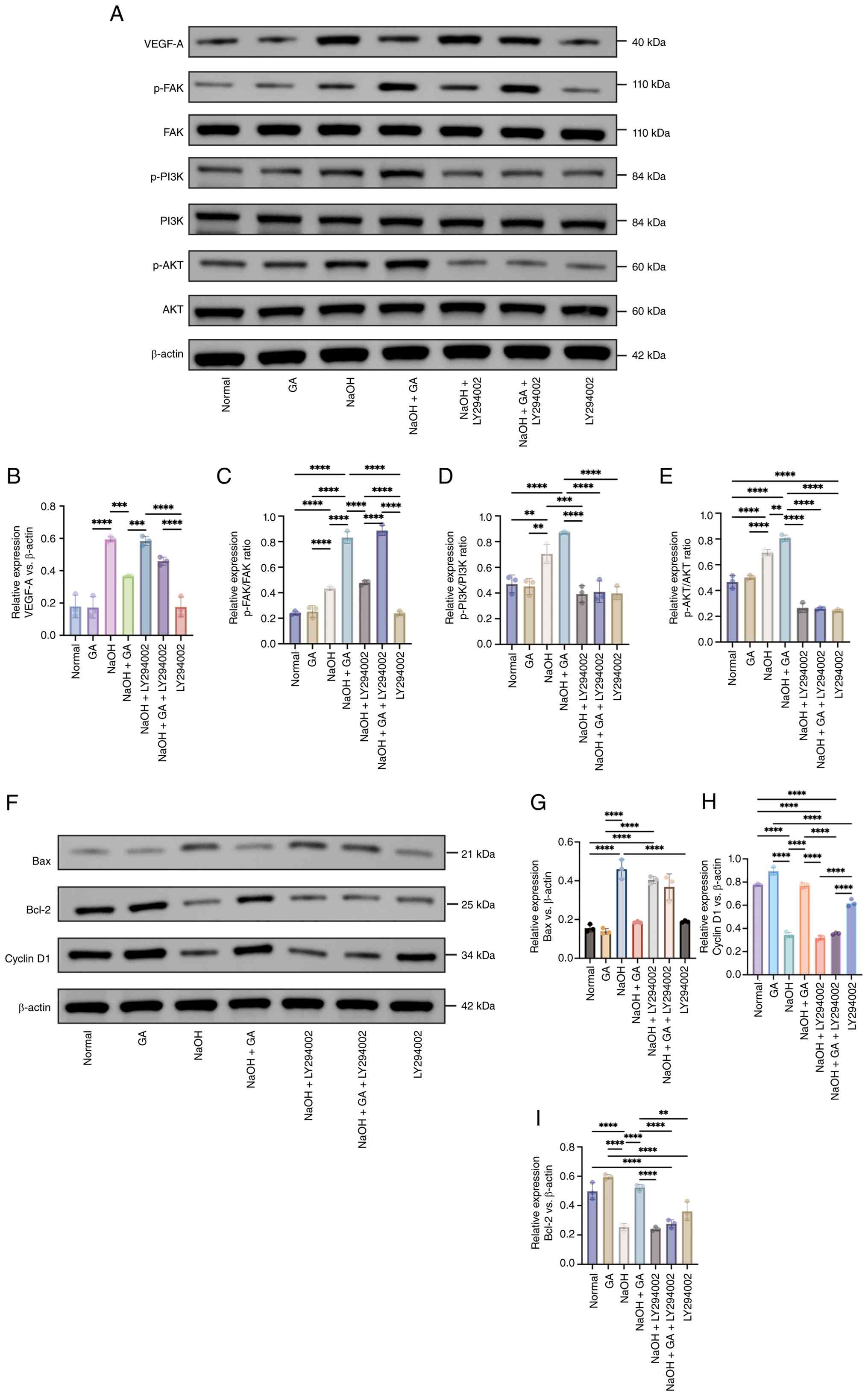 | Figure 8.PI3K inhibitor rescue assays
clarified the hierarchical relationship between VEGF-A and the
FAK/PI3K/AKT pathway in alkali-injured HCECs. (A) Representative
western blot bands showing the protein expression levels of VEGF-A,
p-FAK, FAK, p-PI3K, PI3K, p-AKT, AKT and the internal reference
(β-actin) across seven experimental groups: Normal, GA, NaOH, NaOH
+ GA, NaOH + LY294002, NaOH + GA + LY294002 and LY294002 alone. (B)
Quantitative grayscale analysis of relative VEGF-A expression
normalized to β-actin. Quantitative grayscale analysis of (C)
p-FAK/FAK ratio. (D) p-PI3K/PI3K ratio. (E) Quantitative grayscale
analysis of the p-AKT/AKT ratio. (F) Western blotting detection of
Bax (apoptosis-related protein), Bcl-2 (anti-apoptotic protein) and
cyclin D1 (proliferation marker) in the Normal, GA, NaOH, NaOH +
GA, NaOH + LY294002, NaOH + GA + LY294002 and LY294002 alone.
Statistical quantification of (G) Bax, (H) Bcl-2 and (I) cyclin D1
expression normalized to that of β-actin. Data are presented as
mean ± SD (n=3). **P<0.01; ***P<0.001; ****P<0.0001.
VEGF-A, vascular endothelial growth factor A; FAK, focal adhesion
kinase; HUVECs, human umbilical vein endothelial cells; p-,
phosphorylated; GA, glycyrrhizic acid. |
Overall, GA promotes the proliferation and migration
of NaOH-injured HCECs and inhibits apoptosis by activating the
FAK/PI3K/AKT pathway, thereby exerting cytoprotective effects
during corneal injury repair.
Discussion
The cornea is an avascular and transparent tissue
and its structural integrity is essential for visual acuity. Under
pathological conditions, such as infection, trauma, or corneal
transplantation, multiple insults can trigger CNV and fibrotic
scarring, thereby compromising transparency. Alkali burn induces an
acute and pronounced inflammatory response characterized by limbal
vasodilation, increased vascular permeability, infiltration and
migration of inflammatory cells and activation of the vascular
endothelium, all of which promote neovessel formation (31,32).
Large numbers of pro-inflammatory cells (such as macrophages and
neutrophils) accumulate at the injury site; classically activated
M1 macrophages secrete IL-1β, IL-6, TNF-α and VEGF-A, driving
endothelial proliferation and sprouting (33). In addition, IL-1β, IL-1α,
interferon-γ and TNF-α can induce apoptosis and impede tissue
repair (34–36). Thus, effective therapeutic
strategies should concurrently target inflammation and apoptosis.
Using a murine alkali-burn model, the present study systematically
assessed the effects of GA on corneal inflammation and CNV. GA
markedly attenuated inflammatory changes, reduced corneal haze and
suppressed CNV while mitigating tissue injury by upregulating
cyclin D1 and Bcl-2 expression and downregulating that of Bax and
caspase-3. These data suggest that the protective actions of GA are
closely associated with activation of the FAK/PI3K/AKT signaling
axis (Fig. 9).
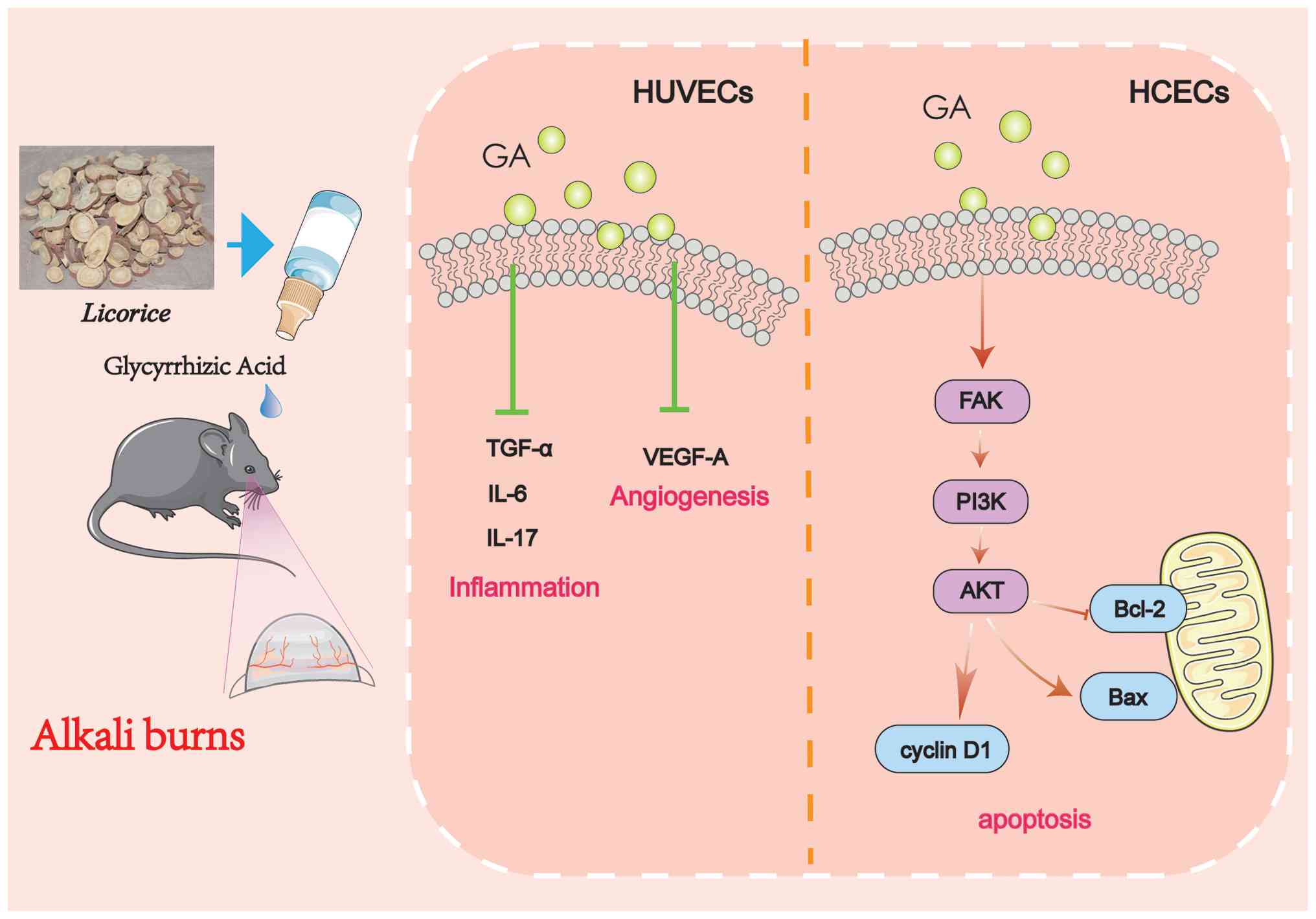 | Figure 9.Proposed mechanism of GA in
alleviating corneal alkali burn-induced inflammation, angiogenesis
and apoptosis. The figure was created and edited using Adobe
Illustrator. GA, glycyrrhizic acid; TGF, transforming growth
factor; VEGF-A, vascular endothelial growth factor A; HUVEC, human
umbilical vein endothelial cells; HCEC, human corneal epithelial
cells; FAK, focal adhesion kinase; PI3K, phosphoinositide 3-kinase;
AKT, protein kinase B; Bcl-2, B-cell lymphoma-2; Bax,
Bcl-2-associated X protein; cyclin D1, cyclin D1. |
GA, a triterpenoid saponin extracted from
Glycyrrhiza roots and rhizomes, is a principal bioactive
constituent exhibiting anti-inflammatory, antioxidative and
immunomodulatory properties, as well as a favorable safety profile.
Previous studies reported no apparent toxicity of GA <200 µg/ml
in macrophages (37) and a safety
range of 12.5–300 µg/ml in AEC II cells (38). Consistently, the present study
found that ≤80 µg/ml GA was safe and effective in HUVECs, whereas
250 µg/ml was optimal in HCECs.
Alkali burn commonly resulted in epithelial loss,
stromal edema, CNV and decreased transparency, which is consistent
with prior reports (5). The
therapeutic efficacy observed in the present study was evaluated
using corneal opacity scores and CNV grading, with Dex serving as a
positive control. As a commonly used corticosteroid for ocular
surface disease, Dex suppresses local inflammation and helps
preserve transparency (39). GA
achieved Dex-comparable effects in reducing edema by inhibiting CNV
and lymphangiogenesis, indicating that GA may act as a natural
anti-inflammatory agent with glucocorticoid-like benefits.
Inflammation-driven angiogenesis is dependent on the
interplay between inflammatory cells and angiogenic mediators.
Alkali injury promotes neutrophil and macrophage infiltration and
enhances the secretion of pro-angiogenic factors, such as VEGF,
further amplifying inflammation (40). Persistent elevation of IL-6 and
TNF-α levels facilitates epithelial migration, proliferation and
tissue remodeling (41). In the
model of the present study, GA markedly reduced the levels of IL-6,
TNF-α, IL-17 and VEGF in corneal tissues, consistent with published
observations (42). FAK, which is
encoded by PTK2, functions as a non-receptor tyrosine kinase
that orchestrates key cellular processes. Aberrant FAK activation
correlates with enhanced proliferation and migration and could
serve as a promising therapeutic target for various diseases
(43). The proteomic and
bioinformatic analyses revealed that GA-responsive DEPs were
markedly enriched in the PI3K/AKT pathway, with a notable
upregulation of FAK and PI3K detected in GA-treated corneas.
Network pharmacology and molecular docking further indicated high
binding affinities between GA constituents and proteins related to
FAK and the PI3K/AKT cascade, suggesting FAK is a putative target
of GA.
The PI3K/AKT pathway governs cell proliferation,
migration, metabolism and survival (44) and GA modulates this pathway in a
context-dependent manner across disease models. For example, GA
suppresses IL-1β-induced PI3K/AKT and NF-κB signaling, thereby
attenuating inflammation (45); in
acute liver injury, GA inhibits HMGB1 and PI3K/mTOR signaling to
reduce inflammatory cytokine release and apoptosis (46); in LPS-stimulated RAW264.7 cells, GA
and its derivative, 18β-glycyrrhetinic acid, target PI3K p110δ/γ
isoforms, suppress NF-κB activation and lower TNF-α, IL-6 and IL-1β
secretion in a dose-dependent manner (47). Tsai et al (48) reported that GA restrains tumor
growth and migration in hepatocellular models by modulating ERK,
AKT and EGFR signaling, consequently triggering
caspase-8/9-mediated mitochondrial apoptosis. In a rat model of
colitis-associated cancer, GA downregulated NF-κB, COX-2, iNOS and
VEGF expression, thereby reducing inflammation, aberrant
proliferation and angiogenesis (49). Collectively, these studies support
the capacity of GA to fine-tune PI3K/AKT signaling.
In ocular disease, the PI3K/AKT pathway status
varies by pathology; it is often downregulated in diabetic
keratopathy and dry eye but activated in infectious and
noninfectious keratitis (50). In
the healthy corneal epithelium, low PI3K/AKT activity maintains
homeostasis. Upon injury, growth factors and neuropeptides (such as
substance P) activate the PI3K/AKT cascade to promote epithelial
proliferation and migration while limiting apoptosis and
inflammation, thereby accelerating repair. Hyperglycemia,
hyperosmolarity, excess cytokines, toxins and infection disrupt
this pathway (51,52). Therefore, targeting the PI3K/AKT
pathway represents a promising strategy for corneal repair. In this
context, the present study observed that GA upregulated FAK
expression and subsequently activated PI3K/AKT signaling, thereby
facilitating epithelial regeneration.
To clarify the causal link among GA, VEGF-A and the
FAK/PI3K/AKT pathway, PI3K inhibitor rescue assays were conducted
in alkali-injured HCECs. Western blotting results showed that
LY294002 blocked GA-induced PI3K/AKT activation but had no impact
on GA-mediated VEGF-A downregulation, confirming that GA-mediated
VEGF suppression is largely independent of PI3K/AKT signaling.
Therefore, GA mainly suppresses pathological CNV by reducing VEGF-A
expression, whereas activated FAK/PI3K/AKT primarily facilitates
epithelial repair and anti-apoptotic effects. GA exerts dual
protective effects after corneal alkali injury via this two-tiered
regulatory axis. Although PTK2 (FAK) was identified via proteomic
analysis and validated in vitro, direct assessment of
phosphorylated FAK in corneal tissues is warranted.
Apoptosis is critical for corneal homeostasis
(53). Bcl-2, a key anti-apoptotic
mediator of the intrinsic mitochondrial pathway, functions by
heterodimerizing with pro-apoptotic Bax; alterations in its
expression can influence epithelial cell lifespan and turnover
(54). Activation of caspase-3 is
a hallmark of apoptosis (55) and
reduced Bcl-2 expression generally correlates with increased
apoptotic activity (56). The
anti-apoptotic capacity of GA has been documented previously: in a
model of Mycoplasma gallisepticum infection, GA suppressed
caspase-3/9 activation and upregulated Bcl-2 and Bcl-xL expression
(38). Consistent with these
findings, the present study's alkali-burn model showed that GA
reversed the injury-induced upregulation of Bax and caspase-3
expression and restored that of Bcl-2, confirming its robust
anti-apoptotic effect (57).
Cyclin D1, which is encoded by CCND1, forms active complexes
with CDK4/6 to phosphorylate retinoblastoma protein and drive the
G1-to-S transition (58). Cyclin D1 plays a pivotal role in
corneal epithelial proliferation and is notably enriched in the
epithelial compartment of pterygium tissues (59); it has also been implicated as a key
downstream mediator of nerve growth factor-regulated HCEC
proliferation (60). The present
data demonstrate, for the first time, that GA promotes corneal
epithelial cell proliferation by upregulating cyclin D1 expression
through activation of the FAK/PI3K/AKT pathway.
In summary, the present study suggested that GA
exerts dual protective effects during corneal alkali burn. On the
one hand, GA suppresses VEGF-A expression and thereby inhibits
pathological CNV; on the other hand, it activates the FAK/PI3K/AKT
pathway to promote epithelial proliferation, migration and
survival. These two mechanisms cooperate in facilitating corneal
repair. Overall, GA exhibited good safety and biocompatibility at
both the cellular and animal levels, supporting its potential as a
natural, low-toxicity anti-inflammatory and pro-repair candidate
for the treatment of sterile keratitis and related corneal
disorders.
The present study systematically demonstrated the
protective effects of GA against alkali burn-induced corneal
injury. GA exerted its therapeutic actions through multiple
coordinated mechanisms. On one hand, GA inhibited VEGF-mediated
angiogenesis, thereby effectively suppressing pathological CNV; on
the other hand, it modulated the expression of apoptosis-associated
molecules, including Bax, Bcl-2 and caspase-3, thereby attenuating
corneal epithelial cell apoptosis and promoting tissue repair.
Moreover, GA activated the FAK/PI3K/AKT signaling pathway, which
contributed to the maintenance of epithelial cell survival and
regeneration, ultimately improving the inflammatory
microenvironment and restoring corneal structural integrity. In
addition, GA exhibited excellent biocompatibility and low
cytotoxicity in both corneal tissues and cell models, confirming
its favorable safety profile. Collectively, these findings
highlighted GA as a naturally derived, multitarget
anti-inflammatory and anti-angiogenic compound with promising
therapeutic potential. GA may serve as a novel candidate for the
prevention and treatment of sterile keratitis, chemical corneal
injury and other ocular surface disorders characterized by
inflammation and neovascularization.
Supplementary Material
Supporting Data
Acknowledgements
Not applicable.
Funding
The present study was supported by the Jiangsu Provincial
Administration of Traditional Chinese Medicine ‘Clinical Research
on Key Diseases in High-Level Hospital Construction’ project (grant
no. GSPYY2023009); the Youth project of Wuxi Municipal Health
Commission (grant no. Q202348); the Natural Science Foundation of
Nanjing University of Chinese Medicine (grant no. XZR2024316); and
the Nantong Municipal Guiding Project of Science and Technology
Plan (grant no. MSZ2025081).
Availability of data and materials
The data generated in the present study may be
requested from the corresponding author.
Authors' contributions
JY conceived the study, prepared all figures, wrote
the original draft and performed the investigation. HQ was
responsible for analyzing the data, designing the methodology,
preparing all figures and writing the original draft. YF was
responsible for conceptualization of the study, organising raw
experimental data and preparing all figures. WX conceived the study
and constructed figures. NW performed the experiments and analyzed
data. ND was responsible for organising raw experimental data and
designing the methodology. JJ was responsible for conceptualization
of the study, formal analysis of the data, and writing, reviewing
and editing of the manuscript. WS conceived and designed the study,
interpreted the data, acquired funding, supervised, verified
experimental results, and reviewed and edited the manuscript. All
authors read and approved the final manuscript. NW and WS confirm
the authenticity of all the raw data.
Ethics approval and consent to
participate
The present study was approved by the Animals Ethics
Committee of the Wuxi Affiliated Hospital of Nanjing University of
Chinese Medicine (Wuxi, China; approval no. 2024-171-01). All
animal experiments were performed following the ARRIVE guidelines
and internationally recognized standards for laboratory animal
welfare to reduce distress. This study adhered to the principles of
transparency, honesty and integrity in data collection, analysis
and reporting.
Patient consent for publication
Not applicable.
Competing interests
The authors declare that they have no competing
interests.
References
|
1
|
Flaxman SR, Bourne RRA, Resnikoff S,
Ackland P, Braithwaite T, Cicinelli MV, Das A, Jonas JB, Keeffe J,
Kempen JH, et al: Global causes of blindness and distance vision
impairment 1990–2020: A systematic review and meta-analysis. Lancet
Glob Health. 5:e1221–e1234. 2017. View Article : Google Scholar : PubMed/NCBI
|
|
2
|
Soiberman U, Kambhampati SP, Wu T, Mishra
MK, Oh Y, Sharma R, Wang J, Al Towerki AE, Yiu S, Stark WJ and
Kannan RM: Subconjunctival injectable dendrimer-dexamethasone gel
for the treatment of corneal inflammation. Biomaterials. 125:38–53.
2017. View Article : Google Scholar : PubMed/NCBI
|
|
3
|
Chen Y, Wang X, Duan Y, Liu L, Chen R,
Huang Y, Tan Y, Lu Q, Lin Q and Shen L: Fluorinated ROS-responsive
polycation enables noninvasive siRNA delivery for the treatment of
corneal neovascularization. Acta Biomater. 212:754–769. 2026.
View Article : Google Scholar : PubMed/NCBI
|
|
4
|
Huang XC, Xu GY, Yang PP, Wang L, Wang H
and Hu Y: Recent progress on topical drug therapy for corneal
neovascularization. ChemMedChem. 20:e2025003662025. View Article : Google Scholar : PubMed/NCBI
|
|
5
|
He M, Li N, Li J and Huang X: Impact of
pterygium excision combined with autologous limbal stem cell
transplantation on microvascular density, tear film stability, and
corneal wound healing. Ann Ital Chir. 96:47–54. 2025. View Article : Google Scholar : PubMed/NCBI
|
|
6
|
Yang T, Wang X, Guo L, Zheng F, Meng C,
Zheng Y and Liu G: Daphnetin inhibits corneal inflammation and
neovascularization on a mouse model of corneal alkali burn. Int
Immunopharmacol. 103:1084342022. View Article : Google Scholar : PubMed/NCBI
|
|
7
|
Wang K, Jiang L, Zhong Y, Zhang Y, Yin Q,
Li S, Zhang X, Han H and Yao K: Ferrostatin-1-loaded liposome for
treatment of corneal alkali burn via targeting ferroptosis. Bioeng
Transl Med. 7:e102762021. View Article : Google Scholar : PubMed/NCBI
|
|
8
|
Wang Y, Shi S, Zhang L, Wang S, Qin H, Wei
Y, Wu X and Zhang M: simpleImatinib@glycymicelles
entrapped in hydrogel: Preparation, characterization, and
therapeutic effect on corneal alkali burn in mice. Drug Deliv
Transl Res. 15:171–184. 2025. View Article : Google Scholar : PubMed/NCBI
|
|
9
|
Yu J, Qu H, Xu W, Fang Y, Wang N and Shi
W: Mechanisms and therapeutic potential of glycyrrhizic acid:
Insights into key signaling pathways and disease modulation
(Review). Mol Med Rep. 33:1262026. View Article : Google Scholar : PubMed/NCBI
|
|
10
|
Wang ZY and Nixon DW: Licorice and cancer.
Nutr Cancer. 39:1–11. 2001. View Article : Google Scholar : PubMed/NCBI
|
|
11
|
Citi V, Neggiani F, Sanso M and Calderone
V: Pharmacological evaluation of innovative eye drop formulations
containing TS-polysaccaride, hyaluronic acid and glycyrrhizin for
irritative ocular diseases using in vitro reconstituted human
corneal epithelium model. Toxicol In Vitro. 75:1051992021.
View Article : Google Scholar : PubMed/NCBI
|
|
12
|
Somayajulu M, McClellan SA, Pitchaikannu
A, Bessert D, Liu L, Steinle J and Hazlett LD: Effects of
glycyrrhizin treatment on diabetic cornea. J Ocul Pharmacol Ther.
37:12–23. 2021. View Article : Google Scholar : PubMed/NCBI
|
|
13
|
Hou Y, Xin M, Li Q and Wu X: Glycyrrhizin
micelle as a genistein nanocarrier: Synergistically promoting
corneal epithelial wound healing through blockage of the HMGB1
signaling pathway in diabetic mice. Exp Eye Res. 204:1084542021.
View Article : Google Scholar : PubMed/NCBI
|
|
14
|
Burillon C, Chiambaretta F and Pisella PJ:
Efficacy and safety of glycyrrhizin 2.5% eye drops in the treatment
of moderate dry eye disease: results from a prospective, open-label
pilot study. Clin Ophthalmol. 12:2629–2636. 2018. View Article : Google Scholar : PubMed/NCBI
|
|
15
|
Ammassam Veettil R, Li W, Pflugfelder SC
and Koch DD: A mouse model for corneal neovascularization by Alkali
Burn. J Vis Exp. Jun 30–2023.(Epub ahead of print). View Article : Google Scholar : PubMed/NCBI
|
|
16
|
Zhang LB, Qiu XY, Zhou YH, Liang ML, Guo
L, Qi F, Wu YT, Meng C and Liu GH: Recombinant Sgp130 attenuates
alkali-burn cornea through inhibition of IL-6 trans-signaling: From
production to therapeutic application. Int J Biol Macromol.
377:1535092026. View Article : Google Scholar : PubMed/NCBI
|
|
17
|
Yan D, Ouyang W, Lin J and Liu Z: Smart
coating by thermo-sensitive Pluronic F-127 for enhanced corneal
healing via delivery of biological macromolecule progranulin. Int J
Biol Macromol. 253((Pt 8)): 1275862023. View Article : Google Scholar : PubMed/NCBI
|
|
18
|
Li H, Li J, Hou C, Li J, Peng H and Wang
Q: The effect of astaxanthin on inflammation in hyperosmolarity of
experimental dry eye model in vitro and in vivo. Exp Eye Res.
197:1081132020. View Article : Google Scholar : PubMed/NCBI
|
|
19
|
Yan D, Yu F, Chen L, Yao Q, Yan C, Zhang
S, Wu N, Gong D, Sun H, Fu Y and Shao C: Subconjunctival injection
of regulatory T cells potentiates corneal healing via orchestrating
inflammation and tissue repair after acute Alkali Burn. Invest
Ophthalmol Vis Sci. 61:222020. View Article : Google Scholar : PubMed/NCBI
|
|
20
|
Yu F, Gong D, Yan D, Wang H, Witman N, Lu
Y, Fu W and Fu Y: Enhanced adipose-derived stem cells with
IGF-1-modified mRNA promote wound healing following corneal injury.
Mol Ther. 31:2454–2471. 2023. View Article : Google Scholar : PubMed/NCBI
|
|
21
|
Liu Z, Liu K, Shi S, Chen X, Gu X, Wang W,
Mao K, Yibulayi R, Wu W, Zeng L, et al: Alkali injury-induced
pathological lymphangiogenesis in the iris facilitates the
infiltration of T cells and ocular inflammation. JCI Insight.
9:e1754792024. View Article : Google Scholar : PubMed/NCBI
|
|
22
|
Varinska L, Faber L, Kello M, Petrovová E,
Balážová Ľ, Solár P, Čoma M, Urdzík P, Mojžiš J, Švajdlenka E, et
al: β-Escin effectively modulates HUVECS proliferation and tube
formation. Molecules. 23:1972018. View Article : Google Scholar : PubMed/NCBI
|
|
23
|
Matsumoto G, Hirohata R, Hayashi K,
Sugimoto Y, Kotani E, Shimabukuro J, Hirano T, Nakajima Y, Kawamata
S and Mori H: Control of angiogenesis by VEGF and
endostatin-encapsulated protein microcrystals and inhibition of
tumor angiogenesis. Biomaterials. 35:1326–1333. 2014. View Article : Google Scholar : PubMed/NCBI
|
|
24
|
Mazari-Arrighi E, Lepine M, Ayollo D,
Faivre L, Larghero J, Chatelain F and Fuchs A: Self-organization of
long-lasting human endothelial capillary-like networks guided by
DLP bioprinting. Adv Healthc Mater. 13:e23028302024. View Article : Google Scholar : PubMed/NCBI
|
|
25
|
Son KN, Lee SM, Shah D, Chaudhary M, Ong
J, Reinhardt C, Shukla D, Han K and Aakalu VK: Histatin-5 improves
in vivo corneal wound healing and immunohistochemical neovascular
markers after acute Alkali injury. Transl Vis Sci Technol.
15:202026. View Article : Google Scholar : PubMed/NCBI
|
|
26
|
Chen B, Xu H, Li B, Cai Y, Xie L and Zou
J: Targeting LRPPRC to improve corneal wound healing via augmenting
autophagy and suppressing TGF-beta/Smad signaling post-alkali burn.
J Transl Med. 24:11212026. View Article : Google Scholar : PubMed/NCBI
|
|
27
|
Guan Y, Jing Y, Yang S, Yan X and Li X:
YTHDF1-mediated m6A modification of NREP promotes corneal fibrosis
via TGF-β-smad signaling. Invest Ophthalmol Vis Sci. 67:512026.
View Article : Google Scholar : PubMed/NCBI
|
|
28
|
Li Q, Bao Q, Wei C, Xie Y, Zhao S, Yan H,
Wu F, Li W, Liu X and Gao H: AIF-1 drives corneal
neovascularization by promoting inflammatory macrophage activation
via the MAPK and PI3K/AKT/mTOR signaling pathways. Invest
Ophthalmol Vis Sci. 67:222026. View Article : Google Scholar : PubMed/NCBI
|
|
29
|
Nogales C, Mamdouh ZM, List M, Kiel C,
Casas AI and Schmidt HHHW: Network pharmacology: curing causal
mechanisms instead of treating symptoms. Trends Pharmacol Sci.
43:136–150. 2022. View Article : Google Scholar : PubMed/NCBI
|
|
30
|
Song S, Wei Q, Tu Y, Zhou S, Zhou J, Tao
J, Chen Y, Zhou H and You M: Chlorpyrifos-induced autism and
depression-like behaviors are ameliorated by trehalose: Integrating
network pharmacology and toxicology analyses and animal
experiments. Environ Pollut. 393:1276702026. View Article : Google Scholar : PubMed/NCBI
|
|
31
|
Herbert SP and Stainier DY: Molecular
control of endothelial cell behaviour during blood vessel
morphogenesis. Nat Rev Mol Cell Biol. 12:551–564. 2011. View Article : Google Scholar : PubMed/NCBI
|
|
32
|
Gao J, Li J, Liu J, Han R, Chen X, Wang L,
Ma Y and Li X: miR-29b-3p promotes the recovery of alkali-burned
cornea by targeting Ddx3×. Exp Eye Res. 264:1108492026. View Article : Google Scholar : PubMed/NCBI
|
|
33
|
Yu J, Shen Y, Luo J, Jin J, Li P, Feng P
and Guan H: Upadacitinib inhibits corneal inflammation and
neovascularization by suppressing M1 macrophage infiltration in the
corneal alkali burn model. Int Immunopharmacol. 116:1096802023.
View Article : Google Scholar : PubMed/NCBI
|
|
34
|
Horwitz V, Dachir S, Cohen M, Gutman H,
Cohen L, Gez R, Buch H, Kadar T and Gore A: Differential expression
of corneal and limbal cytokines and chemokines throughout the
clinical course of sulfur mustard induced ocular injury in the
rabbit model. Exp Eye Res. 177:145–152. 2018. View Article : Google Scholar : PubMed/NCBI
|
|
35
|
Notara M, Lentzsch A, Coroneo M and
Cursiefen C: The role of limbal epithelial stem cells in regulating
corneal (Lymph)angiogenic privilege and the micromilieu of the
limbal niche following UV exposure. Stem Cells Int.
2018:86201722018. View Article : Google Scholar : PubMed/NCBI
|
|
36
|
Yang L, Zhang S, Duan H, Dong M, Hu X,
Zhang Z, Wang Y, Zhang X, Shi W and Zhou Q: Different effects of
pro-inflammatory factors and hyperosmotic stress on corneal
epithelial stem/progenitor cells and wound healing in mice. Stem
Cells Transl Med. 8:46–57. 2019. View Article : Google Scholar : PubMed/NCBI
|
|
37
|
Wang BK, Mao YL, Gong L, Xu X, Jiang SQ,
Wang YB and Li WF: Glycyrrhizic acid activates chicken macrophages
and enhances their Salmonella-killing capacity in vitro. J Zhejiang
Univ Sci B. 19:785–795. 2018. View Article : Google Scholar : PubMed/NCBI
|
|
38
|
Wang Y, Wang L, Luo R, Sun Y, Zou M, Wang
T, Guo Q and Peng X: Glycyrrhizic acid against mycoplasma
gallisepticum-induced inflammation and apoptosis through
suppressing the MAPK pathway in chickens. J Agric Food Chem.
70:1996–2009. 2022. View Article : Google Scholar : PubMed/NCBI
|
|
39
|
Tan Y, Zhang M, Pan Y, Feng H and Xie L:
Suppression of the caspase-1/GSDMD-mediated pyroptotic signaling
pathway through dexamethasone alleviates corneal alkali injuries.
Exp Eye Res. 214:1088582022. View Article : Google Scholar : PubMed/NCBI
|
|
40
|
Shimizu H, Sakimoto T and Yamagami S:
Pro-inflammatory role of NLRP3 inflammasome in experimental sterile
corneal inflammation. Sci Rep. 9:95962019. View Article : Google Scholar : PubMed/NCBI
|
|
41
|
Eslani M, Movahedan A, Afsharkhamseh N,
Sroussi H and Djalilian AR: The role of toll-like receptor 4 in
corneal epithelial wound healing. Invest Ophthalmol Vis Sci.
55:6108–6115. 2014. View Article : Google Scholar : PubMed/NCBI
|
|
42
|
Wang P, Hao P, Chen X, Li L, Zhou Y, Zhang
X, Zhu L, Ying M, Han R, Wang L and Li X: Targeting HMGB1-NFĸb Axis
and miR-21 by glycyrrhizin: Role in amelioration of corneal injury
in a mouse model of Alkali Burn. Front Pharmacol. 13:8412672022.
View Article : Google Scholar : PubMed/NCBI
|
|
43
|
Wang N, Dong SS, Zhuo HF, Wang MW, Xue ZX,
Li Z, Meng L, Qi Y, Yuan X and Hu J: Focal adhesion kinase promotes
biphasic differentiation and induces infiltration and metastasis of
synovial sarcoma. J Transl Med. 24:7012026. View Article : Google Scholar : PubMed/NCBI
|
|
44
|
Yao S, Fu T, Tu Q, Zhang J, Jiang Q, Xu M,
Wang Z, Jiao Y and Tian H: IL-17 promotes the phenotypic
transformation of vascular smooth muscle cells Through PI3K/AKT
signaling pathway. Microcirculation. 33:e700502026. View Article : Google Scholar : PubMed/NCBI
|
|
45
|
Jiang RH, Xu JJ, Zhu DC, Li JF, Zhang CX,
Lin N and Gao WY: Glycyrrhizin inhibits osteoarthritis development
through suppressing the PI3K/AKT/NF-ĸB signaling pathway in vivo
and in vitro. Food Funct. 11:2126–2136. 2020. View Article : Google Scholar : PubMed/NCBI
|
|
46
|
Shen CH, Ma ZY, Li JH, Li RD, Tao YF,
Zhang QB and Wang ZX: Glycyrrhizin improves inflammation and
apoptosis via suppressing HMGB1 and PI3K/mTOR pathway in
lipopolysaccharide-induced acute liver injury. Eur Rev Med
Pharmacol Sci. 24:7122–7130. 2020.PubMed/NCBI
|
|
47
|
Wang CY, Kao TC, Lo WH and Yen GC:
Glycyrrhizic acid and 18β-glycyrrhetinic acid modulate
lipopolysaccharide-induced inflammatory response by suppression of
NF-ĸB through PI3K p110δ and p110ү inhibitions. J Agric Food Chem.
59:7726–7733. 2011. View Article : Google Scholar : PubMed/NCBI
|
|
48
|
Tsai JJ, Pan PJ, Hsu FT, Chung JG and
Chiang IT: Glycyrrhizic acid modulates apoptosis through
extrinsic/intrinsic pathways and inhibits protein kinase B- and
extracellular signal-regulated kinase-mediated metastatic potential
in hepatocellular carcinoma in vitro and in vivo. Am J Chin Med.
48:223–244. 2020. View Article : Google Scholar : PubMed/NCBI
|
|
49
|
Khan R, Rehman MU, Khan AQ, Tahir M and
Sultana S: Glycyrrhizic acid suppresses
1,2-dimethylhydrazine-induced colon tumorigenesis in Wistar rats:
Alleviation of inflammatory, proliferation, angiogenic, and
apoptotic markers. Environ Toxicol. 33:1272–1283. 2018. View Article : Google Scholar : PubMed/NCBI
|
|
50
|
Chen K, Li Y, Zhang X, Ullah R, Tong J and
Shen Y: The role of the PI3K/AKT signalling pathway in the corneal
epithelium: Recent updates. Cell Death Dis. 13:5132022. View Article : Google Scholar : PubMed/NCBI
|
|
51
|
Park JH, Moon SH, Kang DH, Um HJ, Kang SS,
Kim JY and Tchah H: Diquafosol sodium inhibits apoptosis and
inflammation of corneal epithelial cells via activation of Erk1/2
and RSK: In vitro and in vivo dry eye model. Invest Ophthalmol Vis
Sci. 59:5108–5115. 2018. View Article : Google Scholar : PubMed/NCBI
|
|
52
|
Liu Z, Chen D, Chen X, Bian F, Qin W, Gao
N, Xiao Y, Li J, Pflugfelder SC and Li DQ: Trehalose induces
autophagy against inflammation by activating TFEB signaling pathway
in human corneal epithelial cells exposed to hyperosmotic stress.
Invest Ophthalmol Vis Sci. 61:262020. View Article : Google Scholar : PubMed/NCBI
|
|
53
|
Torrealba N, Rodriguez-Berriguete G, Vera
R, Fraile B, Olmedilla G, Martínez-Onsurbe P, Sánchez-Chapado M,
Paniagua R and Royuela M: Homeostasis: Apoptosis and cell cycle in
normal and pathological prostate. Aging Male. 23:335–345. 2020.
View Article : Google Scholar : PubMed/NCBI
|
|
54
|
Robertson DM, Ladage PM, Yamamoto N,
Jester JV, Petroll WM and Cavanagh HD: Bcl-2 and Bax regulation of
corneal homeostasis in genetically altered mice. Eye Contact Lens.
32:3–7. 2006. View Article : Google Scholar : PubMed/NCBI
|
|
55
|
Wang H, Zhu J, Jiang L, Shan B, Xiao P, Ai
J, Li N, Qi F and Niu S: Mechanism of Heshouwuyin inhibiting the
Cyt c/Apaf-1/Caspase-9/Caspase-3 pathway in spermatogenic cell
apoptosis. BMC Complement Med Ther. 20:1802020. View Article : Google Scholar : PubMed/NCBI
|
|
56
|
Harada H and Grant S: Apoptosis
regulators. Rev Clin Exp Hematol. 7:117–138. 2003.PubMed/NCBI
|
|
57
|
Kossl J, Bohacova P, Hermankova B,
Javorkova E, Zajicova A and Holan V: Antiapoptotic properties of
mesenchymal stem cells in a mouse model of corneal inflammation.
Stem Cells Dev. 30:418–427. 2021. View Article : Google Scholar : PubMed/NCBI
|
|
58
|
Qie S and Diehl JA: Cyclin D1, cancer
progression, and opportunities in cancer treatment. J Mol Med
(Berl). 94:1313–1326. 2016. View Article : Google Scholar : PubMed/NCBI
|
|
59
|
Kase S, Takahashi S, Sato I, Nakanishi K,
Yoshida K and Ohno S: Expression of p27(KIP1) and cyclin D1, and
cell proliferation in human pterygium. Br J Ophthalmol. 91:958–961.
2007. View Article : Google Scholar : PubMed/NCBI
|
|
60
|
Wu D, Qian T, Hong J, Li G, Shi W and Xu
J: MicroRNA-494 inhibits nerve growth factor-induced cell
proliferation by targeting cyclin D1 in human corneal epithelial
cells. Mol Med Rep. 16:4133–4142. 2017. View Article : Google Scholar : PubMed/NCBI
|