Introduction
Chronic kidney disease (CKD) represents a notable
global public health challenge, affecting >800 million
individuals worldwide and imposing a burden of morbidity and
mortality, primarily from cardiovascular complications (1,2). In
accordance with the KDIGO 2024 clinical practice guidelines, CKD is
defined as the presence of abnormalities in the kidney structure or
function present for >3 months. It has implications for health,
which can be classified according to the cause, estimated
glomerular filtration rate (eGFR) category (G1-G5) and albuminuria
category (A1-A3) (1). The
traditional pathophysiological focus of CKD progression has been on
hemodynamic factors, including the renin-angiotensin-aldosterone
system and metabolic abnormalities. Conventional therapies, such as
renin-angiotensin-aldosterone system blockade and solute carrier
family 5 member 2 inhibition, remain the mainstay of renoprotection
but only partially delay CKD progression (2,3).
Therefore, the limitations of conventional therapies in curbing
disease progression have encouraged the investigation of novel
pathogenic pathways. The ‘gut-kidney axis’ has emerged as a concept
that can provide an important conceptual framework for
understanding the intricate crosstalk between the host gut
microbial ecosystem and renal physiology (3–5).
This axis posits that disruptions in gut microbial homeostasis,
known as dysbiosis and the subsequent production of gut-derived
metabolites can serve notable roles in the initiation and
aggravation of CKD and its associated complications.
The premise of the gut-kidney axis in CKD is
supported by a growing body of evidence demonstrating consistent
alterations in the gut microbiota of patients with renal
impairment. Studies utilizing advanced sequencing technologies have
reproducibly documented a decline in microbial diversity and a
shift in certain bacterial populations in CKD cohorts (6–8). A
landmark integrative metagenomic and metabolomic analysis by Wu
et al (6) established
severity-specific signatures of gut microbiota, linking the
depletion of beneficial, short-chain fatty acid (SCFA)-producing
families, such as Lachnospiraceae and
Ruminococcaceae, with the accumulation of uremic toxins.
Further reinforcing these findings, Gryp et al (9) reported a lower abundance of the
butyrate-producing genus Butyricicoccus in patients with
advanced CKD, suggesting a progressive loss of protective microbial
elements as renal function declines. This dysbiotic state is
exacerbated by factors inherent to CKD, such as dietary protein and
phosphate restrictions, uremic milieu and frequent antibiotic use,
which collectively create a cycle that perpetuates gut barrier
dysfunction and systemic inflammation (2,10,11).
The most direct mechanistic link between gut
dysbiosis and renal damage is the accumulation of gut-derived
uremic toxins. These compounds, which are normally excreted by
healthy kidneys, reach toxic levels in CKD and directly promote
oxidative stress, inflammation and fibrosis (12,13).
Amongst the most extensively studied are protein-bound uremic
toxins (PBUTs), such as indoxyl sulfate (IS) and p-cresyl sulfate
(PCS) and trimethylamine N-oxide (TMAO). IS and PCS are generated
from the bacterial fermentation of dietary aromatic amino acids
tryptophan and tyrosine, respectively (14,15).
Previous studies by Gryp et al (15,16)
demonstrated that the gut microbiota capacity to generate these
toxins remains active or is altered at different CKD stages, with
specific uremic toxin precursor-generating bacteria (such as
Bacteroides species) being isolated and quantified in
patients. The pathogenicity of these toxins is well-established.
Serum IS levels have been prospectively associated with the
progression of CKD in both adult and pediatric populations
(17,18), whilst mechanistic studies showed
they can directly induce endothelial dysfunction, renal tubular
damage and fibroblast activation (12,19,20).
Concurrently, TMAO, a metabolite derived from
dietary choline and L-carnitine through gut microbial conversion to
TMA and subsequent hepatic oxidation, has garnered notable
attention (21,22). Elevated plasma TMAO levels are
associated with an increased risk of atherosclerotic cardiovascular
disease, a leading cause of mortality in patients with CKD
(23–25). Furthermore, a large prospective
study by Wang et al (26)
provided evidence that higher plasma TMAO concentrations are
associated with incident CKD and a more rapid decline in kidney
function, supporting its role as both a biomarker and a potential
mediator of renal disease. The consistency of these findings across
diverse populations, from children with CKD to adult cohorts,
underscores the broad relevance of this metabolic pathway (27,28).
In response to these aforementioned
pathophysiological insights, targeting the gut microbiome has
emerged as a promising therapeutic frontier. Strategies range from
dietary interventions and pre/pro/synbiotics to more direct
approaches, such as adsorbents and fecal microbiota transplantation
(FMT) (29–31). Supplementation with resistant
starch or other dietary fibers has been shown to increase the
production of beneficial SCFAs (such as butyrate, which can
ameliorate inflammation), improve gut barrier integrity and
directly protect against renal injury in animal models (32–34).
A randomized controlled trial (RCT) by Cosola et al
(35) demonstrated that a
synbiotic formulation containing Lactobacillus casei LC4P1
[2.4×10⁹ colony forming units (CFU)], Bifidobacterium
animalis BLC1 (2.4×10⁹ CFU), fructo-oligosaccharides
(2.5 g), inulin (2.5 g) and a natural antioxidant blend (quercetin
0.064 g, resveratrol 0.023 g and proanthocyanidins 0.013 g), known
as NATUREN G®, successfully decreased free serum IS and
improved gastrointestinal symptoms in patients with stage IIIb-IV
CKD, providing clinical proof-of-concept. However, it is crucial to
critically evaluate the heterogeneity in therapeutic outcomes.
Although some interventions, such as spherical activated carbon
adsorbent (AST-120), do show promise in altering microbial
composition and metabolite profiles (36), others, such as some probiotic
trials, have yielded highly heterogenous results on hard clinical
endpoints (37–39). Specifically, the probiotic trials
reviewed by Favero et al (37) and Tian et al (38) tested numerous Lactobacillus
(including L. acidophilus, L. casei and L. plantarum),
Bifidobacterium (such as B. breve and B.
longum) and Streptococcus thermophilus strains, either
alone or in combination; however, these trials reported
inconsistent effects on uremic toxin levels, inflammatory markers
and renal outcomes, with the evidence for hard clinical endpoints
remaining insufficient (39). This
highlights the need for a deeper understanding of the precise
microbial mechanisms and the development of more personalized,
microbiome-targeted therapies.
Although numerous reviews have addressed the
gut-kidney axis in CKD (3–5,29),
these earlier syntheses have largely focused on isolated
mechanistic or therapeutic aspects rather than integrating
metagenomic, metabolomic and interventional evidence. The present
review distinguishes itself by integrating these three evidence
streams, drawing on the most recent evidence (search period:
January 2015-April 2026), with a particular emphasis on therapeutic
translation. Specifically, the present review aims to achieve the
following: i) Critically assess severity-specific microbial
signatures across diverse populations, including novel data on TMAO
causality and acute kidney injury (AKI)-to-CKD transition; ii)
systematically evaluates the landscape of microbiome-targeted
interventions, presenting a comprehensive summary of clinical
studies in tabulated form (Table
I, Table II, Table III); and iii) identifies key
research gaps and proposes a roadmap for personalized,
microbiota-informed therapeutic strategies. Therefore, the present
review comprehensively compiled current available evidence on the
gut-kidney axis in CKD, whilst critically analyzing the mechanisms
of dysbiosis-induced metabolite toxicity, including the roles of
PBUTs, TMAO and the protective effects of SCFAs. Furthermore, it
systematically evaluated the landscape of novel therapeutic
strategies aimed at manipulating the gut microbiome, discussing
both the compelling preclinical data and the translation of these
findings into clinical practice. By integrating findings from
mechanistic animal studies, observational human cohorts and
interventional trials, the present review aimed to provide the most
up-to-date perspective on how harnessing the gut-kidney axis can
open new avenues for mitigating the progression and complications
of CKD.
 | Table I.Key characteristics of gut microbiota
dysbiosis in patients with chronic kidney disease. |
Table I.
Key characteristics of gut microbiota
dysbiosis in patients with chronic kidney disease.
| Study (author,
year) | Population and
sample size | Sequencing
method | Observed microbial
alterations | Key metabolite
changes | Clinical/functional
correlates | (Refs.) |
|---|
| Sato et al,
2021 | Early CKD (G1-G3a)
and HC; n=150 | 16S rRNA
sequencing | Decreased Microbial
richness in early CKD. Increased Entero-bacteriaceae
(Proteobacteria). | Not assessed in
detail. | Reduced richness
associated with increased serum creatinine. | (7) |
| Gryp et al,
2021 | CKD (stages 3–5)
and HC; n=140 | 16S rRNA
sequencing | Decreased Abundance
ofbutyrate-producing Butyricicoccus. | Decreased Fecal
butyrate. | Progressive loss of
butyrate producers with declining eGFR. | (9) |
|
|
|
| Increased
Proteobacteria/Firmicutes ratio in advanced CKD. |
|
|
|
| Cosola et
al, 2021 (Intervention) | Stage IIIb-IV CKD;
n=30 (RCT) | 16S rRNA sequencing
(subset) | Synbiotic
intervention decreased Bacteroides by 18%. | Decreased Free
serum IS (−26%) and PCS (−21%). | Improved GI
symptoms and intestinal permeability. | (35) |
| Chen et al,
2021 (Meta-analysis) | >1,200 fecal
samples (CKD vs. HC) | Aggregated 16S rRNA
data | Progressive
decrease in Shannon/ Simpson indices from CKD G2. Greatest drop in
ESKD. | Increased Uremic
toxins (associated with decreased diversity). | α-diversity
inversely associated with serum creatinine | (40) |
| Wu et al,
2024 | Advanced CKD (G4-5)
and HC; n=100 | Shotgun
meta-genomics and immune profiling | 41% of species in
HC undetectable or <10% abundant in CKD G4-5. | Increased IS, PCS.
Altered immune and metabolic pathways. | Dysbiosis linked to
impaired host immunity and metabolite clearance. | (41) |
| Peters et
al, 2023 | Multi-ethnic US
cohort (HCHS/SOL); n=2,300+ | 16S rRNA
sequencing | Increased
Proteobacteria, decreased Firmicutes (Clostridia) associated with
decreased eGFR and increased albuminuria. | Not assessed. | Gut microbiome
composition associated with kidney function and damage
cross-sectionally. | (42) |
| Lin et al,
2021 | Hemodialysis
patients; n=180 | 16S rRNA
sequencing | Decreased Microbial
diversity. Increased Proteobacteria. Increased Plasma LPS. | Increased Serum
LPS. Positive association with zonulin. | Low diversity
predicted all-cause mortality (HR 1.18 per 100-unit decreased in
Chao1). | (47) |
| Georgopoulou et
al, 2024 | CKD (non-dialysis)
and HC; n=80 | qPCR &
immunohisto-chemistry | Not primary focus.
Assessed gut barrier. | Serum LPS
increased, association with increased zonulin. | Confirmed gut
barrier disruption and endotoxemia in CKD. | (48) |
| McFarlane et
al, 2022 | CKD (stages 3–5);
n=150 | 16S rRNA
sequencing | Increased
Butyrate-producing genera associated with decreased
inflammation. | Inverse association
between butyrate producers and hs-CRP. | Every 10% increased
in butyrate producers associated with decreased hs-CRP by 0.07
ng/ml. | (49) |
| Hellman et
al, 2025 | Population-based
cohort; n=6,000+ | Shotgun
metagenomics | Baseline dysbiosis
signature predicted future CKD risk. | Not assessed. | Preservation of
butyrate producers linked to 24% decreased in 3-year CV events (HR
0.76). | (50) |
| Table II.Key studies elucidating the role of
the gut-kidney axis in kidney disease progression. |
Table II.
Key studies elucidating the role of
the gut-kidney axis in kidney disease progression.
| Author and
year | Study design |
Population/model | Key
intervention/exposure | Main findings
related to gut-kidney axis | (Refs.) |
|---|
| Wang et al,
2024 | Prospective cohort
(human) | 3,854 adults free
of CKD at baseline | Plasma TMAO
measurement | Highest TMAO
quartile (≥8.3 µM) associated with 2.2-fold higher risk of incident
CKD over 10 years, independent of traditional risk factors. | (26) |
| Lee et al,
2024 | Experimental
(mouse) | Murine IRI
model | Antibiotic-induced
microbiota depletion vs. conventional microbiota | Depletion of
butyrate-producing bacteria worsened AKI severity and fibrosis;
Lactobacillus acidophilus KBL409 restored butyrate and attenuated
injury. | (83) |
| Gharaie et
al, 2023 | Prospective cohort
(human) | Patients in the ICU
(n=88) | Plasma IS measured
within 6h of admission | Early IS levels
associated with tubular injury (KIM-1) and predicted persistent CKD
at 6 months, independent of illness severity. | (85) |
| Gupta et al,
2020 | Experimental
(mouse) | 5/6 nephrectomy CKD
model | Inhibition of TMA
production (DMB) or FMO3 knockout | TMAO reduction
attenuated renal fibrosis by 38% and preserved peritubular
capillaries, supporting mechanistic plausibility. | (90) |
| Kim et al,
2023 | Experimental
(mouse) | Aged vs. young mice
with unilateral IRI | Aging + IRI | Aged mice showed
sustained gut dysbiosis (Increased Enterobacteriaceae, Increased
LPS) and exacerbated fibrosis post-IRI, linking aging microbiome to
AKI-to-CKD transition. | (84) |
| Tungsanga et
al, 2022 | Experimental
(mouse) | 5/6 nephrectomy
model | Post-IRI
administration of Lacticaseibacillus rhamnosus L34 | Probiotic restored
gut barrier (claudin-1), reduced LPS and halved renal fibrosis,
demonstrating modulation of AKI-to-CKD continuum. | (91) |
| Chen et al,
2020 | Longitudinal cohort
(human) | CRIC study
(n=2,391) | Serum IS
levels | Each 1-SD higher IS
associated with faster eGFR decline (−0.24 ml/min/year),
independent of proteinuria and BP. | (92) |
| Watanabe et
al, 2020 | Experimental
(mouse) | UUO model | Gut microbiota
depletion via antibiotics | Microbiota
depletion reduced renal inflammation and injury, highlighting
gut–immune crosstalk in obstructive nephropathy. | (93) |
| Table III.Summary of clinical studies
investigating gut microbiome-targeted interventions in CKD. |
Table III.
Summary of clinical studies
investigating gut microbiome-targeted interventions in CKD.
| Author, year | Intervention
type | Study design | Population (CKD
stage; n) | Intervention
details | Duration | Key findings | (Refs.) |
|---|
| Cosola et
al, 2021 | Synbiotic | RCT, pilot | Stage IIIb-IV;
n=20 |
Lactiplantibacillus plantarum +
resistant dextrin | 6 weeks | Decreased free IS
by 26%, decreased free PCS by 21%; improved intestinal
permeability | (35) |
| Cruz-Mora et
al, 2014 | Synbiotic | Open-label | ESKD on HD;
n=15 | Multi-strain
synbiotic | 4 weeks | Decreased plasma
PCS; increased fecal Bifidobacteria | (94) |
| Guida et al,
2014 | Synbiotic | RCT | CKD (non-dialysis);
n=41 | Synbiotic
(probiotic + FOS) | 4 weeks | Decreased plasma
PCS by 15–20% | (95) |
| Natarajan et
al, 2014 | Probiotic | RCT | Peritoneal
dialysis; n=42 | Renadyl™ (specific
probiotic mix) | 6 months | Decreased serum IS
by 25%; improved quality-of-life scores | (96) |
| Rossi et al,
2016 (SYNERGY) | Synbiotic | RCT, double-blind,
crossover | Stage 3b-4;
n=30 | Synbiotic
yogurt | 6 weeks | Decreased free IS
by 14%; no change in total (protein-bound) IS | (97) |
| Borges et
al, 2018 | Probiotic | RCT,
double-blind | Non-dialysis CKD;
n=46 | Multi-strain
probiotic | 8 weeks | No significant
change in IS, PCS or inflammatory markers | (100) |
| Esgalhado et
al, 2018 | Prebiotic
(resistant starch) | RCT, pilot | Hemodialysis;
n=30 | Resistant starch
type 2 (HAM-RS2), 20 g/day | 6 weeks | Increased fecal
butyrate by 60%; decreased plasma IS (21.4 to 16.8 µM); no change
in PCS | (102) |
| Li et al,
2020 | Prebiotic
(inulin) | RCT, crossover | Peritoneal
dialysis; n=40 | Inulin-type
fructan, 15 g/day | 8 weeks | Decreased fecal IS
by 18%; increased Faecalibacterium prausnitzii 2.3-fold | (103) |
| Ramos et al,
2019 | Prebiotic
(FOS) | RCT | Non-dialysis CKD
(Stage 3–4); n=50 | FOS, 12 g/day | 12 weeks | No significant
change in IS or PCS | (105) |
| Schulman et
al, 2015 (EPPIC) | Oral adsorbent
(AST-120) | RCT,
multi-center | Stage 3–4;
n>1,500 | AST-120 (spherical
activated carbon) | 12 months | Decreased serum IS
by 15%; no difference in composite renal endpoint (doubling Cr,
dialysis and death) | (108) |
| Cha et al,
2016 | Oral adsorbent
(AST-120) | RCT | Advanced CKD (eGFR
15–60); n=203 | AST-120 | 12 months | Trend toward slower
eGFR decline in AST-120 group (not statistically significant) | (109) |
| Kee et al,
2021 | Oral adsorbent
(AST-120) | RCT, Phase IV | Stage 3b-4;
n=210 | AST-120 (6 g/day
vs. 9 g/day) | 12 weeks | 6 g/day: Decreased
IS by 30%; 9 g/day: no additional benefit; increased GI adverse
events dose-dependently | (111) |
| Hung et al,
2025 | Oral adsorbent
(novel) | Pilot RCT | CKD (stage not
specified); n=45 | Bamboo-derived
activated charcoal | 4 weeks | Decreased PCS by
22% (unblinded study) | (112) |
| Kimber et
al, 2020 | Small molecule
(Rifaximin) | RCT,
placebo-controlled | Stage 3–4 CKD;
n=45 | Rifaximin 550 mg
twice daily | 4 weeks | Decreased fecal
TMAO formation by 18%; increased Enterobacteriaceae abundance | (113) |
| Arteaga-Muller
et al, 2024 | Fecal Microbiota
transplantation (FMT) | Phase I,
open-label | Stage 4 CKD;
n=12 | Single nasoduodenal
infusion from lean healthy donor | 1 week
(assessment) | Increased butyrate
producers (Faecalibacterium, Roseburia) from 4 to 15%; decreased
serum IS by 19%;transient increase in TMAO in 2 recipients | (114) |
Literature search strategy
The present narrative review was conducted following
the Scale for the Assessment of Narrative Review Articles
guidelines for quality and transparency (40). A systematic literature search was
performed in the PubMed (https://pubmed.ncbi.nlm.nih.gov/) and MEDLINE
(https://www.nlm.nih.gov/) database for articles
published in English between January 2015 and April 2026. The
search strategy combined the following key words and Medical
Subject Headings (MeSH) terms: ‘chronic kidney disease’ OR ‘CKD’ OR
‘renal insufficiency’ AND ‘gut microbiota’ OR ‘microbiome’ OR
‘dysbiosis’ AND ‘uremic toxins’ OR ‘indoxyl sulfate’ OR ‘p-cresyl
sulfate’ OR ‘trimethylamine N-oxide’ OR ‘TMAO’ AND ‘short-chain
fatty acids’ OR ‘SCFAs’ AND ‘probiotics’ OR ‘prebiotics’ OR
‘synbiotics’ OR ‘fecal microbiota transplantation’ OR ‘FMT’.
Studies were included if they reported original data or
comprehensive reviews on the gut-kidney axis in CKD, with a focus
on mechanistic insights, metabolite toxicity or therapeutic
interventions. In vitro cell studies, preclinical animal
studies, human observational cohorts and interventional trials were
all considered for inclusion. Articles were excluded if they were
not published in English, lacked original data or substantive
synthesis, or focused on non-CKD populations.
Gut microbiota dysbiosis in CKD
A consistent reduction in microbial richness and
diversity is now recognized as a hallmark of CKD, irrespective of
sunderlying etiology (6,40). A meta-analysis of 16S ribosomal RNA
(rRNA) sequencing from >1,200 fecal samples revealed a
progressive decline in Shannon and Simpson indices from stage G2
onward, with the greatest drop in advanced CKD and end-stage kidney
disease (ESKD) (40). Reduced
α-diversity was inversely associated with serum creatinine and
inversely with uremic toxin concentration (7). Parallel shotgun metagenomics also
showed that 41% of the species-level operational taxonomic units
detected in healthy individuals were either undetectable or present
at <10% abundance of healthy individuals in patients with stage
G4-5 CKD (41). Collectively,
these data indicate that loss of microbial richness is not merely
an epiphenomenon but tracks the severity of renal impairment
(Table I). The core features of
CKD-associated dysbiosis, including loss of diversity, phylum-level
shifts and functional changes in metabolite production, are
illustrated in Fig. 1.
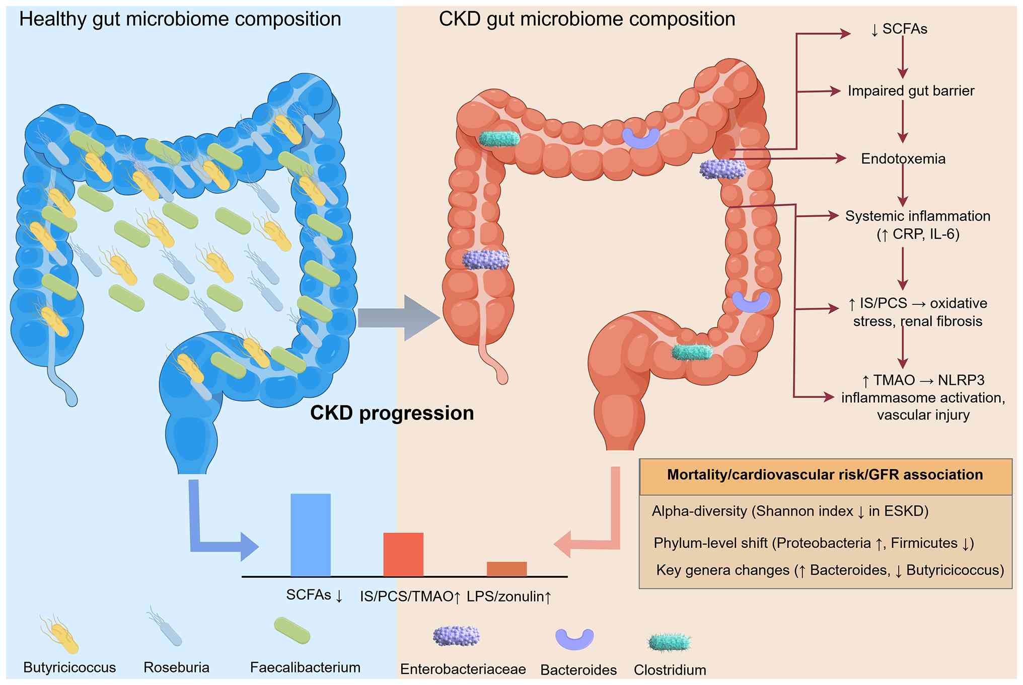 | Figure 1.Gut microbiota dysbiosis in CKD. In
CKD, the gut microbiome exhibits reduced diversity, with a
characteristic shift toward Proteobacteria and uremic
toxin-producing genera (such as Bacteroides), alongside depletion
of butyrate-producing Firmicutes. These changes lead to decreased
SCFAs production, increased generation of protein-bound toxins (IS
and PCS) and TMAO, elevated endotoxemia and impaired intestinal
barrier integrity. The resulting systemic inflammation and direct
renal toxicity accelerate CKD progression and associate with
adverse clinical outcomes, such as cardiovascular events and
increased mortality. Figure created using Figdraw (https://www.figdraw.com/). CKD, chronic kidney
disease; SCFAs, short chain fatty acid; IS, indoxyl sulfate; PCS,
p-cresyl sulfate; TMAO, trimethylamine N-oxide; ESKD, end-stage
kidney disease; CRP, C reactive protein; LPS, lipopolysaccharide;
NLRP3, nod-like receptor family pyrin domain containing 3. |
Phylum-level shifts: Proteobacteria
expansion and butyrate-producer depletion
At the phylum level, three independent Chinese
(40), European (9) and US multi-ethnic cohorts (42) documented a uniform proportional
increase in Proteobacteria (mean relative abundance 28–37% in CKD
vs. 4–9% in controls), which is accompanied by a contraction of
Firmicutes, particularly the Clostridia class. Within Firmicutes,
the butyrate-synthesizing families Lachnospiraceae and
Ruminococcaceae were consistently under-represented, where
pooled random-effects analysis estimated a standardized mean
difference of −0.78 [95% confidence interval (CI), −1.02 to −0.54]
for Lachnospiraceae abundance in CKD vs. controls (40). Butyricicoccus, a genus that
converts dietary fiber to butyrate, fell below the limit of
detection in 64% ESKD samples, whereas it was present in 92% of
healthy controls (9). The
functional consequence is a reduction in fecal butyrate (−38%) and
propionate (−29%), metabolites that normally maintain epithelial
barrier integrity (33) and
suppress renal inflammation through G protein-coupled receptor
(GPR) 43/109A signaling (32).
Uremic toxin-generating taxa:
Concordant enrichment
Culture-independent quantification demonstrated that
bacteria possessing tryptophanase or tyrosine-phenol-lyase genes,
predominantly Bacteroides, Clostridium and some pathogenic
Klebsiella, are selectively expanded in CKD (43). Whole-genome sequencing of 452
Bacteroides isolates recovered from patients showed a
2.3-fold higher copy number of the tryptophanase operon compared
with that of control strains, suggesting genetic as well as
numerical over-representation (43). Consequently, fecal and serum
concentrations of the PBUTs IS and PCS rise in parallel with the
abundance of these genera (44). A
randomized controlled synbiotic trial provided further support for
the biological plausibility of this association (35). An 8-week intervention with the
synbiotic NATUREN G® (containing Lactobacillus
casei LC4P1, Bifidobacterium animalis BLC1,
fructo-oligosaccharides, inulin and a natural antioxidant blend of
quercetin, resveratrol and proanthocyanidins) that reduced
Bacteroides by 18%, simultaneously lowered free serum IS by
26% and PCS by 21%, corroborating earlier observational findings
(35).
TMAO pathway: Divergent findings
explained by diet and geography
TMA producers, including Anaerococcus,
Prevotella and certain Clostridium cluster XIVa, are
generally more abundant in CKD, yet reported plasma TMAO
concentrations vary widely. A pooled analysis previously revealed
that median TMAO was 4.5 µM in European cohorts but 12.8 µM in
East-Asian cohorts (45).
Follow-up metabolomic tracing showed that the difference
disappeared when dietary L-carnitine/choline was restricted to
<5 mg/kg/day, indicating that substrate availability rather than
microbial capacity drives the inter-study discordance (46). Once dietary confounders are
accounted for, renal function remains an independent predictor of
TMAO (45), supporting kidney
clearance as a critical modulator.
Gut-immune interface: Endotoxin,
barrier integrity and inflammation
Expansion of Gram-negative Proteobacteria translates
into elevated fecal and serum lipopolysaccharide (LPS) (47,48).
Mean serum LPS levels in CKD (0.56 EU ml-¹) exceeded control values
(0.21 EU ml−1) by >2.5-fold (47), whereby circulating LPS was
positively associated with zonulin, a marker of tight-junction
disruption (48). Experimental
data corroborate this causality; specifically, LPS infusion in
germ-free mice (animals completely devoid of all microorganisms,
maintained in sterile isolators) reproduced systemic IL-6 and TNF-α
surges, further determining that LPS-driven inflammation occurs
independently of the resident microbiota, whereas butyrate
supplementation (a short-chain fatty acid that upregulates tight
junction proteins and attenuates LPS-induced NF-κB signaling)
restored occludin expression and reduced renal macrophage
infiltration by 34% (33). These
findings align with human studies showing that every 10% increment
in butyrate-producing genera was associated with a 0.07 ng/ml
decrease in high-sensitivity C-reactive protein (CRP) (49). This inverse association is
mechanistically attributable to butyrate, which reinforces
intestinal barrier function by upregulating tight junction proteins
such as occludin and claudin-1, thereby limiting lipopolysaccharide
translocation and systemic CRP elevation (33).
Clinical and prognostic
implications
Low microbial diversity predicted all-cause
mortality in a longitudinal ESKD cohort [hazard ratio (HR), 1.18
per 100-unit decrease in Chao1 index; 95% CI, 1.05–1.33] (47). Conversely, preservation of butyrate
producers conferred a 24% reduction in 3-year cardiovascular events
(HR 0.76; 95% CI, 0.59–0.97) (50). Taken together, current evidence
positions gut dysbiosis not merely as a biochemical curiosity but
as an independent, modifiable risk factor that tracks CKD
progression and amplifies systemic inflammation through toxin
generation and barrier dysfunction.
Confounders and limitations in
microbiome interpretation
Interpretation of CKD-associated microbiome
signatures is complicated by multiple confounding factors,
including dietary restrictions, diabetes, constipation, antibiotic
exposure, phosphate binders, iron supplementation, dialysis
modality and geographic variation (38,51,52).
Dietary protein and fiber intake can directly modulate the
abundance of toxin-producing and SCFA-producing bacteria,
respectively (53,54). In addition, phosphate binders and
iron supplements can alter gut microbial composition and metabolite
profiles (11,51). Furthermore, constipation, which is
prevalent in patients with CKD, promotes the retention and
bacterial fermentation of urea and other nitrogenous substrates,
including dietary aromatic amino acids (including tryptophan and
tyrosine/phenylalanine) that serve as precursors for indoxyl
sulfate and p-cresyl sulfate generation, as well as choline,
L-carnitine and betaine that are converted by gut microbiota to
trimethylamine and subsequently oxidized to trimethylamine N-oxide,
potentially amplifying uremic toxin generation (10,16).
The heterogeneity in sequencing methodologies and analytical
pipelines across studies also complicates direct comparisons
(8,40). Therefore, future studies should
systematically control for these variables and report detailed
medication with the full dietary dataset to enable the more robust
interpretation of gut microbiota alterations in CKD.
In summary, whilst phylum-level shifts
(Proteobacteria expansion and Firmicutes contraction) are
consistently observed across cohorts, genus-level findings vary
markedly, as evidenced by the aforementioned discordant reports
regarding Bacteroides and Clostridium species. Namely, Bacteroides
has been reported as selectively expanded in Taiwanese CKD cohorts
(43), yet no significant
difference in Bacteroidaceae family composition between CKD/ESKD
patients and healthy controls was found in a separate Iranian
cohort (8); similarly, Clostridium
species abundance has been shown to be virtually identical between
CKD/ESRD and healthy groups in one study (5), whereas separate analysis has
implicated Clostridium cluster XIVa as a TMA producer enriched in
CKD (45). These discrepancies
reflect differences in sequencing platforms, analytical pipelines
and population characteristics. The predominance of cross-sectional
designs and inadequate control for dietary and medication
confounders remain the principal limitations. Furthermore, the
clinical significance of these microbial alterations, particularly
whether they represent causal drivers of CKD progression or merely
bystander phenomena, remains uncertain in the absence of
longitudinal intervention studies that modulate specific microbial
targets.
Gut-derived metabolites and their toxic
effects on the kidneys
The accumulation of gut-derived metabolites in CKD
has emerged as a pivotal pathogenic mechanism linking intestinal
dysbiosis to renal injury. As renal clearance declines, microbial
metabolites that are normally excreted by the kidneys accumulate in
the systemic circulation, exerting direct nephrotoxicity and
amplifying systemic inflammation. This section critically reviews
the most clinically relevant gut-derived uremic toxins, IS, PCS and
TMAO, with a focus on their biochemical behavior, renal handling
and toxicological impact across the CKD spectrum. The contrasting
pathogenic and protective mechanisms of key gut-derived metabolites
within the kidney are summarized in Fig. 2.
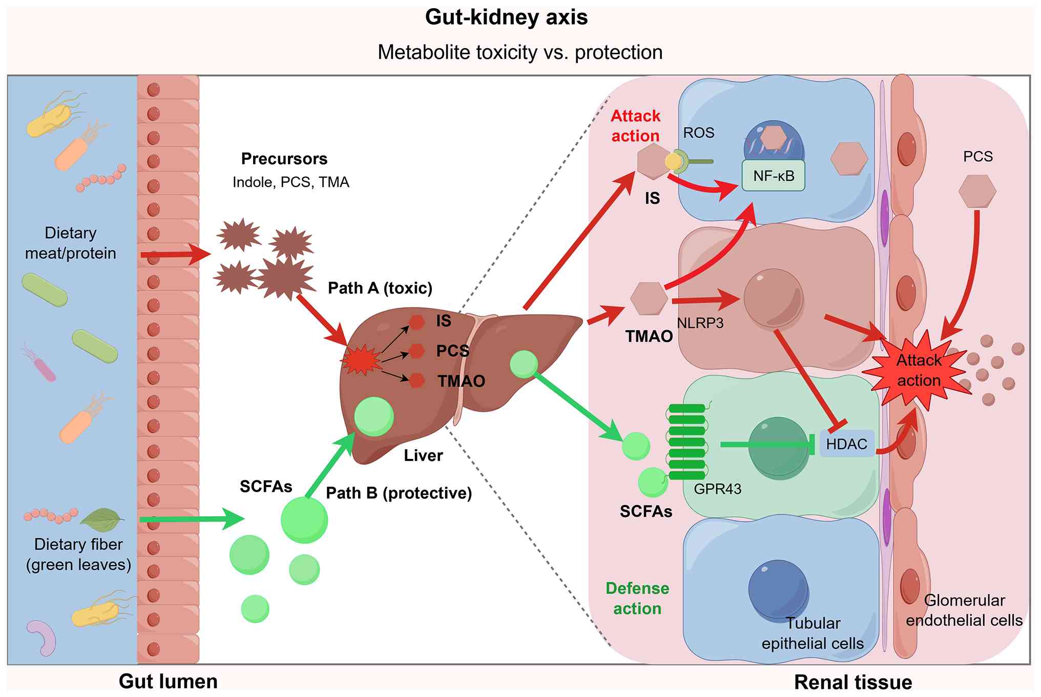 | Figure 2.Gut-kidney axis metabolite toxicity
vs. protection. Dietary protein is fermented by gut microbiota and
transformed in the liver into uremic toxins (PCS, TMAO and IS). In
the kidney, PCS disrupts glomerular endothelial junctions. In
tubular epithelial cells, IS induces ROS and NF-κB activation,
whilst TMAO activates NLRP3. By contrast, dietary fiber-derived
SCFAs exert protective effects on tubular cells by GPR43 binding
and HDAC inhibition to block inflammatory signals. Figure created
using Figdraw (https://www.figdraw.com/). SCFAs, short chain fatty
acid; IS, indoxyl sulfate; PCS, p-cresyl sulfate; TMAO,
trimethylamine N-oxide; TMA, trimethylamine; HDAC, histone
deacetylase; ROS, reactive oxygen species; GPR, G-protein coupled
receptor; NLRP3, nod-like receptor family pyrin domain containing
3. |
PBUT: IS and PCS
IS and PCS are prototypical PBUTs generated through
the bacterial fermentation of dietary tryptophan and tyrosine,
respectively. Their accumulation begins in early CKD stages and
increases exponentially with declining glomerular filtration rate
(55–57). Due to high albumin binding
(>90%), their renal clearance is limited, where conventional
dialysis modalities achieve only modest reductions in their plasma
levels (58–60).
Mechanistically, IS and PCS exert toxicity through
multiple pathways. IS induces the expression of NADPH oxidase 4 and
activates NADPH oxidase activity, triggering oxidative stress and
activating NF-κB-mediated inflammatory cascades in tubular
epithelial cells (61). By
contrast, PCS can disrupt endothelial integrity by phosphorylating
Vascular endothelial-cadherin, increasing vascular permeability and
promoting atherogenesis (62,63).
Furthermore, both toxins can stimulate fibroblast proliferation and
extracellular matrix deposition, accelerating tubulointerstitial
fibrosis (64,65). Notably, IS has been shown to
suppress hypoxia-inducible factor-dependent erythropoietin
expression through activation of the aryl hydrocarbon receptor
pathway, thereby contributing to inadequate erythropoietin
production and erythropoiesis-stimulating agent hyporesponsiveness
in CKD (66).
Clinical studies have consistently associated
elevated IS and PCS levels with adverse renal outcomes. In a
prospective pediatric CKD cohort, IS predicted a faster decline in
eGFR, independent of proteinuria and blood pressure (17). Similarly, PCS levels have been
shown to promote vascular calcification by inducing osteogenic
conversion of vascular smooth muscle cells through reactive oxygen
species (ROS)-dependent activation of the MAPK/NF-κB signaling
pathway and upregulation of runt-related transcription factor 2 and
alkaline phosphatase. In addition, higher PCS levels have been
shown to be independently associated with increased arterial
stiffness in non-dialysis CKD stages 3–5 (67,68).
However, a number of interventional studies have also yielded
ambiguous results. While synbiotic therapy reduced free IS and PCS
levels by ~20–25% (30,35), these reductions did not
consistently translate into improved hard renal endpoints
(including easing of eGFR decline, progression to ESKD, initiation
of dialysis or kidney transplantation), possibly due to the
persistence of protein-bound fractions and the short follow-up
durations.
TMAO: A gut-derived cardiorenal
toxin
TMAO is typically formed from a two-step
microbial-host pathway: Gut bacteria convert dietary choline and
L-carnitine into TMA, which is then oxidized by hepatic flavin
monooxygenases to TMAO (55,69).
Plasma TMAO levels rise markedly in CKD, not only due to impaired
renal excretion but also because of increased microbial TMA
production (26,70). Unlike PBUTs, TMAO is not
protein-bound and is therefore more efficiently cleared by
dialysis. However, its levels remain elevated in patients
undergoing hemodialysis, suggesting sustained microbial generation
(46,71).
The renal toxicity of TMAO is multifaceted. In
rodent models, TMAO administration exacerbated renal fibrosis by
activating the nod-like receptor family pyrin domain containing 3
(NLRP3) inflammasome and enhancing macrophage M2 polarization
through histone H4K12 lactylation (72,73).
It also promotes endothelial dysfunction by inhibiting endothelial
nitric oxide (NO) synthase to reduce NO bioavailability (74). Clinically, elevated TMAO predicts
incident CKD and accelerates eGFR decline in large multi-ethnic
cohorts (26). In children with
CKD, TMAO pathway activity has been associated with subclinical
vascular abnormalities, including increased arterial stiffness and
abnormal ambulatory blood pressure profiles, supporting its
potential role in early vascular remodeling (27).
Dietary interventions targeting TMAO precursors have
shown promise in healthy populations (1,75).
This finding establishes that circulating TMAO may be responsive to
dietary precursor availability, providing a rationale for
investigating whether restricting dietary choline and L-carnitine
intake could lower TMAO burden in patients with CKD (75). In patients with CKD, the strong
association between plasma TMAO and renal function provides a
rationale for investigating dietary strategies to modulate this
pathway (75). However, such diets
are challenging to sustain and may compromise nutritional status.
Pharmacological inhibition of microbial TMA production using
3,3-dimethyl-1-butanol (DMB) was found to attenuate renal fibrosis
in CKD mice (46). However, human
efficacy data for DMB remain limited to in vitro studies
demonstrating that DMB inhibits TMA formation in human fecal
cultures (37), with to the best
of our knowledge, no published clinical trials evaluating its
safety or efficacy in patients with CKD. The absence of human
interventional data represents a key translational gap for this
therapeutic approach.
SCFAs: Protective
counter-regulators
Compared with the aforementioned toxins, SCFAs
(particularly butyrate, propionate and acetate) are microbial
metabolites with documented nephroprotective effects. SCFAs are
produced by the fermentation of dietary fiber by Firmicutes (such
as Lachnospiraceae and Ruminococcaceae), both of
which are depleted in CKD (76).
Butyrate enhances gut barrier integrity by upregulating tight
junction proteins (such as occluding and claudin-1) and suppressing
the systemic translocation of LPS (77).
Experimental data indicate that butyrate attenuates
renal inflammation through GPR43/109A signaling and inhibits
histone deacetylase activity, reducing NF-κB-mediated cytokine
release (32,78). In an RCT, supplementation with
resistant starch increased fecal butyrate levels and reduced serum
IS and PCS concentrations in patients with stage 3b-4 CKD (54). However, the translational potential
of SCFA-augmenting strategies is constrained by inter-individual
variability in microbiota response and the rapid absorption and
metabolism of SCFAs by colonic epithelial cells, which limits their
local availability and systemic delivery (1,5).
Ammonia and hydrogen sulfide
(H2S): Emerging tubular toxins
Beyond the classical PBUTs and TMAO, emerging
evidence implicates ammonia and H2S in CKD-associated tubular
injury. Ammonia is generated by bacterial urease activity that has
been shown to exacerbate tubulointerstitial inflammation by
activating the complement cascade and increasing ammoniagenesis in
residual nephrons (79). H2S is
traditionally viewed as cytoprotective, with endogenous H2S
deficiency implicated in the pathogenesis of CKD (80). However, emerging evidence indicates
that the uremic toxin IS can impair H2S formation in renal tubular
cells by downregulating H2S-producing enzymes, including
cystathionine β-synthase, cystathionine γ-lyase and
3-mercaptopyruvate sulfurtransferase, through aryl hydrocarbon
receptor activation. This in turn compromises the
glutathione-mediated antioxidant defense and contributes to tubular
cell damage (81). Recent
metabolomic profiling identified a marked increase in fecal and
serum H2S levels in CKD stages 4–5, associating with urinary kidney
injury molecule-1 (KIM-1) excretion, a validated marker of tubular
damage (82). However, human data
remain sparse, where the therapeutic modulation of these
metabolites is exploratory at present.
Across these metabolites, the strength of evidence
is strongest for IS and PCS (consistent mechanistic and cohort
data), intermediate for TMAO and weakest for SCFAs (largely
preclinical). Single-time-point measurements, geographic dietary
confounders and the scarcity of studies examining combined
metabolite effects represent pervasive limitations. Notably, the
modest magnitude of toxin reductions achieved by current
interventions (including synbiotics/probiotics and oral adsorbents
such as AST-120; typically 15–30%) raises the question as to
whether such decreases are sufficient to markedly alter disease
trajectory or whether more notable metabolite modulation is
required for clinical benefit (30,35).
Role of the gut-kidney axis in kidney
disease progression
Accumulating evidence is framing the gut-kidney axis
as a self-amplifying circuit rather than a passive uremic toxin
conduit. Intestinal dysbiosis, barrier breakdown and metabolite
accumulation operate sequentially and in parallel to initiate AKI,
propel CKD progression and accelerate the transition to ESKD.
Whilst the primary focus of the present review is CKD, the
inclusion of AKI is justified by the growing recognition that acute
injury can leave a fibrotic legacy that accelerates long-term CKD
progression, where gut dysbiosis induced during AKI can persist
beyond the acute phase, creating a permissive microbiome that fuels
kidney injury persistence (83,84).
Key studies from both experimental models and human cohorts that
have elucidated the causal and progressive role of the gut-kidney
axis are summarized in Table
II.
Dysbiosis as an instigator of AKI
AKI has traditionally been attributed to ischemia,
nephrotoxins or sepsis (3,7). However, the pre-morbid microbial
configuration can independently govern both injury severity and the
fibrotic memory, which can in turn determine the long-term outcome.
Lee et al (83) depleted
murine microbiota with broad-spectrum antibiotics for 3 weeks
before inducing bilateral renal ischemia/reperfusion (IRI).
Antibiotic-treated mice exhibited >50% reduction in fecal
butyrate, a doubling of tubular caspase-3 activity at 24 h and
twice the interstitial collagen III deposition at day-14 compared
with those in vehicle-treated control mice. Subsequent
re-colonization with Lactobacillus acidophilus KBL409
restored butyrate to ≥0.8 μmol/g caecum, halved macrophage
infiltration (CD68+ area, 18.2±2.1 vs. 34.7±3.4%) and preserved
capillary density, indicating that the loss of protective taxa can
worsen AKI independently of systemic infection.
Human data mirror these findings. In a prospective
cohort of 88 patients admitted to the intensive care unit with AKI,
plasma IS measured within 6 h of admission was positively
correlated with urinary KIM-1 at day-3 and predicted persistent CKD
stage ≥3 at 6 months (odds ratio 3.6; 95% CI, 1.4–9.2) after
adjustment for APACHE-II and nephrotoxin exposure (85). Therefore, gut-derived toxins likely
behave as pathophysiological amplifiers rather than the
epiphenomena of reduced renal clearance.
Microbial shifts that propel CKD
progression
Consistent ecological signature across
continents
Meta-analysis of 16S rRNA data from >1,200 fecal
samples revealed a progressive contraction of microbial richness
from CKD G2 onward. Specifically, Shannon index fell by 0.42 (95%
CI, −0.53 to −0.31) in ESKD (86).
In addition, three independent cohorts (China, Europe and the USA)
documented the expansion of Proteobacteria (mean, 28–37 vs. 4–9% in
controls), which is coupled with the depletion of
butyrate-producing Firmicutes (Lachnospiraceae and
Ruminococcaceae) (86–88).
Whole-genome sequencing of 452 Bacteroides isolates from patients
with CKD showed a 2.3-fold duplication of the tryptophanase operon,
paralleling a 2.1-fold higher fecal IS concentration compared with
that in healthy controls (89).
These data support the biological gradient proposal, whereby the
degree of dysbiosis tracks with toxin burden and GFR decline.
TMAO: prospective associations and
mechanistic plausibility
Large-scale metabolomics have closed the causality
loop further. Wang et al (26) followed 3,854 individuals free of
CKD at baseline. Participants in the highest TMAO quartile (≥8.3
µM) displayed a 2.2-fold higher risk of developing incident CKD
(eGFR <60 ml/min) over 10 years after adjustment for protein
intake, diabetes and blood pressure. These observational findings
are complemented by mechanistic animal studies. TMAO activates the
NLRP3 inflammasome in tubular cells, augmenting IL-1β secretion and
epithelial-mesenchymal transition markers (increased vimentin and
decreased E-cadherin) (72).
Genetic ablation of FMO3 or pharmacological inhibition of
microbial TMA-lyases reduced tubulointerstitial fibrosis by 38%
compared with untreated 5/6-nephrectomy control mice and preserved
peritubular capillary density in the 5/6-nephrectomy model
(90). These animal data support
interventional plausibility, but direct human proof of causality in
the form of large randomized trials targeting TMAO production
remains lacking.
AKI-to-CKD transition: A
gut-sensitized window
Complementary murine studies have revealed that
acute uremic dysbiosis can persist beyond the inciting injury,
creating a permissive microbiome that fuels chronicity. Kim et
al (84) demonstrated that
aged mice subjected to unilateral IRI maintained fecal enrichment
of Enterobacteriaceae and elevated systemic LPS levels for
≥4 weeks (0.51±0.06 vs. 0.22±0.04 EU/ml in young mice), paralleling
the sustained tubular hypoxia and exaggerated collagen-I
deposition. By contrast, post-IRI administration of
Lacticaseibacillus rhamnosus L34 restored claudin-1
expression, lowered LPS and halved α-smooth muscle actin-positive
area at day-42, providing direct evidence that microbiota
modulation can interrupt the AKI-to-CKD continuum (91).
Human longitudinal evidence
In the CRIC study (n=2,391), each 1-SD higher serum
IS was associated with a 0.24-ml/min/year faster eGFR decline
independent of proteinuria and blood pressure (92). Similarly, serum TMAO predicted eGFR
loss in children with CKD (ΔeGFR −2.1 vs. −0.8 ml/min/year; highest
vs. lowest TMAO tertile) (93).
These longitudinal cohort findings indicate that higher serum IS
and TMAO levels are associated with a subsequent, faster decline in
eGFR, suggesting that microbial metabolite elevation may precede
and parallel the loss of renal function; however, these
observational data do not establish causality (92,93).
Integrated mechanistic model of
gut-kidney axis-driven CKD progression
The individual pathways aforementioned can be
synthesized into a unified, self-amplifying mechanistic model of
gut-kidney axis-driven CKD progression. This integrated framework
can operate through four interconnected stages.
The initiation phase is characterized by
CKD-associated dysbiosis, wherein the progressive loss of microbial
diversity and depletion of butyrate-producing Firmicutes (such as
Lachnospiraceae and Ruminococcaceae) occur alongside
the expansion of Proteobacteria and uremic toxin-generating taxa,
such as Bacteroides (40,42,43).
This microbial shift reduces SCFA production whilst increasing
generation of toxin precursors. The amplification phase then
involves gut barrier disruption, whereby the depletion of SCFAs
(particularly butyrate) compromises intestinal epithelial integrity
by downregulating tight junction proteins (such as occluding and
claudin-1) and enabling the translocation of bacterial endotoxins
(such as LPS) into the systemic circulation (33,77).
Subsequently, elevated LPS triggers systemic inflammation through
NF-κB activation and cytokine release (IL-6 and TNF-α), further
impairing renal function. The effector phase comes after the
amplication phase, which consists of direct renal injury mediated
by accumulated gut-derived metabolites. IS and PCS activate NADPH
oxidase and NF-κB in tubular epithelial cells, promoting oxidative
stress, inflammation and fibroblast proliferation (61,64).
Concurrently, TMAO activates the NLRP3 inflammasome and promotes
macrophage M2 polarization, accelerating
tubulointerstitial fibrosis (72,73).
Additionally, ammonium accumulation impairs T cell function and
promotes immunosuppressive macrophage polarization, creating an
immunologically permissive environment for progression (11). This is followed by the
self-perpetuation phase to complete the cycle. During this phase,
progressive renal fibrosis reduces glomerular filtration, further
increasing circulating uremic toxin concentrations and perpetuating
dysbiosis, thereby reinforcing the entire cascade (10,13).
This integrated model explains why interventions targeting single
pathways have yielded limited efficacy. The network redundancy and
feedback loops enable compensatory activation at alternative nodes
(5). Accordingly, therapeutic
strategies that simultaneously address multiple components of this
integrated circuit, such as combining dietary fiber to restore SCFA
production, adsorbents to reduce toxin burden and targeted
modulation of microbial metabolism, may be required to disrupt the
self-amplifying cycle and achieve meaningful clinical benefit
(30,39,54).
Novel therapeutic strategies targeting the
gut-kidney axis
Therapeutic efforts have mainly centered on
repopulating the gut with beneficial microbes, supplying
fermentable fibers or limiting systemic uptake of microbial toxins
through adsorbents, enzyme inhibitors (46,90)
or FMT (91). Published studies
have indicated these measures to lower free but not protein bound
uremic solutes, particularly when renal function can still be
measured (3,30,46).
To facilitate critical appraisal, the evidence for each
intervention is stratified in this section according to the
strength of available data: Interventions with human RCT evidence
(mainly biochemical outcomes); interventions with
preclinical/animal evidence only; and experimental approaches. An
overview of clinical studies investigating various gut
microbiome-targeted interventions, including their designs and key
outcomes, is provided in Table
III.
Probiotics and synbiotics
Early open-label studies in Mexican and Italian
hemodialysis cohorts showed that a 4-week course of a multi-strain
synbiotic (Lactobacillus casei, Bifidobacterium breve + 4 g
fructo-oligosaccharide) lowered plasma PCS levels by 15–20% whilst
modestly increasing stool Bifidobacteria counts (94,95).
These findings were corroborated by a 2014 RCT that used a
probiotic mixture specifically formulated for uraemia
(Renadyl™), which observed a 25% reduction in serum IS
together with improved quality-of-life scores in 42 patients
undergoing peritoneal dialysis (96).
The largest trial to date, SYNERGY (n=30 CKD stage
3b-4), employed a double-blind, placebo-controlled, cross-over
design and documented a marked 14% fall in free IS after 6 weeks of
synbiotic yoghurt, although no change in total (protein-bound) IS
was detected (97). A 2021 pilot
study extended these observations to advanced CKD (stage IIIb-IV)
by demonstrating a 26% decrease in free IS and a parallel 21% drop
in PCS following a novel synbiotic capsule containing
Lactiplantibacillus plantarum + resistant dextrin. In
addition, intestinal permeability, as measured by the
lactulose/rhamnose ratio, simultaneously improved (35). Another previous meta-analysis of 13
eligible RCTs (n=550) confirmed a pooled standardized mean
difference of −0.46 (95% CI, −0.72 to −0.20) for PCS and −0.38 (95%
CI, −0.65 to −0.11) for IS, with negligible heterogeneity
(I2=12%) (30).
Notably, the magnitude of toxin reduction was consistently smaller
in patients with an eGFR <20 ml/min, implying that retained
renal clearance contributes to the observed effect.
However, inflammatory endpoints have been less
uniform. A number of previous clinical trials have reported a
10–15% fall in CRP (98,99), whereas others have reported no
significant change (100–102). A 2023 aggregate-level
meta-analysis incorporating 18 RCTs (n=884) concluded that
probiotic/synbiotic supplementation reduced CRP by a modest 0.43
mg/l (95% CI, −0.78 to −0.08) but had no detectable effect on IL-6
or TNF-α (39). The most
comprehensive synthesis to date, the 2023 Cochrane review by Cooper
et al (39), which included
18 RCTs (n=884), concluded that whilst synbiotics, prebiotics and
probiotics may reduce certain uremic toxins and inflammatory
markers, the evidence for clinically meaningful benefits on hard
renal outcomes, such as slowing eGFR decline, reducing
cardiovascular events or mortality, remains insufficient (39). Taken together, current evidence
supports synbiotics as a safe, low-cost modality that lowers free,
but not protein-bound, uremic solutes. The clinical relevance of
the incremental toxin reduction (~20%) remains uncertain,
particularly with respect to hard renal endpoints.
Prebiotics and dietary fiber
interventions
Resistant starch type 2 (HAM-RS2) and inulin-type
fructans have been the most frequently tested prebiotics. A 2018
randomized, double-blind trial in 30 patients undergoing
haemodialysis showed that 20 g/day HAM-RS2 for 6 weeks raised fecal
butyrate levels by 60% and decreased plasma IS from 21.4 to 16.8 µM
without affecting PCS (102).
These findings were essentially reproduced in a crossover study of
patients undergoing peritoneal dialysis using 15 g/day inulin,
where IS fell by 18% and stool Faecalibacterium prausnitzii
increased 2.3-fold (103).
A systematic review and meta-analysis of 19
fiber-feeding RCTs (n=677) reported a pooled reduction in the IS of
4.27 µM (95% CI, −6.38 to −2.16) and in PCS of 5.91 µM (95% CI,
−9.44 to −2.38), with greater efficacy observed when baseline fiber
intake was <15 g/day (104).
By contrast, two trials that provided <10 g/day fermentable
fiber failed to modify either metabolite, underscoring the
importance of dose and individual dietary pattern (105,106). Notably, none of the published
fiber studies have exceeded a duration of 12 weeks. Therefore,
long-term effects on renal function decline remain speculative. In
addition, as with probiotics, the 2023 Cochrane review concluded
that evidence for patient-important outcomes (eGFR decline,
dialysis initiation or mortality) remains insufficient (39).
Oral adsorbents
AST-120 has been evaluated in two multinational
Easing Pelvic Pain Interventions Clinical Research Program (EPPIC)
trials (EPPIC-1 and EPPIC-2) that together randomized 2,035
patients with CKD stages 3–4 (107,108), as well as a separate Korean
multicenter RCT (n=579) in patients with advanced renal dysfunction
(109). The EPPIC program
detected a 15% reduction in serum IS but found no difference in the
composite renal end-point (doubling of creatinine, dialysis or
death) after 12 months (108).
Post-hoc analyses revealed that participants with the highest
baseline IS (>10 µM) experienced a 26% slower annual eGFR
decline (−2.1 vs −2.8 ml/min/year) (110). A more recent Korean
dose-comparison RCT (n=210) confirmed that 6 g/day AST-120 achieved
a 30% IS reduction, yet no additional benefit was observed at 9
g/day and gastrointestinal adverse events increased
dose-dependently (111).
Novel bamboo-derived activated carbon lowered PCS
by 22% in a 2025 pilot study, but the trial was un-blinded and
limited to 4 weeks (112).
Collectively, adsorbents can reproducibly attenuate IS and PCS,
though their capacity to slow CKD progression appears modest and is
largely restricted to patients with elevated baseline toxin
concentrations.
Microbiome-targeted small
molecules
Beyond adsorbents, the first attempts to inhibit
toxin generation have focused on the hepatic enzyme flavin
monooxygenase 3 and on gut microbial TMA-lyases. However, such
approaches currently only have evidence limited to preclinical
studies. DMB is a structural analogue of choline that has been
shown to decrease TMAO by ~33% in 5/6 nephrectomised mice and
attenuated tubulo-interstitial fibrosis, an effect that was lost
when the inhibitor was withdrawn (46). By contrast, rifaximin is an oral
rifamycin derivative with minimal systemic absorption. It was
reported to lower fecal TMAO formation by 18% in a 4-week
placebo-controlled pilot. However, Enterobacteriaceae
abundance doubled, raising concerns about collateral ecological
damage (113). No adverse hepatic
or cardiac signals have been reported for either compound (46,113), but the follow-up period has not
reached >2 months. Therefore, longer, double-blind trials
powered for renal endpoints are needed to define whether
pharmacological suppression of TMAO synthesis translates into
slower CKD progression.
FMT
FMT and engineered probiotics remain experimental
interventions for CKD. In 2024, 12 patients with stage 4 CKD
received a single nasoduodenal infusion of stool from lean healthy
donors after bowel lavage. Within 1 week, butyrate producers, such
as Faecalibacterium and Roseburia, rose from 4 to 15%
of total sequences, accompanied by a 19% fall in serum IS (114). TMAO, however, increased
transiently in two recipients, emphasizing that donor selection and
post-infusion diet may shape metabolite trajectories. A parallel
phase I study using encapsulated frozen donor material reported
similar enrichment of SCFA producers, but no notable biochemical
changes were observed when the grafted microbial community,
including the transferred SCFA-producing taxa, contracted after 4
weeks, likely due to insufficient engraftment or colonization
resistance from the recipient's native microbiota (114). No serious infectious
complications have been documented, but recipients of
immunosuppressed dialysis were excluded. Repeated infusions,
combined with dietary counselling and rigorous donor screening, are
now being evaluated in an ongoing multicenter trial (NCT05923412).
Until these data are available, FMT remains an experimental
intervention best restricted to clinical protocols with close
safety oversight. Similarly, engineered probiotics designed to
specifically degrade uremic toxin precursors (such as
tryptophanase-negative bacteria) and postbiotics (bacterial-derived
inactivated cells or metabolites) represent emerging strategies
that have shown promise in preclinical models but require rigorous
clinical evaluation (115,116).
By comparing these interventions,
probiotic/synbiotic trials are numerous but involve relatively
small sample sizes (n=20-50) and were followed up short-term,
whereas adsorbent trials are larger and longer but failed to show
hard endpoint benefits. Across all approaches, insufficient power
for renal outcomes, reliance on surrogate endpoints and short
follow-up durations remain critical limitations. Notably, the
consistent discordance between biochemical efficacy (toxin
reduction) and clinical efficacy (eGFR preservation) across
multiple intervention types suggests that the relationship between
uremic toxins and CKD progression may be more complex than
currently appreciated. Single-target interventions are now likely
be insufficient to disrupt the multifactorial pathogenic
network.
Future directions and research gaps
Despite considerable advances in elucidating the
gut-kidney axis in CKD, the current understanding remains
fragmented, such that the translation of microbiota-targeted
strategies into routine clinical practice faces notable hurdles.
the majority of interventional studies, whilst reporting modest
efficacy in reducing circulating levels of free uremic toxins (such
as IS and PCS), have largely failed to reveal consistent benefits
on hard clinical endpoints (such as slowing the decline in the eGFR
or reducing cardiovascular events) (30,39,117). This discrepancy underscores
critical research gaps. Although synbiotic and probiotic
interventions in patients undergoing hemodialysis and pre-dialysis
have consistently lowered inflammatory markers, such as CRP and
specific toxins (118,119), their effects are heterogeneous
and frequently uncoupled from meaningful changes in renal function
(120,121). Similarly, dietary fiber
interventions with resistant starch or inulin reliably increase
beneficial SCFA producers, such as Faecalibacterium
prausnitzii and reduce certain uremic toxins (122,123), their long-term impact on CKD
progression remains unknown and responses vary with baseline diet
and microbiota composition (54,124). These inconsistencies highlight
the nascent state of the field and point to several prioritized
avenues for future investigation. To move the field forward, future
research should prioritize a number of key areas.
Key areas needing improvement
Large longitudinal multi-omics cohorts with
standardized collection of dietary and medication data are needed.
Current evidence is largely derived from cross-sectional studies
with heterogeneous populations and incomplete confounder adjustment
(51,52). Prospective cohorts integrating
metagenomic, metabolomic and host genetic data, with detailed
records of diet, medications (including phosphate binders, iron
supplements and antibiotics), dialysis modality and constipation
status, would enable robust identification of causal microbial
signatures and their interactions with host factors (125,126).
Adequately powered RCTs with sufficient duration
and clinically meaningful endpoints are also required. The majority
of published intervention studies have been short-term (≤12 weeks)
and have relied on biochemical surrogate endpoints, such as toxin
reduction or inflammatory markers (39,104). Future trials should be designed
with longer follow-up (≥12 months) and hard outcomes including eGFR
slope, progression to ESKD, cardiovascular events and mortality.
Such trials should also incorporate mechanistic sub-studies to
elucidate the biological pathways through which the interventions
exert their effects (125,127).
Personalized nutrition and microbiome-informed
intervention selection represent a promising frontier. The current
‘one-size-fits-all’ approach fails to account for profound
inter-individual variability in gut microbial composition,
metabolic capacity and host response (51,52).
Emerging evidence suggests that baseline microbial signatures may
predict therapeutic responsiveness. Supporting this, the efficacy
of resistant starch in elevating butyrate and reducing IS appears
more pronounced in patients with low initial fiber intake and
specific microbial configurations (54,124). Machine-learning models trained on
large, well-phenotyped cohorts could identify predictive biomarkers
to guide intervention selection (125,126). However, precision nutrition
strategies, tailoring dietary recommendations based on an
individual's microbiome and metabolomic profile, require rigorous
testing in randomized trials (128).
Safety surveillance for probiotics, FMT and
engineered microbiota must be prioritized. Long-term safety data in
the CKD population are scarce, with isolated reports of bacteremia
linked to probiotic strains in immunocompromised hosts (129). Rigorous post-marketing
surveillance and long-term extension studies of interventional
trials are therefore essential (38,39).
The predictive potential of microbiome profiling also raises
ethical questions regarding data privacy and psychological impact,
while integrating microbiome diagnostics into clinical practice
requires new resources, training and multidisciplinary
collaboration (130,131).
Mechanistic human studies using metabolomics,
organoids and kidney-on-chip models should complement clinical
trials. Animal models, whilst instrumental in establishing causal
links (132,133), inadequately recapitulate the
complexity of human CKD. Human-centric platforms, including
organ-on-a-chip systems incorporating patient-derived renal cells
and microbiota, could bridge the translational gap (125,127). Postbiotics and engineered
probiotics designed to degrade uremic toxin precursors represent
emerging strategies warranting exploration (115,116) and combination approaches (such as
personalized diet with targeted probiotic and adsorbent) may yield
synergistic effects (134,135).
In conclusion, advancing the field requires moving
from broad, non-specific interventions to a precision medicine
paradigm. By addressing these research priorities through
large-scale longitudinal cohorts, rigorous long-term RCTs,
personalized approaches, safety surveillance and mechanistic human
studies, the field can evolve toward delivering targeted, effective
and safe microbiota-based therapies that alter the natural history
of CKD.
Conclusions
The gut-kidney axis is a well-established
pathogenic pathway in CKD, linking intestinal dysbiosis to uremic
toxin accumulation and loss of protective SCFAs. Observational and
mechanistic evidence strongly supports this axis, yet its clinical
translation remains uncertain. Current interventions reduce free
toxin levels only modestly and lack consistent evidence of benefits
for hard outcomes such as decreases in the eGFR or cardiovascular
events. Individual responses vary, and long-term safety data are
scarce. Therefore, routine use of probiotics, prebiotics or
adsorbents to slow CKD progression cannot be recommended outside
supportive dietary care or clinical trials.
Acknowledgements
Not applicable.
Funding
Funding: No funding was received.
Availability of data and materials
Not applicable.
Authors' contributions
YW contributed to the conception and design of the
manuscript, performed the literature search and integrated
mechanistic, clinical and therapeutic findings, whilst also having
drafted the manuscript. HZ contributed to literature screening and
critically assessed study quality and reconciled conflicting
findings, whilst having critically revised the manuscript. NL
participated in literature screening, verified the accuracy of
summarized findings, assisted in assessing the strength of evidence
and assisted in drafting and revising the manuscript. DY conceived
and designed the review, supervised the entire process, critically
revised the manuscript for important intellectual content and
provided final approval of the version to be published. All authors
read and approved the final version of the manuscript. Data
authentication is not applicable.
Ethics approval and consent to
participate
Not applicable.
Patient consent for publication
Not applicable.
Competing interests
The authors declare that they have no competing
interests.
Glossary
Abbreviations
Abbreviations:
|
AKI
|
acute kidney injury
|
|
AST-120
|
spherical activated carbon
adsorbent
|
|
CKD
|
chronic kidney disease
|
|
CRP
|
C-reactive protein
|
|
DMB
|
3,3-dimethyl-1-butanol
|
|
eGFR
|
estimated glomerular filtration
rate
|
|
ESKD
|
end-stage kidney disease
|
|
FMT
|
fecal microbiota transplantation
|
|
GPR
|
G protein-coupled receptor
|
|
H2S
|
hydrogen sulfide
|
|
IS
|
indoxyl sulfate
|
|
LPS
|
lipopolysaccharide
|
|
NLRP3
|
NOD-like receptor family pyrin domain
containing 3
|
|
PBUTs
|
protein-bound uremic toxins
|
|
PCS
|
p-cresyl sulfate
|
|
RCT
|
randomized controlled trial
|
|
SCFAs
|
short-chain fatty acids
|
|
TMA
|
trimethylamine
|
|
TMAO
|
trimethylamine N-oxide
|
References
|
1
|
Colombo I, Aiello-Battan F, Elena R, Ruiz
A, Petraglia L and Musso CG: Kidney-gut crosstalk in renal disease.
Ir J Med Sci. 190:1205–1212. 2021. View Article : Google Scholar : PubMed/NCBI
|
|
2
|
Rysz J, Franczyk B, Ławiński J, Olszewski
R, Ciałkowska-Rysz A and Gluba-Brzózka A: The impact of CKD on
uremic toxins and gut microbiota. Toxins (Basel). 13:2522021.
View Article : Google Scholar : PubMed/NCBI
|
|
3
|
Foresto-Neto O, Ghirotto B and Câmara NOS:
Renal sensing of bacterial metabolites in the gut-kidney axis.
Kidney360. 2:1501–1509. 2021. View Article : Google Scholar : PubMed/NCBI
|
|
4
|
Lowenstein J and Nigam SK: Uremic toxins
in organ crosstalk. Front Med (Lausanne). 8:5926022021. View Article : Google Scholar : PubMed/NCBI
|
|
5
|
Noce A, Marchetti M, Marrone G, Di Renzo
L, Di Lauro M, Di Daniele F, Albanese M, Di Daniele N and De
Lorenzo A: Link between gut microbiota dysbiosis and chronic kidney
disease. Eur Rev Med Pharmacol Sci. 26:2057–2074. 2022.PubMed/NCBI
|
|
6
|
Wu IW, Gao SS, Chou HC, Yang HY, Chang LC,
Kuo YL, Dinh MCV, Chung WH, Yang CW, Lai HC, et al: Integrative
metagenomic and metabolomic analyses reveal severity-specific
signatures of gut microbiota in chronic kidney disease.
Theranostics. 10:5398–5411. 2020. View Article : Google Scholar : PubMed/NCBI
|
|
7
|
Sato N, Kakuta M, Hasegawa T, Yamaguchi R,
Uchino E, Murashita K, Nakaji S, Imoto S, Yanagita M and Okuno Y:
Metagenomic profiling of gut microbiome in early chronic kidney
disease. Nephrol Dial Transplant. 36:1675–1684. 2021. View Article : Google Scholar : PubMed/NCBI
|
|
8
|
Zhao J, Ning X, Liu B, Dong R, Bai M and
Sun S: Specific alterations in gut microbiota in patients with
chronic kidney disease: An updated systematic review. Ren Fail.
43:102–112. 2021. View Article : Google Scholar : PubMed/NCBI
|
|
9
|
Gryp T, Faust K, Van Biesen W, Huys GRB,
Verbeke F, Speeckaert M, Raes J, Vaneechoutte M, Joossens M and
Glorieux G: Gut microbiome profiling uncovers a lower abundance of
Butyricicoccus in advanced stages of chronic kidney disease.
J Pers Med. 11:11182021. View Article : Google Scholar : PubMed/NCBI
|
|
10
|
Ikee R, Sasaki N, Yasuda T and Fukazawa S:
Chronic kidney disease, gut dysbiosis, and constipation: A
Burdensome triplet. Microorganisms. 8:18622020. View Article : Google Scholar : PubMed/NCBI
|
|
11
|
Bhargava S, Merckelbach E, Noels H, Vohra
A and Jankowski J: Homeostasis in the gut microbiota in chronic
kidney disease. Toxins (Basel). 14:6482022. View Article : Google Scholar : PubMed/NCBI
|
|
12
|
Glorieux G, Gryp T and Perna A:
Gut-derived metabolites and their role in immune dysfunction in
chronic kidney disease. Toxins (Basel). 12:2452020. View Article : Google Scholar : PubMed/NCBI
|
|
13
|
Graboski AL and Redinbo MR: Gut-derived
protein-bound uremic toxins. Toxins (Basel). 12:5902020. View Article : Google Scholar : PubMed/NCBI
|
|
14
|
Fernandes ALF, Borges NA, Black AP, Anjos
JD, Silva GSD, Nakao LS and Mafra D: Dietary intake of tyrosine and
phenylalanine, and p-cresyl sulfate plasma levels in non-dialyzed
patients with chronic kidney disease. J Bras Nefrol. 42:307–314.
2020.(In English, Portuguese). View Article : Google Scholar : PubMed/NCBI
|
|
15
|
Gryp T, De Paepe K, Vanholder R, Kerckhof
FM, Van Biesen W, Van de Wiele T, Verbeke F, Speeckaert M, Joossens
M, Couttenye MM, et al: Gut microbiota generation of protein-bound
uremic toxins and related metabolites is not altered at different
stages of chronic kidney disease. Kidney Int. 97:1230–1242. 2020.
View Article : Google Scholar : PubMed/NCBI
|
|
16
|
Gryp T, Huys GRB, Joossens M, Van Biesen
W, Glorieux G and Vaneechoutte M: Isolation and quantification of
uremic toxin precursor-generating gut bacteria in chronic kidney
disease patients. Int J Mol Sci. 21:19862020. View Article : Google Scholar : PubMed/NCBI
|
|
17
|
Holle J, Kirchner M, Okun J, Bayazit AK,
Obrycki L, Canpolat N, Bulut IK, Azukaitis K, Duzova A, Ranchin B,
et al: Serum indoxyl sulfate concentrations associate with
progression of chronic kidney disease in children. PLoS One.
15:e02404462020. View Article : Google Scholar : PubMed/NCBI
|
|
18
|
Sun CY, Li JR, Wang YY, Lin SY, Ou YC, Lin
CJ, Wang JD, Liao SL and Chen CJ: Indoxyl sulfate caused behavioral
abnormality and neurodegeneration in mice with unilateral
nephrectomy. Aging (Albany NY). 13:6681–6701. 2021. View Article : Google Scholar : PubMed/NCBI
|
|
19
|
Filipska I, Winiarska A, Knysak M and
Stompór T: Contribution of gut microbiota-derived uremic toxins to
the cardiovascular system mineralization. Toxins (Basel).
13:2742021. View Article : Google Scholar : PubMed/NCBI
|
|
20
|
Yin L, Li X, Ghosh S, Xie C, Chen J and
Huang H: Role of gut microbiota-derived metabolites on vascular
calcification in CKD. J Cell Mol Med. 25:1332–1341. 2021.
View Article : Google Scholar : PubMed/NCBI
|
|
21
|
Arias N, Arboleya S, Allison J,
Kaliszewska A, Higarza SG, Gueimonde M and Arias JL: The
relationship between choline bioavailability from diet, intestinal
microbiota composition, and its modulation of human diseases.
Nutrients. 12:23402020. View Article : Google Scholar : PubMed/NCBI
|
|
22
|
Naghipour S, Cox AJ, Peart JN, Du Toit EF
and Headrick JP: Trimethylamine N-oxide: Heart of the
microbiota-CVD nexus? Nutr Res Rev. 34:125–146. 2021. View Article : Google Scholar : PubMed/NCBI
|
|
23
|
Lee Y, Nemet I, Wang Z, Lai HTM, de
Oliveira Otto MC, Lemaitre RN, Fretts AM, Sotoodehnia N, Budoff M,
DiDonato JA, et al: Longitudinal plasma measures of trimethylamine
n-oxide and risk of atherosclerotic cardiovascular disease events
in community-based older adults. J Am Heart Assoc. 10:e0206462021.
View Article : Google Scholar : PubMed/NCBI
|
|
24
|
Zeng Y, Guo M, Fang X, Teng F, Tan X, Li
X, Wang M, Long Y and Xu Y: Gut microbiota-derived trimethylamine
N-oxide and kidney function: A systematic review and meta-analysis.
Adv Nutr. 12:1286–1304. 2021. View Article : Google Scholar : PubMed/NCBI
|
|
25
|
Canyelles M, Borràs C, Rotllan N, Tondo M,
Escolà-Gil JC and Blanco-Vaca F: Gut microbiota-derived TMAO: A
causal factor promoting atherosclerotic cardiovascular disease? Int
J Mol Sci. 24:19402023. View Article : Google Scholar : PubMed/NCBI
|
|
26
|
Wang M, Tang WHW, Li XS, de Oliveira Otto
MC, Lee Y, Lemaitre RN, Fretts A, Nemet I, Sotoodehnia N, Sitlani
CM, et al: The gut microbial metabolite trimethylamine N-oxide,
incident CKD, and kidney function decline. J Am Soc Nephrol.
35:749–760. 2024. View Article : Google Scholar : PubMed/NCBI
|
|
27
|
Hsu CN, Chang-Chien GP, Lin S, Hou CY, Lu
PC and Tain YL: Association of trimethylamine, trimethylamine
n-oxide, and dimethylamine with cardiovascular risk in children
with chronic kidney disease. J Clin Med. 9:3362020. View Article : Google Scholar : PubMed/NCBI
|
|
28
|
Li Y, Lu H, Guo J, Zhang M, Zheng H, Liu Y
and Liu W: Gut microbiota-derived trimethylamine N-oxide is
associated with the risk of all-cause and cardiovascular mortality
in patients with chronic kidney disease: A systematic review and
dose-response meta-analysis. Ann Med. 55:22155422023. View Article : Google Scholar : PubMed/NCBI
|
|
29
|
Beker BM, Colombo I, Gonzalez-Torres H and
Musso CG: Decreasing microbiota-derived uremic toxins to improve
CKD outcomes. Clin Kidney J. 15:2214–2219. 2022. View Article : Google Scholar : PubMed/NCBI
|
|
30
|
Chen L, Shi J, Ma X, Shi D and Qu H:
Effects of microbiota-driven therapy on circulating indoxyl sulfate
and P-cresyl sulfate in patients with chronic kidney disease: A
systematic review and meta-analysis of randomized controlled
trials. Adv Nutr. 13:1267–1278. 2022. View Article : Google Scholar : PubMed/NCBI
|
|
31
|
Sumida K, Lau WL, Kalantar-Zadeh K and
Kovesdy CP: Novel intestinal dialysis interventions and microbiome
modulation to control uremia. Curr Opin Nephrol Hypertens.
31:82–91. 2022. View Article : Google Scholar : PubMed/NCBI
|
|
32
|
Li YJ, Chen X, Kwan TK, Loh YW, Singer J,
Liu Y, Ma J, Tan J, Macia L, Mackay CR, et al: Dietary fiber
protects against diabetic nephropathy through short-chain fatty
acid-mediated activation of G protein-coupled receptors GPR43 and
GPR109A. J Am Soc Nephrol. 31:1267–1281. 2020. View Article : Google Scholar : PubMed/NCBI
|
|
33
|
Liu Y, Li YJ, Loh YW, Singer J, Zhu W,
Macia L, Mackay CR, Wang W, Chadban SJ and Wu H: Fiber derived
microbial metabolites prevent acute kidney injury through G-protein
coupled receptors and HDAC inhibition. Front Cell Dev Biol.
9:6486392021. View Article : Google Scholar : PubMed/NCBI
|
|
34
|
Magliocca G, Mone P, Di Iorio BR, Heidland
A and Marzocco S: Short-chain fatty acids in chronic kidney
disease: Focus on inflammation and oxidative stress regulation. Int
J Mol Sci. 23:53542022. View Article : Google Scholar : PubMed/NCBI
|
|
35
|
Cosola C, Rocchetti MT, di Bari I,
Acquaviva PM, Maranzano V, Corciulo S, Di Ciaula A, Di Palo DM, La
Forgia FM, Fontana S, et al: An innovative synbiotic formulation
decreases free serum indoxyl sulfate, small intestine permeability
and ameliorates gastrointestinal symptoms in a randomized pilot
trial in stage IIIb-IV CKD patients. Toxins (Basel). 13:3342021.
View Article : Google Scholar : PubMed/NCBI
|
|
36
|
Hsu CK, Su SC, Chang LC, Yang KJ, Lee CC,
Hsu HJ, Chen YT, Sun CY and Wu IW: Oral absorbent AST-120 is
associated with compositional and functional adaptations of gut
microbiota and modification of serum short and medium-chain fatty
acids in advanced CKD patients. Biomedicines. 10:22342022.
View Article : Google Scholar : PubMed/NCBI
|
|
37
|
Favero C, Ortiz A and Sanchez-Niño MD:
Probiotics for kidney disease. Clin Kidney J. 15:1981–1986. 2022.
View Article : Google Scholar : PubMed/NCBI
|
|
38
|
Tian N, Li L, Ng JK and Li PK: The
potential benefits and controversies of probiotics use in patients
at different stages of chronic kidney disease. Nutrients.
14:40442022. View Article : Google Scholar : PubMed/NCBI
|
|
39
|
Cooper TE, Khalid R, Chan S, Craig JC,
Hawley CM, Howell M, Johnson DW, Jaure A, Teixeira-Pinto A and Wong
G: Synbiotics, prebiotics and probiotics for people with chronic
kidney disease. Cochrane Database Syst Rev.
10:CD0136312023.PubMed/NCBI
|
|
40
|
Chen TH, Liu CW, Ho YH, Huang CK, Hung CS,
Smith BH and Lin JC: Gut microbiota composition and its metabolites
in different stages of chronic kidney disease. J Clin Med.
10:38812021. View Article : Google Scholar : PubMed/NCBI
|
|
41
|
Wu IW, Chang LC, Wu YL, Yang HY, Twu YC,
Tsai PY, Paulus S, Resnick R, Chung WH, Yang CW, et al: Gut flora
metagenomic analysis coupled with metabolic and deep immune
profiling in chronic kidney disease. Nephrol Dial Transplant.
39:1333–1343. 2024. View Article : Google Scholar : PubMed/NCBI
|
|
42
|
Peters BA, Qi Q, Usyk M, Daviglus ML, Cai
J, Franceschini N, Lash JP, Gellman MD, Yu B, Boerwinkle E, et al:
Association of the gut microbiome with kidney function and damage
in the hispanic community health study/study of latinos (HCHS/SOL).
Gut Microbes. 15:21866852023. View Article : Google Scholar : PubMed/NCBI
|
|
43
|
Shivani S, Kao CY, Chattopadhyay A, Chen
JW, Lai LC, Lin WH, Lu TP, Huang IH, Tsai MH, Teng CH, et al:
Uremic toxin-producing bacteroides species prevail in the gut
microbiota of Taiwanese CKD patients: An analysis using the New
Taiwan microbiome baseline. Front Cell Infect Microbiol.
12:7262562022. View Article : Google Scholar : PubMed/NCBI
|
|
44
|
Lin X, Liang W, Li L, Xiong Q, He S, Zhao
J, Guo X, Xiang S, Zhang P, Wang H, et al: The accumulation of gut
microbiome-derived indoxyl sulfate and P-Cresyl sulfate in patients
with end-stage renal disease. J Ren Nutr. 32:578–586. 2022.
View Article : Google Scholar : PubMed/NCBI
|
|
45
|
Cheng E, Hung SC and Lin TY: Association
of trimethylamine N-oxide and metabolites with kidney function
decline in patients with chronic kidney disease. Clin Nutr.
44:239–247. 2025. View Article : Google Scholar : PubMed/NCBI
|
|
46
|
Zhang W, Miikeda A, Zuckerman J, Jia X,
Charugundla S, Zhou Z, Kaczor-Urbanowicz KE, Magyar C, Guo F, Wang
Z, et al: Inhibition of microbiota-dependent TMAO production
attenuates chronic kidney disease in mice. Sci Rep. 11:5182021.
View Article : Google Scholar : PubMed/NCBI
|
|
47
|
Lin TY, Wu PH, Lin YT and Hung SC: Gut
dysbiosis and mortality in hemodialysis patients. NPJ Biofilms
Microbiomes. 7:202021. View Article : Google Scholar : PubMed/NCBI
|
|
48
|
Georgopoulou GA, Papasotiriou M, Bosgana
P, de Lastic AL, Koufou EE, Papachristou E, Goumenos DS, Davlouros
P, Kourea E, Zolota V, et al: Altered expression of intestinal
tight junctions in patients with chronic kidney disease: A
pathogenetic mechanism of intestinal hyperpermeability.
Biomedicines. 12:3682024. View Article : Google Scholar : PubMed/NCBI
|
|
49
|
McFarlane C, Krishnasamy R, Stanton T,
Savill E, Snelson M, Mihala G, Morrison M, Johnson DW and Campbell
KL: Diet quality and protein-bound uraemic toxins: Investigation of
novel risk factors and the role of microbiome in chronic kidney
disease. J Ren Nutr. 32:542–551. 2022. View Article : Google Scholar : PubMed/NCBI
|
|
50
|
Hellman T, Yeo LF, Palmu J, Havulinna A,
Jousilahti P, Laitinen V, Pärnänen K, Salomaa V, Lahti L, Knight R
and Niiranen T: Gut microbiome as a risk factor for future CKD.
Kidney Int Rep. 10:1673–1682. 2025. View Article : Google Scholar : PubMed/NCBI
|
|
51
|
Pantazi AC, Kassim MAK, Nori W, Tuta LA,
Mihai CM, Chisnoiu T, Balasa AL, Mihai L, Lupu A, Frecus CE, et al:
Clinical perspectives of gut microbiota in patients with chronic
kidney disease and end-stage kidney disease: Where do we stand?
Biomedicines. 11:24802023. View Article : Google Scholar : PubMed/NCBI
|
|
52
|
Laiola M, Koppe L, Larabi A, Thirion F,
Lange C, Quinquis B, David A, Le Chatelier E, Benoit B, Sequino G,
et al: Toxic microbiome and progression of chronic kidney disease:
Insights from a longitudinal CKD-microbiome study. Gut.
74:1624–1637. 2025. View Article : Google Scholar : PubMed/NCBI
|
|
53
|
El Amouri A, Snauwaert E, Foulon A, Vande
Moortel C, Van Dyck M, Van Hoeck K, Godefroid N, Glorieux G, Van
Biesen W, Vande Walle J, et al: Dietary Fibre intake is associated
with serum levels of uraemic toxins in children with chronic kidney
disease. Toxins (Basel). 13:2252021. View Article : Google Scholar : PubMed/NCBI
|
|
54
|
Headley SA, Chapman DJ, Germain MJ, Evans
EE, Madsen KL, Miele EM, Kirton K, Loseke J, Cornelius A, Martin B,
et al: Effects of high amylose-resistant starch on gut microbiota
and uremic toxin levels in patients with stage-G3a-G4 chronic
kidney disease: A randomized trial. J Ren Nutr. 35:248–258. 2025.
View Article : Google Scholar : PubMed/NCBI
|
|
55
|
Mishima E, Fukuda S, Mukawa C, Yuri A,
Kanemitsu Y, Matsumoto Y, Akiyama Y, Fukuda NN, Tsukamoto H, Asaji
K, et al: Evaluation of the impact of gut microbiota on uremic
solute accumulation by a CE-TOFMS-based metabolomics approach.
Kidney Int. 92:634–645. 2017. View Article : Google Scholar : PubMed/NCBI
|
|
56
|
Snauwaert E, Van Biesen W, Raes A,
Glorieux G, Van Bogaert V, Van Hoeck K, Coppens M, Roels S, Vande
Walle J and Eloot S: Concentrations of representative uraemic
toxins in a healthy versus non-dialysis chronic kidney disease
paediatric population. Nephrol Dial Transplant. 33:978–986. 2018.
View Article : Google Scholar : PubMed/NCBI
|
|
57
|
Snauwaert E, Van Biesen W, Raes A, Holvoet
E, Glorieux G, Van Hoeck K, Van Dyck M, Godefroid N, Vanholder R,
Roels S, et al: Accumulation of uraemic toxins is reflected only
partially by estimated GFR in paediatric patients with chronic
kidney disease. Pediatr Nephrol. 33:315–323. 2018. View Article : Google Scholar : PubMed/NCBI
|
|
58
|
Maheshwari V, Thijssen S, Tao X,
Fuertinger D, Kappel F and Kotanko P: A novel mathematical model of
protein-bound uremic toxin kinetics during hemodialysis. Sci Rep.
7:103712017. View Article : Google Scholar : PubMed/NCBI
|
|
59
|
Krieter DH, Kerwagen S, Rüth M, Lemke HD
and Wanner C: Differences in dialysis efficacy have limited effects
on protein-bound uremic toxins plasma levels over time. Toxins
(Basel). 11:472019. View Article : Google Scholar : PubMed/NCBI
|
|
60
|
van Gelder MK, Middel IR, Vernooij RWM,
Bots ML, Verhaar MC, Masereeuw R, Grooteman MP, Nubé MJ, van den
Dorpel MA, Blankestijn PJ, et al: Protein-bound uremic toxins in
hemodialysis patients relate to residual kidney function, are not
influenced by convective transport, and do not relate to outcome.
Toxins (Basel). 12:2342020. View Article : Google Scholar : PubMed/NCBI
|
|
61
|
Tanaka S, Watanabe H, Nakano T, Imafuku T,
Kato H, Tokumaru K, Arimura N, Enoki Y, Maeda H, Tanaka M, et al:
Indoxyl sulfate contributes to adipose tissue inflammation through
the activation of NADPH oxidase. Toxins (Basel). 12:5022020.
View Article : Google Scholar : PubMed/NCBI
|
|
62
|
Chen SC, Huang SY, Wu CC and Hsu CF:
P-cresylsulfate, the protein-bound uremic toxin, increased
endothelial permeability partly mediated by Src-induced
phosphorylation of VE-cadherin. Toxins (Basel). 12:622020.
View Article : Google Scholar : PubMed/NCBI
|
|
63
|
Glorieux G, Vanholder R, Van Biesen W,
Pletinck A, Schepers E, Neirynck N, Speeckaert M, De Bacquer D and
Verbeke F: Free p-cresyl sulfate shows the highest association with
cardiovascular outcome in chronic kidney disease. Nephrol Dial
Transplant. 36:998–1005. 2021. View Article : Google Scholar : PubMed/NCBI
|
|
64
|
Chang JF, Hsieh CY, Lu KC, Chen YW, Liang
SS, Lin CC, Hung CF, Liou JC and Wu MS: Therapeutic targeting of
aristolochic acid induced uremic toxin retention, SMAD 2/3 and
JNK/ERK pathways in tubulointerstitial fibrosis: Nephroprotective
role of propolis in chronic kidney disease. Toxins (Basel).
12:3642020. View Article : Google Scholar : PubMed/NCBI
|
|
65
|
Chang JF, Hsieh CY, Liou JC, Liu SH, Hung
CF, Lu KC, Lin CC, Wu CC, Ka SM, Wen LL, et al: Scavenging
intracellular ROS attenuates p-Cresyl sulfate-triggered
osteogenesis through MAPK signaling pathway and NF-κB activation in
human arterial smooth muscle cells. Toxins (Basel). 12:4722020.
View Article : Google Scholar : PubMed/NCBI
|
|
66
|
Asai H, Hirata J and Watanabe-Akanuma M:
Indoxyl glucuronide, a protein-bound uremic toxin, inhibits
hypoxia-inducible factor-dependent erythropoietin expression
through activation of aryl hydrocarbon receptor. Biochem Biophys
Res Commun. 504:538–544. 2018. View Article : Google Scholar : PubMed/NCBI
|
|
67
|
Lai YH, Wang CH, Kuo CH, Lin YL, Tsai JP
and Hsu BG: Serum P-Cresyl sulfate is a predictor of central
arterial stiffness in patients on maintenance hemodialysis. Toxins
(Basel). 12:102019. View Article : Google Scholar : PubMed/NCBI
|
|
68
|
Chang YC, Lin YL, Lai YH, Wang CH and Hsu
BG: Serum P-Cresyl sulfate level is an independent marker of
peripheral arterial stiffness as assessed using brachial-ankle
pulse wave velocity in patients with non-dialysis chronic kidney
disease stage 3 to 5. Toxins (Basel). 14:2872022. View Article : Google Scholar : PubMed/NCBI
|
|
69
|
El-Deeb OS, Atef MM and Hafez YM: The
interplay between microbiota-dependent metabolite trimethylamine
N-oxide, transforming growth factor β/SMAD signaling and
inflammasome activation in chronic kidney disease patients: A new
mechanistic perspective. J Cell Biochem. 120:14476–14485. 2019.
View Article : Google Scholar : PubMed/NCBI
|
|
70
|
Li T, Gua C, Wu B and Chen Y: Increased
circulating trimethylamine N-oxide contributes to endothelial
dysfunction in a rat model of chronic kidney disease. Biochem
Biophys Res Commun. 495:2071–2077. 2018. View Article : Google Scholar : PubMed/NCBI
|
|
71
|
Ribeiro M, Kemp JA, Cardozo L, Vargas D,
Ribeiro-Alves M, Stenvinkel P and Mafra D: Trimethylamine N-oxide
(TMAO) plasma levels in patients with different stages of chronic
kidney disease. Toxins (Basel). 17:152024. View Article : Google Scholar : PubMed/NCBI
|
|
72
|
Kapetanaki S, Kumawat AK, Persson K and
Demirel I: The fibrotic effects of TMAO on human renal fibroblasts
is mediated by NLRP3, caspase-1 and the PERK/Akt/mTOR pathway. Int
J Mol Sci. 22:118642021. View Article : Google Scholar : PubMed/NCBI
|
|
73
|
Tang Y, Li Y, Yang X, Lu T, Wang X, Li Z,
Liu J and Wang J: Intestinal metabolite TMAO promotes CKD
progression by stimulating macrophage M2 polarization through
histone H4 lysine 12 lactylation. Cell Death Differ. 33:314–326.
2026. View Article : Google Scholar : PubMed/NCBI
|
|
74
|
Martelli A, Abate F, Roggia M, Benedetti
G, Caradonna E, Calderone V, Tenore GC, Cosconati S, Novellino E
and Stornaiuolo M: Trimethylamine N-oxide (TMAO) acts as inhibitor
of endothelial nitric oxide synthase (eNOS) and hampers NO
production and acetylcholine-mediated vasorelaxation in rat aortas.
Antioxidants (Basel). 14:5172025. View Article : Google Scholar : PubMed/NCBI
|
|
75
|
Wilcox J, Skye SM, Graham B, Zabell A, Li
XS, Li L, Shelkay S, Fu X, Neale S, O'Laughlin C, et al: Dietary
choline supplements, but not eggs, raise fasting TMAO levels in
participants with normal renal function: A randomized clinical
trial. Am J Med. 134:1160–1169.e3. 2021. View Article : Google Scholar : PubMed/NCBI
|
|
76
|
He M, Wei W, Zhang Y, Xiang Z, Peng D,
Kasimumali A and Rong S: Gut microbial metabolites SCFAs and
chronic kidney disease. J Transl Med. 22:1722024. View Article : Google Scholar : PubMed/NCBI
|
|
77
|
Gonzalez A, Krieg R, Massey HD, Carl D,
Ghosh S, Gehr TWB and Ghosh SS: Sodium butyrate ameliorates insulin
resistance and renal failure in CKD rats by modulating intestinal
permeability and mucin expression. Nephrol Dial Transplant.
34:783–794. 2019. View Article : Google Scholar : PubMed/NCBI
|
|
78
|
Huang W, Man Y, Gao C, Zhou L, Gu J, Xu H,
Wan Q, Long Y, Chai L and Xu Y and Xu Y: Short-chain fatty acids
ameliorate diabetic nephropathy via GPR43-mediated inhibition of
oxidative stress and NF-κB signaling. Oxid Med Cell Longev.
2020:40748322020. View Article : Google Scholar : PubMed/NCBI
|
|
79
|
de la Luz Villela-Torres M, Prado-Uribe
MD, Díaz MÁ, Pablo HQ, Soria-Castro E, Escofet NE, Maldonado CEF
and Paniagua R: Effect of high sodium intake on gut tight
Junctions' structure and permeability to bacterial toxins in a rat
model of chronic kidney disease. Arch Med Res. 55:1029692024.
View Article : Google Scholar : PubMed/NCBI
|
|
80
|
Kasinath BS, Feliers D and Lee HJ:
Hydrogen sulfide as a regulatory factor in kidney health and
disease. Biochem Pharmacol. 149:29–41. 2018. View Article : Google Scholar : PubMed/NCBI
|
|
81
|
Lu CL, Liao CH, Wu WB, Zheng CM, Lu KC and
Ma MC: Uremic toxin indoxyl sulfate impairs hydrogen sulfide
formation in renal tubular cells. Antioxidants (Basel). 11:3612022.
View Article : Google Scholar : PubMed/NCBI
|
|
82
|
Van Mulders L, Vanden Broecke E, De Paepe
E, Mortier F, Vanhaecke L and Daminet S: Metabolomics reveals
alterations in gut-derived uremic toxins and tryptophan metabolism
in feline chronic kidney disease. Vet Q. 45:1–15. 2025. View Article : Google Scholar : PubMed/NCBI
|
|
83
|
Lee J, Lee J, Kim K, Lee J, Jung Y, Hyeon
JS, Seo A, Jin W, Weon B, Shin N, et al: Antibiotic-induced
intestinal microbiota depletion can attenuate the acute kidney
injury to chronic kidney disease transition via NADPH oxidase 2 and
trimethylamine-N-oxide inhibition. Kidney Int. 105:1239–1253. 2024.
View Article : Google Scholar : PubMed/NCBI
|
|
84
|
Kim MG, Cho WY, Chung SM, Choi YE, Fang Y,
Park MS, Park SJ, Ko YS, Lee HY, Yang J, et al: Altered gut
microbiome plays an important role in AKI to CKD transition in aged
mice. Front Med (Lausanne). 10:12389602023. View Article : Google Scholar : PubMed/NCBI
|
|
85
|
Gharaie S, Lee K, Newman-Rivera AM, Xu J,
Patel SK, Gooya M, Arend LJ, Raj DS, Pluznick J, Parikh C, et al:
Microbiome modulation after severe acute kidney injury accelerates
functional recovery and decreases kidney fibrosis. Kidney Int.
104:470–491. 2023. View Article : Google Scholar : PubMed/NCBI
|
|
86
|
Han C, Jiang YH, Li W and Liu Y:
Astragalus membranaceus and Salvia miltiorrhiza ameliorates
cyclosporin A-induced chronic nephrotoxicity through the
‘gut-kidney axis’. J Ethnopharmacol. 269:1137682021. View Article : Google Scholar : PubMed/NCBI
|
|
87
|
Hu X, Xie Y, Xiao Y, Zeng W, Gong Z and Du
J: Longitudinal analysis of fecal microbiome and metabolome during
renal fibrotic progression in a unilateral ureteral obstruction
animal model. Eur J Pharmacol. 886:1735552020. View Article : Google Scholar : PubMed/NCBI
|
|
88
|
Hsieh YH, Hung TW, Chen YS, Huang YN,
Chiou HL, Lee CC and Tsai JP: In vitro and in vivo antifibrotic
effects of fraxetin on renal interstitial fibrosis via the ERK
signaling pathway. Toxins (Basel). 13:4742021. View Article : Google Scholar : PubMed/NCBI
|
|
89
|
Joshi S, McMacken M and Kalantar-Zadeh K:
Plant-based diets for kidney disease: A guide for clinicians. Am J
Kidney Dis. 77:287–296. 2021. View Article : Google Scholar : PubMed/NCBI
|
|
90
|
Gupta N, Buffa JA, Roberts AB, Sangwan N,
Skye SM, Li L, Ho KJ, Varga J, DiDonato JA, Tang WHW and Hazen SL:
Targeted inhibition of gut microbial trimethylamine N-oxide
production reduces renal tubulointerstitial fibrosis and functional
impairment in a murine model of chronic kidney disease.
Arterioscler Thromb Vasc Biol. 40:1239–1255. 2020. View Article : Google Scholar : PubMed/NCBI
|
|
91
|
Tungsanga S, Katavetin P, Panpetch W,
Udompornpitak K, Saisorn W, Praditpornsilpa K, Eiam-Ong S,
Tungsanga K, Tumwasorn S and Leelahavanichkul A:
Lactobacillus rhamnosus L34 attenuates chronic kidney
disease progression in a 5/6 nephrectomy mouse model through the
excretion of anti-inflammatory molecules. Nephrol Dial Transplant.
37:1429–1442. 2022. View Article : Google Scholar : PubMed/NCBI
|
|
92
|
Chen Y, Zelnick LR, Wang K, Hoofnagle AN,
Becker JO, Hsu CY, Feldman HI, Mehta RC, Lash JP, Waikar SS, et al:
Kidney clearance of secretory solutes is associated with
progression of CKD: The CRIC study. J Am Soc Nephrol. 31:817–827.
2020. View Article : Google Scholar : PubMed/NCBI
|
|
93
|
Watanabe IKM, Andrade-Silva M,
Foresto-Neto O, Felizardo RJF, Matheus MAC, Silva RC, Cenedeze MA,
Honda TSB, Perandini LAB, Volpini RA, et al: Gut microbiota and
intestinal epithelial Myd88 signaling are crucial for renal injury
in UUO mice. Front Immunol. 11:5786232020. View Article : Google Scholar : PubMed/NCBI
|
|
94
|
Cruz-Mora J, Martínez-Hernández NE, Martín
del Campo-López F, Viramontes-Hörner D, Vizmanos-Lamotte B,
Muñoz-Valle JF, García-García G, Parra-Rojas I and Castro-Alarcón
N: Effects of a symbiotic on gut microbiota in Mexican patients
with end-stage renal disease. J Ren Nutr. 24:330–335. 2014.
View Article : Google Scholar : PubMed/NCBI
|
|
95
|
Guida B, Germanò R, Trio R, Russo D,
Memoli B, Grumetto L, Barbato F and Cataldi M: Effect of short-term
synbiotic treatment on plasma p-cresol levels in patients with
chronic renal failure: A randomized clinical trial. Nutr Metab
Cardiovasc Dis. 24:1043–1049. 2014. View Article : Google Scholar : PubMed/NCBI
|
|
96
|
Natarajan R, Pechenyak B, Vyas U,
Ranganathan P, Weinberg A, Liang P, Mallappallil MC, Norin AJ,
Friedman EA and Saggi SJ: Randomized controlled trial of
strain-specific probiotic formulation (Renadyl) in dialysis
patients. Biomed Res Int. 2014:5685712014. View Article : Google Scholar : PubMed/NCBI
|
|
97
|
Rossi M, Johnson DW, Morrison M, Pascoe
EM, Coombes JS, Forbes JM, Szeto CC, McWhinney BC, Ungerer JP and
Campbell KL: Synbiotics easing renal failure by improving gut
microbiology (SYNERGY): A randomized trial. Clin J Am Soc Nephrol.
11:223–231. 2016. View Article : Google Scholar : PubMed/NCBI
|
|
98
|
Wang IK, Wu YY, Yang YF, Ting IW, Lin CC,
Yen TH, Chen JH, Wang CH, Huang CC and Lin HC: The effect of
probiotics on serum levels of cytokine and endotoxin in peritoneal
dialysis patients: A randomised, double-blind, placebo-controlled
trial. Benef Microbes. 6:423–430. 2015. View Article : Google Scholar : PubMed/NCBI
|
|
99
|
Soleimani A, Motamedzadeh A, Zarrati
Mojarrad M, Bahmani F, Amirani E, Ostadmohammadi V,
Tajabadi-Ebrahimi M and Asemi Z: The effects of synbiotic
supplementation on metabolic status in diabetic patients undergoing
hemodialysis: A randomized, double-blinded, placebo-controlled
trial. Probiotics Antimicrob Proteins. 11:1248–1256. 2019.
View Article : Google Scholar : PubMed/NCBI
|
|
100
|
Borges NA, Carmo FL, Stockler-Pinto MB, de
Brito JS, Dolenga CJ, Ferreira DC, Nakao LS, Rosado A, Fouque D and
Mafra D: Probiotic supplementation in chronic kidney disease: A
double-blind, randomized, placebo-controlled trial. J Ren Nutr.
28:28–36. 2018. View Article : Google Scholar : PubMed/NCBI
|
|
101
|
Borges NA, Stenvinkel P, Bergman P,
Qureshi AR, Lindholm B, Moraes C, Stockler-Pinto MB and Mafra D:
Effects of probiotic supplementation on trimethylamine-N-oxide
plasma levels in hemodialysis patients: A pilot study. Probiotics
Antimicrob Proteins. 11:648–654. 2019. View Article : Google Scholar : PubMed/NCBI
|
|
102
|
Esgalhado M, Kemp JA, Azevedo R, Paiva BR,
Stockler-Pinto MB, Dolenga CJ, Borges NA, Nakao LS and Mafra D:
Could resistant starch supplementation improve inflammatory and
oxidative stress biomarkers and uremic toxins levels in
hemodialysis patients? A pilot randomized controlled trial. Food
Funct. 9:6508–6516. 2018. View Article : Google Scholar : PubMed/NCBI
|
|
103
|
Li L, Xiong Q, Zhao J, Lin X, He S, Wu N,
Yao Y, Liang W, Zuo X and Ying C: Inulin-type fructan intervention
restricts the increase in gut microbiome-generated indole in
patients with peritoneal dialysis: A randomized crossover study. Am
J Clin Nutr. 111:1087–1099. 2020. View Article : Google Scholar : PubMed/NCBI
|
|
104
|
Wathanavasin W, Cheungpasitporn W,
Thongprayoon C and Fülöp T: Effects of dietary fiber
supplementation on modulating uremic toxins and inflammation in
chronic kidney disease patients: A systematic review and
meta-analysis of randomized controlled trials. Toxins (Basel).
17:572025. View Article : Google Scholar : PubMed/NCBI
|
|
105
|
Ramos CI, Armani RG, Canziani MEF, Dalboni
MA, Dolenga CJR, Nakao LS, Campbell KL and Cuppari L: Effect of
prebiotic (fructooligosaccharide) on uremic toxins of chronic
kidney disease patients: A randomized controlled trial. Nephrol
Dial Transplant. 34:1876–1884. 2019. View Article : Google Scholar : PubMed/NCBI
|
|
106
|
Kemp JA, Dos Santos HF, de Jesus HE,
Esgalhado M, de Paiva BR, Azevedo R, Stenvinkel P, Bergman P,
Lindholm B, Ribeiro-Alves M and Mafra D: Resistant starch type-2
supplementation does not decrease trimethylamine N-oxide (TMAO)
plasma level in hemodialysis patients. J Am Nutr Assoc. 41:788–795.
2022.PubMed/NCBI
|
|
107
|
Wu IW, Hsu KH, Sun CY, Tsai CJ, Wu MS and
Lee CC: Oral adsorbent AST-120 potentiates the effect of
erythropoietin-stimulating agents on Stage 5 chronic kidney disease
patients: A randomized crossover study. Nephrol Dial Transplant.
29:1719–1727. 2014. View Article : Google Scholar : PubMed/NCBI
|
|
108
|
Schulman G, Berl T, Beck GJ, Remuzzi G,
Ritz E, Arita K, Kato A and Shimizu M: Randomized
placebo-controlled EPPIC trials of AST-120 in CKD. J Am Soc
Nephrol. 26:1732–1746. 2015. View Article : Google Scholar : PubMed/NCBI
|
|
109
|
Cha RH, Kang SW, Park CW, Cha DR, Na KY,
Kim SG, Yoon SA, Han SY, Chang JH, Park SK, et al: A Randomized,
controlled trial of oral intestinal sorbent AST-120 on renal
function deterioration in patients with advanced renal dysfunction.
Clin J Am Soc Nephrol. 11:559–567. 2016. View Article : Google Scholar : PubMed/NCBI
|
|
110
|
Schulman G, Berl T, Beck GJ, Remuzzi G,
Ritz E, Shimizu M, Shobu Y and Kikuchi M: The effects of AST-120 on
chronic kidney disease progression in the United States of America:
A post hoc subgroup analysis of randomized controlled trials. BMC
Nephrol. 17:1412016. View Article : Google Scholar : PubMed/NCBI
|
|
111
|
Kee YK, Han SY, Kang DH, Noh JW, Jeong KH,
Kim GH, Kim YW and Kim BS: Comparison of different types of oral
adsorbent therapy in patients with chronic kidney disease: A
multicenter, randomized, phase IV clinical trial. Yonsei Med J.
62:41–49. 2021. View Article : Google Scholar : PubMed/NCBI
|
|
112
|
Hung KC, Lee MY, Hung SY, Sun CY, Wu CC
and Lin CJ: Efficacy of novel activated bamboo charcoal in reducing
uremic toxins and enhancing kidney function in chronic kidney
disease patients: A pilot randomized controlled trial. PeerJ.
13:e190072025. View Article : Google Scholar : PubMed/NCBI
|
|
113
|
Kimber C, Zhang S, Johnson C, West RE III,
Prokopienko AJ, Mahnken JD, Yu AS, Hoofnagle AN, Ir D, Robertson
CE, et al: Randomized, placebo-controlled trial of rifaximin
therapy for lowering gut-derived cardiovascular toxins and
inflammation in CKD. Kidney360. 1:1206–1216. 2020. View Article : Google Scholar : PubMed/NCBI
|
|
114
|
Arteaga-Muller GY, Flores-Treviño S,
Bocanegra-Ibarias P, Robles-Espino D, Garza-González E,
Fabela-Valdez GC and Camacho-Ortiz A: Changes in the progression of
chronic kidney disease in patients undergoing fecal microbiota
transplantation. Nutrients. 16:11092024. View Article : Google Scholar : PubMed/NCBI
|
|
115
|
Jouriani FH, Rezaie N, Ashrafian F,
Aghamohammad S and Rohani M: Native potential probiotics and
postbiotics improve the gut-kidney axis by the modulation of
autophagy signaling pathway. Folia Microbiol (Praha). 71:107–115.
2025. View Article : Google Scholar : PubMed/NCBI
|
|
116
|
Leelahavanichkul A, Phuengmaung P,
Bhunyakarnjanarat T, Kaewduangduen W, Boonnaj P, Tengamnuay P,
Chancharoenthana W, Tungsanga S, Udomkarnjananun S and Tumwasorn S:
Lacticaseibacillus rhamnosus attenuates uremic toxins in
patients with nondialysis chronic kidney disease through the
anti-inflammatory molecules. Sci Rep. 15:279902025. View Article : Google Scholar : PubMed/NCBI
|
|
117
|
Ren Z, Fan Y, Li A, Shen Q, Wu J, Ren L,
Lu H, Ding S, Ren H, Liu C, et al: Alterations of the human gut
microbiome in chronic kidney disease. Adv Sci (Weinh).
7:20019362020. View Article : Google Scholar : PubMed/NCBI
|
|
118
|
de Andrade LS, Sardá FAH, Pereira NBF,
Teixeira RR, Rodrigues SD, de Lima JD, Dalboni MA, Aoike DT, Nakao
LS and Cuppari L: Effect of unripe banana flour on gut-derived
uremic toxins in individuals undergoing peritoneal dialysis: A
randomized, double-blind, placebo-controlled, crossover trial.
Nutrients. 13:6462021. View Article : Google Scholar : PubMed/NCBI
|
|
119
|
Choi E, Yang J, Ji GE, Park MS, Seong Y,
Oh SW, Kim MG, Cho WY and Jo SK: The effect of probiotic
supplementation on systemic inflammation in dialysis patients.
Kidney Res Clin Pract. 41:89–101. 2022. View Article : Google Scholar : PubMed/NCBI
|
|
120
|
Ma X, Wang Y, Wu H, Li F, Feng X, Xie Y,
Xie D, Wang W, Lo ECM and Lu H: Periodontal health
related-inflammatory and metabolic profiles of patients with
end-stage renal disease: Potential strategy for predictive,
preventive, and personalized medicine. EPMA J. 12:117–128. 2021.
View Article : Google Scholar : PubMed/NCBI
|
|
121
|
Cohen C: The highlights of nephrology in
2024. Nephrol Ther. 21((S1)): S5–S10. 2025.(In French).
|
|
122
|
Laffin MR, Tayebi Khosroshahi H, Park H,
Laffin LJ, Madsen K, Kafil HS, Abedi B, Shiralizadeh S and Vaziri
ND: Amylose resistant starch (HAM-RS2) supplementation increases
the proportion of Faecalibacterium bacteria in end-stage
renal disease patients: Microbial analysis from a randomized
placebo-controlled trial. Hemodial Int. 23:343–347. 2019.
View Article : Google Scholar : PubMed/NCBI
|
|
123
|
Dong Y, Gai Z, Han M, Xu J and Zou K:
Reduction in serum concentrations of uremic toxins driven by
Bifidobacterium longum subsp. Longum BL21 is associated with
gut microbiota changes in a rat model of chronic kidney disease.
Probiotics Antimicrob Proteins. 17:1893–1904. 2025. View Article : Google Scholar : PubMed/NCBI
|
|
124
|
Gao F, Cheng C, Li R, Chen Z, Tang K and
Du G: The role of Akkermansia muciniphila in maintaining health: A
bibliometric study. Front Med (Lausanne). 12:14846562025.
View Article : Google Scholar : PubMed/NCBI
|
|
125
|
Bloom PP, Garrett WS, Penniston KL,
Winkler MH, Hazen SL, Agudelo J, Suryavanshi M, Babiker A, Dodd D,
Fischbach MA, et al: Microbiota and kidney disease: The road ahead.
Nat Rev Nephrol. 21:702–716. 2025. View Article : Google Scholar : PubMed/NCBI
|
|
126
|
Wu IW, Liao YC, Tsai TH, Lin CH, Shen ZQ,
Chan YH, Tu CW, Chou YJ, Lo CJ, Yeh CH, et al: Machine-learning
assisted discovery unveils novel interplay between gut microbiota
and host metabolic disturbance in diabetic kidney disease. Gut
Microbes. 17:24735062025. View Article : Google Scholar : PubMed/NCBI
|
|
127
|
Tang Z, Yu S and Pan Y: The gut microbiome
tango in the progression of chronic kidney disease and potential
therapeutic strategies. J Transl Med. 21:6892023. View Article : Google Scholar : PubMed/NCBI
|
|
128
|
Narasaki Y, Rhee CM and Li Z: The role of
precision nutrition in kidney disease. Clin J Am Soc Nephrol.
21:1051–1060. 2026. View Article : Google Scholar : PubMed/NCBI
|
|
129
|
Eze UJ, Lal A, Elkoush MI, Halytska M and
Atif S: Recurrent Lactobacillus rhamnoses bacteremia and
complications in an immunocompromised patient with history of
probiotic use: A case report. Cureus. 16:e548792024.PubMed/NCBI
|
|
130
|
Mahdavi S, Anthony NM, Sikaneta T and Tam
PY: Perspective: Multiomics and artificial intelligence for
personalized nutritional management of diabetes in patients
undergoing peritoneal dialysis. Adv Nutr. 16:1003782025. View Article : Google Scholar : PubMed/NCBI
|
|
131
|
Tsai CC, Wang PC, Hsiung T, Fan YH, Wu JT,
Kan WC and Shiao CC: Sarcopenia in chronic kidney disease: A
narrative review from pathophysiology to therapeutic approaches.
Biomedicines. 13:3522025. View Article : Google Scholar : PubMed/NCBI
|
|
132
|
Samaey A, Vázquez-Castellanos JF,
Caenepeel C, Evenepoel P, Vermeire S, Raes J and Knops N: Effects
of fecal microbiota transplantation for recurrent
Clostridium difficile infection in children on kidney
replacement therapy: A pilot study. Pediatr Nephrol. 39:1201–1212.
2024. View Article : Google Scholar : PubMed/NCBI
|
|
133
|
Zaidan N, Wang C, Chen Z, Lieske JC,
Milliner D, Seide B, Ho M, Li H, Ruggles KV, Modersitzki F, et al:
Multiomics assessment of the gut microbiome in rare hyperoxaluric
conditions. Kidney Int Rep. 9:1836–1848. 2024. View Article : Google Scholar : PubMed/NCBI
|
|
134
|
Cedillo-Flores R, Cuevas-Budhart MA,
Cavero-Redondo I, Kappes M, Ávila-Díaz M and Paniagua R: Impact of
gut microbiome modulation on uremic toxin reduction in chronic
kidney disease: A systematic review and network meta-analysis.
Nutrients. 17:12472025. View Article : Google Scholar : PubMed/NCBI
|
|
135
|
Wang Q, Han Y, Pang L, Zhou Z and Dai L:
Gut microbiome remodeling in chronic kidney disease: Implications
of kidney replacement therapies and therapeutic interventions.
Front Med (Lausanne). 12:16202472025. View Article : Google Scholar : PubMed/NCBI
|

















