Molecular and cellular mechanisms of
testicular aging
Key temporal nodes in testicular
aging
Testicular aging is a gradual process. Using
single-cell transcriptomic analysis, Cui et al (1) identified two critical temporal
junctures for patients, in the thirties and fifties, at which the
testis undergoes ‘cliff-like aging’. In the thirties, testicular
peritubular cells begin to exhibit notable changes, including
thickening of the basement membrane, marking the initiation phase
of testicular aging. This stage primarily affects cellular
structure, establishing a foundation for subsequent functional
decline. In the fifties, testicular cells display substantial
functional alterations, including steroidogenic metabolic
abnormalities in Leydig cells (LCs) and dysregulated immune
responses in macrophages. At this stage, spermatogenesis further
declines, testosterone secretion decreases and fertility is
markedly reduced. These findings reveal that distinct cell types in
the testis exhibit cell type-specific aging patterns at specific
temporal junctures, explaining the phased decline in male fertility
with advancing age (1).
Cell-specific aging alterations in
testicular cells
Sertoli cells
Sertoli cells are the primary structural cells
within the seminiferous tubules; they maintain tubular architecture
and provide nutritional support and a physical barrier for germ
cells. Aging impairs the structure and function of Sertoli cells,
representing a key pathological change underlying testicular
dysfunction (2). Moreover, with
advancing age, the number of Sertoli cells decrease (3,4), and
the cells undergo morphological alterations such as vacuolization
and lipofuscin deposition (5).
Additionally, thickening and fibrosis of the basement membrane,
along with disruption of tight junction structures, compromise
blood-testis barrier (BTB) function (4). Notably, a single-cell sequencing
study identified Sertoli cells as the cell type most sensitive to
aging in the testis, exhibiting the highest number of
aging-associated differentially expressed genes. The transcription
factor Wilms' Tumor 1 (WT1), which is critical for maintaining
Sertoli cell homeostasis, and its target genes are downregulated
during aging. Experimental validation demonstrated that WT1
knockdown in human Sertoli cells resulted in accelerated
senescence, reduced expression of tight junction proteins and
decreased levels of Sertoli cell marker molecules. These findings
suggest that age-related downregulation of WT1 may be a key driver
of homeostatic imbalance in Sertoli cells, manifesting as
accelerated senescence, impaired tight junctions and loss of
cellular identity (3). Deng et
al (6) reported that the
somatic microenvironment of testes from patients with LOH exhibits
more pronounced aging characteristics than the germ cell
compartment, with Sertoli cells showing the most prominent
inflammatory secretory and aging phenotypes (6). Furthermore, Sertoli cells from LOH
patients display marked dysregulation of lysosomal and lipid
metabolism-related genes, accompanied by substantial accumulation
of lysosomes and lipids. Functional analysis of lysosomes revealed
abnormal lysosomal acidity in Sertoli cells from patients with LOH
(6). Recent studies have also
shown that aging leads to progressive accumulation of lipid
droplets in mouse Sertoli cells (5,7).
Collectively, these Sertoli cell-associated changes impair their
role as coordinators of nutrition and metabolism within the testis,
thereby accelerating testicular aging.
LCs
LCs play an essential role in maintaining
spermatogenesis and male fertility through testosterone secretion,
and their function declines markedly with age. This decline is
accompanied by typical aging-related morphological changes,
including mitochondrial dysfunction, reduced cell volume and loss
of smooth endoplasmic reticulum (8). Moreover, studies have reported a
reduction in the steroidogenic capacity of LCs with age (4). Specifically, age-related decreases in
peripheral benzodiazepine receptor expression impair cholesterol
transport into the mitochondria, thereby directly blocking the
initial step of steroidogenesis (9). The responsiveness of LCs to
luteinizing hormone (LH) stimulation is also diminished (10). In aged Brown Norway rats (21–24
months), LH stimulation resulted in reduced cyclic adenosine
monophosphate production (11),
downregulation of the cholesterol transporters Steroidogenic acute
regulatory protein (StAR) and translocator protein (12,13),
and decreased expression of steroidogenic enzymes (14), changes that are closely associated
with oxidative stress (15–17).
Single-cell analyses have further revealed an age-related decline
in LC number (4). In addition,
aging-associated ketogenic dysfunction exacerbates cellular
senescence through the β-hydroxybutyrate-FOXO3a axis (18). A recent study identified cellular
communication network factor 5 (CCN5) as a key biomarker of
testicular aging, revealing a dual mechanism by which CCN5
contributes to testosterone synthesis impairment and cellular
senescence: Extracellularly, it promotes LC fibrosis via the
β-catenin/SMAD signaling pathway, whereas intracellularly, it
downregulates ring finger protein 213 (RNF213), leading to lipid
droplet loss and cholesterol efflux (19).
Spermatogonial stem cells (SSCs)
SSCs are essential for the sustained production of
gametes. Aging affects SSCs through multiple mechanisms, including
increased apoptosis, reduced self-renewal capacity, impaired
differentiation and altered molecular expression profiles (20–22).
In a transplantation study, the transplantation of SSCs isolated
from the testes of 2-year-old aged mice into young busulfan-treated
recipients resulted in reduced germ cell numbers, suggesting that
aging compromises the self-renewal capacity of SSCs (23). Aging is accompanied by molecular
changes in SSCs, including increased expression of BRCA1, which may
affect SC stability and promote depletion of the SC pool. Multiple
genes associated with SSC stemness are downregulated with age,
including Hmga2 (3,24), Tcf7l1, Tcf3, Dmrt1 (3,25),
Gfrα1, Itgβ1, Csf1r, Itgα1, Runx2, Fgf3, Wnt7a and
Wnt5b, indicating a loss of stemness in aged SSCs (25,26).
Single-cell analyses further suggest that SSC numbers may decline
with advancing age (4).
At the cellular level, aged testes exhibit
multinucleated spermatozoa and ultrastructural abnormalities,
including acrosomal defects, redundant nuclear membranes,
intranuclear inclusions, excessive cytoplasmic droplets and
irregular nuclei (27,28). In aged mice, comparative analysis
of epididymal tissue has revealed a reduced sperm count, curved
sperm heads, absent or abnormally arranged mitochondria, impaired
flagellar motility and increased DNA fragmentation (29). Collectively, these cell-intrinsic
aging changes constitute the cellular basis for testicular
functional decline. This process is closely intertwined with
systemic factors such as obesity and chronic diseases (Fig. 1).
 | Figure 1.Cellular changes associated with
aging testes. Testicular aging is characterized by functional and
structural alterations in Sertoli cells, SSCs and Leydig cells,
including impaired cellular homeostasis, reduced stemness and
self-renewal, mitochondrial and lysosomal dysfunction, fibrosis and
defective steroidogenesis. These age-related changes disrupt the
seminiferous microenvironment and spermatogenesis, leading to
increased sperm DNA fragmentation and morphological abnormalities,
and reduced motility. Arrows indicate increased (↑) or decreased
(↓) levels or activities. WT1, Wilms tumor 1; SSC, spermatogonial
stem cell; CCN5, cellular communication network factor 5; RNF213,
ring finger protein 213; SA-β-gal, senescence-associated
β-galactosidase; HMGCS2, 3-hydroxy-3-methylglutaryl-CoA synthase 2;
LH, luteinizing hormone; cAMP, cyclic adenosine monophosphate;
StAR, steroidogenic acute regulatory protein; TSPO, translocator
protein. |
Changes in the testicular immune
microenvironment
The testis has traditionally been regarded as an
immune-privileged organ, maintaining a unique immune
microenvironment. This immune tolerance is sustained by the BTB,
Sertoli cells and local immunosuppressive factors, which
collectively protect germ cells from autoimmune attack (30). However, recent studies have shown
that this immune privilege gradually diminishes with age. Changes
in immune cell populations, increased chronic local inflammation
and impaired immune surveillance collectively define the process
now recognized as testicular immune senescence (31,32).
Notably, single-cell RNA sequencing analysis has
revealed that macrophages in the aged testis do not merely increase
in number but also undergo subset reprogramming. A novel macrophage
subtype (subtype 3) specifically enriched in the aged testis
challenges the traditional M1/M2 classification framework. These
cells not only drive monocyte proliferation and T-cell activation
but also exhibit markedly elevated expression of
inflammation-related genes, highlighting their unique
pro-inflammatory properties (33).
A recent study has shown that restoring macrophage immunological
homeostasis, such as through intercellular mitochondrial transfer
from stem LCs, helps switch macrophages toward an anti-inflammatory
state, thereby alleviating microenvironmental inflammation and
promoting testicular tissue repair (34).
At the broader tissue level, the overall proportion
of testicular immune cells increases substantially from the age of
40 years onward (32).
Classically, aged testicular macrophages exhibit a pronounced shift
toward a pro-inflammatory phenotype, releasing elevated cytokines
such as tumor necrosis factor-α (TNF-α) and interleukin-6 (IL-6),
which directly impair LC steroidogenesis (32). Concurrently, the testicular T-cell
compartment undergoes functional decline: While overall functional
T cell numbers decrease, infiltrating CD8+ T cells
exhibit increased programmed cell death protein 1 expression and
reduced IL-2 secretion, indicative of age-associated immune
exhaustion (32,35). Aging also leads to downregulation
of key junctional proteins in the BTB, disrupting its structural
integrity and thereby triggering immune infiltration and an
imbalance in testicular immune homeostasis (1).
Interaction between obesity and gonadal
dysfunction
Studies have shown that serum testosterone levels in
men begin to decline gradually from the age of 35 years (36,37).
Another study reported that in men aged 40–70 years, total serum
testosterone decreases by 0.4% per year, whereas free testosterone
declines more markedly, at 1.3% per year (37). In addition to age, obesity is a key
factor affecting testicular function. In high-income populations,
the prevalence of severe obesity is projected to double between
2020 and 2035, rising from 10 to 20%, posing a substantial threat
to healthcare systems (38). Among
obese individuals, circulating testosterone concentrations are
further reduced. Data from the European Male Aging Study (EMAS)
indicate that testosterone levels decrease by ~30% in men with a
body mass index (BMI) >30 kg/m2 (37), which may represent a major
contributor to the development of LOH.
The effects of age and obesity on testicular
function are not simply additive but rather synergistic. Emerging
computational biology approaches offer new perspectives for
elucidating this complex interaction; a study combining machine
learning with bioinformatics successfully identified key temporal
nodes at which aging and obesity act synergistically. These
findings indicate that men >45 years of age with a BMI >30
kg/m2 face a higher risk of infertility (31). The underlying mechanism involves a
vicious cycle between obesity and hypogonadism. Declining
testosterone levels promote the accumulation of visceral adipose
tissue in men (39), which in turn
suppresses hypothalamic-pituitary-gonadal (HPG) axis output and
directly impairs LC function through chronic inflammation and
oxidative stress (40),
aromatase-mediated conversion of androgens to estrogens (41) and leptin resistance (42). Together, this bidirectional
interplay drives progressive testicular functional decay and
metabolic derangement (Fig.
2).
 | Figure 2.Diagram illustrating the vicious
cycle between obesity and testosterone deficiency. Adipose tissue
expansion promotes aromatase activity, insulin resistance and
adipokine imbalance, which disrupt hypothalamic-pituitary-gonadal
axis signaling and impair testicular steroidogenesis. In parallel,
endoplasmic reticulum stress, mitochondrial dysfunction, chronic
inflammation and oxidative stress contribute to testicular
dysfunction and reduced testosterone production, forming a vicious
cycle between testosterone deficiency and metabolic dysfunction.
Arrows indicate increased (↑) or decreased (↓) levels or
activities. GnRH, gonadotropin-releasing hormone; FSH,
follicle-stimulating hormone; LH, luteinizing hormone; TNF-α, tumor
necrosis factor-α; ROS, reactive oxygen species. |
Impact of obesity and metabolic
disorders on male reproduction: Mechanisms and implications
LOH is a common clinical syndrome associated with
male aging, characterized by declining serum testosterone levels
and reduced androgen sensitivity. According to the European
Association of Urology (EAU) 2025 guidelines, LOH is clinically
identified by persistent symptoms combined with total testosterone
<12 nmol/l (3.5 ng/ml) confirmed on two separate morning
samples, with a calculated free testosterone (cFT) <220 pmol/l
suggested as an additional threshold in ambiguous cases (43). In obese individuals, markedly
reduced sex hormone-binding globulin (SHBG) can lead to falsely low
total testosterone levels; therefore, current guidelines recommend
evaluating cFT to avoid misdiagnosing low SHBG as organic
hypogonadism (44,45).
The prevalence of LOH in men aged 40–79 years ranges
widely from 2.1 to 25.6% depending on the diagnostic criteria
applied (46), whereas the 2025
EAU guidelines estimate a prevalence of 2–8% in European men
(43). Obesity may represent a
major contributing factor to the development of LOH.
Obesity-induced lipotoxicity impairs male gonadal function
(47); in obese individuals, serum
testosterone levels are frequently reduced, although this reduction
is typically reversible following weight loss (48). Data from the EMAS indicate that men
with a BMI >30 kg/m2 have testosterone levels 30%
lower than those with a BMI <25 kg/m2 (49). Epidemiological surveys have
revealed a higher prevalence of hypogonadism among obese men, with
one meta-analysis reporting a rate of up to 42.8% (50). Mechanistically, obesity may drive
progressive gonadal functional decline through multiple
interconnected pathways, including chronic inflammation, oxidative
stress, and aromatase overexpression, as detailed in the following
subsections.
Obesity-induced hypoxia exacerbates endoplasmic
reticulum (ER) stress by activating the inositol-requiring enzyme 1
and protein kinase R-like endoplasmic reticulum kinase branches of
the unfolded protein response (51). Our previous study in male
Sprague-Dawley rats found that a high-cholesterol diet induces
cholesterol accumulation in LCs, elevates expression of the ER
stress-associated proteins binding immunoglobulin protein and
activating transcription factor 6, and is accompanied by reduced
expression of steroidogenic enzymes such as StAR, 3β-hydroxysteroid
dehydrogenase (3β-HSD), cytochrome P450 side-chain cleavage enzyme
(CYP11A1) and cytochrome P450 17α-hydroxylase/17,20-lyase
(CYP17A1), thereby leading to decreased testosterone production
(52). Furthermore, using a high
fat diet-induced obesity mouse model, we previously demonstrated
that p38 MAPK, a key regulator under oxidative stress, exacerbates
the decline in male fertility (53).
Mitochondrial function is also affected by oxidative
stress. Key proteins and enzymes involved in testosterone
synthesis, including StAR and CYP11A1 (P450scc), require transport
via mitochondria with intact structure and function to facilitate
testosterone biosynthesis; consequently, mitochondrial dysfunction
directly impairs testosterone production (54). Studies have shown that obesity may
generate excessive reactive oxygen species (ROS), leading to
mitochondrial dysfunction and ultimately impairing testicular
function (47,55).
Obesity induces testosterone decline and
hypogonadism through chronic inflammatory pathways, as
comprehensively described in recent reviews (56,57).
Mechanistically, obesity drives white adipose tissue expansion and
M1 macrophage infiltration, releasing pro-inflammatory cytokines
(TNF-α, IL-6 and IL-1β) and establishing chronic low-grade
inflammation. Circulating cytokines reach the testis, activating
resident macrophages and the NLRP3 inflammasome to amplify local
testicular inflammation (56).
TNF-α and IL-6 directly suppress LC steroidogenesis via NF-κB
signaling, downregulating StAR protein and steroidogenic enzymes
(CYP11A1, CYP17A1 and 3β-HSD) and reducing LH receptor expression
(56,57). Meanwhile, IL-1β inhibits
hypothalamic gonadotropin-releasing hormone (GnRH) and pituitary LH
secretion, diminishing gonadotropic drive (40,57).
Together, direct inflammatory injury of LCs and reduced
gonadotropic stimulation cause testosterone deficiency and
obesity-related hypogonadism.
Aromatase is the key rate-limiting enzyme in
estrogen synthesis, irreversibly converting androgens (such as
testosterone and androstenedione) into estrogens (such as estradiol
and estrone). In obese men, this conversion occurs primarily in
expanded white adipose tissue, where both the increased fat mass
and the upregulated aromatase expression per adipocyte, driven by
multiple factors such as TNF-α, IL-6, insulin resistance and
glucocorticoids, contribute to enhanced peripheral aromatization,
increasing the conversion of testosterone to estradiol and thereby
elevating estrogen levels while directly reducing circulating
testosterone (58,59). Moreover, reduced testosterone
further promotes adipose tissue accumulation and upregulates
aromatase expression, creating a self-reinforcing
hypogonadal-obesity cycle (60).
Clinical studies have confirmed that aromatase inhibitors reduce
estradiol and increase testosterone, LH and follicle-stimulating
hormone (FSH) in obese men, supporting a mechanistic contribution
of aromatase to obesity-related hypogonadism (41,60).
Mechanisms of obesity-induced
acceleration of testicular aging
Obesity-mediated systemic chronic low-grade
inflammation and metabolic stress serve as key drivers of cellular
senescence and microenvironmental dyshomeostasis in multiple
organs, including the liver, skeletal muscle and the testes
(61). Specifically, obesity may
promote testicular inflammation and accelerate testicular
senescence through the following mechanisms. Leptin, primarily
produced by adipocytes, suppresses appetite and increases energy
expenditure, thereby regulating energy balance (51). Although leptin normally acts on
kisspeptin neurons to stimulate GnRH release, obesity-associated
hyperleptinemia can lead to functional leptin resistance (42) and inhibition of kisspeptin neurons,
consequently impairing hypothalamic-pituitary function and
subsequent testicular function (42). Obesity and hyperleptinemia
upregulate suppressor of cytokine signaling 3, attenuate JAK2/STAT3
pathway activation in LCs and directly impair steroidogenesis.
Meanwhile, hyperleptinemia sustains a chronic low-grade
inflammatory state. The convergence of leptin resistance,
inflammatory injury and hormonal deprivation accelerates testicular
dysfunction and aging. This progressive leptin resistance further
disrupts metabolic and reproductive regulation, contributing to
obesity-associated gonadal dysfunction (42,51).
Obesity induces white adipose tissue dysfunction and
hypoadiponectinemia, linking metabolic disturbance to testicular
decline (62). Adiponectin shields
LCs through AdipoR1-AMPK activation, which suppresses NF-κB-driven
inflammation and oxidative stress (56). In obesity, adiponectin deficiency
removes this cytoprotection, causing ROS accumulation, reduced
antioxidant capacity and elevated TNF-α/IL-6 in the testicular
interstitium (56,62). ROS further impair LC mitochondrial
function and downregulate StAR and steroidogenic enzymes,
decreasing testosterone biosynthesis (62). Together, adiponectin deficiency,
oxidative stress and inflammation accelerate LC senescence and
premature testicular aging (63).
Insulin resistance is a key physiological mechanism
in metabolic disorders and is considered an important independent
predictor of testosterone deficiency (64). Studies have shown that insulin
resistance markedly elevates plasma levels of the pro-inflammatory
cytokines IL-6 and TNF-α, stimulating the liver to produce
additional cytokines and contributing to chronic systemic
inflammation (64,65). Under such inflammatory conditions,
aromatase activity in adipose tissue is enhanced, promoting the
conversion of testosterone to estradiol and directly reducing
testosterone levels (49), thereby
leading to hypogonadism.
Effects of testosterone deficiency on
obesity and metabolic disorders
Testosterone deficiency not only affects sexual
function but also leads to reduced muscle mass and strength,
increased fatigue, mood disturbances and a marked decline in
overall quality of life (48). A
recent study has shown that aging-associated declines in
testosterone and estrogen contribute to excessive fat accumulation
in men and women, respectively (66). Mechanistically, testosterone
stimulates the expression of glycoprotein hormone β5 (GPHB5) via
the androgen receptor (AR); AR knockout not only induces obesity
but also reduces GPHB5 expression. Conversely, after GPHB5
knockout, even elevated testosterone levels fail to exert their
fat-reducing effects (66).
Testosterone also prevents fat accumulation in males through
combined actions mediated by the estrogen receptor (following its
aromatization to estradiol) and the AR. Multiple lines of evidence
suggest that the inhibitory effect of testosterone on white adipose
tissue (WAT) mass in males is indirectly mediated by AR signaling
in skeletal muscle (67,68). In vitro experiments have
demonstrated that testosterone promotes the differentiation of
multipotent mesenchymal SCs toward the myogenic lineage while
inhibiting their differentiation into the adipogenic lineage
(67). Testosterone deficiency and
insulin resistance exhibit a bidirectional relationship.
Testosterone deficiency induces insulin resistance via
mitochondrial dysfunction (men with low testosterone face a
two-fold increased risk of insulin resistance) whereas obesity
associated with insulin resistance further reduces testosterone
levels (68).
Notably, visceral fat accumulation resulting from
testosterone deficiency itself exacerbates the decline in
testosterone levels. The underlying mechanism involves increased
adipocyte aromatase activity, which irreversibly converts
testosterone to estradiol and elevates ROS production, thereby
depleting circulating testosterone. Concurrently, elevated
estradiol exerts negative feedback to blunt LH pulse amplitude
while directly promoting adipogenesis across subcutaneous, ectopic
and visceral fat depots (52).
Together, this vicious cycle perpetuates peripheral fat
accumulation and accelerates systemic metabolic deterioration.
Clinical management and treatment
strategies
Aging and obesity are key contributors to male
hypogonadism. Clinical treatment should control underlying diseases
while implementing targeted interventions directed at downstream
pathophysiological mechanisms, including inflammation,
hyperestrogenemia and insulin resistance. Clinical management of
LOH and testicular aging is gradually shifting from the
conventional one-size-fits-all model centered on testosterone
replacement to novel individualized therapeutic strategies based on
underlying pathogenetic mechanisms (Fig. 3).
 | Figure 3.Comprehensive treatment strategies
for hypogonadism. Potential interventions for age-related
testicular dysfunction include fertility-oriented treatments,
metabolic disease-related interventions, cross-etiology targeted
molecular therapies, and regenerative medicine and tissue
engineering approaches. These strategies range from lifestyle
modification, dietary and pharmacological interventions, and
hormone replacement to molecular targeting, stem cell-based
therapies, hydrogels and assisted reproductive technologies. GLP-1,
glucagon-like peptide-1; CCN5, cellular communication network
factor 5; TRPM1, transient receptor potential mucolipin 1; HMGCS2,
3-hydroxy-3-methylglutaryl-CoA synthase 2; SLC, stem Leydig
cell. |
However, translating these diverse mechanistic
strategies into evidence-based clinical practice requires a
rigorous evaluation of their efficacy, safety and clinical
maturity.
To provide an evidence-based perspective on the
translational landscape, currently established clinical
interventions and emerging experimental modalities for obesity- and
aging-related male hypogonadism are systematically summarized and
compared using evidence levels adapted from the Oxford Centre for
Evidence-Based Medicine Levels of Evidence (March 2009) (69) in Table
I.
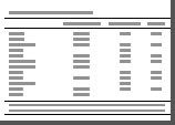 | Table I.Evidence-based comparison of
established and emerging interventions for obesity- and
aging-related male hypogonadism. |
Table I.
Evidence-based comparison of
established and emerging interventions for obesity- and
aging-related male hypogonadism.
| Therapeutic
modality | Intervention | Primary
target/mechanism | Target population
and clinical utility | Evidence level |
|---|
| Lifestyle
modification | Caloric restriction
and exercise | Reduces visceral
adiposity, attenuates aromatase activity and leptin resistance | First-line
management for LOH; restores endogenous HPG axis | Level 1a (70) |
| Weight
management | Sleeve
gastrectomy/RYGB | Rapid visceral fat
loss, normalization of LH/FSH pulse amplitude | Highly effective
for severe obesity (BMI ≥35 kg/m2) with
hypogonadism | Level 2a (71,72) |
|
Pharmacotherapy | Testosterone
replacement therapy | Replenishes
circulating androgens, restores bone/muscle anabolic mass | Indicated for
irreversible/established LOH; contraindicated if desiring
fertility | Level 1a (74,75) |
|
| GLP-1RAs | Enhances insulin
sensitivity, promotes weight loss | Adjunctive option
for obesity-related hypogonadism, not yet guideline-endorsed | Level 2b (78,79) |
|
| Aromatase
inhibitors | Blocks
CYP19A1-mediated testosterone-to-estradiol conversion, relieves
central feedback | Off-label strategy
for obese men with hyperestrogenemia desiring fertility | Level 1b (80) |
| Targeted small
molecules | Piezo1 inhibitors,
TRPML1 agonists | Resolves matrix
stiffness, ribotoxic stress and lysosomal acidity deficits | Translational
candidates for delaying testicular cellular senescence | Level 5 (6,7,83) |
| Natural
antioxidants | Icariin,
Quercetin, | Nrf2 antioxidant
pathway activation, free-radical neutralization | Adjunctive
candidates targeting oxidative stress-induced testicular
damage | Level 5 (84,85) |
| Regenerative
medicine | SLC
transplantation, TdEC hydrogels | Niche
reconstruction, macrophage mitochondrial transfer, tissue
repair | Frontier modalities
for biological restoration of endogenous steroidogenesis | Level 5 (34,87) |
Lifestyle modification and weight
management
Foundational strategies for obesity-associated
hypogonadism include dietary control, lifestyle modification and
bariatric surgery. Studies have shown that calorie restriction can
increase serum testosterone concentrations in overweight or obese
men (70). Bariatric surgery, in
particular, is an effective intervention for hypogonadism secondary
to severe obesity, achieving substantial and sustained weight loss
accompanied by corresponding increases in serum testosterone, LH
and FSH concentrations (71). A
study of 413 men with severe obesity reported that bariatric
surgery markedly ameliorated hypogonadism: Total testosterone
levels increased, and the prevalence of hypogonadism decreased from
69.5% before surgery to 20.1% at 3–6 months after surgery (72).
Historically, lifestyle modifications and weight
management were frequently viewed merely as conservative,
transitional or adjunctive measures prior to pharmacological
interventions. However, over the past decade, major international
guidelines, including those from the European Academy of Andrology
(EAA, 2020) (45), and iterative
updates from the EAU, 2024/2025 (73), have redefined this paradigm. These
consensus frameworks have firmly established and consistently
maintained structured lifestyle intervention and weight loss as the
fundamental, first-line therapeutic strategy for obesity-associated
functional hypogonadism.
Testosterone replacement therapy
(TRT)
For patients with established hypogonadism, TRT
represents a direct and effective pharmacological intervention
(74). Commonly used formulations
include oral testosterone undecanoate, intramuscular testosterone
undecanoate injections and transdermal preparations (gel and patch)
(43). By restoring physiological
serum testosterone levels, TRT effectively improves male sexual
function and enhances bone and muscle mass; however, it confers
critical clinical trade-offs and potential adverse effects
(75). In particular, exogenous
testosterone exerts negative feedback on the HPG axis, suppressing
gonadotropin secretion and severely impairing spermatogenesis,
which renders TRT unsuitable for men desiring to preserve
fertility.
Consequently, contemporary international guidelines
[including EAA (45) and (73) guidelines] strongly caution against
the indiscriminate prescription of TRT for non-organic, age-related
or adiposity-driven reductions in serum testosterone (45,73).
In overweight or obese individuals (BMI ≥27 kg/m2)
without structural HPG pathology, guidelines mandate prioritizing
the aggressive correction of reversible secondary causes, most
notably structured lifestyle modification, substantial weight loss
and the cessation of interfering medications, before considering
lifelong androgen replacement.
Adjuvant pharmacological interventions
for metabolic dysfunction
Studies have shown that exenatide, a glucagon-like
peptide-1 receptor agonist (GLP-1RA), increases serum testosterone
levels in men with type 2 diabetes mellitus who have low baseline
testosterone, but has no such effect in those with normal baseline
levels (76). Compared with
testosterone therapy, a 16-week intervention with liraglutide not
only reduces body weight and improves metabolic parameters but also
effectively elevates serum testosterone levels (77). By promoting weight loss and
improving insulin sensitivity, these agents reverse secondary HPG
axis suppression and restore endogenous testosterone production
without compromising spermatogenesis, thereby serving as promising,
fertility-preserving metabolic interventions for obese men with
functional hypogonadism (78). A
systematic review of five clinical trials presented at the
Endocrine Society 2026 annual meeting reported no detrimental
effect of GLP-1RAs on reproductive hormones, sexual function or
sperm quality, with two trials demonstrating marked improvements
compared with the baseline (79).
Although GLP-1RAs are not yet incorporated into formal clinical
guidelines for hypogonadism, growing evidence supports their
potential inclusion in future recommendations, reshaping the
therapeutic paradigm for this specific patient cohort.
Aromatase inhibitors, such as letrozole, reduce the
conversion of testosterone to estradiol by inhibiting aromatase,
thereby lowering estrogen levels. This may alleviate negative
feedback on the pituitary, increase LH and FSH levels, and
consequently stimulate testicular function (41). Nevertheless, aromatase inhibitors
remain an off-label option for male hypogonadism; while
contemporary guidelines acknowledge their potential to improve
hormonal and seminal parameters, they emphasize the need for
prospective randomized controlled trials to confirm definitive
efficacy (80).
Therapeutic strategies in men desiring
fertility preservation
The reproductive intent of the patient constitutes a
pivotal branch point in clinical decision-making. Authoritative
guidelines, including those from the EAA and the EAU, explicitly
advise against TRT in hypogonadal men with active fertility
desires, as exogenous testosterone suppresses endogenous
gonadotropin secretion and spermatogenesis, potentially resulting
in oligospermia or azoospermia (44,73).
For hypogonadal men with active fertility desires, clinical
practice guidelines recommend a stepwise, fertility-preserving
management algorithm.
i) First-line lifestyle and weight management.
Structured lifestyle interventions and weight reduction serve as
the cornerstone of management, as weight loss can increase serum
testosterone concentrations in overweight or obese men (44,70).
ii) Guideline-recommended pharmacological options.
Aromatase inhibitors (such as letrozole) and selective estrogen
receptor modulators (such as clomiphene citrate and tamoxifen) are
recommended by current guidelines for hypogonadal men desiring to
maintain fertility. By relieving estrogenic negative feedback, they
stimulate endogenous LH/FSH and testosterone secretion while
preserving spermatogenesis (80,81).
iii) GLP-1Ras. Abundant clinical data support the
use of GLP-1RAs in obesity-associated hypogonadism, promoting
weight loss and endogenous testosterone restoration without
compromising spermatogenesis (78,79);
however, they have not yet been incorporated into formal
guidelines.
iv) Gonadotropin-based therapy. In patients with
hypogonadotropic hypogonadism and fertility desires where lifestyle
and medical measures fail, gonadotropin replacement (human
chorionic gonadotropin with or without FSH/human menopausal
gonadotropin) is the guideline-recommended standard of care,
stimulating LCs and Sertoli cells to drive spermatogenesis and
restore fertility (82).
Emerging and investigational
therapeutic strategies
Targeted molecular therapy
Emerging evidence highlights several molecular
targets within the aging testis that may restore LC function and
endogenous testosterone production.
i) Modulating testicular microenvironment stiffness.
Recent studies have revealed that the aging testis undergoes
interstitial fibrosis and extracellular matrix remodeling (19), and that the extracellular matrix of
the aging testis becomes stiffer, leading to increased
Piezo1-mediated calcium influx, which in turn causes mitochondrial
dysfunction and excessive ROS generation. This promotes
ubiquitin-proteasome-mediated degradation of the stem LCs (SLCs),
impairing their proliferation and differentiation (83). The study further demonstrated that
a Piezo1 inhibitor restored SLC function and elevated testosterone
levels, representing a novel therapeutic strategy (83).
ii) Targeting ketone body metabolic deficits.
Hmgcs2, a key enzyme in ketone body synthesis, is downregulated in
aging testicular LCs, resulting in insufficient ketone body
production. Enhancing ketone body production, either through
genetic regulation or oral administration of β-hydroxybutyrate,
alleviates LC senescence and testicular aging in aged mice,
effectively elevating testosterone levels (18).
iii) Restoring lysosomal function. In a high-fat
diet-induced LOH mouse model, intra-seminiferous tubule injection
of a transient receptor potential mucolipin 1 agonist restored
lysosomal function and reversed testosterone deficiency and
associated pathological changes, such as impaired lysosomal
acidification, lipid droplet accumulation and Sertoli cell
senescence (6).
iv) Counteracting fibrosis and steroidogenic
impairment. CCN5, a key biomarker of testicular aging, impairs
steroidogenesis and accelerates LC senescence via
β-catenin/SMAD-mediated fibrosis and RNF213 downregulation-induced
lipid loss and cholesterol efflux. Targeted CCN5 inhibition may
restore endogenous testosterone while avoiding the risks of
exogenous testosterone therapy (19).
v) Targeting cellular senescence and stress. The
ZAKα inhibitor nilotinib alleviates testicular atrophy in aged
mice, restores testosterone levels and ameliorates Sertoli cell
dysfunction (7).
Natural products and metabolic
interventions
i) Flavonoids. Icariin protects spermatogonia and
Sertoli cells against oxidative stress by enhancing estrogen
receptor α expression and activating the nuclear factor erythroid
2-related factor 2 antioxidant pathway (84). Quercetin counteracts various
reproductive toxicities, including impaired sperm function,
reproductive hormone dysfunction, testicular apoptosis, oxidative
stress and inflammation, primarily by inhibiting ROS generation via
its cyclic antioxidant structure and modulating oxidative
stress-induced apoptotic signaling (85).
ii) Other natural extracts. Morinda
citrifolia extract increases serum testosterone levels, reduces
SHBG and dihydrotestosterone concentrations, enhances the
expression of testosterone biosynthesis-associated proteins and
downregulates degradative enzymes (5α-reductase and aromatase),
improving muscle performance and sperm quality (86).
Despite their antioxidant and cytoprotective effects
in aging rodent models, clinical evidence in humans remains limited
owing to poor oral bioavailability and the lack of large-scale
standardized trials.
Regenerative medicine and tissue
engineering
i) Stem cell transplantation and immune modulation.
Transplantation of SLCs into damaged testes promotes endogenous LC
repair, thereby restoring testicular secretory function.
Transplanted SLCs form nanotubes that transfer mitochondria to
testicular macrophages, shifting them from a pro-inflammatory to an
anti-inflammatory phenotype, thereby supporting tissue repair, and
markedly improving fertility in model mice (34).
ii) Biomaterials and organ reconstruction. Chi et
al (87) developed a hydrogel
derived from testicular decellularized extracellular matrix that
mimics the young testicular microenvironment. When co-transplanted
in situ with SLCs into aged testes, this hydrogel promotes
SLC proliferation and differentiation, markedly elevating
endogenous testosterone and ameliorating age-related symptoms
associated with testosterone deficiency, including increased muscle
mass, reduced bone loss, decreased white adipose tissue and
improved cognitive function (87).
Currently, SC transplantation, niche reconstruction
and tissue engineering approaches remain in the early experimental
phase, primarily supported by in vitro cellular systems and
rodent models. With continued preclinical optimization, these
regenerative strategies hold promise as potential therapeutic
modalities to restore endogenous endocrine function and alleviate
hypogonadism-related cardiovascular and systemic functional decline
in the future.
Conclusions and future perspectives
Aging is the intrinsic mechanism underlying the
decline of gonadal function, whereas obesity accelerates this aging
process through chronic inflammation, oxidative stress and hormonal
imbalance. Aging and obesity reinforce each other, forming a
vicious cycle that jointly drives the onset and progression of
chronic hypogonadism. Accordingly, future research should focus on
the interplay between obesity and testicular aging, advancing along
two directions, namely, basic and clinical research.
Future basic research should focus on elucidating
the molecular mechanisms underlying the interaction between obesity
and testicular aging, and on identifying and validating key
targetable molecular nodes, thereby breaking the vicious cycle and
improving gonadal function.
Although abundant population-based evidence
indicates that GLP-1RAs exert beneficial effects on weight loss and
male reproductive function, larger multicenter randomized
controlled trials are still required to further clarify their
efficacy and long-term safety, thereby providing a robust evidence
basis for their eventual incorporation into clinical practice
guidelines. These efforts will ultimately support evidence-based
strategies to preserve gonadal health in aging men.
Acknowledgements
Not applicable.
Funding
This review was supported by grants from the National Natural
Science Foundation of China (nos. 82370849, 82270839, 82200947 and
82400928) and the Natural Science Foundation of Shandong Province
(nos. ZR2023MH008 and ZR2024QH375).
Availability of data and materials
Not applicable.
Authors' contributions
DL and LZ conceived the article and drafted the
manuscript. XQ, QG and FX helped to revise the manuscript
critically for important intellectual content. PL and CY assisted
in designing and organizing the conceptual framework. Data
authentication is not applicable. All authors have read and
approved the final manuscript.
Ethics approval and consent to
participate
Not applicable.
Patient consent for publication
Not applicable.
Competing interests
The authors declare that they have no competing
interests.
References
|
1
|
Cui L, Nie X, Guo Y, Ren P, Guo Y, Wang X,
Li R, Hotaling JM, Cairns BR and Guo J: Single-cell transcriptomic
atlas of the human testis across the reproductive lifespan. Nat
Aging. 5:658–674. 2025. View Article : Google Scholar : PubMed/NCBI
|
|
2
|
Tysoe O: Sertoli cell lysosomal
dysfunction drives age-related testicular degeneration. Nat Rev
Endocrinol. 20:3862024. View Article : Google Scholar : PubMed/NCBI
|
|
3
|
Huang D, Zuo Y, Zhang C, Sun G, Jing Y,
Lei J, Ma S, Sun S, Lu H, Cai Y, et al: A single-nucleus
transcriptomic atlas of primate testicular aging reveals exhaustion
of the spermatogonial stem cell reservoir and loss of Sertoli cell
homeostasis. Protein Cell. 14:888–907. 2023.PubMed/NCBI
|
|
4
|
Nie X, Munyoki SK, Sukhwani M, Schmid N,
Missel A, Emery BR, Stukenborg JB, Mayerhofer A, Orwig KE, Aston
KI, et al: Single-cell analysis of human testis aging and
correlation with elevated body mass index. Dev Cell.
57:1160–1176.e5. 2022. View Article : Google Scholar : PubMed/NCBI
|
|
5
|
Gunes S, Hekim GN, Arslan MA and Asci R:
Effects of aging on the male reproductive system. J Assist Reprod
Genet. 33:441–454. 2016. View Article : Google Scholar : PubMed/NCBI
|
|
6
|
Deng Z, Zhao L, Li S, Chen X, Ling X,
Zheng J, Yu K, Xu J, Yao C, Han S, et al: Targeting dysregulated
phago-/auto-lysosomes in Sertoli cells to ameliorate late-onset
hypogonadism. Nat Aging. 4:647–663. 2024. View Article : Google Scholar : PubMed/NCBI
|
|
7
|
Xu Y, Shi X, Yin Y, Tan X, Ou N, Tang X,
Liu B, Xie H, Chen Y, Han Z, et al: Lipid droplet-driven ribosome
collisions trigger ZAKα-p38 signaling to accelerate testicular
aging. Aging Cell. 25:e703592026. View Article : Google Scholar : PubMed/NCBI
|
|
8
|
Mularoni V, Esposito V, Di Persio S,
Vicini E, Spadetta G, Berloco P, Fanelli F, Mezzullo M, Pagotto U,
Pelusi C, et al: Age-related changes in human Leydig cell status.
Hum Reprod. 35:2663–2676. 2020. View Article : Google Scholar : PubMed/NCBI
|
|
9
|
Li H, Yao Z, Degenhardt B, Teper G and
Papadopoulos V: Cholesterol binding at the cholesterol
recognition/interaction amino acid consensus (CRAC) of the
peripheral-type benzodiazepine receptor and inhibition of
steroidogenesis by an HIV TAT-CRAC peptide. Proc Natl Acad Sci USA.
98:1267–1272. 2001. View Article : Google Scholar : PubMed/NCBI
|
|
10
|
Roelfsema F, Liu PY, Takahashi PY, Yang RJ
and Veldhuis JD: Dynamic interactions between LH and testosterone
in healthy community-dwelling men: Impact of age and body
composition. J Clin Endocrinol Metab. 105:e628–e641. 2020.
View Article : Google Scholar : PubMed/NCBI
|
|
11
|
Chen H, Hardy MP and Zirkin BR:
Age-related decreases in Leydig cell testosterone production are
not restored by exposure to LH in vitro. Endocrinology.
143:1637–1642. 2002. View Article : Google Scholar : PubMed/NCBI
|
|
12
|
Chung JY, Chen H, Midzak A, Burnett AL,
Papadopoulos V and Zirkin BR: Drug ligand-induced activation of
translocator protein (TSPO) stimulates steroid production by aged
brown Norway rat Leydig cells. Endocrinology. 154:2156–2165. 2013.
View Article : Google Scholar : PubMed/NCBI
|
|
13
|
Luo L, Chen H and Zirkin BR: Leydig cell
aging: Steroidogenic acute regulatory protein (StAR) and
cholesterol side-chain cleavage enzyme. J Androl. 22:149–156. 2001.
View Article : Google Scholar : PubMed/NCBI
|
|
14
|
Luo L, Chen H and Zirkin BR: Temporal
relationships among testosterone production, steroidogenic acute
regulatory protein (StAR), and P450 side-chain cleavage enzyme
(P450scc) during Leydig cell aging. J Androl. 26:25–31. 2005.
View Article : Google Scholar : PubMed/NCBI
|
|
15
|
Wang Y, Chen F, Ye L, Zirkin B and Chen H:
Steroidogenesis in leydig cells: Effects of aging and environmental
factors. Reproduction. 154:R111–R122. 2017. View Article : Google Scholar : PubMed/NCBI
|
|
16
|
Beattie MC, Adekola L, Papadopoulos V,
Chen H and Zirkin BR: Leydig cell aging and hypogonadism. Exp
Gerontol. 68:87–91. 2015. View Article : Google Scholar : PubMed/NCBI
|
|
17
|
Ajayi AF, Onaolapo MC, Omole AI, Adeyemi
WJ and Oluwole DT: Mechanism associated with changes in male
reproductive functions during aging process. Exp Gerontol.
179:1122322023. View Article : Google Scholar : PubMed/NCBI
|
|
18
|
Liu C, Peng H, Yu J, Luo P, Xiong C, Chen
H, Fan H, Ma Y, Ou W, Zhang S, et al: Impaired ketogenesis in
Leydig cells drives testicular aging. Nat Commun. 16:42242025.
View Article : Google Scholar : PubMed/NCBI
|
|
19
|
Tan X, Xu Y, Ou N, Chen Y, Xia W, Xing E,
Mo R, Bo H, Han Z, Xu J, et al: CCN5 drives Leydig cell aging and
testicular dysfunction: Insights into fibrosis, lipid
dysregulation, and therapeutic potential. Research. 8:07622025.
View Article : Google Scholar : PubMed/NCBI
|
|
20
|
El-Domyati MM, Al-Din AB, Barakat MT,
El-Fakahany HM, Xu J and Sakkas D: Deoxyribonucleic acid repair and
apoptosis in testicular germ cells of aging fertile men: The role
of the poly(adenosine diphosphate-ribosyl)ation pathway. Fertil
Steril. 91:2221–2229. 2009. View Article : Google Scholar : PubMed/NCBI
|
|
21
|
Liao J, Suen HC, Luk ACS, Yang L, Lee AWT,
Qi H and Lee TL: Transcriptomic and epigenomic profiling of young
and aged spermatogonial stem cells reveals molecular targets
regulating differentiation. PLoS Genet. 17:e10093692021. View Article : Google Scholar : PubMed/NCBI
|
|
22
|
Suzuki N and Withers HR: Exponential
decrease during aging and random lifetime of mouse spermatogonial
stem cells. Science. 202:1214–1215. 1978. View Article : Google Scholar : PubMed/NCBI
|
|
23
|
Zhang X, Ebata KT, Robaire B and Nagano
MC: Aging of male germ line stem cells in mice. Biol Reprod.
74:119–124. 2006. View Article : Google Scholar : PubMed/NCBI
|
|
24
|
Nishino J, Kim I, Chada K and Morrison SJ:
Hmga2 promotes neural stem cell self-renewal in young but not old
mice by reducing p16Ink4a and p19Arf expression. Cell. 135:227–239.
2008. View Article : Google Scholar : PubMed/NCBI
|
|
25
|
Zhang T, Oatley J, Bardwell VJ and
Zarkower D: DMRT1 is required for mouse spermatogonial stem cell
maintenance and replenishment. PLoS Genet. 12:e10062932016.
View Article : Google Scholar : PubMed/NCBI
|
|
26
|
Paul C, Nagano M and Robaire B: Aging
results in molecular changes in an enriched population of
undifferentiated rat spermatogonia. Biol Reprod. 89:1472013.
View Article : Google Scholar : PubMed/NCBI
|
|
27
|
Nistal M, Codesal J and Paniagua R:
Multinucleate spermatids in aging human testes. Arch Androl.
16:125–129. 1986. View Article : Google Scholar : PubMed/NCBI
|
|
28
|
Paniagua R, Nistal M, Amat P, Rodriguez MC
and Martin A: Seminiferous tubule involution in elderly men. Biol
Reprod. 36:939–947. 1987. View Article : Google Scholar : PubMed/NCBI
|
|
29
|
Endo T, Kobayashi K, Matsumura T, Emori C,
Ozawa M, Kawamoto S, Okuzaki D, Shimada K, Miyata H, Shimada K, et
al: Multiple aging effects on testicular/epididymal germ cells lead
to decreased male fertility in mice. Commun Biol. 7:162024.
View Article : Google Scholar : PubMed/NCBI
|
|
30
|
Zhao S, Zhu W, Xue S and Han D: Testicular
defense systems: Immune privilege and innate immunity. Cell Mol
Immunol. 11:428–437. 2014. View Article : Google Scholar : PubMed/NCBI
|
|
31
|
Kaltsas A: Multi-omics perspectives on
testicular aging: Unraveling germline dysregulation, niche
dysfunction, and epigenetic remodeling. Cells. 14:8992025.
View Article : Google Scholar : PubMed/NCBI
|
|
32
|
Jiang Q, Cui L, Nie X, Cai H, Zhang W, Lu
X, Guo Y, Hotaling JM, Cairns BR, Wang X and Guo J: A single-cell
transcriptome atlas characterizes the immune landscape of human
testes during aging. Aging Cell. 24:e700322025. View Article : Google Scholar : PubMed/NCBI
|
|
33
|
Zhang W, Xia S, Xiao W, Song Y, Tang L,
Cao M, Yang J, Wang S, Li Z, Xu C, et al: A single-cell
transcriptomic landscape of mouse testicular aging. J Adv Res.
53:219–234. 2023. View Article : Google Scholar : PubMed/NCBI
|
|
34
|
Chi A, Yang B, Dai H, Li X, Mo J, Gao Y,
Chen Z, Feng X, Ma M, Li Y, et al: Stem Leydig cells support
macrophage immunological homeostasis through mitochondrial transfer
in mice. Nat Commun. 15:21202024. View Article : Google Scholar : PubMed/NCBI
|
|
35
|
Amodio G, Giacomini G, Boeri L, Raffo M,
Cilio S, Pozzi E, Belladelli F, Negri F, Ferrara AM, d'Arma A, et
al: Specific types of male infertility are correlated with T cell
exhaustion or senescence signatures. Nat Commun. 16:9712025.
View Article : Google Scholar : PubMed/NCBI
|
|
36
|
Handelsman DJ, Yeap B, Flicker L, Martin
S, Wittert GA and Ly LP: Age-specific population centiles for
androgen status in men. Eur J Endocrinol. 173:809–817. 2015.
View Article : Google Scholar : PubMed/NCBI
|
|
37
|
Wu FC, Tajar A, Pye SR, Silman AJ, Finn
JD, O'Neill TW, Bartfai G, Casanueva F, Forti G, Giwercman A, et
al: Hypothalamic-pituitary-testicular axis disruptions in older men
are differentially linked to age and modifiable risk factors: The
European male aging study. J Clin Endocrinol Metab. 93:2737–2745.
2008. View Article : Google Scholar : PubMed/NCBI
|
|
38
|
Koliaki C, Dalamaga M and Liatis S: Update
on the obesity epidemic: After the sudden rise, is the upward
trajectory beginning to flatten? Curr Obes Rep. 12:514–527. 2023.
View Article : Google Scholar : PubMed/NCBI
|
|
39
|
Loh NY, Humphreys E, Karpe F, Tomlinson
JW, Noordam R and Christodoulides C: Sex hormones, adiposity, and
metabolic traits in men and women: A Mendelian randomisation study.
Eur J Endocrinol. 186:407–416. 2022. View Article : Google Scholar : PubMed/NCBI
|
|
40
|
Moreira RJ, Oliveira PF, Spadella MA,
Ferreira R and Alves MG: Do lifestyle interventions mitigate the
oxidative damage and inflammation induced by obesity in the testis?
Antioxidants. 14:1502025. View Article : Google Scholar : PubMed/NCBI
|
|
41
|
Colleluori G, Chen R, Turin CG, Vigevano
F, Qualls C, Johnson B, Mediwala S, Villareal DT and
Armamento-Villareal R: Aromatase inhibitors plus weight loss
improves the hormonal profile of obese hypogonadal men without
causing major side effects. Front Endocrinol (Lausanne).
11:2772020. View Article : Google Scholar : PubMed/NCBI
|
|
42
|
Ghaderpour S, Ghiasi R, Heydari H and
Keyhanmanesh R: The relation between obesity, kisspeptin, leptin,
and male fertility. Horm Mol Biol Clin Investig. 43:235–247. 2021.
View Article : Google Scholar : PubMed/NCBI
|
|
43
|
Broul M and Hujová A: Late-onset
hypogonadism in men over 40-how to use the updated EAU 2025
guidelines and new findings on the cardiovascular safety of
testosterone therapy. Cas Lek Cesk. 164:150–155. 2025.PubMed/NCBI
|
|
44
|
Salonia A, Bettocchi C, Boeri L,
Capogrosso P, Carvalho J, Cilesiz NC, Cocci A, Corona G,
Dimitropoulos K, Gül M, et al: European association of urology
guidelines on sexual and reproductive Health-2021 update: Male
sexual dysfunction. Eur Urol. 80:333–357. 2021. View Article : Google Scholar : PubMed/NCBI
|
|
45
|
Corona G, Goulis DG, Huhtaniemi I,
Zitzmann M, Toppari J, Forti G, Vanderschueren D and Wu FC:
European academy of andrology (EAA) guidelines on investigation,
treatment and monitoring of functional hypogonadism in males:
Endorsing organization: European Society of Endocrinology.
Andrology. 8:970–987. 2020. View Article : Google Scholar : PubMed/NCBI
|
|
46
|
Ge P, Zhang H, Liu W, Wang X, Wang J and
Song N: Bibliometric and scientometric analysis of late-onset
hypogonadism in the past two decades: Knowledge mapping and
research trends. J Mens Health. 22:64–76. 2026. View Article : Google Scholar
|
|
47
|
Xu D, Liu L, Zhao Y, Yang L, Cheng J, Hua
R, Zhang Z and Li Q: Melatonin protects mouse testes from palmitic
acid-induced lipotoxicity by attenuating oxidative stress and DNA
damage in a SIRT1-dependent manner. J Pineal Res. 69:e126902020.
View Article : Google Scholar : PubMed/NCBI
|
|
48
|
Muir CA, Wittert GA and Handelsman DJ:
Approach to the patient: Low testosterone concentrations in men
with obesity. J Clin Endocrinol Metab. 110:e3125–e3130. 2025.
View Article : Google Scholar : PubMed/NCBI
|
|
49
|
Kelly DM and Jones TH: Testosterone and
obesity. Obes Rev. 16:581–606. 2015. View Article : Google Scholar : PubMed/NCBI
|
|
50
|
van Hulsteijn LT, Pasquali R, Casanueva F,
Haluzik M, Ledoux S, Monteiro MP, Salvador J, Santini F, Toplak H
and Dekkers OM: Prevalence of endocrine disorders in obese
patients: Systematic review and meta-analysis. Eur J Endocrinol.
182:11–21. 2020. View Article : Google Scholar : PubMed/NCBI
|
|
51
|
Obradovic M, Sudar-Milovanovic E, Soskic
S, Essack M, Arya S, Stewart AJ, Gojobori T and Isenovic ER: Leptin
and obesity: Role and clinical implication. Front Endocrinol
(Lausanne). 12:5858872021. View Article : Google Scholar : PubMed/NCBI
|
|
52
|
Yu C, Jiang F, Zhang M, Luo D, Shao S,
Zhao J, Gao L, Zuo C and Guan Q: HC diet inhibited testosterone
synthesis by activating endoplasmic reticulum stress in testicular
Leydig cells. J Cell Mol Med. 23:3140–3150. 2019. View Article : Google Scholar : PubMed/NCBI
|
|
53
|
Luo D, Qi X, Xu X, Yang L, Yu C and Guan
Q: Involvement of p38 MAPK in Leydig cell aging and age-related
decline in testosterone. Front Endocrinol (Lausanne).
14:10882492023. View Article : Google Scholar : PubMed/NCBI
|
|
54
|
Genchi VA, Rossi E, Lauriola C, D'Oria R,
Palma G, Borrelli A, Caccioppoli C, Giorgino F and Cignarelli A:
Adipose tissue dysfunction and obesity-related male hypogonadism.
Int J Mol Sci. 23:81942022. View Article : Google Scholar : PubMed/NCBI
|
|
55
|
George BT, Jhancy M, Dube R, Kar SS and
Annamma LM: The molecular basis of male infertility in obesity: A
literature review. Int J Mol Sci. 25:1792023. View Article : Google Scholar : PubMed/NCBI
|
|
56
|
Choubey M, Ranjan A and Krishna A:
Adiponectin/AdipoRs signaling as a key player in testicular aging
and associated metabolic disorders. Vitam Horm. 115:611–634. 2021.
View Article : Google Scholar : PubMed/NCBI
|
|
57
|
Behera S, Bhattacharya K, Jal S, Purohit
GK, Sah AK, Agarwal S, Babiker R, Bhardwaj A and Alfeel AHM:
Metabolic syndrome and male infertility: The role of obesity and
chronic inflammation. Biomedicines. 14:17392026. View Article : Google Scholar : PubMed/NCBI
|
|
58
|
Yuxin L, Chen L, Xiaoxia L, Yue L, Junjie
L, Youzhu L, Huiliang Z and Qicai L: Research progress on the
relationship between obesity-inflammation-aromatase axis and male
infertility. Oxid Med Cell Longev. 2021:66127962021. View Article : Google Scholar : PubMed/NCBI
|
|
59
|
Ahmed F, Hetty S, Laterveer R, Surucu EB,
Mathioudaki A, Hornbrinck E, Patsoukaki V, Olausson J, Sundbom M,
Svensson MK, et al: Altered expression of aromatase and estrogen
receptors in adipose tissue from men with obesity or type 2
diabetes. J Clin Endocrinol Metab. 110:e3410–e3424. 2025.
View Article : Google Scholar : PubMed/NCBI
|
|
60
|
Cohen PG: The hypogonadal-obesity cycle:
Role of aromatase in modulating the testosterone-estradiol shunt-a
major factor in the genesis of morbid obesity. Med Hypotheses.
52:49–51. 1999. View Article : Google Scholar : PubMed/NCBI
|
|
61
|
Frasca D and Blomberg BB: Adipose tissue,
immune aging, and cellular senescence. Semin Immunopathol.
42:573–587. 2020. View Article : Google Scholar : PubMed/NCBI
|
|
62
|
Guo Q, Jin S, Hu H, Zhou Y, Yan Y, Zong H,
Wang Y, He H, Oh Y, Liu C and Gu N: Hypoxia in 3T3-L1 adipocytes
suppresses adiponectin expression via the PERK and IRE1 unfolded
protein response. Biochem Biophys Res Commun. 493:346–351. 2017.
View Article : Google Scholar : PubMed/NCBI
|
|
63
|
Feng R, Cheng D, Zhang W, Zhang J, Chen S
and Xia Y: Immune microenvironment dysregulation: A contributing
factor to obesity-associated male infertility. Biomedicines.
13:13142025. View Article : Google Scholar : PubMed/NCBI
|
|
64
|
Pan W, Zhao J, Xu J and Wang L:
Association between estimated glucose disposal rate and
testosterone deficiency in male patients with type 2 diabetes
mellitus. BMC Endocr Disord. 25:2592025. View Article : Google Scholar : PubMed/NCBI
|
|
65
|
Sheikh R, Shakerian S, Fatemi Tabatabaei
SR and Habibi A: Moderate and high-intensity interval training
protect against diabetes-induced modulation of hepatic CD86 and
CD206 expression associated with the amelioration of insulin
resistance and inflammation in rats. Immunobiology. 228:1527452023.
View Article : Google Scholar : PubMed/NCBI
|
|
66
|
Xiao G, Qian A, Gao Z, Dai T, Liang H,
Wang S, Deng M, Yan Y, Zhang X, Zhang X, et al: Reduced
glycoprotein hormone β5 links male aging and testosterone decline
to increased adiposity. J Clin Invest. 136:e1923552026. View Article : Google Scholar : PubMed/NCBI
|
|
67
|
Navarro G, Allard C, Xu W and
Mauvais-Jarvis F: The role of androgens in metabolism, obesity, and
diabetes in males and females. Obesity (Silver Spring). 23:713–719.
2015. View Article : Google Scholar : PubMed/NCBI
|
|
68
|
Hayes FJ: Testosterone and insulin
resistance in men: Evidence for a complex bi-directional
relationship. Trans Am Clin Climatol Assoc. 135:297–305.
2025.PubMed/NCBI
|
|
69
|
Centre for Evidence-Based Medicine, .
Oxford Centre for Evidence-Based Medicine:. Levels of Evidence.
March;2009.https://www.cebm.net/wp-content/uploads/2014/06/CEBM-Levels-of-Evidence.pdfOctober
2–2026
|
|
70
|
Smith SJ, Teo SYM, Lopresti AL, Heritage B
and Fairchild TJ: Examining the effects of calorie restriction on
testosterone concentrations in men: A systematic review and
meta-analysis. Nutr Rev. 80:1222–1236. 2022. View Article : Google Scholar : PubMed/NCBI
|
|
71
|
Emami MR, Safabakhsh M, Khorshidi M,
Moradi Moghaddam O, Mohammed SH, Zarezadeh M and Alizadeh S: Effect
of bariatric surgery on endogenous sex hormones and sex
hormone-binding globulin levels: A systematic review and
meta-analysis. Surg Obes Relat Dis. 17:1621–1636. 2021. View Article : Google Scholar : PubMed/NCBI
|
|
72
|
Bombardieri A, Bufano A, Fralassi N,
Ciuoli C, Benenati N, Dalmiglio C, Voglino C, Tirone A, Vuolo G and
Castagna MG: Assessing the prevalence of male obesity-associated
gonadal dysfunction in severe obesity: A focus on the impact of
bariatric surgery and surgical approaches. Obes Surg. 34:3434–3444.
2024. View Article : Google Scholar : PubMed/NCBI
|
|
73
|
Minhas S, Boeri L, Capogrosso P, Cocci A,
Corona G, Dinkelman-Smit M, Falcone M, Jensen CF, Gül M, Kalkanli
A, et al: European association of urology guidelines on male sexual
and reproductive health: 2025 Update on male infertility. Eur Urol.
87:601–616. 2025. View Article : Google Scholar : PubMed/NCBI
|
|
74
|
Orwoll E: Safety of
testosterone-replacement therapy in older men. N Engl J Med.
389:177–178. 2023. View Article : Google Scholar : PubMed/NCBI
|
|
75
|
Barone B, Napolitano L, Abate M, Cirillo
L, Reccia P, Passaro F, Turco C, Morra S, Mastrangelo F, Scarpato
A, et al: The role of testosterone in the elderly: What do we know?
Int J Mol Sci. 23:35352022. View Article : Google Scholar : PubMed/NCBI
|
|
76
|
Graybill S, Hatfield J, Kravchenko M,
Beckman D, Tate J, Beauvais A, Clerc P, Davila D, Forbes W, Wardian
J, et al: Neutral effect of exenatide on serum testosterone in men
with type 2 diabetes mellitus: A prospective cohort. Andrology.
9:792–800. 2021. View Article : Google Scholar : PubMed/NCBI
|
|
77
|
Jensterle M, Podbregar A, Goricar K,
Gregoric N and Janez A: Effects of liraglutide on
obesity-associated functional hypogonadism in men. Endocr Connect.
8:195–202. 2019. View Article : Google Scholar : PubMed/NCBI
|
|
78
|
Orra SH, Martinez JVN, Porto BC,
Passerotti CC, Sardenberg RAS and Da Cruz JAS: Effect of GLP-1
agonists on testosterone levels: A systematic review and
meta-analysis. BMC Urol. 25:3112025. View Article : Google Scholar : PubMed/NCBI
|
|
79
|
Salvio G, Ciarloni A, Ambo N, Bordoni M,
Perrone M, Rossi S and Balercia G: Effects of glucagon-like peptide
1 receptor agonists on testicular dysfunction: A systematic review
and meta-analysis. Andrology. 13:2022–2034. 2025. View Article : Google Scholar : PubMed/NCBI
|
|
80
|
Del Giudice F, Busetto GM, De Berardinis
E, Sperduti I, Ferro M, Maggi M, Gross MS, Sciarra A and Eisenberg
ML: A systematic review and meta-analysis of clinical trials
implementing aromatase inhibitors to treat male infertility. Asian
J Androl. 22:360–367. 2020. View Article : Google Scholar : PubMed/NCBI
|
|
81
|
Hohl A, Chavez MP, Pasqualotto E, Ferreira
ROM, van de Sande-Lee S and Ronsoni MF: Clomiphene or enclomiphene
citrate for the treatment of male hypogonadism: A systematic review
and meta-analysis of randomized controlled trials. Arch Endocrinol
Metab. 69:e2500932025. View Article : Google Scholar : PubMed/NCBI
|
|
82
|
Gialouris JV, Conway AJ, Idan A, Savkovic
S, Hermosilla R, Muir CA, Bacha F, Zhang T, Hou W, Jayadev V and
Handelsman DJ: Efficacy of gonadotropin therapy to induce
spermatogenesis and fertility in men with pathologic gonadotropin
deficiency. J Clin Endocrinol Metab. 111:1450–1458. 2025.
View Article : Google Scholar : PubMed/NCBI
|
|
83
|
Huang J, Sun L, Yin Y, Yang H, Zeng K,
Huang P, Liang X, Wu J, Xiang AP and Wang J: High matrix stiffness
triggers testosterone decline in aging males by disrupting stem
Leydig cell pool homeostasis. Cell Rep. 44:1162072025. View Article : Google Scholar : PubMed/NCBI
|
|
84
|
Zhao H, You X, Chen Q, Yang S, Ma Q, He Y,
Liu C, Dun Y, Wu J, Zhang C and Yuan D: Icariin improves
age-related testicular dysfunction by alleviating Sertoli cell
injury via upregulation of the ERα/Nrf2-signaling pathway. Front
Pharmacol. 11:6772020. View Article : Google Scholar : PubMed/NCBI
|
|
85
|
Rotimi DE, Olaolu TD and Adeyemi OS:
Pharmacological action of quercetin against testicular dysfunction:
A mini review. J Integr Med. 20:396–401. 2022. View Article : Google Scholar : PubMed/NCBI
|
|
86
|
Kwon HY, Lee H, Choi JS, Lim SH, Kim T,
Bae K, Jang YA, Kim EM, Lee JY, Choung SY, et al: Fermented
Morinda citrifolia extract improves late-onset hypogonadism
in aged rats. Biomed Pharmacother. 189:1182562025. View Article : Google Scholar : PubMed/NCBI
|
|
87
|
Chi A, Yang C, Liu J, Zhai Z and Shi X:
Reconstructing the stem Leydig cell niche via the testicular
extracellular matrix for the treatment of testicular Leydig cell
dysfunction. Adv Sci (Weinh). 12:e24108082025. View Article : Google Scholar : PubMed/NCBI
|