Introduction and nomenclature evolution
With the global pandemic of obesity and type 2
diabetes (T2D), the epidemiological spectrum of chronic liver
disease has undergone profound changes (1,2). To
more accurately reflect the underlying metabolic pathophysiology,
international consensus has updated the nomenclature from
non-alcoholic fatty liver disease to metabolic
dysfunction-associated steatotic liver disease (MASLD) and from
non-alcoholic steatohepatitis (NASH) to metabolic
dysfunction-associated steatohepatitis (MASH) (3–5).
This diagnostic transition represents more than a
semantic revision; it shifts the translational research paradigm
from an exclusionary diagnosis to an inclusionary,
metabolic-centric framework. Clinically, this refinement anchors
hepatic pathology to cardiometabolic risk factors, including
central obesity, hyperglycemia, dyslipidemia and hypertension
(1,3,4).
Age, obesity, insulin resistance and T2D are associated with
advanced fibrosis, whereas elevated ferritin in NAFLD may reflect
hepatic iron burden and should not be interpreted as an independent
fibrosis determinant in isolation (6–8). In
translational research, adopting the MASLD framework encourages
preclinical models to reproduce the systemic metabolic context of
human disease; nevertheless, predictive validity still depends on
how closely an individual model recapitulates the relevant human
phenotype.
Clinically, the pathology advances sequentially from
a healthy liver to MASLD with simple steatosis, MASH, progressive
liver fibrosis (stages F1-F3 according to the NASH Clinical
Research Network [NASH CRN] fibrosis staging system) and cirrhosis
(stage F4) (9). This disease
follows a pathological cascade that may culminate in cirrhosis and
hepatocellular carcinoma (6)
(Fig. 1). Under physiological
conditions, the liver fine-tunes systemic lipid homeostasis by
balancing fatty acid uptake, de novo lipogenesis (DNL), β-oxidation
and very-low-density lipoprotein secretion (10,11).
Chronic nutrient overload and insulin resistance disrupt this
balance, increase DNL and promote hepatic lipid accumulation
(11,12). Hepatic steatosis is conventionally
defined by steatosis affecting >5% of hepatocytes or an
equivalent increase in liver fat content, rather than by >5%
triglycerides within individual hepatocytes (3,6).
Hepatic lipid accumulation is not an isolated event;
mitochondria lie at the intersection of nutrient overload,
lipid-droplet handling and progressive cellular injury (13–15).
Notably, mitochondria are not merely passive recipients of
lipotoxicity; instead, their functional phenotype can shift from
early compensatory adaptation to late decompensated failure during
MASLD progression. Consequently, the present review tracks the
evolution of mitochondrial dysfunction, details the molecular
cross-talk driving liver injury, and evaluates epigenetic and
metabolic regulation by the sirtuin family to identify clinically
relevant therapeutic opportunities.
Mitochondrial dysfunction: The core hub
driving MASLD to MASH progression
Hepatic mitochondrial physiology and
lipid homeostasis
Mitochondria serve as the central hub for regulating
systemic lipid homeostasis. Under physiological conditions,
cytosolic free fatty acids (FFAs) strictly rely on the carnitine
shuttle system to enter the mitochondrial matrix. This system is
coordinately assembled by long-chain acyl-CoA synthetase on the
mitochondrial outer membrane, carnitine palmitoyltransferase 1
(CPT1), carnitine-acylcarnitine translocase and CPT2 within the
mitochondrial matrix (16,17).
Upon entering the matrix, fatty acids undergo
β-oxidation. The NADH and FADH2 generated by this
pathway deliver high-energy electrons into the electronic transport
chain (ETC) (18). While consuming
1–2% of oxygen to generate physiological free radicals, the ETC
establishes a proton gradient across the inner mitochondrial
membrane (IMM), forming an electrochemical transmembrane potential
that ultimately drives ATP synthase to generate energy and
regenerates the NAD+ and FAD required for subsequent
oxidation cycles (19).
Biphase adaptive mechanism: Evolution
from compensatory overload to bioenergetic failure
Mitochondrial involvement in MASLD does not follow a
simple, linear progressive decline; rather, it exhibits a distinct
non-linear biphase kinetic curve characterized by early functional
hypermetabolism followed by late bioenergetic exhaustion.
Early hormetic compensatory adaptation
phase (mitohormesis)
In the initial stages of simple steatosis, systemic
insulin resistance in peripheral adipose tissue drives unrestrained
lipolysis and increases the influx of circulating free fatty acids
into the liver. Hepatic mitochondria can initially respond by
increasing substrate oxidation, tricarboxylic acid (TCA) cycle flux
and respiratory activity; in humans with simple steatosis, this
compensatory increase is lost after progression to steatohepatitis,
while diet-induced models likewise demonstrate elevated TCA cycle
activity during fatty liver development (20,21).
This adaptation may transiently preserve energy production but also
increases reducing-equivalent delivery to the electron transport
chain and can heighten oxidative pressure. Ketogenesis and related
oxidative pathways provide additional outlets for acetyl-CoA,
although whether these responses remain protective depends on
disease stage and metabolic context (14,15).
Transition point and late bioenergetic
collapse phase
When nutrient overload becomes chronic and exceeds
mitochondrial homeostatic capacity, sustained substrate pressure
uncouples TCA cycle activity from efficient oxidative
phosphorylation, increases mitochondrial reactive oxygen species
(mROS) and reduces ATP production. Insulin resistance also sustains
hepatic DNL, further increasing the lipid burden (11,12).
Hepatic PGC-1α expression and activity can become impaired during
MASH, and experimental loss of hepatic PGC-1α reduces mitochondrial
respiration and promotes inflammation and fibrosis (22,23).
In parallel, incomplete lipid disposal favors the accumulation of
diacylglycerols and ceramides that interfere with insulin signaling
and cellular homeostasis (24,25).
The resulting lipotoxic and oxidative injury can impair membrane
integrity, promote hepatocyte ballooning and cell death, and
release damage-associated signals and lipid-peroxidation products
that activate hepatic stellate cells (HSCs) and accelerate
fibrosis.
Oxidative stress outbreak and collapse
of endogenous antioxidant defenses
Uncontrolled oxidative stress is a core mediator of
hepatocyte injury, inflammation and fibrosis (26,27).
Under physiological conditions, ETC complexes I and III exhibit
only minor leaking of electrons (28). However, in the lipotoxic
environment of MASH, this electron leakage worsens notably
(29).
The continuous influx of saturated fatty acids
alters mitochondrial membrane composition and increases lipotoxic
stress. Ceramide accumulation and VDAC1-associated mitochondrial
dysfunction have both been implicated in fatty liver pathology,
while experimental targeting of VDAC1 attenuates steatosis and
NASH-associated injury in mice (25,30,31)
(Fig. 2). These findings support a
role for VDAC1 in mitochondrial injury but do not establish that
free fatty acids directly alter VDAC voltage gating in human
MASH.
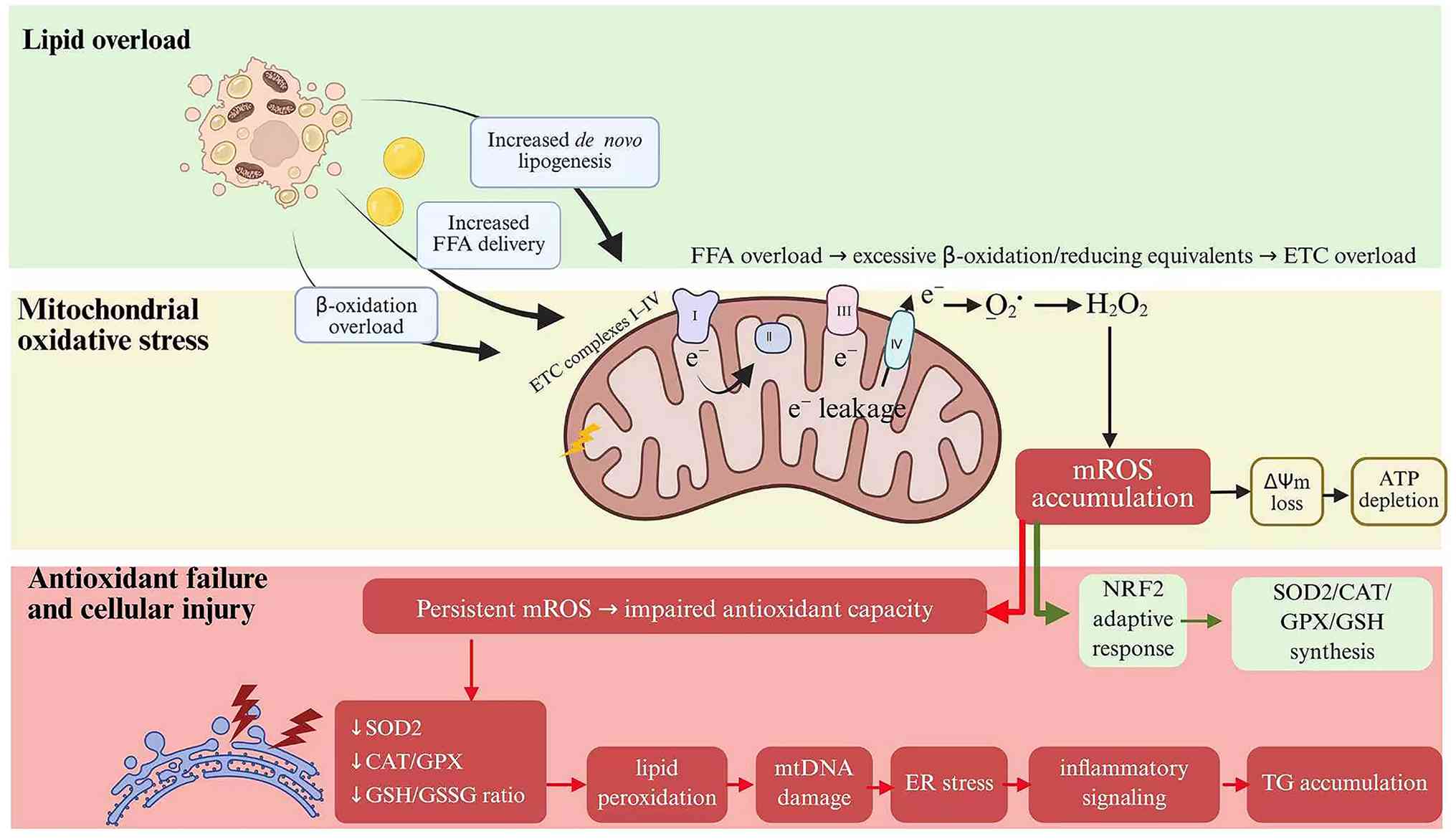 | Figure 2.Evidence-bounded model of
lipid-induced mitochondrial oxidative stress and cellular injury.
Nutrient overload and insulin resistance increase hepatic DNL and
lipid burden (10,11), while ceramides and VDAC1-associated
dysfunction contribute to mitochondrial injury (24,29,30).
Excess mROS can overwhelm antioxidant systems, promote lipid
peroxidation and damage mitochondrial DNA (31–33);
ROS-responsive JNK-Sab signaling can further impair respiration and
promote cell death (34). The
sequence is a conceptual framework and does not establish a
universal linear NRF2-to-antioxidant-exhaustion pathway in human
MASH. DNL, de novo lipogenesis; VDAC1, voltage-dependent
anion channel 1; mROS, mitochondrial reactive oxygen species; JNK,
c-Jun N-terminal kinase; Sab, SH3 domain-binding protein 5; mtDNA,
mitochondrial DNA. |
This lipotoxic mROS burden can overwhelm endogenous
antioxidant defenses, including SOD2, glutathione-dependent
systems, catalase and thioredoxin/peroxiredoxin pathways, thereby
permitting lipid peroxidation and the accumulation of cytotoxic
aldehydes such as 4-hydroxy-2-nonenal and malondialdehyde (32,33).
Oxidative injury also extends to nuclear and mitochondrial DNA;
mitochondrial DNA damage and cytochrome b variation have been
associated with fatty liver disease severity (34).
At the signaling level, ROS-responsive JNK can
interact with the mitochondrial scaffold protein Sab, inhibit
mitochondrial respiration and promote cell death (35). Mitochondrial c-Src independently
regulates phosphorylation of respiratory-chain components and cell
survival, although the available evidence does not establish a
direct chronic JNK-to-c-Src inhibitory pathway in MASH (36). Redox-sensitive signaling in HSCs
contributes to their activation and extracellular matrix deposition
(37,38). In parallel, mitochondria-associated
non-coding RNAs can regulate mROS output: Downregulation of the
mitochondria-localized circRNA SCAR promotes inflammatory signaling
in NASH, and broader evidence implicates lncRNAs and circRNAs in
NAFLD pathobiology (39,40).
Structural quality control and
organelle cross-talk
Mitochondrial dynamics imbalance: From network
fusion to fragmented collapse
Mitochondrial morphology depends on a dynamic
balance between fusion, which mixes mitochondrial contents, and
fission, which segregates damaged regions for clearance. NAFLD
models show excessive fragmentation and altered DRP1-dependent
fission, and experimental reduction of mitochondrial fission
alleviates hepatic steatosis (41,42).
However, the currently cited evidence does not establish DRP1
Ser616 phosphorylation as a universal initiating event in human
MASH.
The fusion and turnover machinery is also impaired
during fatty liver progression. Experimental and clinical studies
report altered mitofusin expression, disturbed mitochondrial
dynamics and reduced markers of mitochondrial turnover in NAFLD
(42,43). At the inner mitochondrial membrane,
cellular stress activates the metalloprotease OMA1, which cleaves
long-form OPA1 and thereby remodels inner-membrane fusion and
cristae organization (44).
Mitophagy impairment and mitochondrial
unfolded protein response (UPRmt) dysregulation: From quality
control to apoptotic switch
Mitochondrial quality control integrates fusion,
lipid droplet-mitochondria communication and selective organelle
turnover. In liver models, MFN2-dependent contacts promote fatty
acid oxidation, BNIP3 regulates mitochondrial function and lipid
metabolism, and ALCAT1 links defective mitophagy to steatosis
(45–47). These data support impaired
mitochondrial quality control in fatty liver disease but do not
justify a universal model in which advanced MASH is caused
specifically by defective proteasomal degradation of PINK1.
To survive mitochondrial proteotoxic stress, cells
activate the UPRmt. Accumulation of misfolded mitochondrial
proteins engages proteases and retrograde signaling, and the
transcription factor ATF5 is required for a mammalian UPRmt program
that induces nuclear-encoded mitochondrial chaperones and other
protective genes (48,49).
Under persistent lipotoxic stress, however, UPRmt
signaling may become insufficient or maladaptive. Reviews of UPRmt
biology and fatty liver disease describe a context-dependent
transition from compensatory proteostasis toward integrated stress
signaling and cell-death pathways involving ATF4 and
CCAAT/enhancer-binding protein homologous protein (CHOP) (49–51).
The precise contribution of CHOP-dependent Bcl-2 and Bax/Bak
regulation to human MASH remains less directly established than the
broader stress-response framework.
Mitochondria-associated endoplasmic
reticulum (ER) membranes (MAMs) and cellular calcium homeostasis
dysregulation
Mitochondria establish tight bidirectional
communication bridges with the ER via specialized contact sites
known as MAMs (52). MAMs serve as
specialized subcellular channels for the direct transfer of
Ca2+ and structural lipids between the two organelles
(53).
In obesity-associated liver, endoplasmic
reticulum-mitochondria contacts are reorganized and can become
chronically enriched, causing mitochondrial Ca2+
overload, impaired oxidative capacity and oxidative stress
(54). MAMs also adapt dynamically
to nutrient and glucose availability and regulate hepatic
mitochondrial metabolism. Thus, MAM remodeling rather than uniform
hyper-formation is the more defensible disease model. Excessive
calcium transfer can predispose mitochondria to permeability
transition and cell-death signaling, but the direction and
magnitude of MAM remodeling are context-dependent and should not be
generalized across all stages of MASH (55,56).
Sirtuin epigenetic metabolic network and
mitochondrial homeostasis regulation
The sirtuin family (SIRT1-SIRT7) comprises
NAD+-dependent deacetylases and deacylases whose activity is
coupled to intracellular NAD+ availability. CD38 can drive NAD+
decline and SIRT3-dependent mitochondrial dysfunction in
age-related metabolic settings, whereas enhancement of de
novo NAD+ synthesis improves mitochondrial function in
experimental models (57,58). These findings make NAD+ depletion a
plausible modifier of the sirtuin network in MASH, although direct
evidence that chronic hepatic inflammation universally causes
CD38-mediated NAD+ collapse remains incomplete. A comprehensive
overview of sirtuin localization, targets and disease-related roles
is summarized in Table I.
 | Table I.Comprehensive landscape of sirtuins
(SIRT1-SIRT7) in MASLD/MASH pathophysiology. |
Table I.
Comprehensive landscape of sirtuins
(SIRT1-SIRT7) in MASLD/MASH pathophysiology.
| Isoform | Subcellular
localization | Key molecular
targets | Expression profile
in MASLD | Core mechanistic
role |
Translational/therapeutic potential | Academic
controversies and context dependencies | (Refs.) |
|---|
| SIRT1 |
Nucleus/cytoplasm | PGC-1α and
autophagy-associated pathways | Frequently reduced
in metabolic liver disease models | Supports fatty acid
oxidation and hepatocyte autophagy; restrains lipotoxicity. | Potential target;
direct MASH efficacy remains to be established. | Effects depend on
NAD+ availability, nutritional state and experimental context. | (58,59) |
| SIRT2 | Cytoplasm | ACLY and NLRP3 | Context- and
stage-dependent | Destabilizes ACLY
through deacetylation and modulates inflam-masome activity. | Preclinical;
liver-selective modulation is required. | Inflammatory and
hepatocyte-autonomous metabolic effects may differ by model. | (60,61) |
| SIRT3 | Mitochon-drial
matrix | IDH2 and
SOD2-associated redox pathways | Often reduced in
lipotoxic liver models | Regulates
mitochondrial redox homeostasis and hepatocyte susceptibility to
lipotoxicity and autophagic stress. | Preclinical; no
selective activator has established clinical efficacy in MASH. | The cited studies
do not establish LCAD activation or cristae preservation as
universal MASH mechanisms. | (62,63) |
| SIRT4 | Mitochon-drial
matrix | MCD; miR-1
30b-5p-associated HSC pathways | Variable by cell
type and disease stage | Suppresses fatty
acid oxidation through MCD inhibition but can restrain HSC
activation and fibrosis. | Emerging,
context-dependent target. | Metabolic and
fibrotic phenotypes can point in opposite directions across cell
types. | (64,65) |
| SIRT5 | Mitochon-drial
matrix | Broad
mito-chondrial succinylated and other acylated proteins |
Context-dependent | Removes succinyl
and related acyl modifications across multiple metabolic
pathways. |
Discovery/preclinical stage. | The cited evidence
does not identify CPT1A or SDHA as defining targets in human
MASH. | (68,69) |
| SIRT6 | Nucleus | LXR, ChREBP, SREBP1
and USP10-associated regulation | Reduced in
experimental steatotic liver | Restrains hepatic
lipogenic transcription and protects against steatosis, insulin
resistance and inflammation. | Preclinical
therapeutic rationale. | Promoter-specific
histone mechanisms do not explain the entire phenotype. | (70,71) |
| SIRT7 |
Nucleus/nucleolus | DDB1/TR4;
NRF1-associated UPRmt in a distinct cell system |
Variable/context-dependent | Regulates hepatic
lipid metabolism through DDB1-TR4; also participates in a
mitochondrial-stress checkpoint outside hepatocyte/MASH
models. | Controversial,
context-dependent target. | NRF1-linked UPRmt
evidence should not be recast as direct SIRT7-GABP-β signaling in
MASH. | (66,67) |
Classical metabolic protective axis
(SIRT1/2/3)
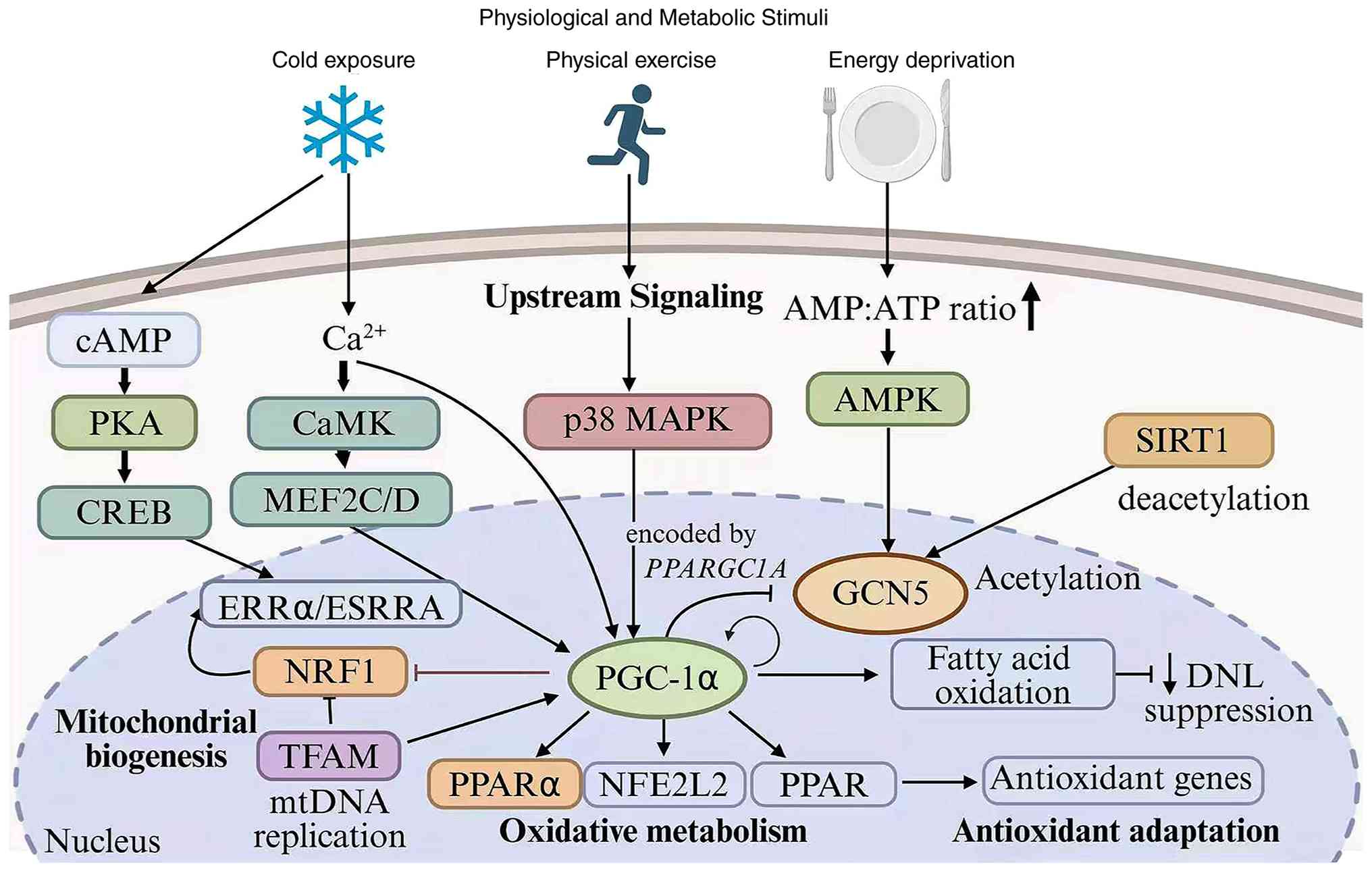
SIRT1 supports hepatic mitochondrial metabolism
through PGC-1α deacetylation and improved fatty acid oxidation, and
SIRT1-dependent autophagy protects hepatocytes from
palmitate-induced lipotoxicity (59,60)
(Fig. 3). The available studies
support PGC-1α regulation and autophagy induction but do not
establish that ATG5 and ATG7 are both the direct hepatic substrates
responsible for mitophagy in MASH.
SIRT2 can restrain inflammatory and lipogenic
signaling through distinct substrates. SIRT2-dependent
deacetylation regulates NLRP3 inflammasome activation in metabolic
inflammation (61), while direct
deacetylation of ATP-citrate lyase at K540, K546 and K554
destabilizes the enzyme and reduces hepatic lipid accumulation
(62).
SIRT3 is a major regulator of mitochondrial redox
homeostasis. It deacetylates isocitrate dehydrogenase 2 and
regulates the mitochondrial redox state (63); in hepatocytes, the SIRT3-SOD2 axis
influences susceptibility to lipotoxicity and autophagic responses
(64) (Fig. 4). These studies support redox and
stress-response functions but do not directly demonstrate that
SIRT3 preserves cristae architecture or that LCAD activation
accounts for all of its effects in MASH.
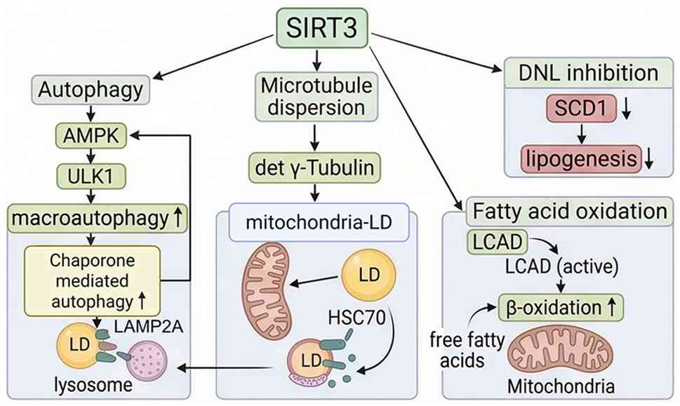 | Figure 4.Evidence-bounded summary of
SIRT3-linked redox and stress-response regulation in hepatocytes.
SIRT3 deacetylates IDH2 and regulates the mitochondrial redox
state, while the hepatocyte SIRT3-SOD2 axis influences
susceptibility to lipotoxicity and autophagic responses (62,63).
Downstream routes involving AMPK-ULK1, LAMP2A/HSC70, lipid
droplet-mitochondria contacts or LCAD should be regarded as
context-dependent hypotheses rather than mechanisms directly
established by the cited MASH studies. IDH2, isocitrate
dehydrogenase 2; SOD2, superoxide dismutase 2; AMPK, adenosine
monophosphate-activated protein kinase; ULK1, Unc-51 like autophagy
activating kinase 1; LAMP2A, lysosome-associated membrane protein
type 2A; HSC70, heat shock cognate 71-kDa protein; LCAD, long-chain
acyl-CoA dehydrogenase. |
Context-dependent isoforms
(SIRT4/7)
SIRT4 can suppress fatty acid oxidation by
deacetylating and inhibiting malonyl-CoA decarboxylase, thereby
favoring malonyl-CoA accumulation and lipid anabolism (65). Conversely, SIRT4 has an
anti-fibrotic role in HSCs, and miR-130b-5p-mediated suppression of
SIRT4 promotes HSC activation and liver fibrosis (66). These findings indicate that the net
effect of SIRT4 depends on substrate metabolism, cell type and
disease stage rather than on degradation of the mitochondrial
trifunctional protein-α subunit.
SIRT7 also has context-dependent nuclear and
mitochondrial stress-response functions. SIRT7-dependent
deacetylation of DDB1 regulates the lipogenic nuclear receptor TR4
and thereby influences hepatic lipid metabolism (67). In a distinct stem-cell system,
SIRT7 participates in an NRF1-associated mitochondrial UPR
metabolic checkpoint, linking nuclear transcriptional control to
mitochondrial proteostasis (68).
The latter mechanism provides a conceptual framework but should not
be presented as direct evidence for SIRT7-GABP-β signaling in
hepatocytes or MASH.
Epigenetic and non-acetylation
modifications (SIRT5/6)
SIRT5 removes succinyl and other acyl modifications
from mitochondrial proteins. Proteomic and biochemical studies
identify SIRT5-dependent desuccinylation across diverse metabolic
pathways, including mitochondrial energy metabolism (69,70).
However, the cited studies are not direct evidence that CPT1A and
succinate dehydrogenase are the defining SIRT5 targets in human
MASH.
SIRT6 restrains hepatic lipogenesis. Experimental
studies show that SIRT6 suppresses the LXR, ChREBP and SREBP1
lipogenic programs, while USP10 stabilizes SIRT6 and protects
against hepatic steatosis, insulin resistance and inflammation
(71,72) (Fig.
5). These data directly support SIRT6-dependent transcriptional
control of lipid metabolism without requiring an unverified claim
that promoter-specific H3K9 and H3K56 deacetylation explains the
entire phenotype.
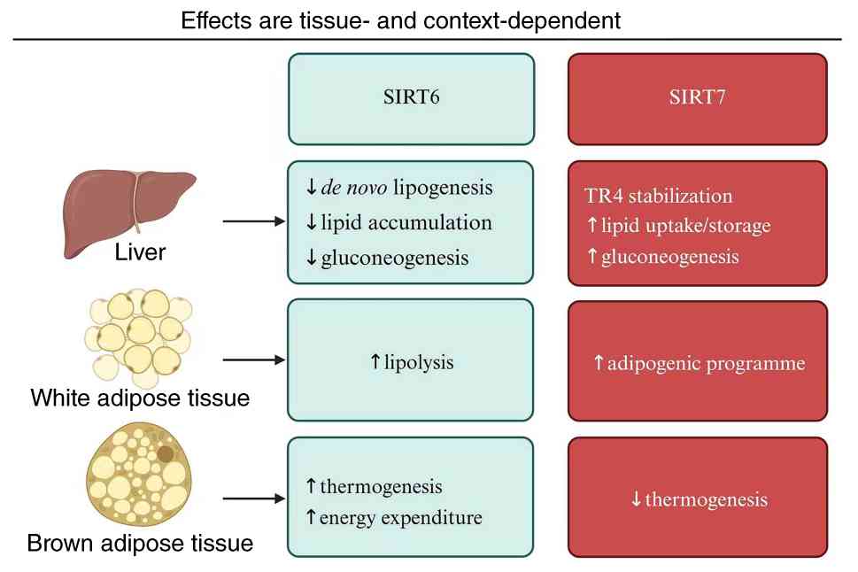 | Figure 5.Hepatic and context-dependent
metabolic functions of SIRT6 and SIRT7. In experimental liver
systems, SIRT6 suppresses LXR-, ChREBP- and SREBP1-associated
lipogenic programs, and USP10-dependent stabilization of SIRT6
protects against steatosis, insulin resistance and inflammation
(70,71). SIRT7-dependent DDB1 deacetylation
regulates TR4 and hepatic lipid metabolism (66), whereas its NRF1-associated
mitochondrial-stress checkpoint was demonstrated in a distinct
stem-cell system (67). Additional
tissue-specific branches depicted in the schematic are contextual
and should not be presented as direct evidence in hepatocytes or
MASH. SIRT, sirtuin; LXR, liver X receptor; ChREBP,
carbohydrate-responsive element-binding protein; SREBP1, sterol
regulatory element-binding protein 1; USP10, ubiquitin-specific
peptidase 10; DDB1, damage-specific DNA-binding protein 1; TR4,
testicular receptor 4; NRF1, nuclear respiratory factor 1. |
Therapeutic interventions targeting
mitochondrial dysfunction and advanced metabolism
The drug development paradigm for MASLD has
undergone a profound shift: From early ‘peripheral empirical
antioxidation’ to modern ‘precise energy metabolic remodeling’. To
provide a rigorous clinical and translational perspective,
contemporary therapeutic strategies must be strictly evaluated
based on their evidence-graded clinical stages and mechanistic
boundaries (Table II).
| Table II.Clinical translational progress of
mitochondrial-targeted and advanced metabolic therapies for
MASLD/MASH. |
Table II.
Clinical translational progress of
mitochondrial-targeted and advanced metabolic therapies for
MASLD/MASH.
| A, Lifestyle
intervention |
|---|
|
|---|
| Core target | Candidates | Mitochondrial and
metabolic mechanisms | Highest clinical
stage | Key clinical
obstacles and limitations |
|---|
| Metabolic
remodeling | Physical
exercise | Stimulates hepatic
mitochondrial biogenesis; restores systemic redox balance. | First-line
recommendation | Low long-term
patient compliance; limited capacity to directly reverse
established advanced fibrosis. |
|
| B, Direct
mitochondrial quality control |
|
| SIRT3-associated
redox pathways | No selective
clinical activator established | IDH2 deacetylation
and SOD2-associated regulation support mitochondrial redox
homeostasis and lipotoxic stress responses. | Preclinical
mechanistic evidence | Requires
liver-selective compounds and direct efficacy testing in MASH. |
| Antioxidant
strategies | Vitamin E;
silybin/phosphati-dylcholine/vitamin E; MitoQ | Conventional
antioxidant regimens have formulation-specific clinical data;
mitochondria-targeted MitoQ reduced oxidative and inflammatory
injury in a mouse NASH model. | Clinical studies
for conventional regimens; MitoQ preclinical in NASH | Clinical
antifibrotic efficacy is inconsistent, and evidence cannot be
transferred across formulations. |
| Mild mito-chondrial
uncoupling | CRMP | Produces controlled
proton leak and reduced hepatic lipid burden in rat and
non-human-primate models. | Preclinical | Human efficacy and
the therapeutic safety window remain unestablished. |
|
| C,
Hepatocyte-autonomous protection |
|
| THR-β agonist | Resmetirom
(MGL-3196) | Selectively targets
hepatic THR-β, reduces liver fat and improves prespecified MASH
histological endpoints. | FDA approved for an
eligible noncirrhotic MASH population | Indication
boundaries, adverse effects and long-term outcomes require
continued monitoring. |
| ACC inhibitor | Firsocostat
(GS-0976) | Inhibits
ACC-dependent DNL and reduces hepatic fat. | Phase II
evidence | Monotherapy can
increase circulating triglycerides; combination benefit is
regimen-specific. |
| Pan-PPAR
agonist | Lanifibranor | Modulates
PPAR-α/γ/δ signaling and improves MASH activity and
fibrosis-related histological endpoints. | Phase IIb efficacy;
late-stage development | Weight gain, fluid
retention and durability of histological benefit require
monitoring. |
|
| D, Systemic
metabolic retrograde driving |
|
| GLP-1-based
therapy |
Semaglutide-containing combination
regimen | Primarily improves
systemic energy balance and reduces substrate delivery to the
liver; direct mitochondrial effects were not established. | Phase II evidence
for the cited combination | The contribution of
each drug and direct hepatocyte-autonomous effects remain
uncertain. |
| Multi-incretin
agonists |
Tirzepatide/retatrutide | Dual or triple
incretin-receptor agonism reduces body weight and hepatic metabolic
burden; trial endpoints differ between agents. | Phase II
(tirzepatide); phase IIa (retatrutide) | Long-term fibrosis
and intrahepatic mitochondrial outcomes require follow-up. |
| FGF21
analogues |
Efruxifermin/pegozafermin | Improve systemic
metabolic measures, liver fat and histological endpoints in
randomized studies. | Phase IIb efficacy;
late-stage development | Chronic injection,
durability, safety and cost remain practical limitations. |
Basic therapies and direct
mitochondrial quality control (preclinical to early clinical)
Physical exercise: The cornerstone of endogenous
mitochondrial adaptation
Regular physical exercise is a first-line
intervention for MASLD and improves hepatic steatosis and insulin
sensitivity. In experimental models, hepatocyte eNOS contributes to
exercise-induced adaptations in hepatic mitochondrial function and
autophagy, while broader evidence links exercise to enhanced
hepatic fatty acid oxidation and reduced steatosis (73,74).
The extent to which exercise alone reverses advanced fibrosis
remains uncertain and adherence limits long-term effectiveness.
Evolution of antioxidants and
mitochondrial-targeted redox strategies
Conventional vitamin E can improve biochemical and
selected histological features in NASH, but treatment response and
fibrosis benefit are variable (75). Silybin combined with
phosphatidylcholine and vitamin E has also been evaluated in a
randomized trial in NAFLD (76).
Mitoquinone is designed to concentrate antioxidant activity within
mitochondria and attenuated steatosis, necroinflammation and
profibrogenic signaling in a mouse NASH model (77). These data support preclinical
mitochondrial targeting but do not yet establish a consistent
antifibrotic benefit in patients.
Modulating the mitochondrial proton motive force
represents another strategy to reduce lipid burden and oxidative
pressure. A controlled-release mitochondrial protonophore reversed
diabetes and steatohepatitis in rats (78) and subsequently reduced dyslipidemia
and hepatic steatosis in dysmetabolic non-human primates (79). These studies provide preclinical
proof of concept; they should not be described as evidence of
established clinical translation.
Sirtuin-related and natural-product
metabolic remodeling
Reduced mitochondrial deacetylase activity and
oxidative stress provide a rationale for targeting
sirtuin-associated pathways, but direct clinical evidence for
selective SIRT3 activators in MASH remains limited.
Among natural products, resveratrol has been studied
across experimental and clinical liver-disease settings and may
influence AMPK, sirtuin and antioxidant pathways (80). The heterogeneity of formulations,
doses and study models precludes assigning a single validated
SIRT1/AMPK/Smad3/microRNA-141 mechanism to its effects in human
MASH.
Hepatic focal metabolic targeting:
Cell-autonomous mitochondrial protection (advanced clinical and
approved stages)
The core of this category lies in
hepatocyte-directed metabolic regulation, in which pharmacological
agents act on hepatic enzymes or nuclear receptors to reduce
substrate overload and improve cellular bioenergetics.
Thyroid hormone receptor (THR)-β
agonist: The milestone breakthrough of resmetirom
Resmetirom (MGL-3196) became the first FDA-approved
pharmacotherapy for adults with noncirrhotic MASH and
moderate-to-advanced fibrosis in 2024 (81). In the phase III MAESTRO-NASH trial,
resmetirom increased the proportions of patients achieving MASH
resolution without worsening of fibrosis and fibrosis improvement
without worsening of disease activity (82). A preceding 36-week phase II
extension also demonstrated improvements in non-invasive efficacy
measures (83). These data
establish clinical efficacy within the studied population without
implying universal reversal of advanced fibrosis.
Combined strategies involving
acetyl-CoA carboxylase (ACC) inhibition
ACC inhibitors such as firsocostat reduce DNL and
hepatic fat, but monotherapy can increase circulating triglycerides
(84). A phase II trial
subsequently evaluated combinations of semaglutide, cilofexor and
firsocostat, supporting combination development while not
establishing a specific synergy with THR-β agonists (85).
PPAR agonists restore metabolic transcription
through multiple receptor isoforms. In the phase IIb NATIVE trial,
the pan-PPAR agonist lanifibranor improved MASH activity and
fibrosis-related histological endpoints (86). Additional analyses showed
improvements in cardiometabolic measures (87). These findings support further
late-stage development while avoiding claims that fibrosis reversal
is already established for all patients.
Systemic metabolic drivers:
Predominantly non-cell-autonomous hepatic protection
Unlike hepatocyte-directed agents, these therapies
primarily remodel metabolic flux in adipose tissue, muscle and the
central nervous system, thereby reducing lipid delivery to the
liver and indirectly relieving mitochondrial substrate
pressure.
Lipid-clearing effects of
glucagon-like peptide-1 receptor agonism and multi-incretin
agonists
Recent phase II studies have demonstrated
substantial improvements in hepatic metabolic and histological
endpoints. Tirzepatide, a dual GIP/GLP-1 receptor agonist, achieved
high rates of MASH resolution without worsening of fibrosis in
patients with F2-F3 fibrosis (88), whereas the triple
GIP/GLP-1/glucagon receptor agonist retatrutide produced marked
reductions in liver fat in patients with MASLD (89).
These agents primarily reduce appetite, body weight,
insulin resistance and systemic free-fatty-acid delivery to the
liver. Although this metabolic unloading can protect hepatocytes
from lipotoxic stress, the cited trials do not directly
characterize autonomous effects on intrahepatic mitochondrial
dynamics (90,91). Their hepatic benefit should
therefore be described as predominantly systemic unless direct
organelle-level evidence is available.
FGF21 analogs: Systemic metabolic
improvement with histological efficacy
Pegozafermin and efruxifermin demonstrated promising
histological efficacy in randomized phase IIb trials in NASH/MASH
(90,91). These results support continued
late-stage development but do not by themselves establish phase III
efficacy.
Earlier phase 1b/2a data for pegozafermin
demonstrated reductions in liver fat and improvements in liver
enzymes, lipids and adiponectin (92). These clinical pharmacodynamic
findings do not directly prove activation of a hepatic PGC-1α/PPAR
axis or brown-adipose thermogenesis; those mechanistic claims have
therefore been removed (Fig.
6).
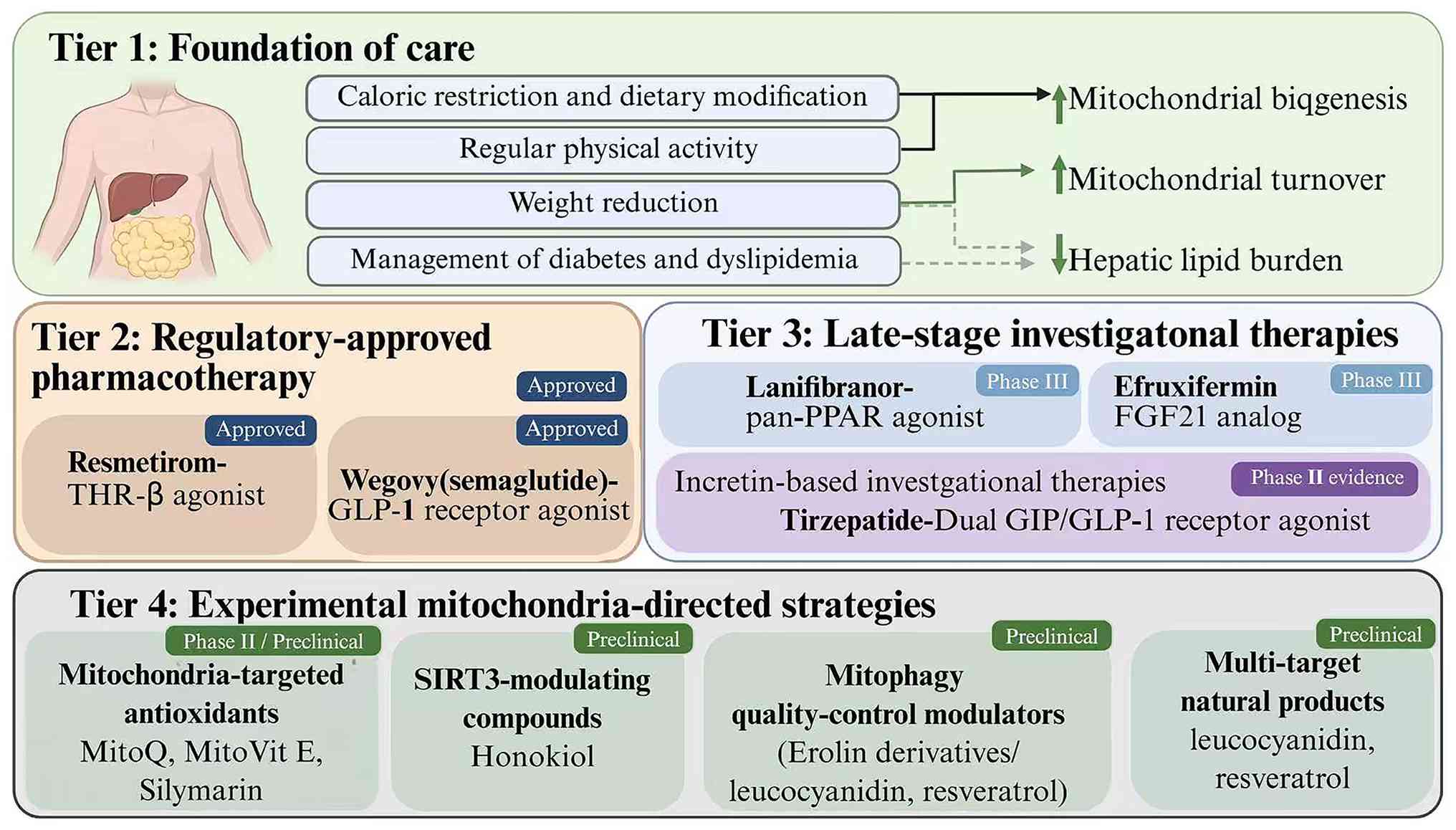 | Figure 6.Evidence-graded therapeutic framework
for MASLD/MASH. Tier 1 comprises lifestyle and cardiometabolic
management, including physical activity and weight reduction
(72,73). Tier 2 contains approved
hepatocyte-directed therapy, represented here by resmetirom for
eligible adults with noncirrhotic MASH and moderate-to-advanced
fibrosis (80,81). Tier 3 comprises agents with
randomized phase II/IIb evidence, including firsocostat-containing
combinations, lanifibranor, tirzepatide, retatrutide, efruxifermin
and pegozafermin (83–91). Tier 4 includes experimental
mitochondria-directed approaches such as MitoQ and
controlled-release mitochondrial protonophores, for which the cited
MASH evidence remains preclinical (76–78).
Conventional vitamin E or silybin-containing regimens and
resveratrol have clinical or mixed translational evidence, but
formulation-specific effects should not be generalized (74,75,79).
MASLD, metabolic dysfunction-associated steatotic liver disease;
MASH, metabolic dysfunction-associated steatohepatitis; THR-β,
thyroid hormone receptor-β; PPAR, peroxisome proliferator-activated
receptor; GIP, glucose-dependent insulinotropic polypeptide; GLP-1,
glucagon-like peptide-1; FGF21, fibroblast growth factor 21; MitoQ,
mitoquinone. |
Discussion and concluding perspectives
The paradigm of MASLD pathogenesis has evolved from
a linear model of lipid overload to a highly interconnected,
non-linear network where mitochondrial dysfunction operates as the
central rheostat. The structural and functional evolution of the
intrahepatic mitochondrial network, progressing through a biphase
curve of early hormetic compensation to late bioenergetic failure,
dictates the clinical transition from simple steatosis to MASH and
progressive fibrosis.
Despite notable advances in characterizing
downstream events such as mROS generation, lipid peroxidation
(4-HNE/MDA formation) and organelle structural collapse
(DRP1-mediated fission overactivation, UPRmt
decompensation and MAM-mediated Ca2+ overload), numerous
key mechanistic gaps and translation barriers remain. First, the
precise upstream triggers that dictate the transition from adaptive
mitochondrial hypermetabolism to decompensated bioenergetic failure
remain incomplete. Second, as highlighted by the sirtuin epigenetic
network, multi-compartmentalized enzymes such as SIRT4 and SIRT7
exhibit profound context-dependency and cell-type specificity,
creating an urgent need for cell-specific and time-resolved genetic
models to reconcile existing academic controversies.
Monotherapies rarely address all pathogenic facets
of this complex metabolic disorder. The current pipeline includes
hepatocyte-directed metabolic agents such as resmetirom and
pan-PPAR agonists, as well as systemic metabolic drivers such as
multi-incretins and FGF21 analogs. While systemic drivers reduce
intrahepatic lipid partly through weight loss and improved
peripheral metabolism, their direct long-term effects on hepatocyte
organelle quality control require longitudinal study.
Consequently, the future of MASLD therapeutics lies
in the rational design of personalized combination protocols.
Pairing cell-autonomous mitochondrial stabilizers with systemic
incretin therapies, tailored precisely to the body mass index,
diabetic status and baseline fibrotic stage of a patient, will
represent the true frontier of clinical precision medicine in
overcoming this global metabolic epidemic.
Acknowledgements
Not applicable.
Funding
The present study was supported by the National Natural Science
Foundation Cultivation Project of Southwest Medical University
(grant no. 2025LCYXZX32), the Special Program for Clinical Medicine
of Southwest Medical University (grant nos. 2024LCYXZX31 and
2025LCYXZX95), the National Natural Science Foundation of China
(grant no. 82570678) and the Sichuan Provincial Natural Science
Foundation for Outstanding Youth Foundation (grant no.
2024NSFJQ0054).
Availability of data and materials
Not applicable.
Authors' contributions
JC conceived and designed the study, performed the
extensive literature review and drafted the primary manuscript as
the principal author. HZ assisted in the literature screening,
designed the thematic tables, prepared the figures and critically
revised the manuscript for important intellectual content. HC
revised the manuscript critically for important intellectual
content and refined the mechanistic frameworks. WF and JL, serving
as senior supervisors, provided overall strategic direction,
critically reviewed the study design, critically revised the
manuscript for important intellectual content and supervised the
final refinement of the manuscript. All authors have read and
approved the final version of the manuscript. Data authentication
is not applicable.
Ethics approval and consent to
participate
Not applicable.
Patient consent for publication
Not applicable.
Competing interests
The authors declare that they have no competing
interests.
References
|
1
|
Younossi ZM, Koenig AB, Abdelatif D, Fazel
Y, Henry L and Wymer M: Global epidemiology of nonalcoholic fatty
liver disease-Meta-analytic assessment of prevalence, incidence,
and outcomes. Hepatology. 64:73–84. 2016. View Article : Google Scholar : PubMed/NCBI
|
|
2
|
Younossi ZM, Golabi P, Paik JM, Henry A,
Van Dongen C and Henry L: The global epidemiology of nonalcoholic
fatty liver disease (NAFLD) and nonalcoholic steatohepatitis
(NASH): A systematic review. Hepatology. 77:1335–1347. 2023.
View Article : Google Scholar : PubMed/NCBI
|
|
3
|
Rinella ME, Lazarus JV, Ratziu V, Francque
SM, Sanyal AJ, Kanwal F, Romero D, Abdelmalek MF, Anstee QM, Arab
JP, et al: A multisociety Delphi consensus statement on new fatty
liver disease nomenclature. Hepatology. 78:1966–1986. 2023.
View Article : Google Scholar : PubMed/NCBI
|
|
4
|
Kanwal F, Neuschwander-Tetri BA, Loomba R
and Rinella ME: Metabolic dysfunction-associated steatotic liver
disease: Update and impact of new nomenclature on the American
association for the study of liver diseases practice guidance on
nonalcoholic fatty liver disease. Hepatology. 79:1212–1219. 2024.
View Article : Google Scholar : PubMed/NCBI
|
|
5
|
Yang A, Zhu X, Zhang L and Ding Y:
Transitioning from NAFLD to MAFLD and MASLD: Consistent prevalence
and risk factors in a Chinese cohort. J Hepatol. 80:e154–e155.
2024. View Article : Google Scholar : PubMed/NCBI
|
|
6
|
Powell EE, Wong VWS and Rinella M:
Non-alcoholic fatty liver disease. Lancet. 397:2212–2224. 2021.
View Article : Google Scholar : PubMed/NCBI
|
|
7
|
Britton LJ, Subramaniam VN and Crawford
DH: Iron and non-alcoholic fatty liver disease. World J
Gastroenterol. 22:8112–8122. 2016. View Article : Google Scholar : PubMed/NCBI
|
|
8
|
Ryan JD, Armitage AE, Cobbold JF, Collier
J, Barnes E, Drakesmith H, Valenti L and Pavlides M: Hepatic iron
is the major determinant of serum ferritin in NAFLD patients. Liver
Int. 38:164–173. 2018. View Article : Google Scholar : PubMed/NCBI
|
|
9
|
Kleiner DE, Brunt EM, Van Natta M, Behling
C, Contos MJ, Cummings OW, Ferrell LD, Liu YC, Torbenson MS,
Unalp-Arida A, et al: Design and validation of a histological
scoring system for nonalcoholic fatty liver disease. Hepatology.
41:1313–1321. 2005. View Article : Google Scholar : PubMed/NCBI
|
|
10
|
Ipsen DH, Lykkesfeldt J and Tveden-Nyborg
P: Molecular mechanisms of hepatic lipid accumulation in
non-alcoholic fatty liver disease. Cell Mol Life Sci. 75:3313–3327.
2018. View Article : Google Scholar : PubMed/NCBI
|
|
11
|
Sanders FWB and Griffin JL: De novo
lipogenesis in the liver in health and disease: More than just a
shunting yard for glucose. Biol Rev Camb Philos Soc. 91:452–468.
2016. View Article : Google Scholar : PubMed/NCBI
|
|
12
|
Smith GI, Shankaran M, Yoshino M,
Schweitzer GG, Chondronikola M, Beals JW, Okunade AL, Patterson BW,
Nyangau E, Field T, et al: Insulin resistance drives hepatic de
novo lipogenesis in nonalcoholic fatty liver disease. J Clin
Invest. 130:1453–1460. 2020. View Article : Google Scholar : PubMed/NCBI
|
|
13
|
Gluchowski NL, Becuwe M, Walther TC and
Farese RV Jr: Lipid droplets and liver disease: From basic biology
to clinical implications. Nat Rev Gastroenterol Hepatol.
14:343–355. 2017. View Article : Google Scholar : PubMed/NCBI
|
|
14
|
Shum M, Ngo J, Shirihai OS and Liesa M:
Mitochondrial oxidative function in NAFLD: Friend or foe? Mol
Metab. 50:1011342021. View Article : Google Scholar : PubMed/NCBI
|
|
15
|
Mansouri A, Gattolliat CH and Asselah T:
Mitochondrial dysfunction and signaling in chronic liver diseases.
Gastroenterology. 155:629–647. 2018. View Article : Google Scholar : PubMed/NCBI
|
|
16
|
Schlaepfer IR and Joshi M: CPT1A-mediated
fat oxidation, mechanisms, and therapeutic potential.
Endocrinology. 161:bqz0462020. View Article : Google Scholar : PubMed/NCBI
|
|
17
|
Wang M, Wang K, Liao X, Hu H, Chen L, Meng
L, Gao W and Li Q: Carnitine palmitoyltransferase system: A new
target for anti-inflammatory and anticancer therapy? Front
Pharmacol. 12:7605812021. View Article : Google Scholar : PubMed/NCBI
|
|
18
|
Spinelli JB and Haigis MC: The
multifaceted contributions of mitochondria to cellular metabolism.
Nat Cell Biol. 20:745–754. 2018. View Article : Google Scholar : PubMed/NCBI
|
|
19
|
Murphy MP and Hartley RC: Mitochondria as
a therapeutic target for common pathologies. Nat Rev Drug Discov.
17:865–886. 2018. View Article : Google Scholar : PubMed/NCBI
|
|
20
|
Koliaki C, Szendroedi J, Kaul K, Jelenik
T, Nowotny P, Jankowiak F, Herder C, Carstensen M, Krausch M,
Knoefel WT, et al: Adaptation of hepatic mitochondrial function in
humans with non-alcoholic fatty liver is lost in steatohepatitis.
Cell Metab. 21:739–746. 2015. View Article : Google Scholar : PubMed/NCBI
|
|
21
|
Satapati S, Sunny NE, Kucejova B, Fu X, He
TT, Méndez-Lucas A, Shelton JM, Perales JC, Browning JD and Burgess
SC: Elevated TCA cycle function in the pathology of diet-induced
hepatic insulin resistance and fatty liver. J Lipid Res.
53:1080–1092. 2012. View Article : Google Scholar : PubMed/NCBI
|
|
22
|
Arconzo M, Piccinin E, Pasculli E,
Cariello M, Loiseau N, Bertrand-Michel J, Guillou H, Matrella ML,
Villani G and Moschetta A: Hepatic-specific Pgc-1α ablation drives
fibrosis in a MASH model. Liver Int. 44:2738–2752. 2024. View Article : Google Scholar : PubMed/NCBI
|
|
23
|
Estall JL, Kahn M, Cooper MP, Fisher FM,
Wu MK, Laznik D, Qu L, Cohen DE, Shulman GI and Spiegelman BM:
Sensitivity of lipid metabolism and insulin signaling to genetic
alterations in hepatic peroxisome proliferator-activated
receptor-gamma coactivator-1alpha expression. Diabetes.
58:1499–1508. 2009. View Article : Google Scholar : PubMed/NCBI
|
|
24
|
Samuel VT and Shulman GI: Nonalcoholic
fatty liver disease as a nexus of metabolic and hepatic diseases.
Cell Metab. 27:22–41. 2018. View Article : Google Scholar : PubMed/NCBI
|
|
25
|
Pagadala M, Kasumov T, McCullough AJ, Zein
NN and Kirwan JP: Role of ceramides in nonalcoholic fatty liver
disease. Trends Endocrinol Metab. 23:365–371. 2012. View Article : Google Scholar : PubMed/NCBI
|
|
26
|
Arroyave-Ospina JC, Wu Z, Geng Y and
Moshage H: Role of oxidative stress in the pathogenesis of
non-alcoholic fatty liver disease: Implications for prevention and
therapy. Antioxidants (Basel). 10:1742021. View Article : Google Scholar : PubMed/NCBI
|
|
27
|
Schwabe RF, Tabas I and Pajvani UB:
Mechanisms of fibrosis development in nonalcoholic steatohepatitis.
Gastroenterology. 158:1913–1928. 2020. View Article : Google Scholar : PubMed/NCBI
|
|
28
|
Brand MD: Mitochondrial generation of
superoxide and hydrogen peroxide as the source of mitochondrial
redox signaling. Free Radic Biol Med. 100:14–31. 2016. View Article : Google Scholar : PubMed/NCBI
|
|
29
|
Sunny NE, Bril F and Cusi K: Mitochondrial
adaptation in nonalcoholic fatty liver disease: Novel mechanisms
and treatment strategies. Trends Endocrinol Metab. 28:250–260.
2017. View Article : Google Scholar : PubMed/NCBI
|
|
30
|
Pittala S, Krelin Y, Kuperman Y and
Shoshan-Barmatz V: A mitochondrial VDAC1-based peptide greatly
suppresses steatosis and NASH-associated pathologies in a mouse
model. Mol Ther. 27:1848–1862. 2019. View Article : Google Scholar : PubMed/NCBI
|
|
31
|
Shoshan-Barmatz V, De Pinto V,
Zweckstetter M, Raviv Z, Keinan N and Arbel N: VDAC, a
multi-functional mitochondrial protein regulating cell life and
death. Mol Aspects Med. 31:227–285. 2010. View Article : Google Scholar : PubMed/NCBI
|
|
32
|
Masarone M, Rosato V, Dallio M, Gravina
AG, Aglitti A, Loguercio C, Federico A and Persico M: Role of
oxidative stress in pathophysiology of nonalcoholic fatty liver
disease. Oxid Med Cell Longev. 2018:95476132018. View Article : Google Scholar : PubMed/NCBI
|
|
33
|
García-Ruiz C and Fernández-Checa JC:
Mitochondrial oxidative stress and antioxidants balance in fatty
liver disease. Hepatol Commun. 2:1425–1439. 2018. View Article : Google Scholar : PubMed/NCBI
|
|
34
|
Pirola CJ, Garaycoechea M, Flichman D,
Castaño GO and Sookoian S: Liver mitochondrial DNA damage and
genetic variability of cytochrome b-a key component of the
respirasome-drive the severity of fatty liver disease. J Intern
Med. 289:84–96. 2021. View Article : Google Scholar : PubMed/NCBI
|
|
35
|
Win S, Than TA, Fernandez-Checa JC and
Kaplowitz N: JNK interaction with Sab mediates ER stress induced
inhibition of mitochondrial respiration and cell death. Cell Death
Dis. 5:e9892014. View Article : Google Scholar : PubMed/NCBI
|
|
36
|
Ogura M, Yamaki J, Homma MK and Homma Y:
Mitochondrial c-Src regulates cell survival through phosphorylation
of respiratory chain components. Biochem J. 447:281–289. 2012.
View Article : Google Scholar : PubMed/NCBI
|
|
37
|
Brunati AM, Pagano MA, Bindoli A and
Rigobello MP: Thiol redox systems and protein kinases in hepatic
stellate cell regulatory processes. Free Radic Res. 44:363–378.
2010. View Article : Google Scholar : PubMed/NCBI
|
|
38
|
Sánchez-Valle V, Chávez-Tapia NC, Uribe M
and Méndez-Sánchez N: Role of oxidative stress and molecular
changes in liver fibrosis: A review. Curr Med Chem. 19:4850–4860.
2012. View Article : Google Scholar : PubMed/NCBI
|
|
39
|
Zhao Q, Liu J, Deng H, Ma R, Liao JY,
Liang H, Hu J, Li J, Guo Z, Cai J, et al: Targeting
mitochondria-located circRNA SCAR alleviates NASH via reducing mROS
output. Cell. 183:76–93.e22. 2020. View Article : Google Scholar : PubMed/NCBI
|
|
40
|
Zeng Q, Liu CH, Wu D, Jiang W, Zhang N and
Tang H: LncRNA and circRNA in patients with non-alcoholic fatty
liver disease: A systematic review. Biomolecules. 13:5602023.
View Article : Google Scholar : PubMed/NCBI
|
|
41
|
Galloway CA, Lee H, Brookes PS and Yoon Y:
Decreasing mitochondrial fission alleviates hepatic steatosis in a
murine model of nonalcoholic fatty liver disease. Am J Physiol
Gastrointest Liver Physiol. 307:G632–G641. 2014. View Article : Google Scholar : PubMed/NCBI
|
|
42
|
Legaki AI, Moustakas II, Sikorska M,
Papadopoulos G, Velliou RI and Chatzigeorgiou A: Hepatocyte
mitochondrial dynamics and bioenergetics in obesity-related
non-alcoholic fatty liver disease. Curr Obes Rep. 11:126–143. 2022.
View Article : Google Scholar : PubMed/NCBI
|
|
43
|
Moore MP, Cunningham RP, Meers GM, Johnson
SA, Wheeler AA, Ganga RR, Spencer NM, Pitt JB, Diaz-Arias A, Swi
AIA, et al: Compromised hepatic mitochondrial fatty acid oxidation
and reduced markers of mitochondrial turnover in human NAFLD.
Hepatology. 76:1452–1465. 2022. View Article : Google Scholar : PubMed/NCBI
|
|
44
|
Baker MJ, Lampe PA, Stojanovski D, Korwitz
A, Anand R, Tatsuta T and Langer T: Stress-induced OMA1 activation
and autocatalytic turnover regulate OPA1-dependent mitochondrial
dynamics. EMBO J. 33:578–593. 2014. View Article : Google Scholar : PubMed/NCBI
|
|
45
|
Bórquez JC, Díaz-Castro F, La Fuente FP,
Espinoza K, Figueroa AM, Martínez-Ruíz I, Hernández V,
López-Soldado I, Ventura R, Domingo JC, et al: Mitofusin-2 induced
by exercise modifies lipid droplet-mitochondria communication,
promoting fatty acid oxidation in male mice with NAFLD. Metabolism.
152:1557652024. View Article : Google Scholar : PubMed/NCBI
|
|
46
|
Glick D, Zhang W, Beaton M, Marsboom G,
Gruber M, Simon MC, Hart J, Dorn GW II, Brady MJ and Macleod KF:
BNip3 regulates mitochondrial function and lipid metabolism in the
liver. Mol Cell Biol. 32:2570–2584. 2012. View Article : Google Scholar : PubMed/NCBI
|
|
47
|
Wang L, Liu X, Nie J, Zhang J, Kimball SR,
Zhang H, Zhang WJ, Jefferson LS, Cheng Z, Ji Q and Shi Y: ALCAT1
controls mitochondrial etiology of fatty liver diseases, linking
defective mitophagy to steatosis. Hepatology. 61:486–496. 2015.
View Article : Google Scholar : PubMed/NCBI
|
|
48
|
Fiorese CJ, Schulz AM, Lin YF, Rosin N,
Pellegrino MW and Haynes CM: The transcription factor ATF5 mediates
a mammalian mitochondrial UPR. Curr Biol. 26:2037–2043. 2016.
View Article : Google Scholar : PubMed/NCBI
|
|
49
|
Zhu L, Luo X, Fu N and Chen L:
Mitochondrial unfolded protein response: A novel pathway in
metabolism and immunity. Pharmacol Res. 168:1056032021. View Article : Google Scholar : PubMed/NCBI
|
|
50
|
Yi HS: Implications of mitochondrial
unfolded protein response and mitokines: A perspective on fatty
liver diseases. Endocrinol Metab (Seoul). 34:39–46. 2019.
View Article : Google Scholar : PubMed/NCBI
|
|
51
|
Shpilka T and Haynes CM: The mitochondrial
UPR: Mechanisms, physiological functions and implications in
ageing. Nat Rev Mol Cell Biol. 19:109–120. 2018. View Article : Google Scholar : PubMed/NCBI
|
|
52
|
Giorgi C, Missiroli S, Patergnani S,
Duszynski J, Wieckowski MR and Pinton P: Mitochondria-associated
membranes: composition, molecular mechanisms, and
physiopathological implications. Antioxid Redox Signal.
22:995–1019. 2015. View Article : Google Scholar : PubMed/NCBI
|
|
53
|
Phillips MJ and Voeltz GK: Structure and
function of ER membrane contact sites with other organelles. Nat
Rev Mol Cell Biol. 17:69–82. 2016. View Article : Google Scholar : PubMed/NCBI
|
|
54
|
Arruda AP, Pers BM, Parlakgül G, Güney E,
Inouye K and Hotamisligil GS: Chronic enrichment of hepatic
endoplasmic reticulum-mitochondria contact leads to mitochondrial
dysfunction in obesity. Nat Med. 20:1427–1435. 2014. View Article : Google Scholar : PubMed/NCBI
|
|
55
|
Rieusset J: Endoplasmic
reticulum-mitochondria calcium signaling in hepatic metabolic
diseases. Biochim Biophys Acta Mol Cell Res. 1864:865–876. 2017.
View Article : Google Scholar : PubMed/NCBI
|
|
56
|
Theurey P, Tubbs E, Vial G, Jacquemetton
J, Bendridi N, Chauvin MA, Alam MR, Le Romancer M, Vidal H and
Rieusset J: Mitochondria-associated endoplasmic reticulum membranes
allow adaptation of mitochondrial metabolism to glucose
availability in the liver. J Mol Cell Biol. 8:129–143. 2016.
View Article : Google Scholar : PubMed/NCBI
|
|
57
|
Camacho-Pereira J, Tarragó MG, Chini CCS,
Nin V, Escande C, Warner GM, Puranik AS, Schoon RA, Reid JM, Galina
A and Chini EN: CD38 dictates age-related NAD decline and
mitochondrial dysfunction through an SIRT3-dependent mechanism.
Cell Metab. 23:1127–1139. 2016. View Article : Google Scholar : PubMed/NCBI
|
|
58
|
Katsyuba E, Mottis A, Zietak M, De Franco
F, van der Velpen V, Gariani K, Ryu D, Cialabrini L, Matilainen O,
Liscio P, et al: De novo NAD+ synthesis enhances
mitochondrial function and improves health. Nature. 563:354–359.
2018. View Article : Google Scholar : PubMed/NCBI
|
|
59
|
Pang J, Yin L, Jiang W, Wang H, Cheng Q,
Jiang Z, Cao Y, Zhu X, Li B, Qian S, et al: Sirt1-mediated
deacetylation of PGC-1α alleviated hepatic steatosis in type 2
diabetes mellitus via improving mitochondrial fatty acid oxidation.
Cell Signal. 124:1114782024. View Article : Google Scholar : PubMed/NCBI
|
|
60
|
Shen C, Dou X, Ma Y, Ma W, Li S and Song
Z: Nicotinamide protects hepatocytes against palmitate-induced
lipotoxicity via SIRT1-dependent autophagy induction. Nutr Res.
40:40–47. 2017. View Article : Google Scholar : PubMed/NCBI
|
|
61
|
He M, Chiang HH, Luo H, Zheng Z, Qiao Q,
Wang L, Tan M, Ohkubo R, Mu WC, Zhao S, et al: An acetylation
switch of the NLRP3 inflammasome regulates aging-associated chronic
inflammation and insulin resistance. Cell Metab. 31:580–591.e5.
2020. View Article : Google Scholar : PubMed/NCBI
|
|
62
|
Guo L, Guo YY, Li BY, Peng WQ, Chang XX,
Gao X and Tang QQ: Enhanced acetylation of ATP-citrate lyase
promotes the progression of nonalcoholic fatty liver disease. J
Biol Chem. 294:11805–11816. 2019. View Article : Google Scholar : PubMed/NCBI
|
|
63
|
Yu W, Dittenhafer-Reed KE and Denu JM:
SIRT3 protein deacetylates isocitrate dehydrogenase 2 (IDH2) and
regulates mitochondrial redox status. J Biol Chem. 287:14078–14086.
2012. View Article : Google Scholar : PubMed/NCBI
|
|
64
|
Li S, Dou X, Ning H, Song Q, Wei W, Zhang
X, Shen C, Li J, Sun C and Song Z: Sirtuin 3 acts as a negative
regulator of autophagy dictating hepatocyte susceptibility to
lipotoxicity. Hepatology. 66:936–952. 2017. View Article : Google Scholar : PubMed/NCBI
|
|
65
|
Laurent G, German NJ, Saha AK, de Boer VC,
Davies M, Koves TR, Dephoure N, Fischer F, Boanca G, Vaitheesvaran
B, et al: SIRT4 coordinates the balance between lipid synthesis and
catabolism by repressing malonyl CoA decarboxylase. Mol Cell.
50:686–698. 2013. View Article : Google Scholar : PubMed/NCBI
|
|
66
|
Wang H, Wang Z, Wang Y, Li X, Yang W, Wei
S, Shi C, Qiu J, Ni M, Rao J and Cheng F: miRNA-130b-5p promotes
hepatic stellate cell activation and the development of liver
fibrosis by suppressing SIRT4 expression. J Cell Mol Med.
25:7381–7394. 2021. View Article : Google Scholar : PubMed/NCBI
|
|
67
|
Karim MF, Yoshizawa T, Sobuz SU, Sato Y
and Yamagata K: Sirtuin 7-dependent deacetylation of DDB1 regulates
the expression of nuclear receptor TR4. Biochem Biophys Res Commun.
490:423–428. 2017. View Article : Google Scholar : PubMed/NCBI
|
|
68
|
Mohrin M, Shin J, Liu Y, Brown K, Luo H,
Xi Y, Haynes CM and Chen D: Stem cell aging. A mitochondrial
UPR-mediated metabolic checkpoint regulates hematopoietic stem cell
aging. Science. 347:1374–1377. 2015. View Article : Google Scholar : PubMed/NCBI
|
|
69
|
Sadhukhan S, Liu X, Ryu D, Nelson OD,
Stupinski JA, Li Z, Chen W, Zhang S, Weiss RS, Locasale JW, et al:
Metabolomics-assisted proteomics identifies succinylation and SIRT5
as important regulators of cardiac function. Proc Natl Acad Sci
USA. 113:4320–4325. 2016. View Article : Google Scholar : PubMed/NCBI
|
|
70
|
Park J, Chen Y, Tishkoff DX, Peng C, Tan
M, Dai L, Xie Z, Zhang Y, Zwaans BM, Skinner ME, et al:
SIRT5-mediated lysine desuccinylation impacts diverse metabolic
pathways. Mol Cell. 50:919–930. 2013. View Article : Google Scholar : PubMed/NCBI
|
|
71
|
Zhu C, Huang M, Kim HG, Chowdhury K, Gao
J, Liu S, Wan J, Wei L and Dong XC: SIRT6 controls hepatic
lipogenesis by suppressing LXR, ChREBP, and SREBP1. Biochim Biophys
Acta Mol Basis Dis. 1867:1662492021. View Article : Google Scholar : PubMed/NCBI
|
|
72
|
Luo P, Qin C, Zhu L, Fang C, Zhang Y,
Zhang H, Pei F, Tian S, Zhu XY, Gong J, et al: Ubiquitin-specific
peptidase 10 (USP10) inhibits hepatic steatosis, insulin
resistance, and inflammation through Sirt6. Hepatology.
68:1786–1803. 2018. View Article : Google Scholar : PubMed/NCBI
|
|
73
|
Cunningham RP, Moore MP, Dashek RJ, Meers
GM, Jepkemoi V, Takahashi T, Vieira-Potter VJ, Kanaley JA, Booth FW
and Rector RS: Hepatocyte-specific eNOS deletion impairs
exercise-induced adaptations in hepatic mitochondrial function and
autophagy. Obesity (Silver Spring). 30:1066–1078. 2022. View Article : Google Scholar : PubMed/NCBI
|
|
74
|
Thyfault JP and Rector RS: Exercise
combats hepatic steatosis: Potential mechanisms and clinical
implications. Diabetes. 69:517–524. 2020. View Article : Google Scholar : PubMed/NCBI
|
|
75
|
Hoofnagle JH, Van Natta ML, Kleiner DE,
Clark JM, Kowdley KV, Loomba R, Neuschwander-Tetri BA, Sanyal AJ
and Tonascia J; Non-alcoholic Steatohepatitis Clinical Research
Network (NASH CRN), : Vitamin E and changes in serum alanine
aminotransferase levels in patients with non-alcoholic
steatohepatitis. Aliment Pharmacol Ther. 38:134–143. 2013.
View Article : Google Scholar : PubMed/NCBI
|
|
76
|
Loguercio C, Andreone P, Brisc C, Brisc
MC, Bugianesi E, Chiaramonte M, Cursaro C, Danila M, de Sio I,
Floreani A, et al: Silybin combined with phosphatidylcholine and
vitamin E in patients with nonalcoholic fatty liver disease: A
randomized controlled trial. Free Radic Biol Med. 52:1658–1665.
2012. View Article : Google Scholar : PubMed/NCBI
|
|
77
|
Turkseven S, Turato C, Villano G,
Ruvoletto M, Guido M, Bolognesi M, Pontisso P and Di Pascoli M:
Low-dose acetylsalicylic acid and mitochondria-targeted antioxidant
mitoquinone attenuate non-alcoholic steatohepatitis in mice.
Antioxidants (Basel). 12:9712023. View Article : Google Scholar : PubMed/NCBI
|
|
78
|
Perry RJ, Zhang D, Zhang XM, Boyer JL and
Shulman GI: Controlled-release mitochondrial protonophore reverses
diabetes and steatohepatitis in rats. Science. 347:1253–1256. 2015.
View Article : Google Scholar : PubMed/NCBI
|
|
79
|
Goedeke L, Peng L, Montalvo-Romeral V,
Butrico GM, Dufour S, Zhang XM, Perry RJ, Cline GW, Kievit P, Chng
K, et al: Controlled-release mitochondrial protonophore (CRMP)
reverses dyslipidemia and hepatic steatosis in dysmetabolic
nonhuman primates. Sci Transl Med. 11:eaay02842019. View Article : Google Scholar : PubMed/NCBI
|
|
80
|
Izzo C, Annunziata M, Melara G, Sciorio R,
Dallio M, Masarone M, Federico A and Persico M: The role of
resveratrol in liver disease: A comprehensive review from in vitro
to clinical trials. Nutrients. 13:9332021. View Article : Google Scholar : PubMed/NCBI
|
|
81
|
Keam SJ: Resmetirom: First approval.
Drugs. 84:729–735. 2024. View Article : Google Scholar : PubMed/NCBI
|
|
82
|
Harrison SA, Bedossa P, Guy CD,
Schattenberg JM, Loomba R, Taub R, Labriola D, Moussa SE, Neff GW,
Rinella ME, et al: A phase 3, randomized, controlled trial of
resmetirom in NASH with liver fibrosis. N Engl J Med. 390:497–509.
2024. View Article : Google Scholar : PubMed/NCBI
|
|
83
|
Harrison SA, Bashir M, Moussa SE, McCarty
K, Pablo Frias J, Taub R and Alkhouri N: Effects of resmetirom on
noninvasive endpoints in a 36-week phase 2 active treatment
extension study in patients with NASH. Hepatol Commun. 5:573–588.
2021. View Article : Google Scholar : PubMed/NCBI
|
|
84
|
Loomba R, Kayali Z, Noureddin M, Ruane P,
Lawitz EJ, Bennett M, Wang L, Harting E, Tarrant JM, McColgan BJ,
et al: GS-0976 reduces hepatic steatosis and fibrosis markers in
patients with nonalcoholic fatty liver disease. Gastroenterology.
155:1463–1473.e6. 2018. View Article : Google Scholar : PubMed/NCBI
|
|
85
|
Alkhouri N, Herring R, Kabler H, Kayali Z,
Hassanein T, Kohli A, Huss RS, Zhu Y, Billin AN, Damgaard LH, et
al: Safety and efficacy of combination therapy with semaglutide,
cilofexor and firsocostat in patients with non-alcoholic
steatohepatitis: A randomised, open-label phase II trial. J
Hepatol. 77:607–618. 2022. View Article : Google Scholar : PubMed/NCBI
|
|
86
|
Francque SM, Bedossa P, Ratziu V, Anstee
QM, Bugianesi E, Sanyal AJ, Loomba R, Harrison SA, Balabanska R,
Mateva L, et al: A randomized, controlled trial of the pan-PPAR
agonist lanifibranor in NASH. N Engl J Med. 385:1547–1558. 2021.
View Article : Google Scholar : PubMed/NCBI
|
|
87
|
Cooreman MP, Butler J, Giugliano RP,
Zannad F, Dzen L, Huot-Marchand P, Baudin M, Beard DR, Junien JL,
Broqua P, et al: The pan-PPAR agonist lanifibranor improves
cardiometabolic health in patients with metabolic
dysfunction-associated steatohepatitis. Nat Commun. 15:39622024.
View Article : Google Scholar : PubMed/NCBI
|
|
88
|
Loomba R, Hartman ML, Lawitz EJ,
Vuppalanchi R, Boursier J, Bugianesi E, Yoneda M, Behling C,
Cummings OW, Tang Y, et al: Tirzepatide for metabolic
dysfunction-associated steatohepatitis with liver fibrosis. N Engl
J Med. 391:299–310. 2024. View Article : Google Scholar : PubMed/NCBI
|
|
89
|
Sanyal AJ, Kaplan LM, Frias JP, Brouwers
B, Wu Q, Thomas MK, Harris C, Schloot NC, Du Y, Mather KJ, et al:
Triple hormone receptor agonist retatrutide for metabolic
dysfunction-associated steatotic liver disease: A randomized phase
2a trial. Nat Med. 30:2037–2048. 2024. View Article : Google Scholar : PubMed/NCBI
|
|
90
|
Loomba R, Sanyal AJ, Kowdley KV, Bhatt DL,
Alkhouri N, Frias JP, Bedossa P, Harrison SA, Lazas D, Barish R, et
al: Randomized, controlled trial of the FGF21 analogue pegozafermin
in NASH. N Engl J Med. 389:998–1008. 2023. View Article : Google Scholar : PubMed/NCBI
|
|
91
|
Harrison SA, Frias JP, Neff G, Abrams GA,
Lucas KJ, Sanchez W, Gogia S, Sheikh MY, Behling C, Bedossa P, et
al: Safety and efficacy of once-weekly efruxifermin versus placebo
in non-alcoholic steatohepatitis (HARMONY): A multicentre,
randomised, double-blind, placebo-controlled, phase 2b trial.
Lancet Gastroenterol Hepatol. 8:1080–1093. 2023. View Article : Google Scholar : PubMed/NCBI
|
|
92
|
Loomba R, Lawitz EJ, Frias JP,
Ortiz-Lasanta G, Johansson L, Franey BB, Morrow L, Rosenstock M,
Hartsfield CL, Chen CY, et al: Safety, pharmacokinetics, and
pharmacodynamics of pegozafermin in patients with non-alcoholic
steatohepatitis: A randomised, double-blind, placebo-controlled,
phase 1b/2a multiple-ascending-dose study. Lancet Gastroenterol
Hepatol. 8:120–132. 2023. View Article : Google Scholar : PubMed/NCBI
|