Introduction
Pregnancy encompasses a coordinated sequence of
maternal and fetal adaptations from conception to parturition
(1). At the center of this
process, the placenta facilitates nutrient and gas exchange between
mother and fetus, forms a selective immunological barrier and,
together with maternal tissues, produces the hormones and growth
factors essential for maintaining gestation (2). However, when placental development or
function is disrupted, the resulting compromise in this interface
can initiate cascades that lead to gestational complications and
have lasting consequences for both mother and child through errors
in developmental programming (3).
This causal chain, from molecular perturbation to clinical
phenotype, highlights the potential of placenta-derived molecular
markers to enable earlier diagnosis of pregnancy complications and
create windows for targeted intervention.
Elabela (ELA) is a small peptide ligand for the
apelin receptor (APJ) (encoded by the APLNR gene) and, together
with apelin, constitutes the apelinergic system. While ELA and
apelin overlap in several biological activities, such as
maintaining cardiovascular homeostasis and regulating fluid
balance, they differ notably in their tissue distribution and
temporal expression profiles (4).
ELA is predominantly expressed during early embryogenesis (e.g., in
human embryonic stem cells and trophoblasts) and is essential for
early heart morphogenesis (5,6),
whereas apelin is primarily expressed in the ovary, adult
cardiovascular tissues, and the placenta later in gestation
(4). These distinct patterns
suggest that ELA serves non-redundant functions in reproduction.
For instance, ELA is critical for endodermal differentiation,
trophoblast invasion and early cardiovascular development (5–7).
Notably, ELA deficiency in mice leads to preeclampsia-like
symptoms, including hypertension and proteinuria, whereas
apelin-deficient mice do not exhibit these severe developmental
defects (4). Consistent with this
view, ELA/APJ signaling has been implicated in both normal
gestational maintenance and a range of obstetric complications,
including preeclampsia (PE), fetal growth restriction (FGR),
gestational diabetes mellitus (GDM), placenta previa, placenta
accreta spectrum and missed abortion (MA) (4). Over the past decade, ELA research in
reproductive medicine has expanded from animal models to clinical
cohorts, with >50 studies published since 2017. However,
clinical findings remain inconsistent and methodological
heterogeneity complicates comparison across studies.
The present narrative review integrates preclinical
and clinical evidence on ELA in maternal-fetal medicine, tracing
the pathway from molecular mechanisms to pregnancy-related
phenotypes. The present review highlights areas where clinical
evidence remains inconsistent and discuss the translational
barriers that must be overcome before ELA can be considered a
clinically actionable biomarker or therapeutic target.
Biological characteristics of ELA:
Biosynthesis, tissue distribution and function
ELA is encoded by the APELA gene (also known as ELA
or toddler) and was first identified in zebrafish as an
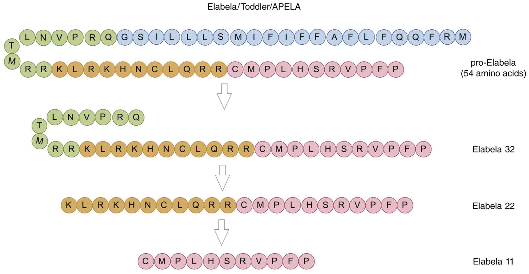
apelin-related peptide (5,6). The 54-amino-acid propeptide undergoes
sequential proteolytic processing to generate three bioactive
isoforms: ELA-32, ELA-22 and ELA-11 (Fig. 1), of which ELA-11 shows the highest
degree of conservation across vertebrates, suggesting an
evolutionarily ancient role (7,8).
ELA-32 exhibits a plasma half-life approximately
10-fold longer than that of apelin (9). ELA is expressed in vascular
endothelial cells, renal and cardiac tissue, and specific tumor
types such as renal cell carcinoma (4), as well as in embryonic stem cells
(ESCs) and induced pluripotent stem cells (10,11),
By contrast, apelin and APJ exhibit much broader tissue
distributions, including the heart, lungs, central nervous system,
adipose tissue, and gastrointestinal tract (4,7,10).
Furthermore, while APJ is widely expressed in adult tissues, ELA
expression in early development is primarily confined to
pluripotent stem cells and the adult kidney (10).
ELA exerts pleiotropic effects that span
embryogenesis, hemodynamic regulation and fluid homeostasis. In the
cardiovascular system, ELA promotes angiogenesis (12–16)
and vasodilatation (11), protects
against myocardial and cerebral ischemia-reperfusion injury
(12–16), and attenuates cardiac hypertrophy,
fibrosis and contractile dysfunction (17). ELA has also been implicated in
neurological (18) and renal
(19) diseases, motivating ongoing
investigation of its potential as a diagnostic or therapeutic
target, particularly in cardiovascular medicine (20,21).
Role of ELA in physiological pregnancy and
early embryogenesis
ELA in early embryogenesis
Early embryogenesis requires tightly coordinated
control of stem-cell renewal, germ-layer specification and cell
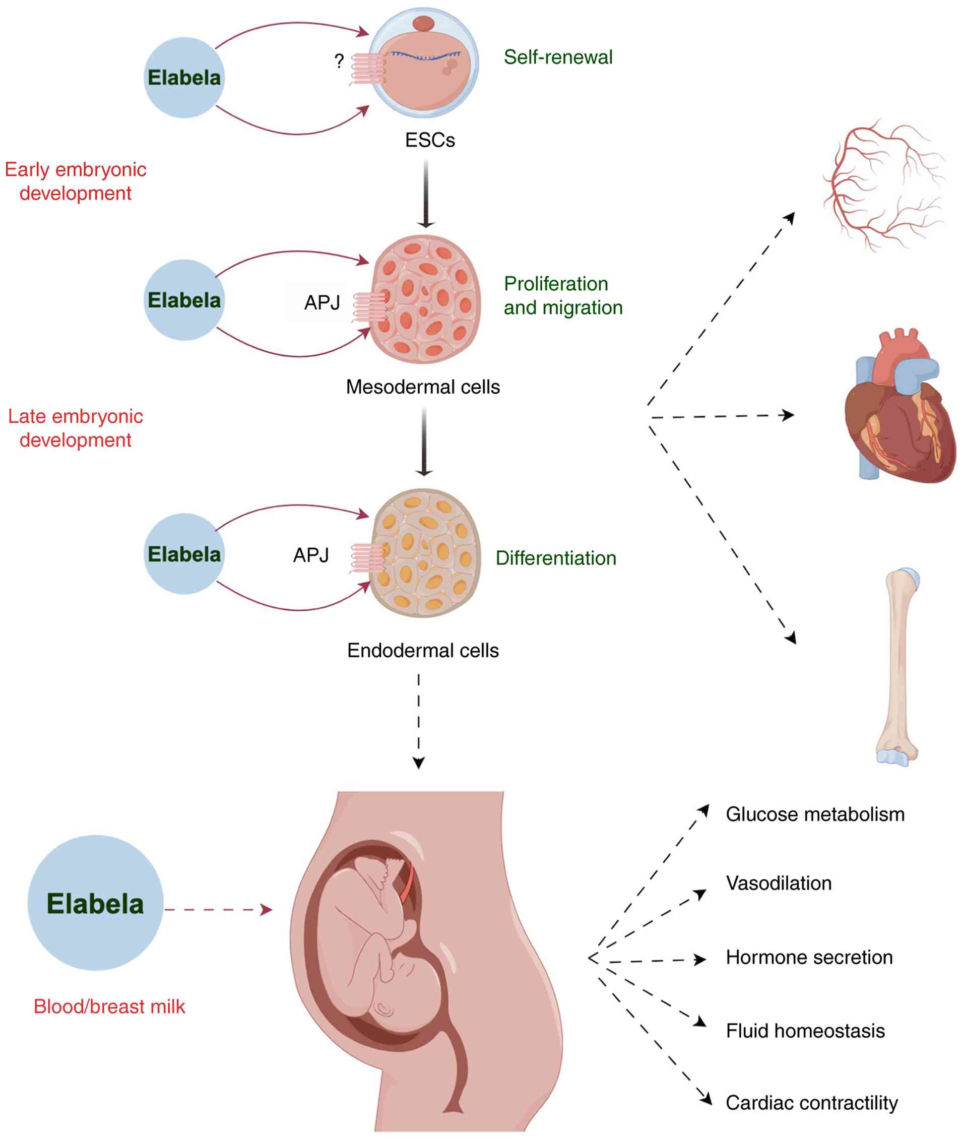
migration (22). ELA acts on ESCs
to promote self-renewal (potentially via an unidentified receptor),
drives APJ-dependent mesodermal cell proliferation and migration
and promotes endodermal differentiation, supporting organogenesis
(vessels, heart and bone) (5,6,23)
(Fig. 2). Additionally, Elabela
exerts systemic effects on glucose metabolism, vasodilation,
hormone secretion, fluid homeostasis and cardiac contractility
during pregnancy (8,9,24–26)
(Fig. 2). The cardiovascular
system, one of the first organ systems to emerge, depends
critically on these processes (27). In human ESCs (hESCs), ELA promotes
protein synthesis, proliferation and self-renewal responses
mediated in part through PI3K/Akt activation. ELA has also been
linked to TGF-β-dependent endodermal differentiation (28,29).
APJ is undetectable in hESCs (4,28),
indicating that ELA-induced pro-survival and self-renewal effects
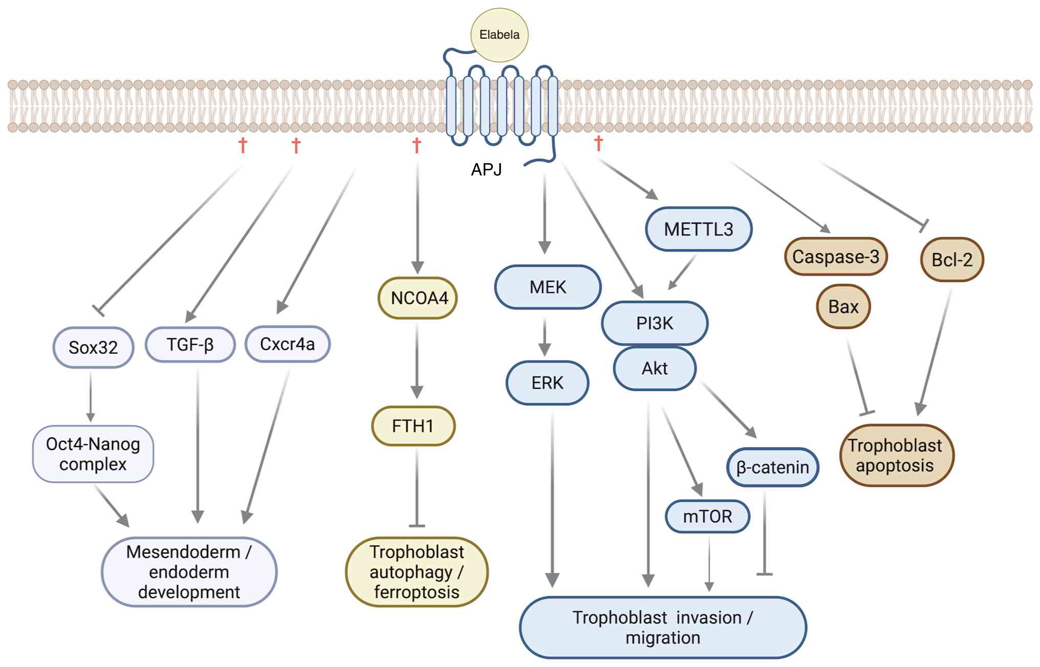
occur independently of canonical APJ signaling. In zebrafish
embryos, ELA governs mesodermal cell movement through Nodal-related
pathways and indirectly controls the migration of the endoderm via
Cxcr4a signaling (Fig. 3)
(30).
ELA is essential for embryonic cardiovascular
development (26). The two APJ
ligands, ELA and apelin, are not functionally interchangeable
during this process. APJ-null mice exhibit marked congenital
cardiac malformations, an observation that initially led to the
discovery of a second endogenous ligand (31), whereas apelin-knockout mice develop
normally and reproduce without severe cardiac defects (32,33).
Consistent with this distinction, ELA deficiency in mice causes
severe cardiovascular abnormalities and embryonic lethality that
closely recapitulate the APJ-null phenotype (7,34,35).
During zebrafish embryogenesis, ELA is the first
detectable APJ ligand, expressed before gastrulation, whereas
apelin expression begins at the end of gastrulation (~5 h after
ELA) (7,36). ELA-mutant embryos perish at early
stages, displaying cardiac malformations, pericardial edema and
endodermal defects (30). At the
molecular level, ELA-APJ signaling coordinates germ-layer
differentiation, heart morphogenesis and vascular progenitor
migration (37). Additionally,
ELA-mediated regulation of Sox32 and the Pou5f3-Nanog complex has
been implicated in mesendodermal cell-fate specification (Fig. 3) (38).
Receptor paradox
The ability of ELA to promote protein synthesis,
proliferation, self-renewal, and TGF-β-dependent endodermal
differentiation in hESCs, despite the absence of detectable APJ in
these cells, raises a receptor paradox: APJ is undetectable in
hESCs, yet ELA elicits clear biological responses in these cells,
implying the existence of an alternative ELA receptor or an
APJ-independent mechanism. G protein-coupled receptor 25 can be
activated by ELA and apelin in pigeons and zebrafish (39), but this finding has not been
extended to humans or other mammalian systems. In APJ-expressing
cells, signaling is inherently context-dependent; APJ engages
multiple G-protein pathways, including Gi/o, Gq/11 and G13
(40), and adopts distinct
receptor-complex configurations, such as heterodimerization with
AT1, kappa opioid, neurotensin 1, or bradykinin 1 receptors
(40). This context-dependency may
explain the diversity of cellular responses ranging from
trophoblast proliferation, apoptosis, invasion, and differentiation
(4) to the regulation of fluid
homeostasis via ERK1/2 phosphorylation (8). However, it does not account for the
APJ-negative hESC phenotype. Therefore, the identity and
physiological relevance of mammalian non-APJ receptors for ELA
remain unknown.
Placental development
Following blastocyst implantation, placental
development proceeds through coordinated trophoblast proliferation,
differentiation, decidual invasion and villous morphogenesis
(2,41). Cytotrophoblasts and
syncytiotrophoblasts fulfil complementary roles in placental growth
and maternal-fetal exchange, and disruption of either lineage can
compromise placental function. Proliferative cytotrophoblasts serve
as the progenitor pool that fuels placental growth and
differentiate into extravillous trophoblasts to remodel maternal
spiral arteries, whereas syncytiotrophoblasts, formed by fusion of
cytotrophoblasts-constitute the primary barrier for gas and
nutrient exchange and serve as the major endocrine source of
placental hormones such as hCG and progesterone (42).
ELA is expressed in both cytotrophoblasts and
syncytiotrophoblasts in human placentas throughout gestation
(43). However, in mice, ELA
expression shifts during development: ELA is initially detected in
trophoblast progenitors and becomes spatially restricted to the
syncytiotrophoblast lineage by mid-gestation, a pattern that
highlights species- and stage-dependent differences in placental
ELA biology (34).
At the functional level, ELA promotes extravillous
trophoblast differentiation (44),
enhances proliferation and reduces apoptosis in BeWo cells via
PI3K/Akt signaling (45), and
upregulates ferritin heavy chain 1 (FTH1) while suppressing nuclear
receptor coactivator 4 (NCOA4)-mediated ferritinophagy and
ferroptosis in trophoblasts (Fig.
3) (46). In pregnant mice,
loss of ELA disrupts signaling between trophoblast-derived ELA and
APJ-expressing fetal endothelial cells (34), leading to increased trophoblast
apoptosis, impaired syncytiotrophoblast differentiation, reduced
labyrinth vascularization and placental hypoxia with inflammation
(11,34). In vitro, ELA enhances the
invasive capacity of JAR cells, a human choriocarcinoma-derived
trophoblast line, further supporting its role in trophoblast
invasion (28,34).
Together, these findings indicate that ELA
contributes to early placentation through effects on trophoblast
differentiation, invasion and placental vascular development,
although the extent to which each effect requires APJ appears to be
cell-context-dependent.
Placental endocrine and nutrient
functions
Syncytiotrophoblasts mediate maternal-fetal nutrient
exchange and secrete hormones essential for pregnancy maintenance,
including human chorionic gonadotropin (hCG), human placental
lactogen (hPL) and placental growth factor (PlGF) (47,48).
This endocrine-nutrient axis is closely coupled to placental
perfusion and trophoblast differentiation. As ELA influences
trophoblast behavior and placental vascular development (28,34,43),
it may indirectly modulate these endocrine and nutrient functions.
However, to the best of our knowledge, direct regulation of hCG,
hPL or PlGF secretion by ELA has not been conclusively
demonstrated. The distinction between indirect modulation via
placental health and direct hormonal regulation should be
maintained when interpreting current evidence (49). Separately, ELA/APJ signaling exerts
neuroendocrine effects involving arginine vasopressin and
corticotropin-releasing hormone; intracerebroventricular injection
of ELA reduces food intake and activates AVP and CRH neurons in the
hypothalamic paraventricular nucleus (PVN), with ELA
dose-dependently increasing cytosolic calcium in single AVP- and
CRH-immunoreactive neurons isolated from the PVN (24), although a direct role in
reproductive hormone axes remains unresolved.
Maternal adaptations: Cardiovascular
system and lactation
In mice experimental models of pregnancy, ELA
deficiency is associated with elevated systolic blood pressure and
endothelial injury (34), whereas
exogenous ELA administration reduces blood pressure and proteinuria
(50). The vasodilatory properties
of ELA may also influence uterine artery hemodynamics and,
indirectly, placental perfusion (11,51).
In healthy pregnant women, circulating ELA levels vary across
gestation, with some cohorts reporting higher concentrations in
early to mid-pregnancy and lower levels in late gestation (25,52),
a pattern consistent with a role in maternal cardiovascular and
renal adaptation, although gestational trajectories are not uniform
across studies. ELA has also been detected at relatively high
concentrations in breast milk, ranging from 4.8 to 7.2 ng/ml across
colostrum, transitional, and mature milk, with the highest levels
observed in colostrum (53–56).
Its physiological significance for the neonate remains incompletely
characterized; however, available evidence suggests a possible role
in sodium and fluid homeostasis (56).
ELA in pregnancy-related diseases
ELA has been increasingly implicated in placental
development and several pregnancy-related disorders. Representative
studies are summarized in Table I,
focusing on sample type, disease subtype, assay method, sample size
or sampling context and major findings (43–46,52,55,57–75).
 | Table I.ELA measurements in pregnancy-related
disorders. |
Table I.
ELA measurements in pregnancy-related
disorders.
| First author(s),
year |
Disease/subtype | Sample | Species | ELA change | Method | Sample size | Sampling
stage/context |
Pathway/mechanism | (Refs.) |
|---|
| Wang et al,
2019 | EoPE | Placenta | Human | Reduced (EoPE) |
Immunohistochemistry, qPCR and western
blotting | N (30); EoPE (both n=30) | Placenta collected
at delivery; exact gestational age NR | PI3K/AKT/mTOR | (43) |
| Yang et al,
2022 | PE | Placenta and
plasma | Human and mice | Reduced |
Immunohistochemistry, qPCR and ELISA | N (20); PE (20) | Sampling stage
NR | Ferroptosis (ATG5
and NCOA4) | (46) |
| Zhou et al,
2019 | PE | Serum, urine and
placenta | Human | Reduced (LoPE) | ELISA | N (22); LoPE/EoPE (22/15) | PE subtype
recorded; exact sampling week NR | - | (57) |
| Yang et al,
2023 | PE | Serum and
placenta | Human | Reduced (placenta
in LoPE) | ELISA | N (60); LoPE (both n=60) | LoPE at clinical
assessment; exact sampling week NR | - | (58) |
| Ma et al,
2021 | Severe PE | Placenta | Human and mice | Reduced | ELISA | N (20); Severe PE (20) | Severe PE;
placental sampling stage NR | Apoptosis
(caspase-3, Bcl-2 and Bax) | (45) |
| Deniz et al,
2019 | PE | Plasma | Human | Reduced | ELISA | N (30); Severe PE/PE (24/28) | PE/severe PE;
sampling stage NR | - | (59) |
| Ali et al,
2023 | PE | Plasma | Human | Reduced (EoPE) | ELISA | N (30); LoPE/EoPE (30/30) | EoPE/LoPE; sampling
stage NR | - | (60) |
| Wang et al,
2026 | EoPE | Placenta | Human | Reduced (EoPE) |
Immunohistochemistry, qPCR and western
blotting | N (30); EoPE (both n=30) | EoPE; placental
tissue at delivery; exact week NR |
METTL3/PI3K/AKT | (61) |
| Khan et al,
2023 | PE | Plasma | Human | Reduced | ELISA | N (30); PE (30) | PE; sampling stage
NR | - | (62) |
| Para et al,
2020 | PE | Plasma | Human | Increased
(LoPE) | ELISA | N (n=108);
LoPE/EoPE (52/54) | EoPE <34 weeks;
LoPE ≥34 weeks | - | (63) |
| Panaitescu et
al, 2018 | PE | Plasma | Human | Increased (LoPE)
and unaffected (EoPE) | ELISA | N (119); LoPE/EoPE
(57/56) | EoPE <34 weeks;
LoPE ≥34 weeks | - | (64) |
| Ma et al,
2020 | PE | Plasma | Human | Unaffected | ELISA | N (132); PE
(16) | First trimester
(10–14+6 weeks) | - | (65) |
| Villié et
al, 2019 | PE | Plasma | Human | Unaffected | ELISA | N (14); PE
(12) | Before clinical
onset of PE; exact week NR | - | (66) |
| Ozgen et al,
2021 | PE | Serum | Human | Unaffected | ELISA | N (47); LoPE
(23) | First trimester
(11–14 weeks) | - | (67) |
| Pritchard et
al, 2018 | PE | Plasma and
placental villi | Human | Unaffected | ELISA and
RT-qPCR | N (82); PE
(82) | Maternal
circulation and placental villi; sampling stage NR | - | (68) |
| Huang et al,
2019 | PE | Plasma | Human | Unaffected | ELISA | N (n=20); PE (1st
trimester, 2nd trimester and 3rd trimester, 29/27/31) | Maternal
circulation; first to the third trimester | - | (69) |
| Georgiadou et
al, 2019 | PE | Placenta, serum and
plasma | Human | Unaffected
(placenta) and reduced (plasma of women with healthy BMI) | ELISA and
immunohistochemistry | ABCD cohort: N
(139); PE (n=66) | Early-pregnancy
cohort; BMI-stratified analysis | - | (44) |
| Wang et al,
2025 | EoPE | Placenta | Human | Reduced (EoPE) |
Immunohistochemistry, qPCR and western
blotting | N (30); EoPE
(30) | EoPE; placental
tissue at delivery; exact week NR | MEK/ERK | (70) |
| Yener et al,
2023 | FGR* | Serum | Human | Increased | ELISA | N (50); IUGR (both
n=50) | FGR/IUGR cohort;
sampling near delivery; exact week NR | - | (71) |
| Behram et
al, 2021 | FGR* | Serum | Human | Reduced | ELISA | N (43); IUGR
(49) |
Gestational-age/BMI-matched FGR/IUGR
cohort; exact week NR | - | (72) |
| Yavuz et al,
2023 | GDM | Breast tissue,
blood and milk | Human | Reduced | ELISA | N (14); GDM
(12) | Pregnancy and
lactation samples; exact gestational stage NR | - | (55) |
| Karagoz et
al, 2022 | GDM | Plasma | Human | Increased | ELISA | N (30); GDM
(30) | Pregnancy with GDM;
sampling stage NR | - | (73) |
| Demir et al,
2025 | Placenta
previa/Accreta | Serum | Human | Increased | ELISA | N (32);
previa/accreta (12/23) | Placenta
previa/accreta cohort; antenatal sampling; exact week NR | - | (74) |
| Akbulut et
al, 2025 | PAS | Serum | Human | Increased | ELISA | N (n=44); PAS
(44) | Antenatal sampling;
gestational-age-matched controls | - | (75) |
| Qi et al,
2022 | MA | Serum | Human | Reduced | ELISA | N (early, middle
and late gestational stage, 35/36/37); MA (early stage, 25) | Early-pregnancy MA;
controls also sampled across pregnancy stages | - | (52) |
PE
PE is a multisystem disorder of pregnancy that
develops after 20 weeks of gestation and remains a leading cause of
maternal and neonatal morbidity and mortality worldwide (76). The global prevalence of PE is ~5%,
with reported estimates of 4.02–5.22% in China (77–80).
Clinically, PE is commonly classified as early-onset PE (EoPE,
developing before 34 weeks of gestation) or late-onset PE (LoPE,
developing at or after 34 weeks of gestation), reflecting
differences in gestational timing and, to some extent, underlying
pathophysiology (80).
The pathophysiology of PE is multifactorial,
involving interacting placental, vascular, metabolic and immune
abnormalities. Impaired placentation and uteroplacental perfusion,
angiogenic imbalance, insulin resistance, systemic inflammation and
endothelial dysfunction collectively contribute to the clinical
syndrome (81). Several of these
processes intersect with established ELA functions, particularly
trophoblast regulation, angiogenesis and vascular tone, making the
peptide a plausible contributor to PE pathogenesis.
Early experimental studies supported a protective
role for ELA against PE-like phenotypes. Ho et al (34) reported that circulating ELA
increases during mouse pregnancy and demonstrated that ELA
deficiency produces hypertension, proteinuria and glomerular
endothelial proliferation, abnormalities that were alleviated by
exogenous ELA administration. ELA-deficient mice also exhibit
placental defects, including reduced labyrinth vascularization,
increased apoptosis, impaired proliferation and delayed
syncytiotrophoblast differentiation (82). In human studies, several groups
have reported reduced placental or circulating ELA in LoPE, severe
PE and EoPE (42,44,57–62).
By contrast, other human studies do not support a
uniform ELA-deficiency model. Maternal ELA concentrations have been
reported to be increased in LoPE (63,64),
unchanged in EoPE and in first-trimester samples from nulliparous
women who subsequently developed PE (65–67),
and unchanged at the placental mRNA or circulating levels in
certain cohorts, including a preterm PE plasma cohort and a term PE
placenta cohort (n=82) (68), the
longitudinal Shanghai Birth Cohort (69). Georgiadou et al (44) further demonstrated marked
interindividual variability and BMI dependence: In a healthy-BMI
subgroup, lower early-pregnancy plasma ELA was associated with
subsequent PE. Taken together, these findings argue against
interpreting a single circulating ELA measurement as a universal
marker of PE.
Mechanistic studies have identified several links
between ELA and trophoblast dysfunction. Rare APELA 5′-UTR variants
have been associated with reduced transcription and increased PE
risk (83). ELA suppresses
trophoblast ferroptosis through an autophagy-related NCOA4/FTH1
pathway (46), although high
concentrations of exogenous ELA can impair trophoblast function
through PI3K/Akt/β-catenin-associated signaling in HTR-8/SVneo
trophoblast cells (84). In EoPE
models, ELA has been linked to PI3K/Akt/mTOR, MEK/ERK and
methyltransferase-like 3/PI3K/Akt signaling pathways that promote
trophoblast invasion and migration (Fig. 3) (43,60,61,70).
These pathways were demonstrated primarily in the HTR-8/SVneo human
trophoblast cell line using ELA knockdown or overexpression, or
treatment with exogenous ELA at defined concentrations. These
findings should not be assumed to be universally active or
uniformly APJ-dependent.
ELA and apelin share APJ as a common receptor and
overlap with regard to several vascular and trophoblast-related
functions, including regulation of vascular tone, angiogenic
responses, oxidative stress and PI3K/Akt-associated cell signaling
(4,85–88).
However, ELA and apelin are not interchangeable ligands. ELA is
expressed earlier during embryogenesis and exhibits distinct
placental and tissue-expression patterns, whereas apelin is more
broadly distributed and becomes prominent later in development (at
the end of gastrulation in zebrafish and at embryonic day 6.5 in
mice) (4,7,34,36).
In PE, experimental supplementation with either ligand has
demonstrated protective effects in preclinical models, including
L-NAME- or RUPP-induced PE rodent models and hypoxia/CoCl2-exposed
HTR-8/SVneo trophoblast cells (29,86–88);
however, human circulating levels of ELA and apelin and their
clinical associations differ across studies. Therefore, findings
provide biological context for the apelinergic system but should
not be treated as direct evidence for ELA. The similarities and
differences between ELA and apelin within the apelinergic system in
pregnancy are summarized in Table
SI.
The conflicting clinical ELA findings in PE are best
interpreted through a multidimensional lens. Disease biology itself
varies notably: EoPE and LoPE represent distinct pathophysiological
entities (89) and measurements
obtained in the first trimester, at diagnosis or near delivery
capture fundamentally different stages of disease progression.
Beyond disease heterogeneity, biological matrices are not
interchangeable; serum, plasma and placental tissue may reflect
different sources of ELA: serum and plasma represent circulating
ELA (which may predominantly derive from maternal sources and
correlate with BMI), whereas placental tissue reflects locally
produced ELA. Emerging evidence suggests the placenta may not be
the major determinant of circulating ELA in all pregnancies
(44). Cohort characteristics
further complicate interpretation, as BMI, parity, ethnicity,
disease severity and concurrent treatments can all modify
associations between circulating ELA levels and PE risk, as well as
analytical and pre-analytical factors, including antibody epitope
specificity, assay platform, rapid proteolytic processing of ELA
and variations in sample handling, storage and freeze-thaw history
(44,58,90).
APJ signaling context may add another layer of biological
heterogeneity, as the receptor can engage different G-protein
pathways (e.g., Gi/o, Gq/11, and G13) and adopt distinct
receptor-complex states (e.g., homodimers or heterodimers with AT1,
kappa opioid, neurotensin 1, or bradykinin 1 receptors) (4,8,40),
although this does not solely explain differences in circulating
ELA. Therefore, longitudinal studies using gestational-age-matched
sampling and standardized assays are required before ELA can be
considered a reliable PE biomarker.
FGR
FGR, termed intrauterine growth restriction (IUGR)
in several cited studies, refers to the failure of a fetus to reach
its genetically determined growth potential and is associated with
increased neonatal morbidity and mortality (91). FGR is commonly linked to fetal,
maternal or placental abnormalities that impair uteroplacental
perfusion and compromise nutrient and oxygen delivery, with
progressive hypoxemia further increasing vascular resistance and
perpetuating growth restriction (92).
Direct clinical evidence linking ELA to FGR is
limited and contradictory. Yener et al (71) reported higher serum ELA
concentrations in pregnancies complicated by IUGR/FGR than in
controls (71), whereas Behram
et al (72) revealed lower
maternal circulating ELA and a positive association between ELA and
birth weight. These studies differed in cohort composition (IUGR
defined as abdominal circumference <10th percentile with
abnormal Doppler vs. estimated fetal weight <3rd percentile) and
sampling context (at delivery vs. at diagnosis), and size were
relatively small (100 and 92 subjects, respectively); therefore,
the direction of the association remains uncertain. Apelin has also
been reported to be reduced in maternal serum and placental
compartments in placental insufficiency and FGR (93). However, as apelin and ELA are
distinct APJ ligands, the reported reduction of apelin is best
viewed as an indirect biological parallel rather than evidence that
ELA is reduced in FGR.
At present, direct evidence is insufficient to
establish a causal role for ELA in human FGR. Although the
ELA/apelin-APJ system influences nitric oxide production and
vascular tone (94), providing a
mechanistic rationale for investigating uteroplacental perfusion,
this does not demonstrate that ELA deficiency causes reduced
uteroplacental blood flow in affected patients. Future studies
should combine standardized circulating ELA measurements with
placental expression analysis, Doppler hemodynamics and
gestational-age-matched longitudinal sampling.
GDM
GDM affects 2–25% of pregnancies worldwide, with
prevalence shaped by diagnostic criteria and population
characteristics (95). Beyond its
immediate obstetric risks, including hypertensive disorders and
cesarean delivery (96,97), GDM reflects a fundamental
disruption of metabolic adaptation, involving insulin resistance,
progressive β-cell failure and low-grade inflammation (98).
Given that the apelinergic system has been
implicated in glucose homeostasis and energy metabolism (40,99,100), a plausible biological connection
exists between ELA signaling and GDM pathogenesis. However, this
connection remains largely inferential. The apelin/APJ literature
provides a mechanistic foundation: Apelin regulates insulin
sensitivity, adipocyte function and pancreatic β-cell survival
(40,99,100). Whether ELA, as a distinct APJ
ligand with different tissue distribution and temporal expression,
shares these metabolic functions remains unknown. Clinical evidence
specific to ELA is sparse and inconsistent. In one study of women
with GDM, circulating ELA was negatively correlated with glucose
and triglyceride levels and positively correlated with apelin-13
and apelin-36 (73), a pattern
suggestive of metabolic involvement but insufficient to establish
causality. Another report described reduced circulating and
breast-milk ELA concentrations in GDM in a human case-control study
(n=12 GDM and n=14 controls) (55), whereas a study using a
streptozotocin-induced type 1 diabetic mouse model have linked ELA
to renal protection via PI3K/Akt/mTOR signaling (101).
Notably, these findings span different biological
matrices (serum, breast milk and kidney tissue) and disease
contexts, complicating direct comparison. Therefore, the current
evidence base for ELA in GDM is preliminary and fragmented. Whether
ELA dysregulation contributes to GDM pathogenesis, represents a
compensatory response or merely reflects metabolic disturbance
remains undetermined. Future longitudinal studies are required with
standardized sampling across gestation, matched metabolic
phenotyping and careful distinction between ELA and apelin
measurements, ideally with isoform-specific assays that can resolve
the contributions of ELA-32, ELA-22 and ELA-11.
Placenta previa and accreta
Placenta previa refers to placental implantation in
the lower uterine segment, with the placenta approaching or
covering the internal cervical os (102). Placenta accreta spectrum (PAS)
involves abnormally adherent or invasive placentation, typically
arising from defective decidualization at the uteroplacental
interface (103). Both conditions
carry notable hemorrhagic risk and require specialized obstetric
management.
Evidence linking ELA to these disorders is emerging
but limited. A 3-year multicenter prospective cohort study revealed
differential circulating ELA profiles between placenta previa and
placenta accreta groups, with higher maternal concentrations in the
previa cohort (75). In a separate
prospective case-control study, maternal serum ELA was elevated in
PAS compared with gestational-age-matched controls, prompting
speculation about diagnostic potential (75). However, these findings represent
preliminary associations; whether ELA can serve as a clinically
predictive biomarker for either condition awaits validation in
larger, independent cohorts.
MA
MA refers to embryonic or fetal demise with retained
products of conception within the uterine cavity. Early
descriptions of MA in twin pregnancies at 10–14 weeks of gestation
noted associated placental villous abnormalities (104), and altered apoptosis-related
signaling has been documented in early MA (105). The only direct clinical evidence
linking ELA to MA is a 2022 study reporting lower maternal
circulating ELA in affected women compared with healthy pregnant
controls (52). Although this
single finding cannot establish causality, the experimental links
between ELA and trophoblast function, placental angiogenesis and
vasodilatation (11,28,34)
provide a mechanistic basis for investigating ELA deficiency in
early pregnancy loss. Prospective studies integrating circulating
ELA measurements with placental expression and vascular phenotyping
would help clarify this association.
ELA: Therapeutic and diagnostic
potential
Diagnostic potential
Clinical studies report decreased, increased or
unchanged ELA concentrations across PE, FGR, GDM and other
pregnancy-related disorders (Table
I), which is consistent with the status of ELA as a candidate
biomarker rather than a validated biomarker. A notable obstacle in
translating ELA into a reliable clinical biomarker or therapeutic
agent is measurement variability. Although ELA-32 undergoes
proteolytic processing in human plasma (90), it exhibits a longer circulating
half-life than apelin in experimental settings, such as ex
vivo incubation in human plasma (where ELA-32 has a half-life
of ~47 min, compared with 2–8 min for apelin) (9). Published studies have used
heterogeneous biological matrices and antibody-based assay
platforms (43–46,52,55,57–75);
therefore, future biomarker studies should adopt standardized
protocols, including predefining serum or plasma as the collection
matrix, recording gestational age and disease subtype at sampling,
processing samples promptly under controlled temperatures, storing
aliquots at −80°C, minimizing freeze-thaw cycles and reporting
assay details (assay kit manufacturer, antibody epitope or
calibrator, analytical precision, and limits of detection and
quantification). Where feasible, orthogonal validation with
complementary analytical methods, such as combining ELISA with
LC-MS/MS, is advisable. An immunochromatographic colloidal-gold
strip for serum ELA detection offers a promising point-of-care
approach (58); however,
independent external validation and harmonized reference ranges
must be established before clinical use.
Therapeutic potential
Preclinical evidence provides a stronger basis for
therapeutic investigation than the current clinical literature
alone would suggest. Exogenous ELA has ameliorated PE-like
phenotypes and placental dysfunction in animal and trophoblast
models, with effects attributed to PI3K/Akt, MEK/ERK and
ferroptosis-related signaling pathways (34,43,46,60,70).
However, ELA-based therapies have not been clinically validated for
obstetric use. Translating these findings to the clinic will
require overcoming several practical barriers: Peptide stability
and in vivo metabolism, selection of the therapeutically
relevant ELA isoform, pharmacokinetic characterization,
dose-response relationships and gestational treatment windows,
maternal-fetal safety, and receptor specificity in APJ-low or
APJ-negative tissues. Placenta-directed or tissue-targeted delivery
strategies may be necessary to limit off-target exposure. These
challenges warrant systematic preclinical investigation before
efficacy trials become feasible.
Conclusion
Current evidence establishes ELA as a key regulator
of embryonic and placental biology through its effects on
trophoblast behavior, vascular development and maternal
cardiovascular adaptation. However, conflicting clinical
measurements in PE and FGR, limited direct evidence across several
other pregnancy-related disorders and unresolved receptor biology
preclude ELA from being considered as an established biomarker or
therapeutic target. A critical unresolved question is the molecular
basis of ELA signaling in APJ-negative or APJ-low cells, which
requires rigorous validation of any non-APJ receptor or
non-canonical mechanism. Beyond this, isoform-, tissue- and
gestational stage-specific functions should be mapped using
longitudinal cohorts, standardized assays, and complementary
placental and circulating measurements. From a translational
perspective, studies must define peptide stability,
pharmacokinetics, dose-response relationships, delivery strategies
and maternal-fetal safety, with particular attention to
placenta-specific targeting. Resolving these questions will
determine whether the compelling preclinical evidence for ELA can
be translated into clinically reproducible outcomes.
Supplementary Material
Supporting Data
Acknowledgements
Not applicable.
Funding
The present research was funded by grants provided by the
National Natural Science Foundation of China (grant nos. 81070125,
81270213 and 81670306), the Science and Technology Foundation in
Guangdong Province (grant no. 2014A020211002), the National Natural
Science Foundation of Guangdong Province (grant no.
2017A030313503), the Science and Technology Foundation in Guangzhou
City (grant no. 201806020084), the Guangdong Basic and Applied
Basic Research Foundation (grant nos. 2023A1515220199 and
2024A1515220088), Fundamental Research Funds for the Central
Universities (grant no. 13ykzd16 and 17ykjc18), the Futian District
Health and Public Welfare Research Project of Shenzhen City (grant
nos. FTWS2019001, FTWS2021016, FTWS2022018 and FTWS2023064), the
Shenzhen Science and Technology Program (grant nos.
JCYJ20190808101405466, JCYJ20210324115003008, JCYJ20220530144404009
and KCXFZ20230731094100002) and the Sanming Project of Medicine in
Shenzhen (grant no. SZSM202402020).
Availability of data and materials
Not applicable.
Authors' contributions
YX, SY and CM collected the data, drafted the
manuscript, and designed the figures and tables. HW, XC and HL
contributed to the revision of the manuscript. TW conceived and
outlined the concept of the review, and directed the overall
structure and preparation of the manuscript. Data authentication is
not applicable. All authors have read and approved the final
version of the manuscript.
Ethics approval and consent to
participate
Not applicable.
Patient consent for publication
Not applicable.
Competing interests
The authors declare that they have no competing
interests.
References
|
1
|
Menon R, Bonney EA, Condon J, Mesiano S
and Taylor RN: Novel concepts on pregnancy clocks and alarms:
Redundancy and synergy in human parturition. Hum Reprod Update.
22:535–560. 2016. View Article : Google Scholar : PubMed/NCBI
|
|
2
|
Khorami-Sarvestani S, Vanaki N, Shojaeian
S, Zarnani K, Stensballe A, Jeddi-Tehrani M and Zarnani AH:
Placenta: An old organ with new functions. Front Immunol.
15:13857622024. View Article : Google Scholar : PubMed/NCBI
|
|
3
|
Ortega MA, Fraile-Martínez O,
García-Montero C, Sáez MA, Álvarez-Mon MA, Torres-Carranza D,
Álvarez-Mon M, Bujan J, García-Honduvilla N, Bravo C, et al: The
pivotal role of the placenta in normal and pathological
pregnancies: a focus on preeclampsia, fetal growth restriction, and
maternal chronic venous disease. Cells. 11:5682022. View Article : Google Scholar : PubMed/NCBI
|
|
4
|
Pécheux O, Correia-Branco A, Cohen M and
Martinez B: The apelinergic system in pregnancy. Int J Mol Sci.
24:80142023. View Article : Google Scholar : PubMed/NCBI
|
|
5
|
Chng SC, Ho L, Tian J and Reversade B:
ELABELA: A hormone essential for heart development signals via the
apelin receptor. Dev Cell. 27:672–680. 2013. View Article : Google Scholar : PubMed/NCBI
|
|
6
|
Pauli A, Norris ML, Valen E, Chew GL,
Gagnon JA, Zimmerman S, Mitchell A, Ma J, Dubrulle J, Reyon D, et
al: Toddler: An embryonic signal that promotes cell movement via
apelin receptors. Science. 343:12486362014. View Article : Google Scholar : PubMed/NCBI
|
|
7
|
Huang SK, Shin K, Sarker M and Rainey JK:
Apela exhibits isoform- and headgroup-dependent modulation of
micelle binding, peptide conformation and dynamics. Biochim Biophys
Acta Biomembr. 1859:767–778. 2017. View Article : Google Scholar : PubMed/NCBI
|
|
8
|
Deng C, Chen H, Yang N, Feng Y and Hsueh
AJ: Apela regulates fluid homeostasis by binding to the APJ
receptor to activate Gi signaling. J Biol Chem. 290:18261–18268.
2015. View Article : Google Scholar : PubMed/NCBI
|
|
9
|
Chapman FA, Maguire JJ, Newby DE,
Davenport AP and Dhaun N: Targeting the apelin system for the
treatment of cardiovascular diseases. Cardiovasc Res.
119:2683–2696. 2023. View Article : Google Scholar : PubMed/NCBI
|
|
10
|
Wang Z, Yu D, Wang M, Wang Q, Kouznetsova
J, Yang R, Qian K, Wu W, Shuldiner A, Sztalryd C, et al:
Elabela-apelin receptor signaling pathway is functional in
mammalian systems. Sci Rep. 5:81702015. View Article : Google Scholar : PubMed/NCBI
|
|
11
|
Yang P, Read C, Kuc RE, Buonincontri G,
Southwood M, Torella R, Upton PD, Crosby A, Sawiak SJ, Carpenter
TA, et al: Elabela/Toddler is an endogenous agonist of the apelin
APJ receptor in the adult cardiovascular system, and exogenous
administration of the peptide compensates for the downregulation of
its expression in pulmonary arterial hypertension. Circulation.
135:1160–1173. 2017. View Article : Google Scholar : PubMed/NCBI
|
|
12
|
Li L, Zeng H and Chen J: Apelin-13
increases myocardial progenitor cells and improves repair
postmyocardial infarction. Am J Physiol Heart Circ Physiol.
303:H605–H618. 2012. View Article : Google Scholar : PubMed/NCBI
|
|
13
|
Peng JY, Fu X, Luo XY, Liu F, Zhang B,
Zhou B, Sun K and Chen AF: Endothelial ELABELA improves
post-ischemic angiogenesis by upregulating VEGFR2 expression.
Transl Res. 270:13–23. 2024. View Article : Google Scholar : PubMed/NCBI
|
|
14
|
Li W, Xu P, Kong L, Feng S, Shen N, Huang
H, Wang W, Xu X, Wang X, Wang G, et al: Elabela-APJ axis mediates
angiogenesis via YAP/TAZ pathway in cerebral ischemia/reperfusion
injury. Transl Res. 257:78–92. 2023. View Article : Google Scholar : PubMed/NCBI
|
|
15
|
XI Y, LI Y, Ren W, Bo W, Ma Y, Pan S, Gong
DW and Tian Z: ELABELA-APJ-Akt/YAP signaling axis: A novel
mechanism of aerobic exercise in cardioprotection of myocardial
infarction rats. Med Sci Sports Exerc. 55:1172–1183. 2023.
View Article : Google Scholar : PubMed/NCBI
|
|
16
|
Madiha SA and Sharma B: Elabela and Apelin
regulate coronary angiogenesis in a competitive manner. MicroPubl
Biol. 2023:0008862023.
|
|
17
|
Xu C: Cardiovascular aspects of ELABELA: A
potential diagnostic biomarker and therapeutic target. Vascul
Pharmacol. 151:1071932023. View Article : Google Scholar : PubMed/NCBI
|
|
18
|
Sato T, Sato C, Kadowaki A, Watanabe H, Ho
L, Ishida J, Yamaguchi T, Kimura A, Fukamizu A, Penninger JM, et
al: ELABELA-APJ axis protects from pressure overload heart failure
and angiotensin II-induced cardiac damage. Cardiovasc Res.
113:760–769. 2017. View Article : Google Scholar : PubMed/NCBI
|
|
19
|
Xiong M, Chen H, Fan Y, Jin M, Yang D,
Chen Y, Zhang Y, Petersen RB, Su H, Peng A, et al: Tubular
Elabela-APJ axis attenuates ischemia-reperfusion induced acute
kidney injury and the following AKI-CKD transition by protecting
renal microcirculation. Theranostics. 13:3387–3401. 2023.
View Article : Google Scholar : PubMed/NCBI
|
|
20
|
Ma Z, Zhao L, Martin S, Zhang Y, Dong Y,
Zhong JC and Yang X: Lower plasma elabela levels in hypertensive
patients with heart failure predict the occurrence of major adverse
cardiac events: A preliminary study. Front Cardiovasc Med.
8:6384682021. View Article : Google Scholar : PubMed/NCBI
|
|
21
|
Liu C, Xiong J, Yi X, Song S, Yang H, Tan
W, Yang X, Zheng L, Yu J and Xu C: Decreased plasma ELABELA level
as a novel screening indicator for heart failure: A cohort and
observational study. Sci Rep. 14:113332024. View Article : Google Scholar : PubMed/NCBI
|
|
22
|
Steventon B, Busby L and Arias AM:
Establishment of the vertebrate body plan: Rethinking gastrulation
through stem cell models of early embryogenesis. Dev Cell.
56:2405–2418. 2021. View Article : Google Scholar : PubMed/NCBI
|
|
23
|
Xu J, Chen L, Jiang Z and Li L: Biological
functions of Elabela, a novel endogenous ligand of APJ receptor. J
Cell Physiol. 233:6472–6482. 2018. View Article : Google Scholar : PubMed/NCBI
|
|
24
|
Santoso P, Maejima Y, Kumamoto K,
Takenoshita S and Shimomura K: Central action of ELABELA reduces
food intake and activates arginine vasopressin and
corticotropin-releasing hormone neurons in the hypothalamic
paraventricular nucleus. Neuroreport. 26:820–826. 2015. View Article : Google Scholar : PubMed/NCBI
|
|
25
|
Guo YY, Li T, Liu H, Tang L, Li YC, Hu HT,
Su YF, Lin Y, Wang YY, Li C, et al: Circulating levels of Elabela
and Apelin in the second and third trimesters of pregnancies with
gestational diabetes mellitus. Gynecol Endocrinol. 36:890–894.
2020. View Article : Google Scholar : PubMed/NCBI
|
|
26
|
Sunjaya AP, Sunjaya AF and Ferdinal F:
Apela/Elabela/Toddler: New perspectives in molecular mechanism of
heart failure. Glob Cardiol Sci Pract. 2019:e2019152019.PubMed/NCBI
|
|
27
|
Lee JG, Yon JM, Kim G, Lee SG, Kim CY,
Cheong SA, Kim HY, Yu J, Kim K, Sung YH, et al: PIBF1 regulates
trophoblast syncytialization and promotes cardiovascular
development. Nat Commun. 15:14872024. View Article : Google Scholar : PubMed/NCBI
|
|
28
|
Ho L, Tan SY, Wee S, Wu Y, Tan SJ,
Ramakrishna NB, Chng SC, Nama S, Szczerbinska I, Chan YS, et al:
ELABELA is an endogenous growth factor that Sustains hESC
Self-Renewal via the PI3K/AKT pathway. Cell Stem Cell. 17:635.
2015. View Article : Google Scholar : PubMed/NCBI
|
|
29
|
Chen S, Chen F, Sun K, Zhou S, Wang J,
Wang Q, Meng J, Peng J, Song W and Zhou Y: Essential Role of the
ELABELA-APJ signaling pathway in cardiovascular system development
and diseases. J Cardiovascular Pharmacol. 75:284–291. 2020.
View Article : Google Scholar : PubMed/NCBI
|
|
30
|
Norris ML, Pauli A, Gagnon JA, Lord ND,
Rogers KW, Mosimann C, Zon LI and Schier AF: Toddler signaling
regulates mesodermal cell migration downstream of Nodal signaling.
Elife. 6:e226262017. View Article : Google Scholar : PubMed/NCBI
|
|
31
|
Eberlé D, Marousez L, Hanssens S, Knauf C,
Breton C, Deruelle P and Lesage J: Elabela and Apelin actions in
healthy and pathological pregnancies. Cytokine Growth Factor Rev.
46:45–53. 2019. View Article : Google Scholar : PubMed/NCBI
|
|
32
|
Kuba K, Zhang L, Imai Y, Arab S, Chen M,
Maekawa Y, Leschnik M, Leibbrandt A, Markovic M, Schwaighofer J, et
al: Impaired heart contractility in apelin Gene-deficient mice
associated with aging and pressure overload. Circ Res. 101:e32–e42.
2007. View Article : Google Scholar : PubMed/NCBI
|
|
33
|
Zhang Y, Wang Y, Lou Y, Luo M, Lu Y, Li Z,
Wang Y and Miao L: Elabela, a newly discovered APJ ligand:
Similarities and differences with Apelin. Peptides. 109:23–32.
2018. View Article : Google Scholar : PubMed/NCBI
|
|
34
|
Ho L, van Dijk M, Chye STJ, Messerschmidt
DM, Chng SC, Ong S, Yi LK, Boussata S, Goh GH, Afink GB, et al:
ELABELA deficiency promotes preeclampsia and cardiovascular
malformations in mice. Science. 357:707–713. 2017. View Article : Google Scholar : PubMed/NCBI
|
|
35
|
Freyer L, Hsu CW, Nowotschin S, Pauli A,
Ishida J, Kuba K, Fukamizu A, Schier AF, Hoodless PA, Dickinson ME
and Hadjantonakis AK: Loss of apela peptide in mice causes low
penetrance embryonic lethality and defects in early mesodermal
derivatives. Cell Rep. 20:2116–2130. 2017. View Article : Google Scholar : PubMed/NCBI
|
|
36
|
Zeng XXI, Wilm TP, Sepich DS and
Solnica-Krezel L: Apelin and its receptor control heart field
formation during zebrafish gastrulation. Dev Cell. 12:391–402.
2007. View Article : Google Scholar : PubMed/NCBI
|
|
37
|
Helker CS, Schuermann A, Pollmann C, Chng
SC, Kiefer F, Reversade B and Herzog W: The hormonal peptide
Elabela guides angioblasts to the midline during vasculogenesis.
Elife. 4:e067262015. View Article : Google Scholar : PubMed/NCBI
|
|
38
|
Perez-Camps M, Tian J, Chng SC, Sem KP,
Sudhaharan T, The C, Wachsmuth M, Korzh V, Ahmed S and Reversade B:
Quantitative imaging reveals real-time Pou5f3-Nanog complexes
driving dorsoventral mesendoderm patterning in zebrafish. Elife.
5:e114752016. View Article : Google Scholar : PubMed/NCBI
|
|
39
|
Zhang J, Wan Y, Fang C, Chen J, Ouyang W,
Li J and Wang Y: The orphan G protein-coupled receptor 25 (GPR25)
is activated by Apelin and Apela in non-mammalian vertebrates.
Biochem Biophys Res Commun. 501:408–414. 2018. View Article : Google Scholar : PubMed/NCBI
|
|
40
|
Chaves-Almagro C, Castan-Laurell I, Dray
C, Knauf C, Valet P and Masri B: Apelin receptors: From signaling
to antidiabetic strategy. Eur J Pharmacol. 763:149–159. 2015.
View Article : Google Scholar : PubMed/NCBI
|
|
41
|
Moghassemi S, Nikanfar S, Dadashzadeh A,
Sousa MJ, Wan Y, Sun F, Colson A, De Windt S, Kwaspen L, Kanbar M,
et al: The revolutionary role of placental derivatives in
biomedical research. Bioact Mate. 49:456–485. 2025.PubMed/NCBI
|
|
42
|
Farah O, Nguyen C, Tekkatte C and Parast
MM: Trophoblast lineage-specific differentiation and associated
alterations in preeclampsia and fetal growth restriction. Placenta.
102:4–9. 2020. View Article : Google Scholar : PubMed/NCBI
|
|
43
|
Wang L, Zhang Y, Qu H, Xu F, Hu H, Zhang Q
and Ye Y: Reduced ELABELA expression attenuates trophoblast
invasion through the PI3K/AKT/mTOR pathway in early onset
preeclampsia. Placenta. 87:38–45. 2019. View Article : Google Scholar : PubMed/NCBI
|
|
44
|
Georgiadou D, Boussata S, Ranzijn WHM,
Root LEA, Hillenius S, Bij de Weg JM, Abheiden CNH, de Boer MA, de
Vries JIP, Vrijkotte TGM, et al: Peptide hormone ELABELA enhances
extravillous trophoblast differentiation, but placenta is not the
major source of circulating ELABELA in pregnancy. Sci Rep.
9:190772019. View Article : Google Scholar : PubMed/NCBI
|
|
45
|
Ma J, Hu H, Lin M, Chen L, Liu M, Li H and
Quan S: ELABELA alleviates syncytiotrophoblast
hypoxia/reoxygenation injury and preeclampsia-like symptoms in mice
by reducing apoptosis. Placenta. 106:30–39. 2021. View Article : Google Scholar : PubMed/NCBI
|
|
46
|
Yang H, Zhang X, Ding Y, Xiong H, Xiang S,
Wang Y, Li H, Liu Z, He J, Tao Y, et al: Elabela: Negative
regulation of ferroptosis in trophoblasts via the ferritinophagy
pathway implicated in the pathogenesis of preeclampsia. Cells.
12:99. 2022. View Article : Google Scholar : PubMed/NCBI
|
|
47
|
Kojima J, Ono M, Kuji N and Nishi H: Human
chorionic villous differentiation and placental development. Int J
Mol Sci. 23:80032022. View Article : Google Scholar : PubMed/NCBI
|
|
48
|
Velicky P, Knöfler M and Pollheimer J:
Function and control of human invasive trophoblast subtypes:
Intrinsic vs. maternal control. Cell Adh Migr. 10:154–162. 2015.
View Article : Google Scholar : PubMed/NCBI
|
|
49
|
Dawid M, Mlyczyńska E, Jurek M, Respekta
N, Pich K, Kurowska P, Gieras W, Milewicz T, Kotula-Balak M and Rak
A: Apelin, APJ, and ELABELA: Role in placental function, pregnancy,
and foetal Development-An overview. Cells. 11:992021. View Article : Google Scholar : PubMed/NCBI
|
|
50
|
Zhang Z, Tang J, Song J, Xie M, Liu Y,
Dong Z, Liu X, Li X, Zhang M, Chen Y, et al: Elabela alleviates
ferroptosis, myocardial remodeling, fibrosis and heart dysfunction
in hypertensive mice by modulating the IL-6/STAT3/GPX4 signaling.
Free Radic Biol Med. 181:130–142. 2022. View Article : Google Scholar : PubMed/NCBI
|
|
51
|
Zhou Y, Zhang X, Liu Z, Wang N, Zhao X and
Guo R: DNMT1 mediates proliferation, migration and invasion of
extravillous trophoblasts by regulating the methylation level of
APLNR. Placenta. 138:33–43. 2023. View Article : Google Scholar : PubMed/NCBI
|
|
52
|
Qi Y, Hou Y, Ma M, Li X and Wu J:
Circulating levels of Elabela in pregnant women with missed
abortion. Gynecol Endocrinol. 38:693–696. 2022. View Article : Google Scholar : PubMed/NCBI
|
|
53
|
Habata Y, Fujii R, Hosoya M, Fukusumi S,
Kawamata Y, Hinuma S, Kitada C, Nishizawa N, Murosaki S, Kurokawa
T, et al: Apelin, the natural ligand of the orphan receptor APJ, is
abundantly secreted in the colostrum. Biochim Biophys Acta.
1452:25–35. 1999. View Article : Google Scholar : PubMed/NCBI
|
|
54
|
Aydin S: The presence of the peptides
apelin, ghrelin and nesfatin-1 in the human breast milk, and the
lowering of their levels in patients with gestational diabetes
mellitus. Peptides. 31:2236–2240. 2010. View Article : Google Scholar : PubMed/NCBI
|
|
55
|
Yavuz A, Aydin MA, Ugur K, Aydin S, Senol
A, Baykus Y, Deniz R, Sahin İ, Yalcin MH, Gencer BT, et al:
Betatrophin, elabela, asprosin, glucagon and subfatin peptides in
breast tissue, blood and milk in gestational diabetes. Biotech
Histochem. 98:243–254. 2023. View Article : Google Scholar : PubMed/NCBI
|
|
56
|
Pietrzak-Fiećko R and Kamelska-Sadowska
AM: The comparison of nutritional value of human milk with other
mammals' Milk. Nutrients. 12:14042020. View Article : Google Scholar : PubMed/NCBI
|
|
57
|
Zhou L, Yang Y, Cheng R, Fan X, Lai SS and
Deng C: ELABELA, as a potential diagnostic biomarker of
preeclampsia, regulates abnormally shallow placentation via APJ. Am
J Physiol Endocrinol Metab. 316:E773–E781. 2019. View Article : Google Scholar : PubMed/NCBI
|
|
58
|
Yang N, Liu K, Zhang W, Li Y, Shen S, Lu
C, Xu K, Peng W, Deng C, Chen M, et al: Predicting late-onset
preeclampsia by detecting ELABELA content using an
immunochromatographic colloidal gold test strip. J Clin Hypertens
(Greenwich). 25:932–942. 2023. View Article : Google Scholar : PubMed/NCBI
|
|
59
|
Deniz R, Baykus Y, Ustebay S, Ugur K,
Yavuzkir Ş and Aydin S: Evaluation of elabela, apelin and nitric
oxide findings in maternal blood of normal pregnant women, pregnant
women with pre-eclampsia, severe pre-eclampsia and umbilical
arteries and venules of newborns. J Obstet Gynaecol. 39:907–912.
2019. View Article : Google Scholar : PubMed/NCBI
|
|
60
|
Ali EA, Nori W, Salman AF, Saeed S, Hameed
BH and Al-Ani RM: Elabela is a reliable biomarker for predicting
early onset preeclampsia: A comparative study. World J Clin Cases.
11:3993–4002. 2023. View Article : Google Scholar : PubMed/NCBI
|
|
61
|
Wang L, Wang X, Wang X, Wu M, Ou M and He
N: ELABELA targets the METTL3/PI3K/AKT axis to enhance trophoblast
invasion in early-onset preeclampsia. BMC Pregnancy Childbirth.
26:2752026. View Article : Google Scholar : PubMed/NCBI
|
|
62
|
Khan H, Uraneb S, Mushtaq R and Bano F:
Effect of serum apelin and elabela levels in pregnancies
complicated with preeclampsia in association to BMI, CRP and
parity. Pak J Pharm Sci. 36:749–753. 2023.PubMed/NCBI
|
|
63
|
Para R, Romero R, Gomez-Lopez N, Tarca AL,
Panaitescu B, Done B, Hsu R, Pacora P and Hsu CD: Maternal
circulating concentrations of soluble Fas and Elabela in early- and
late-onset preeclampsia. J Matern Fetal Neonatal Med. 35:316–329.
2020. View Article : Google Scholar : PubMed/NCBI
|
|
64
|
Panaitescu B, Romero R, Gomez-Lopez N,
Pacora P, Erez O, Vadillo-Ortega F, Yeo L, Hassan SS and Hsu CD:
ELABELA plasma concentrations are increased in women with
late-onset preeclampsia. J Matern Fetal Neonatal Med. 33:5–15.
2018. View Article : Google Scholar : PubMed/NCBI
|
|
65
|
Ma S, Guo C, Zhang J, Long S, Tan H and
You Y: Association between ELABELA serum concentrations in first
trimester and Pregnancy-Induced hypertension. Biomed Res Int.
2020:20517012020. View Article : Google Scholar : PubMed/NCBI
|
|
66
|
Villie P, Lefevre G, Arrestier R, Rousseau
A, Berkane N and Hertig A: ELABELA concentration is not decreased
in maternal plasma before the onset of preeclampsia. Am J Obstet
Gynecol. 220:284–285. 2019. View Article : Google Scholar : PubMed/NCBI
|
|
67
|
Ozgen G, Aydin GA and Ayvaci H: Predictive
role of First-trimester ELABELA levels for late-onset preeclampsia.
J Coll Physicians Surg Pak. 31:916–920. 2021. View Article : Google Scholar : PubMed/NCBI
|
|
68
|
Pritchard N, Kaitu'u-Lino TJ, Gong S,
Dopierala J, Smith GCS, Charnock-Jones DS and Tong S: ELABELA/APELA
levels are not decreased in the maternal circulation or placenta
among women with preeclampsia. Am J Pathol. 188:1749–1753. 2018.
View Article : Google Scholar : PubMed/NCBI
|
|
69
|
Huang R, Zhu J, Zhang L, Hua X, Ye W, Chen
C, Sun K, Wang W, Feng L and Zhang J; Shanghai Birth Cohort study,
: Is ELABELA a reliable biomarker for hypertensive disorders of
pregnancy? Pregnancy Hypertens. 17:226–232. 2019. View Article : Google Scholar : PubMed/NCBI
|
|
70
|
Wang L, Wang X, He N, Yang L, Zou Y, Weng
Z and Guo L: ELABELA targets the MEK/ERK axis to enhance
trophoblast invasion in early-onset preeclampsia. Sci Rep.
15:293572025. View Article : Google Scholar : PubMed/NCBI
|
|
71
|
Yener G, Kavak SB, Gul Y, Celik Kavak E,
Gulcu Bulmus F, Sanli C, Batmaz I and Bulu G: Elabela levels in
pregnancies with intrauterine growth retardation. Ginekol Pol.
94:113–118. 2023. View Article : Google Scholar : PubMed/NCBI
|
|
72
|
Behram M, Cemil Oğlak S and Dağ İ:
Circulating levels of Elabela in pregnant women complicated with
intrauterine growth restriction. J Gynecol Obstet Hum Reprod.
50:1021272021. View Article : Google Scholar : PubMed/NCBI
|
|
73
|
Karagoz ZK and Aydin S, Ugur K, Tigli A,
Deniz R, Baykus Y, Sahin I, Yalcin MH, Yavuz A, Aksoy A and Aydin
S: Molecular communication between Apelin-13, Apelin-36, Elabela,
and nitric oxide in gestational diabetes mellitus. Eur Rev Med
Pharmacol Sci. 26:3289–3300. 2022.PubMed/NCBI
|
|
74
|
Demir Ö, Özalp M, Yaman H and Fındık FM:
The role of maternal blood elabela levels in the prediction of
placenta previa and accreta. Placenta. 159:70–75. 2025. View Article : Google Scholar : PubMed/NCBI
|
|
75
|
Akbulut ÖV, Sümer D, Ağaoğlu RT, Kurt A,
Kından A, Filiz AA, Üçok BS, Çağlar AT and Yücel KY: Elabela as a
predictive biomarker for placenta accreta spectrum, A case-control
study. BMC Pregnancy Childbirth. 25:12782025. View Article : Google Scholar : PubMed/NCBI
|
|
76
|
Chai L, Li S, Yin B, Zhu X, Zhu B and Wu
K: Prevalence, risk factors, and adverse perinatal outcomes in
Chinese women with preeclampsia: A large retrospective cohort
study. J Health Popul Nutr. 44:322025. View Article : Google Scholar : PubMed/NCBI
|
|
77
|
Hassan SS and Gomez-Lopez N: Reducing
maternal mortality: Can elabela help in this fight? Lancet.
394:8–9. 2019. View Article : Google Scholar : PubMed/NCBI
|
|
78
|
Mol BWJ, Roberts CT, Thangaratinam S,
Magee LA, de Groot CJM and Hofmeyr GJ: Pre-eclampsia. Lancet.
387:999–1011. 2016. View Article : Google Scholar : PubMed/NCBI
|
|
79
|
Yang Y, Le Ray I, Zhu J, Zhang J, Hua J
and Reilly M: Preeclampsia prevalence, risk factors, and pregnancy
outcomes in sweden and China. JAMA Netw Open. 4:e2184012021.
View Article : Google Scholar : PubMed/NCBI
|
|
80
|
Khodzhaeva ZS, Kogan YA, Shmakov RG,
Klimenchenko NI, Akatyeva AS, Vavina OV, Kholin AM, Muminova KT and
Sukhikh GT: Clinical and pathogenetic features of early- and
late-onset pre-eclampsia. J Matern Fetal Neonatal Med.
29:2980–2986. 2015. View Article : Google Scholar : PubMed/NCBI
|
|
81
|
Chaiworapongsa T, Chaemsaithong P, Yeo L
and Romero R: Pre-eclampsia part 1: Current understanding of its
pathophysiology. Nat Rev Nephrol. 10:466–480. 2014. View Article : Google Scholar : PubMed/NCBI
|
|
82
|
Liu Y, Wang L and Shi H: The biological
function of ELABELA and APJ signaling in the cardiovascular system
and pre-eclampsia. Hypertens Res. 42:928–934. 2019. View Article : Google Scholar : PubMed/NCBI
|
|
83
|
Yang X, Bian Y, Wan J, Li L, Yang P, Zhao
S and Zhao H: Variants in the 5′-UTR of APELA gene in women with
preeclampsia. Prenat Diagn. 39:308–313. 2019. View Article : Google Scholar : PubMed/NCBI
|
|
84
|
Liu D, Luo D, Ge H, Zhang C, Wei S, Liang
D, Tang D, Li J and Lin Y: Exposure to higher concentrations of
exogenous ELABELA causes HTR-8/SVneo trophoblast cell dysfunction:
A possible pathogenesis of pre-eclampsia. Pregnancy Hypertens.
30:181–188. 2022. View Article : Google Scholar : PubMed/NCBI
|
|
85
|
Kucur M, Tuten A, Oncul M, Acikgoz AS,
Yuksel MA, Imamoglu M, Balci Ekmekci O, Yilmaz N and Madazli R:
Maternal serum apelin and YKL-40 levels in early and late-onset
pre-eclampsia. Hypertens Pregnancy. 33:467–475. 2014. View Article : Google Scholar : PubMed/NCBI
|
|
86
|
Ye L, Huang Y, Liu X, Zhang X, Cao Y, Kong
X, Yuan X, Xu J and Zhu H: Apelin/APJ system protects placental
trophoblasts from hypoxia-induced oxidative stress through
activating PI3K/Akt signaling pathway in preeclampsia. Free Radic
Biol Med. 208:759–770. 2023. View Article : Google Scholar : PubMed/NCBI
|
|
87
|
Wang C, Liu X, Kong D, Qin X, Li Y, Teng X
and Huang X: Apelin as a novel drug for treating preeclampsia. Exp
Ther Med. 14:5917–5923. 2017.PubMed/NCBI
|
|
88
|
Tomruk C, Şirin Tomruk C, Denizlioğlu B,
Olukman M, Ercan G, Duman S, Köse T, Çetin Uyanıkgil EÖ, Uyanıkgil
Y and Uysal A: Effects of apelin on neonatal brain neurogenesis in
L-NAME-induced maternal preeclampsia. Sci Rep. 14:193472024.
View Article : Google Scholar : PubMed/NCBI
|
|
89
|
Burton GJ, Redman CW, Roberts JM and
Moffett A: Pre-eclampsia: Pathophysiology and clinical
implications. BMJ. 366:l23812019. View Article : Google Scholar : PubMed/NCBI
|
|
90
|
Nyimanu D, Kay RG, Kuc RE, Brown AJH,
Gribble FM, Maguire JJ and Davenport AP: In vitro metabolism of
synthetic Elabela/Toddler (ELA-32) peptide in human plasma and
kidney homogenates analyzed with mass spectrometry and validation
of endogenous peptide quantification in tissues by ELISA. Peptides.
145:1706422021. View Article : Google Scholar : PubMed/NCBI
|
|
91
|
Alkalay AL, Graham JM and Pomerance JJ:
Evaluation of neonates born with intrauterine growth retardation:
Review and practice guidelines. J Perinatol. 18:142–151.
1998.PubMed/NCBI
|
|
92
|
Miller SL, Huppi PS and Mallard C: The
consequences of fetal growth restriction on brain structure and
neurodevelopmental outcome. J Physiol. 594:807–823. 2016.
View Article : Google Scholar : PubMed/NCBI
|
|
93
|
Mieghem TV, Doherty A, Baczyk D, Drewlo S,
Baud D, Carvalho J and Kingdom J: Apelin in normal pregnancy and
pregnancies complicated by placental insufficiency. Reprod Sci.
23:1037–1043. 2016. View Article : Google Scholar : PubMed/NCBI
|
|
94
|
Song J, Tang J, Zhang Z, Liu Y and Zhong
J: Targeting the elabela/apelin-apelin receptor axis as a novel
therapeutic approach for hypertension. Chin Med J (Engl).
135:1019–1026. 2022. View Article : Google Scholar : PubMed/NCBI
|
|
95
|
Omori Y and Jovanovic L: Proposal for the
reconsideration of the definition of gestational diabetes. Diabetes
Care. 28:2592–2593. 2005. View Article : Google Scholar : PubMed/NCBI
|
|
96
|
Ostlund I, Haglund B and Hanson U:
Gestational diabetes and preeclampsia. Eur J Obstet Gynecol Reprod
Biol. 113:12–16. 2004. View Article : Google Scholar : PubMed/NCBI
|
|
97
|
HAPO Study Cooperative Research Group and
Metzger BE, ; Lowe LP, Dyer AR, Trimble ER, Chaovarindr U, Coustan
DR, Hadden DR, McCance DR, Hod M, et al: Hyperglycemia and adverse
pregnancy outcomes. N Engl J Med. 358:1991–2002. 2008. View Article : Google Scholar : PubMed/NCBI
|
|
98
|
Li X, Jia T, Wu Y, Peng Y, Feng Y, Gong L,
Dong S, Tian J and Sun L: Multi-omics integration analysis and
association study reveal the potential of ADIPOQ function in
gestational diabetes mellitus. Nutr Diabetes. 15:92025. View Article : Google Scholar : PubMed/NCBI
|
|
99
|
Hu H, He L, Li L and Chen L: Apelin/APJ
system as a therapeutic target in diabetes and its complications.
Mol Genet Metab. 119:20–27. 2016. View Article : Google Scholar : PubMed/NCBI
|
|
100
|
Hu G, Wang Z, Zhang R, Sun W and Chen X:
The role of Apelin/Apelin receptor in energy metabolism and water
homeostasis: A comprehensive narrative review. Front Physiol.
12:6328862021. View Article : Google Scholar : PubMed/NCBI
|
|
101
|
Zhang Y, Wang Y, Luo M, Xu F, Lu Y, Zhou
X, Cui W and Miao L: Elabela protects against podocyte injury in
mice with streptozocin-induced diabetes by associating with the
PI3K/Akt/mTOR pathway. Peptides. 114:29–37. 2019. View Article : Google Scholar : PubMed/NCBI
|
|
102
|
Jenabi E, Salimi Z, Bashirian S, Khazaei S
and Ayubi E: The risk factors associated with placenta previa: An
umbrella review. Placenta. 117:21–27. 2022. View Article : Google Scholar : PubMed/NCBI
|
|
103
|
Ali H and Chandraharan E: Etiopathogenesis
and risk factors for placental accreta spectrum disorders. Best
Pract Res Clin Obstet Gynaecol. 72:4–12. 2021. View Article : Google Scholar : PubMed/NCBI
|
|
104
|
Sebire NJ, Thornton S, Hughes K, Snijders
RJM and Nicolaides KH: The prevalence and consequences of missed
abortion in twin pregnancies at 10 to 14 weeks of gestation. Br J
Obstet Gynaecol. 104:847–848. 1997. View Article : Google Scholar : PubMed/NCBI
|
|
105
|
Xiao Q, Zeng F, Tang G, Lei CY, Zou XX,
Liu XL, Peng BL, Qin S and Li HX: Expression of galectin-3 and
apoptosis in placental villi from patients with missed abortion
during early pregnancy. Exp Ther Med. 17:2623–2631. 2019.PubMed/NCBI
|