Introduction
Epilepsy is a chronic neurological disorder
characterised by abnormal discharges within neuronal networks of
the brain and affects >50 million individuals worldwide.
Although anti-seizure medications can control seizures in a
substantial proportion of patients, ~1/3 fail to achieve sustained
seizure freedom and develop drug-resistant epilepsy, which is
associated with increased risks of injury, cognitive impairment and
sudden unexpected death in epilepsy (1,2). In
genetic epilepsies, conventional therapies, such as sodium channel
blockers or GABA enhancers, primarily modulate neuronal
excitability and do not directly address the underlying molecular
aetiology; therefore, developing aetiology-directed precision
therapies is important (1).
In paediatric populations, the consequences of
genetic epilepsy extend beyond seizure burden. Early-onset seizures
disrupt key periods of neurodevelopment, impairing synaptic
pruning, myelination and network maturation (3). Children with severe genetic
epilepsies, such as Dravet syndrome (DS) and sodium voltage-gated
channel α subunit 8 (SCN8A) encephalopathy, frequently
experience developmental regression, cognitive impairment and motor
dysfunction that progressively worsen over time (4,5).
Globally, ~6.1 million children <14 years are living with
epilepsy, with an estimated 1.23 million new cases diagnosed
annually, with the highest incidence occurring in the first year of
life (6). This substantial disease
burden, reflected in >3.5 million disability-adjusted life years
and nearly 18,200 mortalities per year, underscores the key
importance of early intervention (6). The combined effects of recurrent
seizures, medication-associated adverse effects and the underlying
molecular pathology contribute to a progressive neurodevelopmental
course that becomes increasingly difficult to reverse when
intervention is delayed (7). This
developmental urgency highlights the need for aetiologically
directed therapies that can be implemented during early childhood
to prevent or attenuate neurodevelopmental deterioration (8).
As several genetic epilepsies have a relatively
well-defined molecular basis, they are considered an ideal model
for precision medicine and aetiological intervention. Studies have
demonstrated that mutations in key genes, including sodium
voltage-gated channel α subunit 1 (SCN1A), sodium
voltage-gated channel α subunit 2 (SCN2A), SCN8A,
potassium voltage-gated channel subfamily Q member 2/3
(KCNQ2/3), tuberous sclerosis complex (TSC)1/2,
glutamate ionotropic receptor N-methyl-D-aspartate (NMDA)
type subunit 2A (GRIN2A) and syntaxin binding protein 1
(STXBP1), can directly disrupt the excitatory-inhibitory
balance of neuronal networks by altering ion-channel function,
synaptic transmission or the homeostasis of cellular signalling
pathways (9–16). By contrast, current anti-seizure
medications largely suppress neuronal excitability and therefore
cannot correct specific molecular defects. In certain genetic
epilepsy subtypes, these agents may even exacerbate disease
manifestations. For example, in SCN1A-associated DS,
sodium-channel blockers often worsen clinical symptoms (17). This symptom-oriented rather than
cause-directed therapeutic paradigm, together with the adverse
effects associated with existing medications, remains a major
challenge in the clinical management of epilepsy.
In clinical practice, determining whether a newly
identified variant confers loss-of-function (LOF) or
gain-of-function (GOF) effects is an important first step in
selecting an appropriate therapeutic strategy. This classification
relies on a multi-layered approach. First, variant type and
location should be considered. Nonsense, frameshift and splice-site
variants typically result in LOF through protein truncation or
degradation, whereas missense variants affecting functionally key
domains, such as voltage-sensing or pore-forming regions, often
produce GOF effects. Second, family segregation and inheritance
patterns may reveal pathogenic mechanisms, including
haploinsufficiency and dominant-negative effects. Third, in
vitro functional studies, including electrophysiological
recordings in heterologous expression systems and patient-derived
induced pluripotent stem cell (iPSC) neurons, can directly assess
alterations in channel kinetics, conductance and gating properties.
Fourth, predictive computational tools, including artificial
intelligence (AI)-based algorithms such as Combined
Annotation-Dependent Depletion, Rare Exome Variant Ensemble Learner
and AlphaMissense, can estimate variant pathogenicity and
functional impact, although experimental validation remains key.
Finally, genotype-phenotype correlations may offer additional
evidence. For instance, sodium-channel blockers typically worsen
SCN1A LOF-associated disease but may provide clinical
benefit in SCN8A GOF-associated disorders. Collectively,
this integrative framework enables assigning variants to the most
appropriate therapeutic pathway. However, for a number of
epilepsy-associated genes and variants, pathogenic mechanisms
remain incompletely understood, and the functional consequences of
a substantial proportion of identified variants have yet to be
definitively established (18).
The clinical implementation of high-throughput
sequencing and functional genomics has enabled more accurate
molecular stratification of genetic epilepsies and created new
opportunities to design precision therapies that target
disease-causing mechanisms. By modulating pathogenic genes or
correcting disease-associated sequences at the nucleic acid level,
gene therapy offers a promising strategy for addressing the
diagnostic and therapeutic challenges posed by refractory epilepsy.
In recent years, numerous therapeutic platforms, including
antisense oligonucleotides (ASOs), clustered regularly interspaced
short palindromic repeats (CRISPR)-based transcriptional activation
systems, adeno-associated virus (AAV)-mediated gene delivery, mRNA
replacement therapy and RNA editing technologies, have advanced
considerably. In multiple genetic epilepsy animal models, these
approaches have substantially reduced seizure frequency, improved
neurological function and extended survival, and some have
progressed to early-phase clinical trials (19–21).
Collectively, these advances indicate that epilepsy treatment is
moving beyond symptomatic management towards mechanism-based
precision medicine.
Despite this progress, gene therapy approaches
remain heterogeneous in their mechanisms of action, mutation
suitability and translational readiness. From a molecular
pathophysiological perspective, therapeutic strategies for LOF
variants generally focus on restoring gene activity through
endogenous gene activation or exogenous gene replacement, whereas
GOF variants typically require allele-specific suppression using
approaches such as ASOs, RNA interference or CRISPR interference
(CRISPRi) (22–24). Matching diverse therapeutic
platforms to the complex, often variant-specific functional
consequences of pathogenic mutations has emerged as a major
obstacle to clinical translation. Although gene-correction
technologies, particularly DNA and RNA editing, theoretically offer
the possibility of durable or even one-time therapeutic
interventions, their clinical application remains limited by
challenges including insufficient delivery efficiency, the risk of
off-target effects and uncertainty regarding long-term biosafety
(25).
The present review adopts mutation-specific
functional consequences (LOF vs. GOF) as the principal framework
for therapeutic decision-making and systematically examines gene
therapy strategies for genetic epilepsy. The intervention logic for
LOF and GOF variants is discussed first, followed by a comparative
evaluation of five major therapeutic platforms: ASOs, regulatory
AAV-based systems, CRISPR activation (CRISPRa) and CRISPRi, DNA
editing and RNA editing. These approaches are assessed for
translational maturity, delivery feasibility, therapeutic
durability and safety considerations in paediatric populations.
Finally, the major barriers to clinical translation are summarised,
and a milestone-based translational evaluation framework is
proposed to guide the selection and development of gene therapy
strategies for genetic epilepsy.
Therapeutic strategies oriented to LOF
mutations: Expression enhancement
LOF-associated genetic epilepsies are primarily
caused by haploinsufficiency or dominant-negative variants that
reduce functional protein expression. Consequently, the principal
therapeutic objective is to restore gene dosage or enhance
endogenous gene expression to levels sufficient for normal neuronal
function. The present section focuses on three major therapeutic
approaches designed to achieve this goal: Gene supplementation,
regulatory AAV-mediated enhancement of endogenous expression and
CRISPRa-based endogenous gene upregulation. Each strategy
compensates for reduced gene function through distinct molecular
mechanisms and differs in applicability, durability, delivery
requirements and clinical development stage.
Gene supplementation
Gene supplementation (gene addition) involves
delivering functional cDNA to target cells using viral or non-viral
vectors to compensate for LOF caused by pathogenic variants. This
approach is particularly suitable for disorders in which LOF
mutations result in partial or complete loss of protein
activity.
DS is a prototypical indication for gene
supplementation therapy (17). The
disorder is predominantly caused by heterozygous LOF variants in
SCN1A, leading to Nav1.1 haploinsufficiency in
inhibitory interneurons and disrupting the excitatory-inhibitory
balance within neural networks (26). However, the SCN1A coding
sequence is ~6 kb, exceeding the packaging capacity of a single AAV
vector (~4.7 kb). As a result, SCN1A supplementation
requires split-gene approaches using dual-AAV systems that
reconstitute the full-length transgene after delivery. Mich et
al (27) showed that a
dual-AAV vector platform could mediate SCN1A recombination in
inhibitory interneurons in a mouse model of DS, restoring
Nav1.1 expression to ~60% of wild-type levels and
reducing seizure frequency by ~80%. These findings provide
proof-of-concept evidence supporting the feasibility of gene
supplementation for LOF epilepsies involving large genes.
Despite its therapeutic potential, gene
supplementation faces several important challenges. First, vector
cargo limitations remain a major constraint. For large genes such
as SCN1A and methyl-CpG binding protein 2 (MECP2),
the recombination efficiency, manufacturing complexity and
long-term safety of dual- or triple-AAV systems require further
optimisation. Second, uncontrolled transgene expression may result
in supraphysiological protein levels, potentially causing
neurotoxicity or functional disturbances. Consequently,
incorporating cell-type-specific promoters, such as glutamic acid
decarboxylase 67 (GAD67)-based regulatory elements, is a key
strategy for improving therapeutic precision. Third, although
AAV-mediated transgene expression can persist for months to years,
long-term administration may elicit neutralising antibodies or
cellular immune responses that could affect efficacy and
safety.
Regulatory AAV-mediated enhancement of
endogenous expression
To overcome the cargo limitations and potential
overexpression risks associated with conventional gene
supplementation, regulatory AAV-based approaches have emerged as an
alternative strategy. Rather than delivering a full-length coding
sequence, these systems introduce engineered regulatory elements or
transcriptional modulators that enhance endogenous target gene
expression. This approach restores protein levels while maintaining
the native genomic context and physiological regulatory
mechanisms.
ETX101, developed by Encoded Therapeutics, Inc.,
represents a leading example of this strategy (21,28).
ETX101 is a recombinant AAV9-based regulatory gene therapy designed
to selectively increase endogenous SCN1A expression in
inhibitory interneurons using cell-type-specific regulatory
elements. Preclinical studies demonstrated that ETX101 increased
SCN1A mRNA expression in GABAergic interneurons and elevated
Nav1.1 protein levels in the brains of DS mouse models.
Treatment reduced the mean daily frequency of generalised seizures
by 68% and improved seizure severity as assessed by
video-electroencephalogram recordings (21). Studies in non-human primates
further demonstrated widespread central nervous system distribution
without evident immunotoxicity or dose-limiting adverse effects.
ETX101 has subsequently progressed to a Phase I clinical trial
(NCT05419492), in which a single intracerebroventricular
administration is being evaluated for safety and preliminary
efficacy in paediatric patients with DS (21,28).
This programme represents the first systematic clinical translation
of a regulatory AAV platform for LOF-associated epilepsy.
Regulatory AAV-based therapies offer numerous
notable advantages over conventional gene supplementation
approaches. First, as they do not require delivery of a full-length
coding sequence, they largely circumvent vector cargo limitations
and are therefore particularly attractive for disorders involving
large genes that exceed the packaging capacity of standard AAV
vectors. Second, by enhancing transcription of the endogenous gene
rather than introducing an exogenous transgene, they preserve the
native regulatory environment, allowing gene expression to remain
under the control of endogenous promoters and enhancers and thereby
reducing the risk of supraphysiological protein expression. Third,
cell-type specificity can be achieved through selective promoters
and regulatory elements, restricting therapeutic effects to
disease-relevant neuronal populations while minimising off-target
expression. Such targeted regulation may improve both efficacy and
safety, particularly in neurological disorders in which the
pathogenic mechanism is confined to specific cell types, such as
inhibitory interneurons in DS.
CRISPRa-mediated endogenous gene
activation
CRISPRa is a CRISPR-based transcriptional activation
technology in which nuclease-deactivated Cas9 (dCas9) is fused to
transcriptional activator domains, such as VP64, VPR or the
synergistic activation mediator (SAM) complex, and directed by
guide RNAs (gRNAs) to promoter or enhancer regions of target genes,
thereby increasing endogenous transcription (29–31).
Unlike gene supplementation or regulatory AAV-based approaches,
CRISPRa does not introduce an exogenous protein-coding sequence.
Instead, it enhances endogenous gene expression through
transcriptional and epigenetic regulation, preserving the native
genomic context and potentially providing a more physiologically
controlled therapeutic effect.
In a mouse model of DS, authors delivered a
dCas9-VP64 complex together with a specific gRNA using an AAV
vector to activate the endogenous Scn1a locus. The objective
was to restore Nav1.1 expression from haploinsufficient
levels (~50% of wild-type expression) to near-physiological levels,
rather than inducing overexpression. CRISPRa-mediated activation
restored Nav1.1 expression to near-wild-type levels,
notably reduced seizure frequency and improved motor coordination
in treated animals (31). Fig. 1 illustrates the mechanism and
workflow of CRISPRa-mediated targeted SCN1A activation in
epilepsy.
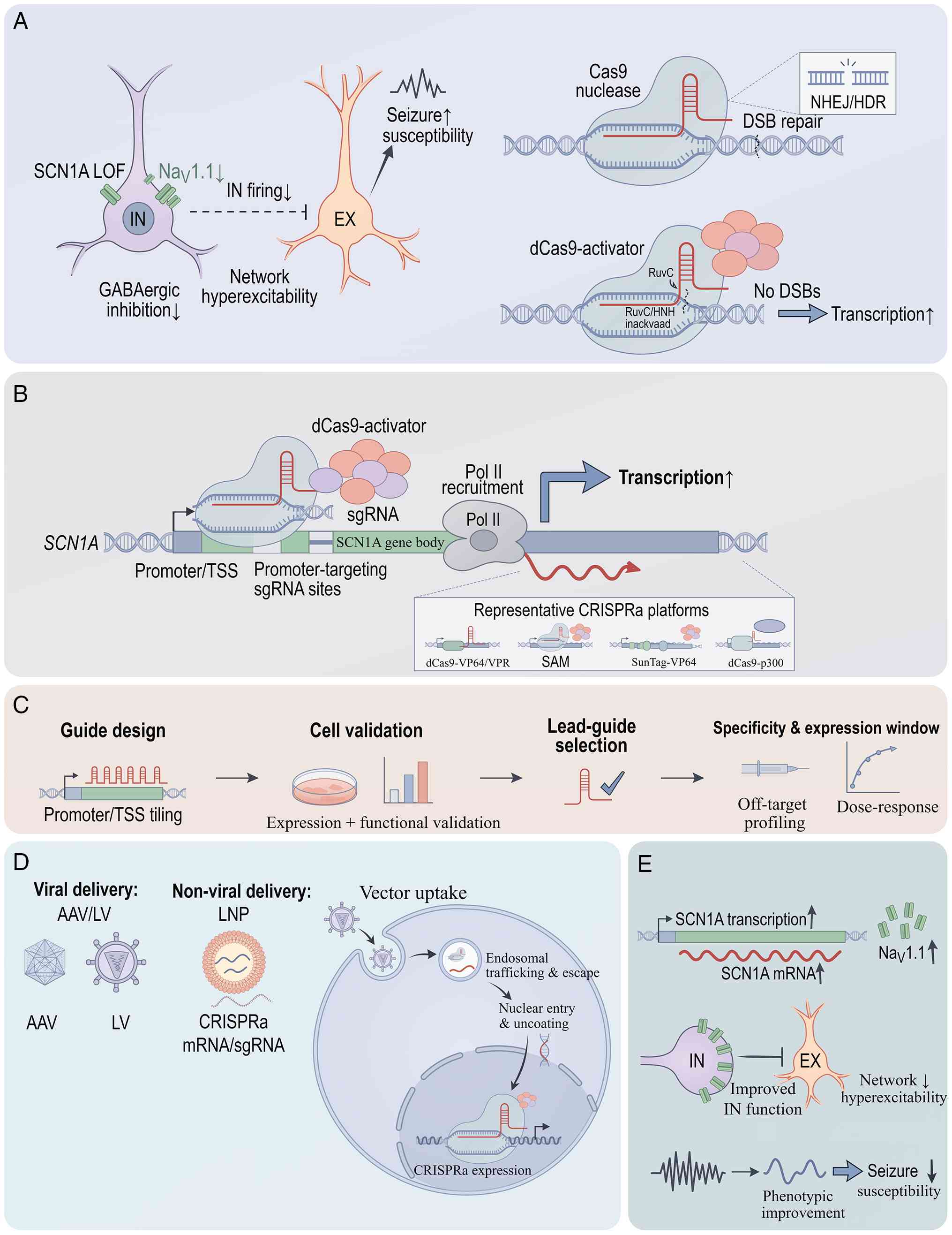 | Figure 1.Mechanisms and workflow of CRISPR
activation for targeted SCN1A upregulation in epilepsy. This
figure illustrates the molecular mechanisms, guide design workflow,
delivery strategies and functional outcomes of CRISPRa for
therapeutic upregulation of SCN1A in genetic epilepsy. (A)
Disease rationale and conceptual framework. LOF or
haploinsufficiency of SCN1A reduces NaV1.1
expression in GABAergic inhibitory interneurons, leading to
impaired inhibitory firing, network hyperexcitability and seizures.
By contrast to wild-type Cas9, which generates DSBs repaired by
NHEJ or HDR, dCas9 retains DNA-targeting capacity but lacks
endonuclease activity, thereby enabling programmable
transcriptional activation without DNA cleavage. (B) Core mechanism
of CRISPRa-mediated SCN1A activation. A dCas9-based
activator complex is directed by an sgRNA to the SCN1A
promoter, TSS region and where applicable validated CREs. Upon
recruitment, the complex recruits transcriptional co-activators and
RNA polymerase II to enhance endogenous SCN1A transcription.
Representative CRISPRa architectures include direct fusion systems
(such as dCas9-VP64), SAM, SunTag-based systems and epigenetic
activators such as dCas9-p300, which act through transcriptional
activation or local chromatin remodelling. (C) Guide design and
optimization workflow. Candidate sgRNAs are designed across the
promoter, TSS-proximal regulatory region and selected CREs, then
validated in cells to identify lead guides with high on-target
activity, appropriate activation strength and acceptable
specificity. This step supports optimization of target engagement,
expression amplitude and dose control before therapeutic
deployment. (D) Delivery strategies and cell-type-targeted
implementation. Therapeutic delivery may use viral (such as AAV) or
non-viral platforms (such as LNPs). Given the large size of CRISPRa
cargoes (~4.2 kb for dCas9-activator fusions plus gRNA expression
cassettes), dual-AAV or split-delivery approaches represent
practical strategies, whereas compact single-vector systems remain
under development. After cellular uptake, endosomal escape and
nuclear targeting, CRISPRa components assemble at the endogenous
SCN1A locus. Interneuron-targeted delivery or
interneuron-specific promoter-driven expression is emphasized to
restore NaV1.1 preferentially in inhibitory neuronal
populations, thereby minimizing the risk of nonspecific activation
in excitatory neurons. (E) Molecular, cellular and functional
consequences. Enhanced SCN1A transcription increases
SCN1A mRNA and NaV1.1 protein expression,
restoring sodium channel function in inhibitory interneurons. This
promotes recovery of interneuron excitability, improves
excitatory-inhibitory balance and provides a mechanistic basis for
seizure reduction and circuit-level rescue. The therapeutic goal is
physiologic restoration of endogenous SCN1A expression,
typically to near-normal levels (80–100% of wild-type), rather than
uncontrolled overexpression. Solid arrows indicate activation,
causal flow, or workflow progression; dashed arrows indicate
validation, optional or emerging steps; T-shaped lines indicate
inhibition. AAV, adeno-associated virus; CRE, cis-regulatory
element; CRISPR, clustered regularly interspaced short palindromic
repeats; CRISPRa, CRISPR activation; DSB, double-strand break;
dCas9, nuclease-deactivated Cas9; HDR, homology-directed repair;
LOF, loss-of-function; LNP, lipid nanoparticle; Nav1.1,
voltage-gated sodium channel Nav1.1; NHEJ,
non-homologous end joining; SAM, synergistic activation mediator;
SCN1A, sodium voltage-gated channel alpha subunit 1; sgRNA,
single guide RNA; TSS, transcription start site. |
Similarly, in a potassium voltage-gated channel
subfamily A member 1 (Kcna1)-deficient mouse model, in which
loss of the Kv1.1 potassium channel increases neuronal
excitability, CRISPRa-mediated activation of Kcna1
successfully restored potassium-channel function, reduced neuronal
hyperexcitability, markedly decreased seizure frequency and
improved cognitive performance (20). These findings provide
proof-of-concept evidence supporting the use of CRISPRa for the
treatment of LOF-associated epilepsies.
Tamura et al (32) extended this therapeutic paradigm to
SCN2A haploinsufficiency. Using AAV-mediated CRISPRa
delivery, the authors restored excitability in neocortical
pyramidal neurons and protected Scn2a+/− mice
against chemoconvulsant-induced seizures. Notably, therapeutic
rescue remained achievable when treatment was administered during
adolescence, suggesting a broader intervention window than
previously assumed.
To enhance cellular specificity, investigators have
incorporated inhibitory neuron-specific promoters, such as GAD67,
to drive dCas9 expression selectively within disease-relevant
neuronal populations. In a mouse model of DS, this strategy enabled
selective activation of SCN1A in inhibitory interneurons,
minimising potential adverse effects from non-specific activation
throughout the brain (31).
CRISPRa offers several theoretical advantages. By
activating endogenous transcription rather than introducing an
exogenous coding sequence, it preserves the physiological
regulatory architecture of the target gene, including native
promoters, enhancers and untranslated regions. Consequently,
CRISPRa may reduce the risk of supraphysiological protein
expression that can occur with conventional cDNA-based gene
supplementation. Furthermore, because CRISPRa targets only
regulatory elements rather than full-length coding sequences, it
may be particularly advantageous for large genes that exceed the
packaging capacity of standard AAV vectors.
However, CRISPRa also presents several important
challenges. Although it avoids exogenous gene replacement, the
approach still depends heavily on efficient delivery of large
genetic constructs. dCas9-activator fusion proteins are ~4.2 kb,
and including gRNA expression cassettes often requires dual-AAV or
alternative delivery systems, such as lipid nanoparticles (LNPs).
In addition, therapeutic efficacy depends on sustained expression
of both dCas9 and gRNAs, raising questions regarding long-term
vector persistence, immunogenicity and durability of gene
activation. Target engagement may also vary with chromatin
accessibility and local epigenetic context, potentially affecting
the consistency of therapeutic responses.
By contrast, conventional gene supplementation
delivers the therapeutic cDNA directly and therefore requires
maintenance only of the transgene itself rather than a
transcriptional activation machinery. However, this approach
remains constrained by vector cargo limitations (33) and the risk of promoter-driven
overexpression. Consequently, the choice between CRISPRa and gene
supplementation balances physiological regulation and technological
complexity. CRISPRa offers endogenous gene control,
cell-type-specific activation and suitability for large genes,
whereas gene supplementation benefits from a more established
translational pathway and a comparatively simpler therapeutic
design. Although CRISPRa remains preclinical, accumulating evidence
suggests it is a promising platform for treating LOF-associated
genetic epilepsies. The principal advantages and challenges of
CRISPRa are summarised in Table I
(19,33,34).
 | Table I.Advantages and challenges of
clustered regularly interspaced short palindromic repeats
activation. |
Table I.
Advantages and challenges of
clustered regularly interspaced short palindromic repeats
activation.
| Advantages | Challenges |
|---|
| Non-integrating;
does not introduce exogenous cDNA | Large delivery
payload (deactivated Cas9 is ~4.2 kb; together with activators and
gRNA, delivery often requires dual adeno-associated virus or lipid
nanoparticle systems) (33,34) |
| Expression levels
are tuneable (through optimization of gRNA design and promoter
selection) | gRNA target-site
design depends on chromatin accessibility and requires
cross-species validation (34) |
| Theoretically
reversible (through cessation of expression or use of inducible
systems) | Long-term
expression may interfere with other genenetworks, and safety still
requires systematic evaluation (19) |
| Preserves the
physiological regulatory architecture of endogenous genes | Still in the
preclinical stage and lacks human data |
Overall, CRISPRa provides a precise therapeutic
strategy for LOF epilepsies without exogenous gene replacement.
However, clinical translation still requires resolving key issues
such as delivery efficiency, long-term safety and cell-type
specificity.
Therapeutic strategies oriented towards GOF
mutations: Gene silencing
GOF epilepsies are caused by increased channel
activity, reduced depolarization thresholds or abnormal persistent
currents, and the therapeutic goal is to selectively suppress
expression of the pathogenic allele to block abnormal neuronal
firing. The present section includes three main strategies:
ASO-mediated silencing, RNA interference [small interfering
(si)RNA] and CRISPRi.
ASO-mediated gene silencing
ASOs are currently the most translationally advanced
gene-silencing platform for GOF epilepsies. ASOs are short,
synthetic, single-stranded oligonucleotides (15–25 nucleotides)
that bind to target RNA through Watson-Crick base pairing (35). Their mechanism of action depends on
the chemical modifications and design of the ASO and can be broadly
categorised into two major classes (36).
Gapmer ASOs [ribonuclease H (RNase
H)-mediated degradation]
Gapmer ASOs contain a central DNA ‘gap’ flanked by
chemically modified (2′-O-methoxyethyl or locked nucleic acid)
‘wings’. Upon binding to target mRNA, the DNA-RNA hybrid duplex
recruits endogenous RNase H, which specifically recognises and
cleaves the RNA strand of the hybrid, degrading the target mRNA and
suppressing pathological gene expression. This mechanism suits GOF
mutations because it reduces mutant transcript abundance. Allele
selectivity can be achieved by designing gapmer ASOs that
specifically target single-nucleotide polymorphisms or
mutation-specific sequences, thereby sparing the wild-type allele.
Examples of gapmer ASOs in epilepsy include the
SCN8A-targeting ASO developed by Lenk et al (37), which reduced mutant SCN8A
mRNA by ~50% while preserving wild-type expression, and the
potassium sodium-activated channel subfamily T member 1
(KCNT1)-targeting ASO developed by Burbano et al
(38), which selectively degraded
mutant KCNT1 transcripts.
Splice-switching ASOs
By contrast, splice-switching ASOs do not recruit
RNase H. Instead, they bind to pre-mRNA at splice sites, exonic
splicing enhancers or intronic splicing silencers, physically
blocking access by splicing factors and thereby modulating
alternative splicing. This mechanism can skip mutated exons,
restore correct reading frames or increase the production of
functional transcripts. Splice-switching ASOs are particularly
valuable for LOF mutations caused by aberrant splicing. A prominent
example in epilepsy is STK-001 (developed by Stoke Therapeutics,
Inc.), which targets a negative splicing enhancer region in
SCN1A pre-mRNA, increasing functional SCN1A mRNA and
restoring Nav1.1 protein expression in DS (39). As splice-switching ASOs do not
degrade RNA, they preserve the transcript pool and are generally
associated with lower toxicity than gapmers (40).
Both ASO classes offer strong allele selectivity
when designed against mutation-specific sequences, controllable
dosing and good reversibility, making them attractive for
paediatric applications that may require dose titration and
treatment adjustment.
SCN8A-associated encephalopathy
GOF mutations in SCN8A enhance
Nav1.6 channel function, triggering spontaneous neuronal
firing and persistent seizures. In an SCN8A-R1872W knock-in
mouse model, Lenk et al (37) demonstrated that
intracerebroventricular administration of a non-allele-specific ASO
reduced mutant mRNA levels by ~50%, decreased Nav1.6
expression by ~50%, reduced seizure frequency by >70% and
extended median survival from 15 days to 7 weeks. The ASO preserved
wild-type allele expression, avoiding potential adverse effects
such as motor dysfunction. Follow-up studies further confirmed that
long-term ASO treatment can durably prevent seizures and prolong
survival (36,41).
KCNT1-associated encephalopathy
Missense KCNT1 mutations (such as
p.Arg428Gln) abnormally enhance the function of the
sodium-activated potassium channel (also known as the Slack
channel), causing severe early-onset developmental and epileptic
encephalopathy. Burbano et al (38) designed an allele-selective ASO
targeting this mutation. Through RNase H-mediated degradation of
mutant mRNA, the ASO markedly reduced expression of the abnormal
channel protein. In a mouse model of KCNT1 encephalopathy,
ASO treatment reduced seizure frequency by >70% without
affecting wild-type allele expression (38). Separately, KCNT1-lowering
ASOs prolonged survival in SCN1A and SCN8A mutant
mice (42). Andreone et al
(43) further validated the
efficacy of divalent siRNA in a preclinical model of KCNT1
genetic epilepsy, demonstrating durable seizure suppression.
SCN2A-associated encephalopathy
ASO-mediated silencing is also applicable to GOF
mutations in SCN2A. Li et al (44) validated the therapeutic efficacy of
ASOs in a mouse model of SCN2A GOF mutations and observed
marked reductions in seizure frequency, along with prolonged
survival. In a landmark clinical study, Kim-McManus et al
(45) reported two N-of-1 trials
using allele-selective ASOs for SCN2A-associated
developmental and epileptic encephalopathy, in which one patient
experienced a 90% reduction in seizure frequency and walked
independently for the first time at 15 years of age, without
serious adverse events. Fig. 2
illustrates the mechanisms of ASO therapy for SCN2A- and
SCN8A-associated epilepsies.
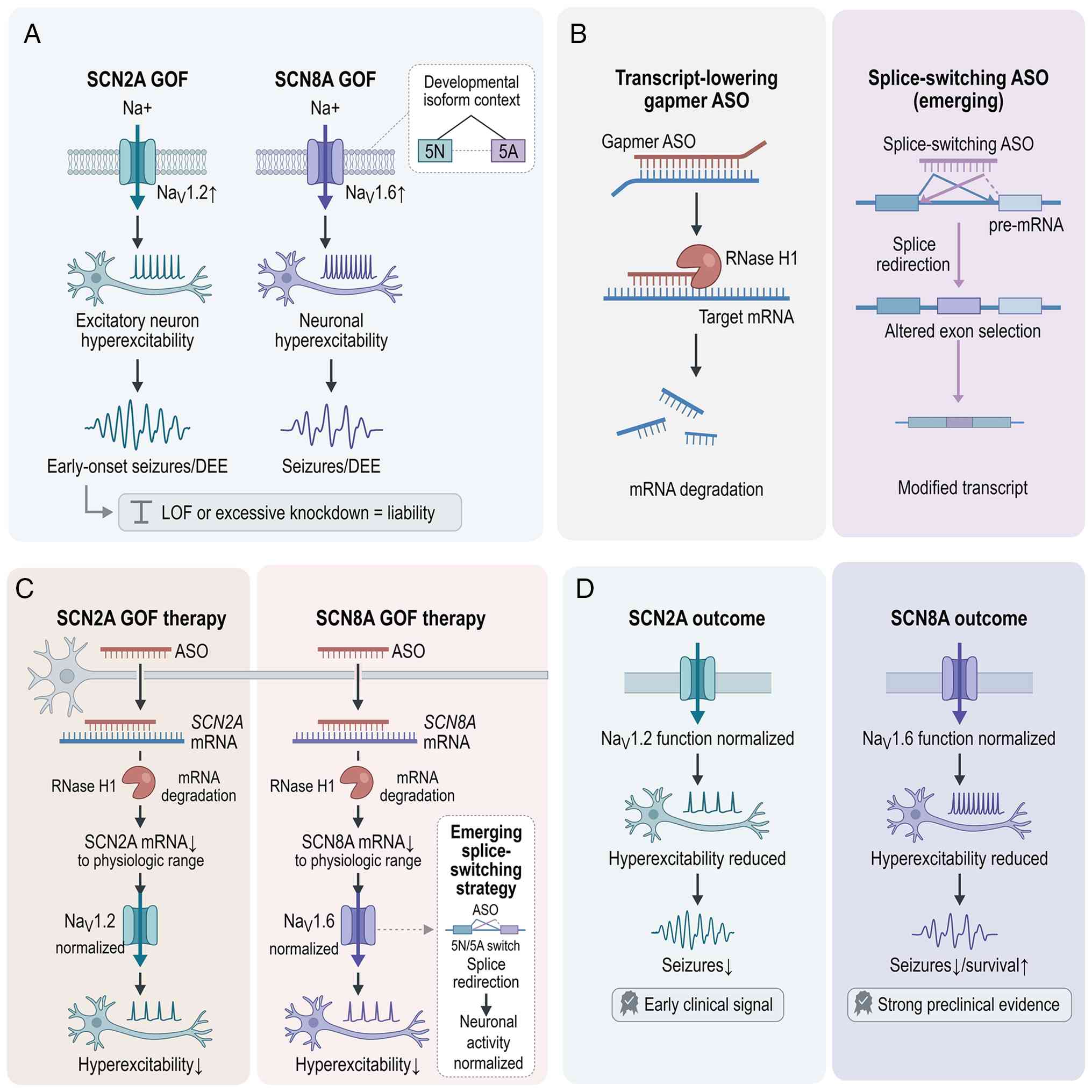 | Figure 2.Mechanisms of antisense
oligonucleotide therapy for SCN2A- and
SCN8A-associated epilepsies. This figure illustrates the
molecular mechanisms, gene-specific applications and therapeutic
outcomes of ASO-based strategies for GOF epilepsies caused by
mutations in SCN2A and SCN8A. (A) Variant
stratification and disease rationale. The figure focuses on GOF
forms of SCN2A- and SCN8A-associated epilepsies, in
which increased NaV1.2 or NaV1.6 activity
drives neuronal hyperexcitability and seizures. A small
developmental isoform context is included to indicate that
alternative splicing contributes to channel biology and therapeutic
opportunities; however, LOF states are presented only as a
cautionary context rather than a primary treatment indication. This
distinction is important because transcript-lowering strategies are
primarily being developed for GOF-associated disease, whereas
excessive suppression of channel expression may itself be
deleterious in LOF contexts. (B) Core ASO mechanisms. The principal
mechanism shown is gapmer ASO-mediated transcript lowering, in
which the ASO binds target mRNA, forms an ASO:mRNA hybrid, recruits
RNase H1 and promotes degradation of the pathogenic transcript. A
secondary, emerging mechanism is splice switching, in which an ASO
binds pre-mRNA and redirects splice-site usage or isoform selection
to favour production of a more functional transcript. Other ASO
mechanisms, such as translational blockade and microRNA modulation,
are not emphasised, as they are not the predominant
evidence-supported therapeutic approaches for these sodium channel
epilepsies. (C) Gene-specific therapeutic mechanisms. For
SCN2A GOF epilepsies, a transcript-lowering ASO reduces
SCN2A mRNA through RNase H1-mediated degradation, thereby
reducing excess NaV1.2 activity towards a physiological
range and decreasing neuronal hyperexcitability. For SCN8A
GOF epilepsies, a similar transcript-lowering strategy reduces
SCN8A mRNA and pathogenic NaV1.6 activity. An
additional, smaller branch illustrates an emerging SCN8A
splice-switching ASO strategy, which redirects pre-mRNA processing
to normalise functionally relevant isoforms. The therapeutic goal
in both settings is controlled normalisation of channel activity
rather than indiscriminate or complete silencing, which could
result in haploinsufficiency or other adverse effects. (D)
Functional rescue and therapeutic outcomes. ASO treatment is
intended to normalise channel activity, reduce pathological
neuronal firing and improve seizure outcomes. The evidence tier
differs between the two genes: SCN2A has early clinical
proof of concept for transcript-lowering therapy in early-onset GOF
developmental and epileptic encephalopathy, with a recent case
report demonstrating ASO treatment in a preterm infant (72); SCN8A currently has strong
preclinical evidence supporting transcript lowering, including
benefit after seizure onset and with repeated dosing in animal
models. A cautionary note highlights that dose titration and
genotype-specific use are required to avoid over-suppression, as
excessive channel reduction may compromise normal neuronal
function. Solid arrows indicate causal flow or therapeutic
progression; dashed arrows indicate emerging or lower-evidence
branches. ASO, antisense oligonucleotide; DEE, developmental and
epileptic encephalopathy; GOF, gain-of-function; LOF,
loss-of-function; mRNA, messenger RNA; pre-mRNA, precursor
messenger RNA; RNase H1, ribonuclease H1; SCN2A, sodium
voltage-gated channel alpha subunit 2; SCN8A, sodium
voltage-gated channel alpha subunit 8. |
Collectively, these studies indicate that
ASO-mediated silencing is an effective, controllable and clinically
advanced therapeutic strategy for GOF channelopathies. At present,
ASO therapy targeting SCN8A has entered a key transition
phase from preclinical research to clinical translation.
siRNA and LNP delivery
RNA interference is a gene-silencing mechanism
mediated by siRNAs. After complementary binding to target mRNA,
siRNAs recruit and activate the RNA-induced silencing complex,
which promotes cleavage and degradation of the target mRNA, thereby
rapidly and efficiently suppressing gene expression (46).
In recent years, LNP-based siRNA delivery systems
have shown substantial improvements in in vivo stability and
tissue targeting. Angiopep-2-modified LNPs (Ang-C2 LNPs), developed
by Tong et al (47), can
effectively cross the blood-brain barrier (BBB), deliver siRNA
efficiently to brain tissue and maintain target-gene suppression
for ~15 days (47,48). Andreone et al (43) further validated the efficacy of
divalent siRNA in a preclinical model of KCNT1-associated
genetic epilepsy and demonstrated durable seizure suppression.
However, compared with ASOs, siRNA delivery to the central nervous
system still faces several challenges.
Shorter half-life
The in vivo half-life of siRNAs is typically
1–2 days, considerably shorter than the 2–4 weeks reported for
ASOs, necessitating more frequent dosing or more sophisticated
delivery systems.
Delivery efficiency
siRNAs depend on delivery vehicles such as LNPs to
cross the BBB, and the cell-type specificity and long-term safety
of these carriers require further optimisation.
Limited clinical evidence
To date, no published study has directly
demonstrated improvement of epileptic phenotypes in SCN2A or
SCN8A animal models using siRNA.
CRISPRi
CRISPRi is an epigenetic repression technology that
does not involve DNA double-strand breaks (DSBs). By fusing
nuclease-dCas9 to a transcriptional repressor, such as the Krüppel
associated box (KRAB) domain, and directing the complex to a gene
promoter using a gRNA, CRISPRi promotes the deposition of
repressive histone marks, including H3K9me3, enabling reversible,
programmable gene silencing (29).
In neuronal systems, AAV-mediated CRISPRi delivery has been shown
to suppress target-gene expression effectively. Duke et al
(49) optimised a neuron-specific
CRISPRi system and achieved efficient repression of endogenous
genes in primary cortical neurons.
Compared with ASOs and siRNA, CRISPRi offers more
durable gene repression as epigenetic modifications are stable and
can simultaneously regulate multiple genes using distinct gRNAs.
However, several challenges continue to limit its clinical
translation: i) Large delivery payload, the dCas9-KRAB fusion
protein is ~4.5 kb and, together with gRNA expression elements,
often requires delivery via dual AAV vectors or LNP-based systems;
cell-type specificity, selective repression of target cell
populations requires cell-type-specific promoters; and iii)
long-term safety, the durability of epigenetic modifications and
their potential off-target effects require further systematic
evaluation (34,50,51).
At present, CRISPRi in epilepsy remains at the preclinical
optimisation stage.
Precision editing strategies: DNA editing
and RNA editing
Precision editing technologies aim to correct
pathogenic mutations at their source and therefore have the
theoretical potential to provide one-time curative treatment.
Depending on the molecular substrate targeted, these approaches can
be divided into DNA editing and RNA editing. These two strategies
differ substantially in their reversibility, durability, safety
profiles and translational maturity and are therefore discussed
separately in this section.
DNA editing
DNA editing corrects mutations by permanently
modifying genomic sequences. The major platforms include
CRISPR/Cas9, base editing and prime editing.
CRISPR/Cas9
Under the guidance of a single-gRNA (sgRNA), the
CRISPR/Cas9 system recognises a target site and induces a DNA DSB,
after which non-homologous end joining (NHEJ) facilitates gene
knockout, whereas homology-directed repair (HDR) using a donor
template enables the correction of point mutations (49,52).
However, HDR is cell cycle-dependent and inefficient in terminally
differentiated neurons, limiting its direct application in adult
neurological disorders. Consequently, in epilepsy, CRISPR/Cas9 has
primarily been employed in knockout-based strategies targeting GOF
mutations.
Base editing
Base editing overcomes the conventional reliance of
conventional gene editing on DNA DSB. By fusing a Cas9 nickase to a
deaminase, it directly mediates targeted single-base substitutions
without cleaving both DNA strands. The most widely used base
editors include cytosine base editors, which mediate C-G to T-A
conversions, and adenine base editors (ABEs), which mediate A-T to
G-C conversions (53).
In SCN8A GOF epilepsy models, base editing
has demonstrated promising therapeutic efficacy. Yu et al
(13) used base editing to target
a dominant mutant SCN8A allele and successfully corrected
the pathogenic variant in a mouse model, thereby protecting the
animals from seizures and premature mortality. Cao et al
(54) subsequently applied prime
editing to correct a pathogenic mutation in a mouse model of
genetic epilepsy, ameliorating neuronal hyperexcitability and
seizure activity. In a separate study, Reever et al
(55) employed adenine base
editing to correct the recurrent SCN8A R1872W mutation,
achieving ~30% conversion of mutant transcripts to the wild-type
sequence. This level of correction was sufficient to notably
prolong survival and completely prevent seizures in 7/11 treated
mice (55).
Prime editing
Prime editing further expands the spectrum of
editable genetic variants. By combining a Cas9 nickase with a
reverse transcriptase and using a prime editing gRNA (pegRNA),
prime editing enables diverse base substitutions, as well as short
insertions and deletions, without donor DNA or the induction of DNA
DSBs (56). Unlike base editing,
which is largely restricted to transition mutations, prime editing
can correct a broader range of pathogenic variants, including
frameshift mutations and short deletions.
Robertson et al (57) optimised a prime-editing platform in
primary mouse neurons and established a methodological framework
for neuronal genome editing relevant to epilepsy research. The
study developed standardised protocols for pegRNA and prime editor
3b (PE3b) nicking guide RNA design, plasmid construction, primary
neuronal nucleofection and quantitative next-generation sequencing
analysis. Although the study did not directly evaluate
anti-epileptic efficacy, it showed that prime editing can precisely
rewrite pathogenic neuronal variants while preserving the
endogenous regulatory environment. These findings provide important
technical guidance for functional modelling and future therapeutic
editing studies involving epilepsy-associated genes such as
SCN1A, SCN2A, SCN8A and GRIN2A (57).
Cerna-Chavez et al (58) subsequently demonstrated efficient
prime editing in human iPSCs (hiPSCs), enabling efficient
site-specific modification of point mutations, small insertions and
small deletions and facilitating the generation of isogenic disease
models for multiple monogenic disorders. Although the study did not
specifically examine epilepsy-associated genes, it suggests that
prime editing is technically feasible for precisely introducing or
correcting pathogenic variants in patient-derived hiPSCs carrying
mutations in genes such as SCN1A, SCN8A and KCNQ2.
Together with subsequent work from the Robertson group (56), which used in utero prime editing to
introduce an ultra-rare GRIN2A epilepsy-associated variant
and induce spontaneous seizures, these findings indicate that prime
editing in epilepsy is extending beyond in vitro disease
modelling toward personalised validation of neurodevelopmental and
seizure-associated phenotypes (59). In addition, Cao et al
(54) applied PE3-mediated prime
editing in Kcnq2R207W mice with genetic epilepsy
to directly correct the pathogenic variant. Site-specific repair
reduced neuronal hyperexcitability and seizure frequency, providing
one of the few preclinical demonstrations of prime editing as a
therapeutic intervention for genetic epilepsy (54).
Core challenges of DNA editing
Despite their transformative potential, DNA-editing
technologies face substantial challenges that temper expectations
for near-term clinical translation. The irreversible nature of
genomic modification requires markedly high editing specificity and
safety, particularly when applied to the developing brain. Delivery
efficiency remains a major obstacle, as base-editing and
prime-editing platforms generally exceed the packaging capacity of
a single AAV vector. In addition, off-target editing at both the
genomic and transcriptomic levels, although reduced compared with
conventional CRISPR/Cas9 systems, has not been completely
eliminated. The long-term consequences of permanent genomic
modification in the paediatric brain remain unknown, while the
therapeutic window for intervention in numerous early-onset
epilepsies are limited. Collectively, these considerations indicate
that, although DNA editing offers the prospect of curative
treatment, its clinical implementation should proceed cautiously
and be restricted to situations in which the anticipated benefits
outweigh the potential risks (60).
RNA editing
RNA editing restores protein function by modifying
bases within mutant mRNA transcripts without permanently altering
genomic DNA. Compared with DNA editing, RNA editing offers greater
reversibility, a lower risk of off-target effects and independence
from cell cycle activity, making it particularly well suited to
non-dividing neurons within the central nervous system (61).
Adenosine deaminase acting on RNA
(ADAR)-mediated A-to-I editing
The most prevalent form of RNA editing is adenosine
deamination mediated by the ADAR family of enzymes. ADAR converts A
to I, which is interpreted as G during translation, thereby
producing the functional equivalent of an A-T to G-C base
substitution (61). Artificial
RNA-editing systems typically achieve site-specific editing by
fusing the catalytic domain of ADAR to a guide RNA-directed
targeting platform or by using engineered oligonucleotides to
recruit endogenous ADAR to the target transcript. i) Recruiting
Endogenous ADAR to Specific Transcripts for
Oligonucleotide-mediated RNA Editing (RESTORE) platform. Merkle
et al (62) developed the
RESTORE system, which employs engineered oligonucleotides to
recruit endogenous ADAR and facilitate efficient RNA repair. As the
platform delivers no exogenous editing enzyme, it is associated
with low immunogenicity.
ii) Leveraging Endogenous ADAR for Programmable
Editing of RNA 2.0. Yi et al (63) utilised engineered circular RNA
molecules to recruit endogenous ADAR, achieving durable and
efficient RNA editing in non-human primates, with editing
efficiencies >50% and persistence for ≥13 weeks (63,64).
In a humanised mouse model of Hurler syndrome, the system
successfully corrected a nonsense mutation, demonstrating
therapeutic potential (63).
iii) Light-controlled RNA editing. Li et al
(65) developed the
photoactivatable RNA ABE system, which uses blue-light stimulation
to regulate ADAR activity and enable spatiotemporal control of RNA
editing. In mice, this approach reduced off-target editing to
baseline levels.
Advantages and challenges of RNA
editing
The principal advantages and challenges of RNA
editing are summarised in Table
II. The reversibility and genomic safety of RNA editing make it
particularly appealing for paediatric applications. However, its
transient nature, inherently associated to mRNA turnover, may
require repeated administration to maintain therapeutic efficacy,
creating challenges for long-term treatment in young children.
Delivery efficiency also remains suboptimal, while
transcriptome-wide off-target A-to-I editing continues to raise
safety concerns that require comprehensive evaluation. Additional
challenges include the potential immunogenicity of exogenous ADAR
enzymes and AAV-based delivery systems, as well as immune responses
directed against edited transcripts.
| Table II.Advantages and challenges of RNA
editing. |
Table II.
Advantages and challenges of RNA
editing.
| Advantages | Challenges |
|---|
| Reversibility:
Editing effects are not permanent and can be adjusted or
terminated | Limited durability:
As RNA has a short half-life, repeated dosing may be required |
| Does not alter the
genome, thereby avoiding the risk of genomic integration | Delivery
efficiency: Adeno-associated virus-orlipid nanoparticle-based
delivery systems still require optimization |
| Independent of cell
division and therefore suitable for terminally differentiated
neurons | Off-target editing:
Transcriptome-wide A-to-I off-target effects require systematic
evaluation |
| Dose is tuneable
and thus suitable for paediatric application | Immunogenicity of
the editing enzyme: Exogenous adenosine deaminase acting on RNA may
trigger immune responses |
Decision logic for DNA vs. RNA
editing
RNA editing may be prioritised when paediatric
safety, dose titration and reversibility are the principal
considerations, particularly in patients undergoing active
neurodevelopment. By contrast, DNA editing may be more appropriate
when efficient delivery systems and long-term monitoring frameworks
are available to support durable, irreversible therapeutic
intervention and when the pathogenic variant has been identified
and the anticipated clinical benefit is substantial. At present,
RNA editing in epilepsy remains at the stage of preclinical
validation. These selection considerations derive from
methodological reviews and safety analyses (60,66),
rather than direct head-to-head clinical comparisons of DNA and RNA
editing in genetic epilepsy.
Translational research progress in
representative epilepsy genes
The preceding sections systematically described the
technical principles underlying three major approaches:
LOF-oriented strategies, GOF-oriented strategies and precision
editing. The present section uses specific genes as illustrative
examples to highlight the current applications, key findings and
translational progress of these strategies in genetic epilepsies.
The discussion focuses primarily on SCN1A (DS),
SCN2A/SCN8A-associated encephalopathies, SCN3A
encephalopathy, KCNQ2/3 mutation-associated epilepsy, mTOR
pathway-associated epilepsy and synapse-associated genes, including
GRIN2A, γ-aminobutyric acid receptor subunit γ-2 (GABRG2)
and STXBP1.
SCN1A: DS-a paradigm of LOF
epilepsy
DS is a prototypical LOF epilepsy. Heterozygous LOF
mutations in SCN1A result in Nav1.1 haploinsufficiency
in inhibitory interneurons, disrupting the excitation-inhibition
balance. As the SCN1A coding sequence is relatively large
(~6 kb), conventional gene supplementation is limited by vector
cargo capacity. Consequently, therapeutic development has shifted
towards strategies that enhance endogenous SCN1A expression
(17).
ASO therapy (STK-001)
STK-001, developed by Stoke Therapeutics, Inc., is
based on the Targeted Augmentation of Nuclear Gene Output platform
and employs a sequence-specific ASO targeting a negative splicing
enhancer within SCN1A pre-mRNA, thereby increasing the
production of functional SCN1A mRNA (39). In Scn1a+/− mice,
intrathecal administration of STK-001 increased brain SCN1A
mRNA levels by ~50%, restored Nav1.1 protein expression
to near-normal levels, reduced spontaneous seizure frequency by
~70% and improved motor coordination and survival (39).
Phase I/II clinical trials (MONARCH, NCT04442295;
SWALLOWTAIL, NCT04740476) have been completed. Publicly available
data indicate that STK-001 is generally well tolerated, with no
dose-limiting toxicities or serious adverse events reported
(67). Some patients achieved a
≥50% reduction in seizure frequency, with trends toward improved
cognitive and behavioural outcomes (68). These findings supported advancing
STK-001 into a global pivotal phase III trial (EMPEROR,
NCT06872125), making it one of the most clinically advanced
gene-targeted therapies for DS (28,69,70).
Regulatory AAV (ETX101)
ETX101, developed by Encoded Therapeutics, Inc.,
uses an engineered AAV9 vector to selectively upregulate endogenous
SCN1A expression in inhibitory interneurons, thereby
avoiding both full-length cDNA cargo constraints and the risk of
exogenous overexpression (21,28).
In DS mouse models, ETX101 markedly increased SCN1A
expression, reduced seizure frequency and improved
electrophysiological function. Studies in non-human primates
demonstrated widespread central nervous system distribution without
evident immunotoxicity (21). A
Phase I clinical trial (NCT05419492) is currently evaluating the
safety and preliminary efficacy of a single intrathecal
administration in paediatric patients. As an early-stage clinical
candidate, ETX101 represents a promising gene-regulatory approach
for DS and is currently undergoing first-in-human evaluation
(21,28).
CRISPRa
In DS mouse models, AAV-delivered dCas9-VP64
complexes activated the endogenous Scn1a gene in inhibitory
interneurons, restoring Nav1.1 expression to near-normal
levels (80–100% of wild-type levels), markedly reducing seizure
frequency and improving motor coordination (31). Human iPSC-derived GABAergic
interneurons have instead been used to map SCN1A
cis-regulatory elements relevant to future gene-activation
strategies (71). Although these
results are encouraging, this approach remains preclinical, with
further optimisation and safety evaluation required before clinical
translation. The major gene-targeted therapeutic strategies for DS
and their translational status are compared in Table III.
| Table III.Summary comparison of Dravet syndrome
strategies. |
Table III.
Summary comparison of Dravet syndrome
strategies.
| Strategy | Representative
agent | Mechanism | Translational
status | Key advantages | Main
challenges |
|---|
| Antisense
oligonucleotides upregulation | STK-001 | Splicing
modulation; increases functional mRNA | Phase III
clinical | Reversible;
dose-controllable; mature delivery | Requires repeated
dosing |
| Regulatory
adeno-associated virus | ETX101 | Enhances endogenous
gene transcription | Phase I
clinical | Durable;
single-dose administration; cell-type specific | Irreversible;
immunogenicity |
| Clustered regularly
interspaced short palindromic repeats activation | Preclinical | Deactivated
Cas9-mediated transcriptional activation | Preclinical | Non-integrating;
preserves physiological regulation | Complex delivery;
unknown long-term safety |
SCN2A/SCN8A-associated
encephalopathies: A paradigm of GOF epilepsy
SCN2A and SCN8A encode the
Nav1.2 and Nav1.6 sodium channels,
respectively. GOF mutations in these genes increase channel
activity and lower the depolarisation threshold, resulting in
early-onset epileptic encephalopathy, developmental delay and
severe neurobehavioral impairment (9). In contrast to the LOF mechanism
associated with SCN1A, these epilepsies are more amenable to
gene-silencing strategies.
SCN8A ASO therapy
In an SCN8A-R1872W knock-in mouse model,
Lenk et al (37)
demonstrated that intracerebroventricular administration of a
non-allele-specific ASO reduced mutant mRNA levels by ~50%,
decreased Nav1.6 expression by ~50%, reduced seizure
frequency by >70%, and extended median survival from 15 days to
7 weeks (37). Expression of the
wild-type allele was preserved, thereby avoiding adverse effects
such as motor dysfunction. Hill et al (41) further demonstrated that long-term
ASO therapy can provide sustained seizure prevention and prolong
survival, supporting the progression of this approach from the
preclinical stage towards clinical translation.
SCN2A ASO therapy
Li et al (44) validated the therapeutic efficacy of
ASOs in a mouse model of SCN2A GOF mutations, demonstrating
marked reductions in seizure frequency and prolonged survival.
Wagner et al (72) reported
a case of a preterm infant with early-onset SCN2A
encephalopathy treated with an ASO, demonstrating the feasibility
of this approach in very-early-stage patients. Collectively, these
findings support advancing SCN2A-targeted ASO therapy from
preclinical studies toward early clinical exploration.
siRNA and CRISPRi
In SCN8A, AAV-delivered short hairpin RNA, a
closely associated RNA interference modality to siRNA, reduced
SCN8A expression, durably prevented seizures and prolonged
survival throughout a 12-month observation period (73). Regarding CRISPRi, Duke et al
(49) showed that a
neuron-specific CRISPRi system efficiently repressed endogenous
genes in primary cortical neurons. Collectively, these findings
support the therapeutic potential of gene-silencing approaches
targeting SCN8A and indicate that both RNA interference and
CRISPRi strategies remain at the preclinical stage.
SCN3A encephalopathy-a complex
phenotype combining LOF and GOF
SCN3A encodes the Nav1.3 sodium
channel. Pathogenic variants can result in either GOF or LOF
effects and are frequently associated with cortical developmental
abnormalities and early-onset epileptic encephalopathy (74). In iPSC-derived neuronal models, GOF
variants, exemplified by p.I875T, recapitulate increased sodium
currents and abnormal persistent neuronal firing (74).
At present, no ASO- or gene-editing strategy
specifically targeting SCN3A has been formally reported.
Clinical management therefore relies primarily on precision
pharmacological approaches. Lacosamide and phenytoin inhibit
Nav1.3 activity and restore electrophysiological
function (74). More recently, Qu
et al (75) reported that a
Nav1.3-selective inhibitor restored abnormal electrical
activity in iPSC-derived neurons.
Several factors have slowed the development of gene
therapies for SCN3A compared with SCN1A, SCN2A and
SCN8A. First, SCN3A was only firmly established as a
cause of epileptic encephalopathy in 2018, whereas SCN1A,
SCN2A and SCN8A had been recognised as major epilepsy
genes >10 years earlier (14–16),
providing substantially more time for preclinical and translational
development. Second, SCN3A-associated disorders exhibit
considerable phenotypic complexity, with manifestations that
include cortical malformations such as polymicrogyria in addition
to epilepsy. Consequently, effective therapies may need to address
both electrophysiological dysfunction and structural
neurodevelopmental abnormalities. Third, SCN3A variants
display marked functional heterogeneity, producing GOF, LOF, or, in
some cases, mixed effects. This complicates therapeutic selection,
whereas SCN1A and SCN8A are associated predominantly
with LOF and GOF mechanisms, respectively. Fourth, disease models
have been comparatively limited, with animal models that faithfully
reproduce both seizure phenotypes and cortical malformations
emerging slowly, and iPSC-based platforms only recently becoming
available for therapeutic evaluation (58). Finally, SCN3A-associated
epilepsies are considerably rarer than SCN1A- or
SCN2A-associated disorders (76,77),
reducing commercial incentives and funding opportunities for
therapeutic development. Collectively, these factors have
positioned SCN3A as a later-stage candidate for gene-based
intervention. However, recent advances in iPSC modelling and the
development of Nav1.3-selective small-molecule
inhibitors provide a foundation for future therapeutic innovation.
The pathogenic pathway underlying SCN3A-associated epileptic
encephalopathy is illustrated in Fig.
3.
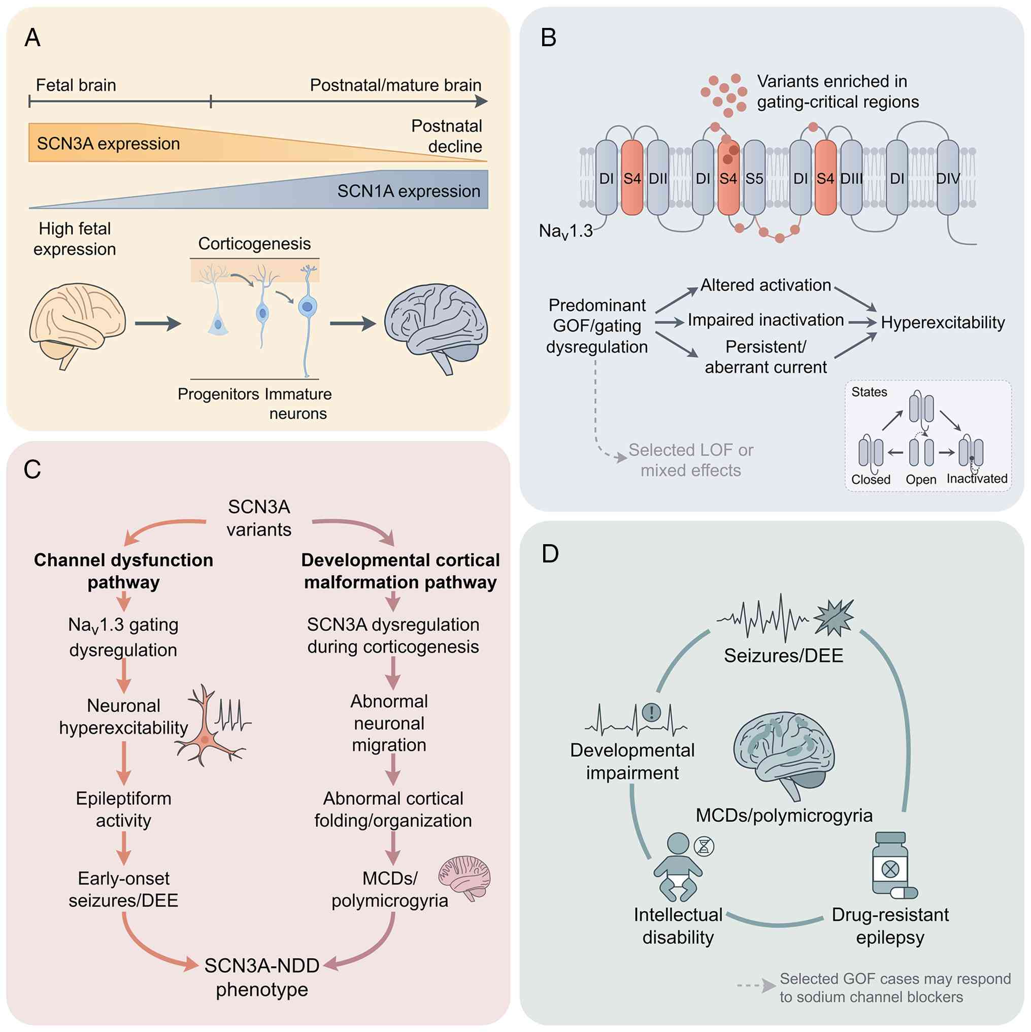 | Figure 3.Pathogenic pathways of
SCN3A-associated epileptic encephalopathy. This figure
illustrates the developmental context, variant clustering, dual
pathogenic mechanisms and integrated clinical spectrum of
SCN3A-associated neurodevelopmental disorders, highlighting
why this condition presents unique challenges for gene therapy
development compared with SCN1A, SCN2A and SCN8A. (A)
Developmental context of SCN3A. SCN3A encodes the
voltage-gated sodium channel NaV1.3, which is expressed
during foetal and early brain development and declines postnatally.
Its expression in cortical progenitors and immature neurons
associates SCN3A dysfunction to early corticogenesis and
helps explain why SCN3A-associated disease frequently
combines epilepsy with MCDs, a phenotypic feature that
distinguishes SCN3A from other sodium channel epilepsies and
complicates gene therapy design. (B) Variant clustering and channel
dysfunction. Pathogenic SCN3A variants are enriched in
gating-critical transmembrane regions, including voltage-sensing
and gating-coupling segments. Numerous disease-associated variants
exert predominantly GOF effects, although variant-specific
biophysical mechanisms are heterogeneous and may include altered
activation, impaired inactivation or other gating abnormalities. A
smaller subset exhibits LOF or mixed functional effects. This
functional heterogeneity poses a major challenge for therapeutic
strategy selection, as GOF mutations may be amenable to silencing
approaches, whereas LOF mutations require enhanced expression, yet
the same gene may harbour both mutation types. (C) Dual pathogenic
pathways. One major branch arises from NaV1.3 gating
dysregulation, which increases neuronal excitability and promotes
epileptiform activity, contributing to DEE. A second major branch
reflects disruption of corticogenesis during foetal brain
development, leading to abnormal neuronal migration, cortical
organisational defects and MCDs, particularly polymicrogyria.
Together, these parallel processes, electrophysiological
dysfunction and structural malformation, generate a epileptogenic
developmental substrate. The coexistence of these two pathogenic
branches distinguishes SCN3A encephalopathy from purely
channelopathic epilepsies and suggests that successful gene therapy
may need to address both electrophysiological and structural
abnormalities. (D) Integrated clinical spectrum. Convergence of
neuronal hyperexcitability and cortical malformation results in a
spectrum that includes early-onset seizures, often drug-resistant
epilepsy, DEE, developmental impairment, intellectual disability
and structural brain abnormalities such as polymicrogyria. Selected
GOF cases may benefit from sodium channel blockers, although
treatment response is variant-dependent and is therefore not
presented as a uniform precision therapy. Therapeutic development
for SCN3A remains at an early exploratory stage; to date, no
ASO-, siRNA- or gene-editing strategy specifically targeting
SCN3A has entered formal preclinical validation, reflecting
the genetic and phenotypic complexity described previously. Solid
arrows indicate causal or pathogenic progression; dashed arrows
indicate secondary or lower-emphasis links. DEE, developmental and
epileptic encephalopathy; GOF, gain of function; LOF, loss of
function; MCD, malformation of cortical development; NDD,
neurodevelopmental disorder; SCN3A, sodium voltage-gated
channel alpha subunit 3. |
Future therapeutic strategies may draw on advances
achieved in SCN8A- and KCNT1-associated disorders.
For GOF variants, allele-specific ASOs could be developed to
selectively suppress mutant transcripts. For LOF variants,
RNA-editing approaches, such as ADAR-mediated A-to-I editing, may
offer a reversible way to restore gene function. Overall,
SCN3A-targeted gene therapy remains at an early exploratory stage,
with research currently focused primarily on pharmacological
intervention and with gene-based approaches yet to enter formal
preclinical validation.
KCNQ2/3 mutation-associated epilepsy:
Exploration of gene therapy for potassium-channel LOF
KCNQ2 and KCNQ3 encode the Kv7.2 and
Kv7.3 potassium channels, which assemble into Kv7.2/Kv7.3
heteromeric complexes and mediate the M current. These channels
serve a key role in maintaining the resting membrane potential and
suppressing neuronal hyperexcitability. LOF mutations in these
genes are among the most common monogenic causes of neonatal
epilepsy, with clinical phenotypes ranging from benign familial
neonatal epilepsy to early infantile epileptic encephalopathy
associated with severe developmental impairment (11).
Gene supplementation strategy
AAV-mediated delivery of wild-type KCNQ2 or
KCNQ3 cDNA may restore potassium channel function.
Electrophysiological studies have demonstrated that channel
dysfunction resulting from KCNQ2/3 mutations can be
partially rescued by supplementation with the corresponding
wild-type gene (10,78,79).
However, the coding regions of KCNQ2 and KCNQ3 are
relatively large (~2.5 and 2.4 kb, respectively). Although each
gene fits within the packaging capacity of a single AAV vector,
co-delivery of both genes remains challenging (80). Consequently, despite encouraging
proof-of-concept data, KCNQ2/3 gene supplementation remains at the
preclinical stage.
Small-molecule activator strategy
Enhancing residual Kv7 channel activity with
small-molecule compounds represents a more advanced clinical
approach. The first-generation drug retigabine was previously
approved for adults with refractory epilepsy but was withdrawn
because of long-term adverse effects, including skin and retinal
pigmentation. Next-generation Kv7 channel activators, such as
XEN1101 (68) and RL648_81
(81), are under development and
exhibit greater selectivity, improved brain penetration and
enhanced safety profiles. XEN1101 has demonstrated favourable
efficacy in a Phase IIb clinical trial (82). As numerous Kv7-channel activators
have progressed into human clinical studies, this therapeutic
strategy is currently in clinical development (83,84).
CRISPRa strategy
In the future, CRISPRa-mediated activation of
endogenous KCNQ2/3 expression may provide a precise, durable way to
restore channel function. However, this approach remains at an
early proof-of-concept stage.
mTOR pathway-associated
epilepsy-synergy between gene repair and pharmacological
inhibition
The mTOR signalling pathway serves a central role
in regulating cell growth, metabolism and synaptic plasticity.
Aberrant activation of this pathway, termed mTORopathy, can lead to
cortical developmental abnormalities and refractory epilepsy, with
mutations in TSC1, TSC2 and DEP domain-containing protein 5
(DEPDC5) among the best-characterised examples (85,86).
Fig. 4 illustrates the pathogenic
mechanisms and precision therapeutic strategies associated with
mTOR-pathway-associated epilepsy.
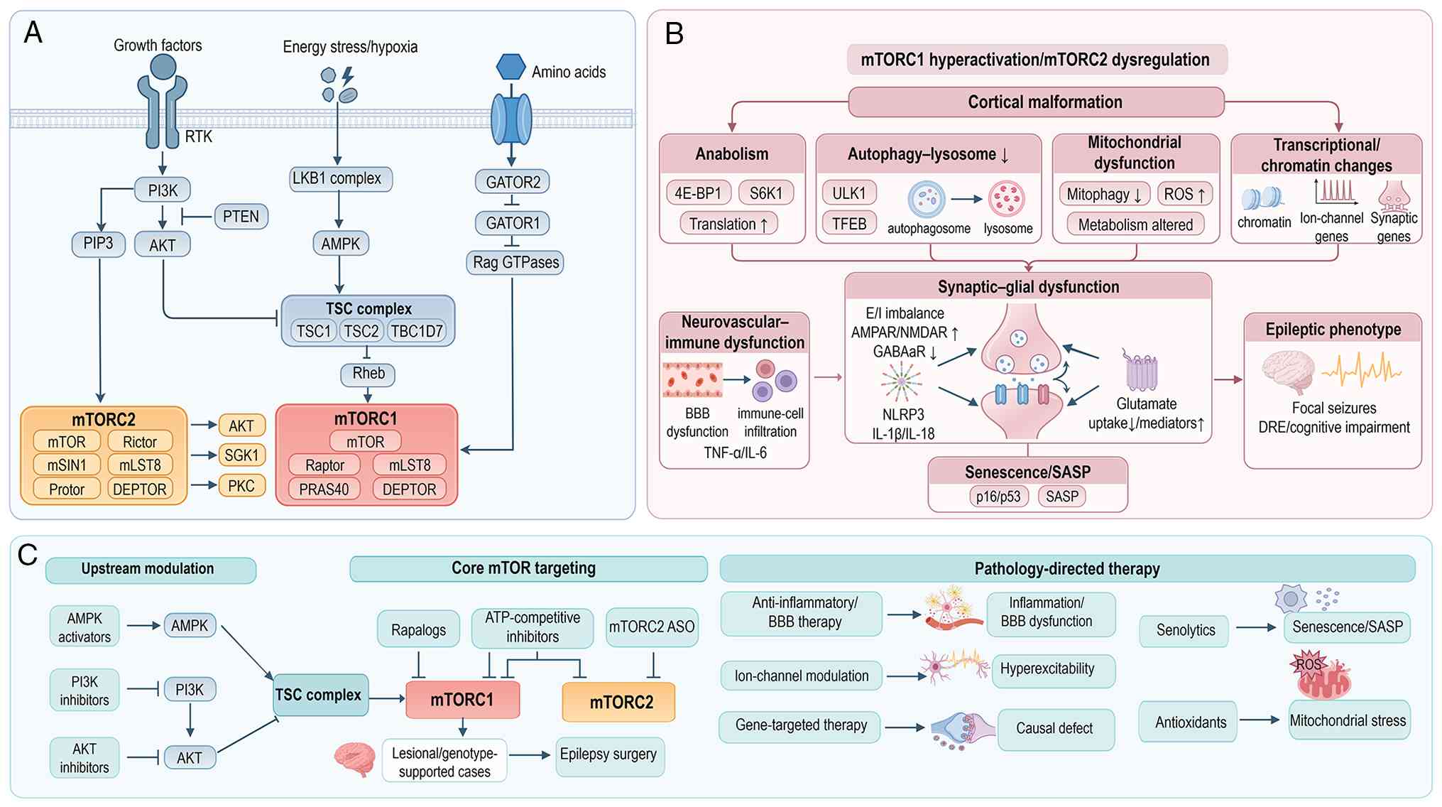 | Figure 4.Pathogenic mechanisms and precision
therapeutic strategies in mTOR pathway-associated epilepsy. This
figure illustrates the complex regulatory architecture of the mTOR
signalling pathway, the pathogenic mechanisms associating mTOR
dysregulation to epileptogenesis and the spectrum of precision
therapeutic strategies, ranging from clinically established to
emerging and preclinical approaches, that target different nodes
within this pathway. (A) Upstream inputs and core regulatory
architecture. Growth factor signalling activates the PI3K-AKT axis,
which PTEN restrains. Energy stress is sensed through the
LKB1-STRAD-MO25-AMPK axis, whereas amino acid availability is
detected through the GATOR2-GATOR1-Rag pathway. The TSC complex
inhibits Rheb and thereby restrains mTOR complex 1 (mTORC1). mTOR
signalling is organised into two functionally distinct complexes:
mTORC1, which regulates cell growth and metabolism, and mTOR
complex 2 (mTORC2), which modulates cytoskeletal organisation and
cell survival. The complexity of this regulatory network, with
multiple inputs converging on two distinct complexes, creates both
therapeutic opportunities (multiple intervention nodes) and
challenges (the potential for compensatory signalling and
off-target effects). (B) Pathogenic mechanisms associating mTOR
dysregulation to epileptogenesis. mTORC1 hyperactivation and mTORC2
dysregulation promote anabolic drive, impaired autophagic-lysosomal
flux, mitochondrial dysfunction, metabolic remodelling and
transcriptional and chromatin dysregulation. These abnormalities
converge on synaptic and glial network dysfunction, including
altered vesicle cycling, excitatory-inhibitory imbalance, astrocyte
dysfunction and microglia-driven neuroinflammation. Downstream
inflammatory cascades, including NLRP3 inflammasome activation,
inflammatory mediators (IL-1β, IL-18, TNF-α and IL-6), and BBB
dysfunction with immune-cell infiltration, further amplify
epileptogenesis. Cellular senescence, characterised by a SASP, adds
another layer of chronic neuroinflammation that sustains the
epileptogenic state. Developmental cortical malformation is shown
as an upstream structural substrate that predisposes to network
hyperexcitability and seizure generation. (C) Precision therapeutic
strategies and major intervention nodes. Therapeutic approaches are
grouped into established and clinically actionable; investigational
and early clinical; and emerging and preclinical categories, and
mapped to upstream signalling nodes, core mTOR complexes or
downstream disease mechanisms. Established and clinically
actionable strategies include everolimus (an mTORC1 inhibitor) for
TSC-associated seizures and epilepsy surgery in selected lesional
or genotype-supported cases. Investigational and early clinical
approaches encompass upstream pathway modulators (AMPK, PI3K and
AKT inhibitors or modulators) and core pathway inhibitors
(rapamycin analogues and ATP-competitive mTOR inhibitors). Emerging
and preclinical strategies include mTORC2-targeting ASOs,
senolytics, anti-inflammatory and BBB-stabilising approaches,
ion-channel modulation and gene-targeted therapies (such as
CRISPR-based correction of TSC1/TSC2 mutations). Importantly,
therapeutic targeting of the mTOR pathway is not one-size-fits-all:
Rapamycin analogues act primarily on mTORC1, whereas
ATP-competitive inhibitors target both mTORC1 and mTORC2, offering
broader suppression but potentially greater toxicity. The diversity
of intervention nodes, from upstream signalling inputs to
downstream inflammatory and metabolic effectors, enables a
multi-tiered therapeutic approach, although the optimal combination
and sequencing of these strategies remain to be determined. Solid
arrows indicate activation or causal flow; T-shaped lines indicate
inhibition; dashed arrows indicate indirect, emerging or
lower-evidence links. AMPA,
α-amino-3-hydroxy-5-methyl-4-isoxazolepropionic acid; AMPK,
AMP-activated protein kinase; AKT, AKT serine/threonine kinase;
ASO, antisense oligonucleotide; BBB, blood-brain barrier; CRISPR,
clustered regularly interspaced short palindromic repeats; GABA,
γ-aminobutyric acid; GATOR, GAP activity towards rags complex; IL,
interleukin; LKB1, liver kinase B1; mTOR, mechanistic target of
rapamycin; mTORC1, mechanistic target of rapamycin complex 1; MO25,
mouse protein 25; mTORC2, mechanistic target of rapamycin complex
2; NMDA, N-methyl-D-aspartate; NLRP3, NOD-like receptor family
pyrin domain containing 3; PI3K, phosphatidylinositol 3-kinase;
Rag, Ras-related GTP-binding protein; Rheb, Ras homolog enriched in
brain; ROS, reactive oxygen species; SASP, senescence-associated
secretory phenotype; STRAD, STE20-related adaptor protein; TNF-α,
tumour necrosis factor; TSC, tuberous sclerosis complex. |
Gene repair strategy
CRISPR/Cas9-mediated correction of pathogenic
variants in TSC1, TSC2 or DEPDC5 can effectively
suppress mTORC1 activity, as reflected by reductions in pS6 levels,
in animal models and iPSC-derived brain organoids, thereby
improving neuronal development and electrophysiological
abnormalities (87). Guo et
al (12) reported successful
CRISPR-mediated correction of pathogenic TSC2 variants in
iPSCs derived from patients with TSC type 2. Despite these
promising findings, gene-repair approaches for
mTOR-pathway-associated epilepsy remain at the preclinical
stage.
mTOR inhibitors (rapalogs)
mTOR inhibitors such as everolimus
pharmacologically suppress mTORC1 activity and have been approved
by the Food and Drug Administration for the treatment of
TSC-associated epilepsy. The Phase III EXIST-3 trial
demonstrated that everolimus notably reduced seizure frequency and
decreased brain lesion burden, while offering the advantages of
oral administration, rapid onset of action and reversibility
(88). However, sustained efficacy
requires long-term treatment, some patients exhibit reduced
responsiveness over time, and therapy may be associated with
immunosuppressive and metabolic adverse effects. Nevertheless,
everolimus represents the most clinically advanced therapeutic
approach targeting the mTOR pathway and is currently approved for
the treatment of TSC-associated epilepsy.
Combination strategies
Each therapeutic approach has inherent limitations.
Consequently, synergistic strategies have been proposed, such as
combining low-dose rapamycin with CRISPR-based correction of
TSC mutations to reduce synaptic hyperactivity while
enhancing the stability of gene-expression regulation or using
pharmacological therapy to control seizure activity and thereby
create a therapeutic window for subsequent gene editing (12,89).
Such combination approaches may be particularly promising for the
treatment of childhood developmental epilepsies.
Emerging synapse-associated genes:
GRIN2A, GABRG2 and STXBP1
Epilepsy genetics research has expanded beyond ion
channel genes to include genes involved in synaptic function and
neurotransmitter regulation. GRIN2A, GABRG2 and
STXBP1 exemplify the key contribution of synaptopathies to
epilepsy pathogenesis.
GRIN2A
GRIN2A encodes the glutamate receptor
ionotropic NMDA 2A subunit of the NMDA-type glutamate receptor, and
pathogenic variants are closely associated with epileptic
encephalopathies, including developmental language disorder and
electrical status epilepticus during sleep (90). Different mutation types can either
enhance or impair NMDA receptor function. GOF variants increase
calcium permeability and persistent current, supporting the
potential use of NMDA receptor antagonists such as memantine. By
contrast, LOF variants may be amenable to CRISPRa-mediated
activation of endogenous GRIN2A expression to restore physiological
pathway activity (91). However,
these therapeutic approaches remain at an early exploratory stage,
with limited evidence currently available beyond proof-of-concept
studies.
GABRG2
GABRG2 encodes the γ2 subunit of the γ-aminobutyric
acid sub-type A receptor, an important component of inhibitory
neurotransmission. Pathogenic variants frequently result in
abnormal subunit localisation, splicing dysregulation or impaired
membrane trafficking, ultimately reducing inhibitory currents while
inducing endoplasmic reticulum stress and protein misfolding
(92). Therapeutic strategies
under investigation include chemical chaperones (93), ASO-mediated splice correction and
CRISPRa-mediated enhancement of gene expression (40), all of which have demonstrated
efficacy in experimental model systems. However, these approaches
remain at the preclinical stage of development and have not yet
advanced to clinical evaluation.
STXBP1
STXBP1 encodes a presynaptic vesicle-fusion
protein that is important for neurotransmitter release. LOF
variants reduce synaptic vesicle exocytosis, impair synaptic
transmission and neural network development and are associated with
early-onset epileptic encephalopathies, including Ohtahara syndrome
and West syndrome (94,95). STXBP1 mouse models
recapitulate a number of key disease features, including recurrent
seizures and cognitive impairment. Preclinical studies have shown
that both ASO-mediated upregulation of STXBP1 expression and
AAV-mediated gene replacement can markedly reduce seizure burden
and improve behavioural outcomes (94,95),
supporting the potential of gene-targeted therapies for
STXBP1-associated disorders. Consistent with these advances,
Konishi et al (96)
extended the base-editing paradigm to protein conformational
epilepsies by demonstrating personalised ABE-mediated correction of
a pathogenic serpin family I member 1 variant causing familial
encephalopathy with neuroserpin inclusion bodies, a progressive
myoclonic epilepsy (96). Despite
these promising results, STXBP1-targeted gene therapies
remain in preclinical development. The therapeutic strategies and
translational status of representative synapse-associated epilepsy
genes are summarised in Table
IV.
| Table IV.Summary of strategies for
synapse-associated genes. |
Table IV.
Summary of strategies for
synapse-associated genes.
| Gene | Protein
function | Major mutational
effect | Therapeutic
strategy | Translational
status |
|---|
| Glutamate
ionotropic receptor NMDA type subunit 2A | NMDA receptor
subunit | GOF and LOF | Antagonists
(GOF)/CRISPRa (LOF) | Early
exploration |
| γ-aminobutyric acid
type A receptor subunit γ2 | γ-aminobutyric acid
receptor subunit | LOF (reduced
inhibition) | Chemical
chaperones, ASOs and CRISPRa | Preclinical |
| Syntaxin binding
protein 1 | Synaptic vesicle
fusion | LOF | ASO upregulation
and adeno-associated virus gene supplementation | Preclinical |
Challenges and limitations
Although gene therapy for epilepsy has made
substantial progress across multiple areas of research, its
clinical translation continues to face complex technical and
regulatory challenges. This section summarises five major
bottlenecks.
Genetic heterogeneity and mechanistic
complexity
The marked genetic heterogeneity of epilepsy
necessitates individualised, mechanism-based therapeutic strategies
(97). GOF mutations are generally
more amenable to gene-silencing approaches, whereas LOF mutations
are more likely to require gene supplementation, gene activation or
genome-editing repair. Although this principle of mechanism-matched
precision therapy is conceptually attractive and has shown
considerable promise in preclinical models, several challenges
limit its clinical implementation.
Incomplete variant annotation
The pathogenic significance of numerous rare
variants remains uncertain, and some variants exhibit mixed
functional effects rather than fitting neatly into LOF or GOF
categories. Recent systematic assessments of in silico
prediction tools have highlighted both the potential and the
limitations of current computational methods for classifying
epilepsy-associated variants (98).
Phenotypic overlap
Different mutations within the same gene may result
in markedly distinct clinical phenotypes. For example,
SCN2A-associated disorders may arise from either LOF or GOF
variants, increasing uncertainty in therapeutic strategy
selection.
Non-ion-channel mechanisms
Certain forms of refractory epilepsy involve
mechanisms beyond ion-channel dysfunction, including
neuroinflammation (99),
inflammatory myeloid-cell involvement (100) and mitochondrial impairment
(101). Gene therapy alone may
not adequately address these pathogenic processes.
Potential solutions
Standardised workflows for functional variant
validation should be established, including high-throughput
electrophysiological screening and evaluation in iPSC-derived
neuronal models. In addition, AI-based predictive platforms may
facilitate rapid in silico functional classification of
novel variants, thereby accelerating therapeutic
decision-making.
Delivery bottlenecks: BBB and vector
cargo limitations
BBB penetration
Efficient delivery of systemically administered
gene therapeutics to the central nervous system remains challenging
because of the restrictive nature of the BBB. Although intrathecal,
intracerebroventricular and intraparenchymal administration can
circumvent this barrier, these approaches are limited by
invasiveness, distribution uniformity and scalability (102).
Vector cargo limitations
AAV vectors remain the most widely used delivery
vehicles for central nervous system gene therapy; however, their
packaging capacity of ~4.7 kb restricts the delivery of large
genes, such as SCN1A (~6 kb) and MECP2 (~10 kb).
Dual- and triple-AAV split-vector strategies can partially overcome
this limitation, but recombination efficiency and long-term safety
remain areas requiring further optimisation (33,103).
Potential solutions
Next-generation AAV capsids, including engineered
variants such as AAV.CAP-B10, may improve BBB penetration and
cell-type-specific targeting. Alternative non-viral delivery
systems, including LNPs (47) and
extracellular vesicles such as exosomes (104), are also being actively
investigated as potential platforms for central nervous system gene
delivery. For disorders caused by mutations in large genes,
strategies such as regulatory AAV approaches or CRISPRa-based
transcriptional activation may be preferable because they do not
require delivery of full-length cDNA.
LNPs have emerged as a promising non-viral platform
for nucleic acid delivery because of their low immunogenicity,
scalable manufacturing and ability to accommodate large or multiple
therapeutic payloads. However, efficient BBB penetration remains an
obstacle for LNP-mediated delivery to the central nervous system.
Several approaches have been developed to enhance LNP-BBB
crossing.
Ligand-mediated transcytosis
Conjugation of targeting ligands that bind
receptors expressed on brain endothelial cells can exploit
receptor-mediated transcytosis to enhance BBB penetration.
Angiopep-2, a peptide ligand for low-density lipoprotein
receptor-related protein 1, has been successfully incorporated into
LNP formulations to improve brain delivery. Tong et al
(47) demonstrated that Ang-C2
LNPs enabled efficient siRNA transport across the BBB and sustained
target-gene suppression for ~15 days. Similarly, ligands targeting
the transferrin receptor, as well as apolipoprotein E-derived
peptides, have been integrated into LNP formulations to facilitate
transcytotic transport into the central nervous system.
Optimisation of surface charge and
lipid composition
The physicochemical properties of LNPs,
particularly surface charge and lipid composition, substantially
influence their interactions with the BBB. Cationic LNPs interact
more strongly with the negatively charged endothelial glycocalyx
but may also increase the risk of immune activation (105). Incorporating helper lipids, such
as dioleoylphosphatidylethanolamine and cholesterol derivatives,
can improve membrane fusion and endosomal escape (106). Furthermore, ionisable lipids that
remain neutral under physiological conditions but become positively
charged within the acidic endosomal environment have been shown to
enhance both BBB penetration and intracellular delivery (107).
Biomimetic strategies
Coating LNPs with cell-derived membranes, including
those obtained from erythrocytes, platelets or exosomes, can
prolong systemic circulation and reduce clearance by the
reticuloendothelial system (108,109). Exosome-based delivery platforms
are particularly attractive because they naturally traffic between
the peripheral circulation and the central nervous system and may
cross the BBB with minimal immunogenicity (110). However, challenges associated
with large-scale production and quality control remain unresolved
(111).
Focused ultrasound-mediated BBB
disruption
Temporary and reversible BBB disruption achieved
through focused ultrasound combined with microbubbles can enhance
LNP accumulation within targeted brain regions. This approach
enables spatially controlled delivery and has been reported to
increase local drug and nucleic acid concentrations several-fold.
Recent studies have shown that combining focused ultrasound with
long-circulating nanoparticle formulations further enhances brain
delivery of nucleic acids, resulting in robust, region-specific
modulation of gene expression (112,113).
Microfluidic-based optimisation
High-throughput microfluidic platforms permit rapid
evaluation of LNP formulation parameters, including lipid
composition, mixing conditions and targeting-ligand density,
facilitating the identification of formulations with improved BBB
penetration (114). Increasingly,
researchers are integrating machine-learning approaches into this
process to predict composition-performance relationships and
accelerate the development of optimised LNP platforms (115,116).
Despite these advances, LNP-mediated delivery to
the brain still faces notable challenges, including limited
cell-type specificity, relatively short circulation half-life and
the potential need for repeated administration. Continued progress
in LNP engineering, together with a more comprehensive
understanding of BBB biology, is expected to substantially improve
the clinical feasibility of LNP-based gene therapies for central
nervous system disorders.
Off-target effects and
immunogenicity
Genome-editing technologies, including CRISPR/Cas9,
base editors and prime editors, may induce unintended modifications
at genomic or transcriptomic sites beyond the intended target. Such
off-target events can disrupt key regulatory elements or inactivate
tumour suppressor genes, raising important safety concerns.
Notably, even base editors, which avoid generating DNA DSBs, have
been reported to produce genome-wide off-target editing events
(117,118).
Multiple components of current gene-therapy
platforms, including AAV vectors, Cas proteins and editing enzymes,
can provoke both innate and adaptive immune responses (119). Pre-existing neutralising
antibodies against AAV capsids may reduce transduction efficiency
and limit the feasibility of repeat dosing. Furthermore, persistent
transgene expression may induce cytotoxic T-cell responses that
eliminate transduced cells, compromising therapeutic efficacy and
long-term safety (120,121).
Several approaches are being explored to mitigate
these risks. Developing specific editing platforms, such as
high-fidelity Cas9 variants and engineered ADAR enzymes with
improved target selectivity, may reduce off-target activity
(117,118). In parallel, researchers are
investigating biodegradable non-viral delivery systems as
alternatives to viral vectors to minimise immunogenicity and
facilitate repeat administration (109). Establishing standardised
protocols for immune monitoring, together with evidence-based
immunosuppressive strategies when appropriate, may further improve
the safety and clinical applicability of gene-based therapies for
epilepsy (119–121).
Paediatric safety and developmental
timing
Most severe genetic epilepsies present during
infancy or early childhood, necessitating therapeutic intervention
during key periods of neurodevelopment. However, the paediatric
nervous system is particularly sensitive to exogenous vectors and
genome-editing technologies, creating distinct challenges for
clinical translation (122).
Irreversibility risk
The long-term consequences of permanent genomic
modification, particularly through DNA-editing approaches, remain
unknown in the developing brain. Given the dynamic nature of
neurodevelopment, even precise genetic interventions may have
unforeseen effects that only become apparent later in life.
Developmental compensation
The timing of intervention is important. Treatment
administered too early may interfere with normal developmental
processes and compensatory mechanisms (123), whereas treatment delivered too
late may fail to prevent the establishment of irreversible
pathological neural circuits and consequently miss the optimal
therapeutic window (124).
Dose design
Pharmacokinetic and pharmacodynamic characteristics
differ substantially between paediatric and adult populations. As a
result, effective and safe dosing strategies require age-specific
modelling rather than simple extrapolation from adult data
(125,126).
Vector dilution during brain
growth
A further challenge unique to paediatric
AAV-mediated gene therapy is the dilution of episomal AAV genomes
during postnatal brain development (127). AAV vectors generally persist as
extrachromosomal episomes that do not replicate during cell
division. As the developing brain undergoes substantial increases
in cell size, synaptic complexity and overall tissue volume, the
effective vector genome copy number per cell may progressively
decline (128). In humans, brain
volume increases ~3-fold between birth and adulthood. This
expansion may reduce transgene dosage and expression over time,
potentially diminishing therapeutic efficacy (129).
Evidence from juvenile animal studies suggests that
AAV-mediated transgene expression can decline relative to brain
growth, reducing therapeutic benefit as development progresses
(130). This issue is of
particular concern for genetic epilepsies that require sustained
lifelong expression of a therapeutic protein, as a dose that is
effective during early childhood may become insufficient during
adolescence or adulthood.
Several strategies have been proposed to address
vector dilution. These include administering higher initial vector
doses, although this may increase the risks of toxicity and
immunogenicity; developing AAV capsids with enhanced transduction
efficiency to generate higher initial genome copy numbers per cell;
incorporating molecular elements that promote episomal
concatemerisation or stable genomic integration, despite the safety
concerns associated with integration; designing therapeutic
approaches with a low efficacy threshold, such that clinical
benefit can be maintained despite partial loss of expression; and
implementing repeat dosing at later developmental stages, although
this approach may be complicated by the development of neutralising
antibodies. Collectively, these considerations highlight the
importance of including long-term monitoring of transgene
expression and clinical efficacy in early-phase paediatric trials
to guide optimisation of dosing strategies.
Potential solutions
Reversible and controllable therapeutic modalities,
such as ASOs and RNA-editing technologies, may be particularly
appropriate for paediatric populations because they allow treatment
adjustment as development progresses. In addition, long-term
follow-up registries should be established to systematically
evaluate the durability, efficacy and safety of gene therapies
throughout childhood and adulthood. Finally, preclinical
development programmes should incorporate juvenile animal models to
assess age-dependent effects and define optimal therapeutic windows
across different stages of neurodevelopment.
Regulation and accessibility of
personalised therapies
As the ‘one patient, one drug’ paradigm continues
to evolve, exemplified by the development of individualised ASOs
for ultra-rare pathogenic variants, the ethical, regulatory and
accessibility challenges associated with personalised therapeutics
are becoming increasingly apparent (131).
High development costs
Designing, developing and manufacturing customised
ASOs or genome-editing platforms for a single patient, or a small
patient population, involves substantial financial and logistical
burdens. These therapies often lack a clear commercial pathway,
limiting investment and large-scale implementation.
Uncertain regulatory pathways
Conventional regulatory frameworks largely rely on
evidence from randomised controlled trials. Such approaches are
often impractical or impossible for personalised N-of-1 therapies,
creating an urgent need for alternative regulatory models that can
evaluate safety and efficacy in ultra-small patient
populations.
Equity and accessibility concerns
Personalised medicines may inadvertently exacerbate
disparities in healthcare access and resource allocation. The
substantial costs and specialised infrastructure required for
development may limit access to a small number of patients and
healthcare systems, raising important questions about fairness and
healthcare equity.
Potential solutions
Accelerated regulatory pathways and shared data
repositories for personalised nucleic acid therapeutics may enable
more efficient development and evaluation of these therapies
(132). Greater involvement of
academic institutions, public-sector organisations and non-profit
foundations may help support therapeutic development for rare
disorders that lack commercial incentives. In parallel,
implementing standardised pipelines for the design, manufacturing,
quality control and clinical evaluation of individualised ASOs
could improve efficiency, reduce costs and promote broader
accessibility. Collectively, these measures may help create a
sustainable framework for translating personalised genetic
therapies from experimental concepts into clinically accessible
treatments.
Future directions
Gene therapy for epilepsy is at an important stage
in the transition from experimental validation to clinical
application. Future development will focus on four major
directions.
Personalized therapy
With advances in high-throughput sequencing and the
expansion of mutation databases, the ‘one patient, one drug’
approach is emerging for rare mutations, including those affecting
SCN1A and SCN2A (133). ASOs, siRNAs and gene-editing
tools designed for individual pathogenic variants have demonstrated
promising efficacy in animal models of neurological disorders and
in ex vivo studies using patient-derived cells (44,132). As synthesis platforms and
delivery technologies continue to mature, a closed-loop process
encompassing rapid diagnosis, sequence design, drug synthesis and
clinical validation is becoming increasingly feasible.
Collaborative initiatives such as the N=1 Collaborative are further
promoting data sharing and standardisation for individualised
nucleic acid therapies (132).
Combination intervention
strategies
Combining gene therapy with small-molecule drugs,
ASOs or protein stabilisers may provide complementary benefits
through structural repair, functional compensation and pathway
stabilisation. Representative examples include TSC-associated
epilepsy. By combining CRISPR-based repair with mTOR inhibitors,
such as low-dose rapamycin, may improve therapeutic durability
while reducing adverse effects associated with monotherapy
(12). For GOF epilepsy, combining
ASO-mediated gene silencing with sodium-channel blockers may
produce synergistic anti-seizure effects. For LOF epilepsy,
combining CRISPRa-mediated activation with protein stabilisers may
increase overall functional protein production. Previous studies
have shown that CRISPRa (134)
and RNA splicing modulation (135) each offer distinct advantages in
enhancing gene expression and limiting toxicity. Their combination
may therefore provide synergistic benefits, although comprehensive
validation in both in vitro and in vivo systems
remains necessary.
AI-assisted design
One of the most immediate applications of AI in
epilepsy gene therapy is addressing the challenge of variant
functional classification (as aforementioned). When clinical
sequencing identifies a novel variant in SCN1A, SCN2A or
another epilepsy-associated gene, AI-based tools can provide rapid
functional predictions to support therapeutic strategy selection.
Machine learning models trained on large-scale mutagenesis and
functional datasets, including deep mutational scanning and
electrophysiological recordings, can predict whether a missense
variant is likely to result in LOF or GOF based on sequence,
structural and evolutionary features. For example, AlphaMissense,
which integrates protein structure prediction with evolutionary
conservation, has demonstrated state-of-the-art performance in
distinguishing pathogenic from benign variants. Likewise,
specialised tools such as Variant Site, Screening for
Non-Acceptable Polymorphisms 2 and Meta Recurrent Neural Network
can generate functional consequence predictions within minutes of
variant identification.
When integrated with clinical phenotype information
and in vitro validation pipelines, these AI-based
predictions can substantially accelerate therapeutic
decision-making. For example, a predicted GOF mutation in
SCN8A would support consideration of ASO-mediated silencing,
whereas a predicted LOF mutation in SCN1A would favour
ASO-mediated upregulation or regulatory AAV approaches. This rapid
triage capability has the potential to reduce the interval between
genetic diagnosis and treatment selection from weeks or months to
only a few days, a particularly important advantage for infants
with severe and rapidly progressive developmental and epileptic
encephalopathies. Beyond variant classification, AI tools are also
being developed to optimise gRNA design for CRISPR-based
applications, predict pegRNA sequences for prime editing (136), identify novel AAV capsids through
machine-learning approaches (22,137) and select ASO target sites with
high on-target activity and minimal off-target effects. These
capabilities are expected to serve an important role in the future
development of personalised therapies.
Integrated AI decision platforms may ultimately
enable an automated workflow spanning mutation diagnosis,
therapeutic design and treatment recommendation, thereby
accelerating the implementation of precision medicine.
Development of translational research
platforms
Standardised and high-fidelity translational
research platforms provide a key bridge between basic research and
clinical application. iPSC-derived neurons enable the reproduction
of electrophysiological phenotypes within patient-specific genetic
backgrounds and have become valuable tools for both drug screening
and gene therapy validation. For example, an iPSC-derived neuronal
model of SCN3A encephalopathy successfully recapitulated
abnormal sodium currents and was subsequently used for therapeutic
screening (75). Beyond 2D
cellular systems, 3D brain organoids more closely mimic the
structural and functional complexity of the developing human brain.
These models can reproduce network dysfunction and cortical
developmental abnormalities associated with pathogenic mutations,
providing a physiologically relevant platform for evaluating the
cell-type specificity and long-term safety of gene therapies.
Animal models continue to serve a central role in
translational development. Humanised mouse and rat models carrying
pathogenic human variants facilitate the assessment of vector
delivery efficiency, behavioural outcomes and immunological safety
under in vivo conditions. At the highest level of
preclinical validation, non-human primate models remain
indispensable for clinical translation because they permit
evaluation of AAV capsid distribution throughout the brain,
long-term safety of gene-editing technologies and
treatment-associated immunogenicity. Together, these complementary
platforms provide an integrated translational pipeline that
supports the progression of gene therapies from mechanistic
investigation to clinical implementation.
Conclusions
Gene therapy for genetic epilepsy is at an
important juncture in its transition from theoretical validation to
clinical application. Using the functional consequences of
mutations (LOF/GOF) as the primary framework for therapeutic
decision-making, the present review systematically outlines the
applicable scenarios, translational maturity and major bottlenecks
associated with five technological approaches. The main conclusions
are as follows.
LOF and GOF mutations require fundamentally
different intervention strategies. LOF epilepsies are suited to
expression-enhancing approaches, such as gene supplementation,
regulatory AAVs and CRISPRa, whereas GOF epilepsies require
gene-silencing technologies, including ASOs, siRNA and CRISPRi.
This principle of matching strategy to mechanism underpins rational
translational decision-making and is a central contribution of the
present review.
The five therapeutic platforms exhibit markedly
different levels of translational and clinical maturity. ASOs and
regulatory AAVs, supported by established delivery systems and
controllable safety profiles, have progressed furthest towards
clinical translation (such as the Phase III trial of STK-001 and
the Phase I trial of ETX101). Endogenous activation technologies
such as CRISPRa provide a genome-editing-independent alternative
for LOF epilepsies but remain at the stage of preclinical
optimisation. Although DNA and RNA editing technologies offer the
potential for durable disease modification and, in some cases,
one-time therapeutic intervention, further evidence regarding
delivery efficiency, specificity and long-term safety is
required.
Clinical translation faces five major systemic
bottlenecks: Genetic heterogeneity and mechanistic complexity; BBB
penetration and vector cargo limitations; off-target effects and
immunogenicity; paediatric safety and developmental timing; and the
regulation and accessibility of personalised therapies. Addressing
these challenges will require coordinated advances in molecular
genetics, neuroscience, vector engineering, clinical medicine,
regulatory science and bioinformatics.
Future progress in the field is likely to be driven
by four major areas: Personalised therapy, combination intervention
strategies, AI-assisted design and standardised translational
research platforms. Through the deep integration of these
approaches, epilepsy treatment may shift from symptom control to
mechanistic correction.
A major contribution of the present review is a
mutation function-guided framework that aligns pathogenic
mechanisms with appropriate therapeutic modalities, facilitating
rational therapeutic selection in genetic epilepsy. By
systematically mapping LOF and GOF mutations to corresponding
intervention strategies, expression enhancement for LOF and gene
silencing for GOF, and by providing a comparative evaluation of
five core technological platforms, this framework offers a
practical decision-support tool for clinicians and researchers
navigating the increasingly complex landscape of gene-based
epilepsy therapies.
Despite these advances, several important knowledge
gaps remain. The relationship between genotype and phenotype in
genetic epilepsy is often non-linear, and numerous variants exhibit
context-dependent functional effects that complicate therapeutic
decision-making. The long-term safety of persistent gene expression
or editing in the developing brain has not been systematically
evaluated. The optimal timing of intervention, balancing early
treatment to prevent developmental deterioration against the risks
associated with intervention in immature neural circuits, remains
unclear. Furthermore, longitudinal investigation is needed to
assess the durability of therapeutic effects and the potential loss
of efficacy from vector dilution, immune responses or epigenetic
silencing. Addressing these gaps will require coordinated efforts
in preclinical modelling, long-term follow-up registries and
iterative refinement of therapeutic platforms.
In summary, the LOF/GOF decision framework and
technology-specific evaluation system proposed in the present
review are intended to provide a reference for selecting gene
therapy strategies for genetic epilepsy while highlighting the key
gaps that must be addressed to advance the field. With continued
optimisation of delivery technologies, editing tools and regulatory
systems, gene therapy has the potential to become an increasingly
important component of future precision treatment strategies for
selected forms of genetic epilepsy, ultimately benefiting a growing
proportion of patients affected by these devastating disorders.
Acknowledgements
Not applicable.
Funding
The present work was supported by Henan Key Project of
International Scientific and Technological Cooperation (grant no.
261111520600).
Availability of data and materials
Not applicable.
Authors' contributions
RQ was responsible for conceptualization of the
present review, writing the original draft, visualization, formal
analysis, data curation, literature collection and validation. XH,
WJ, JW and MZ contributed to literature collection, data curation
and validation, and reviewing and revision of the manuscript for
important intellectual content. ZZ was responsible for reviewing
and editing the manuscript and funding acquisition. DM was
responsible for conceptualization, supervision, review and editing
the manuscript and funding acquisition. All authors read and
approved the final version of the manuscript. Data authentication
is not applicable.
Ethics approval and consent to
participate
Not applicable.
Patient consent for publication
Not applicable.
Competing interests
The authors declare that they have no competing
interests
References
|
1
|
Wahab A: Difficulties in treatment and
management of epilepsy and challenges in new drug development.
Pharmaceuticals (Basel). 3:2090–2110. 2010. View Article : Google Scholar : PubMed/NCBI
|
|
2
|
Kalilani L, Sun X, Pelgrims B, Noack-Rink
M and Villanueva V: The epidemiology of drug-resistant epilepsy: A
systematic review and meta-analysis. Epilepsia. 59:2179–2193. 2018.
View Article : Google Scholar : PubMed/NCBI
|
|
3
|
Guerrini R, Conti V, Mantegazza M,
Balestrini S, Galanopoulou AS and Benfenati F: Developmental and
epileptic encephalopathies: From genetic heterogeneity to
phenotypic continuum. Physiol Rev. 103:433–513. 2023. View Article : Google Scholar : PubMed/NCBI
|
|
4
|
Gardella E and Moller RS: Phenotypic and
genetic spectrum of SCN8A-related disorders, treatment options, and
outcomes. Epilepsia. 60 (Suppl 3):S77–S85. 2019. View Article : Google Scholar : PubMed/NCBI
|
|
5
|
Zhang G, Huang S, Wei M, Wu Y, Xie Z and
Wang J: Dravet syndrome: Novel insights into SCN1A-mediated
epileptic neurodevelopmental disorders within the molecular
diagnostic-therapeutic framework. Front Neurosci. 19:16347182025.
View Article : Google Scholar : PubMed/NCBI
|
|
6
|
Xu K, Qian W, Xu W, Yang W, Yang Y, Song
Y, Li Y, Yuan Z, Zhang S, Zhao F, et al: Global, regional, and
national epidemiology of childhood epilepsy from 1990 to 2021: A
systematic study based on the GBD 2021. Seizure. 131:351–360. 2025.
View Article : Google Scholar : PubMed/NCBI
|
|
7
|
Talwar D and Hammer MF: SCN8A epilepsy,
developmental encephalopathy, and related disorders. Pediatr
Neurol. 122:76–83. 2021. View Article : Google Scholar : PubMed/NCBI
|
|
8
|
Howell KB, White SM, McTague A, D'Gama AM,
Costain G, Poduri A, Scheffer IE, Chau V, Smith LD, Stephenson SEM,
et al: International precision child health partnership (IPCHiP):
An initiative to accelerate discovery and improve outcomes in rare
pediatric disease. NPJ Genom Med. 10:132025. View Article : Google Scholar : PubMed/NCBI
|
|
9
|
Malcolmson J, Kleyner R, Tegay D, Adams W,
Ward K, Coppinger J, Nelson L, Meisler MH, Wang K, Robison R and
Lyon GJ: SCN8A mutation in a child presenting with seizures and
developmental delays. Cold Spring Harb Mol Case Stud.
2:a0010732016. View Article : Google Scholar : PubMed/NCBI
|
|
10
|
Zhou XH, Hui ZY, Shi RM, Song HX, Zhang W
and Liu L: Site-directed mutagenesis of neonatal convulsions
associated KCNQ2 gene and its protein expression. Transl Pediatr.
1:91–98. 2012.PubMed/NCBI
|
|
11
|
Schwake M, Pusch M, Kharkovets T and
Jentsch TJ: Surface expression and single channel properties of
KCNQ2/KCNQ3, M-type K+ channels involved in epilepsy. J Biol Chem.
275:13343–13348. 2000. View Article : Google Scholar : PubMed/NCBI
|
|
12
|
Guo G, Moser M, Chifamba L, Julian D,
Teierle S, Rajappa P, Miller C and Hester ME: CRISPR-Cas9-Mediated
correction of TSC2 pathogenic variants in iPSCs from patients with
tuberous sclerosis complex type 2. CRISPR J. 8:60–70. 2025.
View Article : Google Scholar : PubMed/NCBI
|
|
13
|
Yu W, Hill SF, Huang Y, Zhu L, Demetriou
Y, Ziobro J, Reger F, Jia X, Mattis J and Meisler MH:
Allele-Specific editing of a dominant SCN8A epilepsy variant
protects against seizures and lethality in a murine model. Ann
Neurol. 96:958–969. 2024. View Article : Google Scholar : PubMed/NCBI
|
|
14
|
Escayg A, MacDonald BT, Meisler MH, Baulac
S, Huberfeld G, An-Gourfinkel I, Brice A, LeGuern E, Moulard B,
Chaigne D, et al: Mutations of SCN1A, encoding a neuronal sodium
channel, in two families with GEFS+2. Nature Genetics. 24:343–345.
2000. View Article : Google Scholar : PubMed/NCBI
|
|
15
|
Sugawara T, Tsurubuchi Y, Agarwala KL, Ito
M, Fukuma G, Mazaki-Miyazaki E, Nagafuji H, Noda M, Imoto K, Wada
K, et al: A missense mutation of the Na+ channel alpha II subunit
gene Na(v)1.2 in a patient with febrile and afebrile seizures
causes channel dysfunction. Proc Natl Acad Sci USA. 98:6384–6389.
2001. View Article : Google Scholar : PubMed/NCBI
|
|
16
|
Veeramah KR, O'Brien JE, Meisler MH, Cheng
X, Dib-Hajj SD, Waxman SG, Talwar D, Girirajan S, Eichler EE,
Restifo LL, et al: De novo pathogenic SCN8A mutation identified by
whole-genome sequencing of a family quartet affected by infantile
epileptic encephalopathy and SUDEP. Am J Hum Genet. 90:502–510.
2012. View Article : Google Scholar : PubMed/NCBI
|
|
17
|
Anderson LL, Hawkins NA, Thompson CH,
Kearney JA and George AL Jr: Unexpected efficacy of a novel sodium
channel modulator in dravet syndrome. Sci Rep. 7:16822017.
View Article : Google Scholar : PubMed/NCBI
|
|
18
|
Perucca P and Perucca E: Identifying
mutations in epilepsy genes: Impact on treatment selection.
Epilepsy Res. 152:18–30. 2019. View Article : Google Scholar : PubMed/NCBI
|
|
19
|
Han Z, Chen C, Christiansen A, Ji S, Lin
Q, Anumonwo C, Liu C, Leiser SC, Meena Aznarez I, et al: Antisense
oligonucleotides increase Scn1a expression and reduce seizures and
SUDEP incidence in a mouse model of Dravet syndrome. Sci Transl
Med. 12:eaaz61002020. View Article : Google Scholar : PubMed/NCBI
|
|
20
|
Colasante G, Qiu Y, Massimino L, Di
Berardino C, Cornford JH, Snowball A, Weston M, Jones SP, Giannelli
S, Lieb A, et al: In vivo CRISPRa decreases seizures and rescues
cognitive deficits in a rodent model of epilepsy. Brain.
143:891–905. 2020. View Article : Google Scholar : PubMed/NCBI
|
|
21
|
Tanenhaus A, Stowe T, Young A, McLaughlin
J, Aeran R, Lin IW, Li J, Hosur R, Chen M, Leedy J, et al:
Cell-Selective Adeno-Associated Virus-Mediated SCN1A gene
regulation therapy rescues mortality and seizure phenotypes in a
dravet syndrome mouse model and is well tolerated in nonhuman
primates. Hum Gene Ther. 33:579–597. 2022. View Article : Google Scholar : PubMed/NCBI
|
|
22
|
Nisanov AM, Rivera de Jesús JA and
Schaffer DV: Advances in AAV capsid engineering: Integrating
rational design, directed evolution and machine learning. Mol Ther.
33:1937–1945. 2025. View Article : Google Scholar : PubMed/NCBI
|
|
23
|
Wei PS, Thota N, John G, Chang E, Lee S,
Wang Y, Ma Z, Tsai YH and Mei KC: Enhancing RNA-lipid nanoparticle
delivery: Organ- and cell-specificity and barcoding strategies. J
Control Release. 375:366–388. 2024. View Article : Google Scholar : PubMed/NCBI
|
|
24
|
Li T, Yang Y, Qi H, Cui W, Zhang L, Fu X,
He X, Liu M, Li PF and Yu T: CRISPR/Cas9 therapeutics: Progress and
prospects. Signal Transduct Target Ther. 8:362023. View Article : Google Scholar : PubMed/NCBI
|
|
25
|
Zhang L and Wang Y: Gene therapy in
epilepsy. Biomed Pharmacother. 143:1120752021. View Article : Google Scholar : PubMed/NCBI
|
|
26
|
Catterall WA: Dravet syndrome: A sodium
channel interneuronopathy. Curr Opin Physiol. 2:42–50. 2018.
View Article : Google Scholar : PubMed/NCBI
|
|
27
|
Mich JK, Ryu J, Wei AD, Gore BB, Guo R,
Bard AM, Martinez RA, Luber EM, Liu J, Bishaw YM, et al:
Interneuron-specific dual-AAV SCN1A gene replacement corrects
epileptic phenotypes in mouse models of Dravet syndrome. Sci Transl
Med. 17:eadn56032025. View Article : Google Scholar : PubMed/NCBI
|
|
28
|
Bialer M, Johannessen Landmark C, Koepp
MJ, Perucca E, Perucca P, Tomson T, White HS and Wirrell E:
Progress report on new epilepsy treatments: A summary of the
eighteenth eilat conference on new antiepileptic drugs and devices
(EILAT XVIII). II. Treatments in more advanced clinical
development. Epilepsia. 67:5075–5125. 2026. View Article : Google Scholar : PubMed/NCBI
|
|
29
|
Gilbert LA, Larson MH, Morsut L, Liu Z,
Brar GA, Torres SE, Stern-Ginossar N, Brandman O, Whitehead EH,
Doudna JA, et al: CRISPR-mediated modular RNA-guided regulation of
transcription in eukaryotes. Cell. 154:442–451. 2013. View Article : Google Scholar : PubMed/NCBI
|
|
30
|
Chavez A, Tuttle M, Pruitt BW, Ewen-Campen
B, Chari R, Ter-Ovanesyan D, Haque SJ, Cecchi RJ, Kowal EJK,
Buchthal J, et al: Comparison of Cas9 activators in multiple
species. Nat Methods. 13:563–567. 2016. View Article : Google Scholar : PubMed/NCBI
|
|
31
|
Yamagata T, Raveau M, Kobayashi K,
Miyamoto H, Tatsukawa T, Ogiwara I, Itohara S, Hensch TK and
Yamakawa K: CRISPR/dCas9-based Scn1a gene activation in inhibitory
neurons ameliorates epileptic and behavioral phenotypes of Dravet
syndrome model mice. Neurobiol Dis. 141:1049542020. View Article : Google Scholar : PubMed/NCBI
|
|
32
|
Tamura S, Nelson AD, Spratt PWE, Hamada
EC, Zhou X, Kyoung H, Li Z, Arnould C, Barskyi V, Krupkin B, et al:
CRISPR activation for SCN2A-related neurodevelopmental disorders.
Nature. 646:983–991. 2025. View Article : Google Scholar : PubMed/NCBI
|
|
33
|
Trapani I, Colella P, Sommella A, Iodice
C, Cesi G, de Simone S, Marrocco E, Rossi S, Giunti M, Palfi A, et
al: Effective delivery of large genes to the retina by dual AAV
vectors. EMBO Mol Med. 6:194–211. 2014. View Article : Google Scholar : PubMed/NCBI
|
|
34
|
Li Y, Wu P, Zhu M, Liang M, Zhang L, Zong
Y and Wan M: High-performance delivery of a CRISPR interference
system via Lipid-polymer hybrid nanoparticles combined with
Ultrasound-mediated microbubble destruction for Tumor-specific gene
repression. Adv Healthc Mater. 12:e22030822023. View Article : Google Scholar : PubMed/NCBI
|
|
35
|
Liang XH, Sun H, Nichols JG and Crooke ST:
RNase H1-dependent antisense oligonucleotides are robustly active
in directing RNA cleavage in both the cytoplasm and the nucleus.
Mol Ther. 25:2075–2092. 2017. View Article : Google Scholar : PubMed/NCBI
|
|
36
|
Hill SF and Meisler MH: Antisense
oligonucleotide therapy for neurodevelopmental disorders. Dev
Neurosci. 43:247–252. 2021. View Article : Google Scholar : PubMed/NCBI
|
|
37
|
Lenk GM, Jafar-Nejad P, Hill SF, Huffman
LD, Smolen CE, Wagnon JL, Petit H, Yu W, Ziobro J, Bhatia K, et al:
Scn8a antisense oligonucleotide is protective in mouse models of
SCN8A Encephalopathy and dravet syndrome. Ann Neurol. 87:339–346.
2020. View Article : Google Scholar : PubMed/NCBI
|
|
38
|
Burbano LE, Li M, Jancovski N, Jafar-Nejad
P, Richards K, Sedo A, Soriano A, Rollo B, Jia L, Gazina EV, et al:
Antisense oligonucleotide therapy for KCNT1 encephalopathy. JCI
Insight. 7:e1460902022. View Article : Google Scholar : PubMed/NCBI
|
|
39
|
Wengert ER, Wagley PK, Strohm SM, Reza N,
Wenker IC, Gaykema RP, Christiansen A, Liau G and Patel MK:
Targeted augmentation of nuclear gene output (TANGO) of Scn1a
rescues parvalbumin interneuron excitability and reduces seizures
in a mouse model of dravet syndrome. Brain Res. 1775:1477432022.
View Article : Google Scholar : PubMed/NCBI
|
|
40
|
Takeshima Y: Expansion of Splice-switching
therapy with antisense oligonucleotides. Int J Mol Sci.
26:22702025. View Article : Google Scholar : PubMed/NCBI
|
|
41
|
Hill SF, Yu W, Ziobro J, Chalasani S,
Reger F and Meisler MH: Long-Term downregulation of the sodium
channel gene Scn8a is therapeutic in mouse models of SCN8A
epilepsy. Ann Neurol. 95:754–759. 2024. View Article : Google Scholar : PubMed/NCBI
|
|
42
|
Hill SF, Jafar-Nejad P, Rigo F and Meisler
MH: Reduction of Kcnt1 is therapeutic in mouse models of SCN1A and
SCN8A epilepsy. Front Neurosci. 17:12822012023. View Article : Google Scholar : PubMed/NCBI
|
|
43
|
Andreone BJ, Lin J, Tocci J, Rook M, Omer
A, Carito LM, Yang C, Zhoba H, DeJesus C, Traore M, et al: Durable
suppression of seizures in a preclinical model of KCNT1 genetic
epilepsy with divalent small interfering RNA. Epilepsia.
66:1677–1690. 2025. View Article : Google Scholar : PubMed/NCBI
|
|
44
|
Li M, Jancovski N, Jafar-Nejad P, Burbano
LE, Rollo B, Richards K, Drew L, Sedo A, Heighway J, Pachernegg S,
et al: Antisense oligonucleotide therapy reduces seizures and
extends life span in an SCN2A gain-of-function epilepsy model. J
Clin Invest. 13:120212021.
|
|
45
|
Kim-McManus O, Mignon L, Douville J, Pu H,
Parisien C, Glass S, Bennett CF, Celso J, Robbins K, Ung H, et al:
Individualized antisense oligonucleotides for SCN2A-related
developmental epileptic encephalopathy. Nat Med. 32:2838–2845.
2026. View Article : Google Scholar : PubMed/NCBI
|
|
46
|
Carthew RW and Sontheimer EJ: Origins and
mechanisms of miRNAs and siRNAs. Cell. 136:642–655. 2009.
View Article : Google Scholar : PubMed/NCBI
|
|
47
|
Tong H, Ma Z, Yu J, Li D, Zhu Q, Shi H, Wu
Y, Yang H, Zheng Y, Sun D, et al: Optimizing Peptide-Conjugated
Lipid Nanoparticles for Efficient siRNA Delivery across the
Blood-brain barrier and treatment of glioblastoma multiforme. ACS
Chem Biol. 20:942–952. 2025. View Article : Google Scholar : PubMed/NCBI
|
|
48
|
Ahmed T: Lipid nanoparticle mediated small
interfering RNA delivery as a potential therapy for Alzheimer's
disease. Eur J Neurosci. 59:2915–2954. 2024. View Article : Google Scholar : PubMed/NCBI
|
|
49
|
Duke CG, Bach SV, Revanna JS, Sultan FA,
Southern NT, Davis MN, Carullo NVN, Bauman AJ, Phillips RA III and
Day JJ: An Improved CRISPR/dCas9 Interference Tool for Neuronal
Gene Suppression. Front Genome Ed. 2:92020. View Article : Google Scholar : PubMed/NCBI
|
|
50
|
Loth MK, Mesch KT, Herrera-Garcia C,
Brusman LE and Donaldson ZR: Lentiviral CRISPRa/i in the adult
prairie vole brain: Modulating neuronal gene expression without DNA
cleavage. Front Genome Ed. 7:16029832025. View Article : Google Scholar : PubMed/NCBI
|
|
51
|
Goudy L, Ha A, Borah AA, Umhoefer JM, Chow
L, Tran C, Winters A, Talbot A, Hernandez R, Li Z, et al:
Integrated epigenetic and genetic programming of primary human T
cells. Nat Biotechnol. 44:1470–1481. 2026. View Article : Google Scholar : PubMed/NCBI
|
|
52
|
Jinek M, Chylinski K, Fonfara I, Hauer M,
Doudna JA and Charpentier E: A programmable dual-RNA-guided DNA
endonuclease in adaptive bacterial immunity. Science. 337:816–821.
2012. View Article : Google Scholar : PubMed/NCBI
|
|
53
|
Komor AC, Kim YB, Packer MS, Zuris JA and
Liu DR: Programmable editing of a target base in genomic DNA
without double-stranded DNA cleavage. Nature. 533:420–424. 2016.
View Article : Google Scholar : PubMed/NCBI
|
|
54
|
Cao BR, Huang YM, Tian FY, Li JH, Xu CL,
Wei Y, Liu JK, Guo QB, Xu HY, Zhan L, et al: Prime editing-based
gene correction alleviates the hyperexcitable phenotype and
seizures of a genetic epilepsy mouse model. Acta Pharmacol Sin.
44:2342–2345. 2023. View Article : Google Scholar : PubMed/NCBI
|
|
55
|
Reever CM, Boscia AR, Deutsch TC, Patel
MP, Miralles RM, Kittur S, Fleischel EJ, Buo AM, Yorek MS, Meisler
MH, et al: Base editing rescues seizures and sudden death in a
SCN8A mutation-associated developmental epileptic encephalopathy
model. J Clin Invest. 136:e1964022026. View Article : Google Scholar : PubMed/NCBI
|
|
56
|
Anzalone AV, Randolph PB, Davis JR, Sousa
AA, Koblan LW, Levy JM, Chen PJ, Wilson C, Newby GA, Raguram A and
Liu DR: Search-and-replace genome editing without double-strand
breaks or donor DNA. Nature. 576:149–157. 2019. View Article : Google Scholar : PubMed/NCBI
|
|
57
|
Robertson CD, Richardson RR, Steyert M,
Martin CA, Flynn C and Poulopoulos A: Prime editing of mouse
primary neurons. Methods Mol Biol. 2910:69–84. 2025. View Article : Google Scholar : PubMed/NCBI
|
|
58
|
Cerna-Chavez R, Ortega-Gasco A, Baig HMA,
Ehrenreich N, Metais T, Scandura MJ, Bujakowska K, Pierce EA and
Garita-Hernandez M: Optimized prime editing of human induced
pluripotent stem cells to efficiently generate isogenic models of
mendelian diseases. Int J Mol Sci. 26:1142024. View Article : Google Scholar : PubMed/NCBI
|
|
59
|
Kissling L, Pietrafesa F, Ranucci M, Böck
D, Mathis N, Kulcsár PI, Ioannidi E, Schmidheini L, Tálas A,
Villiger E, et al: Prime editing of a pathogenic Scn1a allele
ameliorates seizure phenotypes in a GEFS+ mouse model. Sci Transl
Med. 18:eadz25572026. View Article : Google Scholar : PubMed/NCBI
|
|
60
|
Tao J, Bauer DE and Chiarle R: Assessing
and advancing the safety of CRISPR-Cas tools: From DNA to RNA
editing. Nat Commun. 14:2122023. View Article : Google Scholar : PubMed/NCBI
|
|
61
|
Cox DBT, Gootenberg JS, Abudayyeh OO,
Franklin B, Kellner MJ, Joung J and Zhang F: RNA editing with
CRISPR-Cas13. Science. 358:1019–1027. 2017. View Article : Google Scholar : PubMed/NCBI
|
|
62
|
Merkle T, Merz S, Reautschnig P, Blaha A,
Li Q, Vogel P, Wettengel J, Li JB and Stafforst T: Precise RNA
editing by recruiting endogenous ADARs with antisense
oligonucleotides. Nat Biotechnol. 37:133–138. 2019. View Article : Google Scholar : PubMed/NCBI
|
|
63
|
Yi Z, Zhao Y, Yi Z, Zhang Y, Tang G, Zhang
X, Tang H, Zhang W, Zhao Y, Xu H, et al: Utilizing AAV-mediated
LEAPER 2.0 for programmable RNA editing in non-human primates and
nonsense mutation correction in humanized Hurler syndrome mice.
Genome Biol. 24:2432023. View Article : Google Scholar : PubMed/NCBI
|
|
64
|
Reautschnig P, Fruhner C, Wahn N, Wiegand
CP, Kragness S, Yung JF, Hofacker DT, Fisk J, Eidelman M,
Waffenschmidt N, et al: Precise in vivo RNA base editing with a
wobble-enhanced circular CLUSTER guide RNA. Nat Biotechnol.
43:545–557. 2025. View Article : Google Scholar : PubMed/NCBI
|
|
65
|
Li H, Qiu Y, Song B, Quan X, Zhang D, Li
X, Yang J, Liu X, Zeng Z, Jing J, et al: Engineering a
photoactivatable A-to-I RNA base editor for gene therapy in vivo.
Nat Biotechnol. 44:291–302. 2026. View Article : Google Scholar : PubMed/NCBI
|
|
66
|
Zhao R, Wang C, Li J, Liao Y, Huang C, Hu
T, Zhang H and Zhang W: DNA and RNA editing for the therapy of
human diseases: Current status, challenges, and future prospects.
Mol Biomed. 7:552006. View Article : Google Scholar
|
|
67
|
Balestrini S and Scheffer IE: Ameliorating
seizures in dravet syndrome: A review of newly approved and
investigational drugs, RNA and Gene-based therapies. CNS Drugs.
40:535–548. 2026. View Article : Google Scholar : PubMed/NCBI
|
|
68
|
Bialer M, Johannessen SI, Koepp MJ,
Perucca E, Perucca P, Tomson T and White HS: Progress report on new
medications for seizures and epilepsy: A summary of the 17th Eilat
Conference on New Antiepileptic Drugs and Devices (EILAT XVII). II.
Drugs in more advanced clinical development. Epilepsia.
65:2858–2882. 2024. View Article : Google Scholar : PubMed/NCBI
|
|
69
|
Laux L, Sullivan J, Perry MS, Brunklaus A,
Desurkar A, Schreiber JM, Roberts CM, Knupp KG, Wheless JW, Wirrell
EC, et al: Zorevunersen in Children and adolescents with dravet
syndrome. N Engl J Med. 394:969–982. 2026. View Article : Google Scholar : PubMed/NCBI
|
|
70
|
Brunklaus A, Perry MS, Sullivan J, Cross
JH, Desurkar A, Laux L, Schreiber JM, Knupp KG, Roberts CM, Wheless
JW, et al: SWALLOWTAIL and LONGWING Open-label Extension Studies
for Children and Adolescents with Dravet Syndrome who Previously
Participated in a Study of Antisense Oligonucleotide Zorevunersen
(STK-001). The 15th European Epilepsy Congress Rome, Italy: Sep
7–11–2024
|
|
71
|
Luginbühl J, Prabhu AV, Yip CW, Ando Y,
Leon J, Yasuzawa K, Hon CC, Moody J, Roudnicky F, Kremer T and Shin
JW: Defining the Cis-regulatory elements of SCN1A in GABAergic
interneurons. Mol Neurobiol. 62:15730–15740. 2025. View Article : Google Scholar : PubMed/NCBI
|
|
72
|
Wagner M, Berecki G, Fazeli W, Nussbaum C,
Flemmer AW, Frizzo S, Heer F, Heinen F, Horton R, Jacotin H, et al:
Antisense oligonucleotide treatment in a preterm infant with
early-onset SCN2A developmental and epileptic encephalopathy. Nat
Med. 31:2174–2178. 2025. View Article : Google Scholar : PubMed/NCBI
|
|
73
|
Wong JC, Makinson CD, Lamar T, Cheng Q,
Wingard JC, Terwilliger EF and Escayg A: Selective targeting of
Scn8a prevents seizure development in a mouse model of mesial
temporal lobe epilepsy. Sci Rep. 8:1262018. View Article : Google Scholar : PubMed/NCBI
|
|
74
|
Zaman T, Helbig I, Božović IB, DeBrosse
SD, Bergqvist AC, Wallis K, Medne L, Maver A, Peterlin B, Helbig
KL, et al: Mutations in SCN3A cause early infantile epileptic
encephalopathy. Ann Neurol. 83:703–717. 2018. View Article : Google Scholar : PubMed/NCBI
|
|
75
|
Qu G, Merchant JP, Clatot J, DeFlitch LM,
Frederick DJ, Tang S, Salvatore M, Zhang X, Li J, Anderson SA and
Goldberg EM: Targeted blockade of aberrant sodium current in a stem
cell-derived neuron model of SCN3A encephalopathy. Brain.
147:1247–1263. 2024. View Article : Google Scholar : PubMed/NCBI
|
|
76
|
Ahn D, Kim D, Sang HD, Cho H, Ko A, Shin
NY, Kang HC, Lee JS, Teng LY and Kim SH: Structural brain MRI
abnormalities in SCN1A-, SCN2A-, SCN3A-, and SCN8A-related
epilepsies: A cohort study. Front Neurol. 16:17061322025.
View Article : Google Scholar : PubMed/NCBI
|
|
77
|
Brunklaus A, Du J, Steckler F, Ghanty II,
Johannesen KM, Fenger CD, Schorge S, Baez-Nieto D, Wang HR, Allen
A, et al: Biological concepts in human sodium channel epilepsies
and their relevance in clinical practice. Epilepsia. 61:387–399.
2020. View Article : Google Scholar : PubMed/NCBI
|
|
78
|
Gomis-Pérez C, Urrutia J, Marcé-Grau A,
Malo C, López-Laso E, Felipe-Rucián A, Raspall-Chaure M, Macaya A
and Villarroel A: Homomeric Kv7.2 current suppression is a common
feature in KCNQ2 epileptic encephalopathy. Epilepsia. 60:139–148.
2019. View Article : Google Scholar : PubMed/NCBI
|
|
79
|
Yang GM, Tian FY, Shen YW, Yang CY, Yuan
H, Li P and Gao ZB: Functional characterization and in vitro
pharmacological rescue of KCNQ2 pore mutations associated with
epileptic encephalopathy. Acta Pharmacologica Sinica. 44:1589–1599.
2023. View Article : Google Scholar : PubMed/NCBI
|
|
80
|
Wu Z, Yang H and Colosi P: Effect of
genome size on AAV vector packaging. Mol Ther. 18:80–86. 2010.
View Article : Google Scholar : PubMed/NCBI
|
|
81
|
Kumar M, Reed N, Liu R, Aizenman E, Wipf P
and Tzounopoulos T: Synthesis and evaluation of potent
KCNQ2/3-Specific channel activators. Mol Pharmacol. 89:667–677.
2016. View Article : Google Scholar : PubMed/NCBI
|
|
82
|
French JA, Porter RJ, Perucca E, Brodie
MJ, Rogawski MA, Pimstone S, Aycardi E, Harden C, Qian J, Rosenblut
CL, et al: Efficacy and Safety of XEN1101, a novel potassium
channel opener, in adults with focal epilepsy: A phase 2b
randomized clinical trial. JAMA Neurol. 80:1145–1154. 2023.
View Article : Google Scholar : PubMed/NCBI
|
|
83
|
Wang H, Qiao Z, Luan K, Xiang W, Chang X,
Zhang Y, Wei N and Wang K: Identification of a new retigabine
derivative with improved photostability for selective activation of
neuronal Kv7 channels and antiseizure activity. Epilepsia.
65:2923–2934. 2024. View Article : Google Scholar : PubMed/NCBI
|
|
84
|
Perucca E and Taglialatela M: Targeting
Kv7 potassium channels for epilepsy. CNS Drugs. 39:263–288. 2025.
View Article : Google Scholar : PubMed/NCBI
|
|
85
|
Lipton JO and Sahin M: The neurology of
mTOR. Neuron. 84:275–291. 2014. View Article : Google Scholar : PubMed/NCBI
|
|
86
|
Moloney PB, Cavalleri GL and Delanty N:
Epilepsy in the mTORopathies: Opportunities for precision medicine.
Brain Commun. 3:fcab2222021. View Article : Google Scholar : PubMed/NCBI
|
|
87
|
Ehninger D, Han S, Shilyansky C, Zhou Y,
Li W, Kwiatkowski DJ, Ramesh V and Silva AJ: Reversal of learning
deficits in a Tsc2+/- mouse model of tuberous sclerosis. Nat Med.
14:843–848. 2008. View
Article : Google Scholar : PubMed/NCBI
|
|
88
|
French JA, Lawson JA, Yapici Z, Ikeda H,
Polster T, Nabbout R, Curatolo P, de Vries PJ, Dlugos DJ, Berkowitz
N, et al: Adjunctive everolimus therapy for treatment-resistant
focal-onset seizures associated with tuberous sclerosis (EXIST-3):
A phase 3, randomised, double-blind, placebo-controlled study.
Lancet. 388:2153–2163. 2016. View Article : Google Scholar : PubMed/NCBI
|
|
89
|
Cox RL, Calderon de Anda F, Mangoubi T and
Yoshii A: Multiple critical periods for rapamycin treatment to
correct structural defects in Tsc-1-suppressed brain. Front Mol
Neurosci. 11:4092018. View Article : Google Scholar : PubMed/NCBI
|
|
90
|
Zhang L, Duan Y, Ma R, Han J, Pan N, Gao L
and Wang Y: Clinical phenotype and functional influence of GRIN2A
variants in epilepsy-aphasia syndrome. Epilepsia Open. 9:2306–2318.
2024. View Article : Google Scholar : PubMed/NCBI
|
|
91
|
Bertocchi I, Eltokhi A, Rozov A, Chi VN,
Jensen V, Bus T, Pawlak V, Serafino M, Sonntag H, Yang B, et al:
Voltage-independent GluN2A-type NMDA receptor Ca2+ signaling
promotes audiogenic seizures, attentional and cognitive deficits in
mice. Commun Biol. 4:592021. View Article : Google Scholar : PubMed/NCBI
|
|
92
|
Di XJ, Wang YJ, Cotter E, Wang M,
Whittsette AL, Han DY, Sangwung P, Brown R, Lynch JW, Keramidas A
and Mu TW: Proteostasis regulators restore function of
Epilepsy-associated GABAA Receptors. Cell Chem Biol. 28:46–59.e7.
2021. View Article : Google Scholar : PubMed/NCBI
|
|
93
|
Wang YJ, Seibert H, Ahn LY, Schaffer AE
and Mu TW: Pharmacological chaperones restore proteostasis of
epilepsy-associated GABA(A) receptor variants. Pharmacol Res.
208:1073562024. View Article : Google Scholar : PubMed/NCBI
|
|
94
|
Kovacevic J, Maroteaux G, Schut D, Loos M,
Dubey M, Pitsch J, Remmelink E, Koopmans B, Crowley J, Cornelisse
LN, et al: Protein instability, haploinsufficiency, and cortical
hyper-excitability underlie STXBP1 encephalopathy. Brain.
141:1350–1374. 2018. View Article : Google Scholar : PubMed/NCBI
|
|
95
|
Ruano-Rodríguez S, Navarro-Alonso M,
Domínguez-Velasco B, Álvarez-Dolado M and Esteban FJ: STXBP1
Syndrome: Biotechnological advances, challenges, and perspectives
in gene therapy, experimental models, and translational research.
BioTech (Basel). 14:112025. View Article : Google Scholar : PubMed/NCBI
|
|
96
|
Konishi CT, Mulaiese N, Butola T, Zhang Q,
Kagan D, Yang Q, Pressler M, Dirvin BG, Devinsky O, Basu J and Long
C: Modeling and correction of protein conformational disease in
iPSC-derived neurons through personalized base editing. Mol Ther
Nucleic Acids. 36:1024412024. View Article : Google Scholar : PubMed/NCBI
|
|
97
|
Lauffer MC, van Roon-Mom W and Aartsma-Rus
A: Possibilities and limitations of antisense oligonucleotide
therapies for the treatment of monogenic disorders. Commun Med
(Lond). 4:62024. View Article : Google Scholar : PubMed/NCBI
|
|
98
|
Montanucci L, Brünger T, Boßelmann CM,
Ivaniuk A, Pérez-Palma E, Lhatoo S, Leu C and Lal D: Evaluating
novel in silico tools for accurate pathogenicity classification in
epilepsy-associated genetic missense variants. Epilepsia.
65:3655–3663. 2024. View Article : Google Scholar : PubMed/NCBI
|
|
99
|
Huang Y, Fan L, Wong MY, Lei Z,
Krishnamachary B, Zhu D, Cadiz MP, Nagiri RK, Ye P, Norman K, et
al: cGAS-mediated type I IFN signaling contributes to disease
progression in drug-refractory epilepsy. Nat Neurosci.
29:2124–2138. 2026. View Article : Google Scholar : PubMed/NCBI
|
|
100
|
Grabon W, Gasmi N, Lang A, Ruiz A, Georges
B, Blot V, Ogier M, Rheims S, Navarro FP and Bezin L:
Brain-infiltrated monocyte macrophages in a rat model of temporal
lobe epilepsy: Revisiting the pro-inflammatory paradigm. Front
Immunol. 16:16958562025. View Article : Google Scholar : PubMed/NCBI
|
|
101
|
Fulton RE, Pearson-Smith JN, Huynh CQ,
Fabisiak T, Liang LP, Aivazidis S, High BA, Buscaglia G, Corrigan
T, Valdez R, et al: Neuron-specific mitochondrial oxidative stress
results in epilepsy, glucose dysregulation and a striking astrocyte
response. Neurobiol Dis. 158:1054702021. View Article : Google Scholar : PubMed/NCBI
|
|
102
|
Pardridge WM: Blood-Brain barrier and
delivery of protein and gene therapeutics to brain. Front Aging
Neurosci. 11:3732019. View Article : Google Scholar : PubMed/NCBI
|
|
103
|
Riedmayr LM, Hinrichsmeyer KS, Thalhammer
SB, Mittas DM, Karguth N, Otify DY, Böhm S, Weber VJ, Bartoschek
MD, Splith V, et al: mRNA trans-splicing dual AAV vectors for
(epi)genome editing and gene therapy. Nat Commun. 14:65782023.
View Article : Google Scholar : PubMed/NCBI
|
|
104
|
Lam BWS, Tan M, Gao C, Pham TT, Tran LTN,
Nguyen LN, Sidik H, Lim MBH, Le AH, Nguyen TTT, et al:
Extracellular vesicles administered via intrathecal injection
mediate safe delivery of nucleic acids to the central nervous
system for gene therapy. J Extracell Vesicles. 14:e701162005.
View Article : Google Scholar : PubMed/NCBI
|
|
105
|
Lonez C, Bessodes M, Scherman D,
Vandenbranden M, Escriou V and Ruysschaert JM: Cationic lipid
nanocarriers activate Toll-like receptor 2 and NLRP3 inflammasome
pathways. Nanomedicine. 10:775–782. 2014. View Article : Google Scholar : PubMed/NCBI
|
|
106
|
Sharma A, Khushika Chaudhary M, Jana PK
and Parveen N: Helper lipids accelerate the mass transfer of
cationic lipid nanoparticles resulting in an efficient gene
delivery. Nanoscale. 18:1420–1432. 2026. View Article : Google Scholar : PubMed/NCBI
|
|
107
|
Zhao Y, Qu Z, Paloncýová M, Wang Z, Weng
B, Zhao Y, Liu L, Song D, Wich D, Otyepka M and Xu Q: Spatial
conformation of ionizable lipids regulates endosomal membrane
disruption. J Am Chem Soc. 147:38265–38274. 2025. View Article : Google Scholar : PubMed/NCBI
|
|
108
|
Zewail MB, Yang G, Fan Y, Hui Y, Zhao CX
and Liu Y: Cell Membrane-coated lipid nanoparticles for drug
delivery. Aggregate. 6:e700542025. View Article : Google Scholar
|
|
109
|
Shafiq T, Khan N, Kausar T, Ahmed W, Zhang
Z, Liang Y and Duan L: Red blood Cell-derived extracellular
vesicles for gene and RNA therapeutics: Biological, engineering,
and translational challenges. Int J Nanomedicine. 21:5799752026.
View Article : Google Scholar : PubMed/NCBI
|
|
110
|
Jana K, Ghosh S, Parua P, Debnath B,
Halder J, Sahoo RK, Rai VK, Pradhan D, Dash P, Das C, et al:
Insights into the versatile role of extracellular vesicles in the
treatment of CNS disorders. Mol Neurobiol. 63:142025. View Article : Google Scholar : PubMed/NCBI
|
|
111
|
Ghaedi A, Saem A, Bazrgar A, Dowran R,
Mallahi A, Zarimeidani F, Rahmati R, Shahvandi E, Goodarzian M,
Vakili S, et al: Exosomes for brain drug delivery: The cutting edge
in nanomedicine. Exp Neurol. 400:1156922026. View Article : Google Scholar : PubMed/NCBI
|
|
112
|
Kwak G, Grewal A, Slika H, Mess G, Li H,
Kwatra M, Poulopoulos A, Woodworth GF, Eberhart CG, Ko HS, et al:
Brain nucleic acid delivery and genome editing via focused
Ultrasound-mediated Blood-brain barrier opening and
Long-circulating nanoparticles. ACS Nano. 18:24139–24153. 2024.
View Article : Google Scholar : PubMed/NCBI
|
|
113
|
Gordon EN, Debski AC, Breza VR, Hoch MR,
Anwar NZ, Imomnazarov K, Batts AJ, DeWitt MR, Sheybani ND and Price
RJ: Focused ultrasound is poised to take Non-viral gene therapies
to the blood-brain barrier- and beyond. Cell Biomater.
2:1004612026. View Article : Google Scholar : PubMed/NCBI
|
|
114
|
Rodrigues Alves MT, Aluotto Scalzo Júnior
SR, Nunes da Silva W, de Almeida Costa RA, Dias Moura Prazeres PH,
de Oliveira Costa RB, Cordeiro Guimarães L, de Oliveira Guarnieri
L, Marta Figueiredo M, de Araujo Moreira F, et al: High-throughput
in vivo and in vitro screening of lipid nanoparticles for nucleic
acid delivery to the brain. Int J Pharm. 686:1262782025. View Article : Google Scholar : PubMed/NCBI
|
|
115
|
Kumar G and Ardekani AM: Machine-Learning
framework to predict the performance of lipid nanoparticles for
nucleic acid delivery. ACS Appl Bio Mater. 8:3717–3727. 2025.
View Article : Google Scholar : PubMed/NCBI
|
|
116
|
Sela M, Chen G, Kadosh H, Kagan T, Nicola
R, Turutov S, Richtman Y, Zhige L, Albalak Menasherov MR, Kagan S,
et al: AI–Validated brain targeted mRNA lipid nanoparticles with
neuronal tropism. ACS Nano. 19:36106–36128. 2025. View Article : Google Scholar : PubMed/NCBI
|
|
117
|
Hwang HY, Lee M, Yi H, Seok C, Lim K, Na
YR, Kang JS, Park JH and Kim D: Engineered Sdd7 cytosine base
editors with enhanced specificity. Nat Commun. 16:58812025.
View Article : Google Scholar : PubMed/NCBI
|
|
118
|
Zhang C, Chen Z, Cao J, Zhang Z, Li WK,
Zhang X, Chen J, Liu J, Yuan Z, Gao F, et al: RNA inosine
sensor-guided TadA mutational scanning for toxicity minimization of
adenine base editors. Mol Ther. 34:380–396. 2026. View Article : Google Scholar : PubMed/NCBI
|
|
119
|
Stigzelius V, Cavallo AL, Chandode RK and
Nitsch R: Peeling back the layers of immunogenicity in Cas9-based
genomic medicine. Mol Ther. 33:4714–4730. 2025. View Article : Google Scholar : PubMed/NCBI
|
|
120
|
Hakim CH, Kumar SRP, Perez-Lopez DO,
Wasala NB, Zhang D, Yue Y, Teixeira J, Pan X, Zhang K, Million ED,
et al: Cas9-specific immune responses compromise local and systemic
AAV CRISPR therapy in multiple dystrophic canine models. Nat
Commun. 12:67692021. View Article : Google Scholar : PubMed/NCBI
|
|
121
|
Charlesworth CT, Deshpande PS, Dever DP,
Camarena J, Lemgart VT, Cromer MK, Vakulskas CA, Collingwood MA,
Zhang L, Bode NM, et al: Identification of preexisting adaptive
immunity to Cas9 proteins in humans. Nat Med. 25:249–254. 2019.
View Article : Google Scholar : PubMed/NCBI
|
|
122
|
Bateman-House A, Cowley K, Fernandez V,
Gilmor M, Hunt C, Nevoret ML, Ward E, Shah LD and Smith JB: Lived
experience of patients and caregivers in rare genetic neurological
gene therapy clinical trials in children. Pediatr Neurol.
163:46–49. 2025. View Article : Google Scholar : PubMed/NCBI
|
|
123
|
Livingston LA and Happé F: Conceptualising
compensation in neurodevelopmental disorders: Reflections from
autism spectrum disorder. Neurosci Biobehav Rev. 80:729–742. 2017.
View Article : Google Scholar : PubMed/NCBI
|
|
124
|
Ramirez CG, Salvador SG, Patel RKR, Clark
S, Miller NW, James LM, Ringelberg NW, Simon JM, Bennett J, Amaral
DG, et al: Regional and cellular organization of the
autism-associated protein UBE3A/E6AP and its antisense transcript
in the brain of the developing rhesus monkey. Front Neuroanat.
18:14107912024. View Article : Google Scholar : PubMed/NCBI
|
|
125
|
Anderson BJ and Holford NHG: Understanding
dosing: Children are small adults, neonates are immature children.
Arch Dis Child. 98:737–744. 2013. View Article : Google Scholar : PubMed/NCBI
|
|
126
|
Kearns GL, Abdel-Rahman SM, Alander SW,
Blowey DL, Leeder JS and Kauffman RE: Developmental
pharmacology-drug disposition, action, and therapy in infants and
children. N Engl J Med. 349:1157–1167. 2003. View Article : Google Scholar : PubMed/NCBI
|
|
127
|
Brommel CM, Cooney AL and Sinn PL:
Adeno-Associated Virus-Based gene therapy for lifelong correction
of genetic disease. Hum Gene Ther. 31:985–995. 2020. View Article : Google Scholar : PubMed/NCBI
|
|
128
|
Vannucci RC, Barron TF and Holloway RL:
Brain growth in modern humans using multiple developmental
databases. Am J Phys Anthropol. 168:247–261. 2019. View Article : Google Scholar : PubMed/NCBI
|
|
129
|
Passini MA and Wolfe JH: Widespread gene
delivery and structure-specific patterns of expression in the brain
after intraventricular injections of neonatal mice with an
adeno-associated virus vector. J Virol. 75:12382–12392. 2001.
View Article : Google Scholar : PubMed/NCBI
|
|
130
|
Garza IT, Eller MM, Holmes SK, Schackmuth
MK and Bailey RM: Expression and distribution of rAAV9
intrathecally administered in juvenile to adolescent mice. Gene
Ther. 32:189–196. 2025. View Article : Google Scholar : PubMed/NCBI
|
|
131
|
Bou-Jaoudeh M, Piaton-Breda G, Pereme F
and Gilbert S: Toward an extensible regulatory framework for N-of-1
to N-of-Few personalized RNA therapy design. Ther Innov Regul Sci.
59:505–518. 2025. View Article : Google Scholar : PubMed/NCBI
|
|
132
|
Belgrad J, McConnell E, Leonard S, Nolen
N, Lauffer MC, Watts JK, Yu T, Yan WX and Aartsma-Rus A: The N=1
Collaborative: Advancing customized nucleic acid therapies through
collaboration and data sharing. Nucleic Acids Res. 53:gkaf3462025.
View Article : Google Scholar : PubMed/NCBI
|
|
133
|
Robinson M, Wettschurack K, Halurkar MS,
Chen X, Zhang Z, Zhang J and Yang Y: Precision medicine for sodium
channelopathy-related autism and epilepsy. Trends Mol Med.
32:338–353. 2026. View Article : Google Scholar : PubMed/NCBI
|
|
134
|
Carroll MS and Giacca M: CRISPR activation
and interference as investigative tools in the cardiovascular
system. Int J Biochem Cell Biol. 155:1063482023. View Article : Google Scholar : PubMed/NCBI
|
|
135
|
Kampmann M: CRISPRi and CRISPRa screens in
mammalian cells for precision biology and medicine. ACS Chem Biol.
13:406–416. 2018. View Article : Google Scholar : PubMed/NCBI
|
|
136
|
Kim HK, Yu G, Park J, Min S, Lee S, Yoon S
and Kim HH: Predicting the efficiency of prime editing guide RNAs
in human cells. Nat Biotechnol. 39:198–206. 2021. View Article : Google Scholar : PubMed/NCBI
|
|
137
|
Guo J, Lin LF, Oraskovich SV, Rivera de
Jesús JA, Listgarten J and Schaffer DV: Computationally guided AAV
engineering for enhanced gene delivery. Trends Biochem Sci.
49:457–469. 2024. View Article : Google Scholar : PubMed/NCBI
|

















