Introduction
Lung cancer is the most frequently diagnosed major
neoplastic disease and the most common cause of cancer mortality in
males and females in the United States, and worldwide (1–3).
Cigarette smoking is the primary cause of lung cancer (1,4). The
exact molecular alterations promoted by smoking in lung tissue that
result in lung cancer development and impact survival have yet to
be elucidated. Lung cancers may appear as non-small cell lung
carcinomas (adenocarcinomas, squamous and large cell carcinomas),
small cell carcinomas or other less frequent mixed types (5). In 2006, the age-standardized mortality
rates for lung cancer in Europe were 64.8 for males and 15.1 for
females per 100,000 individuals (European standard) (1). Concomitantly, the US standard
age-adjusted mortality rates were 67.5 for males and 40.2 for
females per 100,000 persons (2).
Patients with early-stage non-small cell lung cancer (NSCLC), who
undergo curative resection, have a substantial risk of developing
metastases (6). The identification
of sensitive and specific biomarkers predictive of unfavorable
prognosis may therefore have a clinically significant impact on
NSCLC treatment strategies. Such biomarkers may also aid in the
selection of patients for further therapy (7–16).
The lung, as a crucial and specialized organ that
uptakes oxygen and releases carbon dioxide, is simultaneously
vulnerable to numerous insults from inhaled toxic agents. Such
unrelenting physical, chemical and biological insults, including
pollutants, toxins, carcinogens and gases, render the lung
susceptible to varying degrees of oxidative injury. Inhaled toxic
agents stimulate the generation of reactive oxygen/nitrogen species
(ROS/RNS), which in turn provoke inflammatory responses and cause
the release of proinflammatory cytokines and chemokines. These
biomolecules subsequently stimulate the influx of polymorphonuclear
leukocytes (PMNs) and monocytes into the lung, in order to combat
the invading pathogens or toxic agents. However, persistent
inhalation of toxic agents and other pollutants may induce chronic
inflammation and lung injury. During constant inflammation, ROS/RNS
generation is enhanced, resulting in recurring DNA damage,
activation of proto-oncogenes by initiating signal transduction
pathways and inhibition of apoptosis. Therefore, it is likely that
during chronic inflammation constant release of proinflammatory
cytokines and chemokines in the lung predisposes individuals to
lung cancer (17,18).
Labilization of lysosomal enzymes is often
associated with the general process of inflammation (19). The present study investigated the
effect of a tobacco smoking-related carcinogen,
benzo[a]pyrene (B[a]P), on the activity of the
lysosomal enzyme-β-glucuronidase (βG) and its correlation with
other biomarkers of inflammation in the lung. Moderate oxidative
stress (20) was found to rapidly
induce partial lysosomal rupture, followed by apoptosis and further
loss of intact lysosomes. The release of hydrolytic enzymes from
the lysosomal compartment to the cytosol is a crucial initiating
event in the apoptotic process (20). Regarding inflammation, βG is known
to be released from granulocytes, including neutrophils (21). It has been reported that the levels
of proinflammatory cytokines, such as interleukin 1 (IL-1) and
C-reactive protein, circulating markers of inflammation, correlated
well with βG activity in the serum of patients with inflammatory
disorders (20). Published data
(22) add in vivo human
evidence to previous animal data (23–27)
that βG is a potential biomarker useful for monitoring pulmonary
inflammation caused by human exposure to coal dust, asbestos
fibers, crystalline silica dust, diesel engine exhaust, and tobacco
smoke. In a murine model, concomitant with the morphologic changes
noted with the increasing duration of tobacco smoke exposure, the
alveolar and pulmonary macrophage population size increased
approximately 8-fold compared to the control values. Following
smoke exposure, lung βG activities were increased to more than
double those of the control animals. It is suggested that
macrophage changes and elevated activity of lung βG reflect initial
alterations that lead to permanent pulmonary pathology (26).
B[a]P was shown to suppress systemic immunity
in experimental animals, which may contribute to the growth of the
chemically-induced tumors. However, its effects on lung immunity
after inhalation, a common route for human exposure in urban areas,
has yet to be determined. Kong et al (28) examined intratracheal B[a]P
instillation on lung natural killer (NK) cell activity, alveolar
macrophage (AM) functions and susceptibility to tumor cell
challenge in Fischer 344 rats. Although exposure to B[a]P
did not alter cell recovery after lavage, histologic changes were
observed as evidenced by granulomatous inflammation and squamous
metaplasia. A marked suppression of tumor necrosis factor α (TNFα)
and IL-1 secretion in LPS-and/or cytokine-activated alveolar
macrophages occurred. IL-1 was found to be reduced through day 100
following exposure. B[a]P exposure yielded the increased
growth of MADB106 metastatic tumor cells in the lung.
Our long-term aim was to investigate the potential
role of the immune system impairment, including the role of βG and
D-glucaric acid (GA), in the early detection and prevention of lung
cancer. GA is a natural, apparently non-toxic compound produced in
small amounts by mammals, including humans, as well as by certain
plants (29). GA is an end product
of the D-glucuronic acid pathway in mammals (29). The oxidation of D-glucuronic acid or
its lactone results in products that hydrolyze spontaneously in
aqueous solution to yield the potent βG inhibitor,
D-glucaro-1,4-lactone (1,4-GL), non-inhibitory
D-glucaro-1,4-lactone -glucaro-6,3-lactone (6,3-GL) and GA, all of
which are excreted in urine (30).
GA was identified as a normal constituent of urine (30) and serum (31). GA is considered to be a major serum
organic acid (32).
However, significant differences in the urinary
excretion of GA were reported (33,34) in
apparently healthy individuals. Urinary excretion of GA increases
following exposure to xenobiotics. Thus, it is an indirect
indicator of hepatic microsomal enzyme induction by xenobiotic
agents (35). GA excretion in
cancer patients and tumor-bearing rats was found to be
significantly lower than in healthy controls (36). In mice with experimental tumors and
in cancer patients, an uninvolved liver exhibited a reduced GA
level (29). The cancer tissue
itself lacked the GA synthesizing system (29).
The physiological function of GA has yet to be
elucidated. The formation of 1,4-GL, an inhibitor of βG, from one
of the products of its hydrolytic action may be regarded as a
negative feedback mechanism (37,38).
Accumulation in the body of free aglycons, normally excreted as
glucuronides, may not only be aggravated by the elevated βG
activity, but also by the depressed synthesis of 1,4-GL. This
lactone does not directly affect the UDP-glucuronosyltransferase
activity (39). However, by
inhibiting βG, net glucuronidation is enhanced. Therefore, this
lactone shows potential in the chemoprevention of cancer (40–42).
Mounting evidence (41,42) from short and long-term models shows
potential control of the various stages of the carcinogenic process
by the βG inhibitor D-glucaro-1,4-lactone and its precursors, such
as D-glucaric acid salts (D-glucarates) and
2,5-di-O-acetyl-D-glucaro-1,4:6,3-dilactone (DAGDL)
(43). D-glucaric acid and 1,4-GL
are found in certain vegetables and fruits (44,45).
Thus, the consumption of fruits and vegetables naturally rich in
D-glucaric acid, or self-medication with D-glucaric acid
derivatives, offers a novel chemopreventive approach.
The present study aimed to investigate the changes
in the homeostasis of cytokines in the blood serum, as well as the
level of biomarkers of inflammation and apoptosis in the lung
tissue caused by dietary calcium D-glucarate (CG) during early
post-initiation stages of B[a]P-induced lung tumorigenesis.
The inflammatory microenvironment is thought to play a key part in
tumorigenesis, aided by the presence of proinflammatory cytokines.
Cytokines are released in response to a diverse range of cellular
stresses, such as infection and inflammation, including response to
carcinogens (46–49). Interleukins are crucial biomolecules
that regulate inflammatory and immune responses. Interleukin 10
(IL-10) suppresses proinflammatory mediators such as IL-1 and IL-6.
The effects of IL-10 are generally thought to be immunosuppressive,
inhibit the synthesis of a variety of proinflammatory cytokines and
down-regulate the immune response. Numerous proinflammatory
cytokines are found in the microenvironment of various tumors and
cytokines, such as TNFα which promote the transformation of
pre-cancerous cells to malignant ones (47). Proinflammatory cytokines also affect
later stages of tumor progression, including angiogenesis and
metastasis. The longer the inflammation persists, the higher the
risk of associated carcinogenesis.
Our overall hypothesis is that D-glucarate
deficiency and/or high βG levels are markers of an increased risk
for lung cancer, and that D-glucarate supplementation reduces the
risk of lung cancer by suppressing target cell proliferation and
inflammation and inducing apoptosis (41,42,50,51).
Materials and methods
Reagents and diets
B[a]P was obtained from Aldrich Chemical Co.
(Milwaukee, WI, USA). CG and 1,4-GL were purchased from Sigma
Chemical Co. (St. Louis, MO, USA). Experimental diets, based on the
AIN-93G diet, containing 70 or 140 mmol/kg diet of CG (2 and 4%,
w/w) were purchased from Dyets Inc. (Bethleham, PA, USA) and the
AIN-93G diet was used as the control diet.
Animal care and treatment
Female A/J mice, 5–6 weeks of age, were obtained
from The Jackson Laboratory (Bar Harbor, ME, USA). The animals were
housed under conditions of constant temperature and humidity, and
were maintained on a 12-h light/dark cycle with ad libitum
access to food and water. The animal procedures were performed in
accordance with the NIH Guidelines and were approved by the
Institutional Animal Care and Use Committee of the University of
Texas Health Science Center, San Antonio, USA. Briefly, 40 mice
were randomly allocated into four groups (10 mice per group): group
1, vehicle-treated; group 2, B[a]P-treated; group 3,
B[a]P-treated and maintained on a 2% calcium glucarate diet;
and group 4, B[a]P-treated and maintained on a 4% calcium
glucarate diet. The mice were treated by gavage with 3 mg
B[a]P in cotton seed oil. Two doses of 3 mg of B[a]P
were given intragastrically to A/J mice 2 weeks apart. CG
administration in the AIN-93G diet (2 and 4%, w/w) commenced at 2
weeks after the second dose of B[a]P. No change occurred in
the calcium content of CG diets in comparison to the control
AIN-93G diet. The mice were sacrificed at 2, 4, 6 and 10 weeks
after the second dose of B[a]P. Immediately upon sacrifice
by CO2 asphyxiation, blood was collected by cardiac
puncture and the lungs were perfused with cold phosphate-buffered
saline and harvested. Normal lungs from vehicle-treated mice and
the lungs excised from carcinogen-treated mice were frozen in
liquid nitrogen and stored at −80°C.
Cytokine analysis
Whole blood samples, collected in sterile tubes,
were allowed to coagulate for ~2 h at 4°C prior to centrifugation.
The sera were preserved at −70°C until cytokines measurement.
Cytometric bead array mouse inflammation kit (BD Biosciences, San
Diego, CA, USA) was used according to the manufacturer's
instructions to simultaneously detect mouse IL-6, IL-10,
interleukin 12p70 (IL-12p70), interferon-γ (IFN-γ) and TNFα in the
serum. Briefly, dilution of the standards and mixed mouse
inflammation capture beads were prepared according to the
manufacturer's specifications. The reagents and test samples were
transferred to the appropriate assay tubes. A mouse inflammation PE
detection reagent was then added to the assay tubes which were
incubated for 2 h at room temperature. Following incubation, 1 ml
of wash buffer was added and the reaction mixture was centrifuged
at 200 × g for 5 min. The supernatant was discarded and 300 μl of
wash buffer was added to resuspend the bead pellets. The samples
were analyzed at the University of Texas Cytometric Core Facility,
San Antonio, USA. The results were analyzed using the FCAP array
software (BD Biosciences).
Immunohistochemistry
The tissues were prepared for histological
evaluation by using conventional paraffin sections and H&E
staining at the University of Texas Pathology Core Facility, San
Antonio, USA. The murine lung sections were deparaffinized and
rehydrated. Endogenous peroxidase activity was inhibited with 3%
H2O2, followed by antigen retrieval. The
slides were then blocked with 2.5% normal goat serum (Vector
Laboratories, Burlingame, CA, USA). For the immunocytochemical
localization of cleaved caspase-9 and BrdU in the paraffin
sections, the avidin-biotin complex technique (Vector Laboratories)
with 3,3′-diamino-benzidine as a peroxidase substrate (Sigma) were
employed, according to the manufacturer's instructions. Cleaved
caspase-9 antibody detects the endogenous level of the 37 kDa
subunit of mouse caspase-9 only after cleavage at aspartic acid
353. Anti-cleaved caspase-9 and anti-BrdU antibodies were purchased
from Cell Signaling (Danvers, MA, USA) and Lab Vision (Fremont, CA,
USA), respectively. At least 10 sections on each slide were viewed,
counted and photographed using an Olympus BX41 microscope.
Statistical analysis
To verify the statistical significance of the
results, a two-tailed unpaired Student's t-test was conducted. The
B[a]P group was compared to the control group and the CG
groups were compared to the B[a]P group. P<0.05 was
considered to be statistically significant. The results are
expressed as the mean ± SD and were repeated an average of three
times, unless otherwise stated.
Results
The present study aimed to investigate the changes
in the homeostasis of cytokines in the blood serum caused by
dietary CG and the diversities in other biomarkers of inflammation
during early post-initiation stages of B[a]P-induced lung
tumorigenesis. The groups treated with B[a]P and fed CG
diets were compared to the positive controls, treated only with
B[a]P. The B[a]P group was compared to the untreated
control group.
Interleukin 6
IL-6 is a cytokine originally known to be a
regulator of immune and inflammatory responses. An elevated
expression of IL-6 was found to be present in multiple epithelial
tumors (48). IL-6 not only
regulates cell proliferation, cell survival and metabolism, but may
also act on signaling, suggesting it plays a role in tumorigenesis.
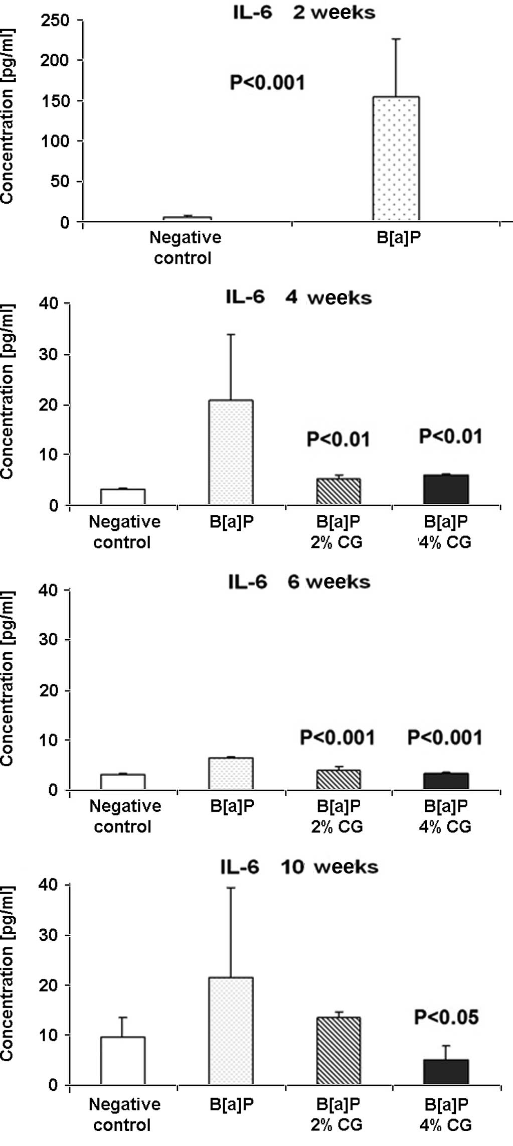
IL-6 levels increased 28-fold 2 weeks after the second dose of
B[a]P in comparison to the negative group (Fig. 1A). Four weeks after the second dose
of B[a]P, included in the diet of 2 and 4% CG, a decrease of
the IL-6 level by 74.3 and 70.6%, respectively, was observed
(Fig. 1B). Six weeks after the
second dose of B[a]P was administered the IL-6 level in the
serum was still lower compared to the B[a]P control. The 2
and 4% CG diet reduced the IL-6 level by 41.4 and 49.5%,
respectively (Fig. 1C). As shown in
Fig. 1D, 10 weeks after the second
dose of B[a]P the 2 and 4% CG diets reduced the IL-6 level
by 37.4 and 76.3%, respectively, compared to the B[a]P group
(Fig. 1D).
Tumor necrosis factor α
The pathways linking inflammation and promotion of
tumor growth are not well characterized, but it is well known that
TNFα is a significant mediator of inflammation. Despite the
apoptotic induction of pathological cells at the site of
inflammation, TNFα stimulates the growth of fibroblasts. However, 2
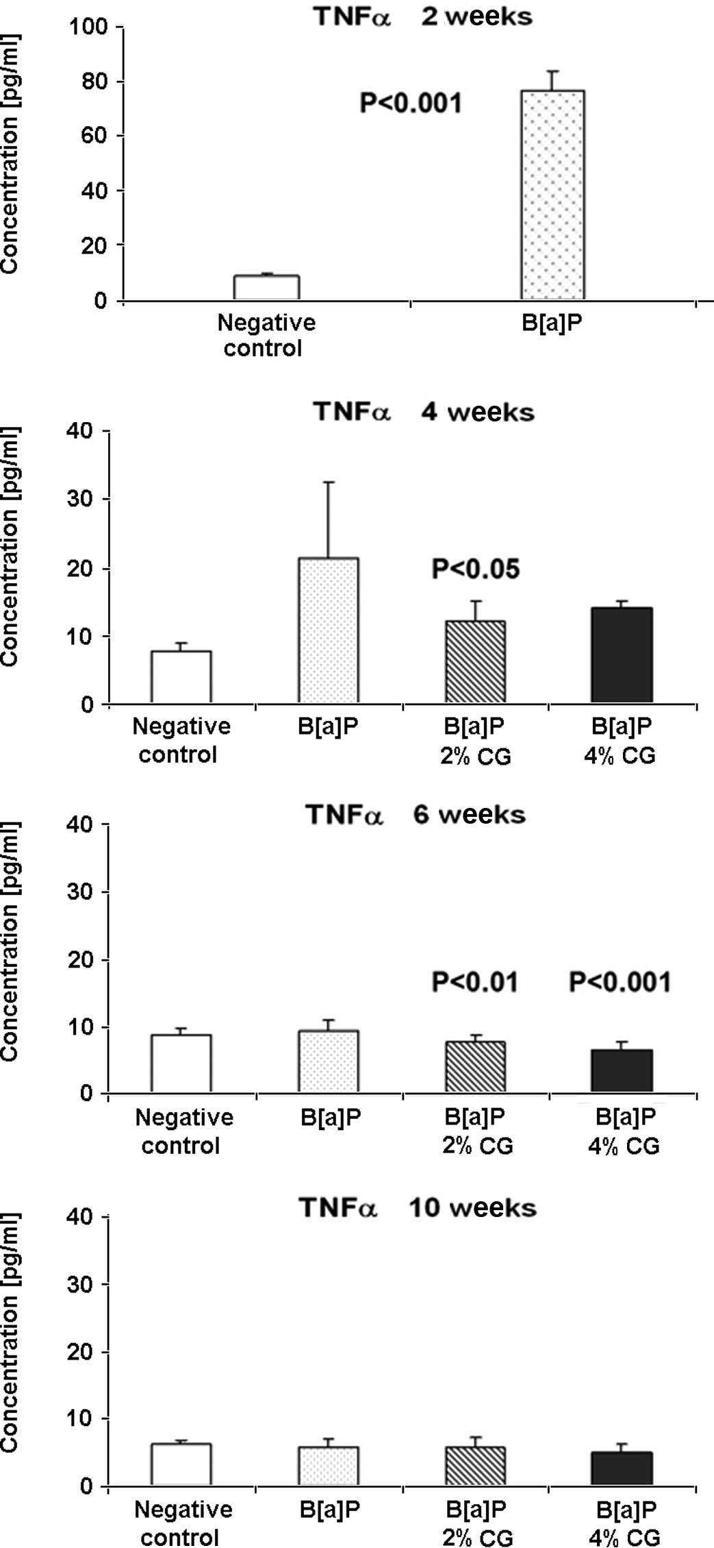
weeks after the second dose of B[a]P, the TNFα level
increased by ~8-fold, compared to the untreated control group
(Fig. 2A). Four weeks after the
last dose of B[a]P and following CG administration in the
diet for 2 weeks, both 2 and 4% CG reduced the TNFα level by 42.4
and 33.8%, respectively (Fig. 2B).
Six weeks following the second dose of B[a]P, the TNFα level
in the serum was reduced in the CG-treated groups. The 2% CG diet
decreased the TNFα level by 16.2% and the 4% CG diet by 28.8%, in
comparison to the B[a]P group (Fig. 2C). Ten weeks after the second dose
of B[a]P, the 4% CG diet reduced the TNFα level by 11.3%
compared to the B[a]P group. Almost no changes occurred in
the TNFα level in the case of the 2% CG diet (Fig. 2D).
Interleukin 12p70
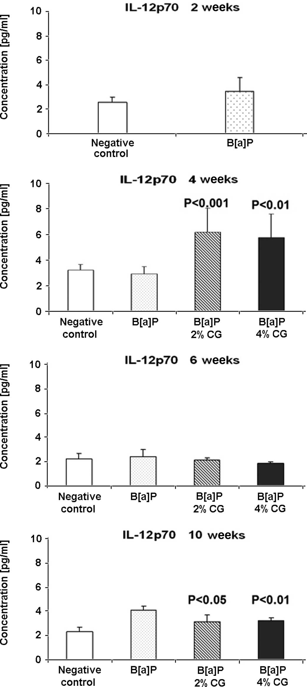
The level of IL-12p70 was evaluated. As shown in
Fig. 3A, the IL-12p70 level was
34.5% higher in the B[a]P group compared to the negative
control, 2 weeks after the second dose of B[a]P. Four weeks
after the second dose of B[a]P, an increase of the IL-12p70
level of 111.49% in the 2% CG diet and one of 97.9% in the 4% CG
diet was noted in mice fed with 2 and 4% CG (Fig. 3B). Six weeks following the last dose
of B[a]P, a decrease of the IL-12p70 level of 10.7 and 21.4%
for the 2 and 4% CG diets, respectively, was observed in the serum
(Fig. 3C). The IL-12p70 level was
reduced 10 weeks after the last treatment with B[a]P by 23.2
and 21.1%, respectively, for the 2 and 4% CG diets (Fig. 3D).
Interleukin-10
In contrast various proinflammatory cytokines, IL-10
indicates potent antitumor activities. IL-10 is synthetized in
monocytes and lymphocytes and is regarded as a key
anti-inflammatory immune-regulating cytokine. A 16.6% decrease of
the IL-10 level in the serum was observed 2 weeks after the second
dose of B[a]P compared to the untreated control group
(Fig. 4A). Four weeks after the
second dose of B[a]P and CG administration in the diet, the
2 and 4% CG diets increased the level of anti-inflammatory cytokine
IL-10, in a dose-related manner, by ~3-fold compared to the
B[a]P group. The IL-10 level was elevated by 249.4 and
288.5% for the 2 and 4% CG in the diet, respectively (Fig. 4B). However, six weeks after the
second dose of B[a]P, the IL-10 level decreased by 51.5 and
42.1% in the cases of the 2 and 4% CG diets, respectively (Fig. 4C). Notably, the IL-10 level was
below detection at 10 weeks after the second dose of
B[a]P.
Interferon-γ
IL-12 was investigated to elucidate its correlation
with IFN-γ, a cytokine that exhibits immunologic functions.
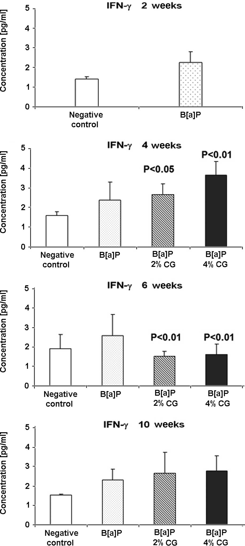
Fig. 5A shows that the IFN-γ level
increased by 58.4% 2 weeks after the last dose of B[a]P. CG
administration in the diet 4 weeks after B[a]P treatment
caused an increase of the IFN-γ level by 12.5 and 54% in the cases
of the 2 and 4% CG diets, respectively (Fig. 5B). However, the same CG
concentration 6 weeks after B[a]P treatment decreased the
IFN-γ level of 40.4 and 36.7%, respectively (Fig. 5C). The IFN-γ level in the serum 10
weeks after the last dose of B[a]P showed an increase of
15.6 and 20.6% in the case of the 2 and 4% CG diets, respectively,
compared to the B[a]P group (Fig. 5D).
Caspase-9 activation
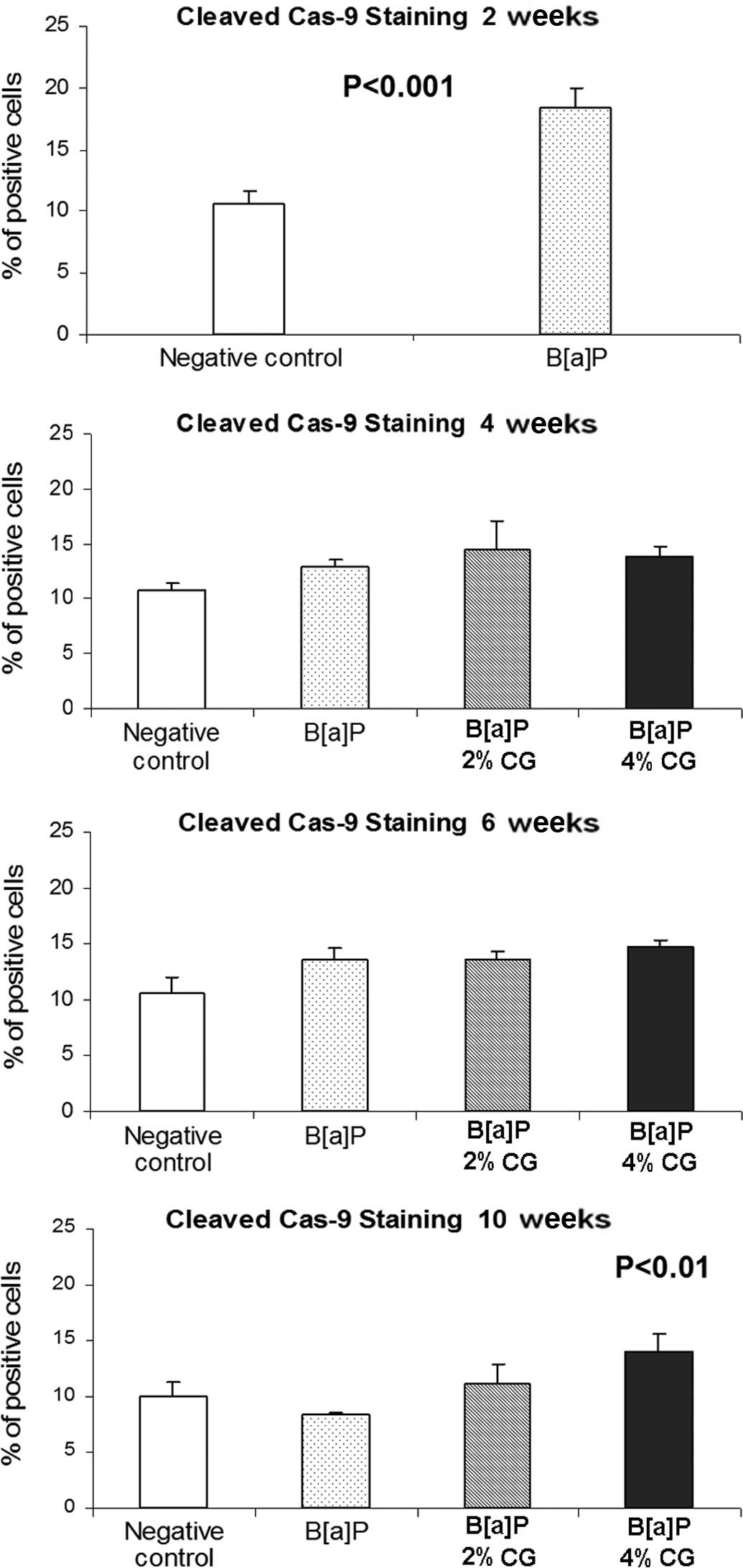
Apoptotic cells were detected by cleaved caspase-9
antibodies identifying the caspase 9 subunit with a molecular
weight of 37 kDa. The percentage of lung cells stained with these
antibodies 2 weeks after the second dose of B[a]P increased
by 73.3% as compared to the untreated control group (Fig. 6A). Feeding the mice for 4 weeks with
a diet containing 2 and 4% CG after the last dose of B[a]P
elevated the percentage of cells stained with the used antibody by
12 and 7.2%, respectively (Fig.
6B). Six weeks following the second dose of B[a]P, the
percentage of cells stained to yield the caspase-9 product
increased by 0.1 and 8.4%, respectively, in the 2 and 4% CG diets
(Fig. 6C). Notably, the highest
percentage of cells immunostained with anticleaved caspase-9
antibody, i.e., 32.3 and 67.5%, was observed 10 weeks after the
second dose of B[a]P after mice were fed with a diet
containing 2 and 4% CG, respectively (Fig. 6D).
Proliferation rate evaluation
The percentage of lung cells stained with anti-BrdU
antibody 2 weeks after the last dose of B[a]P was
significantly elevated, i.e., by 233% as compared to the untreated
control group (Fig. 7A). Results
showed that 4 weeks after the last dose of B[a]P uptake and
administration of 2 and 4% CG in the diet, the population of
stained cells reduced by 13.3 and 35.6%, respectively (Fig. 7B). As shown in Fig. 7C, the duration of mice CG feeding
gradually decreased BrdU staining of the studied cells in the
concentration of the glucaric acid derivative, by 21.2 and 49.2%,
respectively, in the case of the 2 and 4% CG diets. Notably, the
diet containing CG, 10 weeks after the second dose of B[a]P,
significantly reduced the level of the cells stained with anti-BrdU
antibody in the lung tissue by 45.2 and 67.2% in the cases of the 2
and 4% CG diets, respectively (Fig.
7D).
Discussion
The association of various inflammatory cell types
with cancer is not a recent development. Virchow reported dense
areas of inflammatory cells at the periphery of tumor as early as
1863 (49,52). Inflammatory response appears to
contribute to cancer growth and spread, as well as to immune
suppression and antitumor response, concomitantly with emerging
paradigms for the roles of matrix metalloproteinases (MMPs) in
cancer development. A number of independent studies using human
clinical samples show that inflammation has an impact on epithelial
cell turnover (53,54). More significantly, proliferation in
the setting of chronic inflammation predisposes humans to carcinoma
in the breast, large intestine, gastric mucosa and other tissues
(49). Evidence regarding the
importance of inflammation during neoplastic progression originates
from a study concerning cancer risk among long-term users of
aspirin and non-steroidal anti-inflammatory drugs (NSAIDs). A large
body of data indicates that the use of these drugs reduces colon
cancer risk by 40–50% and may be preventive for lung, esophagus and
stomach cancer (52,55). The mechanism(s) underlying the
chemopreventive effects of NSAIDs concerns their ability to inhibit
cyclooxygenases (COX-1 and COX-2). COX-2 converts arachidonic acid
to prostaglandins, which in turn induces inflammatory reactions in
damaged tissues (56). Thus,
inflammatory cells elicit host defense and stimulate progression.
Inflammation is a complex phenomenon, involving the summation of
initiation and maintenance signals originating from inflammatory
cells and the tissue environment (48,49).
The inflammatory response is divided into three
phases: acute, sub-acute and chronic. The acute phase is
characterized by rapid onset, blood vessel dilatation, edema and
leukocyte infiltration. This phase may last only a few days. The
sub-acute phase is characterized by further leukocyte and phagocyte
infiltration, which may continue for 3–4 weeks. Chronic
inflammation persists for longer periods of time and may result
from the persistence of the injuring agent in the tissue. During
the course of chronic inflammation, cytokines attract additional
activated lymphocytes as well as other inflammatory cells to the
site of insult, thereby amplifying and prolonging inflammation. The
inflammatory component of developing neoplasm includes a diverse
leukocyte population, i.e., macrophages, neutrophils, eosinophils
and mast cells. This diverse leukocyte population is intermittently
loaded with an assorted array of cytokines, cytotoxic mediators,
such as ROS, serine-, cysteine- and metallo-proteinase,
membrane-perforating agents and soluble mediators of cell
apoptosis, such as TNFα, ILs and IFNs (54,57,58).
PMNs are the most abundant circulating blood leukocytes. They
provide the first line of defense against infection and release
soluble chemotactic factors and proteases that alter the
microenvironment and guide the recruitment of non-specific and
specific immune effector cells (57).
Mast cells release inflammatory mediators and
factors known to enhance angiogenic phenotypes, including heparin,
heparanase, histamine, metallo- and serine proteinases, and various
polypeptide growth factors, such as tumor growth factor (TGF) and
vascular endothelial growth factor (VEGF) (59). Eosinophils, which possess numerous
bioactive molecules in their granules, are recruited to tissue as a
host defense against parasites or during allergic responses
(60), but are resident in the
mammary gland and contribute to morphogenesis (61). Macrophages produce a number of
potent angiogenic cytokines, growth factors, neutrophil
chemoattractants and proteases. Macrophage infiltration is closely
associated with the depth of invasion of primary melanoma due, in
part, to macrophage-regulated tumor-associated angiogenesis.
Macrophages express numerous bioactive molecules, including
proteases, arachidonate metabolites, TGF-α, TNFα and IL-1 (60–64).
In response to macrophage expression, melanocytes express IL-8 and
VEGF, thereby inducing angiogenesis through paracrine control
(63,65). Notably, macrophages and eosinophils
also contribute to mammary development (61). Macrophages are not unique among
inflammatory cells in the potentiation of neoplastic processes.
PMNs, mast cells and activated T-lymphocytes also contribute to
malignancies by releasing proteases, angiogenic factors and
chemokines (49). Previous data
indicate that mast cells and neutrophils potentiate the actions of
HPV16 oncogenes (59,65) and amplify neoplastic cell
proliferation and angiogenesis largely by the release of MMP-9.
Our previous studies in the mouse lung tumorigenesis
model showed that dietary CG inhibited B[a]P-induced mouse
lung tumorigenesis, in part, by inhibiting the enzyme βG (66), suppressing cell proliferation and
chronic inflammation, and by inducing apoptosis during the late
post-initiation stages of lung tumorigenesis in A/J mice (50). The present study aimed to
investigate the changes in the homeostasis of cytokines in the
serum, as well as the diversities of biomarkers of inflammation and
apoptosis in the lung tissue caused by dietary CG during the early
post-initiation stages of B[a]P-induced lung tumorigenesis.
Although 2 and 4% CG diets (w/w) exerted physiological changes in
lung tissues via the decreased level of B[a]P-induced TNFα,
only 4% CG in the diet significantly decreased the number of
adenomas. Changes likely to occur in the level of IL-6, IL-10 and
TNFα in the serum were investigated using FCAP array analysis. IL-6
was found to be a key mediator of the acute phase response. TNFα
belongs to the same group of stimulators of the acute phase
response, but also causes apoptosis. IL-12p70 is crucial in the
immune response to microorganisms and tumors, activating NK cells
and T-lymphocytes, which in turn initiates IFN-γ production and
antigen-specific Th1 responses (24,25).
Two weeks after the second dose of B[a]P, an
increase of the TNFα and IL-6 levels by approximately 8- and
28-fold, respectively, was observed in the serum along with an
approximately 17% decrease of the IL-10 level compared to the
untreated control group. An increase of the IFN-γ level of 58% and
of the IL-12p70 level of 34% was simultaneously observed in the
B[a]P-treated group, in comparison to the untreated
group.
Four weeks after the second dose of B[a]P and
CG administration in the diet for 2 weeks, the 4% CG diet reduced
the IL-6 and TNFα levels, but also increased the level of
anti-inflammatory cytokines IL-10, IFN-γ and IL-12p70, compared to
the B[a]P group. The levels of IL-6 and TNFα decreased by 70
and 34%, while that of IL-10 increased by approximately 3-fold. The
IFN-γ and IL-12p70 levels increased by 54 and 98%, respectively, in
the 4% CG group. At 6 weeks after the second dose of B[a]P,
the cytokine levels in the serum continued to be decreased in the
CG-treated groups. The 4% CG diet reduced the level of IL-6 by 49%,
TNFα by 29%, IFN-γ by 37%, IL-12p70 by 21% and the level of IL-10
was reduced by 42% compared to the B[a]P group. Ten weeks
after the last treatment with B[a]P, the 4% CG diet reduced
the IL-6, IL-12p70 and TNFα levels by 76, 21 and 11%, respectively,
compared to the B[a]P group. The IFN-γ level in the serum 10
weeks after the last dose of B[a]P showed an increase of 21%
in the case of 4% CG, compared to the B[a]P group.
The percentage of cells stained for cleaved
caspase-9 at 2 weeks after the second dose of B[a]P
increased by 73%, as compared to the untreated control group. At 4
weeks after the second dose of B[a]P, the 4% CG diet
increased the percentage of cells stained with anti-cleaved
caspase-9 antibody in the lung tissue by 7%, and at 6 weeks by 8%.
Ten weeks after the second dose of B[a]P, the 4% CG diet
increased the percentage of cells stained with anti-cleaved
caspase-9 antibody in the lung tissue by 67%.
Regulating cell proliferation is crucial in cancer
prevention, since this process plays a key role in carcinogenesis,
including the initiation and promotion steps. Chemopreventive
agents suppress carcinogen-induced hyper-proliferation of cells in
the target organs during the initiation, as well as the
post-initiation events. Therefore, effective agents usually
suppress cell proliferation and inhibit the occurrence of malignant
lesions (67,68). The percentage of lung cells stained
with anti-BrdU antibody 2 weeks after the last dose of B[a]P
increased by 233%, as compared to the untreated control group. The
results showed that 4 weeks after the last dose of B[a]P
uptake and administration of 4% CG in the diet the population of
stained cells was reduced by 35.6%. Moreover, 6 weeks after the
last dose of B[a]P, this concentration of CG diet gradually
decreased the BrdU-stained cells by 49.2%. The diet containing CG,
10 weeks after the second dose of B[a]P, significantly
decreased the level of cells stained with anti-BrdU antibody in the
lung tissue by 67.2%.
In conclusion, dietary D-glucarate reduces the level
of proinflammatory cytokines and increases the level of
anti-inflammatory cytokine IL-10 during the early post-initiation
stages of B[a]P-induced lung tumorigenesis in A/J mice.
Dietary D-glucarate exhibits proapoptotic effects, as evidenced by
the increased levels of cleaved caspase-9 in the lung tissue.
Moreover, CG may prevent lung cancer in tobacco
smokers and ex-smokers by enhancing apoptosis and suppressing the
acute inflammation and proliferation induced by tobacco-related
carcinogens. CG supplementation has the potential to reduce the
risk of lung cancer development in high-risk individuals.
Acknowledgements
This study was supported by the NIH grants
RO1CA102747, RO1CA079065, P30 CA 54174-16S1 and the American Cancer
Research Center and Foundation (ACRCF).
References
|
1
|
Ferlay J, Autire P, Boniol M, Heanue M,
Colombet M and Boyle P: Estimates of the cancer incidence and
mortality in Europe in 2006. Ann Oncol. 18:581–592. 2007.
View Article : Google Scholar : PubMed/NCBI
|
|
2
|
United States Cancer Statistics. Top Ten
Cancer. 2006
|
|
3
|
Landi MT, Consonni D, Rotunno M, et al:
Environment and Genetics in Lung cancer Etiology (EAGLE) study: an
integrative population-based case-control study of lung cancer. BMC
Public Health. 8:203–213. 2008. View Article : Google Scholar : PubMed/NCBI
|
|
4
|
Miura K, Bowman ED, Simon R, et al: Laser
capture microdissection and microarray expression analysis of lung
adenocarcinoma reveals tobacco smoking- and prognosis-related
molecular profiles. Cancer Res. 62:3244–3250. 2002.
|
|
5
|
Devesa SS, Bray F, Vizcaino AP and Parkin
DM: International lung cancer trends by histologic type:
male:female differences diminishing and adeno-carcinoma rates
rising. Int J Cancer. 117:294–299. 2005. View Article : Google Scholar : PubMed/NCBI
|
|
6
|
Mountain CF: Revisions in the
international system for staging lung cancer. Chest. 111:1710–1717.
1997. View Article : Google Scholar : PubMed/NCBI
|
|
7
|
Ramaswamy S, Ross KN, Lander ES and Golub
TR: A molecular signature of metastasis in primary solid tumors.
Nat Genet. 33:49–54. 2003. View
Article : Google Scholar : PubMed/NCBI
|
|
8
|
Garber ME, Troyanskaya OG, Schluens K, et
al: Diversity of gene expression in adenocarcinoma of the lung.
Proc Natl Acad Sci USA. 98:13784–13789. 2001. View Article : Google Scholar : PubMed/NCBI
|
|
9
|
Bhattacharjee A, Richards WG, Staunton J,
et al: Classification of human lung carcinomas by mRNA expression
profiling reveals distinct adeno-carcinoma subclasses. Proc Natl
Acad Sci USA. 98:13790–13795. 2001. View Article : Google Scholar : PubMed/NCBI
|
|
10
|
Beer DG, Kardia SL, Huang CC, et al:
Gene-expression profiles predict survival of patients with lung
adenocarcinoma. Nat Med. 8:816–824. 2002.PubMed/NCBI
|
|
11
|
Endoh H, Tomida S, Yatabe Y, et al:
Prognostic model of pulmonary adenocarcinoma by expression
profiling of eight genes as determined by quantitative real-time
reverse transcriptase polymerase chain reaction. J Clin Oncol.
22:811–819. 2004. View Article : Google Scholar
|
|
12
|
Xi L, Lyons-Weiler J, Coello MC, et al:
Prediction of lymph node metastasis by analysis of gene expression
profiles in primary lung adenocarcinomas. Clin Cancer Res.
11:4128–4135. 2005. View Article : Google Scholar : PubMed/NCBI
|
|
13
|
Meyerson M and Carbone D: Genomic and
proteomic profiling of lung cancers: lung cancer classification in
the age of targeted therapy. J Clin Oncol. 23:3219–3226. 2005.
View Article : Google Scholar : PubMed/NCBI
|
|
14
|
He P, Varticovski L, Bowman ED, et al:
Identification of carboxypeptidase E and gamma-glutamyl hydrolase
as biomarkers for pulmonary neuro-endocrine tumors by cDNA
microarray. Hum Pathol. 35:1169–1209. 2004.PubMed/NCBI
|
|
15
|
Yanaihara N, Caplen N, Bowman E, et al:
Unique microRNA molecular profiles in lung cancer diagnosis and
prognosis. Cancer Cell. 9:189–198. 2006. View Article : Google Scholar : PubMed/NCBI
|
|
16
|
Potti A, Mukherjee S, Petersen R, et al: A
genomic strategy to refine prognosis in early-stage non-small-cell
lung cancer. New England J Med. 355:570–580. 2006. View Article : Google Scholar
|
|
17
|
Reddy SP: The antioxidant response element
and oxidative stress modifiers in airway diseases. Curr Mol Med.
8:376–383. 2008. View Article : Google Scholar : PubMed/NCBI
|
|
18
|
Walser T, Cui X, Yanagawa J, Lee JM,
Heinrich E, Lee G, Sharma S and Dubinett SM: Smoking and lung
cancer: the role of inflammation. Proc Am Thorac Soc. 5:811–815.
2008. View Article : Google Scholar : PubMed/NCBI
|
|
19
|
Giri SN and Hollinger MA: Effect of
cadmium on lung lysosomal enzymes in vitro. Arch Toxicol.
69:341–345. 1995. View Article : Google Scholar : PubMed/NCBI
|
|
20
|
Shimoi K, Saka N, Nozawa R, et al:
Deglucuronidation of a flavonoid, luteolin monglucuronide, during
inflammation. Drug Metab Disp. 29:1521–1524. 2001.PubMed/NCBI
|
|
21
|
Marshall T, Shult P and Busse WW: Release
of lysosomal enzyme β-glucuronidase from isolated human
eosinophils. J Allergy Clin Immunol. 82:550–555. 1988.
|
|
22
|
Cobben NA, Drent M, De Vries J, Wouters
EF, van Dieijen-Visser MP and Henderson RF: Serum β-glucuronidase
activity in a population of ex-coalminers. Clin Biochem.
32:659–664. 1999.
|
|
23
|
DiMatteo M, Antonini JM, van Dyke K and
Reasor MJ: Characteristics of the acute phase pulmonary response to
silica in rats. J Toxicol Environ Health. 47:93–108. 1996.
View Article : Google Scholar : PubMed/NCBI
|
|
24
|
DiMatteo M and Reasor MJ: Modulation of
silica-induced pulmonary toxicity by dexamethasone-containing
liposomes. Toxicol Appl Pharmacol. 142:411–421. 1997. View Article : Google Scholar : PubMed/NCBI
|
|
25
|
Hirano S, Shimada T, Osugi J, Kodama N and
Suzuki KT: Pulmonary clearance and inflammatory potence
intratracheally instilled or acutely inhaled nickel sulfate in
rats. Arch Toxicol. 68:548–554. 1994. View Article : Google Scholar : PubMed/NCBI
|
|
26
|
Matulionis D and Traurig HH: In situ
response of lung macrophages and hydrolase activities to cigarette
smoke. Lab Invest. 37:314–326. 1977.PubMed/NCBI
|
|
27
|
Pappas P, Sotiropoulou M, Karamanakos P,
et al: Acute-phase response to benzo[a]pyrene and induction of rat
ALDH3A1. Chem Bil Interac. 144:55–62. 2003.
|
|
28
|
Kong LY, Luster MI, Dixon D, O'Grady J and
Rosenthal GJ: Inhibition of lung immunity after tracheal
instillation of benzo[a]pyrene. Am J Resp Cit Care Med.
150:1123–1129. 1994.PubMed/NCBI
|
|
29
|
Levy G and Conchi J: β-Glucuronidase and
the hydrolysis of glucuronides. Glucuronic Acid: Free and Combined.
Academic Press; New York: pp. 301–364. 1966
|
|
30
|
Marsh CA: Metabolism of D-glucarolactone
in mammalian systems. Identification of D-glucaric acid as a normal
constituent of urine. Biochem J. 86:77–86. 1963.PubMed/NCBI
|
|
31
|
Matsui M, Fukuo A, Watanabe Y, Wanibe T
and Okada M: Studies on the glucaric acid pathway in the metabolism
of D-glucuronic acid in mammals. IV. Fluorometric method for the
determination of D-glucaric acid in serum. Chem Phar Bull.
20:845–848. 1972. View Article : Google Scholar : PubMed/NCBI
|
|
32
|
Blumenthal HJ, Lucuta VL and Blumenthal
DC: Specific enzymic assay for D-glucarate in human serum. Anal
Biochem. 185:286–293. 1990. View Article : Google Scholar : PubMed/NCBI
|
|
33
|
Colombi A, Maroni M, Antonini C, Fait A,
Zocchetti C and Foa V: Influence of sex, age and smoking habits on
the urinary excretion of D-glucaric acid. Clin Chim Acta.
128:349–358. 1983. View Article : Google Scholar : PubMed/NCBI
|
|
34
|
Mocarelli P, Brambilla P, Colombo L, et
al: A new method for D-glucaric acid excretion measurement that is
suitable for automated instruments. Clin Chem. 34:2238–2290.
1988.PubMed/NCBI
|
|
35
|
Batt HM and Siest G: Laboratory tests as
indirect indications of the activity of drug metabolizing enzymes
use of glucaric acid and gamma-glutamyl-transpeptidase. Dev Clin
Biochem. 2:178–192. 1980.
|
|
36
|
Yokoyama M, Matsuoka S and Wakui A:
Activation of tegafur and urinary excretion of D-glucaric acid in
tumor-bearing hosts. In: Recent Adv Chemother, Proc Int Congr
Chemother 14th. Univ Tokyo Press; Tokyo: pp. 113–115. 1985
|
|
37
|
Dohrmann RE: β-Glucuronidase.
Springer-Verlag; Berlin: 1969
|
|
38
|
Masaki H: Metabolic pathway of dermal
D-glucaric acid synthesis from D-glucuronolactone. D-Glucaric acid
and D-glucuronolactone dehydrogenase in human skin. β-Gucuronidase
feedback mechanism. Nippon Hifuka Gakkai Zashi. 82:151–158
Chem Abstr. 78:2709b–27062u. 1972.
|
|
39
|
Dutton GJ: Glucuronidation of Drugs and
Other Compounds. CRC Press; Boca Raton, FL: 1980
|
|
40
|
Clark AG, Fischer FJ, Milburn P, Smith RL
and Williams RL: The role of gut flora in the enterohepatic
circulation of stilboestrol in the rat. Biochem J. 112:17–18.
1988.PubMed/NCBI
|
|
41
|
Walaszek Z: Potential use of D-glucaric
acid derivatives in cancer prevention. Cancer Lett. 54:1–8. 1990.
View Article : Google Scholar : PubMed/NCBI
|
|
42
|
Walaszek Z: Chemopreventive properties of
D-glucaric acid derivatives. Cancer Bull. 45:453–457. 1993.
|
|
43
|
Yoshimi N, Walaszek Z, Mori H, Hanausek M,
Szemraj J and Slaga TJ: Inhibition of azoxymethane-induced rat
colon carcinogenesis by potassium hydrogen D-glucarate. Int J
Oncol. 16:43–48. 2000.PubMed/NCBI
|
|
44
|
Walaszek Z, Szemraj J, Hanausek M, Adams
AK and Sherman U: D-Glucaric acid content of various fruits and
vegetables and cholesterol lowering effects of dietary D-glucarate
in the rat. Nutr Res. 16:673–682. 1996. View Article : Google Scholar
|
|
45
|
Zoltaszek R, Hanausek M, Kiliańska ZM and
Walaszek Z: The biological role of D-glucaric acid and its
derivatives potential use in medicine. Adv Hyg Exptl Med.
62:451–462. 2008.PubMed/NCBI
|
|
46
|
Kishimoto T: Interleukin-6: from basic
science to medicine – 40 years in immunology. Annu Rev Immunol.
23:1–21. 2005.
|
|
47
|
Wajant H, Pfizenmaier K and Scheurich P:
Tumor necrosis factor signaling. Cell Death Differ. 10:45–65. 2003.
View Article : Google Scholar : PubMed/NCBI
|
|
48
|
Coussens LM and Werb Z: Inflammatory cells
and cancer: think different! J Exp Med. 193:F23–F26. 2001.
|
|
49
|
Coussens LM and Werb Z: Inflammation and
cancer. Nature. 420:860–867. 2001. View Article : Google Scholar
|
|
50
|
Zoltaszek R, Hanausek M, Slaga TJ and
Walaszek Z: Proapoptotic effects of D-glucarate on chemically
induced lung tumorigenesis in A/J mice. Proc Am Assoc Cancer Res.
48:7952007.
|
|
51
|
Zoltaszek R, Grzelak M, Hanausek M,
Kilianska Z, Slaga TJ and Walaszek Z: Effects of dietary
D-glucarate on biomarkers of inflammation during early
post-initiation stages of benzo[a]pyrene (B[a]P)-induced lung
tumorigenesis in A/J mice. Proc Am Assoc Cancer Res.
49:5322008.PubMed/NCBI
|
|
52
|
Rodriguez-Manzaneque JC, Lane TF, Ortega
MA, Hynes RO, Lawler J and Iruela-Arispe M: Thrombospondin-1
suppresses spontaneous tumor growth and inhibits activation of
matrix metalloproteinase-9 and mobilization of vascular endothelial
growth factor. PNAS USA. 98:12485–12490. 2001. View Article : Google Scholar
|
|
53
|
De Marzo AM, Marchi VL, Epstein JI and
Nelson WG: Proliferative inflammatory atrophy of the prostate:
implications for prostatic carcinogenesis. Am J Pathol.
155:1985–1992. 1999.PubMed/NCBI
|
|
54
|
Kuper H, Adami HO and Trichopoulos D:
Infections as a major preventable cause of human cancer. J Intern
Med. 248:171–173. 2000. View Article : Google Scholar : PubMed/NCBI
|
|
55
|
Baron JA and Sandler RS: Nonsteroidal
anti-inflammatory drugs and cancer prevention. Annu Rev Med.
51:511–523. 2000. View Article : Google Scholar : PubMed/NCBI
|
|
56
|
Williams CS, Mann M and DuBois RN: The
role of cyclooxygenases in inflammation, cancer, and development.
Oncogene. 18:7908–7916. 1999. View Article : Google Scholar : PubMed/NCBI
|
|
57
|
Di Carlo E, Forni G, Lollini P, Colombo
MP, Modesti A and Musiani P: The intriguing role of
polymorphonuclear neutrophils in anti-tumor reactions. Blood.
97:339–345. 2001.
|
|
58
|
Wahl LM and Kleinman HK: Tumor-associated
macrophages as targets for cancer therapy. J Natl Cancer Inst.
90:1583–1584. 1998. View Article : Google Scholar : PubMed/NCBI
|
|
59
|
Coussens LM, Raymond WW, Bergers G, et al:
Inflammatory mast cells up-regulate angiogenesis during squamous
epithelial carcinogenesis. Genes Dev. 13:1382–1397. 1999.
View Article : Google Scholar : PubMed/NCBI
|
|
60
|
Rothenberg ME: Eosinophilia. N Eng J Med.
338:1592–1600. 1998. View Article : Google Scholar : PubMed/NCBI
|
|
61
|
Gouon-Evans V, Rothenberg ME and Pollard
JW: Postnatal mammary gland development requires macrophages and
eosinophils. Development. 127:2269–2282. 2000.PubMed/NCBI
|
|
62
|
Lin EY, Nguyen AV, Russel RG and Pollard
JW: Colony-stimulating factor 1 promotes progression of mammary
tumors to malignancy. J Exp Med. 193:727–740. 2001. View Article : Google Scholar : PubMed/NCBI
|
|
63
|
Ono M, Torisu H, Fukushi J, Nishie A and
Kuwano M: Biological implications of macrophage infiltration in
human angiogenesis. Cancer Chemother Pharmacol. 43:S69–S71. 1999.
View Article : Google Scholar : PubMed/NCBI
|
|
64
|
Torisu H, Ono M, Kiryu H, Furue M, Ohmoto
Y, Nakayama J, Nishioka Y, Sone S and Kuwano M: Macrophage
infiltration correlates with tumor stage and angiogenesis in human
malignant melanoma: possible involvement of TNFalpha and IL-1alpha.
Int J Cancer. 85:182–188. 2000. View Article : Google Scholar
|
|
65
|
Coussens LM, Tinkel CL, Hanahan D and Werb
Z: MMP-9 supplied by bone marrow-derived cells contributes to skin
carcinogenesis. Cell. 103:481–490. 2000. View Article : Google Scholar : PubMed/NCBI
|
|
66
|
Walaszek Z, Hanausek-Walaszek M and Webb
TE: Dietary glucarate-mediated reduction of sensitivity of murine
strains to chemical carcinogenesis. Cancer Lett. 33:25–32. 1986.
View Article : Google Scholar : PubMed/NCBI
|
|
67
|
Gysin R, Azzi A and Visarius T:
Gamma-tocopherol inhibits human cancer cell cycle progression and
cell proliferation by down-regulation of cyclins. FASEB J.
16:1952–1954. 2002.PubMed/NCBI
|
|
68
|
Boivin D, Blanchette M, Barrette S,
Moghrabi A and Béliveau R: Inhibition of cancer cell proliferation
and suppression of TNF-induced activation of NFkappaB by edible
berry juice. Anticancer Res. 27:937–948. 2007.PubMed/NCBI
|


