A case of primary lung cancer lesion demonstrated by F-18 FDG positron emission tomography/computed tomography (PET/CT) one year after the detection of metastatic brain tumor

  • Authors:
    • Yuichi Ozeki
    • Yoshiyuki Abe
    • Hideyuki Kita
    • Katsumi Tamura
    • Ikuko Sakata
    • Jiro Ishida
    • Kikuo Machida
  • View Affiliations

  • Published online on: May 16, 2011     https://doi.org/10.3892/ol.2011.318
  • Pages: 621-623
Metrics: Total Views: 0 (Spandidos Publications: | PMC Statistics: )
Total PDF Downloads: 0 (Spandidos Publications: | PMC Statistics: )


Abstract

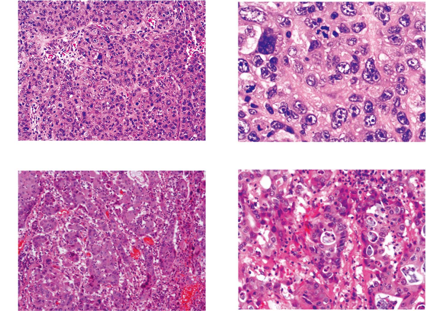
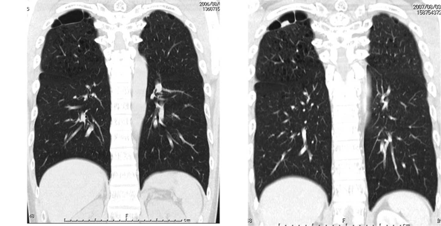
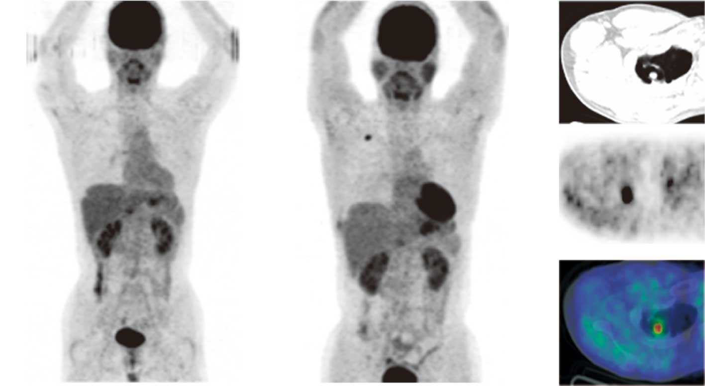
Cancer of unknown primary origin (CUP) is an aggressive disease with a poor prognosis. Metastatic brain tumors occur in approximately 15% of all cancer patients. F-18 2'-deoxy-2fluoro-D-glucose (FDG) positron emission tomography (PET) combined with computed tomography (PET/CT) contributes to the evaluation of cancer staging, although the benefits of PET/CT for detection of CUP origins has yet to be determined. In this study, we present a 37‑year‑old man with a brain tumor detected by magnetic resonance imaging. Surgical biopsy indicated a metastatic undifferentiated carcinoma, while clinical examination and a CT scan did not detect any abnormalities, with the exception of brain metastases. PET/CT did not reveal abnormal FDG uptake. PET/CT revealed abnormal intense FDG uptake in a small nodular lesion in the right lung 1 year following the detection of brain metastasis, and no other abnormal FDG uptake was observed elsewhere in the body. Right upper lobectomy and dissection of mediastinal lymph nodes were performed. The pathological diagnosis was poorly differentiated adenocarcinoma, which was similar to the brain metastatic lesion, and there was no lymph node metastasis. This case revealed an extremely rare lung cancer with primary lesions demonstrated by PET/CT 1 year after the detection of brain metastasis. This case reveals that F-18 FDG PET/CT imaging of CUP origin is capable of positively impacting on the identification of small primary tumor foci.
View Figures
View References

Related Articles

Journal Cover

July-August 2011
Volume 2 Issue 4

Print ISSN: 1792-1074
Online ISSN:1792-1082

Sign up for eToc alerts

Recommend to Library

Copy and paste a formatted citation
x
Spandidos Publications style
Ozeki Y, Abe Y, Kita H, Tamura K, Sakata I, Ishida J and Machida K: A case of primary lung cancer lesion demonstrated by F-18 FDG positron emission tomography/computed tomography (PET/CT) one year after the detection of metastatic brain tumor. Oncol Lett 2: 621-623, 2011
APA
Ozeki, Y., Abe, Y., Kita, H., Tamura, K., Sakata, I., Ishida, J., & Machida, K. (2011). A case of primary lung cancer lesion demonstrated by F-18 FDG positron emission tomography/computed tomography (PET/CT) one year after the detection of metastatic brain tumor. Oncology Letters, 2, 621-623. https://doi.org/10.3892/ol.2011.318
MLA
Ozeki, Y., Abe, Y., Kita, H., Tamura, K., Sakata, I., Ishida, J., Machida, K."A case of primary lung cancer lesion demonstrated by F-18 FDG positron emission tomography/computed tomography (PET/CT) one year after the detection of metastatic brain tumor". Oncology Letters 2.4 (2011): 621-623.
Chicago
Ozeki, Y., Abe, Y., Kita, H., Tamura, K., Sakata, I., Ishida, J., Machida, K."A case of primary lung cancer lesion demonstrated by F-18 FDG positron emission tomography/computed tomography (PET/CT) one year after the detection of metastatic brain tumor". Oncology Letters 2, no. 4 (2011): 621-623. https://doi.org/10.3892/ol.2011.318