Introduction
The incidence and mortality of upper aerodigestive
tract carcinomas remain persistently high regardless of diagnostic
and therapeutic improvements (1).
Exogenous risk factors, such as tobacco smoke and alcohol
consumption, are acknowledged causes of head and neck cancer
(2). However, only a fraction of
smokers are likely to develop head and neck cancer during their
lifetime. Therefore, the importance of endogenous risk factors is
of growing interest in the multifactorial genesis of head and neck
squamous cell cancer (HNSCC). Individual mutagen sensitivity and
DNA repair capacity are likely to be candidates affecting an
individual’s susceptibility to cancer (3).
Tobacco-associated nitrosamines, such as
N-nitrosonor-nicotine (NNN),
4-(methylnitrosamino)-1-(3-pyridyl)-1-butanone (NNK) and
N-nitrosodiethylamine (NDEA) are known to be involved in the
carcinogenesis of HNSCC. NNN is classified as a class 1 carcinogen
by the IARC (4). This nicotine
derivative is found in tobacco smoke, with 200–3000 ng in
cigarettes without filters (5). NNK
is classified as class 1 by the IARC as well, although it was found
to be a stronger carcinogen in animal testing (4,6). NDEA
is a class 2b carcinogen, and its effect is local and systemic,
with 20–70 ng in the smoke of cigarettes without filters (5,7). The
nitrosamines are metabolized in the cells by cytochrome
P-450-dependent hydroxylases into an alkylating agent, which may
induce DNA strand breaks and cause incomplete base excision repair.
Furthermore, NNN and NNK have genotoxic effects through the
formation of oxygen radicals (8).
As in most tumors, the regulatory steps are altered
during multi-step tumor development (9). Regular cell survival is dependent on a
well-balanced relationship between ongoing DNA damage and repair
(10,11). The importance of DNA repair capacity
(DRC) on cancer susceptibility was previously described in lung
cancer patients (12). The ability
to detect and repair DNA damage caused by environmental toxicants
or endogenous processes is assumed to be a significant defense
mechanism against the development of head and neck cancer.
Most studies on DRC are based on lymphocytes as the
surrogate cell type (13,14). However, environmental carcinogens
primarily damage the mucosa of the upper aerodigestive tract, with
epithelial cells being the first target for head and neck
carcinogenesis. Due to previous findings showing poor correlation
between genotoxic sensitivity of peripheral blood lymphocytes and
upper aerodigestive tract epithelia, we included the two cell types
in our investigations to further differentiate systemic and local
effects (15).
The aim of the study was to evaluate mutagen
sensitivity and DRC in human lymphocytes and epithelial cells of
the oropharynx of patients with and without squamous cell carcinoma
(SCC) of the oropharynx. DNA fragmentation following
nitrosamine-induced damage, as well as DNA repair capacities in 15
and 30 min, were measured using the alkaline single cell microgel
electrophoresis (comet) assay. DRC following nitrosamine-induced
DNA damage was presented within matched patient groups using
parameters such as age, gender, tobacco and alcohol
consumption.
Materials and methods
Biopsies and blood samples
Macroscopically healthy epithelial biopsies were
obtained from oropharyngeal resection specimens of SCC patients and
from surgery of the oropharynx in patients without SCC. For the
evaluation of DNA damage and repair using nitrosamines, SCC
patients (n=50, all male, mean age 51.3 years; range 35–62) were
matched to control patients (n=50, all male, average age 50.4
years; range 35–62). Only mucosa that had to be resected for
surgical reasons were used to avoid additional stress for the
patients. Subjects were informed about the experiments and had
signed a written consent statement. Tumor patients were first-time
cancer patients and did not receive any prior treatment, including
radiotherapy or chemotherapy. Control patients underwent
tonsillectomy based on the diagnosis of tonsillitis or obstructive
sleep apnea syndrome (OSAS). Lymphocytes were isolated by
centrifugation of 20 ml heparinized peripheral blood, which was
drawn prior to treatment. Patients included in the study were
matched by gender, age, alcohol and tobacco consumption. All of the
subjects were consumers of alcohol and tobacco smoke. Patient data
are shown in Tables I (controls)
and II (patients with SCC). The
study was approved by the Ethics Commission of the Medical
Department, Ludwig-Maximilians-University Munich (No. 221/04).
 | Table ICharacteristics of male control
patients. |
Table I
Characteristics of male control
patients.
| No. | Age | BMI | Diagnosis | Cig/d | Py | Alcohol /d (g) | Occupational
hazards |
|---|
| 1 | 50 | 27 | Acute
tonsillitis | 10 | 30 | 100 | |
| 2 | 48 | 22 | Sleep apnea | 30 | 23 | 64 | |
| 3 | 40 | 24 | Chronic
tonsillitis | 15 | 20 | 10 | |
| 4 | 47 | 27 | Acute
tonsillitis | 15 | 15 | 25 | |
| 5 | 50 | 22 | Chronic
tonsillitis | 20 | 20 | 20 | |
| 6 | 42 | 29 | Sleep apnea | 40 | 50 | 10 | |
| 7 | 46 | 28 | Sleep apnea | 50 | 36 | 125 | Paints, lubricants,
varnish |
| 8 | 54 | 22 | Chronic
tonsillitis | 25 | 38 | 25 | Metal dust |
| 9 | 40 | 25 | Chronic
tonsillitis | 5 | 3 | 25 | |
| 10 | 42 | 27 | Chronic
tonsillitis | 20 | 25 | 72 | Chemicals |
| 11 | 52 | 30 | Chronic
tonsillitis | 20 | 10 | 175 | |
| 12 | 34 | 22 | Chronic
tonsillitis | 2 | 1 | 50 | Paints |
| 13 | 56 | 22 | Chronic
tonsillitis | 10 | 10 | 20 | |
| 14 | 40 | 22 | Chronic
tonsillitis | 5 | 2 | 37 | |
| 15 | 54 | 30 | Chronic
tonsillitis | 20 | 80 | 10 | Wood dust |
| 16 | 42 | 28 | Chronic
tonsillitis | 60 | 50 | 20 | |
| 17 | 54 | 21 | Chronic
tonsillitis | 20 | 20 | 15 | |
| 18 | 51 | 33 | Chronic
tonsillitis | 15 | 16 | 45 | Paints |
| 19 | 48 | 31 | Chronic
tonsillitis | 10 | 24 | 10 | |
| 20 | 54 | 27 | Sleep apnea | 20 | 40 | 125 | Metal dust |
| 21 | 50 | 27 | Sleep apnea | 40 | 20 | 290 | |
| 22 | 45 | 28 | Sleep apnea | 30 | 15 | 25 | |
| 23 | 49 | 29 | Sleep apnea | 5 | 8 | 20 | |
| 24 | 43 | 25 | Sleep apnea | 12 | 15 | 40 | |
| 25 | 53 | 19 | Chronic
tonsillitis | 10 | 23 | 30 | |
| 26 | 57 | 28 | Sleep apnea | 5 | 15 | 50 | |
| 27 | 52 | 22 | Acute
tonsillitis | 12 | 15 | 30 | |
| 28 | 54 | 20 | Acute
tonsillitis | 20 | 34 | 175 | |
| 29 | 53 | 26 | Chronic
tonsillitis | 5 | 7 | 60 | Wood dust,
glue |
| 30 | 46 | 25 | Chronic
tonsillitis | 12 | 20 | 65 | |
| 31 | 36 | 25 | Acute
tonsillitis | 30 | 21 | 4 | Solvents |
| 32 | 51 | 24 | Chronic
tonsillitis | 10 | 15 | 15 | |
| 33 | 44 | 31 | Sleep apnea | 20 | 13 | 30 | |
| 34 | 44 | 28 | Sleep apnea | 15 | 18 | 40 | |
| 35 | 50 | 31 | Acute
tonsillitis | 10 | 12 | 25 | |
| 36 | 51 | 24 | Sleep apnea | 20 | 35 | 12,5 | |
| 37 | 57 | 28 | Chronic
tonsillitis | 2 | 3 | 69 | Chalk dust |
| 38 | 53 | 27 | Chronic
tonsillitis | 20 | 18 | 120 | |
| 39 | 61 | 27 | Sleep apnea | 30 | 40 | 30 | |
| 40 | 45 | 27 | Sleep apnea | 20 | 5 | 25 | |
| 41 | 47 | 32 | Sleep apnea | 25 | 26 | 50 | |
| 42 | 43 | 23 | Chronic
tonsillitis | 20 | 18 | 10 | |
| 43 | 61 | 33 | Papilloma | 60 | 80 | 100 | Chemicals |
| 44 | 63 | 28 | Chronic
tonsillitis | 25 | 50 | 45 | |
| 45 | 63 | 27 | Chronic
tonsillitis | 40 | 60 | 50 | Wood dust |
| 46 | 61 | 27 | Chronic
tonsillitis | 25 | 53 | 250 | |
| 47 | 61 | 31 | Sleep apnea | 50 | 27 | 66 | Paints |
| 48 | 60 | 26 | Sleep apnea | 20 | 40 | 40 | |
| 49 | 61 | 28 | Sleep apnea | 30 | 25 | 50 | |
| 50 | 62 | 30 | Chronic
tonsillitis | 15 | 30 | 25 | |
| Table IICharacteristics of male SCC
patients. |
Table II
Characteristics of male SCC
patients.
| No. | Age | BMI | Diagnosis | TNM | Grading | Cig/d | py | Alcohol/d (g) | Occupational
hazards |
|---|
| 1 | 50 | 25 | Oropharynx
carcinoma | pT4 pN3 cM0 | G3 | 50 | 75 | 240 | |
| 2 | 50 | 32 | Oropharynx
carcinoma | pT2 pN3 cM0 | G2 | 50 | 120 | 900 | |
| 3 | 40 | 22 | Oropharynx
carcinoma | pT3 pN2a cMx | G3 | 20 | 13 | 175 | |
| 4 | 47 | 24 | Oropharynx
carcinoma | pT2 pN2b cM0 | G2 | 25 | 40 | 100 | |
| 5 | 53 | 24 | Oropharynx
carcinoma | pT2 pT1 cM0 | G3 | 30 | 50 | 125 | Kerosene |
| 6 | 46 | 28 | Oropharynx
carcinoma | pT1 pN1 cM0 | G3 | 40 | 45 | 50 | Wood dust |
| 7 | 48 | 23 | Uvula
carcinoma | pT3 pN2b cM0 | G3 | 20 | 35 | 100 | |
| 8 | 57 | 25 | Oropharynx
carcinoma | pT4 pN0 cM0 | G3 | 35 | 20 | 205 |
Polyvinylchloride |
| 9 | 44 | 19 | Base of tongue
carcinoma | cT4 cN0 cM0 | G1 | 100 | 125 | 200 | |
| 10 | 47 | 23 | Oropharynx
carcinoma | pT3 pN1 cM0 | G3 | 20 | 30 | 150 | |
| 11 | 48 | 31 | Tonsillar
carcinoma | pT2 pN3 cM0 | G2 | 30 | 48 | 300 | Bitumen |
| 12 | 35 | 22 | Oropharynx
carcinoma | cT4 cN2a cMx | G1 | 50 | 40 | 250 | |
| 13 | 56 | 19 | Oropharynx
carcinoma | pT3 cN2c cM0 | G2 | 60 | 60 | 220 | Metal dust |
| 14 | 44 | 20 | Oropharynx
carcinoma | pT4 cN2a cM0 | G3 | 10 | 5 | 10 | |
| 15 | 56 | 27 | Oropharynx
carcinoma | pT4 cN2c cM0 | G3 | 30 | 25 | 100 | |
| 16 | 47 | 19 | Base of tongue
carcinoma | pT4 pN1 cM0 | G3 | 20 | 20 | 900 | |
| 17 | 56 | 23 | Tonsillar
carcinoma | pT2 pN3 cM0 | G3 | 20 | 80 | 60 |
Polyvinylchloride |
| 18 | 54 | 19 | Oropharynx
carcinoma | pT3 pN2b cM0 | G2 | 12 | 20 | 160 | Chemicals |
| 19 | 50 | 27 | Base of tongue
carcinoma | pT1 pN1 cM0 | G1 | 20 | 34 | 123 | |
| 20 | 54 | 23 | Tonsillar
carcinoma | pT2 pN2b cM0 | G2 | 30 | 55 | 200 | Oils |
| 21 | 52 | 24 | Tonsillar
carcinoma | pt1 pN2b cM0 | G3 | 30 | 40 | 144 | |
| 22 | 46 | 22 | Vallecula
carcinoma | pT2 pN0 cM0 | G3 | 20 | 63 | 375 | Paints |
| 23 | 50 | 29 | Oropharynx
carcinoma | pT2 Pn0 cMx | G2 | 25 | 23 | 100 | |
| 24 | 43 | 24 | Tonsillar
carcinoma | pT3 pN1 cM0 | G2 | 25 | 15 | 205 | |
| 25 | 55 | 25 | Oropharynx
carcinoma | pT3 pN1 cM0 | G3 | 4 | 5 | 50 | |
| 26 | 56 | 25 | Tonsillar
carcinoma | pT3 pN2b cM0 | G3 | 30 | 72 | 63 | Solvents |
| 27 | 52 | 32 | Tonsillar
carcinoma | pT2 pN2b cM0 | G2 | 30 | 25 | 8 | |
| 28 | 55 | 22 | Oropharynx
carcinoma | pT2 pN1 cMx | G3 | 40 | 60 | 40 | |
| 29 | 53 | 18 | Oropharynx
carcinoma | pT4 pN3 cM0 | G3 | 10 | 8 | 30 | |
| 30 | 46 | 25 | Oropharynx
carcinoma | pT1 pN3 cM0 | G1 | 15 | 12 | 300 | |
| 31 | 42 | 24 | Oropharynx
carcinoma | pT1 pN2b cM0 | G1 | 20 | 20 | 1000 | |
| 32 | 51 | 24 | Tonsillar
carcinoma | pT2 pN1 cM0 | G3 | 20 | 25 | 400 | |
| 33 | 47 | 26 | Uvula
carcinoma | pT3 pN1 cM0 | G1 | 30 | 15 | 90 | |
| 34 | 42 | 27 | Vallecula
carcinoma | pT2 pN0 cM0 | G2 | 15 | 15 | 100 | |
| 35 | 54 | 21 | Oropharynx
carcinoma | pT4 pN2c cM0 | G3 | 50 | 80 | 100 | |
| 36 | 49 | 20 | Oropharynx
carcinoma | pT1 pN0 cM0 | G1 | 20 | 30 | 125 | Metal dust |
| 37 | 55 | 24 | Tonsillar
carcinoma | pT3 pN2b cM0 | G2 | 35 | 60 | 80 | |
| 38 | 53 | 27 | Oropharynx
carcinoma | pT4 pN2a cM0 | G2 | 20 | 20 | 80 | |
| 39 | 61 | 29 | Uvula
carcinoma | pT2 pN0 cM0 | G2 | 40 | 60 | 100 | Paints |
| 40 | 42 | 21 | Tonsillar
carcinoma | pZ3 pN2a cM0 | G2 | 40 | 20 | 100 | |
| 41 | 67 | 25 | Oropharynx
carcinoma | pT1 pN1 cMx | G3 | 25 | 35 | 132 | Chemicals |
| 42 | 43 | 19 | Oropharynx
carcinoma | pT2 pN0 cM0 | G3 | 20 | 27 | 250 | Cement dust |
| 43 | 62 | 20 | Oropharynx
carcinoma | pT3 pN2a cM0 | G1 | 15 | 23 | 60 | |
| 44 | 62 | 31 | Oropharynx
carcinoma | pT1 pN2a cM1 | G3 | 20 | 10 | 55 | Paints |
| 45 | 62 | 20 | Vallecula
carcinoma | pT1 pN1 cM1 | G3 | 5 | 12 | 35 | |
| 46 | 61 | 27 | Tonsillar
carcinoma | cT4 cN3 cM1 | G3 | 20 | 23 | 100 | |
| 47 | 62 | 22 | Base of tongue
carcinoma | PT2 cN2c cM0 | G2 | 12 | 13 | 50 | |
| 48 | 60 | 25 | Base of tongue
carcinoma | pT3 pN2b cM0 | G2 | 30 | 40 | 20 | |
| 49 | 60 | 29 | Oropharynx
carcinoma | pT4 pN2b cM0 | G2 | 20 | 10 | 25 | Asbestos |
| 50 | 62 | 25 | Base of tongue
carcinoma | pT2 pN2b cM0 | G1 | 25 | 30 | 40 | |
Cell separation
The mucosa samples underwent enzymatic digestion
using collagenase P, hyaluronidase (Boehringer, Mannheim, Germany)
and protease E Type XIV from Streptomyces griseus (Sigma,
Deisenhofen, Germany) for 45 min in a 37°C shaking water bath to
gain single cells (16).
Lymphocytes derived from heparinized blood samples were separated
in equal amounts of Lymphoprep (Nycomed, Oslo, Norway) using
density gradient centrifugation (20 min, 2000 U/min, 20°C).
Viability and cell count were investigated using trypane blue
staining.
Incubation with nitrosamines
Cell aliquots (5×10⁴) of the two cell
types were incubated for 60 min with NNN (10 mM, CAS no.
53759-22-0; Merck, Darmstadt, Germany), NNK (10 mM, CAS no.
64091-91-4; Merck) and NDEA (50 mM, CAS. no. 55-18-5, Merck).
Dimethyl sulfoxide (DMSO, 166 mM; Merck), a solvent of the
nitrosamines, served as a negative control. NDEA, as an exemplary
representative of the nitrosamines, was used to induce damage prior
to DNA repair. After NDEA was washed from the cells using Joklik
medium and centrifugation (24°C, 400 U/min, 5 min), aliquots were
resuspended in Joklik medium and underwent a DNA repair period of
15 and 30 min in a shaking water bath at 37°C. Epithelial cells
were suspended in Joklik medium, whereas lymphocytes were
maintained in PBS (12).
Comet assay
The performance of the comet assay for the two cell
types was mainly based on the protocol of Singh et al
(17). Special slides were designed
for the comet assay with a frosting of 5 mm along the long edges
(76×26 mm, Langenbrinck, Emmendingen, Germany), prepared with 85 μl
of 0.5% normal melting agarose (Biozym, Hameln, Germany). Following
incubation and repair periods, respectively, cell viability was
again examined using trypan blue staining. Having obtained
viabilities between 90 and 100%, the remaining aliquots were
suspended with 75 μl of 0.7% low melting agarose (Biozym) and
applied to the prepared slides. Alkaline lysis (10 ml DMSO, 1 ml
Triton-X® and 89 ml alkaline lysis buffer) followed for
1 h. The slides were dried and placed into a horizontal gel
electrophoresis chamber (Renner, Dannstadt, Germany), and covered
with alkaline buffer solution containing NaOH (10 mM) and
Na2EDTA (200 mM) with pH 13.2. Following a 20 min DNA
unwinding period, the electrophoresis was started with 25 V and 300
mA for 20 min. Following neutralization (0.4 M Tris, pH 7.5,
Merck), the cells were stained with 85 μl ethidium bromide (20
μg/ml, Sigma). The slides were covered with cover slips and stored
for less than three days in humidified boxes at 5°C.
Digital analysis
The cells were investigated using a DMLB microscope
(Leica, Heerbrugg, Switzerland) with an adapted CCD camera (Cohu
Inc., San Diego, CA, USA). A total of 40 representative cells were
investigated per slide, using two slides for each aliquot tested.
For data analysis the median of each subject was used. Cells were
counted and analyzed from different spots on the slide to account
for variability of the signals. The comets were measured using an
image analysis system (Comet++™, Kinetic Imaging, Liverpool, UK).
Comet analysis was performed blinded by one examiner to reduce
observer-based divergence. To quantify DNA damage, the Olive Tail
Moment (OTM: median DNA migration distance × relative amount of DNA
in the tail of the comet) was used (18). DNA repair was calculated by
comparing the initial damage in each subject in relation to the
damage left after 15 and 30 min of repair time.
Statistical analysis
Statistical analysis was performed using SPSS 16.0™.
The OTM values of the patients in the two groups were compared
(Mann-Whitney U test) as well as tail moments within each group
(Wilcoxon test). General acceptance of the level of significance
was p≤0.05. Bonferroni correction was used where necessary.
Standard box-plots (lower, median and upper quartile) were used to
show the results.
Results
The Comet assay was confirmed as being a sensitive
method to quantify DNA damage and DRC. DRC was measured after 15
and 30 min (DRC15 and DRC30). The OTM values provided are medians.
History data and diagnoses of the patients are shown in Tables I (controls) and II (SCC patients).
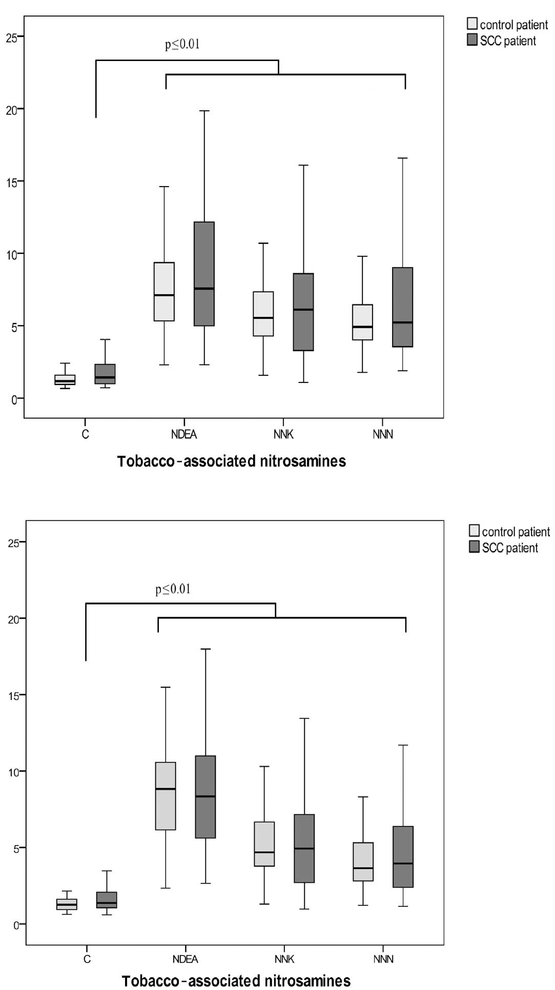
DNA damage in lymphocytes of patients without
carcinoma and patients with oropharyngeal carcinoma following
incubation with nitrosamines is shown in Fig. 1A. While the solvent DMSO as a
negative control did not induce any damage in lymphocyte DNA in
tumor and control patients (OTM 1.0 vs. 1.2), significant DNA
damage was found following incubation with NDEA (OTM 7.3), NNK (OTM
5.8) and NNN (OTM 5.1) in controls. Only OTMs >2 are considered
to reflect relevant DNA damage (16). DNA fragmentation in the SCC group
was comparable with OTMs 7.4, 6.0 and 5.2 following incubation with
NNN (p=0.58), NNK (p=0.48) and NDEA (p=0.67), respectively.
Fig. 1B shows
genotoxicity levels of oropharyngeal epithelial cells following
incubation with DMSO (negative control) and the nitrosamines NNN,
NNK and NDEA. In the control group, OTMs of 1.3 (DMSO), 8.3 (NDEA),
4.7 (NNK) and 3.9 (NNN) were determined. DNA derived from
oropharyngeal epithelial cells of cancer patients yielded OTMs of
7.9 (NDEA), 4.8 (NNK) and 4.0 (NNN) following nitrosamine
incubation with an OTM of the negative control of 1.3 (DMSO). No
significant differences were observed in DNA damage between tumor
patients and patients without malignancy [p=0.55 (NDEA), p=0.95
(NNK) and p=0.67 (NNN)].
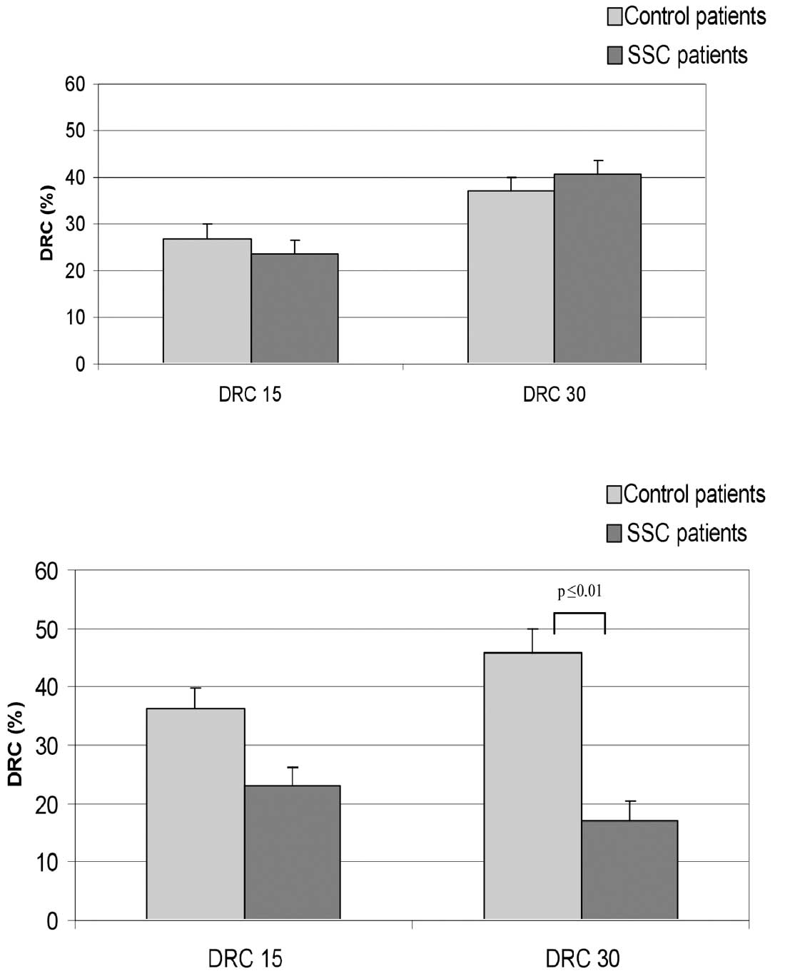
DNA repair in lymphocytes following NDEA incubation
is shown in Fig. 2A. For the
controls, DRC15 was 26.8% and DRC30 was 37.1%. The mean DRC15 and
DRC30 of cancer patients was 23.6 and 40.5%, respectively.
Differences in lymphocyte DNA repair were not observed between the
two groups of donors, with p=0.46 (DRC15) and p=0.62 (DRC30).
However, significant differences were found for the
DNA repair capacity between cases and controls for DRC30. The
results are shown in Fig. 2B.
Following DNA damage caused by NDEA, controls reached DRC levels of
36.2% (DRC15) and 46% (DRC30). Cells of SCC patients yielded a
DRC15 of 23% and a DRC30 of 17.1%. OTM values following damage and
15-min repair were 7.3 and following a 30-min repair period 7.86,
which demonstrates the arrest of repair action in the cells of
carcinoma patients after 20% of repaired damage. Statistical
evaluation of DNA repair of the two groups resulted in p=0.09
(DRC15) and p≤0.01 (DRC30).
Discussion
Besides laryngeal cancer, oropharyngeal SCC is the
most common alcohol- and tobacco-induced head and neck malignancy.
Over 90% of all cases are histopathologically classified as SCC
(1). In 100 cancer and non-cancer
patients, we investigated mutagen sensitivity following exposure to
tobacco-associated nitrosamines. Consecutively to NDEA- induced
damage, we determined DRC after 15 and 30 min. We applied the
alkaline comet assay as an established sensitive method for the
detection of DNA damage and repair and as an accepted biomarker
model (19). The comet assay is
widely used in ecogenotoxicology studies and as a predictor for
tumor therapies, such as radiotherapy (20). While it remains the subject of
controversy, OTM is the most informative measure in the comet assay
(21).
Oropharyngeal epithelium is the first target for
ingested and inhaled carcinogens and therefore employed as test
material in this study. Lymphocytes, which have been used in
numerous other previous studies evaluating DRC with the comet
assay, were also utilized (13,14,22,23).
In the present study, no significant differences in
lymphocytic or mucosal mutagen sensitivity were determined between
cancer and non-cancer patients. Wu et al found mutagen
sensitivity to be a marker for the identification of an increased
risk of developing premalignant lesions in the head and neck, using
lymphocytes exposed to benzo[a]pyrene diol epoxide (BPDE) and
bleomycin (24). In our study,
following incubation of the mucosal cells of SCC patients and
controls with BPDE, OTMs of the initial DNA damage did not
significantly differ. Increased lymphocyte sensitivity to bleomycin
has also been observed in lung cancer patients and proven to be a
potential biomarker for second primary tumor development in the
upper aerodigestive tract (14,25).
Further support for the impact of mutagen sensitivity on the
carcinogenic process results from the findings by Schantz et
al. These authors correlated mutagen sensitivity to chromosome
3p losses known to play a key role in the initiation phase of head
and neck cancer development (26).
Following incubation of lymphocytes with NDEA,
increased mutagen sensitivity was determined in patients suffering
from nasopharyngeal carcinoma, whereas no increased sensitivity was
observed following exposure to sodium dichromate and nickel
sulphate (27). This may be due to
different patient groups and diversity of cells compared to the
present study. Investigations on mucosa of head and neck cancer
patients indicated increased levels of mutagen sensitivity for SCC
patients (16). Studies using the
comet assay revealed significantly higher DNA damage in Barrett’s
epithelium compared with healthy squamous epithelium of the
oesophagus (28). However, although
we compared patients with SCC of the oropharynx to non-tumor
patients in an extended case-control study, including matching of
gender, age, tobacco and alcohol consumption, we did not find any
significant differences in mutagen sensitivity using lymphocytes or
mucosa cells. The results demonstrate the importance of matched
groups. Moreover, testing of mutagen sensitivity appears to depend
on the test system and mutagen used, as well as on the population
and cell type to be investigated.
Notably, mutagen sensitivity did not appear to be a
risk factor for the development of malignant lesions in our test
system. Accordingly, DNA repair was investigated as another
possible endogenous marker in head and neck carcinogenesis. The
comet assay has been adapted for the detection of DNA repair in
lymphocytes (12,29,30).
Certain investigations reported differences in DNA repair following
stimulation, dependent on the type of DNA damage induced (31,32).
Lymphocytes in our study were not stimulated prior to testing, due
to recommendations by Mayer et al after the determination of
a lack of differences in lymphocytic DRC following PHA-stimulation
and γ-irradiation (33).
Consequently, the group considered unstimulated mucosa cells from
fresh biopsy samples. The use of mucosa cells has previously been
described with only minor changes to original lymphocyte protocol
(12) and has been published
earlier (15,29).
In the present study, lymphocyte DRC15 (p=0.455) and
DRC30 (p=0.619) did not differ between cancer patients and patients
without malignancy following incubation with NDEA. However, in
mucosal cells, DRC15 and DRC30 levels were found to be lower in
patients with oropharyngeal tumors vs. controls, although the
significance was only observed in DRC30 (Fig. 2B).
The present study demonstrates that DRC of the upper
aerodigestive tract target cells is a potential endogenous factor
for head and neck cancer susceptibility. However, our data provide
evidence that there is no difference in DNA fragility to cigarette
smoke-related carcinogens. By contrast, overall DRC appears to be
rapidly saturated in HNSCC patients, whereas patients without
malignancy are able to continuously increase the proportion of
repaired DNA damage with time. The findings of other groups support
the role of hereditary reduced DNA repair capacities as risk
factors for head and neck cancer (13,34).
Additionally, gene polymorphisms in proteins, such as XPD and XRCC1
exert a major role in DNA repair, and thus affect head and neck
carcinogenesis (30,35,36).
Due to the diverse findings in the cell systems tested in this
study, we recommend the use of target tissue cells to investigate
DRC in upper aerodigestive tract carcinogenesis. Further studies
are likely to focus on the identification of specific chromosomal
and genetic alterations in mucosal cells of the upper aerodigestive
tract. Previously, a higher sensitivity to tobacco- related
carcinogens was determined in chromosomes 3, 5 and 8 in
oropharyngeal epithelia of head and neck tumor patients compared to
controls (37,38). Further investigation of such data
would provide more detailed information on cancer risks and may be
the basis for new preventive and therapeutic treatments in head and
neck carcinogenesis.
References
|
1
|
Hunter KD, Parkinson EK and Harrison PR:
Profiling early head and neck cancer. NatRevCancer. 5:127–135.
2005.PubMed/NCBI
|
|
2
|
Warnakulasuriya S, Sutherland G and Scully
C: Tobacco, oral cancer, and treatment of dependence. Oral Oncol.
41:244–260. 2005. View Article : Google Scholar
|
|
3
|
Cloos J, Nieuwenhuis EJ, Boomsma DI, Kuik
DJ, van der Sterre ML, Arwert F, Snow GB and Braakhuis BJ:
Inherited susceptibility to bleomycin-induced chromatid breaks in
cultured peripheral blood lymphocytes. J Natl Cancer Inst.
91:1125–1130. 1999. View Article : Google Scholar : PubMed/NCBI
|
|
4
|
IARC. Iarc Monographs on the Evaluation of
the Carcinogenic Risk of Chemicals to Humans: N′-Nitrosonornicotine
(Nnn). IARC; 2007
|
|
5
|
Hecht SS: DNA adduct formation from
tobacco-specific N-nitrosamines. Mutat Res. 424:127–142. 1999.
View Article : Google Scholar : PubMed/NCBI
|
|
6
|
Koppang N, Rivenson A, Dahle HK and
Hoffmann D: A study of tobacco carcinogenesis, Liii:
carcinogenicity of N′-nitrosonornicotine (Nnn) and
4-(methylnitrosamino)-1- (3-pyridyl)-1-butanone (Nnk) in mink
(Mustela Vison). Cancer Lett. 111:167–171. 1997.PubMed/NCBI
|
|
7
|
IARC. Iarc Monographs on the Evaluation of
the Carcinogenic Risk of Chemicals to Humans: N-Nitrosodiethylamine
(Ndea). IARC; 2000
|
|
8
|
Weitberg AB and Corvese D: Oxygen radicals
potentiate the genetic toxicity of tobacco-specific nitrosamines.
Clin Genet. 43:88–91. 1993. View Article : Google Scholar : PubMed/NCBI
|
|
9
|
Compagni A and Christofori G: Recent
advances in research on multistage tumorigenesis. Br J Cancer.
83:1–5. 2000.PubMed/NCBI
|
|
10
|
Hartwell LH and Weinert TA: Checkpoints:
controls that ensure the order of cell cycle events. Science.
246:629–634. 1989. View Article : Google Scholar : PubMed/NCBI
|
|
11
|
Sancar A: Mechanisms of DNA excision
repair. Science. 266:1954–1956. 1994. View Article : Google Scholar : PubMed/NCBI
|
|
12
|
Schmezer P, Rajaee-Behbahani N, Risch A,
Thiel S, Rittgen W, Drings P, Dienemann H, Kayser KW, Schulz V and
Bartsch H: Rapid screening assay for mutagen sensitivity and DNA
repair capacity in human peripheral blood lymphocytes. Mutagenesis.
16:25–30. 2001. View Article : Google Scholar : PubMed/NCBI
|
|
13
|
Cheng L, Eicher SA, Guo Z, Hong WK, Spitz
MR and Wei Q: Reduced DNA repair capacity in head and neck cancer
patients. Cancer Epidemiol Biomarkers Prev. 7:465–468.
1998.PubMed/NCBI
|
|
14
|
Rajaee-Behbahani N, Schmezer P, Risch A,
Rittgen W, Kayser KW, Dienemann H, Schulz V, Drings P, Thiel S and
Bartsch H: Altered DNA repair capacity and bleomycin sensitivity as
risk markers for non-small cell lung cancer. Int J Cancer.
95:86–91. 2001. View Article : Google Scholar : PubMed/NCBI
|
|
15
|
Kleinsasser NH, Wallner BC, Kastenbauer
ER, Muenzenrieder RK and Harreus UA: Comparing the genotoxic
sensitivities of human peripheral blood lymphocytes and mucosa
cells of the upper aerodigestive tract using the comet assay. Mutat
Res. 467:21–30. 2000. View Article : Google Scholar
|
|
16
|
Harreus U, Schmezer P, Kuchenmeister F and
Maier H: Genotoxic effect on human mucous membrane biopsies of the
upper aerodigestive tract. Laryngorhinootologie. 78:176–181.
1999.PubMed/NCBI
|
|
17
|
Singh NP, McCoy MT, Tice RR and Schneider
EL: A simple technique for quantitation of low levels of DNA damage
in individual cells. Exp Cell Res. 175:184–191. 1988. View Article : Google Scholar
|
|
18
|
Olive PL, Durand RE, Le RJ, Olivotto IA
and Jackson SM: Gel electrophoresis of individual cells to quantify
hypoxic fraction in human breast cancers. Cancer Res. 53:733–736.
1993.PubMed/NCBI
|
|
19
|
Glei M, Habermann N, Osswald K, Seidel C,
Persin C, Jahreis G and Pool-Zobel BL: Assessment of DNA damage and
its modulation by dietary and genetic factors in smokers using the
comet assay: a biomarker model. Biomarkers. 10:203–217. 2005.
View Article : Google Scholar : PubMed/NCBI
|
|
20
|
McKeown SR, Robson T, Price ME, Ho ET,
Hirst DG and McKelvey-Martin VJ: Potential use of the alkaline
comet assay as a predictor of bladder tumour response to radiation.
Br J Cancer. 89:2264–2270. 2003. View Article : Google Scholar : PubMed/NCBI
|
|
21
|
Burlinson B, Tice RR, Speit G, Agurell E,
Brendler-Schwaab SY, Collins AR, Escobar P, Honma M, Kumaravel TS,
Nakajima M, Sasaki YF, Thybaud V, Uno Y, Vasquez M and Hartmann A:
Fourth international workgroup on genotoxicity testing: Results of
the in vivo comet assay workgroup. Mutat Res. 627:31–35.
2007. View Article : Google Scholar : PubMed/NCBI
|
|
22
|
Sasiadek M, Schlade-Bartusiak K, Zych M,
Noga L and Czemarmazowicz H: Opposite responses in two DNA repair
capacity tests in lymphocytes of head and neck cancer patients. J
Appl Genet. 43:525–534. 2002.PubMed/NCBI
|
|
23
|
Palyvoda O, Mukalov I, Polanska J, Wygoda
A, Drobot L, Widel M and Rzeszowska-Wolny J: Radiation-induced DNA
damage and its repair in lymphocytes of patients with head and neck
cancer and healthy donors. Anticancer Res. 22:1721–1725.
2002.PubMed/NCBI
|
|
24
|
Wu X, Lippman SM, Lee JJ, Zhu Y, Wei QV,
Thomas M, Hong WK and Spitz MR: Chromosome instability in
lymphocytes: A potential indicator of predisposition to oral
premalignant lesions. Cancer Res. 62:2813–2818. 2002.PubMed/NCBI
|
|
25
|
Cloos J, Leemans CR, van der Sterre ML,
Kuik DJ, Snow GB and Braakhuis BJ: Mutagen sensitivity as a
biomarker for second primary tumors after head and neck squamous
cell carcinoma. Cancer Epidemiol Biomarkers Prev. 9:713–717.
2000.PubMed/NCBI
|
|
26
|
Schantz SP, Huang Q, Shah K, Murty VV, Hsu
TC, Yu G, Andersen PE, Huvos AG and Chaganti RS: Mutagen
sensitivity and environmental exposures as contributing causes of
chromosome 3p losses in head and neck cancers. Carcinogenesis.
21:1239–1246. 2000. View Article : Google Scholar : PubMed/NCBI
|
|
27
|
Kleinsasser NH, Wagner C, Wallner BC,
Harreus UA and Kastenbauer ER: Mutagen sensitivity of
nasopharyngeal cancer patients. Mutat Res. 491:151–161. 2001.
View Article : Google Scholar : PubMed/NCBI
|
|
28
|
Olliver JR, Hardie LJ, Dexter S, Chalmers
D and Wild CP: DNA damage levels are raised in Barrett’s
oesophageal mucosa relative to the squamous epithelium of the
oesophagus. Biomarkers. 8:509–521. 2003.
|
|
29
|
Harreus UA, Wallner BC, Kastenbauer ER and
Kleinsasser NH: DNA repair if mucous membrane cells and lymphocytes
with the comet assay. Laryngorhinootologie. 80:23–26.
2001.PubMed/NCBI
|
|
30
|
Spitz MR, Wei Q, Dong Q, Amos CI and Wu X:
Genetic susceptibility to lung cancer: the role of DNA damage and
repair. Cancer Epidemiol Biomarkers Prev. 12:689–698.
2003.PubMed/NCBI
|
|
31
|
Boerrigter ME and Vijg J: Induction and
disappearance of DNA single-strand breaks in human B and T
lymphocytes after exposure to ethylnitrosourea. Mutat Res.
255:49–55. 1991. View Article : Google Scholar : PubMed/NCBI
|
|
32
|
Leroy T, Lison D and Lauwerys R:
Preliminary in vitro investigation into the use of alkaline
elution assay for the biomonitoring of humans exposed to genotoxic
agents. Hum Exp Toxicol. 14:61–68. 1995.
|
|
33
|
Mayer C, Popanda O, Zelezny O, von Brevern
MC, Bach A, Bartsch H and Schmezer P: DNA repair capacity after
γ-irradiation and expression profiles of DNA repair genes in
resting and proliferating human peripheral blood lymphocytes. DNA
Repair (Amst). 1:237–250. 2002.
|
|
34
|
Schantz SP, Zhang ZF, Spitz MS, Sun M and
Hsu TC: Genetic susceptibility to head and neck cancer: Interaction
between nutrition and mutagen sensitivity. Laryngoscope.
107:765–781. 1997. View Article : Google Scholar : PubMed/NCBI
|
|
35
|
Caldecott KW: Xrcc1 and DNA strand break
repair. DNA Repair (Amst). 2:955–969. 2003. View Article : Google Scholar : PubMed/NCBI
|
|
36
|
Han J, Hankinson SE, Colditz GA and Hunter
DJ: Genetic variation in Xrcc1, sun exposure, and risk of skin
cancer. Br J Cancer. 91:1604–1609. 2004.PubMed/NCBI
|
|
37
|
Harreus UA, Kleinsasser NH, Zieger S,
Wallner B, Reiter M, Schuller P and Berghaus A: Sensitivity to
DNA-damage induction and chromosomal alterations in mucosa cells
from patients with and without cancer of the oropharynx detected by
a combination of Comet assay and fluorescence in situ
hybridization. Mutat Res. 563:131–138. 2004. View Article : Google Scholar : PubMed/NCBI
|
|
38
|
Reiter M, Baumeister P, Zieger S and
Harreus U: Chromosomal alterations and mutagen sensitivity in human
mucosal cells of the oropharynx and lymphocytes caused by Bpde.
Cancer Genomics Proteomics. 6:247–254. 2009.PubMed/NCBI
|