Spandidos Publications Logo
  • About
    • About Spandidos
    • Aims and Scopes
    • Abstracting and Indexing
    • Editorial Policies
    • Reprints and Permissions
    • Job Opportunities
    • Terms and Conditions
    • Contact
  • Journals
    • All Journals
    • Oncology Letters
      • Oncology Letters
      • Information for Authors
      • Editorial Policies
      • Editorial Board
      • Aims and Scope
      • Abstracting and Indexing
      • Bibliographic Information
      • Archive
    • International Journal of Oncology
      • International Journal of Oncology
      • Information for Authors
      • Editorial Policies
      • Editorial Board
      • Aims and Scope
      • Abstracting and Indexing
      • Bibliographic Information
      • Archive
    • Molecular and Clinical Oncology
      • Molecular and Clinical Oncology
      • Information for Authors
      • Editorial Policies
      • Editorial Board
      • Aims and Scope
      • Abstracting and Indexing
      • Bibliographic Information
      • Archive
    • Experimental and Therapeutic Medicine
      • Experimental and Therapeutic Medicine
      • Information for Authors
      • Editorial Policies
      • Editorial Board
      • Aims and Scope
      • Abstracting and Indexing
      • Bibliographic Information
      • Archive
    • International Journal of Molecular Medicine
      • International Journal of Molecular Medicine
      • Information for Authors
      • Editorial Policies
      • Editorial Board
      • Aims and Scope
      • Abstracting and Indexing
      • Bibliographic Information
      • Archive
    • Biomedical Reports
      • Biomedical Reports
      • Information for Authors
      • Editorial Policies
      • Editorial Board
      • Aims and Scope
      • Abstracting and Indexing
      • Bibliographic Information
      • Archive
    • Oncology Reports
      • Oncology Reports
      • Information for Authors
      • Editorial Policies
      • Editorial Board
      • Aims and Scope
      • Abstracting and Indexing
      • Bibliographic Information
      • Archive
    • Molecular Medicine Reports
      • Molecular Medicine Reports
      • Information for Authors
      • Editorial Policies
      • Editorial Board
      • Aims and Scope
      • Abstracting and Indexing
      • Bibliographic Information
      • Archive
    • World Academy of Sciences Journal
      • World Academy of Sciences Journal
      • Information for Authors
      • Editorial Policies
      • Editorial Board
      • Aims and Scope
      • Abstracting and Indexing
      • Bibliographic Information
      • Archive
    • International Journal of Functional Nutrition
      • International Journal of Functional Nutrition
      • Information for Authors
      • Editorial Policies
      • Editorial Board
      • Aims and Scope
      • Abstracting and Indexing
      • Bibliographic Information
      • Archive
    • International Journal of Epigenetics
      • International Journal of Epigenetics
      • Information for Authors
      • Editorial Policies
      • Editorial Board
      • Aims and Scope
      • Abstracting and Indexing
      • Bibliographic Information
      • Archive
    • Medicine International
      • Medicine International
      • Information for Authors
      • Editorial Policies
      • Editorial Board
      • Aims and Scope
      • Abstracting and Indexing
      • Bibliographic Information
      • Archive
  • Articles
  • Information
    • Information for Authors
    • Information for Reviewers
    • Information for Librarians
    • Information for Advertisers
    • Conferences
  • Language Editing
Spandidos Publications Logo
  • About
    • About Spandidos
    • Aims and Scopes
    • Abstracting and Indexing
    • Editorial Policies
    • Reprints and Permissions
    • Job Opportunities
    • Terms and Conditions
    • Contact
  • Journals
    • All Journals
    • Biomedical Reports
      • Information for Authors
      • Editorial Policies
      • Editorial Board
      • Aims and Scope
      • Abstracting and Indexing
      • Bibliographic Information
      • Archive
    • Experimental and Therapeutic Medicine
      • Information for Authors
      • Editorial Policies
      • Editorial Board
      • Aims and Scope
      • Abstracting and Indexing
      • Bibliographic Information
      • Archive
    • International Journal of Epigenetics
      • Information for Authors
      • Editorial Policies
      • Editorial Board
      • Aims and Scope
      • Abstracting and Indexing
      • Bibliographic Information
      • Archive
    • International Journal of Functional Nutrition
      • Information for Authors
      • Editorial Policies
      • Editorial Board
      • Aims and Scope
      • Abstracting and Indexing
      • Bibliographic Information
      • Archive
    • International Journal of Molecular Medicine
      • Information for Authors
      • Editorial Policies
      • Editorial Board
      • Aims and Scope
      • Abstracting and Indexing
      • Bibliographic Information
      • Archive
    • International Journal of Oncology
      • Information for Authors
      • Editorial Policies
      • Editorial Board
      • Aims and Scope
      • Abstracting and Indexing
      • Bibliographic Information
      • Archive
    • Medicine International
      • Information for Authors
      • Editorial Policies
      • Editorial Board
      • Aims and Scope
      • Abstracting and Indexing
      • Bibliographic Information
      • Archive
    • Molecular and Clinical Oncology
      • Information for Authors
      • Editorial Policies
      • Editorial Board
      • Aims and Scope
      • Abstracting and Indexing
      • Bibliographic Information
      • Archive
    • Molecular Medicine Reports
      • Information for Authors
      • Editorial Policies
      • Editorial Board
      • Aims and Scope
      • Abstracting and Indexing
      • Bibliographic Information
      • Archive
    • Oncology Letters
      • Information for Authors
      • Editorial Policies
      • Editorial Board
      • Aims and Scope
      • Abstracting and Indexing
      • Bibliographic Information
      • Archive
    • Oncology Reports
      • Information for Authors
      • Editorial Policies
      • Editorial Board
      • Aims and Scope
      • Abstracting and Indexing
      • Bibliographic Information
      • Archive
    • World Academy of Sciences Journal
      • Information for Authors
      • Editorial Policies
      • Editorial Board
      • Aims and Scope
      • Abstracting and Indexing
      • Bibliographic Information
      • Archive
  • Articles
  • Information
    • For Authors
    • For Reviewers
    • For Librarians
    • For Advertisers
    • Conferences
  • Language Editing
Login Register Submit
  • This site uses cookies
  • You can change your cookie settings at any time by following the instructions in our Cookie Policy. To find out more, you may read our Privacy Policy.

    I agree
Search articles by DOI, keyword, author or affiliation
Search
Advanced Search
presentation
Oncology Letters
Join Editorial Board Propose a Special Issue
Print ISSN: 1792-1074 Online ISSN: 1792-1082
Journal Cover
January 2012 Volume 3 Issue 1

Full Size Image

Sign up for eToc alerts
Recommend to Library

Journals

International Journal of Molecular Medicine

International Journal of Molecular Medicine

International Journal of Molecular Medicine is an international journal devoted to molecular mechanisms of human disease.

International Journal of Oncology

International Journal of Oncology

International Journal of Oncology is an international journal devoted to oncology research and cancer treatment.

Molecular Medicine Reports

Molecular Medicine Reports

Covers molecular medicine topics such as pharmacology, pathology, genetics, neuroscience, infectious diseases, molecular cardiology, and molecular surgery.

Oncology Reports

Oncology Reports

Oncology Reports is an international journal devoted to fundamental and applied research in Oncology.

Experimental and Therapeutic Medicine

Experimental and Therapeutic Medicine

Experimental and Therapeutic Medicine is an international journal devoted to laboratory and clinical medicine.

Oncology Letters

Oncology Letters

Oncology Letters is an international journal devoted to Experimental and Clinical Oncology.

Biomedical Reports

Biomedical Reports

Explores a wide range of biological and medical fields, including pharmacology, genetics, microbiology, neuroscience, and molecular cardiology.

Molecular and Clinical Oncology

Molecular and Clinical Oncology

International journal addressing all aspects of oncology research, from tumorigenesis and oncogenes to chemotherapy and metastasis.

World Academy of Sciences Journal

World Academy of Sciences Journal

Multidisciplinary open-access journal spanning biochemistry, genetics, neuroscience, environmental health, and synthetic biology.

International Journal of Functional Nutrition

International Journal of Functional Nutrition

Open-access journal combining biochemistry, pharmacology, immunology, and genetics to advance health through functional nutrition.

International Journal of Epigenetics

International Journal of Epigenetics

Publishes open-access research on using epigenetics to advance understanding and treatment of human disease.

Medicine International

Medicine International

An International Open Access Journal Devoted to General Medicine.

Journal Cover
January 2012 Volume 3 Issue 1

Full Size Image

Sign up for eToc alerts
Recommend to Library

  • Article
  • Citations
    • Cite This Article
    • Download Citation
    • Create Citation Alert
    • Remove Citation Alert
    • Cited By
  • Similar Articles
    • Related Articles (in Spandidos Publications)
    • Similar Articles (Google Scholar)
    • Similar Articles (PubMed)
  • Download PDF
  • Download XML
  • View XML
Review

Transitional cell carcinoma of the ovary (Review)

  • Authors:
    • Satoshi Ichigo
    • Hiroshi Takagi
    • Kazutoshi Matsunami
    • Takayuki Murase
    • Tsuneko Ikeda
    • Atsushi Imai
  • View Affiliations / Copyright

    Affiliations: Department of Obstetrics and Gynecology, Matsunami General Hospital, Gifu 501-6062, Japan, Department of Pathology, Matsunami General Hospital, Gifu 501-6062, Japan
  • Pages: 3-6
    |
    Published online on: October 21, 2011
       https://doi.org/10.3892/ol.2011.453
  • Expand metrics +
Metrics: Total Views: 0 (Spandidos Publications: | PMC Statistics: )
Metrics: Total PDF Downloads: 0 (Spandidos Publications: | PMC Statistics: )
Cited By (CrossRef): 0 citations Loading Articles...

This article is mentioned in:


Abstract

Transitional cell carcinoma (TCC) of the ovary is a rare recently recognized subtype of ovarian epithelial cancer. Ovarian TCC has a modest response to chemotherapy, and metastatic TCC from the renal pelvis results in mortality. The clinical presentation is indistinguishable from other types of ovarian carcinoma. Histopathological examination remains the first tool used in the diagnosis of these heterogeneous tumors and in the separation of closely related tumors. Since it is generally accepted that surgical resection is the primary therapeutic approach, and patient outcomes following chemotherapy are better than for other types of ovarian cancers, it is a reasonable concept to detect tumors when they are still confined within the ovaries. Thus, the aim of this review was to describe typical cases of primary TCC, and to review the medical literature for information on TCC management in order to determine appropriate diagnostic methods and therapy.

1. Introduction

Ovarian cancer is the most lethal gynecological malignancy. Efforts at early detection and new therapeutic approaches to reduce mortality have largely been unsuccessful, since the origin and pathogenesis of epithelial ovarian cancer are poorly understood (1). Transitional cell carcinoma (TCC), a recently recognized subtype, resembles urothelium rather than ovarian surface epithelium (methothelium) (2,3). A small percentage of ovarian cancer types are accounted for by TCC, which has proven to be a distinct group with various histological and immunohistological patterns. Patients with TCC had better prognoses compared to patients with all other types of ovarian carcinomas following standardized chemotherapy (4). The aim of this review was to describe our typical cases of primary TCC, and to review the medical literature for information on TCC management in order to determine appropriate diagnostic methods and therapy.

2. General considerations

TCC of the ovary is a recently recognized subtype of ovarian surface epithelial cancer. TCC has been described as a primary ovarian carcinoma in which definite urothelial features are present, but no benign, metaplastic and/or proliferating Brenner tumor can be identified. TCC of the ovary was initally defined by Austin and Norris (5). These investigators reported a group of patients who had ovarian tumors presenting with histologic features similar to those observed in a malignant Brenner tumor, but the tumors lacked the associated benign Brenner tumor component. Pure TCC was thus distinguished from malignant Brenner tumor. In addition to not having a benign Brenner tumor component, TCC lacks the prominent stromal calcification (5–7). Since TCC of the ovary has close morphological similarities to TCC of the bladder and it behaves more aggressively than malignant Brenner tumors, Austin and Norris (5) concluded that ovarian TCC arises directly from the pluripotent surface epithelium of the ovary and from cells with urothelial potential, rather than from a benign or proliferative Brenner tumor precursor.

3. Incidence

The true incidence of TCC of the ovary remains unknown. Transitional cell tumors, including TCC, and benign and malignant Brenner tumors of the ovary represent ~2% of all ovarian tumors. Moreover, according to the World Heath Organization (WHO), depending on the histological pattern, these tumors are classified as benign, borderline or malignant Brenner tumors and TCC (8). Silva et al (9) observed the focal or diffuse TCC pattern in 88 of 934 ovarian cancers (9%).

4. Diagnosis

The common presenting symptoms of TCC of the ovary are abdominal pain, abdominal swelling or distension and weight loss. Occasionally, the patient may present with uterine bleeding, back pain, bowel or urinary symptoms, as shown in our cases 
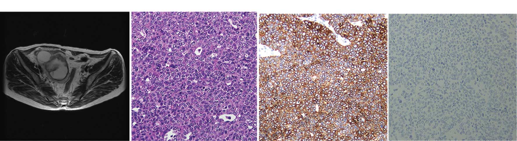
(Figs. 1 and 2). However, the clinical presentation is indistinguishable from other types of ovarian carcinoma (5,10). As described in detail by Eichhorn and Young (10), ovarian TCC typically shows undulating, diffuse, insular and trabecular growth patterns. The tumor cell nuclei were oblong or round, often exhibiting nucleoli with longitudinal grooves. The cytoplasm was often pale and granular, and was rarely clear or eosinophilic (Figs. 1 and 2).
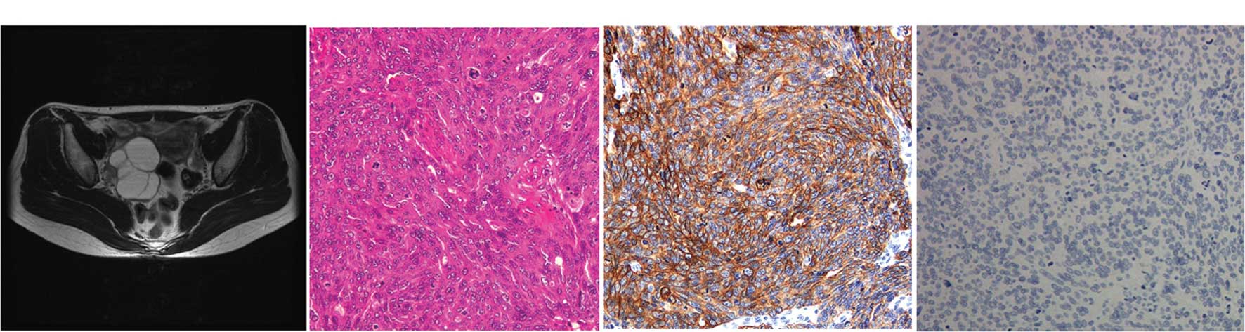
Figure 1

Primary TCC of the ovary (case 1). A 64-year-old postmenopausal woman presented with a 1-year history of progressive enlargement of an abdominal mass. Physical examination showed a pelvic mass. Abdominal ultrasound showed a pelvic mass measuring 6 cm with inhomogeneous echogenicity. (A) Horizontal T2-weighted MRI showed an inhomogeneous cyst on the right side of the pelvis, which was >70 mm in maximal diameter with a solid component. There was no evidence of lymphadenopathy. Prior to surgery, CA125 was elevated to 347 U/ml (normal, <35 U/ml), but other markers were all within normal ranges. Surgical staging procedures including total abdominal hysterectomy, bilateral salpingo-oophorectomy, infracolic omentectomy and pelvic lymph node dissection were performed. The ascites were also sent for cytological examination. (B) Microscopic examination showed a malignant transitional epithelial lining of the right ovarian cyst. The final diagnosis was TCC, grade 3, FIGO stage IIc. Immunohistochemical studies showed that the tumor was (C) positive for CK 7 and (D) negative for CK20. The patient received six cycles of chemotherapy with paclitaxel-carboplatin following surgery, and has been disease-free for 1 year.

Figure 2

Primary TCC of the ovary (case 2). A 44-year-old woman with a right ovarian cyst was referred to the gynecology department. (A) The MRI (T2-weighted, horizontal) revealed a 5×6 cm multiple cystic mass in the right adenexa, suggestive of an ovarian tumor. There was no obvious metastatic foci in other organs. The levels of serum CA72-4 and CA125 were 7.7 U/ml (normal, <10.0 U/ml) and 12.9 U/ml (normal, <10 U/ml), respectively. (B) The right ovarian cyst was laparoscopically resected and diagnosed as TCC, grade 3 with serosal involvement. (C) Immunohistochemical stains for CK7 were positive; (D) but staining for CK20 was negative. The ascites cytology was reported to be positive. The patient was staged as FIGO stage Ic. One month following adenectomy, the patient was submitted for exploratory staging procedures including total abdominal hysterectomy, bilateral salpingo-oophorectomy, infracolic omentectomy and pelvic lymph node dissection. The extensively sampled adnexal tissue was histologically uninvolved by the tumor. After recovering from surgery, the patient received six cycles of chemotherapy with paclitaxel-carboplatin. The patient is presently doing well without any recurrent disease for 2 years.

CA125 is clinically useful as a serum marker of tumor progression and recurrence, although early stages may be CA125-negative (Fig. 2). In the study by Ceauşu et al (11), 13 archived formalin-fixed paraffin-embedded samples of transitional cell tumors of the ovary were assessed using standard hematoxylin-eosin staining and the indirect tristadial ABC peroxidase immunohistochemistry method for 11 antibodies including CA125, cytokeratin (CK) 7 and CEA. Over 50% of the samples were malignant Brenner tumors, CA125 was positive in all malignant tumors (of Brenner type and TCCs), but not in the benign and borderline tumors, while CK7 was positive in approximately 70% of all cases. The two antibodies have shown a high sensitivity and low specificity, but do not correlate with each other. Recent findings (11,12) have shown that p63 is expressed in benign and borderline Brenner tumors, but not in malignant counterparts and TCCs of the ovary, suggesting that this antigen is a marker for the differential diagnosis of malignant Brenner tumors and TCCs, and may also play a role in Brenner carcinogenesis. The aim of these studies was to detect tumors when they are still confined to the ovaries, thereby increasing the likelihood of cure and reducing the mortality of the disease. The modalities that are currently in use to screen women are pelvic examination, imaging modality and measurement of serum CA125 (1) (Figs. 1 and 2), although case 2 was CA125-negative.

6. Treatment

Optimal surgical resectability followed by cisplatin-based chemotherapy may contribute to the survival benefit (4,6,7). The estimated 5-year survival following surgery for 88 patients was 37%, whereas for patients who received chemotherapy, survival was at 41% (6,7). Factors associated with survival for patients who received chemotherapy were the clinical stage, the percentage of the TCC component in the primary tumor and the results of second-look surgery. The predominance of TCC was a favorable prognostic factor and patients with higher clinical stages had poorer prognoses.

7. Prognosis

The relative effects of tumor biology and treatment strategies remain undetermined (6,7). Gershenson et al (13,14) concluded that advanced-stage ovarian TCC was significantly more chemosensitive and associated with better prognosis than poorly differentiated serous carcinoma. Kommoss et al (4) also documented that patients with TCC had better prognoses compared to patients with all other types of ovarian carcinomas following standardized chemotherapy. The metastatic pathways of the tumor simulate TCC of the bladder, which implicates a loss of the integrity of E-cadherin (5–7).

8. Metastatic TCC to the ovaries

The ovaries are common sites for intra-abdominal metastasis (15). Approximately 6% of ovarian cancers found at laparotomy are secondary tumors from other sites (15,16). Metastatic TCC from the urinary bladder, or elsewhere within the urinary system, involving the ovary is extremely rare (16). There have been six cases reported thus far, as described by Lee et al (17). In all cases, secondary ovarian tumors are unilateral. The time interval to the appearance of ovarian metastases varied from synchronous to 4 years. In the study by Lee et al (17), all cases received surgery, with the overall survival ranging from 3 months to 7 years. These cases favor metastatic ovarian tumors for the following reasons: definite histological evidence of a primary renal tumor, and deep stromal invasion. The origin of primary lesions has prognostic significance as TCC of the ovary has a modest response to chemotherapy (10) and metastatic TCC from the renal pelvis results in mortality (17).

Microscopically, metastatic TCC of the ovary resembles a primary ovarian TCC. Primary TCC accounts for 1–2% of all ovarian tumors (10,17). TCC of the ovary is a recently recognized subtype of ovarian surface epithelial-stromal cancer, and studies of its morphology are rare. The presence of a component of benign or borderline Brenner tumor confirms an ovarian primary tumor. TCC of the ovaries has mucin pools and thick papillae with smooth luminal borders, in contrast to the pseudo-papillae of tumor cell necrosis that is common in metastatic TCC (10,17). Another study has shown that the morphological similarity between transitional cell carcinoma of the ovary and its counterpart from the urinary bladder does not indicate any histogenic similarity, but CK7 and CK20, together with uroplakin III and WT1 may prove useful in distinguishing primitive TCCs of the ovary, and metastases from invasive TCC of the bladder to the ovary, the former being a variant morphology in the spectrum of surface epithelial carcinomas (11,18).

9. Conclusion

Microscopic examination remains the first tool in the diagnosis of these heterogeneous tumors and in the separation of closely related tumors. Primary TCC of the ovary is a relatively rare subtype of epithelial ovarian cancer. Surgical resection is the primary therapeutic approach, and patient outcomes following chemotherapy are better than for other types of ovarian cancers.

References

1 

Kurman R and Shih I-M: The origin and pathogenesis of epithelial ovarian cancer: A proposed unifying theory. Am J Surg Pathol. 34:433–443. 2010. View Article : Google Scholar : PubMed/NCBI

2 

Cuatrecasas M, Catasus L, Palacios J and Prat J: Transitional cell tumors of the ovary: A comparative clinicopathologic, immunohistochemical, and molecular genetic analysis of Brenner tumors and transitional cell carcinomas. Am J Surg Pathol. 33:556–567. 2009. View Article : Google Scholar

3 

Oh S, Rha S, Jung S, Lee Y, Choi B, Byun J, Ku Y and Jung C: Transitional cell tumor of the ovary: computed tomographic and magnetic resonance imaging features with pathological correlation. J Comput Assist Tomogr. 33:106–112. 2009. View Article : Google Scholar : PubMed/NCBI

4 

Kommoss F, Kommoss S, Schmidt D, Trunk M, Pfisterer J and du Bois A: Survival benefit for patients with advanced-stage transitional cell carcinomas vs. other subtypes of ovarian carcinoma after chemotherapy with platinum and paclitaxel. Gynecol Oncol. 97:195–199. 2005. View Article : Google Scholar : PubMed/NCBI

5 

Austin R and Norris H: Malignant Brenner tumor and transitional cell carcinoma of the ovary: A comparison. Int J Gynecol Pathol. 6:29–39. 1987. View Article : Google Scholar : PubMed/NCBI

6 

Tazi E, Lalya I, Tazi M, Ahellal Y, M’rabti H and Errihani H: Transitional cell carcinoma of the ovary: a rare case and review of literature. World J Surg Oncol. 8:98–101. 2010. View Article : Google Scholar : PubMed/NCBI

7 

Lin C, Liu F and Ho E: Transitional cell carcinoma of the ovary. Taiwan J Obstet Gynecol. 45:268–271. 2006. View Article : Google Scholar

8 

World Health Organization. Classification of tumours: Pathology and genetics of tumors of the breast and female genital organs. IARC Press; Lyon: pp. 140–143. 2003

9 

Silva E, Robey-Cafferty S, Smith T and Gershenson D: Ovarian carcinomas with transitional cell carcinoma pattern. Am J Clin Pathol. 93:457–465. 1990.PubMed/NCBI

10 

Eichhorn J and Young R: Transitional cell carcinoma of the ovary: a morphologic study of 100 cases with emphasis on differential diagnosis. Am J Surg Pathol. 28:453–463. 2004. View Article : Google Scholar : PubMed/NCBI

11 

Ceauşu M, Terzea D, Georgescu A, Dobrea C, Mihai M, Iosif C, Vasilescu F and Ardeleanu C: Transitional cell tumors of the ovary: a compact group with a heterogeneous histological and immunophenotypical pattern. Rom J Morphol Embryol. 49:513–516. 2008.PubMed/NCBI

12 

Liao X, Xue W, Shen D, Ngan H, Siu M and Cheung A: P63 expression in ovarian tumours: a marker for Brenner tumours but not transitional cell carcinomas. Histopathology. 51:477–783. 2007. View Article : Google Scholar : PubMed/NCBI

13 

Gershenson D, Morris M, Burke T, Levenback C, Kavanagh J, Fromm G, Silva E, Warner D and Wharton J: Combined cisplatin and carboplatin chemotherapy for treatment of advanced epithelial ovarian cancer. Gynecol Oncol. 58:349–355. 1995. View Article : Google Scholar : PubMed/NCBI

14 

Gershenson D, Silva E, Mitchell M, Atkinson E and Wharton J: Transitional cell carcinoma of the ovary: a matched control study of advanced-stage patients treated with cisplatin-based chemotherapy. Am J Obstet Gynecol. 168:1178–1185. 1993. View Article : Google Scholar : PubMed/NCBI

15 

Valappil S, Toon P and Anandaram P: Ovarian metastasis from primary renal cell carcinoma: report of a case and review of literature. Gynecol Oncol. 94:846–849. 2004. View Article : Google Scholar : PubMed/NCBI

16 

Skírnisdóttir I, Garmo H and Holmberg L: Non-genital tract metastases to the ovaries presented as ovarian tumors in Sweden 1990–2003: occurrence, origin and survival compared to ovarian cancer. Gynecol Oncol. 105:166–171. 2007.PubMed/NCBI

17 

Lee M, Jung Y, Kim S, Kim S and Kim Y: Metastasis to the ovaries from transitional cell carcinoma of the bladder and renal pelvis: a report of two cases. J Gynecol Oncol. 21:59–61. 2010. View Article : Google Scholar : PubMed/NCBI

18 

Logani S, Olova E, Amin M, Folpe A, Cohen C and Young R: Immunoprofile of ovarian tumors with putative transitional cell (urothelial) differentiation using novel urothelial markers: histogenetic and diagnostic implications. Am J Surg Pathol. 27:1434–1441. 2003. View Article : Google Scholar

Related Articles

  • Abstract
  • View
  • Download
  • Twitter
Copy and paste a formatted citation
Spandidos Publications style
Ichigo S, Takagi H, Matsunami K, Murase T, Ikeda T and Imai A: Transitional cell carcinoma of the ovary (Review). Oncol Lett 3: 3-6, 2012.
APA
Ichigo, S., Takagi, H., Matsunami, K., Murase, T., Ikeda, T., & Imai, A. (2012). Transitional cell carcinoma of the ovary (Review). Oncology Letters, 3, 3-6. https://doi.org/10.3892/ol.2011.453
MLA
Ichigo, S., Takagi, H., Matsunami, K., Murase, T., Ikeda, T., Imai, A."Transitional cell carcinoma of the ovary (Review)". Oncology Letters 3.1 (2012): 3-6.
Chicago
Ichigo, S., Takagi, H., Matsunami, K., Murase, T., Ikeda, T., Imai, A."Transitional cell carcinoma of the ovary (Review)". Oncology Letters 3, no. 1 (2012): 3-6. https://doi.org/10.3892/ol.2011.453
Copy and paste a formatted citation
x
Spandidos Publications style
Ichigo S, Takagi H, Matsunami K, Murase T, Ikeda T and Imai A: Transitional cell carcinoma of the ovary (Review). Oncol Lett 3: 3-6, 2012.
APA
Ichigo, S., Takagi, H., Matsunami, K., Murase, T., Ikeda, T., & Imai, A. (2012). Transitional cell carcinoma of the ovary (Review). Oncology Letters, 3, 3-6. https://doi.org/10.3892/ol.2011.453
MLA
Ichigo, S., Takagi, H., Matsunami, K., Murase, T., Ikeda, T., Imai, A."Transitional cell carcinoma of the ovary (Review)". Oncology Letters 3.1 (2012): 3-6.
Chicago
Ichigo, S., Takagi, H., Matsunami, K., Murase, T., Ikeda, T., Imai, A."Transitional cell carcinoma of the ovary (Review)". Oncology Letters 3, no. 1 (2012): 3-6. https://doi.org/10.3892/ol.2011.453
Follow us
  • Twitter
  • LinkedIn
  • Facebook
About
  • Spandidos Publications
  • Careers
  • Cookie Policy
  • Privacy Policy
How can we help?
  • Help
  • Live Chat
  • Contact
  • Email to our Support Team