Spandidos Publications Logo
  • About
    • About Spandidos
    • Aims and Scopes
    • Abstracting and Indexing
    • Editorial Policies
    • Reprints and Permissions
    • Job Opportunities
    • Terms and Conditions
    • Contact
  • Journals
    • All Journals
    • Oncology Letters
      • Oncology Letters
      • Information for Authors
      • Editorial Policies
      • Editorial Board
      • Aims and Scope
      • Abstracting and Indexing
      • Bibliographic Information
      • Archive
    • International Journal of Oncology
      • International Journal of Oncology
      • Information for Authors
      • Editorial Policies
      • Editorial Board
      • Aims and Scope
      • Abstracting and Indexing
      • Bibliographic Information
      • Archive
    • Molecular and Clinical Oncology
      • Molecular and Clinical Oncology
      • Information for Authors
      • Editorial Policies
      • Editorial Board
      • Aims and Scope
      • Abstracting and Indexing
      • Bibliographic Information
      • Archive
    • Experimental and Therapeutic Medicine
      • Experimental and Therapeutic Medicine
      • Information for Authors
      • Editorial Policies
      • Editorial Board
      • Aims and Scope
      • Abstracting and Indexing
      • Bibliographic Information
      • Archive
    • International Journal of Molecular Medicine
      • International Journal of Molecular Medicine
      • Information for Authors
      • Editorial Policies
      • Editorial Board
      • Aims and Scope
      • Abstracting and Indexing
      • Bibliographic Information
      • Archive
    • Biomedical Reports
      • Biomedical Reports
      • Information for Authors
      • Editorial Policies
      • Editorial Board
      • Aims and Scope
      • Abstracting and Indexing
      • Bibliographic Information
      • Archive
    • Oncology Reports
      • Oncology Reports
      • Information for Authors
      • Editorial Policies
      • Editorial Board
      • Aims and Scope
      • Abstracting and Indexing
      • Bibliographic Information
      • Archive
    • Molecular Medicine Reports
      • Molecular Medicine Reports
      • Information for Authors
      • Editorial Policies
      • Editorial Board
      • Aims and Scope
      • Abstracting and Indexing
      • Bibliographic Information
      • Archive
    • World Academy of Sciences Journal
      • World Academy of Sciences Journal
      • Information for Authors
      • Editorial Policies
      • Editorial Board
      • Aims and Scope
      • Abstracting and Indexing
      • Bibliographic Information
      • Archive
    • International Journal of Functional Nutrition
      • International Journal of Functional Nutrition
      • Information for Authors
      • Editorial Policies
      • Editorial Board
      • Aims and Scope
      • Abstracting and Indexing
      • Bibliographic Information
      • Archive
    • International Journal of Epigenetics
      • International Journal of Epigenetics
      • Information for Authors
      • Editorial Policies
      • Editorial Board
      • Aims and Scope
      • Abstracting and Indexing
      • Bibliographic Information
      • Archive
    • Medicine International
      • Medicine International
      • Information for Authors
      • Editorial Policies
      • Editorial Board
      • Aims and Scope
      • Abstracting and Indexing
      • Bibliographic Information
      • Archive
  • Articles
  • Information
    • Information for Authors
    • Information for Reviewers
    • Information for Librarians
    • Information for Advertisers
    • Conferences
  • Language Editing
Spandidos Publications Logo
  • About
    • About Spandidos
    • Aims and Scopes
    • Abstracting and Indexing
    • Editorial Policies
    • Reprints and Permissions
    • Job Opportunities
    • Terms and Conditions
    • Contact
  • Journals
    • All Journals
    • Biomedical Reports
      • Information for Authors
      • Editorial Policies
      • Editorial Board
      • Aims and Scope
      • Abstracting and Indexing
      • Bibliographic Information
      • Archive
    • Experimental and Therapeutic Medicine
      • Information for Authors
      • Editorial Policies
      • Editorial Board
      • Aims and Scope
      • Abstracting and Indexing
      • Bibliographic Information
      • Archive
    • International Journal of Epigenetics
      • Information for Authors
      • Editorial Policies
      • Editorial Board
      • Aims and Scope
      • Abstracting and Indexing
      • Bibliographic Information
      • Archive
    • International Journal of Functional Nutrition
      • Information for Authors
      • Editorial Policies
      • Editorial Board
      • Aims and Scope
      • Abstracting and Indexing
      • Bibliographic Information
      • Archive
    • International Journal of Molecular Medicine
      • Information for Authors
      • Editorial Policies
      • Editorial Board
      • Aims and Scope
      • Abstracting and Indexing
      • Bibliographic Information
      • Archive
    • International Journal of Oncology
      • Information for Authors
      • Editorial Policies
      • Editorial Board
      • Aims and Scope
      • Abstracting and Indexing
      • Bibliographic Information
      • Archive
    • Medicine International
      • Information for Authors
      • Editorial Policies
      • Editorial Board
      • Aims and Scope
      • Abstracting and Indexing
      • Bibliographic Information
      • Archive
    • Molecular and Clinical Oncology
      • Information for Authors
      • Editorial Policies
      • Editorial Board
      • Aims and Scope
      • Abstracting and Indexing
      • Bibliographic Information
      • Archive
    • Molecular Medicine Reports
      • Information for Authors
      • Editorial Policies
      • Editorial Board
      • Aims and Scope
      • Abstracting and Indexing
      • Bibliographic Information
      • Archive
    • Oncology Letters
      • Information for Authors
      • Editorial Policies
      • Editorial Board
      • Aims and Scope
      • Abstracting and Indexing
      • Bibliographic Information
      • Archive
    • Oncology Reports
      • Information for Authors
      • Editorial Policies
      • Editorial Board
      • Aims and Scope
      • Abstracting and Indexing
      • Bibliographic Information
      • Archive
    • World Academy of Sciences Journal
      • Information for Authors
      • Editorial Policies
      • Editorial Board
      • Aims and Scope
      • Abstracting and Indexing
      • Bibliographic Information
      • Archive
  • Articles
  • Information
    • For Authors
    • For Reviewers
    • For Librarians
    • For Advertisers
    • Conferences
  • Language Editing
Login Register Submit
  • This site uses cookies
  • You can change your cookie settings at any time by following the instructions in our Cookie Policy. To find out more, you may read our Privacy Policy.

    I agree
Search articles by DOI, keyword, author or affiliation
Search
Advanced Search
presentation
Oncology Letters
Join Editorial Board Propose a Special Issue
Print ISSN: 1792-1074 Online ISSN: 1792-1082
Journal Cover
March 2012 Volume 3 Issue 3

Full Size Image

Sign up for eToc alerts
Recommend to Library

Journals

International Journal of Molecular Medicine

International Journal of Molecular Medicine

International Journal of Molecular Medicine is an international journal devoted to molecular mechanisms of human disease.

International Journal of Oncology

International Journal of Oncology

International Journal of Oncology is an international journal devoted to oncology research and cancer treatment.

Molecular Medicine Reports

Molecular Medicine Reports

Covers molecular medicine topics such as pharmacology, pathology, genetics, neuroscience, infectious diseases, molecular cardiology, and molecular surgery.

Oncology Reports

Oncology Reports

Oncology Reports is an international journal devoted to fundamental and applied research in Oncology.

Experimental and Therapeutic Medicine

Experimental and Therapeutic Medicine

Experimental and Therapeutic Medicine is an international journal devoted to laboratory and clinical medicine.

Oncology Letters

Oncology Letters

Oncology Letters is an international journal devoted to Experimental and Clinical Oncology.

Biomedical Reports

Biomedical Reports

Explores a wide range of biological and medical fields, including pharmacology, genetics, microbiology, neuroscience, and molecular cardiology.

Molecular and Clinical Oncology

Molecular and Clinical Oncology

International journal addressing all aspects of oncology research, from tumorigenesis and oncogenes to chemotherapy and metastasis.

World Academy of Sciences Journal

World Academy of Sciences Journal

Multidisciplinary open-access journal spanning biochemistry, genetics, neuroscience, environmental health, and synthetic biology.

International Journal of Functional Nutrition

International Journal of Functional Nutrition

Open-access journal combining biochemistry, pharmacology, immunology, and genetics to advance health through functional nutrition.

International Journal of Epigenetics

International Journal of Epigenetics

Publishes open-access research on using epigenetics to advance understanding and treatment of human disease.

Medicine International

Medicine International

An International Open Access Journal Devoted to General Medicine.

Journal Cover
March 2012 Volume 3 Issue 3

Full Size Image

Sign up for eToc alerts
Recommend to Library

  • Article
  • Citations
    • Cite This Article
    • Download Citation
    • Create Citation Alert
    • Remove Citation Alert
    • Cited By
  • Similar Articles
    • Related Articles (in Spandidos Publications)
    • Similar Articles (Google Scholar)
    • Similar Articles (PubMed)
  • Download PDF
  • Download XML
  • View XML
Article

Extraskeletal myxoid chondrosarcoma of the thigh with a t(9;17) translocation

  • Authors:
    • Taketoshi Yasuda
    • Takeshi Hori
    • Kayo Suzuki
    • Masahiko Kanamori
    • Shigeharu Nogami
    • Yasuhito Yahara
    • Tomoatsu Kimura
  • View Affiliations / Copyright

    Affiliations: Department of Orthopaedic Surgery, University of Toyama, Toyama 930-0194, Japan, Department of Orthopaedic Surgery, Takaoka City Hospital, Toyama 933-8550, Japan
  • Pages: 621-624
    |
    Published online on: December 19, 2011
       https://doi.org/10.3892/ol.2011.526
  • Expand metrics +
Metrics: Total Views: 0 (Spandidos Publications: | PMC Statistics: )
Metrics: Total PDF Downloads: 0 (Spandidos Publications: | PMC Statistics: )
Cited By (CrossRef): 0 citations Loading Articles...

This article is mentioned in:


Abstract

Extraskeletal myxoid chondrosarcomas (EMC) are relatively rare. We report a case of EMC of the thigh. A 41-year-old man presented with a tumor history of more than 4 months. Following open biopsy, wide resection of the tumor was performed. Histopathologically, the tumor had a multinodular architecture consisting of myxomatous areas demarcated by fibrous septa. Proliferation of uniform, round tumor cells with oval nuclei was observed. Well-formed hyaline cartilage and rhabdoid-like cells were not visible. Immunohistochemically, the tumor cells were positive for vimentin and S-100. The composite karyotype was 46,XY,t(9;17)(q22;q11),t(9;21)(q21;p13), and the diagnosis of EMC was made. No recurrence of the mass or metastasis was observed during a follow-up period of 4 years and 7 months. Only 50 cytogenetic cases of EMC, including our case, have been reported in the English literature thus far. Clinical presentation, radiological features and histopathological and cytogenetic findings are described, and the relevant literature is reviewed.

Introduction

The concept of extraskeletal myxoid chondrosarcoma (EMC) was described by Enzinger and Shiraki in 1972 (1). As cartilaginous areas are not common despite the name ‘EMC’, making a diagnosis based solely on histopathological findings is often difficult. At present, the number of reported cytogenetic studies of EMC remains small due to the rarity of the tumor (2–21). We report an additional case of EMC and review the literature on cytogenetic studies. This study was conducted following a clinical research review by our ethics committee. The patient was informed that data from the case would be submitted for publication and gave his consent.

Case report

A 41-year-old man presented with more than a 4-month history of a tumor in the left thigh. No history of disease or trauma was noted. The size of the tumor was 5×10 cm. The surface was smooth with no signs of inflammation, but the borders were unclear. Radiological studies revealed an expansion of the soft tissue, but no bony changes or calcifications.

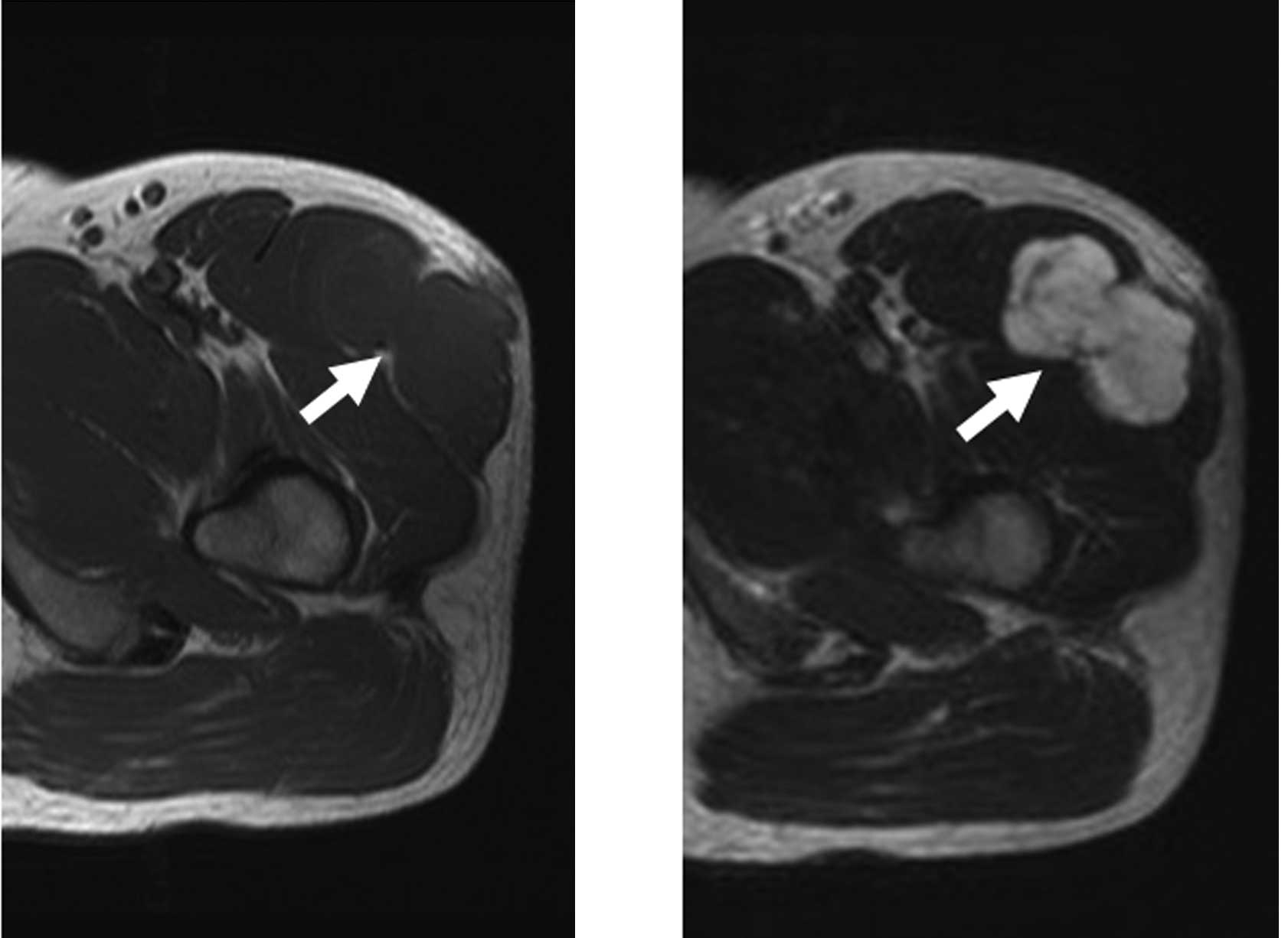
Magnetic resonance imaging (MRI) revealed a low-intensity lesion with T1-weighted imaging and a high-intensity lesion with T2-weighted imaging (Fig. 1A and B). The tumor exhibited enhancement following intravenous administration of gadolinium. The inside was heterogeneous, and the border was ill-defined (Fig. 1C). 67Ga scintigraphy did not show abnormal uptake. Chest radiography and computed tomography showed no evidence of lung metastasis. Laboratory findings were normal.

Figure 1

MRI findings. (A) T1-weighted axial imaging; (B) T2-weighted axial imaging. MRI shows an intramuscular, low signal intensity mass located in the rectus femoris and tensor fascia latae (arrow). (C) T1-weighted frontal MRI image after contrast enhancement shows a multinodular, heterogeneous, high signal intensity mass (arrow).

Open biopsy was performed. Uniform round- to spindle-like tumor cells proliferated with myxomatous stroma. Well-formed hyaline cartilage and rhabdoid-like cells were not observed. The specimen was submitted for cytogenetic analysis. 
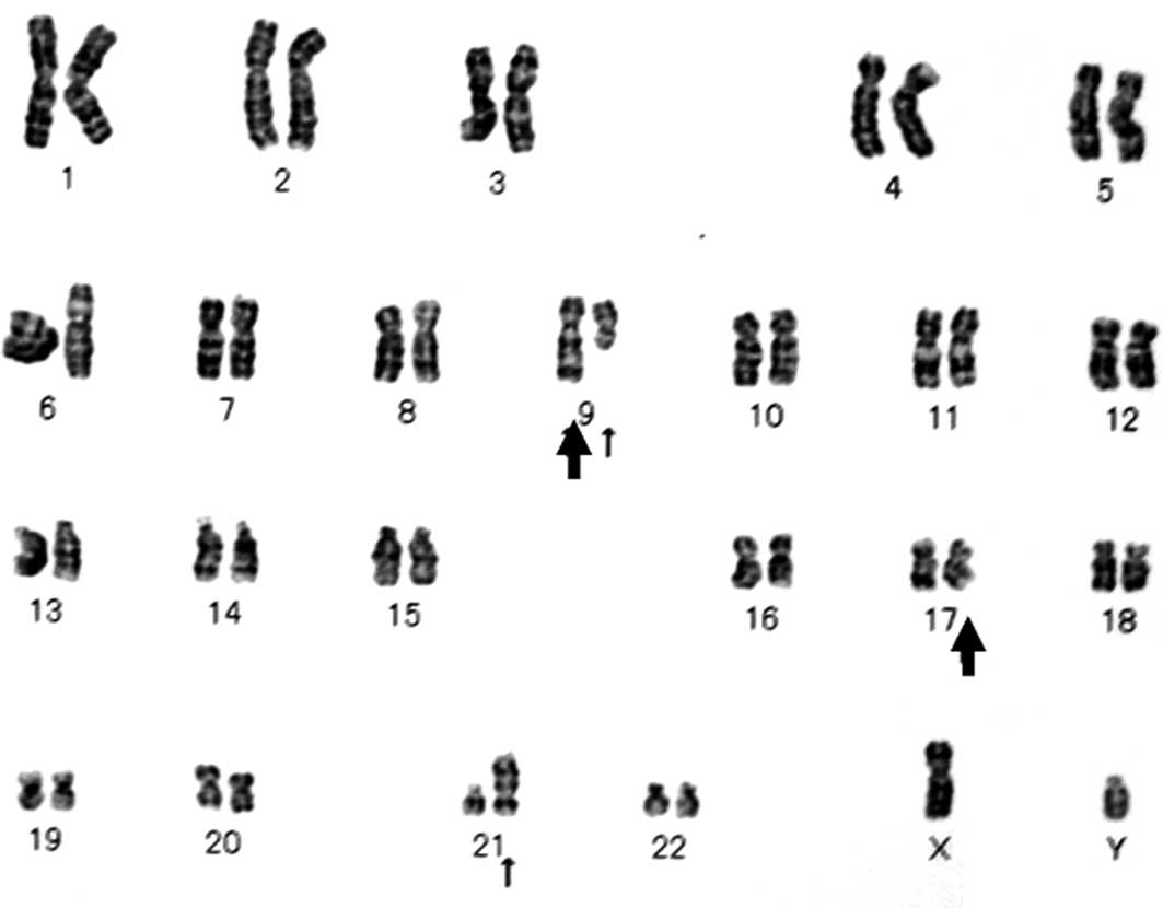
Karyotype analysis was performed with the G-banded method. Karyotypes were described using the International System for Human Cytogenetic Nomenclature (22). The composite karyotype was 46,XY,t(9;17)(q22;q11),t(9;21)(q21;p13)[16]/46,XY[4] (Fig. 2). The diagnosis of EMC was made based on the histopathological and cytogenetic findings.

Figure 2

G-banded karyotype. The translocation t(9;17)(q22;q12) was identified (large arrows). The translocation t(9;21)(q21;p13) was also observed in this case (small arrows).

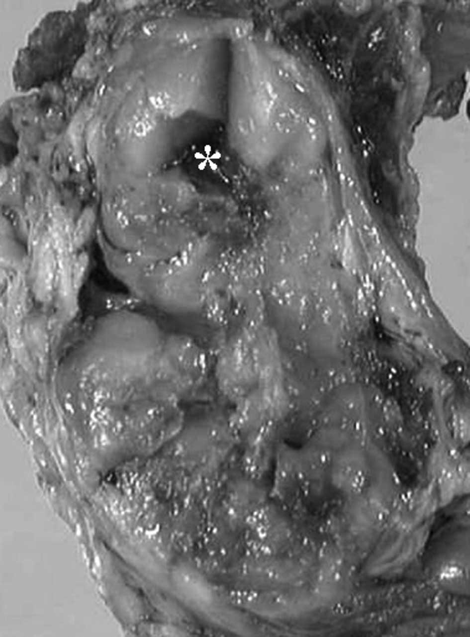
Wide resection of the tumor was performed in July, 2006. The tumor was completely resected along with a portion of the rectus femoris, tensor fascia latae and the fascia of vastus lateralis. The tumor was contained within a pseudocapsule and was 
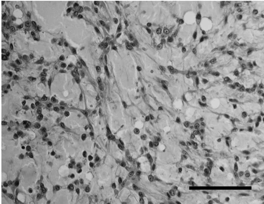
multinodular and yellowish in color (Fig. 3). Histopathologically, the tumor had a multinodular architecture consisting of myxomatous areas demarcated by fibrous septa. Each lobule was composed of proliferating tumor cells and showed a lace-like appearance with an abundant myxoid matrix (Fig. 4A). The proliferation of uniform, round tumor cells with oval nuclei was observed (Fig. 4B). Mitoses were rare. Immunohistochemically, tumor cells were positive for vimentin and S-100, and negative for epithelial membrane antigen. Chemotherapy and radiotherapy were not performed. No masses recurred during the follow-up period of 4 years and 7 months.

Figure 3

Gross appearance of surgical specimens. The tumor was encased by a thin capsule and divided into multiple gelatinous nodules by fibrous tissue. The hemorrhagic area is shown (asterisk).

Figure 4

Histological appearance of surgical specimen. (A) Lace-like appearance with an abundant myxoid matrix is shown (hematoxylin and eosin; scale bar, 200 μm). (B) Diffuse proliferation of small, uniform, round cells with hyperchromatic nuclei is observed. Mitoses are rare (hematoxylin and eosin; scale bar, 50 μm).

Discussion

EMC is a rare tumor, accounting for less than 3% of primary soft tissue sarcomas (23) and most commonly affecting individuals older than 35 years of age (1). Males are affected approximately twice as often as females. EMC arises in the deep soft tissues of proximal extremities. Patients with EMC tumors have long survival and prolonged follow-up, but EMC leads to a high disease-associated mortality rate (24,25). Local recurrence and metastasis each occur in approximately half of cases. Meis-Kindblom et al (24) reported 5-, 10-, and 15-year survival rates of 90, 70, and 60%, respectively. Therefore, EMC should be considered an intermediate-grade rather than a low-grade malignant tumor, and long follow-up is necessary.

Based on histopathological and immunohistochemical findings, EMC is difficult to distinguish from myxoid tumors including myxomas, myxofibrosarcomas (also known as myxoid malignant fibrous histiocytomas), and myxoid liposarcomas. Myxomas exhibit a similar paucity of vascular structures, but are less cellular. Myxofibrosarcomas have a similar fibrous septum and myxoid stroma but show a broad spectrum of cellularity and pleomorphism. Most myxofibrosarcomas are negative for S-100. Myxoid liposarcomas exhibit a marked plexiform vascular pattern and contain lipoblasts. S-100 positivity is found in approximately 40% of myxoid liposarcomas (26) and 50% of EMCs (27). Thus, S-100 does not help distinguish myxoid liposarcomas from EMC. Therefore, we performed a cytogenetic examination. The karyotype was t(9;17)(q22;q11), and the diagnosis of EMC was made in this case.

At present, only 50 cytogenetic cases of EMC, including our case, have been reported in the English language literature (2–21). Cytogenetic findings of EMC are occasionally complex (12%). In addition, a diploid or near-diploid chromosomal complement has been observed. Recurrent chromosomal aberrations in EMC include t(9;22)(q22;q12), t(9;17)(q22;q11), and trisomies 7, 8, 12, 18, 19 and 22. t(9;22)(q22;q12) is the most specific chromosomal aberration in EMC and was observed in 29 cases (58%). t(9;22)(q22;q12) results in the fusion of NR4A3, located at 9q22, to EWSR1, located at 22q12, to form the abnormal fusion gene EWSR1/NR4A3, which is thought to play a primary role in the causation and development of EMC (9).

t(9;17)(q22;q11) is less common than t(9;22)(q22;q12), but is a specific chromosomal aberration in EMC. Of the 50 cases, t(9;17)(q22;q11) was observed in nine cases (18%). t(9;17)(q22;q11) results in the fusion of TAF15, located at 17q11, to NR4A3, located at 9q22, to form the abnormal fusion gene TAF15/NR4A3 (15,19,27). t(9;22)(q22;q12) and t(9;17)(q22;q11) have not been found in any other tumors. t(9;15)(q22;q21), which results in the TCF12/NR4A3 fusion gene (16,27), and t(3;9)(q12;q22), which results in the TFG/NR4A3 fusion gene (28), were reported in only one case each. It is unclear whether the fusion genes identified in EMC are associated with particular morphological features and clinical significance.

We reviewed the site, size and prognosis of tumors with t(9;22)(q22;q12) and t(9;17)(q22;q11). EMC tumors with t(9;17)(q22;q11) tended to be larger than those with t(9;22)(q22;q12). However, we did not observe a significant difference due to the small number of cases. In our case, t(9;17)(q22;q11) and t(9;21)(q21;p13) were observed. t(9;21)(q21;p13) has not previously been reported in EMC or any other tumor. A number of genes located at 9q21, including ALDH1A1, NTRK2 and GAS1 were identified, but tumor-related genes located at 21p13 have not yet been identified.

EMC usually lacks overtly cartilaginous areas. Therefore, EMC was provisionally classified as a tumor of uncertain differentiation in the revised version of the World Health Organization classification of tumors of soft tissue and bone (29). Although a possible association between t(9;17)(q22;q11) and neural differentiation has been suggested (30), the number of cases examined is insufficient to draw any conclusions. Comprehensive studies examining the nature of the tumor and biological properties are required in EMC.

References

1 

Enzinger FM and Shiraki M: Extraskeletal myxoid chondrosarcoma. An analysis of 34 cases. Hum Pathol. 3:421–435. 1972. View Article : Google Scholar : PubMed/NCBI

2 

Hinrichs SH, Jaramillo MA, Gumerlock PH, Gardner MB, Lewis JP and Freeman AE: Myxoid chondrosarcoma with a translocation involving chromosomes 9 and 22. Cancer Genet Cytogenet. 14:219–226. 1985. View Article : Google Scholar : PubMed/NCBI

3 

Turc-Carel C, Dal Cin P, Rao U, Karakousis C and Sandberg AA: Recurrent breakpoints at 9q31 and 22q12.2 in extraskeletal myxoid chondrosarcoma. Cancer Genet Cytogenet. 30:145–150. 1988. View Article : Google Scholar : PubMed/NCBI

4 

Bridge JA, Sanger WG and Neff JR: Translocations involving chromosomes 2 and 13 in benign and malignant cartilaginous neoplasms. Cancer Genet Cytogenet. 38:83–88. 1989. View Article : Google Scholar : PubMed/NCBI

5 

Orndal C, Carlén B, Akerman M, et al: Chromosomal abnormality t(9;22)(q22;q12) in an extraskeletal myxoid chondrosarcoma characterized by fine needle aspiration cytology, electron microscopy, immunohistochemistry and DNA flow cytometry. Cytopathology. 2:261–270. 1991. View Article : Google Scholar

6 

Bridge JA, Bhatia PS, Anderson JR and Neff JR: Biologic and clinical significance of cytogenetic and molecular cytogenetic abnormalities in benign and malignant cartilaginous lesions. Cancer Genet Cytogenet. 69:79–90. 1993. View Article : Google Scholar : PubMed/NCBI

7 

Stenman G, Andersson H, Mandahl N, Meis-Kindblom JM and Kindblom LG: Translocation t(9;22)(q22;q12) is a primary cytogenetic abnormality in extraskeletal myxoid chondrosarcoma. Int J Cancer. 62:398–340. 1995. View Article : Google Scholar : PubMed/NCBI

8 

Hirabayashi Y, Ishida T, Yoshida MA, et al: Translocation (9;22)(q22;q12). A recurrent chromosome abnormality in extraskeletal myxoid chondrosarcoma. Cancer Genet Cytogenet. 81:33–37. 1995. View Article : Google Scholar : PubMed/NCBI

9 

Sciot R, Dal Cin P, Fletcher C, et al: t(9;22)(q22-31;q11-12) is a consistent marker of extraskeletal myxoid chondrosarcoma: evaluation of three cases. Mod Pathol. 8:765–768. 1995.PubMed/NCBI

10 

Rao UN, Surti U, Hoffner L, et al: Extraskeletal and skeletal myxoid chondrosarcoma: A multiparameter analysis of three cases including cytogenetic analysis and fluorescence in situ hybridization. Mol Diagn. 1:99–107. 1996. View Article : Google Scholar : PubMed/NCBI

11 

Kilpatrick SE, Inwards CY, Fletcher CD, Smith MA and Gitelis S: Myxoid chondrosarcoma (chordoid sarcoma) of bone: a report of two cases and review of the literature. Cancer. 79:1903–1910. 1997. View Article : Google Scholar : PubMed/NCBI

12 

Day SJ, Nelson M, Rosenthal H, Vergara GG and Bridge JA: Der(16)t(1;16)(q21;q13) as a secondary structural aberration in yet a third sarcoma, extraskeletal myxoid chondrosarcoma. Genes Chromosomes Cancer. 20:425–427. 1997. View Article : Google Scholar : PubMed/NCBI

13 

Sjögren H, Meis-Kindblom JM, Kindblom LG, Åman P and Stenman G: Fusion of the EWS-related gene TAF2N to TEC in extraskeletal myxoid chondrosarcoma. Cancer Res. 59:5064–5067. 1999.PubMed/NCBI

14 

Attwooll C, Tariq M, Harris M, Coyne JD, Telford N and Varley JM: Identification of a novel fusion gene involving hTAFII68 and CHN from a t(9;17)(q22;q11.2) translocation in an extraskeletal myxoid chondrosarcoma. Oncogene. 18:7599–7601. 1999. View Article : Google Scholar : PubMed/NCBI

15 

Bjerkehagen B, Dietrich C, Reed W, et al: Extraskeletal myxoid chondrosarcoma: multimodal diagnosis and identification of a new cytogenetic subgroup characterized by t(9;17)(q22;q11). Virchows Arch. 435:524–530. 1999. View Article : Google Scholar : PubMed/NCBI

16 

Sjögren H, Wedell B, Meis-Kindblom J, Kindblom LG and Stenman G: Fusion of the NH2-terminal domain of the basic helix-loop-helix protein TCF12 to TEC in extraskeletal myxoid chondrosarcoma with translocation t(9;15)(q22;q21). Cancer Res. 60:6832–6835. 2000.PubMed/NCBI

17 

Panagopoulos I, Mertens F, Isaksson M, et al: Molecular genetic characterization of the EWS/CHN and RBP56/CHN fusion genes in extraskeletal myxoid chondrosarcoma. Genes Chromosomes Cancer. 35:340–52. 2002. View Article : Google Scholar : PubMed/NCBI

18 

Reid R, de Silva MV and Paterson L: Poorly differentiated extraskeletal myxoid chondrosarcoma with t(9;22)(q22;q11) translocation presenting initially as a solid variant devoid of myxoid areas. Int J Surg Pathol. 11:137–141. 2003. View Article : Google Scholar

19 

Sjögren H, Meis-Kindblom JM, Örndal C, et al: Studies on the molecular pathogenesis of extraskeletal myxoid chondrosarcoma: cytogenetic, molecular genetic, and cDNA microarray analyses. Am J Pathol. 162:781–792. 2003.PubMed/NCBI

20 

Domanski HA, Carlén B, Mertens F and Akerman M: Extraskeletal myxoid chondrosarcoma with neuroendocrine differentiation: a case report with fine-needle aspiration biopsy, histopathology, electron microscopy, and cytogenetics. Ultrastruct Pathol. 27:363–368. 2003. View Article : Google Scholar

21 

Nilsson M, Meza-Zepeda LA, Mertens F, Forus A, Myklebost O and Mandahl N: Amplification of chromosome 1 sequences in lipomatous tumors and other sarcomas. Int J Cancer. 109:363–369. 2004. View Article : Google Scholar : PubMed/NCBI

22 

Shaffer LG and Tommerup N: An International System for Human Cytogenetic Nomenclature. S Karger; Basel: 2005

23 

Tsuneyoshi M, Enjoji M, Iwasaki H and Shinohara N: Extraskeletal myxoid chondrosarcoma. A clinicopathologic and electron microscopic study. Acta Pathol Jpn. 31:439–447. 1981.PubMed/NCBI

24 

Meis-Kindblom JM, Bergh P, Gunterberg B and Kindblom LG: Extraskeletal myxoid chondrosarcoma. A reappraisal of its morphologic spectrum and prognostic factors based on 117 cases. Am J Surg Pathol. 23:636–650. 1999. View Article : Google Scholar : PubMed/NCBI

25 

Saleh G, Evans HL, Ro JY and Ayala AG: Extraskeletal myxoid chondrosarcoma. A clinicopathologic study of ten patients with long-term follow-up. Cancer. 702:827–830. 1992.PubMed/NCBI

26 

Hashimoto H, Daimaru Y and Enjoji M: S-100 protein distribution in liposarcoma. An immunoperoxidase study with special reference to the distinction of liposarcoma from myxoid malignant fibrous histiocytoma. Virchows Arch A Pathol Anat Histopathol. 405:1–10. 1984. View Article : Google Scholar

27 

Okamoto S, Hisaoka M, Ishida T, et al: Extraskeletal myxoid chondrosarcoma: a clinicopathologic, immunohistochemical, and molecular analysis of 18 cases. Hum Pathol. 32:1116–1124. 2001. View Article : Google Scholar : PubMed/NCBI

28 

Hisaoka M, Ishida T, Imamura T and Hashimoto H: TFG is a novel fusion partner of NOR1 in extraskeletal myxoid chondrosarcoma. Genes Chromosomes Cancer. 40:325–328. 2004. View Article : Google Scholar : PubMed/NCBI

29 

Lucas DR and Heim S: Extraskeletal myxoid chondrosarcoma. WHO Classification of Tumours, Pathology and Genetics of Tumours of Soft Tissue and Bone. Fletcher CDM, Unni K and Martens F: IARC Press; Lyon: pp. 213–215. 2002

30 

Harris M, Coyne J, Tariq M, et al: Extraskeletal myxoid chondrosarcoma with neuroendocrine differentiation: a pathologic, cytogenetic, and molecular study of a case with a novel translocation t(9;17)(q22;q11.2). Am J Surg Pathol. 24:1020–1026. 2000. View Article : Google Scholar : PubMed/NCBI

Related Articles

  • Abstract
  • View
  • Download
  • Twitter
Copy and paste a formatted citation
Spandidos Publications style
Yasuda T, Hori T, Suzuki K, Kanamori M, Nogami S, Yahara Y and Kimura T: Extraskeletal myxoid chondrosarcoma of the thigh with a t(9;17) translocation. Oncol Lett 3: 621-624, 2012.
APA
Yasuda, T., Hori, T., Suzuki, K., Kanamori, M., Nogami, S., Yahara, Y., & Kimura, T. (2012). Extraskeletal myxoid chondrosarcoma of the thigh with a t(9;17) translocation. Oncology Letters, 3, 621-624. https://doi.org/10.3892/ol.2011.526
MLA
Yasuda, T., Hori, T., Suzuki, K., Kanamori, M., Nogami, S., Yahara, Y., Kimura, T."Extraskeletal myxoid chondrosarcoma of the thigh with a t(9;17) translocation". Oncology Letters 3.3 (2012): 621-624.
Chicago
Yasuda, T., Hori, T., Suzuki, K., Kanamori, M., Nogami, S., Yahara, Y., Kimura, T."Extraskeletal myxoid chondrosarcoma of the thigh with a t(9;17) translocation". Oncology Letters 3, no. 3 (2012): 621-624. https://doi.org/10.3892/ol.2011.526
Copy and paste a formatted citation
x
Spandidos Publications style
Yasuda T, Hori T, Suzuki K, Kanamori M, Nogami S, Yahara Y and Kimura T: Extraskeletal myxoid chondrosarcoma of the thigh with a t(9;17) translocation. Oncol Lett 3: 621-624, 2012.
APA
Yasuda, T., Hori, T., Suzuki, K., Kanamori, M., Nogami, S., Yahara, Y., & Kimura, T. (2012). Extraskeletal myxoid chondrosarcoma of the thigh with a t(9;17) translocation. Oncology Letters, 3, 621-624. https://doi.org/10.3892/ol.2011.526
MLA
Yasuda, T., Hori, T., Suzuki, K., Kanamori, M., Nogami, S., Yahara, Y., Kimura, T."Extraskeletal myxoid chondrosarcoma of the thigh with a t(9;17) translocation". Oncology Letters 3.3 (2012): 621-624.
Chicago
Yasuda, T., Hori, T., Suzuki, K., Kanamori, M., Nogami, S., Yahara, Y., Kimura, T."Extraskeletal myxoid chondrosarcoma of the thigh with a t(9;17) translocation". Oncology Letters 3, no. 3 (2012): 621-624. https://doi.org/10.3892/ol.2011.526
Follow us
  • Twitter
  • LinkedIn
  • Facebook
About
  • Spandidos Publications
  • Careers
  • Cookie Policy
  • Privacy Policy
How can we help?
  • Help
  • Live Chat
  • Contact
  • Email to our Support Team