Introduction
Primary prevention of breast cancer is a necessity.
Previous studies have demonstrated the effectiveness of drugs, such
as selective estrogen receptor modulators (SERMs) and aromatase
inhibitors (AIs) for the chemoprevention of breast cancer (1–4).
The utilization of SERMs is known to be effective in
reducing the risk of breast cancer. Moreover, recent studies using
AIs suggest that these drugs may be considerably more effective
than SERMs in post-menopausal women. These agents are useful in
cases of breast tumors that are hormone receptor (HR)-positive,
since in negative cases no benefit is observed (5).
Li and Brown performed a study in transgenic mice
that developed estrogen receptor (ER)-negative breast cancer,
demonstrating that rexinoids (retinoid analogs), tyrosine kinase
and cyclooxygenase-2 (COX-2) inhibitors have chemopreventive
properties for these specific tumors (5).
Cyclooxygenase is an enzyme that catalyzes the
conversion of arachidonic acid into prostaglandins (PGs) and is
expressed as two isoenzymes: COX-1 and COX-2. The first is
constitutive and mediates normal physiological functions, while the
second is undetectable in most normal tissues but is induced by
cytokines, growth factors, oncogenes and tumor promoters, thus
contributing to the synthesis of PGs in inflammatory diseases and
tumor tissues (6,7).
In breast cancer, COX-2 plays several roles, among
which is the inhibition of apoptosis through the synthesis of PGE2,
leading to the increased expression of anti-apoptotic protein
B-cell lymphoma/leukemia-2 (Bcl-2) and decreased pro-apoptotic
B-cell lymphoma 2-associated X protein (Bax), as well as the
promotion of neoangiogenesis (8).
Apoptosis is dependent on the balance between the anti-apoptotic
protein Bcl-2 and the pro-apoptotic proteins Bax and Bak. In breast
cancer, Bax plays a key role as a facilitator of apoptosis, a
function that ensures the balance against the main anti-apoptotic
member of this family, Bcl-2 (9).
Certain investigators have studied the correlation
between the expression of COX-2 and the proteins Bcl-2 and Bax.
Singh et al (10) observed
that in MCF-7 breast cancer cell cultures overexpressing COX-2,
high levels of Bcl-2 are also found and that these cells behave
like more aggressive tumors. Zhu et al (11) analyzed the effects of the
non-selective COX-2 inhibitor Aspisol® (a salicylic acid
derivative) on breast cancer cells in vitro and in
vivo, observing that the antitumor effect of Aspisol on breast
cancer cells is possibly mediated by the induction of apoptosis,
and may be correlated with the downregulation of COX-2 or Bcl-2
expression and the upregulation of caspase-3 or Bax.
COX-2 positively correlates with the cell
proliferation marker Ki-67. Ristimäki et al (12) analyzed the expression of COX-2 in
1,576 infiltrating ductal carcinoma (IDC) cases and found that in
instances of COX-2 overexpression (37.4% of cases) rates were high
for Ki-67 (p<0.0001), indicating a more aggressive tumor. In
another study, Boland et al (13) observed that in ductal carcinoma
in situ (DCIS), which exhibited high levels of Ki-67, COX-2
expression was also positively correlated in 79% of cases.
The type I insulin-like growth factor (IGF) receptor
(IGF1-R) is considered to be a crucial prognostic factor in breast
cancer as it is expressed in most breast cancer epithelial cells
and is essential for the malignant phenotype (14,15).
Using cell culture assays, Levitt et al (16) found a positive correlation between
COX-2 and IGF1-R, and that the use of celecoxib (a selective COX-2
inhibitor) determines a decreased expression of COX-2 and the
subsequent downregulation of IGF1-R, resulting in decreased cell
proliferation. Põld et al (17) studied lung cancer cell cultures and
observed that cells that overexpress COX-2 exhibit an increase in
anti-apoptotic and mitogenic signals induced by IGF-1.
Given the scarcity of studies evaluating the
interaction between COX-2 and the proliferation markers IGF1-R and
Ki-67, and the apoptosis markers Bcl-2 and Bax, the aim of this
study was to evaluate the correlation between these proteins in
DCIS and IDC in order to obtain information that may assist in the
prevention and treatment of breast carcinoma.
Materials and methods
Patients
We selected 110 patients (age range, 26–90)
surgically treated for breast cancer from the Mastology Clinics of
the Department of Obstetrics and Gynecology of the Santa Casa
Hospital, Brazil, between August 2002 and January 2008. Only the
patients with IDC and DCIS in the same surgical specimen were
selected for our study. The study was approved by the ethics
committee of the hospital and informed consent was obtained from
the patients who participated.
Histopathology and tissue microarray
immunohistochemistry
Following the study of macroscopic specimens,
selected fragments were dehydrated in ethanol, cleared by xylene
and embedded in paraffin to create the blocks. The blocks were cut
using a microtome calibrated for 4-mm cuts. The histological
sections were stained with hematoxylin and eosin (H&E) and were
read using a light microscope.
The DCIS cases were classified according to the
presence or absence of comedonecrosis in comedocarcinomas and
non-comedocarcinomas. The criteria proposed by Dabbs (18) and Elston and Ellis (19) were used for the classification of
DCIS and IDC using the nuclear and histological grades,
respectively.
The cases with IDC and adjacent DCIS were selected
for the preparation of the tissue microarray. Two regions from each
histological type were selected for composing the microarray; thus,
each case was represented by four areas, two from DCIS and two from
IDC from the same patient, comprising a total of 440 regions for
analysis.
In the immunohistochemistry analysis we evaluated
the expression of COX-2, Bcl-2, Bax, Ki-67 and IGF1-R using
specific antibodies that were detected using the chromogenic
substrate, diaminobenzidine. The sections were counterstained with
Harris hematoxylin, followed by dehydration and mounting in
Entellan with cover slips. The primary antibodies used in
immunohistochemistry reactions were: COX-2 clone CX-294 (Dako;
Glostrup, Denmark), Bcl-2 clone 124 (Dako), Bax clone 43-61 (Dako),
Ki-67 clone MIB-1 (Dako) and IGF1-R clone 24-31 DBS®
(Pleasanton, CA, USA) at dilutions of 1/80, 1/2000, 1/3000, 1/80
and 1/80, respectively.
Evaluation of the expression of COX-2,
Bcl-2, Bax, Ki-67 and IGF1-R
The expression of COX-2, Bcl-2, Bax, Ki-67 and
IGF1-R were evaluated by scores from two independent examiners.
We used the same criteria employed by Ristimäki
et al (12) for the analysis
of COX-2: score 0, no stained cells observed; score 1, cytoplasm
and cell membrane stained diffusely and weakly (at least 10% of
stained cells with strong intensity); score 2, cytoplasmic granular
staining of the cell membrane and moderate to strong staining in
10–90% of cells; score 3, >90% of cells stained with strong
intensity.
To classify the immunohistochemical expression of
Bcl-2, Bax, IGF1-R and Ki-67 we used the same criteria for
determining the score of the HER-2 according to the
HercepTest® (Dako) (20): score 0, no staining observed or
membrane staining observed in <10% of the tumor cells; score 1,
a faint/barely perceptible membrane staining in >10% of tumor
cells and the cells exhibit incomplete membrane staining; score 2,
a weak to moderate complete staining observed in >10% of tumor
cells; score 3, a strong complete membrane staining observed in
>10% of tumor cells.
We used the same criteria as those used by Ristimäki
et al (12), Boland et
al (13), Shim et al
(21) and Oliveira et al
(22) to classify the
immunohistochemical expression as positive or negative, 0 and 1
were regarded as negative and 2 or 3 as positive.
Statistical analysis
The correlation between COX-2, Bcl-2, Bax, IGF1-R
and Ki-67 was assessed according to the Spearman's correlation. The
Chi-square test was used to analyze nuclear grade, histological
grade, age group, tumor size, lymph node status and hormonal
status. We set 5% as the rejection level for the null hypothesis
for all the parameters we evaluated (23). The data were evaluated using the
statistical program SPSS® (Statistical Package for
Social Sciences) version 14.0 for Microsoft Windows.
Results
Patient characteristics
The aim of the present study was to evaluate the
correlation between COX-2, IGF1-R, Ki-67, Bcl-2 and Bax levels in
DCIS and IDC, present in the same surgical specimen. In addition,
we observed whether there was any correlation between the
expression of these biomarkers and age (younger or older than 50
years), tumor size (≤2 cm or >2 cm), histological grade, nuclear
grade, axillary lymph node status and hormonal status. The ages of
the patients at diagnosis ranged from 26 to 90 years old, with a
mean age of 56.4 years, a standard deviation of 12.81 and median
age of 55 years.
Evaluation of COX-2 expression
A total of 110 cases were evaluated by
immunohistochemistry and the scores were defined from 0 to 3,
according to the intensity and number of stained cells. The
COX-2-positive expression was similar in DCIS and IDC in 87% of
cases, demonstrating a high correlation (p<0.001). No
statistically significant difference was found when we analyzed the
expression of COX-2 with histological grade, nuclear grade, age,
axillary lymph node status and hormonal status.
Immunoreactivity to Bcl-2, Bax, Ki-67 and
IGF1-R
Bcl-2 was positive in 55% of the cases in IDC and
DCIS; Bax in 23% of cases in IDC and 19% of cases in DCIS; IGF-1 in
24% of cases in IDC and DCIS and Ki-67 in 81% of cases in IDC and
DCIS. Results of the statistical analyses of these data showed a
statistically significant correlation between their expression in
IDC and DCIS (p<0.001). As in the analysis of COX-2, no
statistically significant difference was observed when we analyzed
the expression of these biomarkers with histological grade, nuclear
grade, age, axillary lymph node status and hormonal status
(Table I).
 | Table IExpression of biomarkers according to
scores. |
Table I
Expression of biomarkers according to
scores.
| Scores | COX-2 (%) | Bcl-2 (%) | Bax (%) | Ki-67 (%) | IGF1-R (%) |
|---|
|
|
|
|
|
|
|---|
| IDC | DCIS | IDC | DCIS | IDC | DCIS | IDC | DCIS | IDC | DCIS |
|---|
| 1 | 12.7 | 12.7 | 45.5 | 45.5 | 77.3 | 80.9 | 19.1 | 19.1 | 76.4 | 76.4 |
| 2 | 38.2 | 38.2 | 36.4 | 36.4 | 18.2 | 15.5 | 70 | 70 | 20 | 20 |
| 3 | 49.1 | 49.1 | 18.2 | 18.2 | 4.5 | 3.6 | 10.9 | 10.9 | 3.6 | 3.6 |
Correlation between the expression of
COX-2, IGF1-R, Ki-67, Bcl-2 and Bax
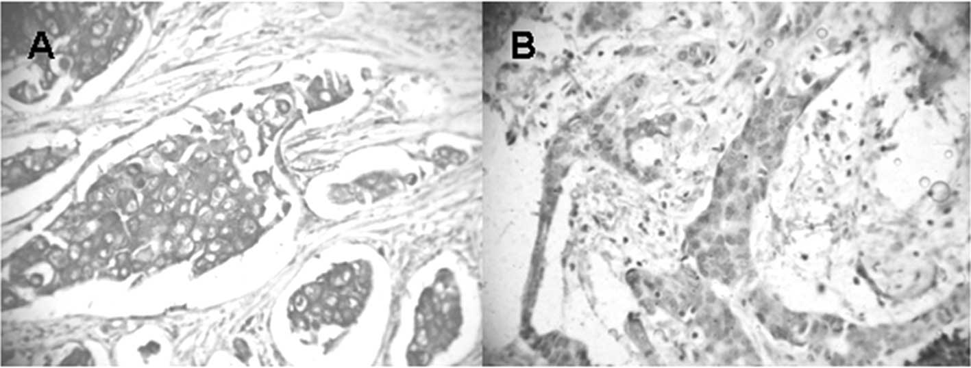
The expression of COX-2 in IDC and DCIS was
positively correlated with the expression of IGF1-R (p=0.045;
Fig. 1). We also found a negative
correlation between the expression of Ki-67 and IGF1-R in IDC and
DCIS (p=0.013). No statistically significant difference was
observed when we analyzed the expression of COX-2, Bcl-2, Bax,
Ki-67 and IGF1-R with histological grade, nuclear grade, age,
axillary lymph node status and hormonal status (Tables II and III).
| Table IICorrelation between the expression of
COX-2, Bcl-2, Bax, IGF-1 and Ki-67 in IDC in 110 cases. |
Table II
Correlation between the expression of
COX-2, Bcl-2, Bax, IGF-1 and Ki-67 in IDC in 110 cases.
| Variant | Statistic | COX-2 | Bcl-2 | Bax | IGF1-R | Ki-67 |
|---|
| COX-2 | Correlation | 1 | 0.138 | 0.113 | 0.191a | 0.029 |
| Significance
(p) | - | 0.152 | 0.241 | 0.045 | 0.766 |
| Bcl-2 | Correlation | 0.138 | 1 | -0.036 | 0.065 | 0.007 |
| Significance
(p) | 0.152 | - | 0.712 | 0.497 | 0.940 |
| Bax | Correlation | 0.113 | −0.036 | 1 | −0.055 | −0.019 |
| Significance
(p) | 0.241 | 0.712 | - | 0.570 | 0.847 |
| IGF1-R | Correlation | 0.191a | 0.065 | −0.055 | 1 | −0.236a |
| Significance
(p) | 0.045 | 0.497 | 0.570 | - | 0.013 |
| Ki-67 | Correlation | 0.029 | 0.007 | −0.019 | −0.236a | 1 |
| Significance
(p) | 0.766 | 0.940 | 0.847 | 0.013 | - |
| Table IIICorrelation between the expression of
COX-2, Bcl-2, Bax, IGF-1 and Ki-67 in DCIS in 110 cases. |
Table III
Correlation between the expression of
COX-2, Bcl-2, Bax, IGF-1 and Ki-67 in DCIS in 110 cases.
| Variant | Statistic | COX-2 | Bcl-2 | Bax | IGF1-R | Ki-67 |
|---|
| COX-2 | Correlation | 1 | 0.138 | 0.113 | 0.191a | 0.029 |
| Significance
(p) | - | 0.152 | 0.172 | 0.045 | 0.766 |
| Bcl-2 | Correlation | 0.138 | 1 | 0.039 | 0.065 | 0.007 |
| Significance
(p) | 0.152 | - | 0.688 | 0.497 | 0.940 |
| Bax | Correlation | 0.113 | 0.039 | 1 | −0.018 | 0.124 |
| Significance
(p) | 0.172 | 0.688 | - | 0.221 | 0.198 |
| IGF1-R | Correlation | 0.191a | 0.065 | −0.018 | 1 | −0.236a |
| Significance
(p) | 0.045 | 0.497 | 0.221 | - | 0.013 |
| Ki-67 | Correlation | 0.029 | 0.007 | 0.124 | −0.236a | 1 |
| Significance
(p) | 0.766 | 0.940 | 0.198 | 0.013 | - |
Discussion
The overexpression of the COX-2 enzyme in breast
tumors is a significant biological marker. Its expression has been
reported in numerous studies aimed at demonstrating its role in the
carcinogenesis and natural history of breast cancer through
mechanisms involving cytokines, growth factors, apoptotic agents
and tumor promoters (21,24,25).
Epidemiological studies have suggested that non-steroidal
anti-inflammatory drugs confer a moderate degree of benefit against
breast cancer (26).
In the present study, we found COX-2 (as detected by
immunohistochemistry) in 87% of the cases of IDC and DCIS
evaluated. Similar results were found by Davies et al
(27), who observed COX-2
expression in 79% of the 80 cases of IDC, and Shim et al
(21), who found COX-2 in IDC and
DCIS in 81.6% of the 38 cases they studied. Conversely, Half et
al (28) found the presence of
COX-2 in 43% of 42 cases of pure IDC evaluated. Similar numbers
were observed by Ristimäki et al (12) who analyzed 1,576 cases of IDC, and
found COX-2 expression in 39.9% of cases.
Oliveira et al (22) analyzed the expression of COX-2 in
IDC, DCIS and normal epithelium and correlated these levels with
nuclear grade, histological grade and the presence of
comedonecrosis. These results are similar to those found in the
present study, demonstrating positive results for COX-2 in 87% of
cases of IDC, 85% of cases of DCIS and 74.5% of normal control
patients. These data demonstrated a high correlation between the
expression of COX-2 in IDC, DCIS and normal epithelium
(p<0.001).
In the analysis of COX-2 expression in DCIS we found
a positivity rate of 87%. This rate is similar to that found by
other authors. Shim et al (21) analyzed 46 cases of pure DCIS, and
observed a positivity rate of of 85%. Shim et al (21), Half et al (28), Tan et al (29), Boland et al (13) and Perrone et al (30), found positivity rates of 63, 67, 80,
76 and 88%, respectively. This finding demonstrates homogeneity in
the expression of COX-2 in DCIS, which is not observed in invasive
disease.
The mechanisms by which COX-2 is upregulated in
breast cancer are unknown, but one possibility is that tumor cells
express a more active enzyme owing to intrinsic mechanisms. The
inactivation of tumor suppressor genes, such as p53, and the
activation of proto-oncogenes, including Ras and HER-2/neu are
involved in these mechanisms (12,31,32).
Recent studies indicate that anti-inflammatory drugs
may inhibit cell proliferation and induce apoptosis in tumor cells.
In an experimental study, Wang et al (33) examined how celecoxib, a COX-2
inhibitor, is associated with the NF-κB pathway and apoptosis in
breast cancer cell cultures treated with celecoxib. As a result,
they deduced that cell proliferation was significantly decreased
(p<0.05) in the celecoxib-treated group when compared with the
control group, who received no medication, leading to the
conclusion that celecoxib may inhibit cell proliferation and induce
apoptosis through downregulation of COX-2 and NF-κB.
We showed that in the presence of two tumor
components in the same surgical specimen (IDC and DCIS), the rate
of positivity in IDC and DCIS was similar, probably as a result of
the paracrine action of one component over the other. Our findings
denote a positive correlation between the expression of COX-2 in
DCIS and IDC. It appears that the expression of COX-2 in the in
situ component upregulates the enzyme in the infiltrating
carcinoma, since its levels are generally higher in DCIS.
A comparison of the expression of COX-2 according to
tumor size did not yield statistically significant differences
between the two groups, data consistent with the findings obtained
by Half et al (28).
However, these data are not equivalent to those obtained by
Ristimäki et al (12) and
Shim et al (21), who found
a greater expression directly proportional to tumor size. These
results may be explained by the specific analysis of invasive
tumors by these authors, as in our study and that carried out by
Half et al (28), where
samples containing DCIS and IDC in the same specimen were
evaluated.
Evaluation of COX-2 expression according to the
patients' age showed no difference between the groups. These data
were similar to those obtained by Ristimäki et al (12).
An analysis of the correlation between the
expression of COX-2 and axillary lymph node status did not yield
any difference between the lymph node groups of patients with COX-2
expression, these data being consistent with the results obtained
by Shim et al (21). Li
et al (34) analyzed the
correlation between the expression of COX-2 and lymph node
metastases and found a higher rate of axillary lymph nodes in the
group of patients with overexpressed COX-2 (p=0.012). This
difference in results may be explained by the small number of
COX-2-negative patients in our study.
When we compared the expression of COX-2 with HR, no
significant difference between the two groups was observed, which
was similar to the findings by Half et al (28) and Shim et al (21).
An analysis of the expression of Bcl-2 and Bax
revealed that Bcl-2 was positive in 55% of cases for IDC and DCIS,
while Bax was present in 23% of IDC and 19% of DCIS cases. Our rate
of positivity in DCI for Bcl-2 was similar to that found by Yang
et al (35) who observed 48%
of cases overexpressing Bcl-2, but differs from the Bax analysis
where the results showed 54% of cases with positivity. We observed
a statistically significant correlation between the expression of
Bcl-2 in DCIS and IDC, and the same was noted for Bax (p<0.001).
These data differ from those found by Mintz et al (36), who observed a higher expression of
Bcl-2 in DCIS than in IDC.
The balance between the expression of the
anti-apoptotic Bcl-2 and pro-apoptotic Bax gene is considered a
good indicator of apoptotic activity in tumor cells.
Martínez-Arribas et al (37)
studied the apoptosis associated with the two proteins in 86
specimens of breast tumors, where 14 patients had received
neoadjuvant chemotherapy. Their results revealed that Bcl-2
overexpression correlated with the presence of estrogen and
progesterone receptors in cases without previous treatment, and
high rates of apoptosis were significantly correlated with the
expression of the progesterone receptor (p=0.037). Our results
found no correlation between the expression of Bcl-2 and Bax and
the hormonal receptors.
There was also no correlation between the expression
of COX-2 and Bcl-2 and Bax, which is similar to findings described
by Arun et al (38). In
their study on the apoptosis induction in human breast cancer
cells, Michael et al (39)
found an association between a high expression of COX-2 and a low
expression of Bcl-2.
In the analysis of Bax we found 23% positivity for
IDC and 19% for DCIS, which is a significant correlation
(p<0.001). We found no correlation between the expression of
Bcl-2 and the expression of Bax, which differs from results
reported by Martínez-Arribas et al (37), who found a statistically significant
association between the two proteins (p=0.0063).
We observed a high expression of Ki-67 (81%) for IDC
and DCIS (p=0.001). This positivity correlation is consistent with
the results reported by Hoque et al (40), who concluded that there is no
increased expression of Ki-67 in IDC associated with DCIS. However,
Mylonas et al (41) reported
a higher expression of Ki-67 in IDC (p=0.05) than in DCIS and
attributed this finding to the evidence that DCIS has a lower
potential for malignancy than IDC.
We found no correlation between the expression of
Ki-67 and COX-2, which is not consistent with the study by Boland
et al, who found a positive association between the group
with high positivity for Ki-67 and COX-2 (p<0.001) for IDC and
DCIS, respectively (13).
We observed a positive correlation between the
expression of Ki-67 and hormonal status (p<0.01) in DCIS and
IDC. These data are consistent with the findings of Faratian et
al (42); however, Boland et
al (13) observed the opposite,
showing that cases with a high Ki-67 expression positively
correlated with negative HR (p=0.003).
Our analysis of the expression of IGF1-R yielded 24%
positivity for IDC and DCIS (p<0.001). When analyzing the
expression of IGF1-R in positive HR cases we observed a decrease in
IGF1-R expression to 20% in ER-positive and 19% in PR-positive
cases. As in our study, Shimizu et al (43) found no correlation between the
overexpression of IGF1-R and the variables of age, axillary lymph
node status, histological grade and hormonal status, concluding
that its prognostic value is limited.
IGF1-R is a transmembrane tyrosine kinase
involved in breast cancer proliferation, survival and metastasis
(44). The activities of IGF-1 and
2 are tightly regulated by a network of binding proteins and
targeted degradation mechanisms. This complex regulatory system is
disrupted in breast cancer, leading to excess IGF1-R signaling
(45).
In the present study, when we compared the
expression of IGF1-R with hormonal status, the correlation was
negative, although data from the literature does not show the
association between IGF1-R and HR. Shimizu et al (43) and Law et al (46) found no association between the
overexpression of IGF1-R and ER/PR.
When we compared COX-2 and IGF1-R expression we
found a statistically significant correlation (p<0.05). This
finding is consistent with that of Henriksen et al (47), who, by using the semi-quantitative
method as recommended by the HercepTest (quantifying their results
in scores 0, 1, 2 and 3), found a positive correlation between
COX-2 and IGF1-R (p<0.01).
Põld et al (17) reported that high systemic levels of
IGF-1 and the correlation with the insulin-like growth
factor-binding protein 3 (IGFBP-3) has emerged as a potential risk
marker for tumors with COX-2 overexpression.
There is a hypothesis that COX-2 increases the
viability and proliferation of tumor cells that express IGF being
accompanied by the processes of facilitating auto-phosphorylation
of IGF1-R and a decreased expression of IGFBP-3. All these actions
may be interpreted as amplifiers of the mitogenic process and as
crucial for maintaining the activity of cellular immortality
determined by IGF-1 and IGF-2, being consistent, thus explaining
the link between COX-2 and IGF1-R (17).
Due to the interrelation between COX-2 and IGF1-R by
the mechanisms described previously, the use of a combination
therapy with COX-2 and tyrosine kinase inhibitors correlated with
IGF1-R may be promising (17).
Several experimental and epidemiological studies
have demonstrated the role of COX-2 inhibitors in the prevention of
breast carcinoma. In a study that examined the growth inhibition of
breast cancer epithelial cells treated with the COX-2 inhibitor,
celecoxib, Levitt and Pollak (48)
observed that following application of this drug, the induction of
apoptosis occurred in these cells as well as a decrease in IGF-1,
concluding that these two biomarkers are closely related and may
assist in the prevention and treatment of breast cancer.
The development of effective medications that
modulate the carcinogenesis of breast tumors is crucial for the
prevention and treatment of breast cancer. Recent data suggest that
the use of tyrosine kinase, HER-2 and IGF1-R inhibitors is a good
strategy for the treatment of breast cancer (49). New studies suggest that the use of
metformin inhibits tumor growth in cells in vitro. In an
experimental study, Zakikhani et al (50) found that metformin is capable of
inducing apoptosis and inhibiting cellular proliferation in breast
cancer cells. There are few population studies concerning this
drug. Jiralerspong et al (51) evaluated patients who underwent
neoadjuvant chemotherapy and who used metformin, and observed a
better response when compared with the group that did not use
metformin, demonstrating the antitumor effect of this drug.
There is currently a lack of studies involving COX-2
inhibitors and drugs that inhibit activation of IGF1-R. Our results
have demonstrated the interrelation between COX-2 and IGF1-R.
However, further studies are required to confirm the effectiveness
and elucidation of the mechanisms and pathways involved in the use
of COX-2 inhibitors, in combination with metformin and IGF1-R
inhibitors, in order that they may be used in the prevention and
treatment of breast cancer.
References
|
1
|
Fisher B, Costantino JP, Wickerham DL,
Redmond CK, Kavanah M and Cronin WM: Tamoxifen for prevention of
breast cancer: report of the National Surgical Adjuvant Breast and
Bowel Project P-1 Study. J Natl Cancer Inst. 90:1371–1388. 1998.
View Article : Google Scholar : PubMed/NCBI
|
|
2
|
Powles T, Eeles R, Ashley SE, Easton D,
Chang J and Dowsett L: Interim analysis of the incidence of breast
cancer in the Royal Marsden Hospital tamoxifen randomized
chemoprevention trial. Lancet. 352:98–101. 1998. View Article : Google Scholar : PubMed/NCBI
|
|
3
|
Veronesi U, Maisonneuve P, Sacchini V,
Rotmensz N and Boyle P: Tamoxifen for breast cancer among women
hysterectomised. Lancet. 359:1122–1124. 2002. View Article : Google Scholar : PubMed/NCBI
|
|
4
|
Forbes JF, Cuzick J, Buzdar A, Howell A,
Tobias JS and Baum M: Arimidex, tamoxifen, alone or in combination
(ATAC) trialists' group. Effect of anastrozole and tamoxifen as
adjuvant treatment for early-stage breast cancer: 100-month
analysis of the ATAC trial. Lancet Oncol. 9:45–53. 2008.
|
|
5
|
Li Y and Brown PH: Prevention of
ER-negative breast cancer. Recent Results Cancer Res. 181:121–134.
2009. View Article : Google Scholar : PubMed/NCBI
|
|
6
|
Fosslien E: Molecular pathology of
cyclooxygenase-2 in neoplasia. Ann Clin Lab Sci. 30:3–21.
2000.PubMed/NCBI
|
|
7
|
Dempke W, Rie C, Grothey A and Schmoll H:
Cyclooxygenase-2: a novel target for cancer chemotherapy. J Cancer
Res Clin Oncol. 127:411–417. 2001. View Article : Google Scholar : PubMed/NCBI
|
|
8
|
Krcova Z, Ehrmann J, Krejci V, Eliopoulos
A and Kolar Z: TPL-2/COT and COX-2 in breast cancer. Biomed Pap Med
Fac Univ Palacky Olomouc Czech Repub. 152:21–25. 2008. View Article : Google Scholar : PubMed/NCBI
|
|
9
|
Martínez-Arribas F, Martín-Garabato E,
Zapardiel I, Sánchez J, Lucas AR, Tejerina A and Schneider J: Bax
expression in untreated breast cancer: an immunocytometric study of
255 cases. Anticancer Res. 28:2595–2598. 2008.PubMed/NCBI
|
|
10
|
Singh B, Cook KR, Vincent L, Hall CS,
Berry JA, Multani AS and Lucci A: Cyclooxygenase-2 induces genomic
instability, BCL2 expression, doxorubicin resistance, and altered
cancer-initiating cell phenotype in MCF7 breast cancer cells. J
Surg Res. 147:240–246. 2008. View Article : Google Scholar : PubMed/NCBI
|
|
11
|
Zhu XG, Tao L, Mei ZR, Wu HP and Jiang ZW:
Aspisol inhibits tumor growth and induces apoptosis in breast
cancer. Exp Oncol. 30:289–294. 2008.PubMed/NCBI
|
|
12
|
Ristimäki A, Sivula A, Lundin J, Lundin M,
Salminen T, Haglund C, Joensuu H and Isola J: Prognostic
significance of elevated cyclooxygenase-2 expression in breast
cancer. Cancer Res. 62:632–635. 2002.PubMed/NCBI
|
|
13
|
Boland GP, Butt IS, Prasad R, Knox WF and
Bundred NJ: COX-2 expression is associated with an aggressive
phenotype in ductal carcinoma in situ. Br J Cancer. 90:423–429.
2004. View Article : Google Scholar : PubMed/NCBI
|
|
14
|
Sheen-Chen SM, Chou FF, Hsu W, Huang CC,
Eng HL and Tang RP: Lack of prognostic value of insulin-like growth
factor-1 in patients with breast cancer: analysis with tissue
microarray. Anticancer Res. 27:3541–3544. 2007.PubMed/NCBI
|
|
15
|
Werner H and Bruchim I: The insulin-like
growth factor-1 receptor on the oncogene. Arch Physiol Biochem.
115:58–71. 2009. View Article : Google Scholar
|
|
16
|
Levitt RJ, Buckley J, Blouin MJ, Schaub B,
Triche TJ and Pollak M: Growth inhibition of breast epithelial
cells by celecoxib is associated with up-regulation of insulin-like
growth factor binding protein-3 expression. Biochem Biophys Res
Commun. 316:421–428. 2004. View Article : Google Scholar : PubMed/NCBI
|
|
17
|
Põld M, Krysan K, Põld A, Dohadwala M,
Heuze-Vourc'h N, Mao JT, Riedl KL, Sharma S and Dubinett SM:
Cyclooxygenase-2 modulates the insulin-like growth factor axis in
non-small-cell lung cancer. Cancer Res. 64:6549–6555.
2004.PubMed/NCBI
|
|
18
|
Dabbs DJ: Ductal carcinoma of the breast:
nuclear grade as a predictor of S-phase fraction. Human Pathol.
24:652–656. 1993. View Article : Google Scholar : PubMed/NCBI
|
|
19
|
Elston CW and Ellis IO: Pathological
prognostic factors in breast cancer I. The value of histological
grade in breast cancer: experience from a large study with
long-term follow-up. Histopathology. 19:403–410. 1991. View Article : Google Scholar
|
|
20
|
Wolff AC, Hammond ME, Schwartz JN, Hagerty
KL, Allred DC, Cote RJ, Dowsett M, Fitzgibbons PL, Hanna WM, Langer
A, et al; American Society of Clinical Oncology; College of
American Pathologists. American Society of Clinical
Oncology/College of American Pathologists guideline for human
epidermal growth factor receptor 2 testing in breast cancer. J Clin
Oncol. 25:118–145. 2006. View Article : Google Scholar
|
|
21
|
Shim JY, Jung An H, Lee YH, Kim SK, Lee KP
and Lee KS: Over-expression of Cyclooxygenase-2 is associated with
breast carcinoma and its poor prognostic factors. Mod Pathol.
16:1199–1204. 2003. View Article : Google Scholar : PubMed/NCBI
|
|
22
|
Oliveira VM, Piato S and Silva MA:
Correlation of cyclooxygenase-2 and aromatase immunohistochemical
expression in invasive ductal carcinoma, ductal carcinoma in situ,
and adjacent normal epithelium. Breast Cancer Res Treat.
95:235–241. 2006. View Article : Google Scholar
|
|
23
|
Bernard R: Fundamentals of Biostatistics.
2nd edition. Buxbury Press; Boston, MA: 1986
|
|
24
|
Hwang D, Scollard D, Byrne J and Levine E:
Expression of cyclooxygenase-1 and cyclooxygenase-2 in human breast
cancer. J Natl Cancer Inst. 90:455–460. 1998. View Article : Google Scholar : PubMed/NCBI
|
|
25
|
Buskens CJ, Sivula A, van Rees BP, Haglund
C, Offerhaus GJ, van Lanschot JJ and Ristimäki A: Comparison of
cyclooxygenase 2 expression in adenocarcinomas of the gastric
cardia and distal esophagus. Gut. 52:1678–1683. 2003. View Article : Google Scholar : PubMed/NCBI
|
|
26
|
Singh-Ranger G, Salhab M and Mokbel K: The
role of cyclooxygenase-2 in breast cancer: review. Breast Cancer
Res Treat. 109:189–198. 2008. View Article : Google Scholar : PubMed/NCBI
|
|
27
|
Davies G, Salter J, Hills F, Martin L,
Sacks N and Dowset M: Correlation of cyclooxygenase-2 expression
and angiogenesis in human breast cancer. Clin Cancer Res.
9:2652–2656. 2003.
|
|
28
|
Half E, Tang XT, Gwyn K, Sahin A, Wathen K
and Sinicrope FA: Cyclooxygenase-2 expression in human breast
cancers and adjacent ductal carcinoma in situ. Cancer Res.
62:1676–1681. 2002.PubMed/NCBI
|
|
29
|
Tan KB, Yong WP and Putti TC:
Cyclooxygenase-2 expression: a potential prognostic and predictive
marker for high-grade ductal carcinoma in situ of the breast.
Histopathology. 44:24–28. 2004. View Article : Google Scholar : PubMed/NCBI
|
|
30
|
Perrone G, Santini D, Vincenzi B, Zagami
M, La Cesa A, Bianchi A, Altomare V, Primavera A, Battista C,
Vetrani A, Tonini G and Rabitti C: COX-2 expression in DCIS:
correlation with VEGF, HER-2/neu, prognostic molecular markers and
clinicopathological features. Histopathology. 46:561–568. 2005.
View Article : Google Scholar : PubMed/NCBI
|
|
31
|
Howe LR, Subbaramaiah K, Brown AM and
Dannenberg AJ: Cyclooxygenase-2: a target for prevention and
treatment of breast cancer. Endocr Relat Cancer. 8:97–114. 2001.
View Article : Google Scholar : PubMed/NCBI
|
|
32
|
Benoit V, Relic B, Leval X, Chariot A,
Merville MP and Bours V: Regulation of HER-2 oncogene expression by
cyclooxygenase-2 and prostaglandin E2. Oncogene. 23:1631–1635.
2004. View Article : Google Scholar : PubMed/NCBI
|
|
33
|
Wang L, Liu LH, Shan BE, Zhang C, Sang MS
and Li J: Celecoxib promotes apoptosis of breast cancer cell line
MDA-MB-231 through down-regulation of the NK-κB pathway. Chin J
Cancer. 28:569–574. 2009.PubMed/NCBI
|
|
34
|
Li RX, Shi F, Wu YY, Wu Y, Guo JJ and Dong
DF: The relationship between lymphatic metastasis and serum
vascular endothelial growth factor C and cyclooxygenase 2
expression in breast cancer. Zhonghua Yi Xue Za Zhi. 88:88–91.
2008.PubMed/NCBI
|
|
35
|
Yang L, Zhu X and Ran L: Correlations of
HER-2, PCNA, Bcl-2, and Bax expression to prognosis of breast
cancer. Ai Zheng. 26:756–761. 2007.(In Chinese).
|
|
36
|
Mintz PJ, Habib NA, Jones LJ, Giamas G,
Lewis JS, Bowen RL, Coombes RC and Stebbing J: The phosphorylated
membrane estrogen receptor and cytoplasmic signaling and apoptosis
proteins in human breast cancer. Cancer. 113:1489–1495. 2008.
View Article : Google Scholar : PubMed/NCBI
|
|
37
|
Martínez-Arribas F, Nuñez-Villar MJ, Lucas
AR, Sanchez J, Tejerina A and Schneider J: Immunofluorometric study
of Bcl-2 and BAX expression in clinical fresh tumor samples from
breast cancer patients. Anticancer Res. 23:565–568. 2003.PubMed/NCBI
|
|
38
|
Arun B, Kilic G, Yen C, et al: Loss of
FHIT expression in breast cancer is correlated with poor prognostic
markers. Cancer Epidemiol Biomarkers Prev. 14:1681–1685. 2005.
View Article : Google Scholar : PubMed/NCBI
|
|
39
|
Michael MS, Badr MZ and Badawi AF:
Inhibition of cyclooxygenase-2 and activation of peroxisome
proliferator-activated receptor-γ synergistically induces apoptosis
and inhibits growth of human breast cancer cells. Int J Mol Med.
11:733–736. 2003.
|
|
40
|
Hoque A, Menter DG, Sahin AA, Sneige N and
Lippman SM: No increased Ki67 expression in ductal carcinoma in
situ associated with invasive breast cancer. Cancer Epidemiol
Biomarkers Prev. 10:153–154. 2001.PubMed/NCBI
|
|
41
|
Mylonas I, Makovitzky J, Jeschke U, Briese
U, Friese K and Gerber B: Expression of Her2/neu, steroid receptors
(ER and PR), Ki67 and p53 in invasive mammary ductal carcinoma
associated with ductal carcinoma in situ (DCIS) versus invasive
breast cancer alone. Anticancer Res. 25:1719–1723. 2005.
|
|
42
|
Faratian D, Munro A, Twelves C and
Bartlett JM: Membranous and cytoplasmic staining of Ki67 is
associated with HER2 and ER status in invasive breast carcinoma.
Histopathology. 54:254–257. 2009. View Article : Google Scholar : PubMed/NCBI
|
|
43
|
Shimizu C, Hasegawa T, Tani Y, Takahashi
F, Takeuchi M, Watanabe T, Ando M, Katsumata N and Fujiwara Y:
Expression of insulin-like growth factor 1 receptor in primary
breast cancer: immunohistochemical analysis. Hum Pathol.
35:1537–1542. 2004. View Article : Google Scholar : PubMed/NCBI
|
|
44
|
Zhang H, Sachdev D, Wang C, Hubel A,
Gaillard-Kelly M and Yee D: Detection and downregulation of type I
IGF receptor expression by antibody-conjudated quantum dots in
breast cancer cells. Breast Cancer Res Treat. 114:277–285. 2009.
View Article : Google Scholar : PubMed/NCBI
|
|
45
|
Ellis MJ, Jenkins S, Hanfelt J, Redington
ME, Taylor M, Leek R, Siddle K and Harris A: Insulin-like growth
factors in human breast cancer. Breast Cancer Res Treat.
52:175–184. 1998. View Article : Google Scholar : PubMed/NCBI
|
|
46
|
Law JH, Habibi G, Hu K, Masoudi H, Wang
MY, Stratford AL, Park E, Gee JM, Finlay P, Jones HE, et al:
Phosphorylated insulin-like growth factor-1/insulin receptor is
present in all breast cancer subtypes and is related to poor
survival. Cancer Res. 68:10238–103246. 2008. View Article : Google Scholar : PubMed/NCBI
|
|
47
|
Henriksen KL, Rasmussen BB, Lykkesfeldt
AE, Moller S, Ejlertsen B and Mouridsen HT: Semi-quantitative
scoring of potentially predictive markers for endocrine treatment
of breast cancer: a comparison between whole sections and tissue
microarrays. J Clin Pathol. 60:397–404. 2007. View Article : Google Scholar
|
|
48
|
Levitt R and Pollak M: Insulin-like growth
factor-I antagonizes the antiproliferative effects of
cyclooxygenase-2 inhibitors on BxPC-3 pancreatic cancer cells.
Cancer Res. 62:7372–7376. 2002.PubMed/NCBI
|
|
49
|
Jin Q and Esteva FJ: Cross-talk between
the ErbB/HER family and the type I insulin-like growth factor
receptor signaling pathway in breast cancer. J Mammary Gland Biol
Neoplasia. 13:485–498. 2008. View Article : Google Scholar : PubMed/NCBI
|
|
50
|
Zakikhani M, Blouin MJ, Piura E and Pollak
MN: Metformin and rapamycin have distinct effects on the AKT
pathway and proliferation in breast cancer cells. Breast Cancer Res
Treat. 123:271–279. 2010. View Article : Google Scholar : PubMed/NCBI
|
|
51
|
Jiralerspong S, Palla SL, Giordano SH,
Meric-Bernstam F, Liedtke C, Barnett CM, Hsu L, Hung MC, Hortobagyi
GN and Gonzalez-Angulo AM: Metformin and pathologic complete
responses to neoadjuvant chemotherapy in diabetic patients with
breast cancer. J Clin Oncol. 27:3297–3302. 2009. View Article : Google Scholar : PubMed/NCBI
|