Introduction
Osteosarcoma is the most common malignant tumor of
bone in childhood and adolescence (1). It is an aggressive tumor that
metastasizes primarily to the lung. Clinical efforts, including the
development of protocols for effective chemotherapy, have
significantly improved the 5-year survival rate of osteosarcoma
patients from 15 to 70% (2).
However, 30-40% of patients still succumb to the disease, mainly
due to distant metastasis to the lung (3). Despite intensive research for new
therapies, the outcome in patients with metastasis remains
extremely poor.
The S100 protein family consists of 20
calcium-binding proteins with EF hand motifs (4). S100 proteins have been shown to have
intracellular and extracellular roles in the regulation of diverse
processes, including protein phosphorylation, cell growth and
motility, cell-cycle regulation, transcription, differentiation and
cell survival (5). S100 proteins
are normally expressed in a tissue-specific manner and their
dysregulation has been causally linked to numerous diseases,
including several types of cancer (6). For example, S100A4 is involved in
proliferation, cell cycle progression and the metastasis of a
number of malignant tumors, including osteosarcoma (7). In a screening study for S100 proteins
potentially involved in the migration of osteosarcoma cells, we
identified S100A7 as a candidate.
S100A7 (Psoriasin) was originally identified as a
protein that is upregulated in psoriatic skin (8). Several S100 proteins, including
S100A7, S100A8/9, S100A12 and S100A15 (koebnerisin) were found to
be upregulated in psoriatic skin (4,9) and
these S100 proteins are localized within the epidermal
differentiation complex (EDC) on human chromosome 1 (1q21)
(10). Among the S100 family genes
in the EDC, S100A7 and S100A15 are almost identical in sequence
(<90%). Despite their similarity, S100A7 and S100A15 are
distinct in tissue distribution, regulation and function, being
exemplary for the diversity within the S100 family (9). In skin tissue, S100A7 interacts with
epidermal fatty acid-binding protein and the expression of S100A7
is elevated under pathological conditions, including inflammation
and hyperproliferation (4). Wolf
et al reported that S100A7 acts through its putative
receptor, the receptor for advanced glycation end products (RAGE),
in the inflammatory epidermis (9).
However, the roles of S100A7 in other biological/pathological
contexts, including its effects on tumor cells, remain unclear.
In the present study, we investigated the effects of
S100A7 on the migration and invasion of osteosarcoma cells.
Materials and methods
Cell culture
The human osteosarcoma cell line Saos-2 (11) and the human breast cancer cell line
MCF-7 (12) were obtained from the
American Type Culture Collection (Rockville, MD, USA). The normal
human fibroblast cell line OUMS-24 (13) and the human osteosarcoma cell line
HuO9 (14) were established in our
department. Saos-2, MCF-7 and HuO9 were cultured in RPMI-1640
(Invitrogen, Carlsbad, CA, USA). OUMS-24 was maintained in
Dulbecco's modified EM (Nissui, Tokyo, Japan). All the media were
supplemented with 10% FBS (Invitrogen), 100 μg/ml kanamycin (Meiji
Seika, Tokyo, Japan) and 0.5 μg/ml Fungizone (Invitrogen). Primary
normal human keratinocytes (NHK) were purchased from Kurabo (Osaka,
Japan) and were cultured in HuMedia-KB2 (Kurabo) with Human
Keratinocyte Growth Supplement (Invitrogen). To determine the
effect of S100A7 on cell growth, Saos-2 cells were inoculated at
1×10⁵ cells/well and cultured in a serum-free medium
containing indicated recombinant proteins (1 μg/ml). The 10%
FBS-containing medium was used as a positive control.
Preparation of recombinant proteins
S100A7 (NM002963) cDNA was amplified by PCR and
cloned into pGEX6P1 (GE Healthcare Bio-Sciences, Piscataway, NJ,
USA). The nucleotide sequence of the cDNA was confirmed by DNA
sequencing. Escherichia coli (BL21) cells were transformed
by the vector and the recombinant GST-fusion protein was purified
by glutathione-agarose affinity chromatography using a Sepharose 4B
column (GE Healthcare Bio-Sciences) under conventional conditions.
GST was released by cleaving with PreScission protease (GE
Healthcare Bio-Sciences) and removed from the final preparations
using the Sepharose 4B column. Recombinant GST protein was also
prepared and used as a negative control.
Pull-down assay and western blot
analysis
Biotinylated recombinant S100A7 or GST protein (5
μg) was incubated with 5 μg of human RAGE Fc (R&D Systems,
Minneapolis, MN, USA) and the complex of proteins was pulled down
using streptavidine agarose (Invitrogen). After washing the agarose
beads, the bound proteins were eluted and fractionated by SDS-PAGE.
Streptavidine-HRP (R&D Systems) was used to detect the applied
biotinylated S100A7 and GST proteins. Bound RAGE protein was
detected by western blotting. Western blot analysis was performed
under conventional conditions using 10 μg of protein extracts
(15). The antibodies used were as
follows: mouse anti-RAGE antibody (R&D Systems), mouse
anti-tubulin antibody (Sigma, St. Louis, MO, USA) and HRP-linked
anti-mouse IgG (Cell Signaling Technologies, Beverly, MA, USA).
RNA interference
siRNA against human RAGE (siGENOME SMART pool siRNA
targeting AGER, Thermo Scientific Dharmacon, Lafayette, CO, USA)
was transfected into cells using RNAi MAX reagent (Invitrogen). A
control siRNA with no known mammalian homology (siGENONE
non-targeting siRNA 1, Thermo Scientific Darmacon) was used as a
negative control. The cells were incubated for 72 h and used for
various assays.
Transmigration assay and invasion
assay
The transmigration and invasion potentials of the
cells were assayed in vitro under conditions similar to
those described previously (16).
Briefly, 50,000 osteosarcoma cells were inoculated into top wells
of pre-coated Boyden chambers (pore size, 8 μm; BD Biosciences,
Bedford, MA, USA). Following incubation, the filters were stained
and the cells on the bottom surface were counted. For the
transmigration assay, the chambers were coated with 2 μg of human
fibronectin (Sigma) on the bottom and the incubation time was 8 h.
For the invasion assay, 2 μg of human fibronectin and 10 μg of
Matrigel (BD Biosciences) were applied onto the upper and lower
sides, respectively, and the cells were incubated for 24 h. Mean
values were obtained from 10 visual fields.
Gelatin zymography
Saos-2 cells were treated with siRNA for 24 h,
washed twice and incubated with 10 ml of Opti-MEM (Invitrogen)
supplemented with either S100A7 or GST. The conditioned medium was
collected and concentrated into 100 μl by centrifugation using a
Centricon (Amicon Ultra-15 Ultracell-10k; Millipore, Billerica, MA,
USA) and then mixed with 50 μl of 3X SDS sample buffer. The samples
(10 μl) were applied onto an 8% SDS-polyacrylamide slab gel
containing 0.5% gelatin (Sigma). Following electrophoresis, the gel
was washed with water to remove the SDS, soaked in protein
refolding buffer (2.5% Triton X-100, 10% glycerol, 0.5 mM
CaCl2, 100 mM NaCl, 50 mM Tris-HCl/pH 7.4) for 1 h and
incubated in 50 mM Tris-HCl/pH 7.4 containing 0.5 mM
CaCl2 for 12 h at 37°C. The gel was then stained with 1%
Coomassie brilliant blue (CBB) and further treated with 10%
methanol and 5% acetic acid to destain it. The gelatinolytic
activity was detected as clear bands on a blue background of the
CBB-stained gel. The recombinant matrix metalloproteinase (MMP)-2
and -9 proteins (R&D Systems) were used as positive
controls.
Statistical analysis
Each experiment was repeated a minimum of three
times. The results are expressed as the mean ± SD. The Student's
t-test was used to compare the two groups. P<0.05 was considered
to indicate a statistically significant result.
Results and Discussion
S100A7 promoted the migration of Saos-2
cells
We screened for S100 proteins that promote the
migration of osteosarcoma cells. The cDNA of 20 S100 family members
was cloned and GST-fused recombinant proteins were prepared. The
proteins were applied to Saos-2 cells in the wound migration assay
in vitro. Among the member proteins, S100A7 was identified
as the most promising candidate (data not shown).
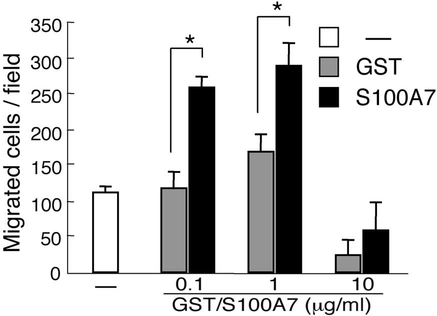
We confirmed the function of S100A7 in Saos-2 cells
by the transmigration assay (Fig.
1). The number of cells that migrated was significantly
increased by the S100A7 protein and the extent of the increase was
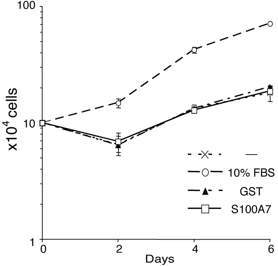
dose-dependent. However, the growth of the Saos-2 cells was not
affected by the addition of S100A7 protein (Fig. 2).
RAGE was necessary in S100A7-dependent
promotion of Saos-2 cell migration
S100 proteins are recognized as damage-associated
molecular pattern proteins (DAMPs) due to their release from
damaged cells under conditions of cell stress (17). Certain DAMPs, including S100
proteins, are thought to be ligands for the multiligand receptor
RAGE. RAGE is a type I transmembrane protein belonging to the
immunoglobulin superfamily (18)
and is involved in a broad range of inflammatory, degenerative and
hyperproliferative diseases, including sepsis, rheumatoid
arthritis, diabetic nephropathy/angiopathy, atherosclerosis, cancer
and neurological disorders (19,20).
The receptor is composed of an extracellular region, a hydrophobic
transmembrane-spanning domain and a short cytoplasmic tail. The
extracellular domain binds a number of ligands, including advanced
glycation end products, high-mobility group box 1, S100 family
proteins and amyloid fibrils (21).
Wolf et al (9) revealed the
interaction between S100A7 and RAGE by a competitive ligand binding
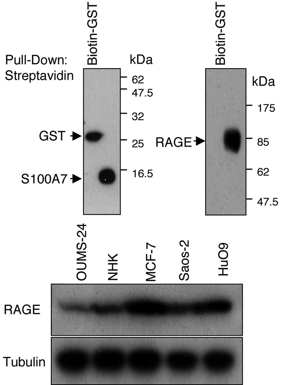
assay. We performed a pull-down assay using recombinant RAGE and
biotinylated S100A7 proteins (Fig.
3A). The RAGE protein was co-precipitated with biotinylated
GST-S100A7 but not with biotinylated GST. The measurement of the
surface plasmon resonance of these proteins also supported their
interaction (data not shown). RAGE is expressed in various types of
human cancer, including brain, breast, colon, lung, oral squamous
cell and ovarian cancer (22). The
expression of RAGE was examined in osteosarcoma cell lines, and the
results showed that compared with normal fibroblasts and
keratinocytes, which are known to express RAGE protein,
osteosarcoma cells (Saos-2 and HuO9) expressed RAGE protein at high
levels (Fig. 3B).
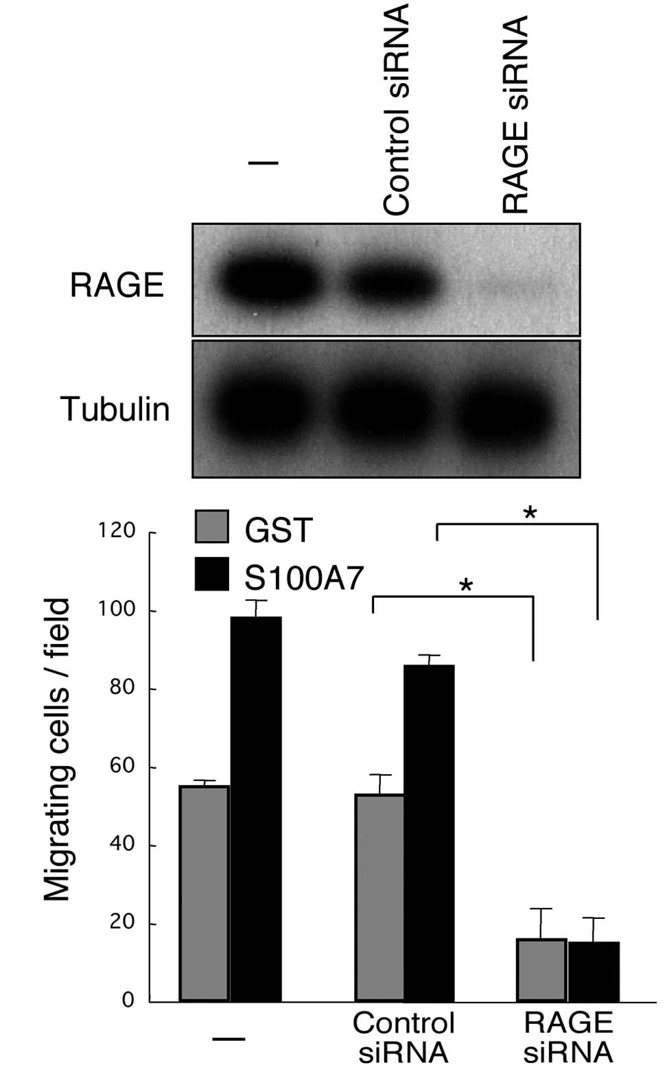
The functional involvement of RAGE in the
S100A7-induced promotion of migration of osteosarcoma cells was
also assessed. The expression of RAGE was successfully
downregulated by the specific siRNA in Saos-2 cells (Fig. 4A). The downregulation of RAGE
resulted in marked eradication of the S100A7-promoted
transmigration of Saos-2 cells (Fig.
4B). Notably, RAGE downregulation suppressed not only the
migration of S100A7-stimulated Saos-2 cells but also that of
unstimulated cells, suggesting a potential role of endogenous
S100A7 and/or other RAGE ligands in transmigration capacity.
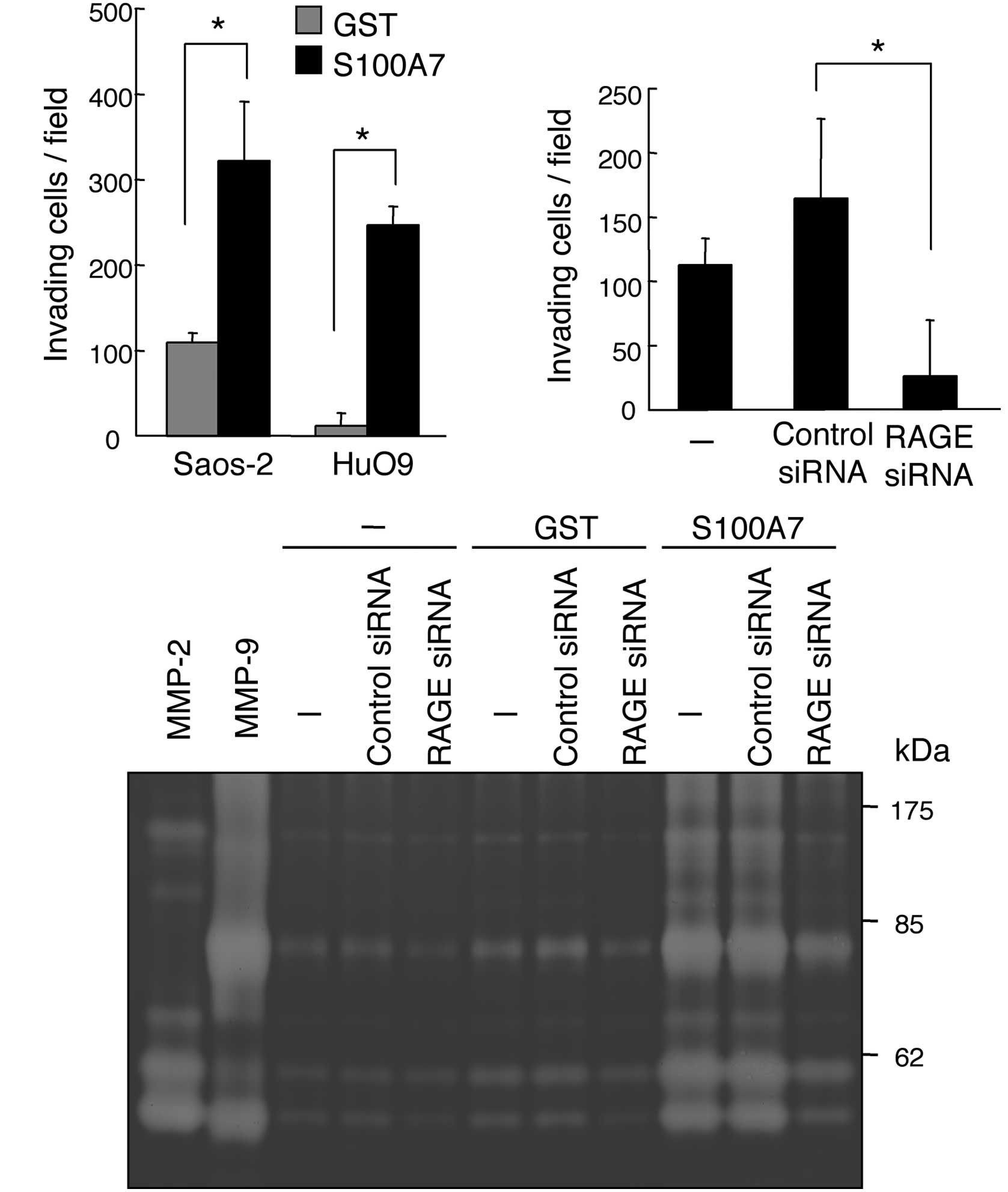
S100A7 promoted the invasion and MMP
activity of osteosarcoma cells
The coordination of cell migration and the
degradation of matrix proteins is crucial for the invasion of
malignant cells. We therefore performed an invasion assay that
mimicks the invasion of cancer cells passing through the basement
membrane (23). The addition of
S100A7 significantly increased the invasion capacity of Saos-2 and
HuO9 osteosarcoma cells (Fig. 5A).
The downregulation of RAGE by siRNA demonstrates that the invasion
of Saos-2 cells also depends on the expression of RAGE (Fig. 5B).
MMPs are known to be involved in the invasion of
various types of cancer cells, including osteosarcoma cells
(24,25). We analyzed the activity of MMPs by
gelatin zymography (Fig. 5C). The
application of S100A7 to Saos-2 cells resulted in a marked
enhancement of the activity of MMP-2 and MMP-9 (Fig. 5C). Collectively, our results
indicate that the S100A7-RAGE signal transdution pathway is
involved in the invasion of osteosarcoma cells.
S100A7 is known to promote breast cancer progression
(26). However, S100A7 has been
reported to act as a negative regulator of breast cancer invasion
(27). Our data have demonstrated
that S100A7 promoted the migration and invasion of osteosarcoma
cells in vitro. A crucial question is whether S100A7
functions to promote the invasion and/or metastasis of osteosarcoma
in vivo. Hiratsuka et al (28) revealed that S100A8 and S100A9
indirectly act on the lung to accelerate the migration of primary
tumor cells to lung tissues. Andresen et al (29) reported that the expression of S100A7
was detected in the normal lung and that it was increased under
pathological conditions. An increased production of S100A7 in the
lung may enable circulating cancer cells to settle and grow
invasively in the lung. Although further studies are needed, the
S100A7-RAGE axis may be a new target for preventing the invasion
and/or metastasis of osteosarcoma.
Acknowledgements
This study was supported in part by grants from the
Ministry of Education, Culture, Sports, Science, and Technology of
Japan (70379840 to M. Sakaguchi and 21591699 to K. Kataoka).
Abbreviations:
|
RAGE
|
receptor for advanced glycation end
products
|
|
siRNA
|
small interfering RNA
|
|
CBB
|
Coomassie brilliant blue
|
|
MMP
|
matrix metalloproteinase
|
References
|
1
|
Longhi A, Errani C, De Paolis M, Mercuri M
and Bacci G: Primary bone osteosarcoma in the pediatric age: state
of the art. Cancer Treat Rev. 32:423–436. 2006. View Article : Google Scholar : PubMed/NCBI
|
|
2
|
Provisor AJ, Ettinger LJ, Nachman JB,
Krailo MD, Makley JT, Yunis EJ, Huvos AG, Betcher DL, Baum ES,
Kisker CT and Miser JS: Treatment of nonmetastatic osteosarcoma of
the extremity with preoperative and postoperative chemotherapy: a
report from the Children's Cancer Group. J Clin Oncol. 15:76–84.
1997.
|
|
3
|
Ferguson WS and Goorin AM: Current
treatment of osteosarcoma. Cancer Invest. 19:292–315. 2001.
View Article : Google Scholar : PubMed/NCBI
|
|
4
|
Eckert RL, Broome AM, Ruse M, Robinson N,
Ryan D and Lee K: S100 proteins in the epidermis. J Invest
Dermatol. 123:23–33. 2004. View Article : Google Scholar : PubMed/NCBI
|
|
5
|
Donato R: Intracellular and extracellular
roles of S100 proteins. Microsc Res Tech. 60:540–551. 2003.
View Article : Google Scholar : PubMed/NCBI
|
|
6
|
Salama I, Malone PS, Mihaimeed F and Jones
JL: A review of the S100 proteins in cancer. Eur J Surg Oncol.
34:357–364. 2008. View Article : Google Scholar : PubMed/NCBI
|
|
7
|
Boye K and Maelandsmo GM: S100A4 and
metastasis: a small actor playing many roles. Am J Pathol.
176:528–535. 2010. View Article : Google Scholar : PubMed/NCBI
|
|
8
|
Madsen P, Rasmussen HH, Leffers H, Honoré
B, Dejgaard K, Olsen E, Kiil J, Walbum E, Andersen AH, Basse B, et
al: Molecular cloning, occurrence, and expression of a novel
partially secreted protein ‘psoriasin’ that is highly up-regulated
in psoriatic skin. J Invest Dermatol. 97:701–712. 1991.PubMed/NCBI
|
|
9
|
Wolf R, Howard OM, Dong HF, Voscopoulos C,
Boeshans K, Winston J, Divi R, Gunsior M, Goldsmith P, Ahvazi B, et
al: Chemotactic activity of S100A7 (Psoriasin) is mediated by the
receptor for advanced glycation end products and potentiates
inflammation with highly homologous but functionally distinct
S100A15. J Immunol. 181:1499–1506. 2008. View Article : Google Scholar : PubMed/NCBI
|
|
10
|
Engelkamp D, Schäfer BW, Mattei MG, Erne P
and Heizmann CW: Six S100 genes are clustered on human chromosome
1q21: identification of two genes coding for the two previously
unreported calcium-binding proteins S100D and S100E. Proc Natl Acad
Sci USA. 90:6547–6551. 1993. View Article : Google Scholar
|
|
11
|
Rodan SB, Imai Y, Thiede MA, Wesolowski G,
Thompson D, Bar-Shavit Z, Shull S, Mann K and Rodan GA:
Characterization of a human osteosarcoma cell line (Saos-2) with
osteoblastic properties. Cancer Res. 47:4961–4966. 1987.PubMed/NCBI
|
|
12
|
Soule HD, Vazguez J, Long A, Albert S and
Brennan M: A human cell line from a pleural effusion derived from a
breast carcinoma. J Natl Cancer Inst. 51:1409–1416. 1973.PubMed/NCBI
|
|
13
|
Bai L, Mihara K, Kondo Y, Honma M and
Namba M: Immortalization of normal human fibroblasts by treatment
with 4-nitroquinoline 1-oxide. Int J Cancer. 53:451–456. 1993.
View Article : Google Scholar : PubMed/NCBI
|
|
14
|
Kawai A, Ozaki T, Ikeda S, Oda T, Miyazaki
M, Sato J, Taketa K and Inoue H: Two distinct cell lines derived
from a human osteosarcoma. J Cancer Res Clin Oncol. 115:531–536.
1989. View Article : Google Scholar : PubMed/NCBI
|
|
15
|
Yamamoto K, Sakaguchi M, Medina RJ, Niida
A, Sakaguchi Y, Miyazaki M, Kataoka K and Huh NH: Transcriptional
regulation of a brown adipocyte-specific gene, UCP1, by KLF11 and
KLF15. Biochem Biophys Res Commun. 400:175–180. 2010. View Article : Google Scholar : PubMed/NCBI
|
|
16
|
Kataoka K and Huh NH: A novel
beta1,3-N-acetylglucosaminyltransferase involved in invasion of
cancer cells as assayed in vitro. Biochem Biophys Res Commun.
294:843–848. 2002. View Article : Google Scholar : PubMed/NCBI
|
|
17
|
Foell D, Wittkowski H, Vogl T and Roth J:
S100 proteins expressed in phagocytes: a novel group of
damage-associated molecular pattern molecules. J Leukoc Biol.
81:28–37. 2007. View Article : Google Scholar : PubMed/NCBI
|
|
18
|
Neeper M, Schmidt AM, Brett J, Yan SD,
Wang F, Pan YC, Elliston K, Stern D and Shaw A: Cloning and
expression of a cell surface receptor for advanced glycosylation
end products of proteins. J Biol Chem. 267:14998–15004.
1992.PubMed/NCBI
|
|
19
|
Yan SF, Ramasamy R and Schmidt AM:
Mechanisms of disease: advanced glycation end-products and their
receptor in inflammation and diabetes complications. Nat Clin Pract
Endocrinol Metab. 4:285–293. 2008. View Article : Google Scholar : PubMed/NCBI
|
|
20
|
Rojas A, Figueroa H and Morales E: Fueling
inflammation at tumor microenvironment: the role of
multiligand/RAGE axis. Carcinogenesis. 31:334–341. 2010. View Article : Google Scholar : PubMed/NCBI
|
|
21
|
Sparvero LJ, Asafu-Adjei D, Kang R, Tang
D, Amin N, Im J, Rutledge R, Lin B, Amoscato AA, Zeh HJ and Lotze
MT: RAGE (Receptor for Advanced Glycation Endproducts), RAGE
ligands, and their role in cancer and inflammation. J Transl Med.
7:172009. View Article : Google Scholar : PubMed/NCBI
|
|
22
|
Riehl A, Németh J, Angel P and Hess J: The
receptor RAGE: Bridging inflammation and cancer. Cell Commun
Signal. 7:122009. View Article : Google Scholar : PubMed/NCBI
|
|
23
|
Albini A and Benelli R: The chemoinvasion
assay: a method to assess tumor and endothelial cell invasion and
its modulation. Nat Protoc. 2:504–511. 2007. View Article : Google Scholar : PubMed/NCBI
|
|
24
|
Gialeli C, Theocharis AD and Karamanos NK:
Roles of matrix metalloproteinases in cancer progression and their
pharmacological targeting. FEBS J. 278:16–27. 2011. View Article : Google Scholar : PubMed/NCBI
|
|
25
|
Xin ZF, Kim YK and Jung ST: Risedronate
inhibits human osteosarcoma cell invasion. J Exp Clin Cancer Res.
28:1052009. View Article : Google Scholar : PubMed/NCBI
|
|
26
|
Emberley ED, Murphy LC and Watson PH:
S100A7 and the progression of breast cancer. Breast Cancer Res.
6:153–159. 2004. View
Article : Google Scholar : PubMed/NCBI
|
|
27
|
Krop I, März A, Carlsson H, Li X,
Bloushtain-Qimron N, Hu M, Gelman R, Sabel MS, Schnitt S, Ramaswamy
S, et al: A putative role for psoriasin in breast tumor
progression. Cancer Res. 65:11326–11334. 2005. View Article : Google Scholar : PubMed/NCBI
|
|
28
|
Hiratsuka S, Watanabe A, Sakurai Y,
Akashi-Takamura S, Ishibashi S, Miyake K, Shibuya M, Akira S,
Aburatani H and Maru Y: The S100A8-serum amyloid A3-TLR4 paracrine
cascade establishes a pre-metastatic phase. Nat Cell Biol.
10:1349–1355. 2008. View
Article : Google Scholar : PubMed/NCBI
|
|
29
|
Andresen E, Lange C, Strodthoff D,
Goldmann T, Fischer N, Sahly H, Branscheid D and Heine H:
S100A7/psoriasin expression in the human lung: unchanged in
patients with COPD, but upregulated upon positive S. aureus
detection. BMC Pulm Med. 11:102011. View Article : Google Scholar : PubMed/NCBI
|