Introduction
Multiple myeloma (MM) is the second most commonly
diagnosed hematologic malignancy (1). Historically, the prognosis of patients
with MM has been poor due to a lack of effective therapies. Over
the past decade, although the introduction of new drugs, including
bortezomib and lenalidomide, has improved the treatment landscape
for MM patients, almost all patients continue to experience disease
relapse (2). Therefore, further
investigations to find a novel anti-myeloma drug should be
conducted.
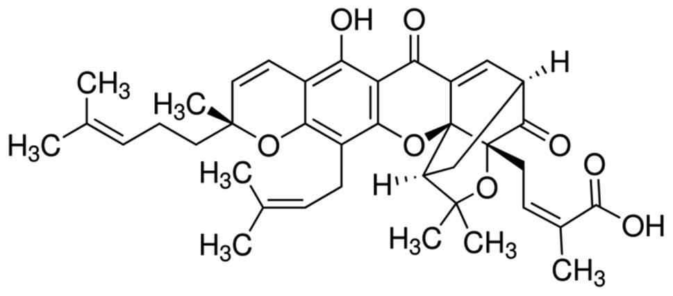
Gambogic acid (GA,
C38H44O8, Fig. 1) is the main active ingredient of
gamboges secreted from the Garcinia hanburryi tree, which
mainly grows in Southeast Asia. GA is known to have extensive
antitumor activities in certain solid tumors (3,4). Over
the last decade, our group has confirmed that GA exhibits
cytotoxicity in various types of hematological malignancy (5,6).
Various mechanisms by which GA exhibits extensive anticancer
activity have been reported, including the downregulation of Bcl-2
(3,4), activation of p53 (7) and caspase-3 (4) and the downregulation of the HERG
potassium channel (5). However,
which point is the key target of GA in the induction of the
apoptosis of cancer cells remains uncertain. As the changes in
these proteins were correlated with the generation of reactive
oxygen species (ROS) (8–11), it is suggested that GA contributes
to ROS accumulation, which causes changes in the proteins mentioned
above as the downstream targets of ROS.
The molecular structure of GA includes an
α,β-unsatured ketone, which is present in certain drugs that induce
apoptosis by generating ROS (12)
and ROS accumulation contributes to the apoptosis of human hepatoma
SMMC-7721 cells treated with GA (13). Based on these theories, we
hypothesized that GA has the potential to induce the apoptosis of
MM RPMI-8226 cells via ROS accumulation.
ROS are generally highly reactive and short-lived
molecules, including superoxide anion, hydroxyl, peroxyl, alkoxyl
and O2-derived non-radical species such as hydrogen
peroxide that are generated by the incomplete one-electron
reduction of oxygen (14). As
products or by-products of cell metabolism, ROS act either as
signaling molecules or as cell toxicants depending on the site of
generation, spatial distribution, pulse concentration and temporal
duration (9). ROS-generating
enzymes are usually controlled at the gene and protein level
(15,16). ROS levels are balanced by
non-enzymatic antioxidants (e.g., glutathione) and antioxidant
enzymes (e.g., superoxide dismutase and catalase) (17). When redox signaling and control are
disrupted, or the balance between oxidants and antioxidants tips
towards the oxidant side, the concentration of ROS rises,
contributing to the damage of biomolecules, including DNA, proteins
and lipids, by oxidative modification (18). The damage that occurs may lead to
disease (19), but also contributes
to the anticancer activity of chemotherapy (20).
SIRT1, a NAD+-dependent deacetylase, acts
on numerous substrates to control cell senescence, proliferation
and apoptosis (21). There is a
close association between the cellular redox status and SIRT1
function, as the overexpression of SIRT1has been reported to block
oxidant-induced apoptosis via the inhibition of p53 activity
(22). Under nutrient depletion,
SIRT1 may protect MM RPMI-8226 cells from apoptosis (23). Moreover, SIRT1 is relevant to
tumorigenesis and chemotherapy resistance (24), and SRT1720, which targets SIRT1, has
been shown to inhibit growth and induce apoptosis in MM cells
resistant to conventional and bortezomib therapies (25). This observation indicates that SIRT1
may be a new target in anticancer research, particularly in MM,
which has a high relapse rate.
In the present study, we examined the effect of GA
on the proliferation and apoptosis of RPMI-8226 cells and explored
the correlation between ROS generation, SIRT1 expression and
anticancer activity. We demonstrated that GA had the potential to
inhibit the proliferation of RPMI-8226 cells and induce apoptosis.
These properties were mainly dependent on ROS accumulation, the
activation of the downstream targets of GA and SIRT1
downregulation.
Materials and methods
Reagents and cell culture
GA (C38H44O8,
molecular weight 628.75 g/mol), dimethyl sulfoxide (DMSO), Hoechst
33258, 3-(4,5-dimethyl-2-thiazolyl)-2,5-diphenyl-2H- tetrazolium
(MTT) and scavenger N-acetylcysteine (NAC) were purchased from
Sigma-Aldrich (St. Louis, MO, USA).
2′,7′-dichlorofluorescein-diacetate (DCFH-DA) was purchased from
Beyotime (Jiangsu, China). RPMI-1640 medium was purchased from
Gibco Co. (Carlsbad, CA, USA), while the fetal bovine serum (FBS)
was from Hangzhou Sijiqing Biological Engineering Materials Co.,
Ltd. (Hangzhou, China). The Annexin V-fluorescein isothiocyanate
(FITC)/propidium iodide (PI) reagent kit was purchased from Nanjing
Key-Gen Biotech Co., Ltd. (Nanjing, China). The anti-activated
caspase-3, anti-PARP and anti-γ-tubulin antibodies were from Cell
Signaling Technology, Inc. (Danvers, MA, USA). The anti-SIRT1
antibody was purchased from Santa Cruz Biotechnology (Santa Cruz,
CA, USA). The anti-rabbit and anti-mouse secondary antibodies were
from Jackson ImmunoResearch Laboratories, Inc. (West Grove, PA,
USA). The BCA Protein Assay kit, chemiluminescence reagent kit and
PVDF membranes were provided by Pierce Biotechnology, Inc.
(Rockford, IL, USA). Briefly, GA was dissolved in DMSO,
equivalently packed, stored at −20°C and thawed prior to use. The
RPMI-8226 cells were donated by the Department of Immunology,
Tongji Medical College, Huazhong University of Science and
Technology (Wuhan, China) and were cultured in RPMI-1640 medium
supplemented with 10% FBS and placed in a humidified incubator with
95% air and 5% CO2 at 37°C.
MTT assay
The effect of GA on the proliferation of RPMI-8226
cells was analyzed using the MTT assay. Briefly, cells
(2×10⁴) were seeded in a 96-well plate and treated with
0.5, 1.0, 1.5, 2.0 or 2.5 μM GA for 12 h. Following incubation, 20
μl MTT (5 mg/ml) was added to each well and the cells were
incubated for a further 3 h at 37°C. The supernatant was discarded,
150 μl DMSO was added and the plate was gently agitated until the
blue crystals were dissolved. Absorbance (A) at a wavelength of 490
nm was measured using a plate microreader (Tecan Spectra,
Männedorf, Switzerland). The cell proliferation inhibition rate (%)
was calculated using the formula: [1-(A of experimental samples/A
of the control)]x100.
Annexin V-FITC/PI double-labeled flow
cytometry
To detect the apoptotic ratio of cells treated with
GA (1.0, 1.5 or 2.0 μM) alone or with NAC for 12 h, the expression
of Annexin V-FITC and the exclusion of PI were detected using
two-color flow cytometry (FCM). RPMI-8226 cells were collected
using EP tubes, washed twice with PBS and resuspended in 500 μl
binding buffer. The samples were incubated with 5 μl Annexin V-FITC
for 10 min at room temperature and then 5 μl PI was added. Each
sample was incubated for a further 10 min at room temperature in
the dark before the fluorescence intensity was quantitated using a
flow cytometer (Becton-Dickinson, Franklin Lakes, NJ, USA).
Hoechst 33258 staining
The nuclear fragmentation in RPMI-8226 cells treated
with 2.0 μM GA for 12 h was visualized using Hoechst 33258
staining. RPMI-8226 cells were plated in 12-well plates at a
density of 1×10⁵ cells/well and incubated with GA. After
12 h, the cells were collected and washed twice in PBS. The cells
were then fixed in 4% paraformaldehyde for 10 min at room
temperature and re-suspended in PBS prior to deposition on
polylysine-coated slides. After 30 min, the adhered cells were
permeabilized with 0.1% Triton X-100 for 5 min at 4°C and incubated
with Hoechst 33258 for 30 min at room temperature. Following
washing with PBS, the cells were mounted with glycerol and covered
with a cover slip. The images of the nuclei were captured using an
Olympus BH-2 fluorescence microscope (Tokyo, Japan).
Detection of intracellular ROS
levels
The levels of ROS in RPMI-8226 cells were detected
using DCFH-DA. The cells were plated in 12-well plates and treated
with GA at different concentrations (1.0, 1.5 or 2.0 μM) in the
absence or presence of 1.5 mM NAC. When the interruption point was
reached, the cells were collected and washed three times in PBS.
DCFH-DA (500 μl; 10 μM) was added to each sample and the cells were
incubated at 37°C for 30 min. During the incubation period, each
sample was agitated every 10 min to ensure that the reagent reacted
sufficiently with the ROS. To reduce the fluorescence background,
each sample was washed twice in PBS before detecting the
fluorescence intensity of DCF using FCM (Becton Dickinson).
Western blot analysis
RPMI-8226 cells treated with 1.5 or 2.0 μM GA in the
absence or presence of 1.5 mM NAC for 12 h were collected and lysed
in lysis buffer (150 mM NaCl, 50 mM Tris with pH 7.4, 1% NP40, 0.1%
SDS, 0.5 sodium deoxycholate) supplemented with protease
inhibitors, followed by centrifugation at 12,000 × g for 15 min at
4°C. The protein concentration in each sample extract was detected
using the BCA assay. SDS-PAGE was performed on 15% polyacrylamide
gels, with 40 μg of protein samples per lane. Following
electrophoresis, the proteins were transferred to PVDF membranes
and incubated in 5% non-fat milk at room temperature for 2 h.
Subsequently, the membranes were incubated with a specific primary
antibody overnight at 4°C. After being washed three times using
PBS, the membranes were incubated with an appropriate concentration
of horseradish peroxidase (HRP)-conjugated anti-mouse or
anti-rabbit secondary antibody for 2 h. After being washed a
further three times with PBS, the specific protein band was
visualized using the ECL kit.
Statistical analysis
Experiments were repeated three times. The data were
processed using SPSS 13.0 statistical software for Windows (SPSS,
Chicago, IL, USA) and shown as the mean ± SD. Comparisons among the
groups were analyzed using one-way ANOVA and the
Student-Newman-Keuls (SNK) test. P<0.05 was considered to
indicate a statistically significant result.
Results
GA inhibited the proliferation of
RPMI-8226 cells
The MTT assay was used to identify the effect of GA
on the proliferation inhibition rate of RPMI-8226 cells. The
inhibition rates of the cells treated with 0.5, 1.0, 1.5, 2.0 and
2.5 μM were found to be 5.99±3.39, 19.41±2.95, 36.26±4.34,
43.69±3.07 and 52.28±1.24%, respectively, which were significantly
higher than those of the untreated cells (Fig. 2).
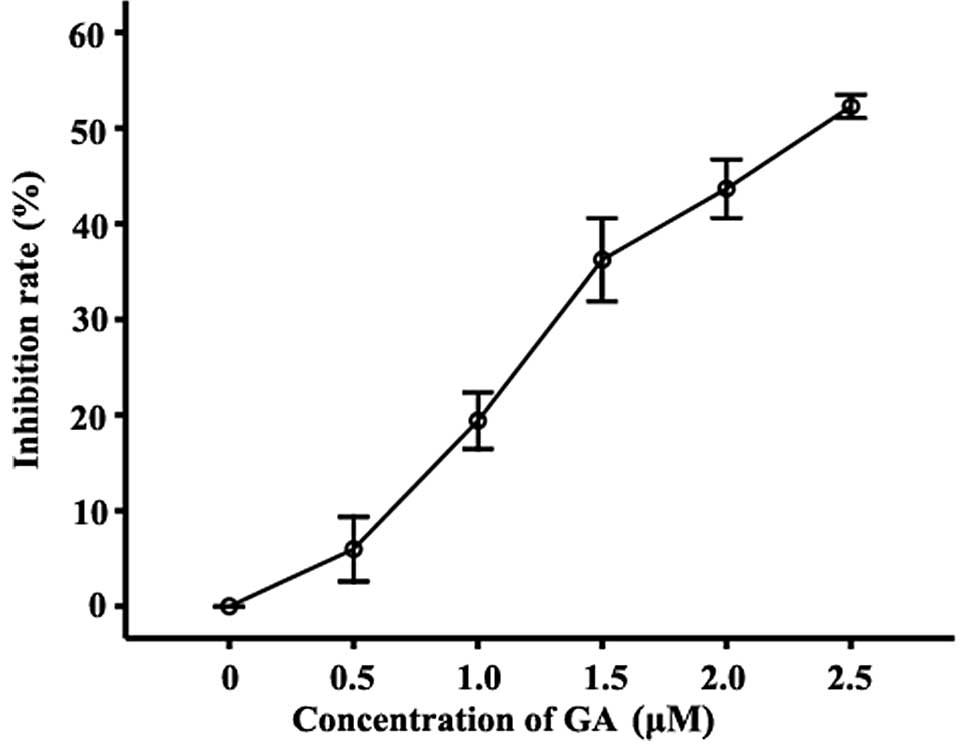 | Figure 2Effect of GA on the proliferation
inhibition ratio in RPMI-8226 cells. Inhibition of the growth of
RPMI-8226 cells treated with 0.5, 1.0, 1.5, 2.0 or 2.5 μM GA for 12
h was detected by MTT assay. Compared with the untreated group, the
inhibition rates of cells treated with 0.5, 1.0, 1.5, 2.0 and 2.5
μM GA were 5.99±3.39, 19.41±2.95, 36.26±4.34, 43.69±3.07 and
52.28±1.24%, respectively. Data were presented as the mean ± SD of
three independent experiments. GA, gambogic acid. |
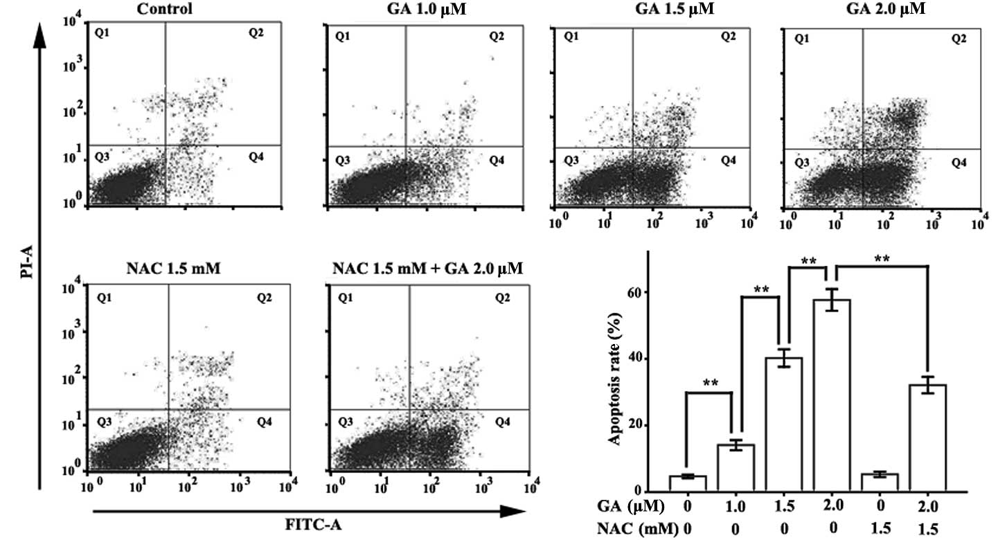
GA induced apoptosis in RPMI-8226
cells
Annexin V-FITC/PI double-labeled FCM was applied to
detect the apoptosis of RPMI-8226 cells treated with various
concentrations of GA (0, 1.0, 1.5 or 2.0 μM) for 12 h. The Annexin
V-FITC-positive and PI-negative cells are early apoptotic cells and
the Annexin V-FITC and PI-positive cells are late apoptotic cells.
The total apoptotic rate is the sum of the early and late apoptotic
rates. Few Annexin V-FITC-positive cells were observed in the
control group (Fig. 3). However,
when the concentration of GA increased to 1.0 μM, the Annexin
V-positive cells were detected. The total apoptotic rates of
RPMI-8226 cells treated with 1.0, 1.5 and 2.0 μM GA were
14.10±1.51, 40.23±2.63 and 57.67±3.25%, respectively, which were
significantly higher than those of the control group
(4.73±0.51%).
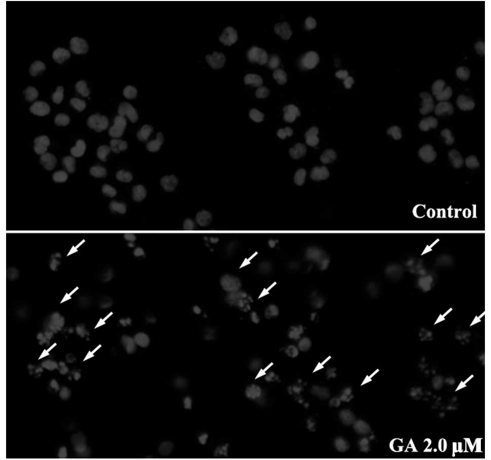
Hoechst 33258 staining was used to detect changes in
nuclear morphology in RPMI-8226 cells treated with 1.5 μM GA for 12
h. Normal RPMI-8226 cells had intact plasma membranes and ordered
chromatin folding (Fig. 4).
Following treatment with 1.5 μM GA for 12 h, apoptotic bodies were
found to be present in the RPMI-8226 cells, in which the chromatin
became condensed, the nuclear envelopes were lytic and the
cytoplasm had decreased in size.
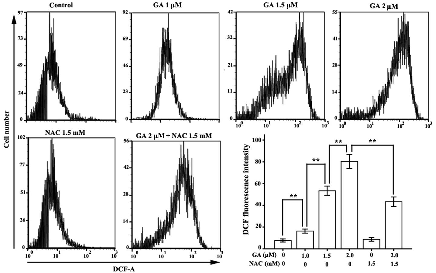
GA contributed to ROS accumulation in
RPMI-8226 cells
FCM analysis of DCF fluorescence intensity was
applied to monitor the level of intracellular ROS in RPMI-8226
cells treated with GA for 12 h. The mean fluorescence intensities
of DCF in RPMI-8226 cells treated with GA (1.0, 1.5 or 2.0 μM) were
16.30±1.94, 53.38±4.24 and 80.42±6.48, respectively, which were
significantly higher than those of the untreated group (7.70±1.41)
(Fig. 5).
NAC reduced the apoptosis rate in
RPMI-8226 cells treated with GA
We applied ROS scavenger NAC to investigate the
correlation between ROS accumulation and apoptosis induction in
RPMI-8226 cells. As predicted, compared with the normal group, NAC
did not affect the ROS level or the apoptotic rate in RPMI-8226
cells, but it significantly decreased GA-induced ROS accumulation
in RPMI-8226 cells. Compared with the 2.0 μM GA group, the mean DCF
florescence intensity of RPMI-8226 cells treated with 1.5 mM NAC
and 2.0 μM GA was markedly decreased, from 80.42±6.48 to
43.30±4.46. The apoptotic ratio was also significantly reduced,
from 57.67±3.25 to 32.10±2.47.
ROS accumulation leads to the activation
of caspase-3 and cleavage of poly (ADP-ribose) polymerase
(PARP)
ROS accumulation is known to activate caspase-3 in
human hepatoma SMMC-7721 cells (13). The results of the present study
indicate that GA increased the amount of activated caspase-3 in a
dose-dependent manner. PARP, a 116 kDa protein and a major
substrate of activated caspase-3, is cleaved to form an
amino-terminal DNA binding fragment (24 kDa) and a carboxy-terminal
catalytic fragment (89 kDa) (26).
Following the activation of caspase-3, the level of the 89 kDa
fragment of PARP was increased (Fig.
6A). Consistent with the changes in the apoptotic rate and the
ROS level, NAC significantly reduced the activation of caspase-3
and the cleavage of PARP in RPMI-8226 cells treated with 2.0 μM GA
for 12 h.
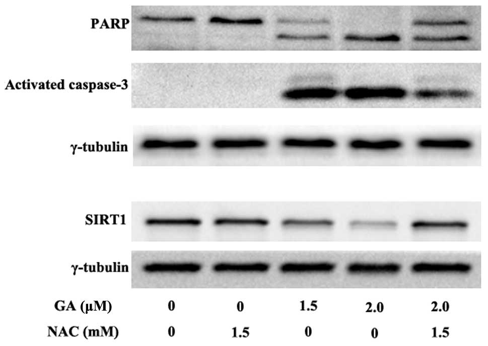 | Figure 6Effect of GA on caspase-3, PARP and
SIRT1. Cells were incubated at various concentrations (1.0, 1.5 or
2.0 μM) of GA alone or with 1.5 mM NAC for 12 h. Western blot
analysis was used to detect the amount of activated caspase-3, PARP
(the major substrate of activated caspase-3) and SIRT1. The result
shows that GA contributes to the activation of caspase-3, followed
by the cleavage of PARP in a dose-dependent manner. GA also
decreases the expression of SIRT1. NAC blocks the ability of GA to
activate caspase-3 and inhibit SIRT1 expression, whereas 1.5 mM NAC
alone has no effect on the activation of caspase-3, cleavage of
PARP, and SIRT1 downregulation in RPMI-8226 cells. GA, gambogic
acid; PARP, poly (ADP-ribose) polymerase; NAC,
N-acetylcysteine. |
ROS accumulation leads to the
downregulation of SIRT1
Western blot analysis was used to detect the
expression of SIRT1 in RPMI-8226 cells treated with 1.5 or 2.0 μM
GA for 12 h. GA was found to downregulate SIRT1 expression in a
dose-dependent manner (Fig. 6B).
Compared with the normal group, treatment with 1.5 mM NAC alone did
not affect the expression of SIRT1, whereas NAC significantly
blocked GA-induced SIRT1 downregulation in RPMI-8226 cells. These
data indicate that ROS accumulation mediates SIRT1 reduction.
Discussion
GA, the major active ingredient of gamboges secreted
from the Garcinia hanburryi tree, has antitumor properties
in solid tumors of various derivations in vitro and in
vivo (27). The mechanisms of
the anticancer activities of GA are complex and the most
significant contributor has yet to be identified. At present,
little is known as to how GA affects MM cells and the possible
mechanisms by which it occurs, thus, further investigations should
be conducted to determine these mechanisms.
Based on the theory that the structure of GA
includes an α,β-unsatured ketone and GA contributes to the
accumulation of ROS mentioned above (13), we explored the anticancer potential
of GA in MM RPMI-8226 cells and investigated its basic molecular
mechanism. First, GA inhibited the proliferation rate of RPMI-8226
cells (Fig. 2). We also found that
GA has the potential to induce ROS accumulation in RPMI-8226 cells
in a dose-dependent manner. Following treatment with 2.0 μM GA for
12 h, the mean fluorescence intensity of DCF, which represents the
level of intracellular ROS, was 80.42±6.48, which was approximately
ten times that of the normal control group (7.70±1.41). Consistent
with this phenomenon, GA also induced the apoptosis of RPMI-8226
cells in a dose-dependent manner. The apoptosis rate of RPMI-8226
cells treated with 2.0 μM GA was 57.67±3.25%, which was
approximately thirteen times that of the control group
(4.73±0.51%).
To confirm the correlation between GA-induced ROS
accumulation and apoptosis in RPMI-8226 cells, we added ROS
scavenger NAC to the GA group and detected the changes in ROS
levels and apoptotic rates with or without NAC. The results showed
that NAC significantly reduced the ROS accumulation induced by GA
(from 80.42±6.48 to 43.30±4.46). NAC also reduced the apoptotic
rate of RPMI-8226 cells treated with GA (from 57.67±3.25 to
32.10±2.47%). These results demonstrate that ROS accumulation is
the major cause of apoptosis in RPMI-8226 cells treated with GA and
that reducing ROS accumulation protects the vitality of RPMI-8226
cells from GA treatment.
Caspase-3, as the most significant executioner
caspase, induces apoptosis via the cleavage of substrates,
including DNA repair- and cell cycle-related proteins, structural
proteins and the mediators and regulators of apoptosis (28). ROS are known to activate caspases,
which are constitutively expressed in the cytosol as inactive
proenzyme monomers, via proteolysis at internal sites (29). ROS also induce the collapse of MMP,
followed by the release of the pro-apoptotic factor Cyt c from the
inner mitochondrial space to the cytosol, which in turn activates
apoptosis executioner caspase-3 via the activation of apoptosis
initiator caspase-9 (30).
Moreover, the release of Cyt c contributes to ROS accumulation
(30) and activated caspase-3
enhances caspase-9 processing by amplification via the promotion of
caspase-2 and -6 activation (31).
The results of the present study have shown that GA
may also induce the activation of caspase-3 in RPMI-8226 cells in a
dose-dependent manner. Since ROS scavenger NAC reduced caspase-3
activation and the apoptotic rate, we concluded that GA induces
apoptosis in RPMI-8226 cells mainly through the accumulation of
ROS, which activates caspase-3. To verify this conclusion, we
investigated changes in the level of PARP, which is involved in DNA
repair in response to extracellular stress and is one of the major
cleavage targets of caspase-3 in vivo (26). Consistent with the activation of
caspase-3 in RPMI-8226 cells treated with GA, the amount of 89 kDa
fragments of 116 kDa PARP was elevated depending on the drug
concentration. NAC also blocked the cleavage of PARP in cells
treated with GA.
We found that GA had the potential to downregulate
the expression of SIRT1 in a dose-dependent manner. Mammalian
SIRT1, as the closest homolog of the yeast Sir2, is extensively
involved in regulating cell processes, including cell senescence,
aging and neuronal protection, as well as having anti-apoptotic
properties (24). SIRT1 is
upregulated in various types of cancer, including leukemia,
lymphomas, soft-tissue sarcomas, prostate cancer and lung and colon
carcinomas (32–35). Moreover, a high level of expression
of SIRT1 has been reported to protect cancer cells from
chemotherapy (36) and ionizing
radiation (37). The mechanisms of
SIRT1 that contribute to tumorigenesis and resistance to
chemotherapy and radiotherapy are complex and include inhibitory
effects on FOXO3a, p53, E2F1 and Ku70 (24). For example, SIRT1 overexpression may
block oxidant-induced apoptosis via the inhibition of p53-mediated
nuclear transactivation (22). The
SIRT1-FOXO-3a interaction reportedly increases the transcription of
stress-resistant genes and decreases the expression of
FOXO-3a-dependent pro-apoptotic genes during oxidative stress
(38). SIRT1 may thus be a new
target in cancer therapy. To clarify, SIRT1 RNAi knockdown induced
apoptosis and senescence, inhibited invasion and enhanced
chemosensitivity in pancreatic cancer cells (39). SIRT1720 also inhibited growth and
induced apoptosis in MM cells resistant to conventional and
bortezomib therapies by targeting SIRT1 (25). In the present study, we found that
GA downregulates SIRT1 expression via ROS generation, whereas NAC
reduces the downregulation of SIRT1 via the elimination of the
accumulation of ROS (Figs. 5 and
6B).
In conclusion, our results demonstrate that GA
induces apoptosis in RPMI-8226 cells via ROS accumulation.
Caspase-3, which is located downstream of ROS and executes
apoptosis, was activated during the apoptosis of RPMI-8226 cells
treated with GA. Moreover, high levels of ROS downregulated the
expression of SIRT1, which is also relevant to apoptosis. As SIRT1
is significant in protecting cancer cells from chemotherapy, and
chemotherapy resistance is the main cause of cancer relapse, we
predict that GA may have the potential, not only to induce the
apoptosis of MM cells, but also to decrease the relapse rate of
MM.
Acknowledgements
This study was supported by the National Natural
Science Foundation of China (no. 30871036/H1616 and 81070429).
References
|
1
|
Kasenda B, Ruckert A, Farthmann J, et al:
Management of multiple myeloma in pregnancy: strategies for a rare
challenge. Clin Lymphoma Myeloma Leuk. 11:190–197. 2011. View Article : Google Scholar : PubMed/NCBI
|
|
2
|
Siegel DS, Vij R and Jakubowiak AJ:
Clinical roundtable monograph. Emerging treatment options for
relapsed and refractory multiple myeloma. Clin Adv Hematol Oncol.
9:1–15. 2011.PubMed/NCBI
|
|
3
|
Gu H, Rao S, Zhao J, et al: Gambogic acid
reduced bcl-2 expression via p53 in human breast MCF-7 cancer
cells. J Cancer Res Clin Oncol. 135:1777–1782. 2009. View Article : Google Scholar : PubMed/NCBI
|
|
4
|
Xu X, Liu Y, Wang L, et al: Gambogic acid
induces apoptosis by regulating the expression of Bax and Bcl-2 and
enhancing caspase-3 activity in human malignant melanoma A375
cells. Int J Dermatol. 48:186–192. 2009. View Article : Google Scholar : PubMed/NCBI
|
|
5
|
Cui G, Shu W, Wu Q and Chen Y: Effect of
Gambogic acid on the regulation of hERG channel in K562 cells in
vitro. J Huazhong Univ Sci Technolog Med Sci. 29:540–545. 2009.
View Article : Google Scholar : PubMed/NCBI
|
|
6
|
Wang Y, Chen Y, Chen Z, Wu Q, Ke WJ and Wu
QL: Gambogic acid induces death inducer-obliterator 1-mediated
apoptosis in Jurkat T cells. Acta Pharmacol Sin. 29:349–354. 2008.
View Article : Google Scholar : PubMed/NCBI
|
|
7
|
Rong JJ, Hu R, Song XM, et al: Gambogic
acid triggers DNA damage signaling that induces p53/p21 (Waf1/CIP1)
activation through the ATR-Chk1 pathway. Cancer Lett. 296:55–64.
2010. View Article : Google Scholar : PubMed/NCBI
|
|
8
|
Nanduri J, Wang N, Bergson P, Yuan G,
Ficker E and Prabhakar NR: Mitochondrial reactive oxygen species
mediate hypoxic down-regulation of hERG channel protein. Biochem
Biophys Res Commun. 373:309–314. 2008. View Article : Google Scholar : PubMed/NCBI
|
|
9
|
Liu B, Chen Y and St Clair DK: ROS and
p53: a versatile partnership. Free Radic Biol Med. 44:1529–1535.
2008. View Article : Google Scholar : PubMed/NCBI
|
|
10
|
Azad N, Iyer A, Vallyathan V, et al: Role
of oxidative/nitrosative stress-mediated Bcl-2 regulation in
apoptosis and malignant transformation. Ann NY Acad Sci. 1203:1–6.
2010. View Article : Google Scholar : PubMed/NCBI
|
|
11
|
Borutaite V and Brown GC: Caspases are
reversibly inactivated by hydrogen peroxide. FEBS Lett.
500:114–118. 2001. View Article : Google Scholar : PubMed/NCBI
|
|
12
|
Chen YC, Shen SC and Tsai SH:
Prostaglandin D(2) and J(2) induce apoptosis in human leukemia
cells via activation of the caspase 3 cascade and production of
reactive oxygen species. Biochim Biophys Acta. 1743:291–304. 2005.
View Article : Google Scholar : PubMed/NCBI
|
|
13
|
Nie F, Zhang X, Qi Q, et al: Reactive
oxygen species accumulation contributes to gambogic acid-induced
apoptosis in human hepatoma SMMC-7721 cells. Toxicology. 260:60–67.
2009. View Article : Google Scholar : PubMed/NCBI
|
|
14
|
Scherz-Shouval R and Elazar Z: Regulation
of autophagy by ROS: physiology and pathology. Trends Biochem Sci.
36:30–38. 2011. View Article : Google Scholar : PubMed/NCBI
|
|
15
|
Bokoch GM: Regulation of the human
neutrophil NADPH oxidase by the Rac GTP-binding proteins. Curr Opin
Cell Biol. 6:212–218. 1994. View Article : Google Scholar : PubMed/NCBI
|
|
16
|
Sundaresan M, Yu ZX, Ferrans VJ, et al:
Regulation of reactive-oxygen-species generation in fibroblasts by
Rac1. Biochem J. 318(Pt 2): 379–382. 1996.PubMed/NCBI
|
|
17
|
Terada LS: Specificity in reactive oxidant
signaling: think globally, act locally. J Cell Biol. 174:615–623.
2006. View Article : Google Scholar : PubMed/NCBI
|
|
18
|
Jones DP: Redefining oxidative stress.
Antioxid Redox Signal. 8:1865–1879. 2006. View Article : Google Scholar
|
|
19
|
Valko M, Leibfritz D, Moncol J, Cronin MT,
Mazur M and Telser J: Free radicals and antioxidants in normal
physiological functions and human disease. Int J Biochem Cell Biol.
39:44–84. 2007. View Article : Google Scholar : PubMed/NCBI
|
|
20
|
Chen Y, Jungsuwadee P, Vore M, Butterfield
DA and St Clair DK: Collateral damage in cancer chemotherapy:
oxidative stress in nontargeted tissues. Mol Interv. 7:147–156.
2007. View
Article : Google Scholar : PubMed/NCBI
|
|
21
|
Smith BC, Hallows WC and Denu JM:
Mechanisms and molecular probes of sirtuins. Chem Biol.
15:1002–1013. 2008. View Article : Google Scholar : PubMed/NCBI
|
|
22
|
Luo J, Nikolaev AY, Imai S, et al:
Negative control of p53 by Sir2alpha promotes cell survival under
stress. Cell. 107:137–148. 2001. View Article : Google Scholar : PubMed/NCBI
|
|
23
|
Zeng R, He J, Peng J, et al: The
time-dependent autophagy protects against apoptosis with possible
involvement of Sirt1 protein in multiple myeloma under nutrient
depletion. Ann Hematol. 91:407–417. 2012. View Article : Google Scholar : PubMed/NCBI
|
|
24
|
Olmos Y, Brosens JJ and Lam EW: Interplay
between SIRT proteins and tumour suppressor transcription factors
in chemotherapeutic resistance of cancer. Drug Resist Updat.
14:35–44. 2011. View Article : Google Scholar : PubMed/NCBI
|
|
25
|
Chauhan D, Bandi M, Singh AV, et al:
Preclinical evaluation of a novel SIRT1 modulator SRT1720 in
multiple myeloma cells. Br J Haematol. 155:588–598. 2011.
View Article : Google Scholar : PubMed/NCBI
|
|
26
|
Lazebnik YA, Kaufmann SH, Desnoyers S,
Poirier GG and Earnshaw WC: Cleavage of poly(ADP-ribose) polymerase
by a proteinase with properties like ICE. Nature. 371:346–347.
1994. View
Article : Google Scholar : PubMed/NCBI
|
|
27
|
Prasad S, Pandey MK, Yadav VR and Aggarwal
BB: Gambogic acid inhibits STAT3 phosphorylation through activation
of protein tyrosine phosphatase SHP-1: potential role in
proliferation and apoptosis. Cancer Prev Res (Phila). 4:1084–1094.
2011. View Article : Google Scholar : PubMed/NCBI
|
|
28
|
Degterev A, Boyce M and Yuan J: A decade
of caspases. Oncogene. 22:8543–8567. 2003. View Article : Google Scholar : PubMed/NCBI
|
|
29
|
Salvesen GS and Abrams JM: Caspase
activation stepping on the gas or releasing the brakes? Lessons
from humans and flies. Oncogene. 23:2774–2784. 2004. View Article : Google Scholar : PubMed/NCBI
|
|
30
|
Circu ML and Aw TY: Reactive oxygen
species, cellular redox systems, and apoptosis. Free Radic Biol
Med. 48:749–762. 2010. View Article : Google Scholar : PubMed/NCBI
|
|
31
|
Slee EA, Harte MT, Kluck RM, et al:
Ordering the cytochrome c-initiated caspase cascade: hierarchical
activation of caspases-2, -3, -6, -7, -8, and -10 in a
caspase-9-dependent manner. J Cell Biol. 144:281–292. 1999.
View Article : Google Scholar : PubMed/NCBI
|
|
32
|
Fraga MF, Agrelo R and Esteller M:
Cross-talk between aging and cancer: the epigenetic language. Ann
NY Acad Sci. 1100:60–74. 2007. View Article : Google Scholar : PubMed/NCBI
|
|
33
|
Fraga MF and Esteller M: Epigenetics and
aging: the targets and the marks. Trends Genet. 23:413–418. 2007.
View Article : Google Scholar : PubMed/NCBI
|
|
34
|
Hida Y, Kubo Y, Murao K and Arase S:
Strong expression of a longevity-related protein, SIRT1, in Bowen's
disease. Arch Dermatol Res. 299:103–106. 2007. View Article : Google Scholar : PubMed/NCBI
|
|
35
|
Lim CS: Human SIRT1: a potential biomarker
for tumorigenesis? Cell Biol Int. 31:636–637. 2007. View Article : Google Scholar : PubMed/NCBI
|
|
36
|
Chu F, Chou PM, Zheng X, Mirkin BL and
Rebbaa A: Control of multidrug resistance gene mdr1 and cancer
resistance to chemotherapy by the longevity gene sirt1. Cancer Res.
65:10183–10187. 2005. View Article : Google Scholar : PubMed/NCBI
|
|
37
|
Matsushita N, Takami Y, Kimura M, et al:
Role of NAD-dependent deacetylases SIRT1 and SIRT2 in radiation and
cisplatin-induced cell death in vertebrate cells. Genes Cells.
10:321–332. 2005. View Article : Google Scholar : PubMed/NCBI
|
|
38
|
Brunet A, Sweeney LB, Sturgill JF, et al:
Stress-dependent regulation of FOXO transcription factors by the
SIRT1 deacetylase. Science. 303:2011–2015. 2004. View Article : Google Scholar : PubMed/NCBI
|
|
39
|
Zhao G, Cui J, Zhang JG, et al: SIRT1 RNAi
knockdown induces apoptosis and senescence, inhibits invasion and
enhances chemosensitivity in pancreatic cancer cells. Gene Ther.
18:920–928. 2011. View Article : Google Scholar : PubMed/NCBI
|