Introduction
Papillary thyroid cancer (PTC) is the most common
well-differentiated cancer of the thyroid. It represents
approximately 80–85% of well-differentiated thyroid cancers
(1). However, some cases show
relatively early recurrence, severe invasion, multiple lymph node
metastasis or distant metastasis (2). Therefore, it is important to identify
those characteristics of PTC that have a high risk for invasion and
metastasis.
The amyloid precursor protein (APP) is a
transmembrane protein that contains a large N-terminal ectodomain
and a short C-terminal endodomain and is expressed in the central
nervous system and peripheral tissues, including epidermis,
pancreas and thyroid (3–5). A proteomic study revealed an increased
expression of APP in benign cold thyroid nodules compared with
normal thyroid tissue (6). In a
previous study, the upregulation of APP expression was induced by
TSH in differentiated thyroid cells and by insulin in thyroid
cancer cells in vitro; thyroid cancers are characterized by
APP upregulation, increased membrane targeting of the APP
ectodomain and significantly increased mRNA levels of the APP
scaffold proteins (7). However,
correlations between APP expression and clinicopathological
parameters have yet to be investigated in PTC.
The present study aimed to investigate the
expression levels of the APP in PTC and to examine
clinicopathological correlations such as invasive and metastatic
characteristics.
Materials and methods
Case selection
Tumor specimens for quantitative real time-PCR were
obtained from 10 PTC patients who underwent total thyroidectomy at
the Department of Surgery, Shanghai Jiao Tong University Affiliated
Sixth People's Hospital, China. Non-tumor thyroid tissues (n=10)
were obtained from tissue adjacent to each PTC exhibiting
apparently non-tumor morphology as a control.
Tissues for immunohistochemistry were randomly
selected from 90 PTC patients who underwent surgery in the same
department between 2008 and 2010. Non-tumor thyroid tissues (n=90)
were obtained from adjacent tissue of PTC patients exhibiting
apparently non-tumor morphology as a control. These patients
included 20 males and 70 females, and the average age was 45.2
years. The study was approved by the Ethics Committee of the Sixth
People's Hospital Affiliated to Shanghai Jiao Tong University.
Tissue sample preparation and tissue
microarray
Following surgical resection, specimens from the 90
PTC patients were fixed with 10% formalin. Sections of PTC tissue
cores were stained with hematoxylin and eosin to identify areas of
tumor and non-tumor tissue. When the areas of interest were
identified, the recipient tissue array block was constructed using
manual tissue array equipment (IHC World, Woodstock, MD, USA).
Cores (2-mm) were placed in the recipient block, which was heated
to fix the samples and a paraffin layer was applied to ensure
proper facing. To facilitate blind grading, an Excel spreadsheet
(Microsoft Corporation, Redmond, WA, USA) was constructed using
sample accession numbers but without information regarding the
final pathological finding.
Immunohistochemistry
Sectioned slides were deparaffinized three times in
xylene for 20 min each and rehydrated using a graded alcohol
solution. Antigen retrieval was performed in citrate buffer in a
microwave. Endogenous peroxidase activity was quenched by
incubating in peroxidase-blocking reagent. Sections were incubated
in the primary antibody of APP (22C11, 1:50, Millipore, Billerica,
MA, USA). The second antibody was subsequently incubated for 30 min
and counterstained with hematoxylin.
Quantitative real-time-PCR
Total RNA was prepared from tissues using the RNeasy
extraction kit (GE Healthcare, Amersham, UK) and reverse
transcribed using high-capacity cDNA reverse transcription kits
(Applied Biosystems, Melbourne, Australia) according to the
manufacturer's instructions. Quantitative real-time PCR (qRT-PCR)
was performed on a 7300 Fast Real-Time PCR System (Applied
Biosystems) using SYBR-Green PCR Master mix (Applied Biosystems).
The human-specific intron spanning primer pairs for APP were:
forward: 5′-CCGATGATGACGAGGACGAT-3′ and reverse:
5′-GTGGTGGTGGTGGCAATG-3′. The primer pairs used for GAPDH were:
forward: 5′-CAATGACCCCTTCATTGACC-3′ and reverse:
5′-TGATGACAAGCTTCCCGTTC-3′. Cycle conditions were as follows: 1
cycle at 50°C for 2 min, followed by 1 cycle at 95°C for 10 min, 40
cycles at 95°C for 15 sec and 60°C for 1 min. Specificity of PCR
products was tested by dissociation curves. Relative values of
transcripts were calculated using the equation, 2−ΔΔCt,
where ΔCt is equal to the difference in threshold cycles for the
target and reference. Each experiment was performed in
triplicate.
Immunohistological scores and
clinicopathological parameters
A surgical pathologist blinded to the identity of
the specimens examined the percentage of positivity and intensity
of immunostained slides and scored them as previously described
(8). The percentage of positivity
was the number of cells showing positive staining (grade 0, 0;
grade 1, 1–33%; grade 2, 34–66%; and grade 3, 67–100%,
respectively). The intensity grade 0 was defined as no
immunoreaction, grade 1 as weak immunoreaction, grade 2 as moderate
immunoreaction, and grade 3 as strong immunoreaction. The total
scores in PTC (tumor score) and non-tumor tissues (non-tumor score)
were determined as the sum of the positivity and intensity
grades.
Based on the clinical and pathological record, a
retrospective analysis was performed on the following variables:
age, gender, tumor size, lymph node metastasis, extracapsular
invasion, multifocality, distant metastasis, and clinical stage.
Clinical stage was determined according to the pathological TNM
system (NCCN Guidelines, Thyroid Carcinoma, Version 2.2011).
Immunohistochemical results were correlated with the
clinicopathological parameters to evaluate the prognostic
significance.
Statistical analysis
Scores were shown as the mean ± standard deviation.
Statistical analysis was performed using SPSS statistical software
(version 17.0; SSPS, Chicago, IL, USA). The Mann-Whitney U test was
used to compare the expression levels of the APP gene by
quantitative PCR between PTC and non-tumor tissue. The independent
samples t-test was used to compare average scores of tumor marker
using immunohistochemistry and clinicopathological variables. The
number of positive immunoreactions (tumor score >0) in PTC and
normal thyroid tissues were evaluated using the Chi-square test.
P<0.05 was accepted as indicative of a significant
difference.
Results
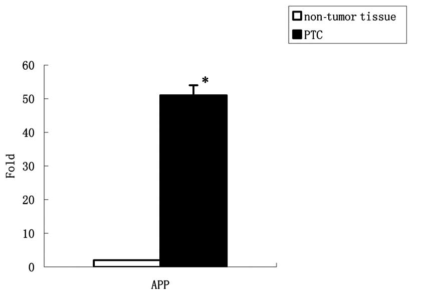
Expression levels of APP mRNA in PTC and
non-tumor thyroid tissues
To compare the gene expression in PTC and non-tumor
thyroid tissues, APP mRNA expression was analyzed by quantitative
real time-PCR. APP mRNA increased approximately 50-fold in PTC
compared with non-tumor thyroid tissues (Fig. 1).
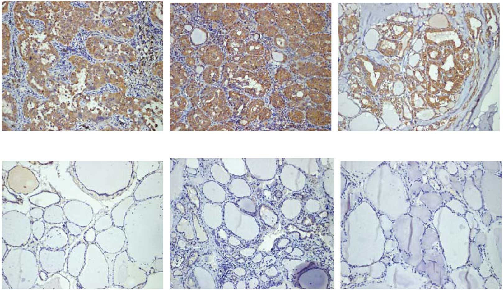
Immunohistochemical expression of APP in
PTC and non-tumor thyroid tissues
Tissue microarrays were constructed to investigate
the differences in APP protein expression between PTC and non-tumor
thyroid tissues (Fig. 2).
In the tumor regions, positive immunoreactions for
APP (tumor score >0) were present in 90 of 90 cases (100%). In
the non-tumor regions, positive immunoreactions for APP (non-tumor
score >0) were present in 36 of 90 cases (40%). APP protein
expression levels were increased in the tumor regions compared to
the non-tumor regions (P<0.001; Table I).
 | Table IImmunohistochemical analysis of APP in
tumor regions and non-tumor regions from the same thyroidectomy
specimens according to the scoring system and positive
immunoreactivity. |
Table I
Immunohistochemical analysis of APP in
tumor regions and non-tumor regions from the same thyroidectomy
specimens according to the scoring system and positive
immunoreactivity.
| n | Tissues with positive
immunoreactivity (%) | P-value |
|---|
| Tumor score | | 100 (100) | <0.001 |
| 0 | 0 | | |
| 1–2 | 10 | | |
| 3–4 | 35 | | |
| 5–6 | 45 | | |
| Non-tumor score | | 36 (40) | |
| 0 | 54 | | |
| 1–2 | 25 | | |
| 3–4 | 11 | | |
| 5–6 | 0 | | |
Correlation between the
immunohistochemical scores and clinicopathological parameters
APP high scores significantly correlated with and
clinicopathological data such as large tumor size, extracapsular
invasion, and lymph node metastasis (Table II).
| Table IIPrevalence of APP tumor scores and
clinicopathological parameters. |
Table II
Prevalence of APP tumor scores and
clinicopathological parameters.
| n (%) | APP score | P-value |
|---|
| Age (years) |
| <45 | 50 (55) | 3.25±2.01 | NS |
| ≥45 | 40 (45) | 3.30±1.90 | |
| Gender |
| Male | 20 (20) | 3.78±1.98 | NS |
| Female | 70 (80) | 3.77±1.85 | |
| Size (cm) |
| ≤2 | 54 (60) | 2.82±2.30 | 0.005 |
| >2 | 36 (40) | 4.52±2.12 | |
| Extracapsular
invasion |
| No | 63 (70) | 3.58±1.45 | 0.024 |
| Yes | 27 (30) | 4.64±2.50 | |
| Multifocality |
| No | 75 (83) | 3.79±2.45 | NS |
| Yes | 15 (17) | 3.84±1.45 | |
| LN metastasis |
| No | 68 (75) | 2.98±2.46 | 0.004 |
| Yes | 22 (25) | 4.95±1.55 | |
| Distant
metastasis |
| No | 81 (90) | 3.74±2.21 | NS |
| Yes | 9 (10) | 3.80±2.25 | |
| TNM stage |
| I and II | 72 (80) | 3.71±2.25 | NS |
| III and IV | 18 (20) | 3.74±1.91 | |
Discussion
APP undergoes several types of proteolytic
processing, one of which results in the release of the secretory
N-terminal portion of APP (sAPP), which carries a number of
biologically relevant domains. The ability of APP to induce
growth-promoting effects and morphological alterations occurs not
only through effects of the sAPP but may also occur through effects
of the cytosolic domain of APP, whether cleaved or intact. The
presence of multiple cytosolic interactors of APP has been
identified, including JIP1b, Fe65, ShcA and APPBP1, many of which
may function in a neoplastic system to induce cell proliferation
and migration (5).
Overexpression of APP was observed in other human
cancer types, such as prostate (9),
colon (10), parathyroid (11), pancreatic (5), and oral cancers(12). The present study showed that APP
expression was upregulated in PTC compared with non-tumor thyroid
tissues at the transcription and protein levels. APP acts at the
cell surface to bind and activate signaling proteins via its
intracellular domain (AICD); Fe65, an AICD-binding protein, has
been shown to stimulate APP trafficking and APP proteolysis, which
in turn leads to increased sAPP secretion (13). Combined with the results of the
present study, this finding indicates that a high expression of the
APP in PTC may increase sAPP secretion. Thyrotropin (TSH)
reportedly induced APP expression and sAPP release in
differentiated thyroid cells (FRTL-5), and promoted change from
differentiation to proliferation in thyroid epithelial cells
(14), indicating that sAPP
operates as an autocrine growth factor mediating the proliferative
effect of TSH on neighboring thyroid epithelial cells. Therefore,
the present results indicate that APP expression may be associated
with thyroid carcinogenesis and be of use as a diagnostic
marker.
Findings of certain studies have shown that APP and
its secreted forms promote adhesion, migration, neurite outgrowth,
and general growth-promoting properties (15). Therefore, we investigated the
correlation between the expression levels of APP and
clinicopathological parameters such as invasive and metastatic
characteristics in PTC. Our results showed that high APP scores
significantly correlated with large tumor size, extracapsular
invasion, and lymph node metastasis in PTC. Several studies have
shown that patients with increased APP levels have a significantly
lower survival rate and APP has therefore been suggested as a
potential biomarker for the evaluation of cancer prognosis
(5,9,10,12).
For PTC patients, tumor size, lymph node metastasis and
extracapsular invasion are recognized as important prognostic
factors (16). Therefore, the
results of the present study indicate that a high APP expression
may be correlated with poor prognosis in patients with PTC.
In conclusion, our results confirm previous
observations that APP is a crucial mediator of tumor growth in
general, and provides evidence of a role for APP in PTC diagnosis
and prognosis. APP may therefore provide a molecular target for the
therapeutic intervention of PTC. More studies regarding the role of
APP and intracellular signaling pathways in PTC are required.
References
|
1
|
Davies L and Welch HG: Increasing
incidence of thyroid cancer in the United States, 1973–2002. JAMA.
295:2164–2167. 2006.
|
|
2
|
Hay ID, Thompson GB, Grant CS, et al:
Papillary thyroid carcinoma managed at the Mayo Clinic during six
decades (1940–1999): temporal trends in initial therapy and
long-term outcome in 2444 consecutively treated patients. World J
Surg. 26:879–885. 2002.PubMed/NCBI
|
|
3
|
Hoffmann J, Twiesselmann C, Kummer MP,
Romagnoli P and Herzog V: A possible role for the Alzheimer amyloid
precursor protein in the regulation of epidermal basal cell
proliferation. Eur J Cell Biol. 79:905–913. 2000. View Article : Google Scholar : PubMed/NCBI
|
|
4
|
Graebert KS, Lemansky P, Kehle T and
Herzog V: Localization and regulated release of Alzheimer amyloid
precursor-like protein in thyrocytes. Lab Invest. 72:513–523.
1995.PubMed/NCBI
|
|
5
|
Hansel DE, Rahman A, Wehner S, Herzog V,
Yeo CJ and Maitra A: Increased expression and processing of the
Alzheimer amyloid precursor protein in pancreatic cancer may
influence cellular proliferation. Cancer Res. 63:7032–7037.
2003.PubMed/NCBI
|
|
6
|
Krause K, Karger S, Schierhorn A, Poncin
S, Many MC and Fuhrer D: Proteomic profiling of cold thyroid
nodules. Endocrinology. 148:1754–1763. 2007. View Article : Google Scholar : PubMed/NCBI
|
|
7
|
Krause K, Karger S, Sheu SY, et al:
Evidence for a role of the amyloid precursor protein in thyroid
carcinogenesis. J Endocrinol. 198:291–299. 2008. View Article : Google Scholar : PubMed/NCBI
|
|
8
|
Nam KH, Noh TW, Chung SH, et al:
Expression of the membrane mucins MUC4 and MUC15, potential markers
of malignancy and prognosis, in papillary thyroid carcinorma.
Thyroid. 21:745–750. 2011. View Article : Google Scholar : PubMed/NCBI
|
|
9
|
Takayama K, Tsutsumi S, Suzuki T, et al:
Amyloid precursor protein is a primary androgen target gene that
promotes prostate cancer growth. Cancer Res. 69:137–142. 2009.
View Article : Google Scholar : PubMed/NCBI
|
|
10
|
Meng JY, Kataoka H, Itoh H and Koono M:
Amyloid β protein precursor is involved in the growth of human
colon carcinoma cell in vitro and in vivo. Int J Cancer. 92:31–39.
2001.
|
|
11
|
Haven CJ, Howell VM, Eilers PH, et al:
Gene expression of parathyroid tumors: molecular subclassification
and identification of the potential malignant phenotype. Cancer
Res. 64:7405–7411. 2004. View Article : Google Scholar : PubMed/NCBI
|
|
12
|
Ko SY, Lin SC, Chang KW, Wong YK, Liu CJ,
Chi CW and Liu TY: Increased expression of amyloid precursor
protein in oral squamous cell carcinoma. Int J Cancer. 111:727–732.
2004. View Article : Google Scholar : PubMed/NCBI
|
|
13
|
King GD and Scott Turner R: Adaptor
protein interactions: modulators of amyloid precursor protein
metabolism and Alzheimer's disease risk? Exp Neurol. 185:208–219.
2004. View Article : Google Scholar : PubMed/NCBI
|
|
14
|
Pietrzik CU, Hoffmann J, Stöber K, et al:
From differentiation to proliferation: the secretory amyloid
precursor protein as a local mediator of growth in thyroid
epithelial cells. Proc Natl Acad Sci USA. 95:1770–1775. 1998.
View Article : Google Scholar : PubMed/NCBI
|
|
15
|
Thinakaran G and Koo EH: Amyloid precursor
protein trafficking, processing, and function. J Biol Chem.
283:29615–29619. 2008. View Article : Google Scholar : PubMed/NCBI
|
|
16
|
Ito Y and Miyauchi A: Prognostic factors
of papillary and follicular carcinomas in Japan based on data of
Kuma hospital. J Thyroid Res. 2012:9734972012.PubMed/NCBI
|