Introduction
Previous studies have reported that the oncogene
RhoA is involved in the regulation of a number of biological
processes, including stress fiber formation, membrane transport,
gene transcription, focal adhesion and tumor progression (1–4). RhoA
is a small G protein and is therefore inactive when GDP-bound and
active when GTP-bound, with GDP/GTP exchange or GTPase reactions
converting one form to the other (5). The results of previous studies have
shown that lysophosphatidic acid (LPA) is able to activate RhoA and
induce its nuclear translocation and that the subcellular
distribution of RhoA is correlated with its activity (6). Previous studies have reported that
RhoA plays a significant role in CNS injuries and is a potential
target of non-steroidal anti-inflammatory drugs (NSAIDs) in
treating CNS injuries (7). This
finding suggests a role of RhoA as an anti-inflammatory factor.
Bacterial lipopolysaccharide (LPS) is found in the
outer membrane of Gram-negative bacteria and is able to activate a
number of mammalian cell types and intracellular signaling
pathways. For instance, it has been reported that LPS activates
nuclear factor (NF)-κB, inducing the production and release of
numerous pro-inflammatory mediators, including interleukin (IL)-1,
IL-6, IL-8 and tumor necrosis factor (TNF)-α. The synthesis and
release of pro-inflammatory cytokines in response to LPS depends on
inducible gene expression, which is mediated by the activation of
transcription factors, including NF-κB. NF-κB regulates various
genes involved in immune and acute phase inflammatory responses and
in cell survival (8).
Pro-inflammatory stimuli lead to the activation of NF-κB via the
phosphorylation of inhibitors of κB (IκBs) by the IκB kinase (IKK)
signalosome complex (9). This frees
NF-κB and allows it to translocate to the nucleus of the cell where
it induces the transcription of pro-inflammatory mediators,
including iNOS, COX-2, TNF-α, IL-1, IL-6 and IL-8, by binding to κB
binding sites in the promoter regions of the target genes (10). However, the detailed molecular
anti-inflammatory mechanism has not yet been studied. Since LPS is
able to activate NF-κB and trigger its nuclear translocation and
alter the activity of RhoA, we suggested a link between RhoA and
the LPS/NF-κB signaling pathway.
In the present study, we investigated the activity
and nuclear distribution of RhoA following LPS stimulation and
revealed for the first time that LPS triggers the nuclear
translocation of RhoA, which is dependent on NF-κB. Furthermore, we
confirmed that RhoA is critical for the LPS/NF-κB signaling pathway
in epithelial inflammation.
Materials and methods
Cell line
The human lung cancer cell line A549 was provided by
the Institute of Cell Biology (Shanghai, China).
Reagents
Dulbecco’s modified Eagle’s medium (DMEM) and fetal
bovine serum (FBS) were purchased from Gibco (Grand Island, NY,
USA). Antibodies against RhoA (Cat. No. sc-418) and NF-κB P50 (Cat.
No. sc-114) were purchased from Santa Cruz Biotechnology, Inc.
(Santa Cruz, CA, USA). FITC, TRITC and horseradish peroxidase
(HRP)-conjugated secondary antibodies were purchased from Jackson
ImmunoResearch Laboratories (West Grove, PA, USA). The antibody
against glyceraldehyde phosphate dehydrogenase (GAPDH) and the
Nuclear and Cytoplasmic Extract kit (Cat. No. KC-435) were
purchased from Kangcheng Bio-tech (Hangzhou, China). RhoA small
interfering RNA (siRNA) (Cat. No. sc-29471), NF-κB P50 siRNA (Cat.
No. sc-29407) and transfection reagent (Cat. No. sc-29528) were
purchased from Santa Cruz Biotechnology, Inc.. Nuclear fluorochrome
Hoechst 33342 was purchased from Sigma (St. Louis, MO, USA),
Electrochemiluminescence (ECL) reagents were purchased from
Amersham Biosciences (Buckinghamshire, UK).
Cell culture and transfections
A549 cells were maintained in DMEM supplemented with
10% FBS and penicillin/streptomycin (100 IU/ml and 100 mg/ml,
respectively) and incubated at 37°C and 5% CO2. The
cells were seeded in six-well plates at a density of 80% confluence
and transfected the following day. The transfection of siRNA was
performed according to the manufacturer’s instructions.
Preparation of cell extracts
The cells were harvested at various times by
aspiration of the media and direct addition of 2X SDS sample
buffer. The cell lysate was scraped and transferred to tubes,
heated for 5 min at 95°C and stored at −20°C.
Nucleus and cytosol preparation
The cells were seeded in six-well plates at a
density of 80% confluence and treated with or without 10 μg/ml LPS
for 4 h. The nuclear extracts were prepared according to the
instructions of the manufacturer of the Nuclear and Cytoplasmic
Extract kit.
RhoA activation assay
RhoA activity was determined as described in a
previous study (11). This
pull-down assay uses the RhoA-binding domain (RBD) from the
effector protein Rhotekin as a probe to specifically isolate the
active forms of RhoA. Briefly, A549 cells were cultured in 100 mm
culture dishes with complete medium (DMEM with 10% FBS and
antibiotics) until 90% confluence was achieved. Following 24 h of
starvation, the cells were stimulated with 10 μg/ml LPS for 5 min
and then lysed in lysis buffer (25 mM HEPES, pH 7.5, 150 mM NaCl,
1% Igepal CA-630, 10 mM MgCl2, 1 mM EDTA, 2% glycerol, 2
μg/ml each of leupeptin and aprotinin, 1 μl/ml
phenylmethylsulphonyl fluoride and 0.5 μl/ml DTT). The cellular
proteins were harvested by scraping with a rubber policeman. The
lysates were centrifuged at 4°C at 14,000 × g for 5 min to remove
particulate material and the protein concentrations were determined
using the Bradford assay. The protein extracts (25 μg of each) were
incubated while rotating at 4°C for 3 h with an equal volume of
Rhotekin-RBD bound to glutathione-agarose beads. The beads were
washed three times with washing buffer and activated RhoA bound to
the beads or total RhoA in cell extracts was detected using western
blot analysis with a monoclonal antibody against RhoA.
Western blot assay
The sample proteins were separated by SDS-PAGE and
blotted onto a polyvinyl difluoride (PVDF) membrane. The PVDF
membrane was blocked with 3% (w/v) bovine serum albumin (BSA) in
TBS-T for 1 h at room temperature. The membrane was incubated at
4°C overnight with the primary antibody and at room temperature for
1 h with the secondary antibody, with three washes following each
incubation. ECL reagents were used to visualize the positive bands
on the membrane. The bands were detected by Typhoon 9400 (GE
Healthcare, Piscataway, NJ, USA).
Immunofluorescence assay
Forty-eight hours after transfection, control and
NF-κB P50 siRNA-transfected cells grown on coverslips were treated
with or without 10 μg/ml LPS for 4 h, incubated with 0.2 mM Hoechst
33342 for 10 min to reveal the nuclei and then fixed with freshly
prepared 40 g/l paraformaldehyde in PBS at 4°C overnight. After
being penetrated with 30 ml/l Triton X-100 and blocked with 30 g/l
BSA, the cells were incubated with primary antibodies at 4°C
overnight and then with cy3-conjugated second antibodies for 1 h at
room temperature, with three washes following each incubation. The
morphological changes of the cells were analyzed using a
fluorescence microscope.
Determination of IL-8 and IL-6
Forty-eight hours after transfection, control, RhoA
and NF-κB P50 siRNA-transfected A549 cells were treated with or
without LPS for 24 h. The supernatants were then harvested and
assessed for IL-8 and IL-6 production by ELISA (R&D Systems,
Minneapolis, MN, USA).
Statistical analysis
Values are shown as the mean ± SE (n=6). The
Student’s t-test was used for comparisons of two sample means.
P<0.05 was considered to indicate a statistically significant
result.
Results
LPS activates RhoA and triggers its
nuclear translocation in A549 human lung cancer cells
Pro-inflammatory cytokines and bacterial endotoxins
activate the Rho GTPase signaling pathway, which mediates the
pro-inflammatory responses and aggressive carcinogenesis (12–14).
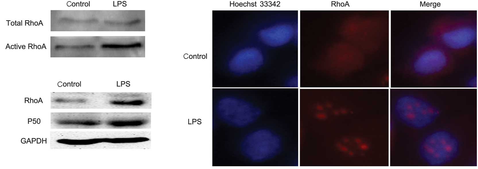
To determine whether LPS was able to activate RhoA in A549 cells,
we performed a pull-down assay to detect the activity of RhoA
following LPS stimulation. The A549 cells were harvested and the
active RhoA in the extract was isolated using the pull-down assay
following treatment with 10 μg/ml LPS for 10 min and analyzed by
western blotting with an antibody against RhoA. The results
revealed that the level of active RhoA increased following LPS
treatment (Fig. 1A). As there was a
correlation between RhoA activity and its subcellular distribution,
we determined the level of expression of RhoA in the nucleus
following LPS stimulation. A549 cells were treated with or without
10 μg/ml LPS for 4 h. An immunofluorescence assay was performed to
show the level of expression of RhoA in the nucleus with an
antibody against RhoA (Fig. 1B).
The nuclear extracts were then prepared following treatment with 10
μg/ml LPS for 4 h and analyzed by western blotting with antibodies
against RhoA, NF-κB P50 and GAPDH (Fig.
1C). The results showed that LPS triggered the nuclear
translocation of RhoA while increasing its activity, similar to the
effect of LPS on NF-κB.
LPS-induced RhoA nuclear translocation is
dependent on NF-κB
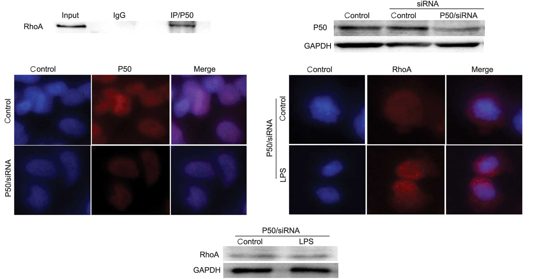
As LPS was able to trigger the nuclear translocation
of both RhoA and NF-κB, we investigated whether the LPS-induced
RhoA nuclear translocation was dependent on NF-κB P50, since NF-κB,
and not RhoA, contains nuclear localization signals (NLS). Firstly,
we investigated if there was binding between RhoA and NF-κB. A
co-immunoprecipitation (Co-IP) assay was performed with an antibody
against NF-κB P50 to reveal any interaction. The results showed
that there was an association between RhoA and NF-κB (Fig. 2A). Secondly, we detected the effect
of NF-κB P50 depletion on LPS-induced RhoA nuclear translocation.
We used NF-κB P50 siRNA to deplete NF-κB P50 in A549 cells. The
effect of the NF-κB P50 siRNA on the expression of NF-κB was
detected by western blotting and an immunofluorescence assay using
an antibody against NF-κB P50. The results of the western blotting
(Fig. 2B) and immunofluorescence
assay (Fig. 2C) revealed that the
expression of NF-κB P50 decreased markedly. Cells with NF-κB P50
depletion were treated with or without 10 μg/ml LPS for 4 h and
western blotting and an immunofluorescence assay with an antibody
against RhoA were performed to detect the nuclear expression of
RhoA. The results of the immunofluorescence assay (Fig. 2D) and western blotting (Fig. 2E) revealed that the nuclear
expression of RhoA did not increase following LPS stimulation in
NF-κB P50-depleted cells. These data demonstrate that LPS-induced
nuclear translocation of RhoA was dependent on NF-κB.
Depletion of RhoA inhibits LPS-induced
IL-6 and IL-8 secretion in A549 human lung cancer cells
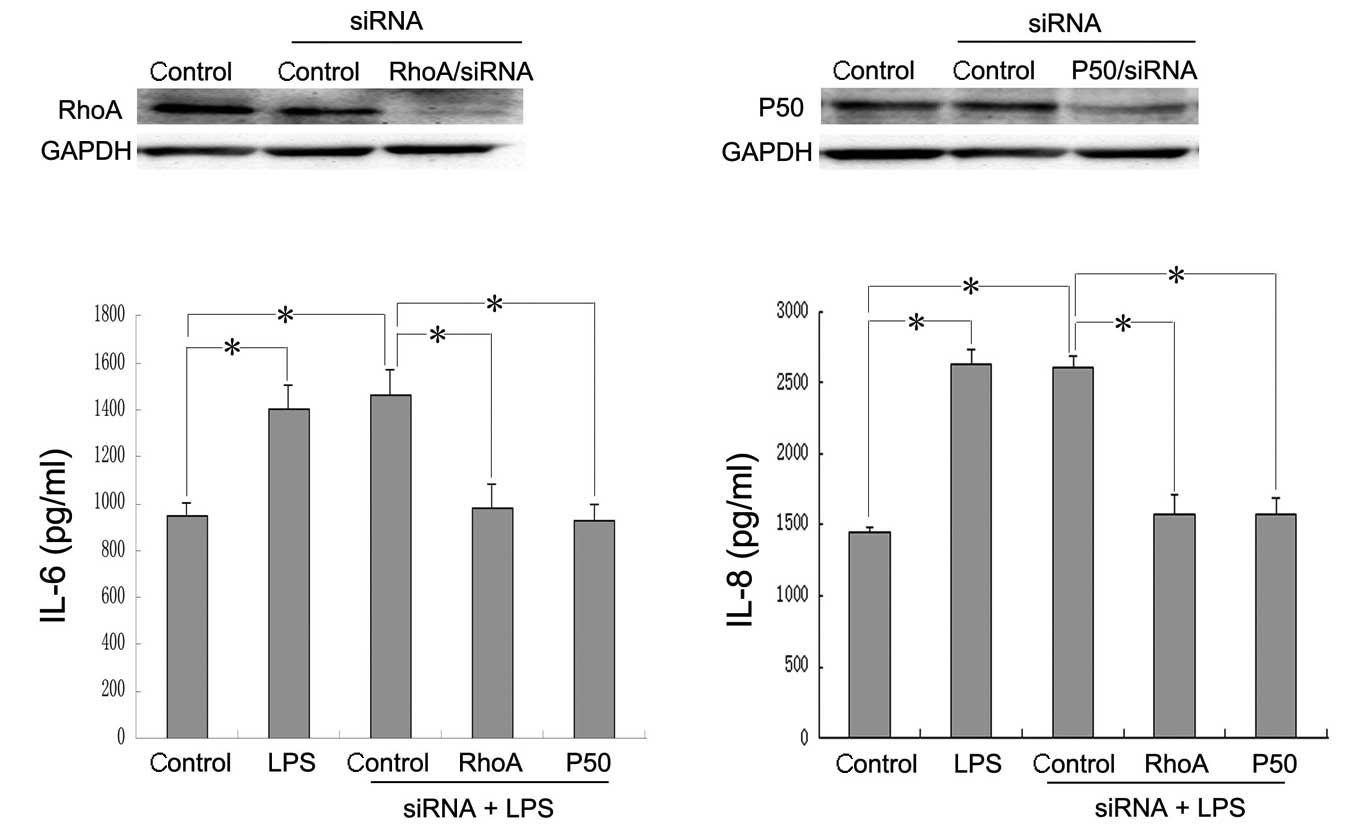
Since LPS had a similar effect on the activity and
nuclear distribution of RhoA and NF-κB and active NF-κB was able to
mediate the transcriptional activation of pro-inflammatory genes,
including IL-6 and IL-8, we investigated whether RhoA was involved
in the LPS/NF-κB pathway. We applied the NF-κB P50 and RhoA siRNAs
to deplete the expression of NF-κB P50 and RhoA in A549 cells,
respectively. The effect of the RhoA and NF-κB P50 siRNAs on the
expression of RhoA (Fig. 3A) and
NF-κB P50 (Fig. 3B) were detected
by western blotting with antibodies against RhoA, NF-κB P50 and
GAPDH. Forty-eight hours after siRNA transfection, the cells were
treated with 10 μg/ml LPS for 24 h and the concentration of IL-6
and IL-8 in the supernatants was measured by ELISA. The results
showed that the depletion of RhoA inhibited the LPS-induced release
of IL-6 (Fig. 3C) and IL-8
(Fig. 3D), similar to the effect of
NF-κB depletion.
Discussion
The ability of Rho GTPases to function in signaling
pathways has been reported to depend partly on the location of the
proteins within the cell (15).
However, the activity of GTPases is also regulated by three types
of proteins: guanine nucleotide exchange factors (GEFs) catalyze
the exchange of GDP for GTP and thereby activate Rho (16); GTPase activating proteins (GAPs)
enhance the hydrolytic ability of GTPases thereby inactivating the
proteins (17); and guanine
nucleotide dissociation inhibitors (GDIs) also inactivate the
GTPases by removing them from the plasma membrane (PM) and
maintaining them in an inactive form in the cytosol (18). The majority of Rho-GEFs are
localized to the cytoplasm or the PM (16), but two GEFs, Net1 and Ect2, have
been reported to localize to the nucleus at a steady state
(19,20). These two GEFs contain an NLS that is
necessary for the proteins to be targeted to the nucleus (21,22).
As RhoA has no NLS, it was of note for us to reveal how the nuclear
translocation occurred. In the present study, following treatment
with LPS, the nuclear translocation of RhoA occurred and its
activity increased. Since results of previous studies showed that
LPS is able to activate NF-κB and induce its nuclear translocation,
and as NF-κB contains an NLS, we hypothesized that the LPS-induced
nuclear translocation of RhoA was mediated by NF-κB. The results of
the present study show that the depletion of NF-κB by siRNA
inhibited the LPS-induced nuclear translocation of RhoA. These data
demonstrate that LPS-induced RhoA nuclear translocation is
dependent on NF-κB.
LPS is a major inflammatory molecule that triggers
the production of pro-inflammatory toxins and cytokines, including
iNOS, COX-2, TNF-α and IL-1, in various cell types (23). As LPS activates RhoA and induces its
nuclear translocation, it was important for us to investigate
whether RhoA is involved in LPS-induced inflammatory cytokine
secretion. In the present study, we have demonstrated that the
depletion of RhoA by siRNA inhibits the LPS-induced secretion of
IL-6 and IL-8.
In conclusion, we have shown a significant link
between RhoA and the LPS/NF-κB signaling pathway. LPS activates
RhoA and triggers its nuclear translocation, which is dependent on
NF-κB; RhoA plays a significant role during LPS-induced
inflammatory cytokine secretion and the depletion of RhoA markedly
inhibits the LPS-induced secretion of IL-6 and IL-8, similar to the
effect of NF-κB depletion.
Acknowledgements
This study was supported by the National Natural
Science Foundation of China, Nos. 31100974, 31040002 and 81001100
and the Specialized Research Fund for Senior Personnel Program of
Jiangsu University, No. 11JDG032.
References
|
1
|
Price LS and Collard JG: Regulation of the
cytoskeleton by Rho-family GTPases: implications for tumour cell
invasion. Semin Cancer Biol. 11:167–173. 2001. View Article : Google Scholar : PubMed/NCBI
|
|
2
|
Sotiropoulos A, Gineitis D, Copeland J and
Treisman R: Signal-regulated activation of serum response factor is
mediated by changes in actin dynamics. Cell. 98:159–169. 1999.
View Article : Google Scholar : PubMed/NCBI
|
|
3
|
Kjoller L and Hall A: Signaling to Rho
GTPases. Exp Cell Res. 253:166–179. 1999. View Article : Google Scholar
|
|
4
|
Narumiya S: The small GTPase Rho: cellular
functions and signal transduction. J Biochem. 120:215–228. 1996.
View Article : Google Scholar : PubMed/NCBI
|
|
5
|
Hall A: The cellular functions of small
GTP-binding proteins. Science. 249:635–640. 1990. View Article : Google Scholar : PubMed/NCBI
|
|
6
|
Tao Y, Chen YC, Li YY, Yang SQ and Xu WR:
Localization and translocation of RhoA protein in the human gastric
cancer cell line SGC-7901. World J Gastroenterol. 14:1175–1181.
2008. View Article : Google Scholar : PubMed/NCBI
|
|
7
|
Fu Q, Hue J and Li S: Nonsteroidal
anti-inflammatory drugs promote axon regeneration via RhoA
inhibition. J Neurosci. 27:4154–4164. 2007. View Article : Google Scholar : PubMed/NCBI
|
|
8
|
Li Q and Verma IM: NF-kappaB regulation in
the immune system. Nat Rev Immunol. 2:725–734. 2002. View Article : Google Scholar : PubMed/NCBI
|
|
9
|
Karin M and Delhase M: The I kappa B
kinase (IKK) and NF-kappa B: key elements of proinflammatory
signaling. Semin Immunol. 12:85–98. 2000. View Article : Google Scholar : PubMed/NCBI
|
|
10
|
Baeuerle PA and Baltimore D: NF-kappa B:
ten years after. Cell. 87:13–20. 1996.PubMed/NCBI
|
|
11
|
Sawada K, Morishige K, Tahara M, Kawagishi
R, Ikebuchi Y, Tasaka K and Murata Y: Alendronate inhibits
lysophosphatidic acid-induced migration of human ovarian cancer
cells by attenuating the activation of rho. Cancer Res.
62:6015–6020. 2002.
|
|
12
|
Knaus UG: Rho GTPase signaling in
inflammation and transformation. Immunol Res. 21:103–109. 2000.
View Article : Google Scholar : PubMed/NCBI
|
|
13
|
Shimada H and Rajagopalan LE: Rho kinase-2
activation in human endothelial cells drives lysophosphatidic
acid-mediated expression of cell adhesion molecules via NF-kappaB
p65. J Biol Chem. 285:12536–12542. 2010. View Article : Google Scholar : PubMed/NCBI
|
|
14
|
Wu M, Wu ZF, Rosenthal DT, Rhee EM and
Merajver SD: Characterization of the roles of RHOC and RHOA GTPases
in invasion, motility, and matrix adhesion in inflammatory and
aggressive breast cancers. Cancer. 116:2768–2782. 2010. View Article : Google Scholar : PubMed/NCBI
|
|
15
|
Mor A and Philips MR: Compartmentalized
Ras/MAPK signaling. Annu Rev Immunol. 24:771–800. 2006. View Article : Google Scholar : PubMed/NCBI
|
|
16
|
Rossman KL, Der CJ and Sondek J: GEF means
go: turning on RHO GTPases with guanine nucleotide-exchange
factors. Nat Rev Mol Cell Biol. 6:167–180. 2005. View Article : Google Scholar : PubMed/NCBI
|
|
17
|
Moon SY and Zheng Y: Rho GTPase-activating
proteins in cell regulation. Trends Cell Biol. 13:13–22. 2003.
View Article : Google Scholar : PubMed/NCBI
|
|
18
|
DerMardirossian C and Bokoch GM: GDIs:
central regulatory molecules in Rho GTPase activation. Trends Cell
Biol. 15:356–363. 2005. View Article : Google Scholar : PubMed/NCBI
|
|
19
|
Chalamalasetty RB, Hümmer S, Nigg EA and
Silljé HH: Influence of human Ect2 depletion and overexpression on
cleavage furrow formation and abscission. J Cell Sci.
119:3008–3019. 2006. View Article : Google Scholar : PubMed/NCBI
|
|
20
|
Schmidt A and Hall A: The Rho exchange
factor Net1 is regulated by nuclear sequestration. J Biol Chem.
277:14581–14588. 2002. View Article : Google Scholar : PubMed/NCBI
|
|
21
|
Alberts AS and Treisman R: Activation of
RhoA and SAPK/JNK signaling pathways by the RhoA-specific exchange
factor mNET1. EMBO J. 17:4075–4085. 1998. View Article : Google Scholar : PubMed/NCBI
|
|
22
|
Chan AM, Takai S, Yamada K and Miki T:
Isolation of a novel oncogene, NET1, from neuroepithelioma cells by
expression cDNA cloning. Oncogene. 12:1259–1266. 1996.PubMed/NCBI
|
|
23
|
MacMicking J, Xie QW and Nathan C: Nitric
oxide and macrophage function. Annu Rev Immunol. 15:323–350. 1997.
View Article : Google Scholar : PubMed/NCBI
|