Introduction
KG-1 and KG-1a leukemia cell lines have been used in
research in numerous laboratories. KG-1, a cell line established in
1978 by Koeffler and Golde (1), was
derived from the bone marrow of a male individual with
erythroleukemia that developed into acute myeloid leukemia (AML).
The KG-1 cells are mostly at the myeloblast or promyelocyte stage
of maturation.
KG-1a is a less differentiated subline of KG-1
developed after 35 passages. The karyotypes of the two lines were
initially identical (2). The
comparative karyotypic analysis of the two cell lines performed in
subsequent years by other authors using various cytogenetic and
molecular techniques (3,4) revealed that the cell lines had
acquired additional karyotypical abnormalities and, although
sharing several identical structural aberrations, differed in the
presence of certain typical chromosomal rearrangements.
Particularly, Mrózek et al (4) used G-banding, spectral karyotyping
(SKY) and fluorescence in situ hybridization (FISH) analyses
to produce a detailed description of chromosome aberrations in the
KG-1 and KG-1a cell lines. Comparative analysis of the two
karyotypes is extremely useful for authentication of the two cell
lines.
To obtain a characterization and cytogenetic
authentication of the two cell lines prior to use, their karyotype
was analyzed by combining DAPI- and CMA-chromosome bandings and a
FISH-based approach. For FISH analyses a number of BAC clones
useful for the identification of chromosome regions of interest
were employed, including the MYC, retinoic acid receptor
(RARA), promyelocytic leukemia zinc finger (PLZF) and
breakpoint cluster region (BCR) genes. Evidence suggests
that the MYC copy number gain has a role in human myeloid
leukemia (5 and refs. cited
therein). The RARA, PLZF and BCR genes are
known to be involved in translocations generating fusion proteins
that play a critical role in leukemogenesis (6). A number of BAC clones mapped to five
known common fragile sites (CFS) were also used. CFS are
preferential loci for double strand DNA breaks under stressful
growing cell conditions. In cancer cells, CFS are frequently
involved in recurrent chromosome rearrangements (7). A number of BAC clones mapped on
chromosome 1 were used to characterize the marker chromosome
der(1) that we observed in the cell
line KG-1a. This study reveals the results of our analysis.
Materials and methods
Cell lines
The analyzed KG-1 and KG-1a cell lines were obtained
from the American Type Culture Collection (Manassas, VA, USA)
approximately 20 years ago. The cells had not been maintained in
culture for long periods of time but were refreshed a certain
number of times. The cells were cultured in RPMI-1640 with 2 mM
L-glutamine, 10% FCS at 37°C in 5% CO2.
Chromosome analysis
Cell harvesting, DAPI- and CMA-chromosome bandings
were performed using standard methods. The BAC clones used for the
FISH analysis were: RP11-440N18 mapped at 8q24.21 that contains the
entire MYC gene; RP11-1006G20 (11q23.2), the majority of the
PLZF gene; RP11-1152A10 (17q21.2), the entire RARA
gene; and RP11-434O9 (22q11.23), the first part of the BCR
gene. BAC clones mapped to five known CFS were also used:
RP11-158L8 (FRA2G, 2q31), RP11-48E21 (FRA3B, 3p14.2), RP11-425P5
(FRA7B, 7p22.1), RP11-36B6 (FRA7H, 7q32.3) and RP11-264L1 (FRA16D,
16q23.2). We also used a telomeric probe (TTAGGG)n, generated by
PCR (8) and a set of BAC clones
mapped onto chromosome 1 to characterize the marker chromosome
der(1) that was observed in the
cell line KG-1a: RP11-319A11 (1p36.32), RP11-79E5 (1q12),
RP11-434B7 (1q32.3), RP11-385F5 (1q43) and RP11-438F14 (1q44). The
BAC stabs were obtained from CHORI (Oakland, CA, USA), and a number
of them were supplied by the Cytogenetic Unit of the University of
Bari (Italy). FISH experiments were performed as described in a
previous study (9).
Results
KG-1 cell line karyotype
The karyotype of the KG-1 and KG-1a cell lines
revealed a pseudodiploid modal number of chromosomes. The karyotype
of the KG-1 cell line analyzed was the same as that described by
Mrózek et al (4):
46,der(4)t(4;8)(q31;p21),−5,del(7)(q22q35), der(8)t(8;12)(p11;q13),+idic(8)(p11)x2,−12,
der(17)(5pter→5p11::5q13→5q31::17p11.2→cen→17qter),der(20)t(12;20)(?;p13).
Therefore the MYC gene was present in six copies: one on a
normal chromosome 8, four on the two copies of the idic(8)(p11) and one on the der(8)t(8;12); the PLZF and BCR
genes were present in the normal number and chromosomal location
and the RARA gene was present in two copies: one localized
on a normal chromosome 17, the other on the short arm of the
der(17)t(5;17).
KG-1a cell line karyotype
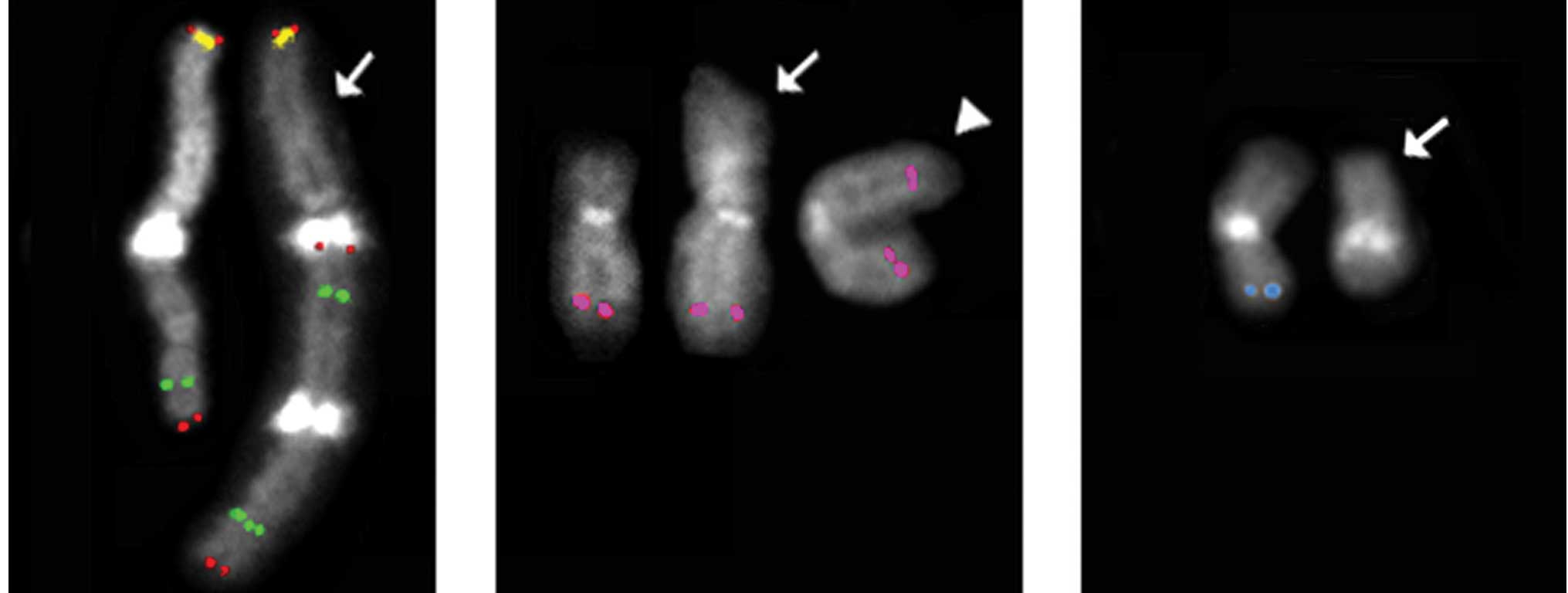
Differing from KG-1, the karyotype of the KG-1a cell
line showed specific differences from that described by Mrózek
et al (4): the presence,
besides a normal chromosome 1, of a chromosome der(1) that has the duplication of the entire
long arm (Fig. 1A); chromosome
der(8;12) (Fig. 1B, arrow) instead
of the der(8;22); only one normal copy of the chromosome 11 besides
the i(11)(q10); a deleted
chromosome 16q (Fig. 1C); two
normal copies of chromosome 22 and lack of the chromosome Y.
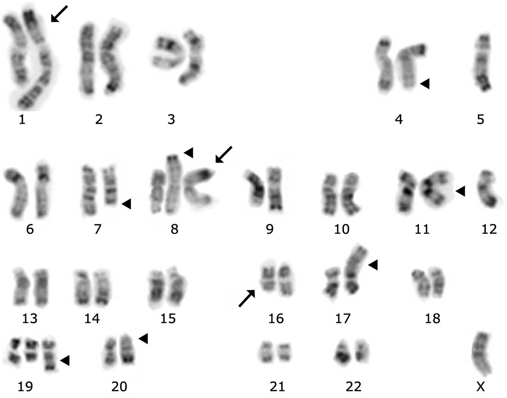
Accordingly, the description of the KG-1a karyotype observed
(Fig. 2) is:
45-47,X,-Y,der(1)(pter→cen→q12::qter→q12::q12→qter),
der(4)t(4;8)(q31;p21),−5,del(7)(q22q35),der(8)t(8;12)(p11;q13), +idic(8)(p11),i(11)(q10),−12,del(16)(q13~21),
der(17)(5pter→5p11::5q13→5q31::17p11.2→cen→17qter),
+der(19)t(14;19)(q13;q13.4),der(20)t(12;20)(?;p13).
Therefore, in the KG-1a karyotype the MYC gene was
present in four copies: one on a normal chromosome 8, two on the
idic(8)(p11) and one on the
der(8)t(8;12) (Fig. 1B). We also observed that the
PLZF gene was present in three copies: one on a normal
chromosome 11 and two on the i(11)(q10). The RARA gene was present
in two copies: one localized on a normal chromosome 17, the other
on the short arm of the der(17)t(5;17). The BCR gene was
present in normal number and chromosomal location on two
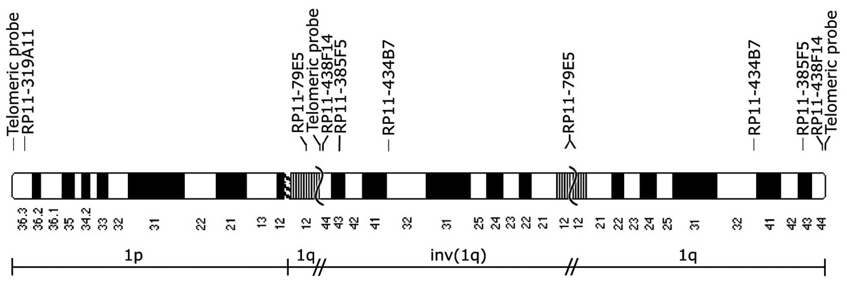
chromosomes 22. Of note is that due to the presence of the
chromosome der(1) bearing two
copies of the long arm of the chromosome 1, three copies of the 54
genes indicated as involved in cancer (10) localized on the long arm of this
chromosome are present (Fig.
3).
Discussion
Koeffler et al (2) reported the presence of a large
submetacentric chromosome (MAR-1) in the karyotype of the KG-1 and
KG-1a cell lines, besides two normal copies of chromosome 1.
Moreover, Furley et al (3)
described the presence of a der(1)
(mar 1) only partially identified and slightly different in the two
cell lines, besides two normal copies of chromosome 1. Therefore,
it is likely that the KG-1 cell line examined by Mrózek et
al (4) and by the authors of
this study has lost the chromosome MAR-1 observed by Koeffler et
al (2) and by Furley et
al (3) and has maintained two
copies of chromosome 1. Conversely, the KG-1a cell line examined by
Mrózek et al (4) lost the
chromosome MAR-1 and maintained the two copies of chromosome 1,
whereas the KG-1a cell line examined in the present study has lost
one normal chromosome 1 and maintained the der(1) described in the present study.
The differences between the KG-1a karyotype
described by Mrózek et al (4) and the one described in the present
study demonstrate again that although the established cancer cell
lines maintained in culture or refreshed numerous times in
different laboratories derive from the same original cell lines,
they may have different genotypic characteristics due either to the
selection of subclones or to intervening mutations. Therefore, the
use of established cancer cell lines for research purposes in
cancer biology, and in any other field, should be preceded by a
proper cytogenetic and molecular characterization (11).
References
|
1
|
Koeffler HP and Golde DW: Acute
myelogenous leukemia: a human cell line responsive to
colony-stimulating activity. Science. 200:1153–1154. 1978.
View Article : Google Scholar : PubMed/NCBI
|
|
2
|
Koeffler HP, Billing R, Lusis AJ, et al:
An undifferentiated variant derived from the human acute
myelogenous leukemia cell line (KG-1). Blood. 56:265–273.
1980.PubMed/NCBI
|
|
3
|
Furley AJ, Reeves BR, Mizutani S, et al:
Divergent molecular phenotypes of KG1 and KG1a myeloid cell lines.
Blood. 68:1101–1107. 1986.PubMed/NCBI
|
|
4
|
Mrózek K, Tanner SM, Heinonen K and
Bloomfield CD: Molecular cytogenetic characterization of the KG-1
and KG-1a acute myeloid leukemia cell lines by use of spectral
karyotyping and fluorescence in situ hybridization. Genes
Chromosomes Cancer. 38:249–252. 2003.PubMed/NCBI
|
|
5
|
Jones L, Wei G, Sevcikova S, et al: Gain
of MYC underlies recurrent trisomy of the MYC chromosome in acute
promyelocytic leukemia. J Exp Med. 207:2581–2594. 2010. View Article : Google Scholar : PubMed/NCBI
|
|
6
|
Guidez F, Parks S, Wong H, et al:
RARalpha-PLZF overcomes PLZF-mediated repression of CRABPI,
contributing to retinoid resistance in t(11;17) acute promyelocytic
leukemia. Proc Natl Acad Sci USA. 104:18694–18699. 2007. View Article : Google Scholar : PubMed/NCBI
|
|
7
|
Arlt MF, Casper AM and Glover TW: Common
fragile sites. Cytogenet Genome Res. 100:92–100. 2003. View Article : Google Scholar
|
|
8
|
Ijdo WJ, Wells RA, Baldini A and Reeders
ST: Improved telomere detection using a telomere repeat probe
(TTAGGG)n generated by PCR. Nucleic Acids Res. 19:47801991.
View Article : Google Scholar : PubMed/NCBI
|
|
9
|
Pelliccia F, Bosco N and Rocchi A:
Breakages at common fragile sites set boundaries of amplified
regions in two leukemia cell lines K562 - Molecular
characterization of FRA2H and localization of a new CFS FRA2S.
Cancer Lett. 299:37–44. 2010. View Article : Google Scholar : PubMed/NCBI
|
|
10
|
Dessen P, Knuutila S and Huret JL:
Chromosome. Atlas Cytogenet Oncol Haematol. Jul. 2004, Updated
2010. http://AtlasGeneticsOncology.org.
|
|
11
|
Hughes P, Marshall D, Reid Y, et al: The
costs of using unauthenticated, over-passaged cell lines: how much
more data do we need? Biotechniques. 43:575–586. 2007. View Article : Google Scholar : PubMed/NCBI
|