Introduction
Pancreatic cancer is one of the most aggressive
malignancies worldwide. It is ranked as the fourth leading cause of
cancer mortality in the United States and the sixth leading cause
of cancer mortality in China (1). A
2011 analysis of the global incidence of cancer mortality revealed
that there are approximately 138,100 and 127,900 annual male and
female mortalities worldwide, respectively (2). Due to its high propensity for local
invasion, distant metastases and a lack of early detection methods
when diagnosed, more than two-thirds of cases demonstrate advanced
or unresectable forms of the disease. Thus, the five-year survival
rate for pancreatic cancer patients remains less than 5% (3). The pathogenesis of pancreatic cancer
is a complex, multistage and multigenetic process, of which the
precise mechanisms are not yet fully understood. Gemcitabine, a
cytotoxic nucleoside analogue, is the current therapeutic strategy
for advanced pancreatic cancer, but it provides no significant
survival advantage to patients (4).
Accordingly, there is great demand to identify novel oncogenes and
clinically applicable molecular targets for the diagnosis and
treatment of this disease (5).
The metastasis-associated in colon cancer-1 (MACC1)
gene was identified by a genome-wide search for genes
differentially expressed by analyzing normal tissues, primary
tumors and metastastic lesions in colon cancer (6). MACC1 has been found to induce tumor
proliferation, invasion and metastasis in cell culture, as well as
in patients with colon cancer. Moreover, MACC1 expression is
upregulated at the crucial step of transition from adenoma to
carcinoma. Furthermore, in distant metastases, MACC1 mainly
translocates into the nucleus from the cytoplasm. Notably,
hepatocyte growth factor (HGF) treatment may also cause MACC1
nuclear translocation and following this, MACC1 binds to the
promoter of the receptor tyrosine kinase Met and activates its
transcription. The resulting upregulation of Met enhances the
HGF/Met pathway and MACC1 acts as a key regulator (7). MACC1 may be a target gene of the MAPK
signaling pathway (6). MACC1 was
reported to be elevated in various cancer tissues, including colon
cancer (6,8), hepatocarcinoma (9) and lung adenocarcinoma (10,11),
and upregulation of its gene expression was also observed in
gastric cancer (12). However,
until recently, there has been no study demonstrating that serum
MACC1 may be used as a biomarker in cancer, and little is known
regarding its role in pancreatic cancer development and drug
resistance.
In the present study, we have focused on evaluating
the clinical significance of serum MACC1 for the diagnosis of
pancreatic cancer. In addition to this, we investigated the effect
of MACC1 inhibition on pancreatic cancer proliferation, metastasis
and sensitivity to gemcitabine. We also explored the possible
underlying mechanisms that may be involved in gemcitabine
resistance induced by MACC1.
Patients and methods
Patients and serum
A total of 109 serum samples were analyzed. The
study included 60 patients with pancreatic cancer and 49 healthy
controls who were hospitalized in the Department of Surgery at The
Second Affiliated Hospital of Zhejiang University, Zhejiang, China,
between January 2010 and December 2011. The serum samples had been
obtained the day before surgical procedures or before the
initiation of neoadjuvant chemotherapy, and stored at −80°C until
analysis. The clinical characteristics of the patients, including
gender, age and TNM stage are summarized in Table I. The clinical stage of the tumors
was determined according to the Union for International Cancer
Control (UICC) 2002 TNM staging of pancreatic cancer (13). The present study was approved by the
Ethical Committee within our hospital and informed consent was
obtained from each patient prior to surgery or collection of blood
samples.
 | Table I.Correlation between serum MACC1
expression levels and clinicopathological parameters of pancreatic
cancer. |
Table I.
Correlation between serum MACC1
expression levels and clinicopathological parameters of pancreatic
cancer.
| | MACC1 expression
| | |
|---|
| Clinicopathological
parameters | No. of cases | Low | % | High | % | χ2 | P-value |
|---|
| Gender | | | | | | 0.141 | 0.707 |
| Female | 20 | 13 | 65.0 | 7 | 35.0 | | |
| Male | 40 | 24 | 60.0 | 16 | 40.0 | | |
| Age (years) | | | | | | 0.984 | 0.321 |
| ≤60 | 23 | 16 | 69.6 | 7 | 30.4 | | |
| >60 | 37 | 21 | 56.8 | 16 | 43.2 | | |
| Invasion depth | | | | | | 0.242 | 0.623 |
| T1+T2 | 11 | 8 | 72.7 | 3 | 27.3 | | |
| T3+T4 | 49 | 29 | 59.2 | 20 | 40.8 | | |
| Lymph node
metastasis | | | | | | 4.467 | 0.035 |
| No | 14 | 12 | 85.7 | 2 | 14.3 | | |
| Yes | 46 | 25 | 54.3 | 21 | 45.7 | | |
| Distant
metastasis | | | | | | 7.944 | 0.005 |
| No | 24 | 20 | 83.3 | 4 | 16.7 | | |
| Yes | 36 | 17 | 47.2 | 19 | 52.8 | | |
| TNM stages | | | | | | 5.107 | 0.024 |
| I+II | 18 | 15 | 83.3 | 3 | 16.7 | | |
| III+IV | 42 | 22 | 52.4 | 20 | 47.6 | | |
Measurement of MACC1 in serum
samples
The serum levels of MACC1 were measured by ELISA
(Uscn Life Science Inc., Wuhan, China) according to the
manufacturer’s instructions, and all measurements were conducted in
duplicate. A 100 μl measurement of each dilution of standard
(provided by the manufacturer), sample and blank was added to a
96-well plate that had been coated with MACC1 antibody and
incubated for 2 h at 37°C. Each well was aspirated and subsequently
washed with washing buffer (provided by the manufacturer).
Following this, 100 μl of detection reagent A was added to
each well and the plate was incubated for 1 h at 37°C. Once any
unbound antibody had been removed, 100 μl of detection
reagent B was added to each well and the plate was incubated at
37°C for 30 min. Following the washing process, 90 μl of
substrate solution was added to the wells and the plate was
incubated for 20 min at 37°C in a dark environment. The reaction
was then terminated with 50 μl stop solution. The optical
density of each well was determined immediately using a microplate
reader at 450 nm. Standard curves generated by a two-parameter
logistic curve fit analysis were used to determine the MACC1
concentrations of the samples.
Cell culture and reagents
The human pancreatic cancer cell lines (CFPAC-1,
AsPC-1, PANC-1, MIA PaCa-2 and BxPC-3) and the human colon cancer
cell lines (SW480 and SW620) were purchased from the American Type
Culture Collection (Manassas, VA, USA). CFPAC-1 cells were
maintained in Iscove’s Modified Dulbecco’s Medium (IMDM), PANC-1
and MIA PaCa-2 cells were maintained in Dulbecco’s modified Eagle’s
medium and AsPC-1, BxPC-3, SW480 and SW620 cells were maintained in
RPMI-1640 medium. All media were supplemented with 10% fetal bovine
serum (FBS), 100 U/ml penicillin and 100 mg/ml streptomycin. The
cells were grown in a monolayer culture in a humidified incubator
containing 5% CO2 in air at 37°C. Gemcitabine was
purchased from Eli Lilly (Bad Homburg, Germany) and dissolved in
sterile 0.9% sodium chloride to obtain a stock solution.
siRNA transfection
Three siRNA sequences targeting human MACC1 and a
negative control siRNA were obtained from Shanghai Genepharma Co.,
Ltd. (Shanghai, China). The sequences of MACC1 and control siRNA
were as follows: MACC1 siRNA1 sense, 5′-CAGGCUAUUUGCAGAA GAATT-3′
and antisense, 5′-UUCUUCUGCAAAUAGCCU GTT-3′; MACC1 siRNA2 sense,
5′-GAGCAAGUAAU GUUUAUGUTT-3 and antisense, 5′-ACAUAAACAUU
ACUUGCUCTT-3′; MACC1 siRNA3 sense, 5′-GCAGUGC UAAGACAAAGCATT-3′ and
antisense, 5′-UGCUUUGUC UUAGCACUGCTT-3′; negative control siRNA
sense, 5′-UUC UCCGAACGUGUCACGUTT-3′ and antisense, 5′-ACGUGA
CACGUUCGGAGAATT-3′. The cells were seeded in 6-well cell culture
plates at a density of 2x10⁵ cells/well and cultured to
40% confluence on the day before transfection. siRNA transfection
was performed using Lipofectamine 2000 (Invitrogen, Carlsbad, CA,
USA) according to the manufacturer’s instructions. The effects of
the transfection on MACC1 reduction were assayed by western blot
analysis and flow cytometry analysis.
RNA preparation and quantitative
real-time PCR (qRT-PCR)
Total RNA was isolated from cell cultures with
TRIzol reagent (Invitrogen) according to the manufacturer’s
instructions. RNA concentrations were determined by
spectrophotometry. Complementary DNA was synthesized from 5
μg of total RNA using M-MLV Reverse Transcriptase (Promega,
Madison, WI, USA). The expression of MACC1 mRNA relative to
glyceraldehyde-3-phosphate dehydrogenase (GAPDH) was measured 3
times by qRT-PCR using the Light Cycler real-time PCR system
(Roche, Mannheim, Germany). The primers for MACC1 and GAPDH were as
follows: MACC1 forward, 5′-TCTGTATGAACTTATTGTGGCTC-3′ and reverse,
5′-CATAGGCAGGTTTCCACATC-3′; GAPDH forward,
5′-TGCACCACCAACTGCTTAG-3′ and reverse,
5′-GAGGCAGGGATGATGTTC-3′.
Cell proliferation assay
Cell viability was determined using the
3-(4,5-dimethyl-thiazol-2-yl)-2,5-diphenyltetrazolium bromide (MTT)
assay (Sigma, St. Louis, MO, USA). Cells were plated in a 96-well
plate at 3000 cells/well. Following this, 20 μl of MTT
solution (5 mg/ml) was added to each well and the plate was
incubated at 37°C for 4 h. The medium was then discarded, and the
purple-blue MTT formazan precipitate was dissolved in 150 μl
dimethyl sulfoxide. The plates were mixed for 10 min on a gyratory
shaker and absorbance at 570 nm was measured by an ELISA reader
(ELx800; Bio Tek Instruments, Inc., Winooski, VT, USA).
Migration assay
The migration assays were performed in 24-well
plates using transwell filters with 8.0 μm pore size and 6.5
mm diameter (Corning Inc., Corning, NY, USA). On the day following
transfection, CFPAC-1 cells were trypsinized and seeded at a
density of 1x10⁵ cells/well. The upper chamber contained
300 μl of IMDM (0.5% FBS) and the lower chamber contained
600 μl of IMDM (10% FBS). Following 48-h incubation, the
cells were fixed with 4% paraformaldehyde for 10 min and stained
with eosin for 10 min. The non-migrated cells on the upper side of
the membranes were removed using wet cotton swabs. The stained
cells on the lower side of the membranes were counted in at least 5
fields under the microscope at a magnification of x200.
Western blot analysis
The cultured cells were harvested and lysed in cold
RIPA lysis buffer (Beyotime Institute of Biotechnology, Jiangsu,
China) supplemented with a complete protease inhibitor cocktail
(Roche) composed of 50 mmol/l NaF and 1 mmol/l
Na3VO4. The supernatants were separated by
centrifugation at 12,000 x g for 20 min at 4°C, and the protein
concentrations were determined using a BCA Protein Assay kit
(Thermo, USA). Following this, 40 mg of protein was resolved by
electrophoresis on 8–12% SDS-PAGE and transferred onto
polyvinylidene difluoride (PVDF) membranes (Bio-Rad, Hercules, CA,
USA). The transblotted membranes were blocked with TBST containing
5% skimmed milk for 2 h at room temperature, and then incubated
with the appropriate antibodies. Antibodies used in this study were
as follows: MACC1 (Abcam, Cambridge, MA, USA), Bcl-2, caspase-3,
caspase-9, E-cadherin, vimentin, β-actin (Cell Signaling
Technology, Inc., Danvers, MA, USA), PARP-1, Gab2, p-ERK1/2, ERK1/2
(Santa Cruz Biotechnology, Inc., Santa Cruz, CA, USA) and Ras
(Epitomics, Inc., Burlingame, CA, USA). The secondary antibodies
were purchased from Beijing Zhongshan Golden Bridge Biotechnology
Co., Ltd., Beijing, China. Bands were visualized using an enhanced
chemiluminescence (ECL) reagent kit (Beyotime) and exposed to X-ray
film.
Flow cytometric analysis of
apoptosis
CFPAC-1 cells (2x10⁵) were seeded in
6-well plates for 24 h and transfected with siRNA. Following
transfection, CFPAC-1 cells were treated with or without 0.5
μg/ml gemcitabine for 48 h. The CFPAC-1 cells were
collected, washed with phosphate-buffered saline (PBS) containing
0.1% bovine serum albumin and re-suspended in 500 μl binding
buffer. Following this, the cells were incubated with 5 μl
annexin V-FITC and 10 μl PI solution for 15 min at room
temperature in a dark environment. Subsequently, the samples were
evaluated for apoptosis using flow cytometry (FACSCalibur; BD
Biosciences, Franklin Lakes, NJ, USA).
Statistical analysis
Each experiment was performed in triplicate. The
data were shown as the mean ± standard deviation (SD). All
statistical tests were performed using Statistical Program for
Social Sciences (SPSS) software 18.0 (SPSS Inc., Chicago, IL, USA).
Correlations between MACC1 expression and clinicopathological
features were analyzed using the χ2 test. P<0.05 was
considered to indicate a statistically significant difference.
Results
Correlations between serum MACC1 levels
and clinicopathological features of pancreatic cancer patients
The serum samples from 60 patients with
pathologically confirmed pancreatic cancer and 49 healthy controls
were analyzed. The demographic characteristics of the patients are
summarized in Table I. We measured
serum MACC1 levels using a commercial ELISA kit. To ensure that the
immunoassay was suitable for detecting clinical serum samples, the
linearity was examined to demonstrate excellent linearity with
serial dilutions. The minimum detectable dose of human MACC1
measured by this kit was typically <0.312 ng/ml. The MACC1
levels in the healthy subjects were too low to be measured
precisely using this ELISA kit. In our study, when the MACC1 level
was < 0.312 ng/ml it was considered negative and when >0.312
ng/ml it was considered positive. To determine the association
between the serum MACC1 levels and clinical characteristics,
patients were divided into low level (<4 ng/ml) and high level
(≥4 ng/ml) group. We determined the cut-off level according to the
‘minimum P-value approach’ (14).
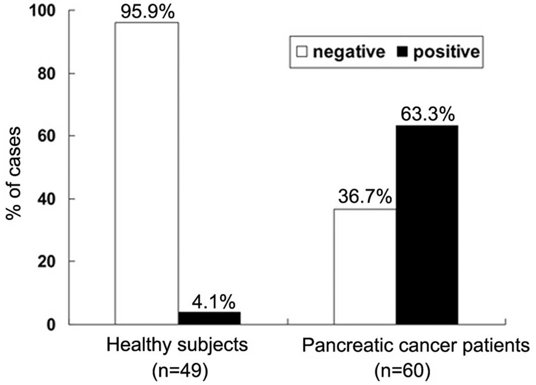
The rate of positive MACC1 expression was 63.3% (38/60) in the
pancreatic cancer patients and 4.1% (2/49) in the healthy subjects
(Fig. 1). A significant difference
in the level of MACC1 expression was demonstrated between the
pancreatic cancer patients and the healthy subjects
(χ2=40.763; P<0.001). Table I shows the correlations between the
serum MACC1 level and the clinicopathological characteristics of
individuals with pancreatic cancer. In these patients, no
significant difference was found in serum MACC1 levels according to
gender (P=0.707), age (P=0.321) and invasion depth (P=0.623).
However, a high level of serum MACC1 expression was positively
correlated with lymph node metastasis (P=0.035), distant metastasis
(P=0.005) and later TNM stages (P=0.024). These data indicated that
serum MACC1 may be a novel diagnostic marker for pancreatic
cancer.
Expression of MACC1 in pancreatic cancer
cell lines
Previous studies have indicated that MACC1
demonstrates overexpression in SW620 cells and almost no expression
in SW480 cells, despite SW60 cells being derived from a metastasis
of the same tumor from which the SW480 cell line is derived
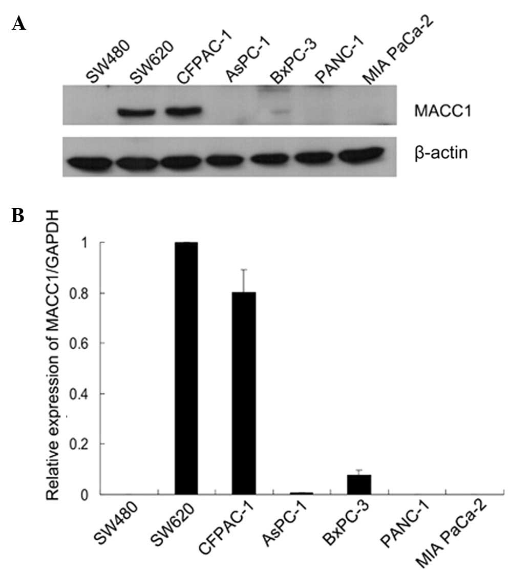
(6). Consequently, we examined the
expression of MACC1 in 5 human pancreatic cancer cell lines. The
MACC1 protein expression was high in CFPAC-1 cells, which are
derived from a metastatic lesion of ductal adenocarcinoma, and low
in BxPC-3 cells. The remaining 3 pancreatic cancer cell lines
demonstrated almost no MACC1 expression (Fig. 2A). The human colon cancer cell line,
SW480, was used as the negative control and the SW620 cell line as
the positive control. We also examined the expression at the mRNA
level using qRT-PCR. The qRT-PCR amplification results demonstrate
that MACC1 mRNA was not detected in the AsPC-1, PANC-1 and MIA
PaCa-2 cell lines. The mRNA expression of MACC1 in BxPC-3 cells was
low, whereas a high expression was noted in the highly metastatic
pancreatic cancer cell line CFPAC-1, second to that in the SW620
cell line (Fig. 2B). In comparison
with non-metastatic cell lines, the metastatic cell lines presented
high levels of MACC1, indicating that MACC1 had a high correlation
with metastasis. Therefore, a CFPAC-1 cell line was selected for
the subsequent experiments.
MACC1 inhibition by siRNA in CFPAC-1
cells
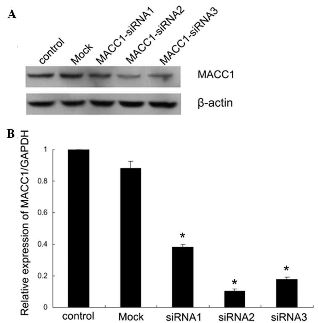
The present study focused on investigating the
potential functions of MACC1 in pancreatic cancer cells. The MACC1
silencing experiment was conducted using 400 pmol of MACC1-siRNA,
and the effects of transfection were confirmed 72 h later by
qRT-PCR and western blot analysis. Our data indicated that in
comparison to the normal control and mock-transfected cells, the
expression of MACC1 was reduced in the 3 siRNA-transfected cells,
particularly in the MACC1-siRNA2-transfected cells (Fig. 3A). The results were consistent with
the experimental data of qRT-PCR (Fig.
3B) where MACC1-siRNA2 provided the highest inhibitory rate.
Downregulation of MACC1 was also visible 4 days following the
initial siRNA transfection. In the following experiments,
MACC1-siRNA2 was used to downregulate the expression of MACC1 in
CFPAC-1 cells.
Downregulation of MACC1 suppressed
pancreatic cancer cell proliferation, migration and inhibited
epithelial-mesenchymal transition (EMT)
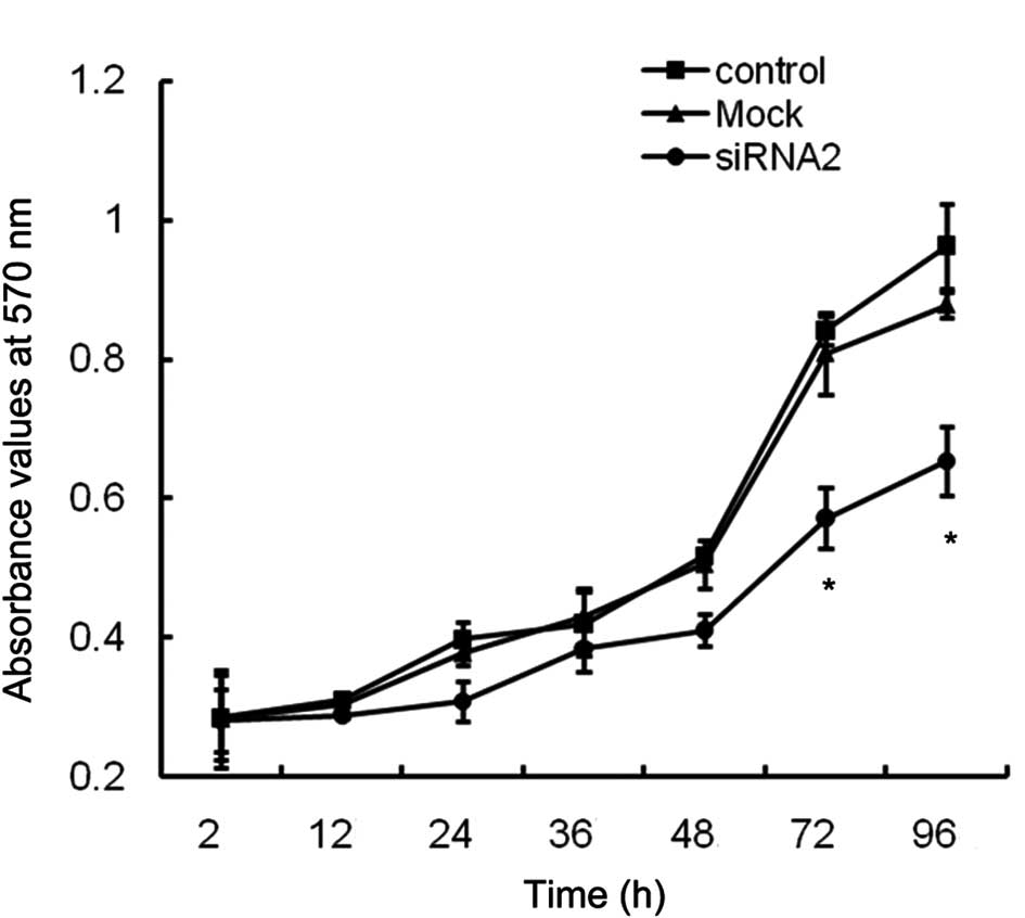
To evaluate the effects of MACC1 down-regulation on
CFPAC-1 cell growth, normal, mock-transfected and
siRNA2-transfected cells were seeded in 96-well plates at a density
of 3000 cells/well and their growth was monitored using an MTT
assay for the following 4 days. In comparison with the control
cells, the mock-transfected cells produced a similar growth rate,
but the MACC1-siRNA2-transfected cells exhibited a significantly
lower growth rate (Fig. 4),
indicating that the inhibition of MACC1 may suppress the
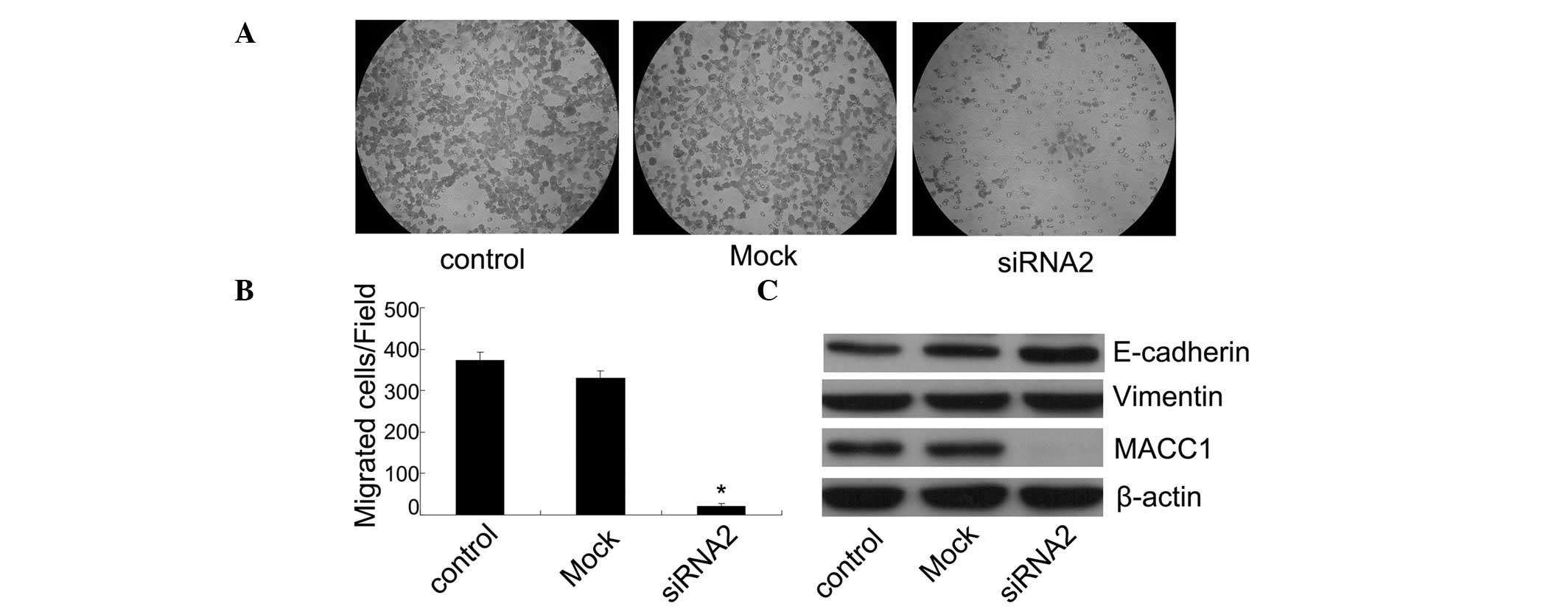
proliferation of CFPAC-1 cells. To examine the effects of
MACC1-siRNA on cell migration, a transwell chamber assay was
performed. Following a 48 h incubation period, the number of
migrated cells in the control group was 374±18, the
mock-transfected group was 331±17 and the MACC1-siRNA2 transfected
group was 20±8. Therefore, there was a reduction in the migration
rate of the MACC1-siRNA2 group in comparison with the control and
mock-transfected groups following MACC1 inhibition by 95% and 94%,
respectively (Fig. 5A and B).
Western blot analyses were performed to confirm that the EMT
phenotype was suppressed by MACC1 inhibition in CFPAC-1 cells.
Following 48 h of transfection, the level of E-cadherin protein was
significantly upregulated in MACC1-siRNA2-transfected cells
relative to the control and mock-transfected cells, while the
vimentin level did not appear to change (Fig. 5C). Collectively, these results
indicated that MACC1 was involved in the EMT of pancreatic cancer
cells and its downregulation may inhibit EMT and suppress cancer
cell migration.
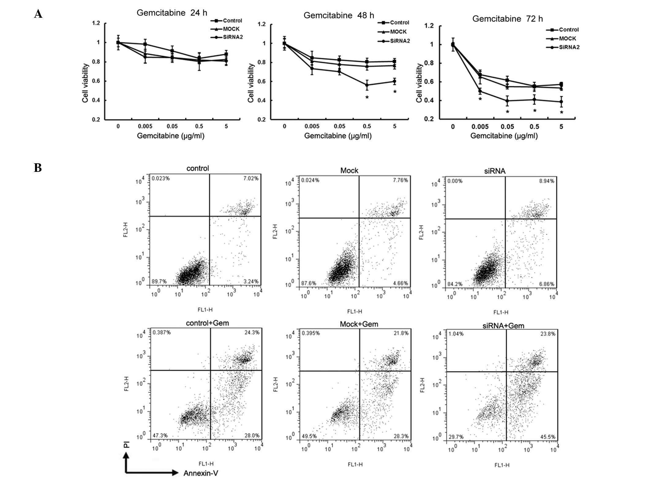
Downregulation of MACC1 sensitized
CFPAC-1 cells to gemcitabine
At present, the best chemotherapeutic agent
available for the treatment of advanced pancreatic cancer is
gemcitabine (15). To verify that
MACC1 was directly involved in the mediation of chemoresistance,
CFPAC-1 cells were transfected with siRNA against MACC1 to
downregulate its expression prior to gemcitabine treatment.
MACC1-siRNA2 was used in the subsequent experiments and cell
viability was evaluated using the MTT assay. Following 24 h of
gemcitabine incubation, no significant difference in the inhibitory
rate was observed among the 3 groups. However, following 48 h of
gemcitabine incubation, there was a significant growth inhibition
in MACC1-siRNA2-transfected cells in comparison with the control
and mock-transfected cells at the concentrations of 0.5
μg/ml and 5 μg/ml, respectively (Fig. 6A). Following 72 h of gemcibatine
incubation at these concentrations, MACC1 downregulation resulted
in an increased inhibitory effect on the proliferation of CFPAC-1
cells. Furthermore, we investigated the degree of cell death in
response to gemcitabine treatment in the 3 cell groups using flow
cytometry. In absence of gemcitabine, suppression of MACC1 caused a
slight reduction in surviving cells and an increased percentage of
apoptotic and dead cells. Following incubation with gemcitabine
(0.5 μg/ml) for 48 h, MACC1 downregulation resulted in a
strong apoptotic effect. The percentage of live cells in the
MACC1-siRNA-transfected, control and mock-transfected groups was
29.7, 47.3 and 49.5%, respectively. The apoptotic rates of the
control and mock- transfected groups at 0.5 μg/ml
gemcitabine were 28.0 and 28.3%, respectively, while the apoptotic
rate in the MACC1 silenced group was 45.5% (Fig. 6B).
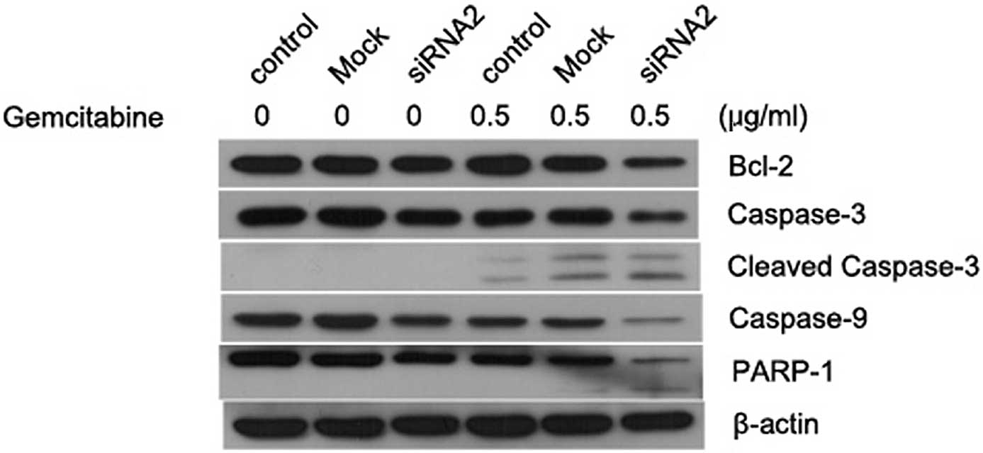
As revealed by immunoblotting analysis (Fig. 7), MACC1 knockdown induced
significant apoptosis in CFPAC-1 cells following treatment with
gemcitabine. This result was verified by a decreased expression of
pro-caspase-3, pro-caspase-9 and PRAP proteins and an increased
production of cleaved caspase-3. siRNA-mediated knockdown of MACC1
also reduced the expression of Bcl-2, an anti-apoptotic protein,
following gemcitabine stimulation. Such chemosensitization effects
of MACC1 knockdown were not evident without gemcitabine treatment.
In normal conditions, knockdown of MACC1 alone induced slight cell
apoptosis, suggesting a potential role of sensitization under
stressful situations.
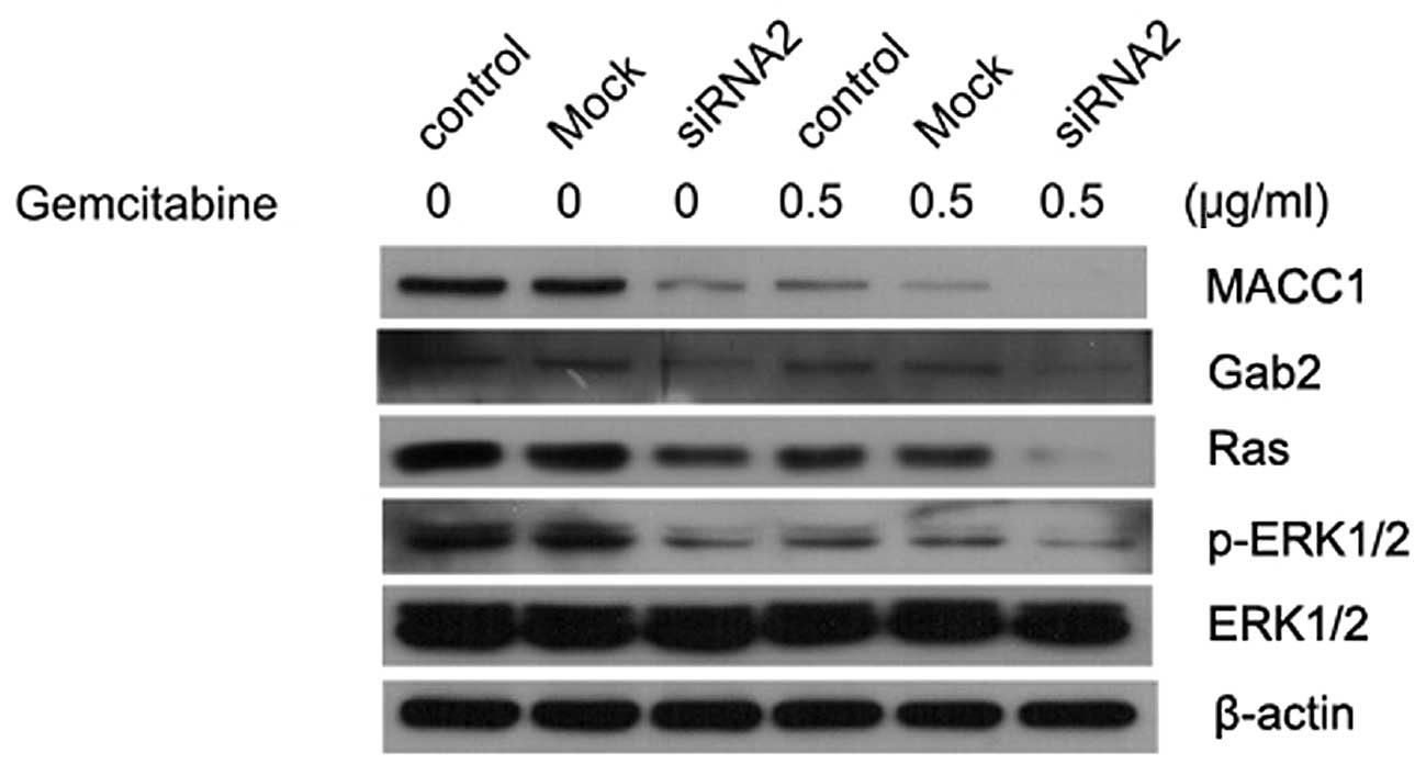
MACC1 regulates the Ras/ERK pathway in
pancreatic cancer cells
We investigated whether MACC1 exhibited any effects
on the Ras/ERK pathway. Knockdown of MACC1 decreased the expression
of Ras and pERK1/2, where such effects were more evident during
gemcitabine treatment (Fig. 8).
Silencing MACC1 also led to the downregulation of Gab2, an adaptor
protein that potentiated the activation of the Ras/ERK and PI3K/AKT
pathways and is associated with mammary tumorigenesis and
metastasis. We found that gemcitabine treatment alone reduced the
activation of Gab2, Ras and pERK, which may be attributed to the
gemcitabine-mediated MACC1 down-regulation. However, further
studies are required to prove such correlations between MACC1 and
the Ras/ERK pathway.
Discussion
MACC1 was first identified using qRT-PCR and
databank analysis, as a colon cancer-associated gene, by Stein
et al (6). These authors
revealed that MACC1 was a prognostic marker for colon cancer
metastasis and metastasis-free survival (6). Since then, the association between
MACC1 levels and the various cancer risks and prognosis have been
analyzed using immunohistochemistry and qRT-PCR. MACC1 has been
demonstrated to be a metastatic or poor prognosis marker for colon
cancer (6,8), gastric cancer (12), lung adenocarcinoma (10,11),
hepatocellular carcinoma (9,16) and
ovarian cancer (17).
Stein et al indicated that MACC1 was located
on the human chromosome 7 (7p21.1) and its role was to markedly
induce cell migration, invasion, colony formation and proliferation
(6). It was observed that in
metastatic lesions MACC1 was mainly expressed in the nuclei of
tumor cells, while in non-metastasizing tumor cells, MACC1 was
predominantly observed in the cytoplasm. Additionally, when treated
with HGF, MACC1 was able to translocate to the nucleus from the
cytoplasm and then bind to the Met promoter to regulate its
expression. These findings demonstrated that MACC1 served as the
major regulator of the HGF/Met signaling pathway. Furthermore,
Stein et al indicated that MACC1 may be involved in the MAPK
signaling pathway (6).
To the best of our knowledge, this is the first
study to investigate serum MACC1 expression in human pancreatic
cancer patients using ELISA methods. In the present study, we found
that serum MACC1 values were significantly elevated in pancreatic
cancer patients, but were weakly expressed or not expressed in
healthy subjects (P<0.001). Furthermore, we found that high
levels of MACC1 often correlated with enhanced lymph node
metastasis, distant metastasis and a later TNM stage. However,
MACC1 serum levels had no significant associations with gender, age
or depth of invasion. Therefore, serum MACC1 levels are considered
to be correlated with more aggressive pancreatic cancers and may be
useful for diagnosis.
The role of MACC1 in pancreatic cancer has not been
reported, therefore, we examined the expression of MACC1 in two
colon cancer cell lines and five pancreatic cancer cell lines. The
CFPAC-1 cell line, which presented the highest level of MACC1, was
selected for the subsequent experiments. In our study, we revealed
that downregulation of MACC1 expression by RNA interference was
able to suppress the proliferation and migration of pancreatic
cancer cells, as well as enhance gemcitabine-induced
cytotoxicity.
EMT was first identified as a culmination of
transcriptional and protein modification events in early embryonic
morphogenesis (18). Emerging
evidences indicated that the EMT was a developmental process that
involved the reduction of intercellular adhesions and the
acquisition of mesenchymal cell characteristics. EMT plays a
significant role in tumor metastasis, invasion and carcinogenesis
(19). Previous studies have
demonstrated that gemcitabine-resistant pancreatic cancer cells
acquired the EMT phenotype, and that EMT may maintain drug
resistance in human pancreatic cancer cells (20–22).
In the present study, we found that the suppression of MACC1 not
only inhibits the migration of CFPAC-1 cells, but also causes the
upregulation of E-cadherin, a marker for epithelial cell phenotype.
We revealed for the first time that inhibiting MACC1 reversed the
EMT phenotype and may increase drug sensitivity. However, other EMT
markers should be investigated to confirm the results.
The Ras/ERK pathway is involved in the regulation of
a variety of biological functions, including cell proliferation,
survival and apoptosis. In addition to this, the Ras/ERK pathway
has been identified to be mutated in approximately 30% of all types
of cancer, with the highest incidences found in pancreatic cancer
(90%) (23). Ras proteins are
distributed in a variety of plasma membrane microdomains and
translocated between cell compartments (24). Activated K-ras mutations are
considered to be the initial step of pancreatic ductal
carcinogenesis (25), while the
knockdown of K-ras by RNA interference enhances gemcitabine-induced
apoptosis in pancreatic cancer (26). In the present study, suppression of
MACC1 was able to inhibit the growth of CFPAC-1 cells, and induce
enhanced apoptosis when combined with gemcitabine treatment. We
demonstrated the induction of apoptotic cell death using multiple
assays, including MTT, flow cytometry and western blot analysis. We
also found that the suppression of MACC1 combined with gemcitabine
significantly inhibited the activity of the Ras/ERK signaling
pathway. This was identified by investigating several downstream
targets of the Ras/ERK pathway, including Bcl-2, caspase-3 and
caspase-9. It was also found that treatment with MACC1-siRNA
significantly decreased the expression of Gab2, a member of the Gab
family of scaffolding adaptors that also contains Gab1 and Gab3
(27). The majority of Gab
protein-receptor interactions are mediated indirectly via Grb2, and
the direct recruitment has been demonstrated only between Gab1 and
c-Met (28). Previous studies have
demonstrated that Gab-Shp2 complexes act as ‘amplifiers’ of the
Ras/ERK signaling pathway activation (29–30).
Therefore, we considered that the Gab family may be involved in the
process of the MACC1-mediated activation of the Ras/ERK signaling
pathway, but the clear mechanisms require further
investigation.
In this study, we present evidence that MACC1
over-expression is detectable in the serum of pancreatic cancer
patients and correlated with tumor metastasis. Therefore, serum
MACC1 expression has value in the diagnosis of pancreatic cancer.
However, the sample size used in our study is small and
investigations should be conducted to confirm our findings in a
larger cohort of patients. MACC1 may also play a significant role
in chemoresistance through regulating the Ras/ERK signaling
pathway. In conclusion, MACC1 may prove to be a novel therapeutic
target for gemcitabine treatment; however, ongoing studies are
required to fully explore the molecular mechanisms involved in
tumor metastasis and chemoresistance.
Acknowledgements
This study was supported by grants
from the National Natural Science Foundation of China (nos.
81172158, 81001094, 81071960, 30901445 and 81100549).
References
|
1.
|
A JemalR SiegelJ XuE WardCancer
statisticsCA Cancer J Clin602773002010
|
|
2.
|
A JemalF BrayMM CenterJ FerlayE WardD
FormanGlobal cancer statisticsCA Cancer J
Clin616990201110.3322/caac.20107
|
|
3.
|
K PliarchopoulouD PectasidesPancreatic
cancer: Current and future treatment strategiesCancer Treat
Rev35431436200910.1016/j.ctrv.2009.02.00519328630
|
|
4.
|
A MaitraRH HrubanPancreatic cancerAnnu Rev
Pathol3157188200810.1146/annurev.pathmechdis.3.121806.154305
|
|
5.
|
J ShenMD PersonJ ZhuJL AbbruzzeseD
LiProtein expression profiles in pancreatic adenocarcinoma compared
with normal pancreatic tissue and tissue affected by pancreatitis
as detected by two-dimensional gel electrophoresis and mass
spectrometryCancer
Res6490189026200410.1158/0008-5472.CAN-04-3262
|
|
6.
|
U SteinW WaltherF ArltMACC1, a newly
identified key regulator of HGF-MET signaling, predicts colon
cancer metastasisNat Med155967200910.1038/nm.188919098908
|
|
7.
|
F ArltU SteinColon cancer metastasis:
MACC1 and Met as metastatic pacemakersInt J Biochem Cell
Biol4123562359200910.1016/j.biocel.2009.08.00119666136
|
|
8.
|
A ShirahataK ShinmuraY KitamuraMACC1 as a
marker for advanced colorectal carcinomaAnticancer
Res3026892692201020682999
|
|
9.
|
A ShirahataW FanK SakurabaMACC 1 as a
marker for vascular invasive hepatocellular carcinomaAnticancer
Res31777780201121498695
|
|
10.
|
G ChundongH UramotoT OnitsukaMolecular
diagnosis of MACC1 status in lung adenocarcinoma by
immunohistochemical analysisAnticancer
Res3111411145201121508357
|
|
11.
|
H ShimokawaH UramotoT
OnitsukaOverexpression of MACC1 mRNA in lung adenocarcinoma is
associated with postoperative recurrenceJ Thorac Cardiovasc
Surg141895898201110.1016/j.jtcvs.2010.09.04421093878
|
|
12.
|
A ShirahataM SakataY KitamuraMACC 1 as a
marker for peritoneal-disseminated gastric carcinomaAnticancer
Res3034413444201020944120
|
|
13.
|
LH SobinWC WittekindTNM Classification of
Malignant Tumours (UICC)6th editionWiley-LissNew York93962002
|
|
14.
|
DG AltmanB LausenW SauerbreiM
SchumacherDangers of using ‘optimal’ cutpoints in the evaluation of
prognostic factorsJ Natl Cancer Inst868298351994
|
|
15.
|
A ArltA GehrzS MuerkosterRole of NF-kappaB
and Akt/PI3K in the resistance of pancreatic carcinoma cell lines
against gemcitabine-induced cell
deathOncogene2232433251200310.1038/sj.onc.120639012761494
|
|
16.
|
J QiuP HuangQ LiuIdentification of MACC1
as a novel prognostic marker in hepatocellular carcinomaJ Transl
Med9166201110.1186/1479-5876-9-16621955323
|
|
17.
|
R ZhangH ShiZ ChenQ WuF RenH HuangEffects
of metastasis-associated in colon cancer 1 inhibition by small
hairpin RNA on ovarian carcinoma OVCAR-3 cellsJ Exp Clin Cancer
Res3083201110.1186/1756-9966-30-8321923915
|
|
18.
|
D ShookR KellerMechanisms, mechanics and
function of epithelial-mesenchymal transitions in early
developmentMech
Dev12013511383200310.1016/j.mod.2003.06.00514623443
|
|
19.
|
J YangRA WeinbergEpithelial-mesenchymal
transition: at the crossroads of development and tumor
metastasisDev
Cell14818829200810.1016/j.devcel.2008.05.00918539112
|
|
20.
|
AN ShahJM SummyJ ZhangSI ParkNU ParikhGE
GallickDevelopment and characterization of gemcitabine-resistant
pancreatic tumor cellsAnn Surg
Oncol1436293637200710.1245/s10434-007-9583-517909916
|
|
21.
|
Z WangY LiD KongAcquisition of
epithelial-mesenchymal transition phenotype of
gemcitabine-resistant pancreatic cancer cells is linked with
activation of the notch signaling pathwayCancer
Res6924002407200910.1158/0008-5472.CAN-08-4312
|
|
22.
|
T ArumugamV RamachandranKF
FournierEpithelial to mesenchymal transition contributes to drug
resistance in pancreatic cancerCancer
Res6958205828200910.1158/0008-5472.CAN-08-281919584296
|
|
23.
|
JL BosRas oncogenes in human cancer: a
reviewCancer Res494682468919892547513
|
|
24.
|
F CalvoL Agudo-IbanezP CrespoThe Ras-ERK
pathway: understanding site-specific signaling provides hope of new
anti-tumor
therapiesBioessays32412421201010.1002/bies.20090015520414899
|
|
25.
|
T DeramaudtAK RustgiMutant KRAS in the
initiation of pancreatic cancerBiochim Biophys
Acta175697101200516169155
|
|
26.
|
S RejibaS WackM AprahamianA HajriK-ras
oncogene silencing strategy reduces tumor growth and enhances
gemcitabine chemotherapy efficacy for pancreatic cancer
treatmentCancer
Sci9811281136200710.1111/j.1349-7006.2007.00506.x
|
|
27.
|
K NishidaT HiranoThe role of Gab family
scaffolding adapter proteins in the signal transduction of cytokine
and growth factor receptorsCancer
Sci9410291033200310.1111/j.1349-7006.2003.tb01396.x14662016
|
|
28.
|
H GuBG NeelThe ‘Gab’ in signal
transductionTrends Cell Biol131221302003
|
|
29.
|
A YartM LaffargueP MayeuxA critical role
for phosphoinositide 3-kinase upstream of Gab1 and SHP2 in the
activation of ras and mitogen-activated protein kinases by
epidermal growth factorJ Biol
Chem27688568864200110.1074/jbc.M00696620011134009
|
|
30.
|
JM CunnickS MengY RenRegulation of the
mitogen-activated protein kinase signaling pathway by SHP2J Biol
Chem27794989504200210.1074/jbc.M11054720011779868
|