Introduction
Chemotherapy is one of the key tools used in the
treatment of cancer, but drug resistance and severe adverse
side-effects are significant obstacles (1–3). New
drugs with improved features are urgently required to overcome drug
resistance in various types of cancer. Natural products have been a
source in the search for anticancer molecules and their further
development into anticancer drugs. Traditional Chinese medicine is
proving to be a promising resource for identifying drugs with
anti-cancer activity (4,5). In recent years, interest in using
natural products as therapeutic agents for cancer has
increased.
Marsdenia tenacissima, the stem of
Marsdenia tenacissima (Roxb.) Wight et Arn. (Family
Asclepiadaceae), is grown widely in the southern provinces
of China. It is used as a herbal medicine for the treatment of
asthma, cancer, trachitis, tonsillitis, pharyngitis, cystitis and
pneumonia (6,7). There are two major active constituents
in M. tenacissima: phenolic acid and C21 steroidal
glycosides (8). Preliminary
clinical studies in China suggest that M. tenacissima is
beneficial for treating patients with cancers such as esophageal
cancer, gastric cancer and lung cancer, and has no significant
side-effects (9,10). Extracts of M. tenacissima
have been reported to have anticancer activity, inhibiting cancer
growth and inducing apoptosis in several cancer cell lines in
vitro(11–14). However, whether M.
tenacissima inhibits tumor angiogenesis and its underlying
mechanism(s) in endothelial cells remains unknown.
Angiogenesis is a crucial step for tumor growth and
progression (15,16). It is a complex multistep process
which includes the destabilization of established vessels,
endothelial cell proliferation, migration and tubulogenesis
(17). Tumor angiogenesis is a
process wherein a network of blood vessels penetrates a cancerous
growth to supply nutrients and oxygen and remove metabolic waste
from the tumor. Tumor growth and metastasis depends heavily on
angiogenesis. The induction of angiogenesis is mediated by a
variety of molecules secreted from the cells within the tumors. It
is well known that vascular endothelial growth factor (VEGF) is
crucial in regulating angiogenesis (18) and has become a key focus of
antiangiogenic therapy.
In the present study, the effect of M.
tenacissima extract (MTE) on the antiangiogenic response was
studied using in vitro and in vivo angiogenesis
models. Furthermore, the possibility of MTE reacting with the
vascular endothelial cells specifically through VEGF receptors was
also investigated.
Materials and methods
Reagents
MTE was provided by Nanjing Sanhome pharmeceutical
Co. Ltd. (Nanjing, China). RPMI-1640 medium, Dulbecco’s modified
Eagle’s medium (DMEM), fetal bovine serum (FBS),
penicillin-streptomycin, trypsin-EDTA and TRIzol reagent were
purchased from Invitrogen (Carlsbad, CA, USA). The cell cycle assay
kit was purchased from BD Biosciences (San Jose, CA, USA), the
SuperScript II reverse transcriptase kit from Promega (Madison, WI,
USA) and the in vitro angiogenesis assay kit from Millipore
(Billerica, MA, USA). Human VEGF-A and VEGF receptor-2 (VEGFR-2;
KDR) ELISAs were obtained from R&D Systems (Minneapolis, MN,
USA). All other chemicals were purchased from Sigma Chemicals (St.
Louis, MO, USA).
Cell culture
Human umbilical vein endothelial (HUVECs) and human
hepatoma cells (HepG2) were obtained from the American Type Culture
Collection (ATCC, Manassas, VA, USA). HUVECs and HepG2 cells were
grown in RPMI-1640 and DMEM, respectively, supplemented with 10%
(v/v) FBS, 100 U/ml penicillin and 100 μg/ml streptomycin
and incubated at 5% CO2 in a 37°C incubator.
The study was approved by the Ethics Committee of
Fujian University of Traditional Chinese Medicine, Fuzhou,
China.
Cell viability assay
Cell viability was evaluated using the
3-(4,5-dimethylthiazol-2-yl)-2, 5-diphenyl tetrazolium bromide
(MTT) colorimetric assay. HUVECs were seeded into 96-well plates at
1×10⁴ cells/well. The cells were treated with 0, 2.5, 5
and 7.5 mg/ml of MTE for 24 h. At the end of the treatment, 20
μl of the MTT (5 mg/ml) was added to each well. After 4 h,
MTT was removed and 100 μl of DMSO was added to each well.
The absorbance was measured at 490 nm with a microplate reader
(BioTek, Winooski, VT, USA). The cell viability was calculated as
follows: cell viability (%) = (average absorbance of MTE-containing
serum group / average absorbance of blank group) × 100.
Evaluation of cell confluency
HUVECs were seeded at a concentration of
2×10⁵ cells/well into a 6-well plate. The cells were
treated with 0, 2.5, 5 and 7.5 mg/ml of MTE for 24 h. Cell
confluency was evaluated using a phase-contrast microscope at a
magnification of ×200 (Olympus, Tokyo, Japan).
Cell cycle analysis
After incubating the HUVECs with various
concentrations of MTE for 24 h, the cells were harvested and
adjusted to a concentration of 1×10⁶ cells/ml. Cell
cycle analysis was evaluated using flow cytometry and the
percentages of G0/G1-phase, S-phase and G2/M-phase were calculated
with the ModFit software (BD Biosciences).
Wound-healing assay
HUVECs were seeded at a concentration of
2×10⁵ cells/well, into a 12-well plate. Following 24 h
of incubation, cells were scraped away vertically in each well
using a pipette tip. Three randomly selected views along the
scraped line were photographed on each well using a phase-contrast
inverted microscope at ×100 magnification. The cells were further
treated with various concentrations of MTE (0, 2.5, 5, 7.5 mg/ml)
for 24 h and another set of images was recorded. A reduction in the
scraped area was considered to be the indicative of wound-healing
and cell migration.
Capillary-like tube formation assay
Tube formation by HUVECs was evaluated using the
in vitro angiogenesis assay kit, according to the
manufacturer’s instructions. After the HUVECs were treated with
MTE, cells were harvested and diluted to 1×10⁴ cells in
50 μl medium. The cells were then seeded onto a solid gel of
basement proteins (ECMatrix gel) in 12-well plates and incubated
for 9 h at 37°C. Cellular morphology and the development of
capillary tube networks were evaluated using a phase-contrast
inverted microscope. Images were obtained at a magnification of
×100.
VEGF-A and VEGFR-2 RT-PCR analysis
HUVECs or HepG2 cells were seeded at a concentration
of 2×10⁵ cells into a 6-well plate and treated with
various concentrations of MTE for 24 h. Total RNA was isolated with
TRIzol reagent (Invitrogen). Oligo(dT)-primed RNA (1 μg) was
reverse-transcribed with SuperScript II reverse transcriptase and
the cDNA was used to determine the amount of VEGF-A or VEGFR-2 mRNA
by PCR with Taq DNA polymerase (Fermentas, Waltham, MA, USA) using
the VEGF-A (S: GCC TTG CCT TGC TGC TCT A, AntiS: GAT GTC CAC CAG GG
TCT CG), VEGFR-2 (S: ACG CCG ATT ATG TGA GA, AntiS: AGGC AGG AGT
TGA GTA TGT) and GAPDH (S: GTC ATC CAT GAC AAC TTT GG, AntiS: GAG
CTT GAC AAA GTG GTC GT) primers.
VEGF-A and VEGFR-2 ELISA assay
HUVECs and HepG2 cells were seeded at a
concentration of 2×10⁵ cells into a 6-well plate and
treated with various concentrations of MTE for 24 h. Cells were
collected to measure the secretion level of VEGF-A in the two cell
lines and cell lysates were used to determine the protein
expression levels of VEGFR-2 in the HUVECs. The measurements were
performed using the Quantikine ELISA kit, according to the
manufacturer’s instructions.
Chick chorioallantoic membrane (CAM)
assay
A CAM assay was performed to determine the in
vivo anti-angiogenic activity of MTE. MTE (1 mg) was loaded on
to 0.5 cm-diameter Whatman filter paper and then applied to the CAM
of a seven-day-old embryo. Following incubation for 72 h at 37°C,
the angiogenesis around the filter was recorded. The number of
blood vessels in a circular perimeter surrounding the implants, at
a distance of 0.25 cm from the edge of the filter was counted
manually.
Statistical analysis
All data are the mean of three replicates, with the
exception of the CAM assays in which 10 replicates were performed
for each data point. The data were analyzed using the SPSS software
(Version 11.5). Statistical analysis of the data was performed
using the Student’s t-test and analysis of variance (ANOVA).
P<0.05 was considered to indicate statistically significant
differences.
Results
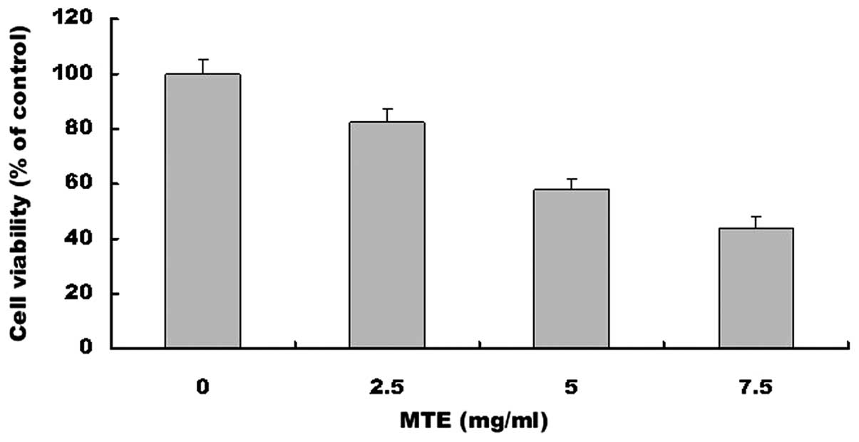
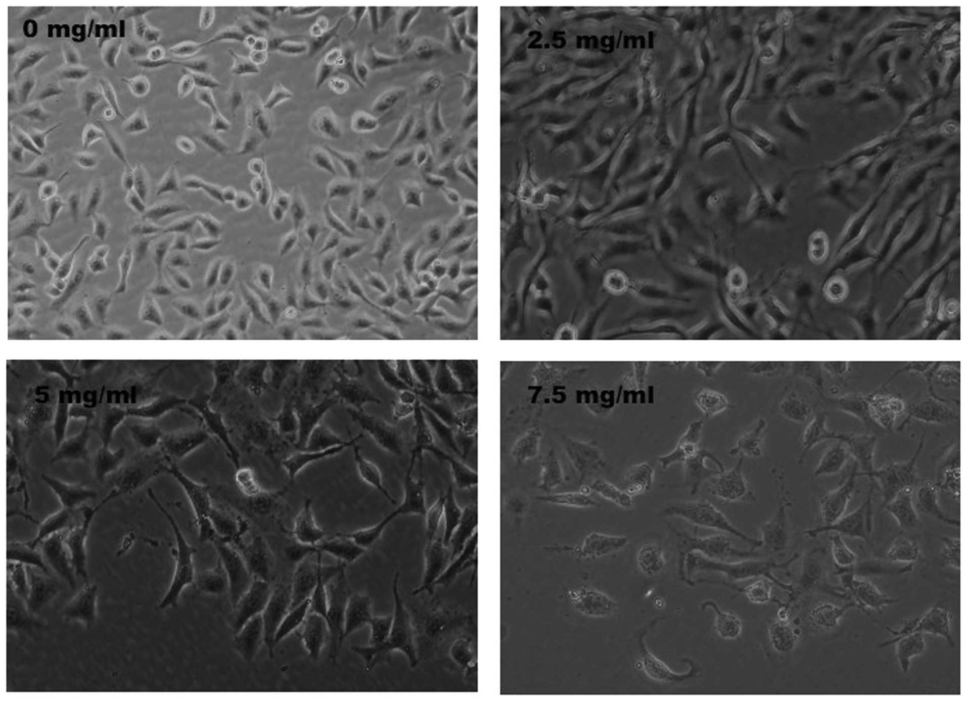
MTE inhibits the proliferation of
HUVECs
HUVEC viability was determined following treatment
with various concentrations of MTE for 24 h. Treatment with 2.5 to
7.5 mg/ml of MTE for 24 h dose dependently reduced the cell
viability from 56 to 17%, when compared with the control cells
(P<0.01; Fig. 1). To further
verify these results, the effect of MTE on HUVEC confluency was
observed via phase-contrast microscopy. MTE treatment led to a
gradual decrease in the confluency of the monolayer with the
increase in drug concentration (Fig.
2).
MTE blocks cell cycle progression in
HUVECs
To test whether the treatment of cells with MTE was
able to cause cell cycle arrest, the cell cycle distribution was
analyzed by flow cytometry following the treatment of the HUVECs
with 0, 2.5, 5 and 7.5 mg/ml of MTE for 24 h. As shown in Fig. 3A and B, the percentage proportions
of S phase cells following treatment with 0, 2.5, 5 and 7.5 mg/ml
of MTE were 41.51±5.2, 37.47±4.5, 28.42±3.5 and 25.75±3.2%,
respectively (P<0.01), suggesting that MTE inhibits HUVEC
proliferation by blocking the cell cycle progression from G1 to
S.
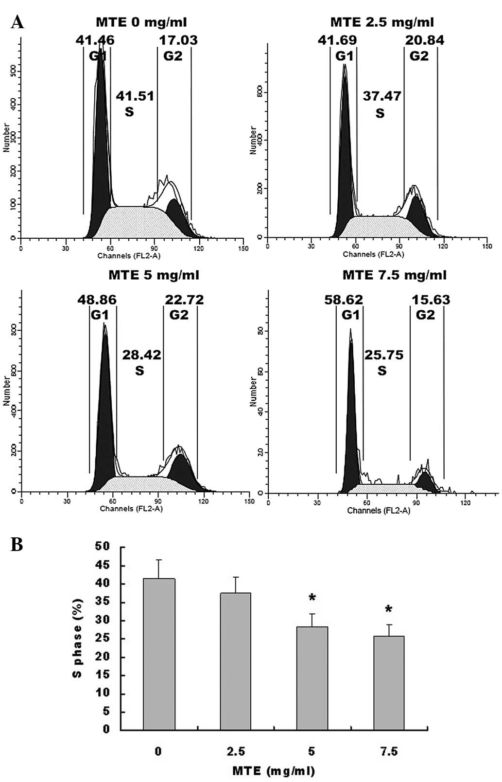 | Figure 3Effect of MTE on HUVEC cell cycle
progression. (A) The cells were treated with 0, 2.5, 5 and 7.5
mg/ml of MTE for 24 h, stained with PI and analyzed by FACS. The
proportion of cells in each phase of the cell cycle was calculated
using ModfitLT Version 3.0 Software. Representative assays are
shown for each concentration of MTE. (B) The percentages of cells
in the S phase, following the treatment with 0, 2.5, 5 and 7.5
mg/ml of MTE were compared. Data shown are the mean ± SD (error
bars) from three independent experiments. *P<0.01,
vs. control cells. MTE, Marsdenia tenacissima extract;
HUVEC, human umbilical vein endothelial cell; PI, propidium
iodide. |
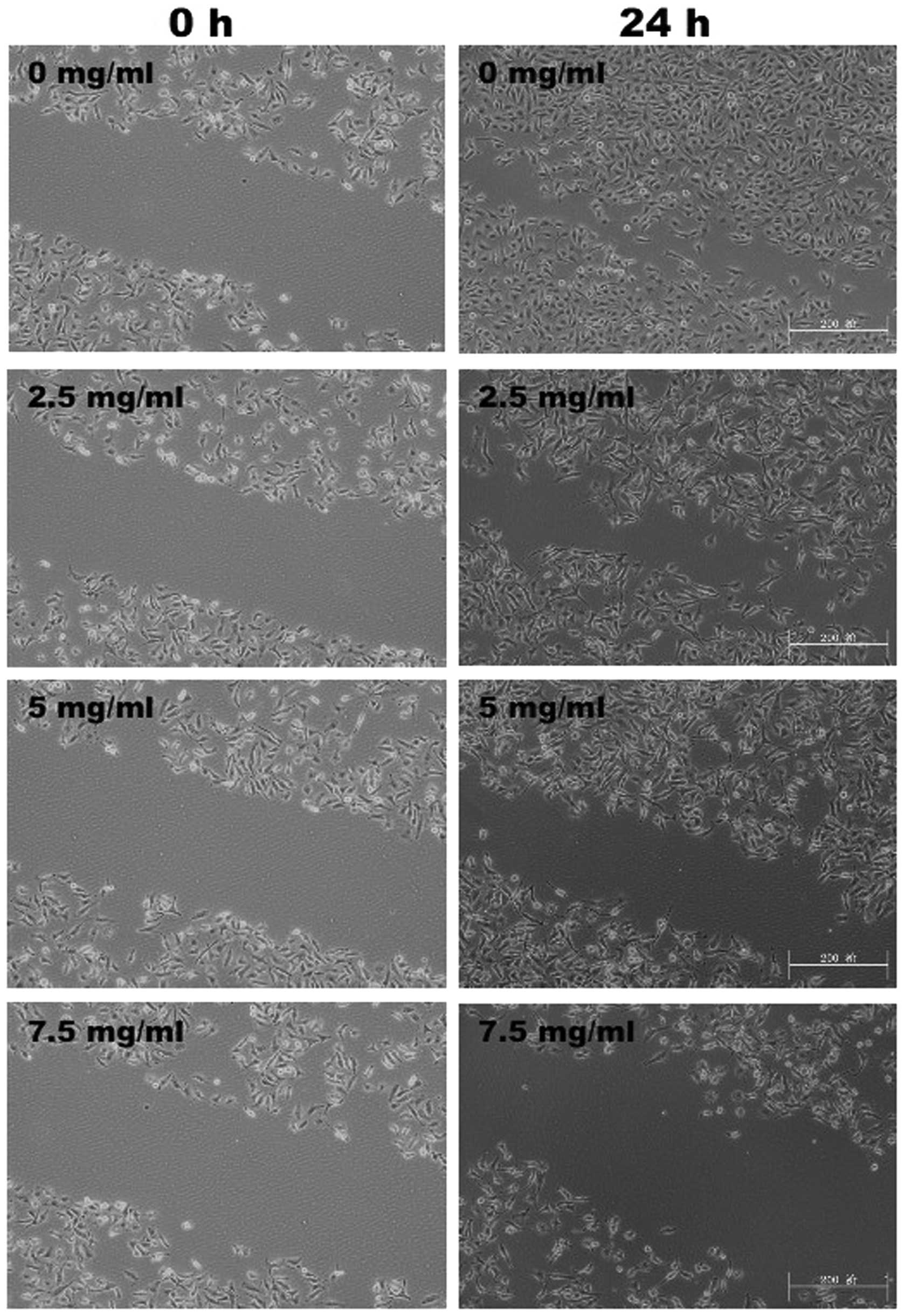
MTE inhibits HUVEC migration and
tubulogenesis
To assess the antiangiogenic properties of MTE in
vitro, its inhibitory effects on the chemotactic motility of
HUVECs were investigated using the wound-healing migration assay.
As shown in Fig. 4, 24 h
post-wounding, untreated HUVECs migrated into the wounded (clear)
area of the cell monolayer, whereas MTE treatment markedly
inhibited the HUVEC migration in a dose-dependent manner.
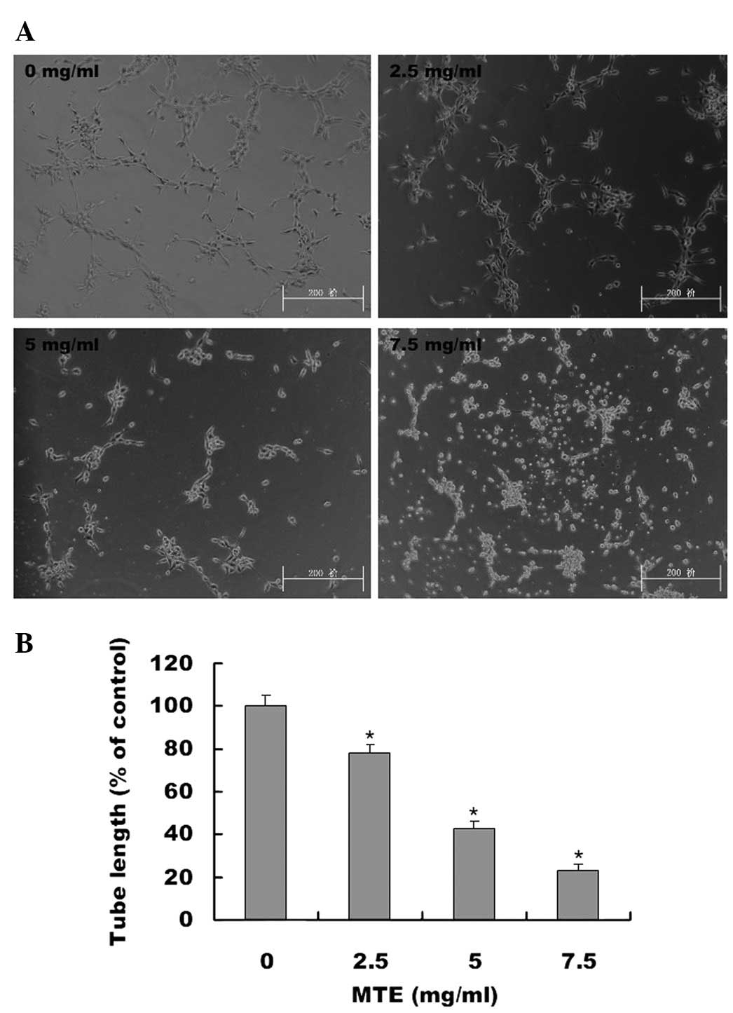
Furthermore, how MTE regulates the capillary tubule formation of
HUVECs was studied. As shown in Fig.
5A and B, untreated HUVECs formed elongated tube-like
structures. By contrast, MTE treatment resulted in a significant
decrease in capillary tube formation in a dose-dependent
manner.
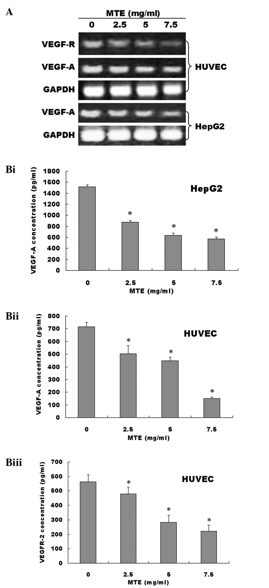
MTE suppresses the expression of VEGF-A
and VEGFR-2
To further investigate the underlying mechanism of
MTE’s anti-angiogenic activity, the effects of MTE on VEGF-A
expression and secretion in HUVECs and HepG2 cells and the
expression of VEGFR-2 in HUVECs were investigated. The results of
the RT-PCR assay showed that MTE treatment reduced VEGF-A mRNA
expression in HepG2 cells and HUVECs in a dose-dependent manner, as
well as suppressing VEGFR-2 mRNA expression in HUVECs (Fig. 6A). Moreover, the protein expression
patterns of VEGF-A and VEGFR-2 showed similar changes, as the mRNA
levels decreased according to the MTE treatment (Fig. 6B).
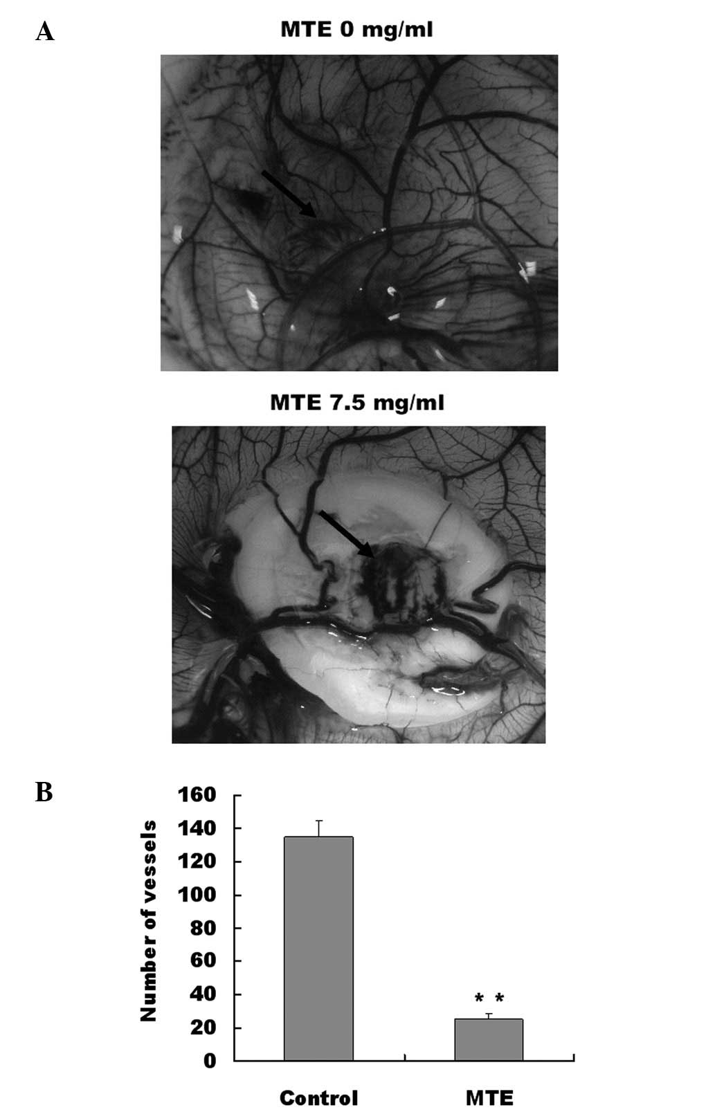
MTE inhibits angiogenesis in vivo
For tumors to grow they must be able to integrate
successfully with normal host tissues at the primary tumor site and
metastastic sites. Tumors require a blood supply to sustain growth.
Therefore, the effect of MTE on microvessel formation in
vivo was studied in the CAM model. The number of vessel branch
points was determined in the absence and presence of MTE. As shown
in Fig. 7A and B, MTE treatment
significantly reduced the total number of blood vessels in the
chicken embryos when compared with the untreated control. This
result provided evidence that the anti-angiogenic effects of MTE
may be important in its anti-cancer activity.
Discussion
The present study demonstrates that MTE is highly
effective at inhibiting cell proliferation in HUVECs by blocking
the cell cycle progression from G1 to S. In addition, the present
study showed that MTE inhibits endothelial cell migration and
prevents vascular formation in vitro. Collectively, the
present data suggest that MTE affects angiogenesis by targeting
endothelial cells directly. MTE was also able to suppress the
growth of blood vessels in CAM models in vivo.
Angiogenesis is important in providing nutrients and
oxygen to the growing tumor (19).
The process of cancer-associated angiogenesis is highly regulated,
involving several growth factors and cytokines. Studies suggest
that tumor growth and dissemination are dependent on the
development of a neovasculature and that VEGF is a primary
stimulator of angiogenesis (20).
VEGF regulates vascular formation through the initiation of vessel
growth, incorporation of hematopoietic and endothelial progenitor
cells into the developing vasculature and inhibition of endothelial
cell apoptosis (21). VEGF exerts
its biological effects primarily through interaction with its
specific receptors, VEGFRs (22,23).
Although VEGF is a ligand for VEGFR-1 and VEGFR-2, VEGF signaling
in angiogenesis is mainly mediated through VEGFR-2, a receptor
tyrosine kinase expressed at elevated levels in endothelial and
cancerous cells (24). Inhibition
of VEGF/VEGFR is a key strategy in anti-angiogenic therapy for
cancer. The present study showed that MTE inhibited VEGF-A and
VEGFR-2 expression in HUVECs. The selective inhibition of
angiogenic factors suggests that MTE has an anti-angiogenic effect,
specifically through VEGF suppression. However, this requires
further study, to understand whether MTE inhibits VEGF expression
through other pathways.
In the present study, MTE inhibited angiogenesis
in vivo and suppressed the key steps involved in
angiogenesis, including proliferation, survival, migration and
tubulogenesis in endothelial cells. To the authors’ knowledge, this
is the first study demonstrating the expression of VEGF/VEGFR in
HUVECs in response to MTE.
In conclusion, the present study demonstrated for
the first time that MTE is able to inhibit tumor angiogenesis in
vitro and in vivo by targeting VEGF/VEGFR in the signal
pathway. The present observation of the anticancer effects of M.
tenacissima not only supports its ethnopharmacological value,
but also confirms its potential as a new anti-angiogenesis
agent.
References
|
1
|
Boos G and Stopper H: Genotoxicity of
several clinically used topoisomerase II inhibitors. Toxicol Lett.
116:7–16. 2000. View Article : Google Scholar : PubMed/NCBI
|
|
2
|
Longley DB, Allen WL and Johnston PG: Drug
resistance, predictive markers and pharmacogenomics in colorectal
cancer. Biochim Biophys Acta. 1766:184–196. 2006.PubMed/NCBI
|
|
3
|
Nakanishi T: Drug transporters as targets
for cancer chemotherapy. Cancer Genomics Proteomics. 4:241–254.
2007.PubMed/NCBI
|
|
4
|
Ji HF, Li XJ and Zhang HY: Natural
products and drug discovery. Can thousands of years of ancient
medical knowledge lead us to new and powerful drug combinations in
the fight against cancer and dementia? EMBO Rep. 10:194–200.
2009.PubMed/NCBI
|
|
5
|
Zhao J, Jiang P and Zhang WD: Molecular
networks for the study of TCM pharmacology. Brief Bioinform.
11:417–430. 2010. View Article : Google Scholar : PubMed/NCBI
|
|
6
|
Editorial Committee of the Pharmacopoeia
of People’s Republic of China: The Pharmacopoeia of People’s
Republic of China, Part 1. Chemical Industry Press; Beijing, China:
pp. 442–443. 2005
|
|
7
|
State Administration of Traditional
Chinese Medicine: Selected Works of Chinese Bencao (No. 4).
Scientific and Technical Publishers; Shanghai: pp. 71999
|
|
8
|
Qiu SX, Luo SQ, Lin LZ and Cordell GA:
Further polyoxypregnanes from Marsdenia tenacissima.
Phytochemistry. 41:1385–1388. 1996. View Article : Google Scholar
|
|
9
|
Wang ZL, Fan QX and Fan KS: Clinical study
of Xiaoaiping in the treatment of cancer. Acta Acad Med Henan.
29:79–80. 1994.
|
|
10
|
Yuan XY, Fang ZG and Huang XY:
Observations On the treatment of 14 cases of advanced gastric
cancer with Inj. Xiao-Ai-Ping. J Shanghai Med Pharm. 6:12–13.
1996.
|
|
11
|
Sun J, Shen JH, Zhang MH, Li CH and Fan
ZZ: Experimental study on therapeutic function of
‘carcinoma-eliminating injection’ on cell of human gastric
carcinoma. Acta Acad Shanghai Tradit Chin Med. 14:41–43. 2000.
|
|
12
|
Li MQ, Shen JH, Xu B and Chen J: The
Mechanism of Laboratory research for Xiaoaiping treating SGC-7901
gastric carcinoma cellular strains. J Interv Radiol. 10:228–231.
2001.
|
|
13
|
Li D, Ouyang J, Li CP and Chen JH:
Marsdensia tenacissima induces apoptosis of human U937, HL60
leukemic cells. Chin J of Biochem Pharm. 29:33–37. 2008.
|
|
14
|
Chen B, Li CP, Chen JH and Ouyang J:
Effect of extract from Marsdenia tenacissima on Jurkat, Raji
and RPMI8226 cells in vitro. Chin J of Biochem Pharmc. 30:174–177.
2009.
|
|
15
|
Folkman J: Seminars in medicine of the
Beth Israel Hospital, Boston. Clinical applications of research on
angiogenesis. N Engl J Med. 333:1757–1763. 1995. View Article : Google Scholar : PubMed/NCBI
|
|
16
|
Folkman J and Shing Y: Angiogenesis. J
Biol Chem. 267:10931–10934. 1992.
|
|
17
|
Risau W: Mechanisms of angiogenesis.
Nature. 386:671–674. 1997. View
Article : Google Scholar : PubMed/NCBI
|
|
18
|
Yi ZF, Cho SG, Zhao H, et al: A novel
peptide from human apolipoprotein(a) inhibits angiogenesis and
tumor growth by targeting c-Src phosphorylation in VEGF induced
human umbilical endothelial cells. Int J Cancer. 124:843–852. 2009.
View Article : Google Scholar : PubMed/NCBI
|
|
19
|
Bergers G and Benjamin LE: Tumorigenesis
and the angiogenic switch. Nat Rev Cancer. 3:401–410. 2003.
View Article : Google Scholar
|
|
20
|
Hicklin DJ and Ellis LM: Role of the
vascular endothelial growth factor pathway in tumor growth and
angiogenesis. J Clin Oncol. 23:1011–1027. 2005. View Article : Google Scholar : PubMed/NCBI
|
|
21
|
Ellis LM and Hicklin DJ: VEGF-targeted
therapy: mechanisms of anti-tumor activity. Nat Rev Cancer.
8:579–591. 2008. View
Article : Google Scholar : PubMed/NCBI
|
|
22
|
Ferrara N: Role of vascular endothelial
growth factor in physiologic and pathologic angiogenesis:
therapeutic implications. Semin Oncol. 29(Suppl 16): 10–14. 2002.
View Article : Google Scholar : PubMed/NCBI
|
|
23
|
Ferrara N, Gerber HP and LeCouter J: The
biology of VEGF and its receptors. Nat Med. 9:669–676. 2003.
View Article : Google Scholar : PubMed/NCBI
|
|
24
|
Shibuya M and Claesson-Welsh L: Signal
transduction by VEGF receptors in regulation of angiogenesis and
lymphangiogenesis. Exp Cell Res. 312:549–560. 2006. View Article : Google Scholar : PubMed/NCBI
|