Introduction
The survival rate of patients with pancreatic ductal
adenocarcinoma (PDAC) is worse than that of patients with other
gastrointestinal malignancies. The principal reasons for this poor
prognosis include difficulties in diagnosing PDAC at a localized
resectable stage and the propensity of the tumor for early
metastasis to the regional lymph nodes and liver. The presence or
absence of lymph node metastasis is one of the key prognostic
factors for patients with PDAC. Therefore, an assessment of the
status of the regional lymph nodes is required to affect the
survival outcomes and therapeutic methods of choice (1,2).
However, the mechanisms by which tumor cells detach
from the primary tumor, invade lymphatic vessels and metastasize to
regional lymph nodes are complex. Previous data has suggested that
chemokine receptors may direct the lymphatic spread and
additionally affect the sites of the metastatic growth of various
tumors (3). Originally, chemokines
and their G protein-coupled receptors were reported to mediate
various pro- and anti-inflammatory responses. CCR7, the receptor
for the chemokine CCL21, is expressed on naive T cells, memory T
cells, B cells and mature dendritic cells, and is considered to be
important in lymphocyte cell trafficking and homing to the lymph
nodes (4,5). The chemokine receptor CXCR4 was
initially described as being able to regulate the homing of
lymphocytes in inflammatory tissues (6). The natural ligand of CXCR4, stromal
cell-derived factor 1a (SDF-1a), is highly expressed in tissues of
metastatic growth, including those of the lung, liver and lymph
nodes, and also attracts lymphocytes to these organs (7). Vascular endothelial growth factor
(VEGF)-C, a member of the VEGF family, has been reported to be a
lymphatic-specific growth factor (8,9).
VEGF-C is the first ligand to be identified for VEGFR-3 (8). Since the expression of VEGFR-3 is
predominantly restricted to the lymphatic endothelium in adults
(10), the major function of VEGF-C
appears to be the regulation of lymphatic vessel growth.
These two chemokine receptors, along with VEGF-C,
have been brought into focus with regard to their role in the
spreading of tumors. CCR7 expression has been shown to be
positively correlated with lymphatic metastasis and a poor
prognosis in squamous cell, oral and oropharyngeal squamous cell
carcinoma and breast, colorectal, esophageal and prostate cancers
(11–15). High CXCR4 expression has been
associated with lymph node metastases in breast cancer and oral
squamous cell carcinoma (16,17).
The levels of VEGF-C in primary tumors have been significantly
correlated with lymph node metastasis in a variety of cancer types,
including oral squamous cell cancer, squamous cell carcinomas of
the head and neck, non-small cell lung carcinoma, cervical cancer
and colorectal cancer (18–22). Gastric cancer, which exhibited a
co-expression of CCR7 and CXCR4, was revealed to be more likely to
include lymph node metastasis (23). High VEGF-C and CXCR4 expression
levels in hepatocellular carcinoma have also been associated with
lymph node metastasis (24). It is
possible that CCR7, CXCR4 and VEGF-C interact with each other in
the process of the metastatic spread of tumor cells to distant
regional lymph nodes.
However, no data are currently available with regard
to the co-expression of CCR7, CXCR4 and VEGF-C in PDAC and their
association with each other. Therefore, the present study evaluated
the mRNA and protein expression levels of CCR7, CXCR4 and VEGF-C in
PDAC and correlated the results with the patients’
clinicopathological parameters.
Materials and methods
Patients and tissue specimens
Tumor tissue was collected for RNA extraction from
24 patients with PDAC who underwent curative surgery between 2006
and 2008, and for immunohistochemistry from 65 patients with PDAC
who underwent curative surgery between 2004 and 2008 at the
Department of Surgery, Zhongshan Affiliated Hospital of Fudan
University (Shanghai, China). Written informed consent was obtained
from each individual. The study was approved by the biomedical
research Ethics Committee of Affiliated Zhongshan Hospital of Fudan
University (Shanghai, China). Patients were excluded if they had
received neoadjuvant chemotherapy or radiotherapy. The freshly
removed PDAC tissues for RNA extraction were immediately frozen in
liquid nitrogen and stored at −80°C until further use. The baseline
characteristics of these patients are shown in Table I. The tissue samples that were to be
used for immunohistochemistry and HE staining were fixed in
formalin and then embedded in paraffin. The baseline
characteristics of these patients are shown in Table II.
 | Table IBaseline characteristics of patients
whose samples were used for RNA extraction (n=24). |
Table I
Baseline characteristics of patients
whose samples were used for RNA extraction (n=24).
| Variable | Value |
|---|
| Total, n | 24 |
| Median age, years (n
± SD) | 63.87±11.22 |
| Age range, years | 42–80 |
| Gender, n (%) | |
| Male | 16 (67) |
| Female | 8 (33) |
| Histological grade, n
(%) | |
| Poorly
differentiated | 14 (58) |
| Well/moderately
differentiated | 10 (42) |
| Tumour stage, n
(%) | |
| T1–T2 | 18 (75) |
| T3–T4 | 6 (25) |
| UICC stage, n
(%) | |
| I + IIA | 8 (33) |
| IIB + III + IV | 16 (67) |
| Lymph node
metastasis, n (%) | |
| Yes | 16 (67) |
| No | 8 (33) |
| Table IIBaseline characteristics of patients
whose samples were used for immunohistochemistry (n=65). |
Table II
Baseline characteristics of patients
whose samples were used for immunohistochemistry (n=65).
| Variable | Value |
|---|
| Total, n | 65 |
| Median age, years (n
± SD) | 63.18±9.23 |
| Age range, years | 44–81 |
| Gender, n (%) | |
| Male | 49 (75) |
| Female | 16 (25) |
| Histological grade, n
(%) | |
| Poorly
differentiated | 33 (51) |
| Well/moderately
differentiated | 32 (49) |
| Tumour stage, n
(%) | |
| T1–T2 | 60 (92) |
| T3–T4 | 5 (8) |
| UICC stage, n
(%) | |
| I + IIA | 39 (60) |
| IIB + III + IV | 26 (40) |
| Lymph node
metastasis, n (%) | |
| Yes | 27 (42) |
| No | 38 (58) |
Real-time reverse transcription (RT)-PCR
analysis
Real-time RT-PCR tumor tissue blocks of ∼1×1×1 cm,
wrapped in silver paper, were rapidly frozen in liquid nitrogen for
∼1 min, then stored in a −80°C refrigerator until the target mRNA
was detected. Total RNA was purified from fresh soft tissues using
TRIzol according to the manufacturer’s instructions. The purity of
the RNA was measured and determined with a UV spectrophotometer and
the OD 260/280 value was 1.8–2.1. To normalize the expressed
cytokine mRNA, the internal housekeeping β-actin gene was used. The
gene transcription of CCR7, CXCR4, VEGF-C and β-actin was analyzed
by two-step RT-PCR. Reverse transcription was performed with 500 ng
RNA (10 μl total volume; SYBR® PrimeScript™
RT-PCR kit, Takara Biotechnology Co. Ltd., Dalian, China) according
to the manufacturer’s instructions. The first-standard cDNA
solution was used as a template for the specific PCR reactions. The
primers used were as follows: CCR7 sense,
5′-CTCCAGGCACGCAACTTTGA-3′ and antisense,
5′-CACAGGTGCTACTGGTGATGTTGA-3′ (145-bp fragment); CXCR4 sense,
5′-GCCAACGTCAGTGAGGCAGA-3′ and antisense,
5′-GCCAACCATGATGTGCTGAAAC-3′ (99-bp fragment); VEGF-C sense,
5′-CAGCACGAGCTACCTCAGCAAG-3′ and antisense,
5′-TTTAGACATGCATCGGCAGGAA-3′ (115-bp fragment); and β-actin sense,
5′-TGAGATGCGTTGTTACAGGA-3′ and antisense,
5′-ACGAAAGCAATGCTATCACC-3′ (119-bp fragment). The cycling
conditions of the PCRs were as follows: An initial denaturation for
10 sec at 95°C, followed by 40 cycles of denaturation for 5 sec at
95°C and annealing for 30 sec at 60°C. Following the last cycle, a
final extension of 10 sec at 60°C was completed and thereafter the
samples were maintained at 4°C. The products (15 μl) were
run on a 2.5% agarose gel, stained with ethidium bromide and
analyzed under UV light.
Quantitative real-time RT-PCR was performed with an
IQ5TM Sequence Detection System (Bio-Rad, Hercules, CA, USA),
according to the manufacturer’s instructions. The reaction mixture
was composed of SYBR Green Mastermix, 12.5 μl of each primer
and cDNA with a total volume of 25 μl. For each gene, a
standard curve was used to calculate the gene expression levels,
thus correcting for the different primer efficiencies. To calculate
the data, the comparative Ct method (2−ΔΔCt
method) was used for the relative quantification, which described
the change in the expression of the target gene in a test sample
and provided accurate comparisons with the initial levels of the
template in each sample. The data were analyzed with SPSS software
version 17.0.
Immunohistochemistry and staining
evaluation
Immunohistochemical studies were performed for
comparison with the results from the mRNA expression analysis
obtained by real-time RT-PCR. The paraffin-embedded tissue sections
(3–5-μm thick) were subjected to immunostaining for CCR7,
CXCR4 (EliVision™ plus kit; Maixin Bio, Fuzhou, China) and VEGF-C
(EliVision™ plus kit; ZSGB Bio Systems, Beijing, China). The
sections were mounted on positively charged slides, incubated for
45 min at 60°C and deparaffinized. Antigen retrieval was performed
with a microwave to boil the tissue sections in 10 mM sodium
citrate buffer (pH 6.0) for 20 min. Subsequent to the endogenous
peroxidase activity being blocked with a 3% aqueous
H2O2 solution for 10 min, the tissue was
incubated with the primary antibodies for CCR7 (ab32527, rabbit
monoclonal IgG; Abcam Cambridge Chemical Co., Cambridge, MA, USA)
at a 1:200 dilution, for CXCR4 (ab2074, rabbit polyclonal IgG Abcam
Cambridge Chemical Co.) and VEGF-C (AF752, goat polyclonal IgG;
R&D Systems, Minneapolis, MN, USA) at 1:100 dilution
respectively, for 1 h. The tissue slides were then incubated with a
reinforcing agent for 20 min. The slides were rinsed with washing
buffer and color was developed with a DAB detection kit (Maixin
Bio, Fuzhou, China) following incubation with the anti-rabbit
antibodies for CCR7 and CXCR4 and the anti-goat antibodies for
VEGF-C. For the negative controls, PBS buffer was substituted for
the primary antibody. The intensity, staining percentage and
pattern of staining were assessed.
The immunostaining was evaluated in a manner that
was blinded to the patient outcome and all clinicopathological
findings. Quantification of the immunostaining was performed by
digital image analysis with the Image-Pro Plus 6.0 software (Media
Cybernetics, Inc., Silver Spring, MD, USA). This method uses areas
of specific staining from the various images to determine the
positivity discrimination plane, minimizing the possible visual
variation in the detection of immunostained areas over time when an
interactive discrimination plane is used. To count the total area,
the discrimination plane was set at 0–30 in the H channel and 0–255
in the S and I channels.
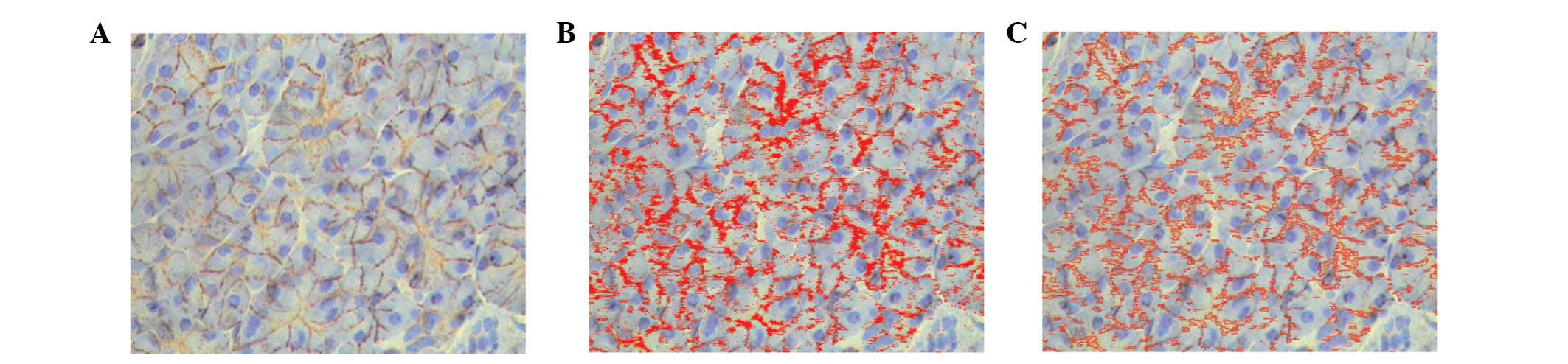
Identical settings were used for each field. A total
of five fields selected from hot-spot areas (400X objective lens)
were acquired per slide. The integrated optical density (IOD) of
all the positive staining in each field and area of interest (AOI)
was measured. The IOD was used to evaluate the area and intensity
of the positive staining. The mean density (IOD/AOI) represented
the concentration of specific protein per unit area (Fig. 1A–C).
Statistical analysis
SPSS 17.0 software (SPSS, Inc., Chicago, IL, USA)
was used for the statistical analysis. Categorical variables were
evaluated by the Wilcoxon rank-sum and Pearson’s correlation tests.
P<0.05 was considered to indicate statistically significant
differences.
Results
Expression of CCR7, CXCR4 and VEGF-C mRNA
and clinicopathological factors
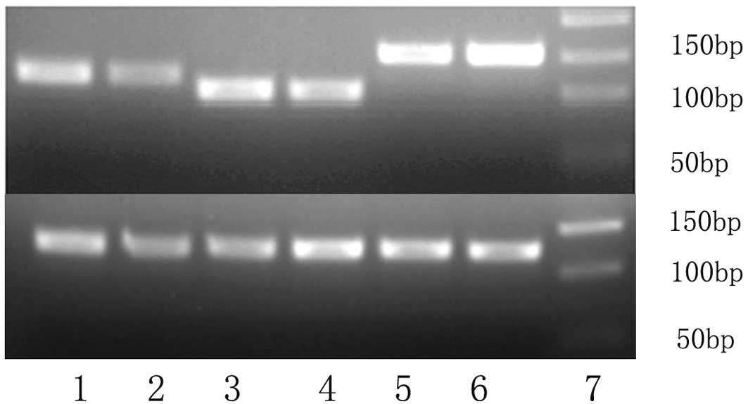
A total of 24 PDAC specimens and 24 adjacent normal
tissues were examined for the expression of CCR7, CXCR4 and VEGF-C
mRNA (Fig. 2). The expression
levels of CCR7, CXCR4 and VEGF-C mRNA in the cancer samples were
all significantly higher than those in the adjacent normal tissue.
There were no significant differences between the CCR7, CXCR4 and
VEGF-C mRNA expression levels and the age, gender or tumor grading
(P>0.05). However, the expression of the CCR7 and VEGF-C mRNA
was significantly correlated with lymph node metastasis and the
advanced International Union Against Cancer (UICC) stage (P=0.001
for lymph node metastasis and for staging). However, the
correlation between the expression of CXCR4 mRNA and lymph node
metastasis or UICC stage was not statistically significant
(P>0.05).
Correlations among the expression of
CCR7, CXCR4 and VEGF-C mRNA
Spearman’s rank correlation analyses showed that
there were correlations among the mRNA expression levels of CCR7,
CXCR4 and VEGF-C. There was a significant positive linear
correlation between the expression levels of CCR7 and VEGF-C
(r=0.915, P<0.001), but not between CCR7 and CXCR4 or CXCR4 and
VEGF-C (P>0.05).
Expression of CCR7, CXCR4 and VEGF-C
protein, and clinicopathological factors
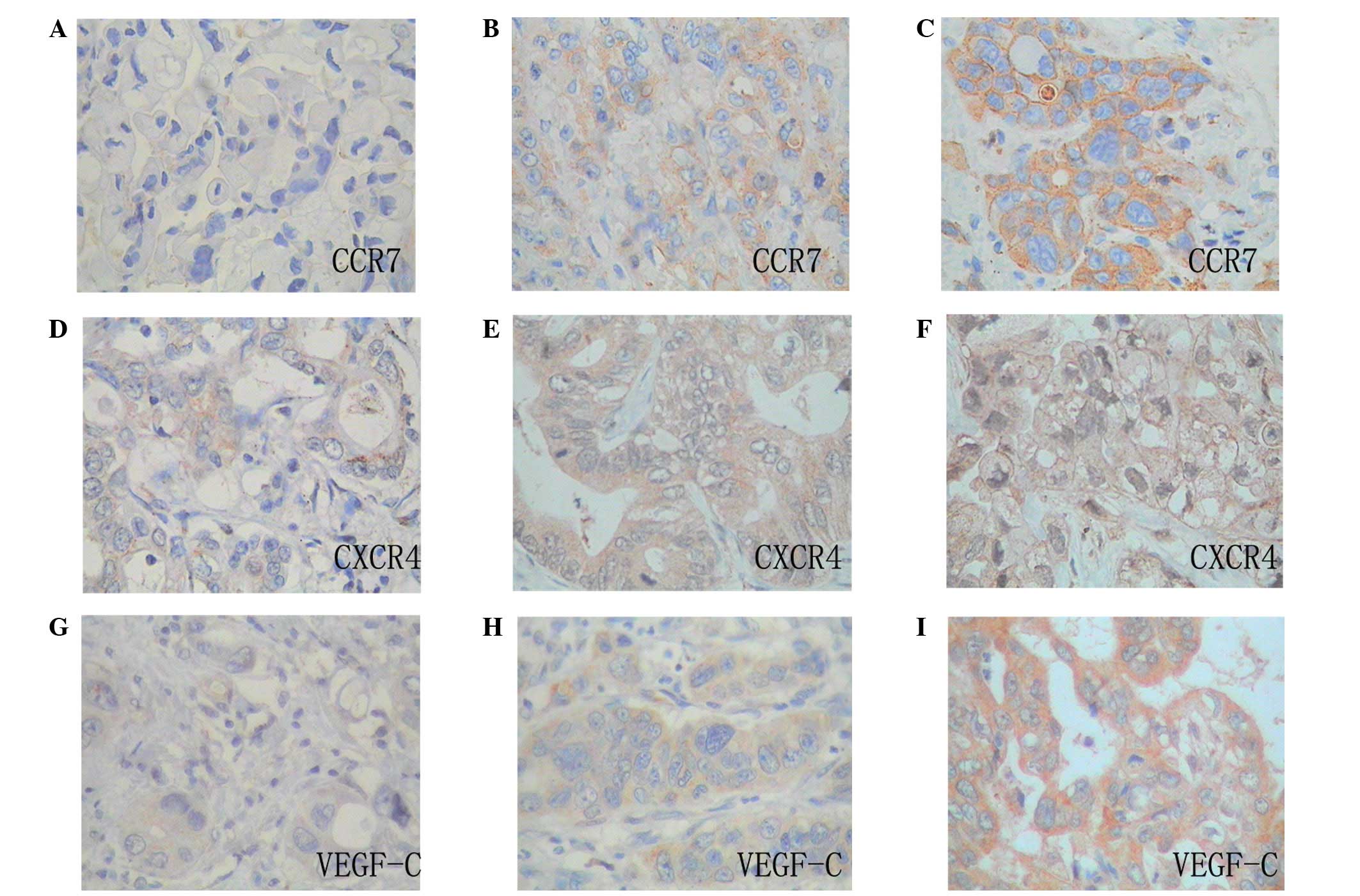
Staining for the CCR7 protein was identified in the
cytoplasm and cell membrane of the cancer cells and was not
detected in the cytoplasm of the normal pancreatic cells obtained
from the non-cancerous regions of the PDAC tissue. Staining for the
CXCR4 protein was also identified in the cytoplasm and cell nucleus
of the cancer cells, but not in the cell nucleus of the normal
pancreatic cells obtained from the non-cancerous regions of the
PDAC tissue. The immunohistological localization of VEGF-C was
cytoplasmic in the cancer and normal pancreatic cells obtained from
the non-cancerous regions of the PDAC tissue. In the cancer
specimens with positive expression, the number of immunoreactive
cells ranged from a few to almost all of the tumor cells (Fig. 3).
The expression levels of CCR7, CXCR4 and VEGF-C in
the cancer cells were all significantly higher than those in the
non-cancerous regions (P<0.05). Furthermore, the patients with a
higher CCR7 and VEGF-C expression in their cancer cells had
significantly higher incidences of lymphatic metastasis
(P<0.01). The greater the number of metastatic lymph nodes, the
higher the level of CCR7 expression. Patients with a higher CCR7
and/or VEGF-C expression also had a higher UICC stage (P<0.05).
However, the correlation between the expression of the CXCR4
protein and lymph node metastasis or UICC was not statistically
significant (P>0.05). The expression levels of CCR7, CXCR4 and
VEGF-C did not correlate with the clinicopathological parameters,
including those of age, gender, tumor size and histological grade
(P>0.05).
Correlations among the expression levels
of CCR7, CXCR4 and VEGF-C protein
Spearman’s rank correlation analyses showed that
there were correlations among the levels of CCR7, CXCR4 and VEGF-C
expression. There were significant positive linear correlations
between the expression levels of CCR7 and CXCR4 or VEGF-C (r=0.449,
P<0.001; r=0.770, P<0.05), but not between CXCR4 and VEGF-C
(P>0.05).
Discussion
PDAC is a type of malignant tumor with marked
invasive characteristics and a high incidence of lymph node
metastasis. Of the patients with small cell PDAC, 30–40% have lymph
node micrometastases which may be observed by immunohistochemistry
or other molecular biological methods (25). At present, surgery remains the
primary treatment for resectable PDAC, although the effect of this
surgery is not completely satisfactory. Previous clinical studies
have shown that lymph node metastasis is the main cause of tumor
recurrence following pancreaticoduodenectomy (1). Even patients who have undergone
extended regional lymph node dissection may experience lymphatic
metastatic recurrence (26,27).
Therefore, adopting a targeted adjuvant therapy to
control lymphatic metastatic recurrence following surgery is one of
the key approaches to improve the survival rate of patients with
PDAC. The identification of post-operative factors associated with
lymph node metastasis is likely to be of great clinical
significance for the application of targeted adjuvant therapy.
Usually, tumor prognosis is predicted from the TNM classification,
although this modality lacks sensitivity and accuracy. Thus,
identifying molecular biomarkers with the potential to predict the
prognosis of PDAC may potentially compensate for the lack of
efficacy of conventional methods.
It is well-known that tumor chemotactic migration
and lymphangiogenesis are directly correlated with lymph node
metastasis. A study by Müller et al revealed that tumor
cells with CCR7-positive expression preferentially transfer to the
lymph nodes that are rich in the ligand CCL21 (28). This provided a theoretical basis
behind the phenomenon of certain tumor cells preferentially
transferring to regional lymph nodes. Nakata et al(29) reported that the high expression of
CCR7 was correlated with lymph node metastasis and a poor prognosis
in PDAC. However, lymphangiogenesis is a precondition for lymph
node metastasis. The current hypothesis is that VEGF-C may be
expressed in a variety of solid tumors and that it is able to
induce lymphatic endothelial cell mitosis by binding to the
receptors VEGFR-3 and VEGFR-2, which are important in
lymphangiogenesis. The expression level of VEGF-C is positively
correlated with lymph node metastasis. Previous studies have shown
that the expression of VEGF-C in PDAC is also closely correlated
with lymph node metastasis (30,31).
However, it is unknown whether the expression of CCR7 and VEGF-C
are correlated with each other and if together they may cause lymph
node metastasis in PDAC. Whether CXCR4 is involved in this process
also remains unclear.
The present study is the first to detect the
expression of CCR7, CXCR4 and VEGF-C in tumor tissues using
real-time RT-PCR and immunohistochemistry assays in a large series
of human PDAC specimens. It was demonstrated that the expression
levels of CCR7, CXCR4 and VEGF-C mRNA and protein were all
significantly higher in the cancer specimens compared with those in
the adjacent normal tissue. The CCR7 and VEGF-C mRNA and protein
expression levels were significantly higher in patients with cancer
types exhibiting lymph node metastasis and a more advanced UICC
stage. Furthermore, the greater the number of metastatic lymph
nodes, the higher the level of CCR7 expression. There was a
significant positive linear correlation between the mRNA and
protein expression levels of CCR7 and VEGF-C. This indicates that
CCR7 and VEGF-C mRNA and protein expression are upregulated in
cases with a greater number of metastasis-positive nodes and may
contribute to lymph node metastasis occurring in PDAC. The present
data are consistent with previous studies that describe a positive
correlation between CCR7 expression and lymph node metastasis in
cases of breast, colorectal, esophageal and prostate cancer and
oral and oropharyngeal squamous cell carcinoma (11–15). A
positive correlation has also been reported between VEGF-C
expression and lymph node metastasis in cases of oral squamous cell
cancer, squamous cell carcinomas of the head and neck, non-small
cell lung carcinoma, cervical cancer and colorectal cancer
(18–22). Although the role of chemokines and
their receptors in human cancers is complex, the chemokine
receptors CCR7 and VEGF-C may be critical in determining lymph node
metastasis in these types of tumors. Wehler et al(32) reported that marked CXCR4 expression
was significantly associated with advanced UICC stages and also
revealed a correlation with hematogenous metastasis. Studies have
shown that CXCR4 is involved in pancreatic cancer progression
through the promotion of angiogenesis (33). However, the present study showed
that the correlation between the expression of CXCR4 and UICC was
not statistically significant. In the present study, the samples
were divided into two groups (I and IIA or IIB, III and IV)
according to the UICC stage, which is different to the grouping
method within the literatur. It is possible that patients with
stage II PDAC had hematogenous metastasis. Thus, the difference of
the result could be interpreted and at the same time it was
conferred that CXCR4 did not play a key role in the lymph node
metastasis of PDAC.
In accordance with previous studies conducted on
other types of cancer, the staining for CCR7 was localized in the
membrane and cytoplasm of the tumor cells, while VEGF-C was
predominantly cytoplasmic. Staining for CCR7 was observed in the
membrane of the normal cells. This may represent the functional
status of the receptor since binding to a specific ligand induces
receptor internalization. The necessity of internalization for
chemotaxis and signaling remains controversial. Endosomes are
gaining considerable attention as scaffolds for signaling
complexes. The assembly of signaling complexes on intracellular
endosomal membranes indicates that the intracellular trafficking
itinerary of chemokine receptors may have significant implications
for signaling (34).
In conclusion, surgery remains the primary treatment
for PDAC in China. Although there are still a range of views with
regard to whether patients who have undergone complete resection of
PDAC should receive adjuvant therapy, due to the high incidence of
lymphatic metastatic recurrence after pancreaticoduodenectomy, we
recommend that patients at a high risk of lymphatic metastatic
recurrence receive targeted adjuvant therapy following surgery. The
present study suggested that the positive expression of CCR7 and
VEGF-C are closely correlated with lymphatic metastatic recurrence
in PDAC. Therefore, these two molecular indicators may become a
reference index for the clinical assessment of lymphatic metastatic
recurrence and poor outcome in patients who should receive
additional treatment, including molecular targeted therapy and
follow-up examinations following surgical treatment. The present
study has laid the groundwork for research into the molecular
mechanism and targeted adjuvant therapy for lymphatic metastases of
PDAC. However, the limitations of the present study include the use
of a small number of patients and limited research conditions, and
consequently, the results do not permit final conclusions to be
drawn. We plan to conduct a prospective study of a large number of
cases to confirm these results.
References
|
1
|
Schnelldorfer T, Ware AL, Sarr MG, Smyrk
TC, Zhang L, Qin R, Gullerud RE, Donohue JH, Nagorney DM and
Farnell MB: Long-term survival after pancreatoduodenectomy for
pancreatic adenocarcinoma: is cure possible? Ann Surg. 247:456–462.
2008. View Article : Google Scholar : PubMed/NCBI
|
|
2
|
House MG, Gönen M, Jarnagin WR, et al:
Prognostic significance of pathologic nodal status in patients with
resected pancreatic cancer. J Gastrointest Surg. 11:1549–1555.
2007. View Article : Google Scholar : PubMed/NCBI
|
|
3
|
Arya M, Patel HR and Williamson M:
Chemokines: key players in cancer. Curr Med Res Opin. 19:557–564.
2003. View Article : Google Scholar
|
|
4
|
Dieu MC, Vanbervliet B, Vicari A, Bridon
JM, Oldham E, Aït-Yahia S, Brière F, Zlotnik A, Lebecque S and Caux
C: Selective recruitment of immature and mature dendritic cells by
distinct chemokines expressed in different anatomic sites. J Exp
Med. 188:373–386. 1998. View Article : Google Scholar : PubMed/NCBI
|
|
5
|
Hirao M, Onai N, Hiroishi K, Watkins SC,
Matsushima K, Robbins PD, Lotze MT and Tahara H: CC chemokine
receptor-7 on dendritic cells is induced after interaction with
apoptotic tumor cells: critical role in migration from the tumor
site to draining lymph nodes. Cancer Res. 60:2209–2217.
2000.PubMed/NCBI
|
|
6
|
Murdoch C: CXCR4: chemokine receptor
extraordinaire. Immunol Rev. 177:175–184. 2000. View Article : Google Scholar : PubMed/NCBI
|
|
7
|
Phillips RJ, Burdick MD, Lutz M, Belperio
JA, Keane MP and Strieter RM: The stromal derived
factor-1/CXCL12-CXC chemokine receptor 4 biological axis in
non-small cell lung cancer metastases. Am J Respir Crit Care Med.
167:1676–1686. 2003. View Article : Google Scholar : PubMed/NCBI
|
|
8
|
Joukov V, Pajusola K, Kaipainen A, Chilov
D, Lahtinen I, Kukk E, Saksela O, Kalkkinen N and Alitalo K: A
novel vascular endothelial growth factor, VEGF-C, is a ligand for
the Flt4 (VEGFR-3) and KDR (VEGFR-2) receptor tyrosine kinases.
EMBO J. 15:290–298. 1996.
|
|
9
|
Orlandini M, Marconcini L, Ferruzzi R and
Oliviero S: Identification of a c-fos-induced gene that is related
to the platelet-derived growth factor/vascular endothelial growth
factor family. Proc Natl Acad Sci USA. 93:11675–11680. 1996.
View Article : Google Scholar : PubMed/NCBI
|
|
10
|
Kaipainen A, Korhonen J, Mustonen T, van
Hinsbergh VW, Fang GH, Dumont D, Breitman M and Alitalo K:
Expression of the fms-like tyrosine kinase FLT4 gene becomes
restricted to lymphatic endothelium during development. Proc Natl
Acad Sci USA. 92:3566–3570. 1995. View Article : Google Scholar : PubMed/NCBI
|
|
11
|
Cabioglu N, Yazici MS, Arun B, Broglio KR,
Hortobagyi GN, Price JE and Sahin A: CCR7 and CXCR4 as novel
biomarkers predicting axillary lymph node metastasis in T1 breast
cancer. Clin Cancer Res. 11:5686–5693. 2005. View Article : Google Scholar : PubMed/NCBI
|
|
12
|
Günther K, Leier J, Henning G, et al:
Predictor of lymph node metastasis in colorectal carcinoma by
expression of chemokine receptor CCR7. Int J Cancer. 116:726–733.
2005.PubMed/NCBI
|
|
13
|
Ding Y, Shimada Y, Maeda M, Kawabe A,
Kaganoi J, Komoto I, Hashimoto Y, Miyake M, Hashida H and Imamura
M: Association of CC chemokine receptor 7 with lymph node
metastasis of esophageal squamous cell carcinoma. Clin Cancer Res.
9:3406–3412. 2003.PubMed/NCBI
|
|
14
|
Heresi GA, Wang J, Taichman R, Chirinos
JA, Regalado JJ, Lichtstein DM and Rosenblatt JD: Expression of the
chemokine receptor CCR7 in prostate cancer presenting with
generalized lymphadenopathy: report of a case, review of the
literature, and analysis of chemokine receptor expression. Urol
Oncol. 23:261–267. 2005. View Article : Google Scholar
|
|
15
|
Tsuzuki H, Takahashi N, Kojima A, Narita
N, Sunaga H, Takabayashi T and Fujieda S: Oral and oropharyngeal
squamous cell carcinomas expressing CCR7 have poor prognoses. Auris
Nasus Larynx. 33:37–42. 2006. View Article : Google Scholar : PubMed/NCBI
|
|
16
|
Kato M, Kitayama J, Kazama S and Nagawa H:
Expression pattern of CXC chemokine receptor-4 is correlated with
lymph node metastasis in human invasive ductal carcinoma. Breast
Cancer Res. 5:R144–R150. 2003. View
Article : Google Scholar : PubMed/NCBI
|
|
17
|
Uchida D, Begum NM, Almofti A, Nakashiro
K, Kawamata H, Tateishi Y, Hamakawa H, Yoshida H and Sato M:
Possible role of stromal-cell-derived factor-1/CXCR4 signaling on
lymph node metastasis of oral squamous cell carcinoma. Exp Cell
Res. 290:289–302. 2003. View Article : Google Scholar : PubMed/NCBI
|
|
18
|
Sedivy R, Beck-Mannagetta J, Haverkampf C,
Battistutti W and Hönigschnabl S: Expression of vascular
endothelial growth factor-C correlates with the lymphatic
microvessel density and the nodal status in oral squamous cell
cancer. J Oral Pathol Med. 32:455–460. 2003. View Article : Google Scholar : PubMed/NCBI
|
|
19
|
Neuchrist C, Erovic BM, Handisurya A,
Fischer MB, Steiner GE, Hollemann D, Gedlicka C, Saaristo A and
Burian M: Vascular endothelial growth factor C and vascular
endothelial growth factor receptor 3 expression in squamous cell
carcinomas of the head and neck. Head Neck. 25:464–474. 2003.
View Article : Google Scholar : PubMed/NCBI
|
|
20
|
Arinaga M, Noguchi T, Takeno S, Chujo M,
Miura T and Uchida Y: Clinical significance of vascular endothelial
growth factor C and vascular endothelial growth factor receptor 3
in patients with nonsmall cell lung carcinoma. Cancer. 97:457–464.
2003. View Article : Google Scholar : PubMed/NCBI
|
|
21
|
Van Trappen PO, Steele D, Lowe DG, Baithun
S, Beasley N, Thiele W, Weich H, Krishnan J, Shepherd JH, Pepper
MS, Jackson DG, Sleeman JP and Jacobs IJ: Expression of vascular
endothelial growth factor (VEGF)-C and VEGF-D, and their receptor
VEGFR-3, during different stages of cervical carcinogenesis. J
Pathol. 201:544–554. 2003.PubMed/NCBI
|
|
22
|
Hanrahan V, Currie MJ, Gunningham SP,
Morrin HR, Scott PA, Robinson BA and Fox SB: The angiogenic switch
for vascular endothelial growth factor (VEGF)-A, VEGF-B, VEGF-C,
and VEGF-D in the adenoma-carcinoma sequence during colorectal
cancer progression. J Pathol. 200:183–194. 2003. View Article : Google Scholar
|
|
23
|
Arigami T, Natsugoe S, Uenosono Y,
Yanagita S, Arima H, Hirata M, Ishigami S and Aikou T: CCR7 and
CXCR4 expression predicts lymph node status including
micrometastasis in gastric cancer. Int J Oncol. 35:19–24. 2009.
View Article : Google Scholar : PubMed/NCBI
|
|
24
|
Xiang Z, Zeng Z, Tang Z, Fan J, Sun H, Wu
W and Tan Y: Increased expression of vascular endothelial growth
factor-C and nuclear CXCR4 in hepatocellular carcinoma is
correlated with lymph node metastasis and poor outcome. Cancer J.
15:519–525. 2009. View Article : Google Scholar : PubMed/NCBI
|
|
25
|
Nakao A, Takeda S, Sakai M, Kaneko T,
Inoue S, Sugimoto H and Kanazumi N: Extended radical resection
versus standard resection for pancreatic cancer: the rationale for
extended radical resection. Pancreas. 28:289–292. 2004. View Article : Google Scholar : PubMed/NCBI
|
|
26
|
Büchler MW, Kleeff J and Friess H:
Surgical treatment of pancreatic cancer. J Am Coll Surg. 205(4
Suppl): S81–S86. 2007.
|
|
27
|
Koliopanos A, Avgerinos C, Farfaras A,
Manes C and Dervenis C: Radical resection of pancreatic cancer.
Hepatobiliary Pancreat Dis Int. 7:11–18. 2008.
|
|
28
|
Müller A, Homey B, Soto H, Ge N, Catron D,
Buchanan ME, McClanahan T, Murphy E, Yuan W, Wagner SN, Barrera JL,
Mohar A, Verástegui E and Zlotnik A: Involvement of chemokine
receptors in breast cancer metastasis. Nature. 410:50–56.
2001.PubMed/NCBI
|
|
29
|
Nakata B, Fukunaga S, Noda E, Amano R,
Yamada N and Hirakawa K: Chemokine receptor CCR7 expression
correlates with lymph node metastasis in pancreatic cancer.
Oncology. 74:69–75. 2008. View Article : Google Scholar : PubMed/NCBI
|
|
30
|
Tang RF, Wang SX, Peng L, Wang SX, Zhang
M, Li ZF, Zhang ZM, Xiao Y and Zhang FR: Expression of vascular
endothelial growth factors A and C in human pancreatic cancer.
World J Gastroenterol. 12:280–286. 2006.PubMed/NCBI
|
|
31
|
Kurahara H, Takao S, Maemura K, Shinchi H,
Natsugoe S and Aikou T: Impact of vascular endothelial growth
factor-C and -D expression in human pancreatic cancer: its
relationship to lymph node metastasis. Clin Cancer Res.
10:8413–8420. 2004. View Article : Google Scholar
|
|
32
|
Wehler T, Wolfert F, Schimanski CC, Gockel
I, Herr W, Biesterfeld S, Seifert JK, Adwan H and Berger MR: Strong
expression of chemokine receptor CXCR4 by pancreatic cancer
correlates with advanced disease. Oncol Rep. 16:1159–1164.
2006.PubMed/NCBI
|
|
33
|
Niu ZX, Fei LM and Wang CL: Expression of
CXCL12-CXCR4 and its association with angiogenesis in pancreatic
cancer. Zhonghua Zhong Liu Za Zhi. 31:286–287. 2009.(In
Chinese).
|
|
34
|
Neel NF, Schutyser E, Sai J, Fan GH and
Richmond A: Chemokine receptor internalization and intracellular
trafficking. Cytokine Growth Factor Rev. 16:637–658. 2005.
View Article : Google Scholar : PubMed/NCBI
|