Introduction
As surgery and adjuvant chemotherapy are associated
with low survival rates, there is an urgent requirement to identify
novel therapeutic agents for the treatment of gastric cancer.
Recently, traditional Chinese medicines have been receiving
increasing attention due to their anticancer properties.
Tetramethylpyrazine (TMPZ), a major bioactive component of the
traditional Chinese medicine Chuanxiong, has been widely used in
the clinical treatment of neurovascular and cardiovascular diseases
(1,2). Although a number of studies have
demonstrated that TMPZ inhibits proliferation and induces apoptosis
in rat C6 glioma and human adenocarcinoma cells (3,4), to
the best of our knowledge, there are no studies reporting its
effects in gastric cancer. The exact mechanism of action of TMPZ is
not fully understood.
AMP-activated protein kinase (AMPK) is a
metabolic-sensing protein kinase that is important in regulating
cellular homeostasis and protecting cells under conditions of
metabolic stress (5). AMPK is
activated in response to the phosphorylation of the critical amino
acid residue Thr172(6).
Several studies have identified a notable pro-apoptotic potential
for AMPK in its activated form (7,8). It
has been observed that the activation of AMPK is frequently
accompanied by an increase in the levels of reactive oxygen species
(ROS); however, the correlation between the levels of ROS and AMPK
activation in TMPZ-induced apoptosis is unclear.
In the present study, we examined the effects of
TMPZ on the SGC7901 human gastric cancer cell line originating from
a human gastric adenocarcinoma. This cell line is widely used in
China. Additionally, we examined the key signaling pathways that
are stimulated during apoptotic cell death following TMPZ
treatment, and investigated whether the inhibition of AMPK
activation through pharmacological methods affects these signaling
pathways.
Materials and methods
Materials
TMPZ was acquired from the National Institute for
the Control of Pharmaceutical and Biological Products (Beijing,
China). TMPZ was dissolved in distilled water to produce a stock
solution of 1 M concentration. RPMI-1640 medium was purchased from
Gibco-BRL (Carlsbad, CA, USA). Fetal calf serum (FCS) was obtained
from HyClone (Logan, UT, USA). Compound C was purchased from Merck
KGaA (Darmstadt, Germany). N-acetylcysteine (NAC), trypsin,
3-(4,5-dimethylthiazol-2-yl)-2,5-diphenyl tetrazolium bromide
(MTT), sodium dodecyl sulfate (SDS), Triton X-100, HEPES, NP-40,
phenylmethylsulfonyl fluoride (PMSF), aprotinin and leupeptin were
obtained from Sigma (St. Louis, MO, USA). The anti-phospho-AMPK
(Thr172), anti-AMPKα, anti-cytochrome c and
anti-Bax antibodies were purchased from Cell Signaling Technology,
Inc. (Danvers, MA, USA). The anti-caspase-3 and -9 and anti-β-actin
antibodies were purchased from Santa Cruz Biotechnology, Inc.
(Santa Cruz, CA, USA). All the reagents used in the study were of
the highest purity.
Cell culture
The SGC7901 human gastric adenocarcinoma cell line
was obtained from the Chinese Academy of Medical Sciences Cell
Center of Basic Medicine (Beijing, China). The cells were
maintained in RPMI-1640 medium containing 10% FCS at 37°C, in a
humidified incubator with a mixture of 95% air and 5%
CO2.
Cell viability assay
Cell viability was analyzed using the MTT assay.
Briefly, SGC7901 cells were seeded at 1×10⁴ cells/well
in flat-bottomed 96-well plates. At 24 h following TMPZ treatment,
100 μl MTT was added to each well and the cells were
incubated at 37°C for 4 h, resulting in the formation of MTT
formazan crystals. Following incubation, the supernatants were
removed and the formazan crystals were solubilized in
dimethylsulfoxide. The plates were thoroughly shaken and the
absorbance of each well was measured at 570 nm using a Bio-Tek
microplate reader (Bio-Rad, Hercules, CA, USA).
Analysis of cell apoptosis by flow
cytometry
Apoptosis was identified by flow cytometry with an
Annexin V-FITC Apoptosis Detection kit (BD Biosciences, San Diego,
CA, USA) according to the manufacturer’s instructions. The cells
were washed twice with cold phosphate-buffered saline (PBS) and
were then resuspended in a buffer solution at a final concentration
of 1×10⁶ cells/ml. Annexin V-FITC (5 μl) and PI
(5 μl) were added to the cells, which were resuspended in
500 μl of 1X binding buffer. The cells were gently vortexed
and incubated in the dark at room temperature for 15 min. The cells
were then analyzed using a FACSAria™ flow cytometer (BD
Biosciences) within 1 h. A minimum of 10,000 cells was analyzed in
each treatment group and the analysis of apoptotic cells was
performed using CellQuest software (BD Biosciences).
Western blot analysis
The cells were rinsed twice with ice-cold PBS and
then incubated for 30 min in lysis buffer (50 mM Tris-HCl, pH 8.0;
1% sodium deoxycholate; 1% NP-40; 0.1% SDS; 1 mM PMSF; 10
μg/ml aprotinin and 10 μg/ml leupeptin). The cells
were then scraped and centrifuged at 12,000 × g for 10 min at 4°C.
Extracts of the cytoplasm and mitochondria were prepared using the
Mitochondria/Cytosol Isolation kit (Applygen Technologies, Beijing,
China) according to the manufacturer’s instructions. The protein
concentration in the lysates was quantified using a BCA Protein
Assay kit (Pierce Biotechnology, Inc., Rockford, IL, USA). Cell
lysates and fractionated extracts were separated by
SDS-polyacrylamide gel electrophoresis and transferred onto
nitrocellulose membranes. The membranes were blocked with 5%
non-fat dried milk in 10 mM Tris (pH 7.5), 100 mM NaCl and 0.1%
Tween-20 for 2 h at room temperature. The proteins were probed with
antibodies against the target proteins and the protein bands were
visualized by exposure to X-ray film. The bands were quantified by
scanning the film on a GDS-8000 UVP photo scanner (Bio-Rad). The
monoclonal anti-β-actin antibody was used as an internal control
for loading.
Measurement of intracellular ROS
The levels of ROS were measured using a Reactive
Oxygen Species Assay kit (Applygen Technologies). Briefly, the
intracellular formation of ROS in the SGC7901 cells was determined
using 2′7′-dichlorodihydrofluorescein diacetate (DCFH-DA) molecular
probes. DCFH-DA is a membrane-permeable indicator of ROS levels,
and is non-fluorescent until the acetate groups are removed by
intracellular esterases and oxidation occurs within the cell. Thus,
a reaction between these probes and intracellular ROS yields the
fluorescent molecule DCF, which may be used as a measure of
intracellular ROS levels. Briefly, SGC7901 cells were harvested,
washed with PBS and loaded with 10 μM DCFH-DA in serum-free
DMEM in the dark for 30 min at 37°C. The excess dye was flushed off
and the cells were resuspended in serum-free DMEM. Fluorescence
intensity was quantified using a BD FACSVantage™ SE (BD
Biosciences).
Detection of the mitochondrial membrane
potential (ΔΨm)
The ΔΨm was evaluated using a Mitochondrial Membrane
Potential Assay kit with JC-1 (Beyotime Biotechnology Co., Jiangsu,
China). JC-1 is a mitochondria-specific probe and a fluorescent
cationic dye that exhibits potential-dependent accumulation in
mitochondria by a fluorescence emission shift from green (530 nm)
to red (590 nm). Consequently, mitochondrial depolarization is
indicated by a decrease in the red-green fluorescence intensity
ratio. The cells were plated at a density of 5×10⁵
cells/well in 6-well plates. Following treatment, the cells were
harvested, washed with PBS and incubated with culture medium
(without FBS) containing JC-1 probes (2 mM) for 30 min at 37°C in
the dark. Following the removal of the JC-1 probes, the cells were
washed with PBS, harvested by trypsinization and resuspended in
PBS. The amount of JC-1 retained by 10,000 cells per sample was
measured at 530 nm (green fluorescence) and 590 nm (red
fluorescence) with a flow cytometer and analyzed with CellQuest
Alias software (BD Biosciences). CCCP-treated samples were used to
perform standard compensation analysis. Data are presented as the
ratio of red to green signals (590/530 nm).
Statistical analysis
Each value is expressed as the mean ± standard
deviation from at least three independent experiments. The
differences in the means between groups were tested by one-way
analysis of variance followed by the Student-Newman-Keuls test
(comparisons among multiple groups). P<0.05 was considered to
indicate a statistically significant difference.
Results
Effects of TMPZ on cell viability and
apoptosis in SGC7901 cancer cells
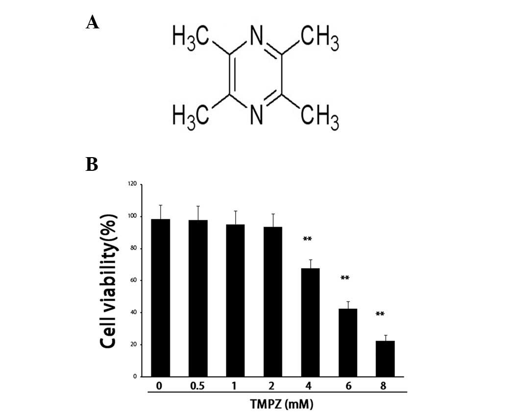
To investigate the antitumor effect of TMPZ on
SGC7901 gastric cancer cells in vitro, cell viability and
apoptosis were assessed. Following the administration of TMPZ (0–8
mM for 24 h) to SGC7901 cells, a contrasting effect of TMPZ towards
tumorigenic cells was observed. Treatment with TMPZ at the higher
doses of 4, 6 and 8 mM for 24 h resulted in reduced cell
viabilities of 67.8±5.4, 42.6±4.5 and 22.5±3.5% in SGC7901 cells
(P<0.01), respectively, whereas treatment with lower doses of
TMPZ (0.5, 1 and 2 mM) demonstrated no effect on cell viability
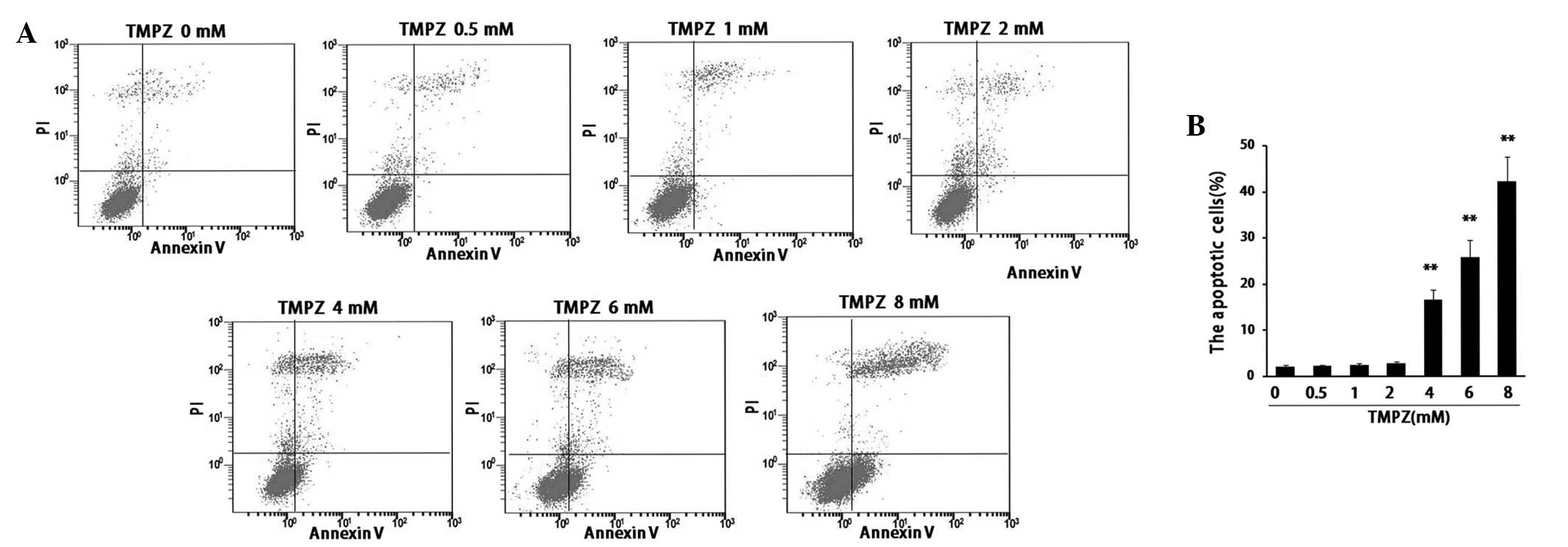
(Fig. 1). Furthermore, the analysis
of apoptosis was concordant with the results observed in the cell
viability experiments. The percentages of apoptotic cells were
16.7±2.1, 25.8±3.7 and 42.3±5.2% in SGC7901 cells treated with the
respective doses of TMPZ (4, 6 and 8 mM) for 24 h (Fig. 2). These findings confirmed that a
high dose of TMPZ confers significant cytotoxicity, which may be
beneficial for anticancer treatment. Therefore, the higher doses of
TMPZ (4, 6 and 8 mM) were selected for further experimentation.
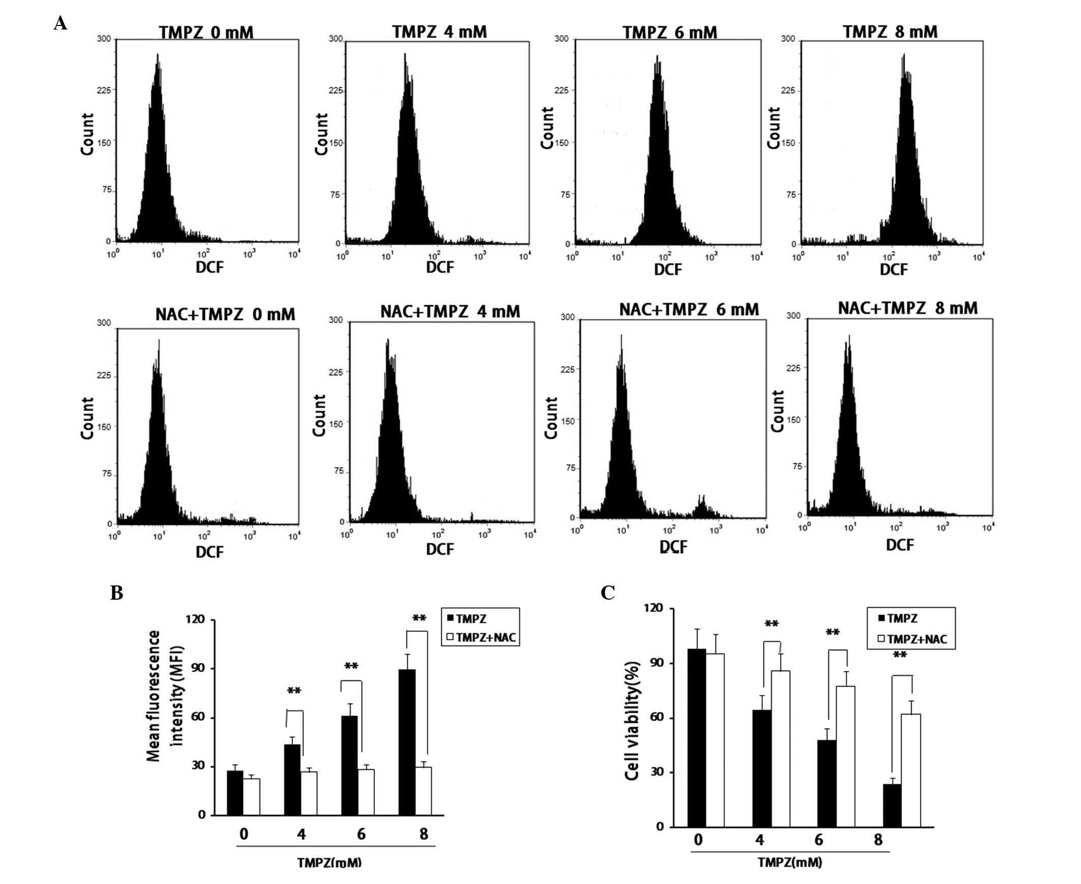
TMPZ initiates the accumulation of ROS in
a dose-dependent manner
A number of natural compounds for the treatment of
cancer have been demonstrated to cause increased levels of cellular
ROS generation (9,10). To determine whether this mechanism
is responsible for the antitumor effect of TMPZ, the generation of
ROS was investigated. The results demonstrated that TMPZ treatment
significantly elevated the ROS levels in a dose-dependent manner in
SGC7901 cells. These data were further confirmed by incubating the
cells with NAC, which is a scavenger of ROS (11). Treatment with NAC almost completely
abrogated the release of ROS in SGC7901 cells and inhibited
TMPZ-induced cytotoxicity (Fig.
3).
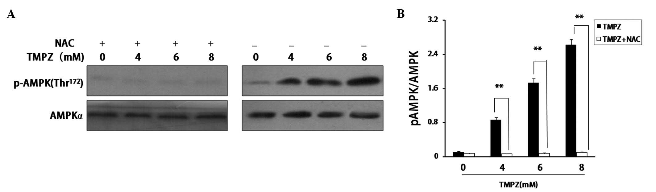
TMPZ-induced activation of AMPK is
mediated by ROS
Numerous studies have implicated AMPK in cancer
growth and metabolism (?). It has previously been suggested that
stimulating the release of ROS may lead to the activation of AMPK
(12); therefore, we investigated
whether AMPK activation may result from TMPZ treatment. The results
from this study revealed that high doses of TMPZ significantly
upregulated the phosphorylation of AMPK in SGC7901 cells in a
dose-dependent manner. To determine whether the activation of AMPK
is involved in the accumulation of ROS, the cells were incubated
with TMPZ in the presence of 5 mM NAC for 24 h. NAC almost
completely inhibited TMPZ-induced AMPK phosphorylation,
demonstrating that AMPK was activated by TMPZ-induced ROS
accumulation (Fig. 4).
TMPZ-induced activation of the
mitochondrial apoptotic pathway is mediated by AMPK
To verify whether the AMPK signaling pathway was
responsible for the antitumor effect of TMPZ that was observed in
SGC7901 cells, compound C, an inhibitor of AMPK was used (13). Pretreatment with 10 μM
compound C for 15 min inhibited TMPZ-induced apoptosis (Fig. 5A). We also examined the mechanism by
which AMPK mediated TMPZ-induced apoptosis. Compound C did not
suppress TMPZ-induced ROS accumulation (Fig. 5B), indicating that compound C does
not inhibit TMPZ-induced cell death by suppressing ROS
accumulation. It is well-known that the majority of intrinsic death
signals converge onto the translocation of Bax, a pro-apoptotic
member of the Bcl-2 family, from the cytosol to the mitochondria
(14). Our results demonstrated
that TMPZ induced the translocation of Bax from the cytosol to the
mitochondria and that this translocation was significantly
inhibited by compound C. In addition, compound C significantly
inhibited TMPZ-induced mPTP opening and depolarization of the
mitochondria. The JC-1 red/green fluorescence intensity ratios were
23.4±1.5 and 82.7±4.7% in SGC7901 cells treated with 8 mM TMPZ
alone and 8 mM TMPZ combined with compound C pretreatment,
respectively (Fig. 5C). It is
well-known that the majority of intrinsic death signals converge
onto the translocation of Bax, a pro-apoptotic member of the Bcl-2
family, from the cytosol to the mitochondria (14). Our results demonstrated that TMPZ
induced the translocation of Bax from the cytosol to the
mitochondria and that this translocation was significantly
inhibited by compound C. Furthermore, compound C significantly
inhibited the TMPZ-induced release of cytochrome c and the
activation of caspase-9 and -3 (Fig.
5D). Thus, ROS-induced AMPK activation is important in
initiating the mitochondria-mediated apoptosis of SGC7901 cells
treated with TMPZ.
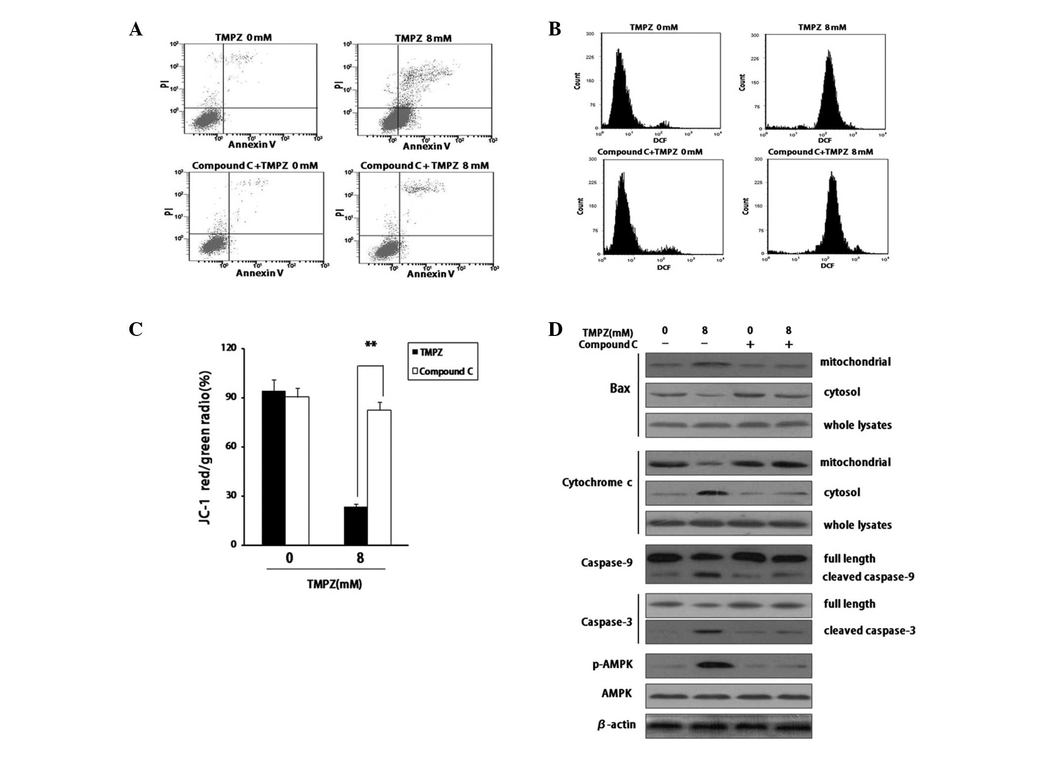 | Figure 5.AMPK mediates TMPZ-induced apoptosis
by initiating the mitochondrial apoptotic pathway. SGC7901 cells
were treated with 8 mM TMPZ for 24 h with or without pretreatment
with 10 μM compound C. (A) Compound C mediates TMPZ-induced
apoptosis. Following 24 h of treatment with TMPZ, cells were
stained with PI and Annexin V, and analyzed by flow cytometry.
Representative measurements from at least three independent
experiments are shown. (B) Compound C does not suppress
TMPZ-induced ROS accumulation. Cells were stained with DCFH-DA and
analyzed by flow cytometry. Representative measurements are shown.
The relative levels of ROS geometry fluorescence are shown as the
mean fluorescence intensity. Each experiment was performed in
triplicate. (C) AMPK mediates mitochondrial depolarization induced
by TMPZ. Cells were stained with JC-1 and analyzed by flow
cytometry. The ratio of JC-1 red/green fluorescence intensity was
normalized by comparing the data to the control group and
represents the loss of mitochondrial membrane potential. Each
experiment was performed in triplicate and each value reported
represents the mean ± SD. (D) AMPK mediates TMPZ-induced apoptosis
by initiating the mitochondrial apoptotic signaling pathway. The
mitochondrion and cytoplasm were separated, and proteins were
extracted from each fraction. The alteration and subcellular
localization of Bax and cytochrome c were analyzed by
western blot analysis. In addition, caspase-9 and -3, pAMPK, AMPK
and β-actin from whole cell lysates were analyzed by western blot
analysis. **P<0.01. AMPK, AMP-activated protein
kinase; TMPZ, tetramethylpyrazine; PI, propidium iodide; ROS,
reactive oxygen species; DCFH-DA, 2′7′-dichlorodihydrofluorescein
diacetate. |
Discussion
The induction of apoptosis in malignant cells is an
important mechanism of action for anticancer drugs (15,16).
TMPZ, one of the active components of the Chinese herbal medicine
Chuanxiong, has been traditionally used in the treatment of
neurovascular and cardiovascular diseases. Recently, an increasing
number of studies have indicated that TMPZ possesses anticancer
properties (17,18). However, the function and mechanism
of TMPZ in gastric cancer have yet to be elucidated. In the present
study, we demonstrated that TMPZ induces cytotoxicity by causing
the accumulation of ROS. The accumulation of ROS led to the
activation of AMPK, which then induced apoptosis. The effects of
TMPZ on the accumulation of ROS and apoptosis were observed in
SGC7901 cells. Therefore, we conclude that ROS represent a novel
intermediate in mediating TMPZ-induced cytotoxicity.
As a metabolic-sensing protein kinase, AMPK is
important as an energy sensor, mainly in ATP-deprived conditions
(19). It was previously reported
that AMPK is phosphorylated and activated by LKB1 (20). However, we observed that AMPK was
activated by TMPZ-induced ROS accumulation. Several studies have
indicated that AMPK activation reprograms metabolism and also
enforces a metabolic checkpoint in the cell cycle, which suggests
that AMPK-activating drugs may be useful as cancer therapeutics
(7,21). Recently, metformin has been shown to
inhibit the growth of a wide variety of tumor cells in culture in
an AMPK-dependent manner, and the activation of AMPK by
aminoimidazole carboxamide ribonucleotide has been observed to
suppress the growth of tumors (22,23).
The results of our study demonstrated that AMPK is activated
following treatment with TMPZ in SGC7901 cells. Furthermore, we
also demonstrated that TMPZ-induced activation of AMPK is dependent
on the release of ROS, as co-treatment of SGC7901 cells with NAC, a
scavenger of ROS, inhibited the TMPZ-induced activation of
AMPK.
The exact mechanism by which TMPZ-induced AMPK
activation results in apoptosis is unknown. The majority of
intrinsic death signals converge onto the activation of the
mitochondrial apoptotic pathway (24,25).
The mitochondria-dependent pathway for apoptosis involves the
release of cytochrome c from the mitochondrion into the
cytosol, a process that is controlled by Bcl-2 family members
(26). Genetic and biochemical
studies indicate that Bax functions as a major component of the
intrinsic apoptotic pathway in mitochondria (27). Bax is an effective, multidomain,
pro-apoptotic protein that resides in the cytoplasm as an inactive
monomer in healthy cells. Upon treatment with apoptotic stimuli,
Bax undergoes conformational activation leading to its
translocation into the mitochondrion. Several studies have
previously implicated the conformational activation of Bax as an
early event that contributes to the release of cytochrome c
from mitochondria and subsequent caspase cascade activation in the
mitochondria-dependent apoptotic signaling pathway (28,29).
In the present study, TMPZ induced the translocation of Bax from
the cytosol into mitochondria. To investigate whether AMPK
activation was accountable for the translocation of Bax in SGC7901
cells, compound C was utilized. The results confirmed that AMPK
activation induced by TMPZ treatment was responsible for the
translocation of Bax. In addition, AMPK activation induced by TMPZ
treatment resulted in a disruption of the ΔΨm and mitochondrial
swelling. This event was accompanied with the release of cytochrome
c and subsequently induced the activation of the caspase
cascade. This role of AMPK was further supported by data indicating
that compound C significantly inhibited the loss of ΔΨm and the
subsequent mitochondrial apoptotic pathway.
To the best of our knowledge, our results
demonstrate for the first time that TMPZ induces apoptosis in a
gastric cancer cell line. Additionally, our data provide the first
evidence that TMPZ-induced apoptosis occurs via the accumulation of
ROS. Accumulation of ROS levels leads to AMPK activation, which is
important in apoptosis in gastric cancer cells through a signaling
cascade that includes the translocation of Bax from the cytosol
into mitochondria, resulting in mitochondrial dysfunction followed
by cytochrome c release. The release of cytochrome c
stimulates the activation of the caspase cascade. Therefore, these
results contribute to the understanding of the cell apoptotic
pathways induced by TMPZ, and to the development of TMPZ as a
potential treatment for human gastric cancer.
Acknowledgements
This study was supported by the
Natural Scientific Foundation of China (nos. 81160309, 81100104 and
30860271), the Natural Scientific Foundation of Jiangxi Province
(no. 20114BAB215034) and the Jiangxi Province Technology Support
and Social Development Projects (no. 2010BSA13900).
References
|
1.
|
Liu X, Liu H, Zeng Z, Zhou W, Liu J and He
Z: Pharmacokinetics of ligustrazine ethosome patch in rats and
anti-myocardial ischemia and anti-ischemic reperfusion injury
effect. Int J Nanomedicine. 6:1391–1398. 2011. View Article : Google Scholar : PubMed/NCBI
|
|
2.
|
Liang X, Zhou H, Ding Y, Li J, Yang C, Luo
Y, Li S, Sun G, Liao X and Min W: TMP prevents retinal
neovascularization and imparts neuroprotection in an oxygen-induced
retinopathy model. Invest Ophthalmol Vis Sci. 53:2157–2169. 2012.
View Article : Google Scholar : PubMed/NCBI
|
|
3.
|
Yu K, Chen Z, Pan X, Yang Y, Tian S, Zhang
J, Ge J, Ambati B and Zhuang J: Tetramethylpyrazine-mediated
suppression of C6 gliomas involves inhibition of chemokine receptor
CXCR4 expression. Oncol Rep. 28:955–960. 2012.PubMed/NCBI
|
|
4.
|
Zheng CY, Xiao W, Zhu MX, Pan XJ, Yang ZH
and Zhou SY: Inhibition of cyclooxygenase-2 by tetramethylpyrazine
and its effects on A549 cell invasion and metastasis. Int J Oncol.
40:2029–2037. 2012.PubMed/NCBI
|
|
5.
|
Carling D, Thornton C, Woods A and Sanders
MJ: AMP-activated protein kinase: new regulation, new roles?
Biochem J. 445:11–27. 2012. View Article : Google Scholar : PubMed/NCBI
|
|
6.
|
Webster I, Friedrich SO, Lochner A and
Huisamen B: AMP kinase activation and glut4 translocation in
isolated cardiomyocytes. Cardiovasc J Afr. 21:72–78.
2010.PubMed/NCBI
|
|
7.
|
Park JB, Lee MS, Cha EY, Lee JS, Sul JY,
Song IS and Kim JY: Magnolol-induced apoptosis in HCT-116 colon
cancer cells is associated with the AMP-activated protein kinase
signaling pathway. Biol Pharm Bull. 35:1614–1620. 2012.PubMed/NCBI
|
|
8.
|
Liu L, Ulbrich J, Müller J, Wüstefeld T,
Aeberhard L, Kress TR, Muthalagu N, Rycak L, Rudalska R, Moll R, et
al: Deregulated MYC expression induces dependence upon AMPK-related
kinase 5. Nature. 483:608–612. 2012. View Article : Google Scholar : PubMed/NCBI
|
|
9.
|
Chen G, Zhang X, Zhao M, Wang Y, Cheng X,
Wang D, Xu Y, Du Z and Yu X: Celastrol targets mitochondrial
respiratory chain complex I to induce reactive oxygen
species-dependent cytotoxicity in tumor cells. BMC Cancer.
11:1702011. View Article : Google Scholar
|
|
10.
|
Miki H, Uehara N, Kimura A, Sasaki T, Yuri
T, Yoshizawa K and Tsubura A: Resveratrol induces apoptosis via
ROS-triggered autophagy in human colon cancer cells. Int J Oncol.
40:1020–1028. 2012.PubMed/NCBI
|
|
11.
|
Zheng F, Yang WJ, Sun KJ, Wan XM, Man N
and Wen LP: Hoechst 33342-induced autophagy protected HeLa cells
from caspase-independent cell death with the participation of ROS.
Free Radic Res. 46:740–749. 2012. View Article : Google Scholar : PubMed/NCBI
|
|
12.
|
Jang KY, Jeong SJ and Kim SH, Jung JH, Kim
JH, Koh W, Chen CY and Kim SH: Activation of reactive oxygen
species/AMP activated protein kinase signaling mediates
fisetin-induced apoptosis in multiple myeloma U266 cells. Cancer
Lett. 319:197–202. 2012. View Article : Google Scholar : PubMed/NCBI
|
|
13.
|
Benziane B, Björnholm M, Pirkmajer S,
Austin RL, Kotova O, Viollet B, Zierath JR and Chibalin AV:
Activation of AMP-activated protein kinase stimulates Na+,K+-ATPase
activity in skeletal muscle cells. J Biol Chem. 287:23451–23463.
2012.
|
|
14.
|
Landes T and Martinou JC: Mitochondrial
outer membrane permeabilization during apoptosis: the role of
mitochondrial fission. Biochim Biophys Acta. 1813:540–545. 2011.
View Article : Google Scholar : PubMed/NCBI
|
|
15.
|
Sampieri K and Fodde R: Cancer stem cells
and metastasis. Semin Cancer Biol. 22:187–193. 2012. View Article : Google Scholar
|
|
16.
|
Zhao J, Lu Y and Shen HM: Targeting p53 as
a therapeutic strategy in sensitizing TRAIL-induced apoptosis in
cancer cells. Cancer Lett. 314:8–23. 2012. View Article : Google Scholar : PubMed/NCBI
|
|
17.
|
Wang P, She G, Yang Y, Li Q, Zhang H, Liu
J, Cao Y, Xu X and Lei H: Synthesis and biological evaluation of
new ligustrazine derivatives as anti-tumor agents. Molecules.
17:4972–4985. 2012. View Article : Google Scholar : PubMed/NCBI
|
|
18.
|
Zhang Y, Liu X, Zuo T, Liu Y and Zhang JH:
Tetramethylpyrazine reverses multidrug resistance in breast cancer
cells through regulating the expression and function of
P-glycoprotein. Med Oncol. 29:534–538. 2012. View Article : Google Scholar : PubMed/NCBI
|
|
19.
|
Hardie DG: AMP-activated protein kinase:
an energy sensor that regulates all aspects of cell function. Genes
Dev. 25:1895–1908. 2011. View Article : Google Scholar : PubMed/NCBI
|
|
20.
|
Alexander A and Walker CL: The role of
LKB1 and AMPK in cellular responses to stress and damage. FEBS
Lett. 585:952–957. 2011. View Article : Google Scholar : PubMed/NCBI
|
|
21.
|
El-Masry OS, Brown BL and Dobson PR:
Effects of activation of AMPK on human breast cancer cell lines
with different genetic backgrounds. Oncol Lett. 3:224–228.
2012.PubMed/NCBI
|
|
22.
|
Kourelis TV and Siegel RD: Metformin and
cancer: new applications for an old drug. Med Oncol. 29:1314–1327.
2012. View Article : Google Scholar : PubMed/NCBI
|
|
23.
|
Petti C, Vegetti C, Molla A, Bersani I,
Cleris L, Mustard KJ, Formelli F, Hardie GD, Sensi M and Anichini
A: AMPK activators inhibit the proliferation of human melanomas
bearing the activated MAPK pathway. Melanoma Res. 22:341–350. 2012.
View Article : Google Scholar : PubMed/NCBI
|
|
24.
|
Scatena R: Mitochondria and cancer: a
growing role in apoptosis, cancer cell metabolism and
dedifferentiation. Adv Exp Med Biol. 942:287–308. 2012. View Article : Google Scholar : PubMed/NCBI
|
|
25.
|
Jourdain A and Martinou JC: Mitochondrial
outer-membrane permeabilization and remodelling in apoptosis. Int J
Biochem Cell Biol. 41:1884–1889. 2009. View Article : Google Scholar : PubMed/NCBI
|
|
26.
|
Leibowitz B and Yu J: Mitochondrial
signaling in cell death via the Bcl-2 family. Cancer Biol Ther.
9:417–422. 2010. View Article : Google Scholar : PubMed/NCBI
|
|
27.
|
Walensky LD and Gavathiotis E: BAX
unleashed: the biochemical transformation of an inactive cytosolic
monomer into a toxic mitochondrial pore. Trends Biochem Sci.
36:642–652. 2011. View Article : Google Scholar : PubMed/NCBI
|
|
28.
|
Lalier L, Cartron PF, Juin P, Nedelkina S,
Manon S, Bechinger B and Vallette FM: Bax activation and
mitochondrial insertion during apoptosis. Apoptosis. 12:887–896.
2007. View Article : Google Scholar : PubMed/NCBI
|
|
29.
|
Zeng L, Li T, Xu DC, Liu J, Mao G, Cui MZ,
Fu X and Xu X: Death receptor 6 induces apoptosis not through type
I or type II pathways, but via a unique mitochondria-dependent
pathway by interacting with Bax protein. J Biol Chem.
287:29125–29133. 2012. View Article : Google Scholar : PubMed/NCBI
|