Introduction
Radiation therapy is the most effective treatment
method for nasopharyngeal carcinoma. However, the auditory pathway
inevitably suffers from radiation exposure during the therapy
process. Therefore radiation-induced ear injury, particularly
sensorineural hearing loss (SNHL), is a common complication
following radiotherapy for patients with nasopharyngeal carcinoma
(1–2). Thus far, no effective strategy has
been proposed to treat radiation-induced SNHL, which seriously
deteriorates the quality of life for these patients.
Generally, radiation-induced SNHL is regarded as a
result of damage to the auditory sensory hair cells in the cochlea
(3). Animal experiments using
radiation regimens similar to those applied clinically have
demonstrated cochlear hair cell degeneration in the absence of
damage to vascular and supporting structures (4,5). Low
et al(6) reported that the
cochlear OC-K3 cell line demonstrated dose-dependent cell apoptosis
and was identified to upregulate p53-related genes by micro-array
studies under γ-radiation. The data suggest that radiation-induced
cochlear hair cell death may play a role in SNHL.
Numerous studies have investigated the protective
effects of agents against ototoxicity induced by antibiotics or
cisplatin, but not radiation-induced ototoxicity. Currently there
is no effective drug to protect against radiation-induced
ototoxicity. Tanshinone is a derivative of phenanthrene-quinone
with anti-oxidant and anti-inflammation properties that is isolated
from Salvia miltiorrhiza Bunge (7–9).
Tanshinone IIA significantly attenuates aminoglycoside-induced free
radical formation in vitro and ototoxicity in
vivo(10). Furthermore,
tanshinone IIA is able to inhibit the radiation-induced activation
of nuclear factor (NF)-κB in microglia BV-2 cells (11). Hence, the present study aimed to
investigate the in vitro effect of tanshinone IIA on
radiation-induced apoptosis and cell death in the cochlea. Using
the HEI-OC1 cell line, the release of the apoptosis-inducing
factors, p53 and p21, and the subsequent activation of the NF-κB
pathway, were observed in the irradiated HEI-OC1 cells.
Materials and methods
Cell culture and radiation exposure
The HEI-OC1 cells were cultured in Dulbecco’s
modified Eagle’s medium (DMEM), supplemented with 10% fetal bovine
serum (FBS) at 33°C under 10% CO2 in an incubator. The
HEI-OC1 cells were irradiated using a 6-MV linear accelerator
(LINAC; 2300EX; Varian Co., Palo Alto, CA, USA) at a dose rate of
4.0 Gy/min. All the irradiations were performed at room temperature
(18–25°C). Subsequent to being irradiated, the culture plates were
returned to an incubator under the same conditions as previously
described (33°C, 10% CO2).
Cell viability assay
The cytotoxicity of tanshinone IIA was determined by
an MTT assay. The HEI-OC1 cells were seeded in 96-well plates at a
density of 1×10⁴ cells/well in 200 μl complete medium.
Subsequent to being incubated overnight, the medium in each well
was discarded and replaced with fresh medium containing various
concentrations of tanshinone IIA (1–64 μg/ml). The cells that were
not treated with tanshinone IIA were used as controls. Following a
24-h incubation period, 20 μl 5 mg/ml MTT was added to each well
and cultivated for an additional 4 h. The supernatant was removed,
150 μl/well dimethyl sulfoxide (DMSO) was added and the samples
were shaken for 15 min. The optical density (OD) was measured at
490 nm and the wells that did not contain cells were used as
blanks. The protective effect of tanshinone IIA on
radiation-induced ototoxicity was also determined using an MTT
assay. The cells were incubated with or without tanshinone IIA and
exposed to 0 and 16 Gy radiation, respectively. The cell viability
was measured following irradiation for 24, 72 and 120 h. All the
experiments were performed in triplicate.
Cell morphology observation
The HEI-OC1 cells were irradiated and subsequently
incubated for 72 h. Then, the cell morphology was observed by
inversion microscopy. The nuclear morphology was observed under a
fluorescence microscope (Olympus, Tokyo, Japan) subsequent to the
cells being stained with 4′,6-diamidino-2-phenylindole (DAPI).
Annexin V-fluorescin
isothiocyanate/propidium iodide (FITC/PI) staining
Annexin V-FITC/PI staining was performed to quantify
the percentage of apoptotic cells at 72 h post-irradiation. The
cells were stained using the Annexin V-FITC Apoptosis Detection kit
(Invitrogen, Inc., Carlsbad, CA, USA) following the manufacturer’s
instructions. The cells were washed with phosphate-buffered saline
(PBS) three times and resuspended in 1X binding buffer.
Subsequently, the cells were incubated with Annexin V-FITC and PI
for 10–15 min at room temperature and analyzed using flow
cytometry.
Colocalization of γH2AX and
p65/NF-κB
The HEI-OC1 cells were planted onto
polylysine-coated cover glasses and treated with irradiation.
Subsequently, the samples were fixed in 4% paraformaldehyde for 30
min, permeabilized in 0.25% triton X-100 and blocked for 30 min in
1% goat serum. Subsequent to being blocked overnight at 4°C,
anti-γH2AX primary antibody (1:100 dilution; Abcam Inc., Cambridge,
MA, USA) and anti-p65/NF-κB primary antibody (1:200 dilution; CST
Inc., Mount Carmel, IL, USA) were applied. Following this, the
cells were washed three times for 5 min each in PBS, then rabbit
anti-mouse AlexaFlour-488 secondary antibody (1:200 dilution) and
goat anti-rabbit AlexaFluor-568 secondary antibody (1:200 dilution;
Invitrogen, Inc.) were applied for 1 h at room temperature in the
dark, followed by three 5-min washes in PBS. The samples were then
mounted in fluorescence mounting medium with DAPI. The cells were
observed using a fluorescence microscope (Olympus).
RNA isolation and quantitative
(q)PCR
Total RNA was purified from the cultured cells using
TRIzol reagent (Invitrogen), according to the manufacturer’s
instructions. Each RNA sample was reverse-transcribed to cDNA for
15 min at 37°C and 5 sec at 85°C using a PrimeScript® RT
reagent kit (Takara, Inc., Kyoto, Japan). qPCR was performed using
the Stratgene MX3005P qPCR system with a SYBR® Premix Ex
Taq™ II kit (Takara Inc.). A total of 40 cycles of amplification
were performed at 95°C for 30 sec, 60°C for 15 sec and 72°C for 15
sec. The fluorescence signal was detected at the end of each cycle.
A melting curve analysis was used to confirm the specificity of the
products. The 2−ΔΔCt method was performed to analyze the
results. The relative expression levels of each mRNA was assessed
using the 2−ΔΔCt method by normalizing to GAPDH and
comparing it with the control samples.
Western blot analysis
The cells were washed and suspended in lysis buffer
(KeyGen Biotech, Inc., Nanjing, Jiangsu, China). The proteins were
solubilized by sonication. Equal amounts of protein were separated
by SDS-PAGE and transferred onto polyvinylidene difluoride
membranes (Millipore Corp., Bedford, MA, USA). The membranes were
blocked in PBS containing 0.1% Tween-20 and 5% powdered milk and
probed with primary antibodies. Primary antibodies against
phosphorylated-p21 (p-p21; Epitomics, Inc., Burlingame, CA, USA),
p-p53 and β-actin (Bioworld technology, Inc., St Louis Park, MN,
USA) were used at a dilution of 1:500.
Statistical analysis
All values are presented as the mean ± SD. The data
analysis was performed using the SPSS 13.0 statistical program
(SPSS, Inc., Chicago, IL, USA). A one-way ANOVA was used to
determine the statistical significance among the various groups and
the LSD method was used for the pairwise comparisons. P<0.05 was
considered to indicate a statistically significant difference.
Results
Effects of tanshinone IIA on the cell
morphological changes and viability induced by irradiation
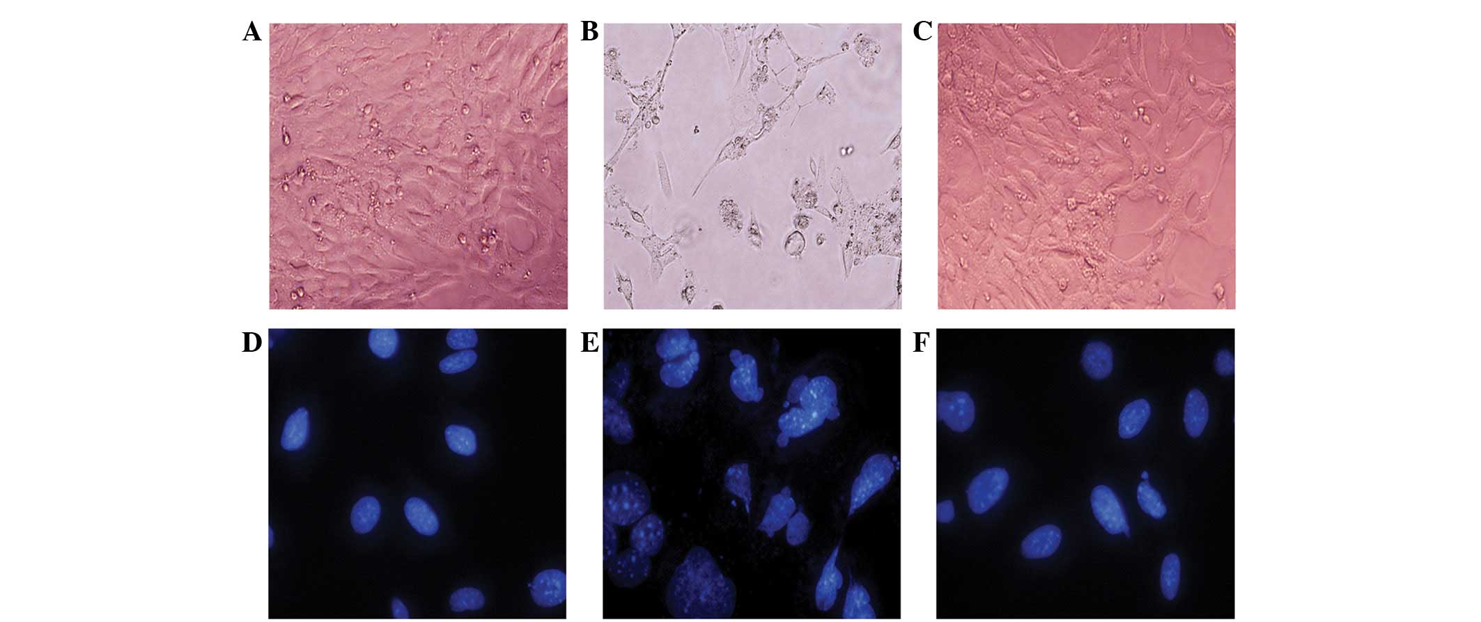
In the absence of irradiation, the HEI-OC1 cells
showed a normal polygon and fusiform morphology (Fig. 1A), with oval, uniformly sized nuclei
(Fig. 1D). There was no significant
morphological change in the HEI-OC1 cells following 2 or 4 Gy
irradiation. However, when treated with 16 Gy, the HEI-OC1 cell
morphology was changed to a dendritic or amoeboid appearance with
numerous highly ramified processes and body swelling (Fig. 1B). The cell nucleus demonstrated
shrinkage, fragmentation, megakaryocytes and deformation (Fig. 1E). In contrast, the majority of the
irradiated HEI-OC1 cells that were pre-treated with tanshinone IIA
preserved their polygon and fusiform morphology rather than change
to the dendritic or amoeboid appearance (Fig. 1C), while the nuclei appeared to be
oval-shaped and of a relatively homogeneous size (Fig. 1F).
To investigate the effect of tanshinone IIA on cell
viability, the cells were exposed to tanshinone IIA for 24 h. As
shown in Fig. 2A, the
concentrations (1–8 μg/ml) of tanshinone IIA that were used had no
effect on the viability of the HEI-OC1 cells. However, tanshinone
IIA at concentrations of >16 μg/ml resulted in cytotoxicity.
According to the preliminary results obtained from the MTT assay,
the maximum concentrations of 8 μg/ml tanshinone IIA were not used
for further experiments to avoid the tanshinone IIA cytotoxicity
effect on the HEI-OC1 cell line. The results also showed that the
irradiation decreased the viability of the HEI-OC1 cells in a time-
and dose-dependent manner (Fig.
2B). To verify whether tanshinone IIA was able to prevent
radiation-induced cytotoxicity, the HEI-OC1 cells were incubated
with 8 μg/ml tanshinone IIA and exposed to 0 and 16 Gy irradiation.
As shown in Fig. 2C, the treatment
with tanshinone IIA significantly protected the HEI-OC1 cells from
the irradiation.
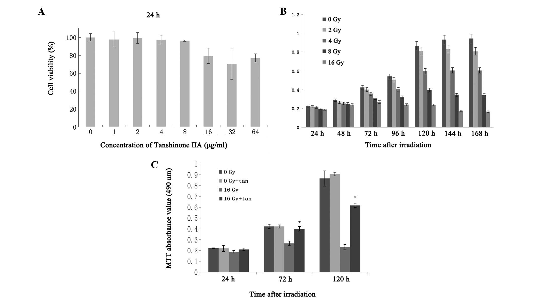 | Figure 2(A) Effect of tanshinone IIA on the
cell viability of the HEI-OC1 cells. The HEI-OC1 cells were seeded
into culture plates and treated with 1, 2, 4, 8, 16, 32 and 64
μg/ml tanshinone IIA for 24 h, then the cell viability was
determined using an MTT assay. (B) The post-irradiation viability
of the cells was dose dependent. MTT absorbance values for the
different doses of irradiation at various time-points are shown.
(C) Effect of tanshinone IIA on the cell viability of the HEI-OC1
cells. When the cultured cells were exposed to 16 Gy irradiation,
the cell viability was 84.68, 62.88 and 26.91% following 24, 72 and
120 h of irradiation, respectively. However, when the cells were
exposed to 16 Gy irradiation following treatment with 8 mg/ml
tanshinone IIA, the cell viability was 94.59, 94.56 and 71.25% at
the various time-points, respectively, and significantly higher
than those without tanshinone IIA (P<0.01). There were no
significant changes in the groups that were exposed to 0 Gy with or
without tanshinone IIA at the various time-points. Each value
represents the mean ± SD of three separate experiments. |
Tanshinone IIA inhibits radiation-induced
apoptosis in HEI-OC1 cells
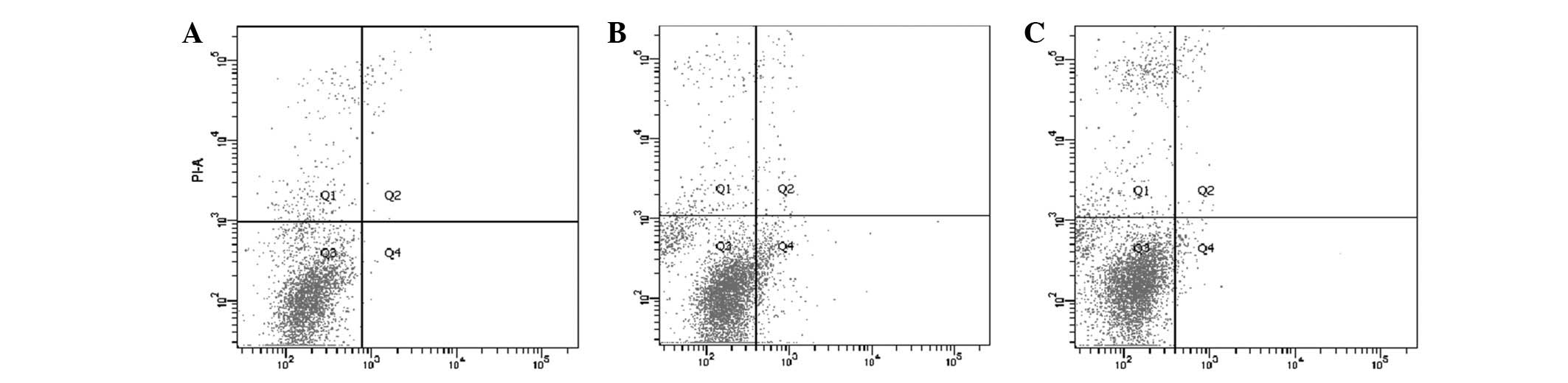
Flow cytometry was used to analyze the percentage of
apoptotic cells that were treated with radiation in the absence or
presence of tanshinone IIA (Fig.
3). Radiation accompanied by tanshinone IIA treatment
significantly decreased the number of apoptotic cells (mean, 3.36%)
compared with the cells that were treated with radiation only
(mean, 9.09%; P<0.05), while there was no significant difference
between the control group and the group with tanshinone IIA
treatment alone. These results indicate that radiation promotes
cell apoptosis and that radiation-induced apoptosis may be
inhibited by a pre-treatment with tanshinone IIA.
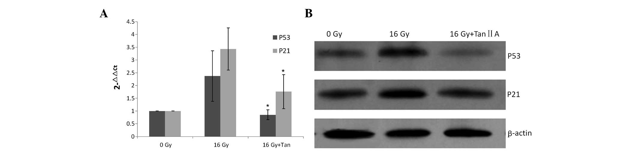
Effects of tanshinone IIA on
radiation-induced p21 and p53 expression
qPCR and western blotting were used to investigate
the changes in p21 and p53 expression following irradiation with or
without tanshinone IIA. As shown in Fig. 4, the levels of p21 and p53 after
being stimulated by irradiation were significantly higher than
those of the control group. When the cells were irradiated with
tanshinone IIA, the upregulation of p21 and p53 was attenuated.
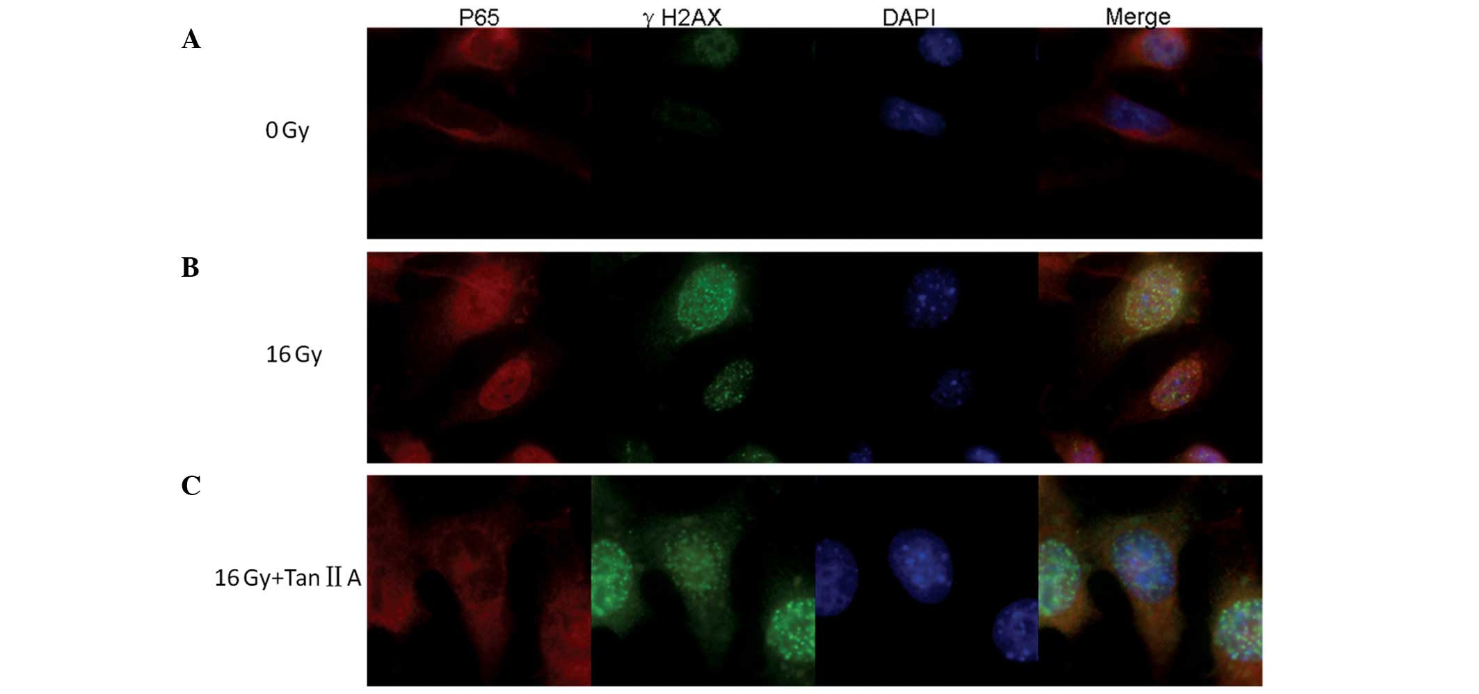
Inhibitory effects of tanshinone IIA are
mediated by p65/NF-κB pathway suppression in the radiation-induced
DNA damage response
Double immunofluorescence staining for γH2AX and p65
was established to investigate the repair of double-strand breaks
(DSBs) and the translocation of p65/NF-κB in the HEI-OC1 cells
following irradiation (Fig. 5). The
expression of the γH2AX foci was almost completely absent and that
of the p65 was mainly located in the cytoplasm in the cells of the
control group (Fig. 5A). Following
16 Gy irradiation for 6 h, distinct nuclear γH2AX foci were
observed and the nuclei of the HEI-OC1 cells were stained red,
indicating the translocation of p65 (Fig. 5B). However, when using the
pre-treatment with 8 mg/ml tanshinone IIA, p65 protein
translocation was inhibited and the cytoplasm was stained red
(Fig. 5C)
Discussion
Radiation damages tumor DNA by direct and/or
indirect effects on the tumor cells (12), leading to cell apoptosis and cell
death, which is the biological basis of radiotherapy. However, in
the course of radiotherapy, normal tissues that surround the tumor
are inevitably subjected to a certain dose of irradiation. Although
a vast amount information is available with regard to the
radiation-induced apoptosis and cell death of tumor cells, there is
limited information on the effects of radiation on normal cells,
particularly on the auditory hair cells of the cochlea.
Furthermore, information with regard to the agents that may protect
against radiation-induced ototoxicity is also limited.
In the present study, by observing the morphology of
cells and using an MTT assay and flow cytometry, the cells with
body swelling, nuclear fragmentation, decreased viability and an
increased percentage of apoptotic cells were regarded as evidence
of apoptosis and cell death in radiation-induced toxicity. This is
consistent with the previous conclusion that ototoxicity induced by
irradiation leads to cell apoptosis and death (3). In the present study, tanshinone IIA
accompanied by irradiation treatment decreased the changes in
radiation-induced cell morphology and cell viability. Furthermore,
tanshinone IIA attenuated the irradiation-induced cell
apoptosis.
Radiation induces the apoptotic death of various
cells through the activation of a number of intracellular signaling
pathways. NF-κB has been suggested to be activated in response to
radiation-induced DNA damage. DSBs are the most deleterious form of
DNA damage following ionizing radiation. H2AX phosphorylation is an
early step in the response to DNA damage, and it has been verified
that enumerating γH2AX foci may be a method used to measure the
induction and repair of radiation-induced DSBs (13,14).
Ataxia telangiectasia mutated (ATM) is a crucial component of the
DNA DSB signaling cascade and has been suggested to be able to
activate NF-κB in response to DNA damage (15,16).
The NF-κB signaling pathway mediates a variety of significant
cellular functions by regulating apoptosis and inflammatory
responses. In unstimulated cells, NF-κB is in the form of a
heterodimer of p65/p50 binding to the inhibitor proteins, namely
Iκβ. Following stimulation, the release of p65/p50 from the Iκβ-α
protein and the degradation of Iκβ-α are necessary for p65
translocation into the nucleus to regulate gene transcription. In
the nucleus, NF-κB may induce a number of genes that activate
intracellular programs, leading to either apoptosis or cell death.
Among the apoptotic genes activated by NF-κB is p53, which may
activate the transcription of other genes, including p21, to
progress the apoptosis pathway (17). In the present study, it was
identified that irradiation was able to activate the translocation
of p65 and lead to an increase in the expression of p53/p21, which
plays a significant role in apoptosis. The present study also
indicated that tanshinone IIA exerted anti-apoptosis properties by
suppressing the translocation of p65 and the transcription of
p53/p21 through the NF-κB signaling pathway.
Although the results of the present study indicate
that tanshinone IIA reduces radiation-induced ototoxicity, its
clinical effect remains unclear. Since the present study was based
on the HEI-OC1 cell line, which was harvested from the cochlea of
immortal mice, tanshinone IIA is presumed to have an ability to
protect the cochlea in radiation-induced ototoxicity. Future
studies may focus on in vivo experiments to reveal the
effect of tanshinone IIA on cochlea function.
To the best of our knowledge, this is the first
study on auditory hair cells to investigate the protective effects
of tanshinone IIA against radiation-induced ototoxicity. The
results of the present study provide evidence of the anti-apoptotic
effects of tanshinone IIA. Tanshinone IIA was also observed to
enhance HEI-OC1 cell viability and prevent NF-κB translocation to
inhibit p53/p21 activation when the cells underwent
irradiation.
Acknowledgements
This study was supported by grants from the External
Scientific and Technological Cooperation Projects of Guangdong (no.
2010B050700021) and the President Foundation of Nanfang Hospital
(no. 2011Z004). The authors would like to thank Professor F.
Kalinec (House Ear Institute, Los Angeles, CA, USA) for providing
the HEI-OC1 cell line.
References
|
1
|
Chan SH, Ng WT, Kam KL, et al:
Sensorineural hearing loss after treatment of nasopharyngeal
carcinoma: a longitudinal analysis. Int J Radiat Oncol Biol Phys.
73:1335–1342. 2009. View Article : Google Scholar : PubMed/NCBI
|
|
2
|
Petsuksiri J, Sermsree A, Thephamongkhol
K, et al: Sensorineural hearing loss after concurrent
chemoradiotherapy in nasopharyngeal cancer patients. Radiat Oncol.
6:192011. View Article : Google Scholar : PubMed/NCBI
|
|
3
|
Low WK, Burgess R, Fong KW and Wang DY:
Effect of radiotherapy on retro-cochlear auditory pathways.
Laryngoscope. 115:1823–1826. 2005. View Article : Google Scholar : PubMed/NCBI
|
|
4
|
Kelemen G: Radiation and ear. Experimental
studies. Acta Otolaryngol. 184(Suppl 184): 1–48. 1963.PubMed/NCBI
|
|
5
|
Gamble JE, Peterson EA and Chandler JR:
Radiation effects on the inner ear. Arch Otolaryngol. 88:156–161.
1968. View Article : Google Scholar : PubMed/NCBI
|
|
6
|
Low WK, Tan MG, Sun L, et al:
Dose-dependent radiation-induced apoptosis in a cochlear cell-line.
Apoptosis. 11:2127–2136. 2006. View Article : Google Scholar : PubMed/NCBI
|
|
7
|
Dong H, Mao S, Wei J, et al: Tanshinone
IIA protects PC12 cells from β-amyloid(25–35)-induced apoptosis via
PI3K/Akt signaling pathway. Mol Biol Rep. 39:6495–6503. 2012.
|
|
8
|
Zhu B, Zhai Q and Yu B: Tanshinone IIA
protects rat primary hepatocytes against carbon tetrachloride
toxicity via inhibiting mitochondria permeability transition. Pharm
Biol. 48:484–487. 2010. View Article : Google Scholar
|
|
9
|
Xia WJ, Yang M, Fok TF, et al: Partial
neuroprotective effect of pretreatment with tanshinone IIA on
neonatal hypoxia-ischemia brain damage. Pediatr Res. 58:784–790.
2005. View Article : Google Scholar : PubMed/NCBI
|
|
10
|
Wang AM, Sha SH, Lesniak W and Schacht J:
Tanshinone (Salviae miltiorrhizae extract) preparations attenuate
aminoglycoside-induced free radical formation in vitro and
ototoxicity in vivo. Antimicrob Agents Chemother. 47:1836–1841.
2003. View Article : Google Scholar
|
|
11
|
Dong X, Dong J, Zhang R, et al:
Anti-inflammatory effects of tanshinone IIA on radiation-induced
microglia BV-2 cells inflammatory response. Cancer Biother
Radiopharm. 24:681–687. 2009. View Article : Google Scholar : PubMed/NCBI
|
|
12
|
Shinomiya N: New concepts in
radiation-induced apoptosis: ‘premitotic apoptosis’ and
‘postmitotic apoptosis’. J Cell Mol Med. 5:240–253. 2001.
|
|
13
|
Löbrich M, Rief N, Kühne M, et al: In vivo
formation and repair of DNA double-strand breaks after computed
tomography examinations. Proc Natl Acad Sci USA. 102:8984–8989.
2005.PubMed/NCBI
|
|
14
|
Rübe CE, Dong X, Kühne M, et al: DNA
double-strand break rejoining in complex normal tissues. Int J
Radiat Oncol Biol Phys. 72:1180–1187. 2008.PubMed/NCBI
|
|
15
|
Hadian K and Krappmann D: Signals from the
nucleus: activation of NF-kappaB by cytosolic ATM in the DNA damage
response. Sci Signal. 4:pe22011.PubMed/NCBI
|
|
16
|
Wu ZH, Shi Y, Tibbetts RS and Miyamoto S:
Molecular linkage between the kinase ATM and NF-kappaB signaling in
response to genotoxic stimuli. Science. 311:1141–1146. 2006.
View Article : Google Scholar : PubMed/NCBI
|
|
17
|
Grilli M and Memo M: Possible role of
NF-kappaB and p53 in the glutamate-induced pro-apoptotic neuronal
pathway. Cell Death Differ. 6:22–27. 1999. View Article : Google Scholar : PubMed/NCBI
|