Introduction
DNA methylation is a significant regulator of gene
transcription, and its role in carcinogenesis has become a topic of
considerable interest in the last few years. DNA cytosine
methylation has been widely studied, with investigations often
focusing on the methylation level of CpG dinucleotides in promoter
regions that usually have higher concentrations of CpGs, known as
CpG islands (1). The methylation of
normally unmethylated CpG islands in the promoter regions of DNA
repair genes is correlated with a loss of expression of these genes
(2–5), which occurs in the early stages of
colorectal cancer (CRC) development (6–8).
Methylation of DNA mismatch repair gene mutator L
homologue 1 (MLH1) and DNA repair gene
O6-methylguanine-DNA methyltransferase (MGMT), is
known to cause high-degree microsatellite instability (MSI-H)
(4) and guanine to adenine
mutations in KRAS, TP53(9) and PIK3CA(6), respectively. Methylation of the
MLH1 promoter has been reported in sporadic MSI tumors and
is associated with the loss of protein expression (4,7,8).
MGMT encodes a DNA repair enzyme that removes the mutagenic
adduct from O6-methylguanine (10). Alterations in the MGMT gene
impair the ability of the MGMT protein to remove the
mutagenic adduct from O6-methylguanine, thereby
increasing the mutation rate (6,9) and
the risk of cancer (11).
To date, studies with regard to the methylation of
genes have mainly focused on the methylation level of tumor tissues
(12–15). Although the majority of CpG islands
are unmethylated in normal tissues, the methylation changes of a
small subset of genes may be observed under physiological
conditions in normal colonic mucosa (16–18).
In a previous study, samples of colorectal mucosa collected from
healthy individuals undergoing screening colonoscopies were
analyzed for MLH1 and MGMT promoter methylation and
low background methylation levels were subsequently identified
(0.1–18.8%) (19). The results of
another study that analyzed 13 types of normal somatic tissues,
placenta, sperm and an immortalized cell line, indicated that ~18%
of the genomic regions exhibited a significant difference in DNA
methylation levels among the 16 tissues analyzed and were
classified as tissue-specific differentially methylated regions
(20). Furthermore, studies have
focused on the detection of methylated DNA in peripheral blood and
normal tissues (21,22), and MLH1(23–26)
and MGMT(27–29) gene methylation has been reported in
peripheral blood leukocytes.
Previous studies (16–18) on
DNA methylation typically used paired tumor and normal surrounding
tissues from cancer-bearing individuals. To the best of our
knowledge, no studies with regard to the correlation between DNA
methylation levels in peripheral blood leukocytes and colorectal
tissue specimens, including colorectal tumor and normal colorectal
tissues, from each matched patient have been published. Therefore,
the present study aimed to determine whether there was a
correlation between the MLH1 and MGMT methylation
levels in patient-matched peripheral blood leukocytes and
colorectal tissue DNA samples.
Materials and methods
Individuals and study samples
Samples (5 ml) of peripheral blood and colorectal
tumor and normal tissues were obtained from 44 patients with CRC
who underwent surgery in the Department of Surgery of the Tumor
Hospital (Harbin, China). Informed consent was obtained from the
surgeons and patients. No patients were administered pre-operative
radiation or chemotherapy. The normal colorectal mucosa specimens
were obtained from colorectal tissues at the margins of the
resected specimens (≥10 cm away from the tumor). Approval for this
study was obtained from the Human Subjects Committee, Harbin
Medical University.
The methylation status of MLH1 and
MGMT was examined in the peripheral blood leukocyte DNA of
the CRC cases. Based on the MLH1 and MGMT methylation
results that were detected in the peripheral blood leukocytes (0%
methylation as a cut-off value), 19 individuals with methylation of
either gene were selected as positive subjects and another 25
individuals without methylation for both genes were selected as
negative subjects.
Sodium bisulfite conversion
The genomic DNA was extracted from the blood samples
and colorectal tissue specimens, including colorectal tumor and
normal colorectal tissues, using a TIAN-amp Genomic DNA kit
(Tiangen, Beijing, China), according to the manufacturer’s
instructions. Sodium bisulfite conversion of the genomic DNA was
performed as described previously (29). DNA (1 μg) was bisulfite-modified
using the EZ DNA Methylation-Gold kit (Zymo Research, Orange
County, CA, USA). The eluted DNA (10 μl volume) was used for the
methylation-sensitive high-resolution melting (MS-HRM)
analysis.
Methylation analysis
Methylation of the MGMT and MLH1
promoter was assessed using MS-HRM (30). The primers used were those designed
by Wojdacz and Dobrovic (30). For
MGMT, the published primer sequences (31) and the designed sequences for
MLH1 were 5′-TTTTTTTAGGAGTGAAGGAGG-3′ and
5′-AACRCCACTACRAAACTAAA-3′. The reactions were performed in 96-well
LightCycler® 480 plates (Roche, Mannheim, Germany) using
the LightCycler 480 High Resolution Melting Master mix, which
contains a DNA intercalating dye in a final volume of 10 μl. The
reaction mixture contained 1× LightCycler 480 High Resolution
Melting Master mix, 200 nmol/l each primer and 1 μl
bisulfite-modified DNA, with 3.0 mmol/l final MgCl2 for
MLH1 and MGMT. Each reaction was performed in
duplicate. The cycling conditions that were used for the two assays
were as follows: SYBR Green 1 detection format; 1 cycle at 95°C for
10 min, 50 cycles at 95°C for 10 sec, a touch down from 64°C to
58°C for 10 sec (1°C/cycle) and 72°C for 20 sec, followed by an HRM
step at 95°C for 1 min, 40°C for 1 min, 74°C for 5 sec and
continuous acquisition to 90°C at 25 acquisitions per 1°C. Each
plate included multiple water blanks for a negative control.
Methylated and unmethylated genomic templates were used to
calibrate the quantitative measurements of methylation. CpGenome
Universal Methylated DNA (Zymo Research) was used as 100%
methylated control DNA. CpGenome Universal Unmethylated DNA (Zymo
Research) was used as unmethylated control DNA. Methylation
standards were constructed by diluting 100% methylated
bisulfite-modified control DNA in a pool of bisulfite-modified
unmethylated control DNA at levels of 50, 25, 5 and 1%. These
standards were included in each experimental run. Based on the
standard curves, the patient data were classified into various
methylation categories by two independent observers. Disagreements
were settled by consensus or a third review for adjudication.
Statistical analysis
The data were analyzed using non-parametric Friedman
and χ2 tests for the comparison of methylation levels in
the peripheral blood leukocyte, colorectal tumor and normal
colorectal tissue DNA. Spearman’s rank correlation coefficient was
used for analyzing the associations of the methylation levels
between the three groups. The magnitude of the correlation was
specified as weak (0.00–0.39) moderate (0.40–0.79) and strong
(0.80–1.00). Agreements of the levels of methylation between the
peripheral blood leukocytes and normal colorectal tissues were
determined using generalized weighted κ-statistics (32) Agreement was classified as excellent
(κ>0.80), good (0.61≤κ≤0.80), moderate (0.41≤κ≤0.60), fair
(0.21≤κ≤0.40) or poor (κ<0.20). SPSS (version 16.0; SPSS, Inc.,
Chicago, IL, USA) was used to analyze the data. P≤0.05 was
considered to indicate a statistically significant difference.
Results
Patient characteristics
A total of 44 patients, 29 males and 15 females
(mean age, 55 years; range, 28–79 years), were selected for the
present study. The basic characteristics of the patients are shown
in Table I.
 | Table IDemographic and clinical
characteristics of 44 CRC patients. |
Table I
Demographic and clinical
characteristics of 44 CRC patients.
| Patient
characteristics | No. of cases
(%) |
|---|
| Age, years |
| ≤60 | 26 (59.1) |
| >60 | 18 (40.9) |
| Total | 44 (100.0) |
| Gender |
| Female | 15 (34.1) |
| Male | 29 (65.9) |
| Tumor location |
| Proximal
colona | 8 (18.2) |
| Distal
colonb | 6 (13.6) |
| Rectum | 30 (68.2) |
| Tumor stage |
| I | 3 (6.8) |
| II | 22 (50.0) |
| III | 18 (40.9) |
| IV | 1 (2.3) |
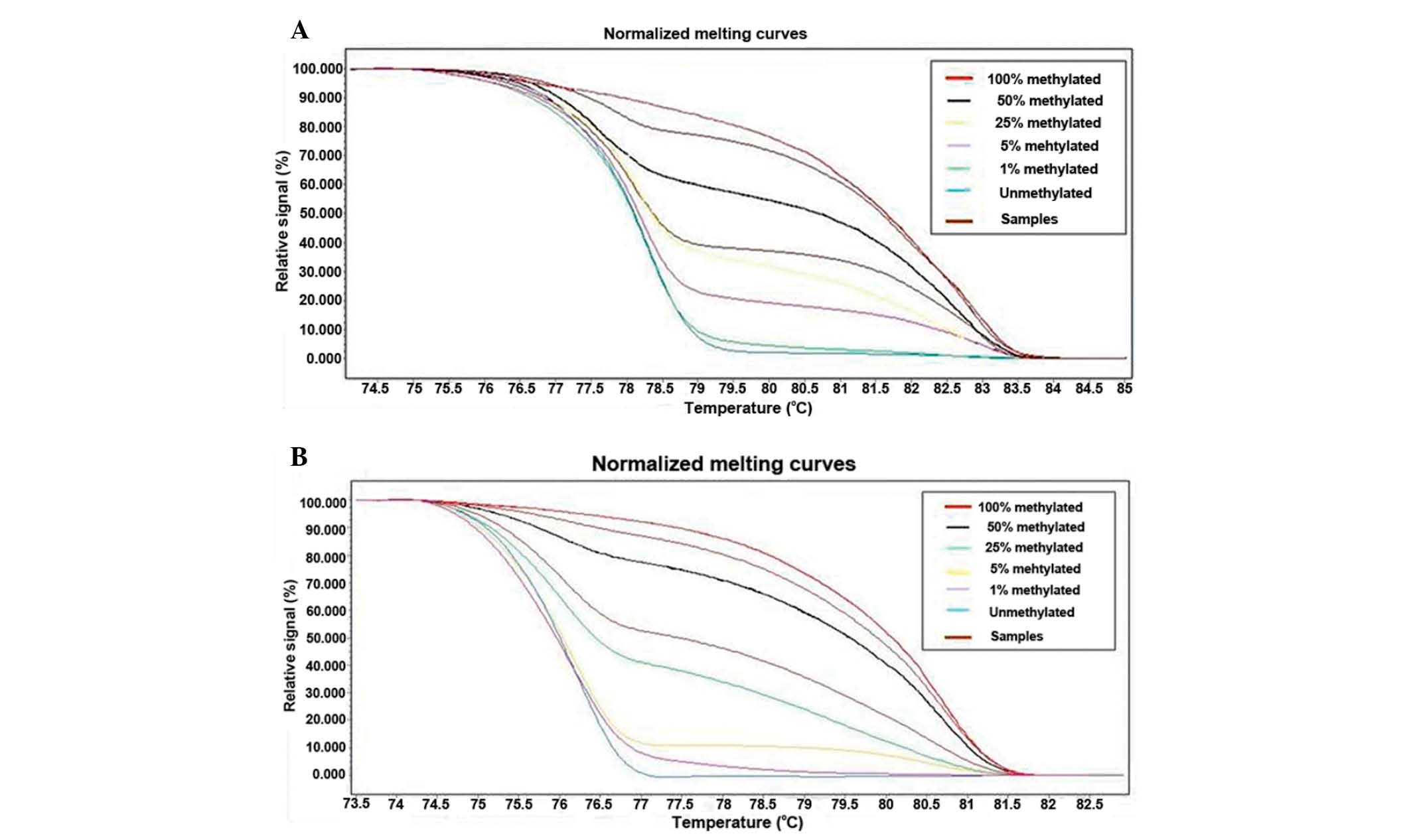
Comparing methylation status in
patient-matched peripheral blood leukocyte and colorectal tissue
DNA
The methylation analysis results of the 44 patients
are shown in Table II and
illustrated in Fig. 1A and 1B.
Differences in the levels of MGMT and MLH1
methylation were examined in patient-matched peripheral blood
leukocyte, colorectal tumor and normal colorectal tissue DNA
(Table II). There were no
significant differences in the levels of MGMT and
MLH1 methylation between the three groups (Friedman test,
P=0.260 and P=0.464, respectively).
| Table IIMS-HRM assay of peripheral blood and
colorectal tissue samples of CRC patients. |
Table II
MS-HRM assay of peripheral blood and
colorectal tissue samples of CRC patients.
| MGMT, n | MLH1, n |
|---|
|
|
|
|---|
| Frequencies of
methylation (%) | Leukocytes | Normal tissue | Tumor tissue | Leukocytes | Normal tissue | Tumor tissue |
|---|
| 0 | 37 | 35 | 31 | 32 | 30 | 31 |
| 0–1 | 0 | 2 | 1 | 0 | 2 | 2 |
| 1–5 | 5 | 4 | 2 | 7 | 1 | 2 |
| 5–25 | 0 | 3 | 3 | 2 | 9 | 5 |
| 25–50 | 1 | 0 | 0 | 3 | 1 | 2 |
| 50–100 | 1 | 0 | 2 | 0 | 0 | 1 |
| 100 | 0 | 0 | 5 | 0 | 1 | 1 |
Various cut-off methylation levels were used for the
analysis (Table III). The level
of methylation was classified as positive at a cut-off value of
0–1% methylation and no statistical significant differences were
observed in the levels of MGMT and MLH1 methylation
among the three groups (Table
III). When a level of methylation of >5% was classified as
positive, there was a significant difference in the levels of
MGMT methylation among the three groups (P=0.014;
χ2 test), but no significant difference in the levels of
MLH1 methylation (P=0.251; χ2 test). Further
analysis revealed that a significant difference in MGMT
methylation existed between colorectal tumor tissue DNA and
leukocytes or normal colorectal tissue DNA (P=0.013 and P=0.035,
respectively; χ2 test), but not between leukocyte and
normal colorectal tissue DNA (P=0.645; χ2 test).
| Table IIIMS-HRM assay of peripheral blood and
colorectal tissue samples of CRC patients using various cut-off
values. |
Table III
MS-HRM assay of peripheral blood and
colorectal tissue samples of CRC patients using various cut-off
values.
| MGMT | | | MLH1 | |
|---|
|
| | |
| |
|---|
| Cut-off (%) | Leukocytes, n
(%) | Normal tissue, n
(%) | Tumor tissue, n
(%) | P-value* | P-value* | Leukocytes, n
(%) | Normal tissue, n
(%) | Tumor tissue, n
(%) | P-value* |
|---|
| 0 | 7 (15.9) | 9 (20.5) | 13 (29.5) | 0.282 | - | 12 (27.3) | 14 (31.8) | 13 (29.5) | 0.897 |
| 1 | 7 (15.9) | 7 (15.9) | 12 (27.3) | 0.302 | - | 12 (29.5) | 12 (29.5) | 11 (25.0) | 0.962 |
| 5 | 2 (4.5) | 3 (6.8) | 10 (22.7) | 0.014 | 0.645a,
0.035b, 0.013c | 5 (11.4) | 11 (25.0) | 9 (20.5) | 0.251 |
Spearman rank correlation
coefficients
Positive correlations were observed between the
peripheral blood leukocyte and normal colorectal tissue DNA in the
levels of MGMT and MLH1 methylation (r=0.475, P=0.001
and r=0.362, P=0.016, respectively). However, there were no
positive correlations between colorectal tumor tissue and
peripheral blood leukocyte or normal colorectal tissue DNA, based
on the methylation levels of the assessments of the two genes
(Table IV).
| Table IVSpearman’s rank correlation
coefficients (P-values) of MGMT and MLH1 methylation
levels in case-matched DNA with CRC. |
Table IV
Spearman’s rank correlation
coefficients (P-values) of MGMT and MLH1 methylation
levels in case-matched DNA with CRC.
| DNA source | Leukocytes | Normal tissues | Tumor tissues |
|---|
| MGMT |
| Leukocytes | - | 0.475 (0.001) | −0.033 (0.833) |
| Normal
tissues | 0.475 (0.001) | - | 0.025 (0.873) |
| Tumor tissues | −0.033 (0.833) | 0.025 (0.873) | - |
| MLH1 |
| Leukocytes | - | 0.362 (0.016) | 0.215 (0.161) |
| Normal
tissues | 0.362 (0.016) | - | 0.293 (0.054) |
| Tumor tissues | 0.215 (0.161) | 0.293 (0.054) | - |
Agreement
The agreement between the peripheral blood leukocyte
and normal colorectal tissue DNA with CRC on the levels of
MGMT and MLH1 methylation were calculated using κ
coefficients (Table V). The
agreement of the MGMT gene methylation levels in the leukocytes of
the peripheral blood and normal colorectal tissue was graded as
fair (κ=0.299). The agreement of the MLH1 gene methylation levels
in the leukocytes of the peripheral blood and normal colorectal
tissue was graded as poor (κ=0.126) (Table V).
| Table VFrequencies of MGMT and
MLH1 methylation in the peripheral blood and the normal
colorectal tissues. |
Table V
Frequencies of MGMT and
MLH1 methylation in the peripheral blood and the normal
colorectal tissues.
| Frequencies of
normal colorectal tissues methylation (%) | |
|---|
|
| |
|---|
| Frequencies of
blood methylation (%) | 0 | 0–1 | 1–5 | 5–25 | 25–50 | 50–100 | 100 | n |
|---|
| MGMT |
| 0 | 32 | 2 | 3 | 0 | 0 | 0 | 0 | 37 |
| 0–1 | 0 | 0 | 0 | 0 | 0 | 0 | 0 | 0 |
| 1–5 | 3 | 0 | 1 | 1 | 0 | 0 | 0 | 5 |
| 5–25 | 0 | 0 | 0 | 0 | 0 | 0 | 0 | 0 |
| 25–50 | 0 | 0 | 0 | 1 | 0 | 0 | 0 | 1 |
| 50–100 | 0 | 0 | 0 | 1 | 0 | 0 | 0 | 1 |
| 100 | 0 | 0 | 0 | 0 | 0 | 0 | 0 | 0 |
| n | 35 | 2 | 4 | 3 | 0 | 0 | 0 | 44 |
| κ | 0.299
(P=0.002) | |
| MLH1 |
| 0 | 24 | 2 | 1 | 5 | 0 | 0 | 0 | 32 |
| 0–1 | 0 | 0 | 0 | 0 | 0 | 0 | 0 | 0 |
| 1–5 | 5 | 0 | 0 | 2 | 0 | 0 | 0 | 7 |
| 5–25 | 1 | 0 | 0 | 1 | 0 | 0 | 0 | 2 |
| 25–50 | 0 | 0 | 0 | 1 | 1 | 0 | 1 | 3 |
| 50–100 | 0 | 0 | 0 | 0 | 0 | 0 | 0 | 0 |
| 100 | 0 | 0 | 0 | 0 | 0 | 0 | 0 | 0 |
| n | 30 | 2 | 1 | 9 | 1 | 0 | 1 | 44 |
| κ | 0.126
(P=0.098) | |
Discussion
DNA promoter methylation has previously been shown
to be a well-characterized event in tumor biology and has been
extensively documented in CRC (33,34).
However, few studies have compared DNA promoter methylation in DNA
from various patient-matched sources. The present results revealed
that the levels of MLH1 and MGMT methylation were not
significantly different in patient-matched peripheral blood
leukocyte, colorectal tumor tissue and normal colorectal tissue DNA
as original semi-quantitatively rank data. Since low background
methylation levels have previously been reported for various genes
in normal samples (35,36), several studies have used 0.1–10% as
a cut-off for the scoring criteria of gene methylation using the
MS-HRM assay (31,37–40)
and there has not been a unified standard to define methylation.
Therefore, in the present study, in order to identify the various
methylation levels between cancer and normal samples, a range of
cut-off methylation levels were used. When samples with >5%
methylation were considered as methylated, distinctive MGMT
gene promoter methylation levels were identified between colorectal
tumor tissue and leukocyte or normal colorectal tissue DNA, but no
significant differences were observed between leukocyte and normal
colorectal tissue DNA. Thus, in samples containing >5%
methylation, the analysis of the MGMT gene appeared to
increase the sensitivity for discriminating cancer from normal
colorectal tissues or leukocytes.
Previous studies have focused on DNA methylation
measured in the leukocytes (41,42) or
the normal mucosa tissues (17,43,44),
rarely reporting the correlation between the two. Ally et al
reported significant positive correlations between the estrogen
receptor-α methylation index in leukocytes and normal colonic
tissue in CRC patients (r=0.570; P=0.003) (45). However, the samples were not case
matched. Spearman rank correlations were performed to investigate
the correlation of the MLH1 and MGMT methylation
levels in patient-matched peripheral blood leukocyte, colorectal
tumor and normal colorectal tissue DNA samples. The most
significant positive correlations were observed between the
leukocyte and normal colorectal tissue DNA for methylation
detection of MGMT and MLH1 (r=0.475 and r=0.362,
respectively).
In human studies, ethical and practical barriers may
make it difficult or impossible to collect specimens from the
target tissue. Hence, the use of surrogate samples, including DNA
derived from easily accessible peripheral blood, is widely accepted
when the target tissue is unobtainable. Several studies with regard
to DNA methylation biomarkers tested in leukocytes suggest the
suitability of epigenetic biomarkers for the detection of several
cancers relative to controls (46–48).
Widschwendter et al identified that particular methylation
patterns in peripheral blood DNA may serve as surrogate markers for
the risk of breast cancer (49). To
investigate the use of blood as a surrogate for DNA methylation in
tissues, the present study measured the MGMT and MLH1
gene methylation levels in the leukocytes of the peripheral blood
and normal colorectal tissue, which were graded as fair and poor
(κ=0.299 and 0.126, respectively); therefore, blood-derived DNA
methylation level measurements may not always represent the levels
of target colorectal tissue methylation (50). While all somatic cells in a given
individual are genetically identical, differing cell types form
highly distinct anatomical structures and perform a wide range of
disparate physiological functions (51). It has been conjectured that during
tissue differentiation and development, transcription-relevant
control regions in the genome become selectively de- or
upmethylated to enable the transcription of a restricted set of
genes within a given tissue (52).
It is also plausible that methylation patterns in DNA obtained from
blood may be more ‘plastic’ compared with that of other tissues,
due to the close proximity of the blood to environmental
influences, such as nutrition and smoking (50).
A limitation of the present study is the fact that
the subjects that were selected. The methylation results may not
reflect the natural frequencies of methylation in leukocytes or
colorectal tissues. Another limitation of the study is a lack of
inclusion of healthy controls. Studies using cancer subjects may
not exclude the possibility of disseminated tumor cells or the
effect the disease itself may have on the systemic methylation
status in leukocytes and normal colorectal tissues. Further studies
with disease-free individuals and an investigation of tumor
suppressor gene methylation are required to clarify this issue.
In summary, the correlation of MGMT and
MLH1 methylation levels between patient-matched leukocytes
and normal colorectal tissues was classified as moderate and weak,
respectively. Blood-derived DNA methylation measurements may not
always represent the levels of normal colorectal tissue
methylation.
Acknowledgements
This study was supported by grants from the
Education Bureau of Heilongjiang Province (11531100).
References
|
1
|
Jones PA and Baylin SB: The fundamental
role of epigenetic events in cancer. Nat Rev Genet. 3:415–428.
2002.PubMed/NCBI
|
|
2
|
Herman JG, Graff JR, Myöhänen S, Nelkin BD
and Baylin SB: Methylation-specific PCR: a novel PCR assay for
methylation status of CpG islands. Proc Natl Acad Sci USA.
93:9821–9826. 1996. View Article : Google Scholar : PubMed/NCBI
|
|
3
|
Jones PA and Laird PW: Cancer epigenetics
comes of age. Nat Genet. 21:163–167. 1999. View Article : Google Scholar : PubMed/NCBI
|
|
4
|
Baylin SB and Herman JG: DNA
hypermethylation in tumorigenesis: epigenetics joins genetics.
Trends Genet. 16:168–174. 2000. View Article : Google Scholar : PubMed/NCBI
|
|
5
|
Herman JG and Baylin SB: Gene silencing in
cancer in association with promoter hypermethylation. N Engl J Med.
349:2042–2054. 2003. View Article : Google Scholar : PubMed/NCBI
|
|
6
|
Derks S, Postma C, Moerkerk PT, et al:
Promoter methylation precedes chromosomal alterations in colorectal
cancer development. Cell Oncol. 28:247–257. 2006.PubMed/NCBI
|
|
7
|
Esteller M: Epigenetic lesions causing
genetic lesions in human cancer: promoter hypermethylation of DNA
repair genes. Eur J Cancer. 36:2294–2300. 2000. View Article : Google Scholar : PubMed/NCBI
|
|
8
|
Frigola J, Solé X, Paz MF, et al:
Differential DNA hypermethylation and hypomethylation signatures in
colorectal cancer. Hum Mol Genet. 14:319–326. 2005. View Article : Google Scholar : PubMed/NCBI
|
|
9
|
Esteller M and Herman JG: Generating
mutations but providing chemosensitivity: the role of
O6-methylguanine DNA methyltransferase in human cancer. Oncogene.
23:1–8. 2004. View Article : Google Scholar : PubMed/NCBI
|
|
10
|
Pegg AE: Mammalian O6-alkylguanine-DNA
alkyltransferase: regulation and importance in response to
alkylating carcinogenic and therapeutic agents. Cancer Res.
50:6119–6129. 1990.PubMed/NCBI
|
|
11
|
Povey AC, Badawi AF, Cooper DP, et al: DNA
alkylation and repair in the large bowel: animal and human studies.
J Nutr. 132:3518S–3521S. 2002.PubMed/NCBI
|
|
12
|
Anacleto C, Leopoldino AM, Rossi B, et al:
Colorectal cancer ‘methylator phenotype’: fact or artifact?
Neoplasia. 7:331–335. 2005.
|
|
13
|
Anacleto C, Rossi B, Lopes A, et al:
Development and application of a multiplex PCR procedure for the
detection of DNA methylation in colorectal cancer. Oncol Rep.
13:325–328. 2005.PubMed/NCBI
|
|
14
|
Fox EJ, Leahy DT, Geraghty R, et al:
Mutually exclusive promoter hypermethylation patterns of hMLH1 and
O6-methylguanine DNA methyltransferase in colorectal cancer. J Mol
Diagn. 8:68–75. 2006. View Article : Google Scholar : PubMed/NCBI
|
|
15
|
Kim YH, Petko Z, Dzieciatkowski S, et al:
CpG island methylation of genes accumulates during the adenoma
progression step of the multistep pathogenesis of colorectal
cancer. Genes Chromosomes Cancer. 45:781–789. 2006. View Article : Google Scholar : PubMed/NCBI
|
|
16
|
Shen L, Kondo Y, Rosner GL, et al: MGMT
promoter methylation and field defect in sporadic colorectal
cancer. J Natl Cancer Inst. 97:1330–1338. 2005. View Article : Google Scholar : PubMed/NCBI
|
|
17
|
Nagasaka T, Goel A, Notohara K, et al:
Methylation pattern of the O6-methylguanine-DNA methyltransferase
gene in colon during progressive colorectal tumorigenesis. Int J
Cancer. 122:2429–2436. 2008. View Article : Google Scholar : PubMed/NCBI
|
|
18
|
Lee KH, Lee JS, Nam JH, et al: Promoter
methylation status of hMLH1, hMSH2, and MGMT genes in colorectal
cancer associated with adenoma-carcinoma sequence. Langenbecks Arch
Surg. 396:1017–1026. 2011. View Article : Google Scholar : PubMed/NCBI
|
|
19
|
Menigatti M, Truninger K, Gebbers JO,
Marbet U, Marra G and Schär P: Normal colorectal mucosa exhibits
sex- and segment-specific susceptibility to DNA methylation at the
hMLH1 and MGMT promoters. Oncogene. 28:899–909. 2009. View Article : Google Scholar : PubMed/NCBI
|
|
20
|
Rakyan VK, Down TA, Thorne NP, et al: An
integrated resource for genome-wide identification and analysis of
human tissue-specific differentially methylated regions (tDMRs).
Genome Res. 18:1518–1529. 2008.
|
|
21
|
Lofton-Day C, Model F, Devos T, et al: DNA
methylation biomarkers for blood-based colorectal cancer screening.
Clin Chem. 54:414–423. 2008. View Article : Google Scholar : PubMed/NCBI
|
|
22
|
Grützmann R, Molnar B, Pilarsky C, et al:
Sensitive detection of colorectal cancer in peripheral blood by
septin 9 DNA methylation assay. PLoS One. 3:e37592008.PubMed/NCBI
|
|
23
|
Gazzoli I, Loda M, Garber J, Syngal S and
Kolodner RD: A hereditary nonpolyposis colorectal carcinoma case
associated with hypermethylation of the MLH1 gene in normal tissue
and loss of heterozygosity of the unmethylated allele in the
resulting microsatellite instability-high tumor. Cancer Res.
62:3925–3928. 2002.
|
|
24
|
Miyakura Y, Sugano K, Akasu T, et al:
Extensive but hemiallelic methylation of the hMLH1 promoter region
in early-onset sporadic colon cancers with microsatellite
instability. Clin Gastroenterol Hepatol. 2:147–156. 2004.
View Article : Google Scholar : PubMed/NCBI
|
|
25
|
Sheng JQ, Zhang H, Ji M, et al: Genetic
diagnosis strategy of hereditary non-polyposis colorectal cancer.
World J Gastroenterol. 15:983–989. 2009. View Article : Google Scholar : PubMed/NCBI
|
|
26
|
Zhou HH, Yan SY, Zhou XY, et al: MLH1
promoter germline-methylation in selected probands of Chinese
hereditary non-polyposis colorectal cancer families. World J
Gastroenterol. 14:7329–7334. 2008. View Article : Google Scholar : PubMed/NCBI
|
|
27
|
Psofaki V, Kalogera C, Tzambouras N, et
al: Promoter methylation status of hMLH1, MGMT, and CDKN2A/p16 in
colorectal adenomas. World J Gastroenterol. 16:3553–3560. 2010.
View Article : Google Scholar : PubMed/NCBI
|
|
28
|
Candiloro IL and Dobrovic A: Detection of
MGMT promoter methylation in normal individuals is strongly
associated with the T allele of the rs16906252 MGMT promoter single
nucleotide polymorphism. Cancer Prev Res (Phila). 2:862–867. 2009.
View Article : Google Scholar
|
|
29
|
Vineis P, Chuang SC, Vaissière T, et al:
DNA methylation changes associated with cancer risk factors and
blood levels of vitamin metabolites in a prospective study.
Epigenetics. 6:195–201. 2011. View Article : Google Scholar : PubMed/NCBI
|
|
30
|
Wojdacz TK and Dobrovic A:
Methylation-sensitive high resolution melting (MS-HRM): a new
approach for sensitive and high-throughput assessment of
methylation. Nucleic Acids Res. 35:e412007. View Article : Google Scholar : PubMed/NCBI
|
|
31
|
Balic M, Pichler M, Strutz J, et al: High
quality assessment of DNA methylation in archival tissues from
colorectal cancer patients using quantitative high-resolution
melting analysis. J Mol Diagn. 11:102–108. 2009. View Article : Google Scholar
|
|
32
|
Landis JR and Koch GG: The measurement of
observer agreement for categorical data. Biometrics. 33:159–174.
1977. View Article : Google Scholar : PubMed/NCBI
|
|
33
|
Esteller M, Sparks A, Toyota M, et al:
Analysis of adenomatous polyposis coli promoter hypermethylation in
human cancer. Cancer Res. 60:4366–4371. 2000.PubMed/NCBI
|
|
34
|
Chan AO, Broaddus RR, Houlihan PS, Issa
JP, Hamilton SR and Rashid A: CpG island methylation in aberrant
crypt foci of the colorectum. Am J Pathol. 160:1823–1830. 2002.
View Article : Google Scholar : PubMed/NCBI
|
|
35
|
Ahlquist T, Lind GE, Costa VL, et al: Gene
methylation profiles of normal mucosa, and benign and malignant
colorectal tumors identify early onset markers. Mol Cancer.
7:942008. View Article : Google Scholar : PubMed/NCBI
|
|
36
|
Nakagawa H, Nuovo GJ, Zervos EE, et al:
Age-related hypermethylation of the 5′ region of MLH1 in normal
colonic mucosa is associated with microsatellite-unstable
colorectal cancer development. Cancer Res. 61:6991–6995. 2001.
|
|
37
|
Meng W, Huebner A, Shabsigh A, Chakravarti
A and Lautenschlaeger T: Combined RASSF1A and RASSF2A promoter
methylation analysis as diagnostic biomarker for bladder cancer.
Mol Biol Int. 2012:7018142012. View Article : Google Scholar : PubMed/NCBI
|
|
38
|
Morandi L, Franceschi E, de Biase D, et
al: Promoter methylation analysis of O6-methylguanine-DNA
methyltransferase in glioblastoma: detection by locked nucleic acid
based quantitative PCR using an imprinted gene (SNURF) as a
reference. BMC Cancer. 10:482010. View Article : Google Scholar : PubMed/NCBI
|
|
39
|
Avraham A, Uhlmann R, Shperber A, et al:
Serum DNA methylation for monitoring response to neoadjuvant
chemotherapy in breast cancer patients. Int J Cancer.
131:E1166–E1172. 2012. View Article : Google Scholar : PubMed/NCBI
|
|
40
|
Kristensen LS, Mikeska T, Krypuy M and
Dobrovic A: Sensitive Melting Analysis after Real Time- Methylation
Specific PCR (SMART-MSP): high-throughput and probe-free
quantitative DNA methylation detection. Nucleic Acids Res.
36:e422008. View Article : Google Scholar : PubMed/NCBI
|
|
41
|
Lim U, Flood A, Choi SW, et al: Genomic
methylation of leukocyte DNA in relation to colorectal adenoma
among asymptomatic women. Gastroenterology. 134:47–55. 2008.
View Article : Google Scholar : PubMed/NCBI
|
|
42
|
Pufulete M, Al-Ghnaniem R, Leather AJ, et
al: Folate status, genomic DNA hypomethylation, and risk of
colorectal adenoma and cancer: a case control study.
Gastroenterology. 124:1240–1248. 2003. View Article : Google Scholar : PubMed/NCBI
|
|
43
|
Lind GE, Thorstensen L, Løvig T, et al: A
CpG island hypermethylation profile of primary colorectal
carcinomas and colon cancer cell lines. Mol Cancer. 3:282004.
View Article : Google Scholar : PubMed/NCBI
|
|
44
|
Lee S, Hwang KS, Lee HJ, Kim JS and Kang
GH: Aberrant CpG island hypermethylation of multiple genes in
colorectal neoplasia. Lab Invest. 84:884–893. 2004. View Article : Google Scholar : PubMed/NCBI
|
|
45
|
Ally MS, Al-Ghnaniem R and Pufulete M: The
relationship between gene-specific DNA methylation in leukocytes
and normal colorectal mucosa in subjects with and without
colorectal tumors. Cancer Epidemiol Biomarkers Prev. 18:922–928.
2009. View Article : Google Scholar : PubMed/NCBI
|
|
46
|
Moore LE, Pfeiffer RM, Poscablo C, et al:
Genomic DNA hypomethylation as a biomarker for bladder cancer
susceptibility in the Spanish Bladder Cancer Study: a case-control
study. Lancet Oncol. 9:359–366. 2008. View Article : Google Scholar : PubMed/NCBI
|
|
47
|
Wang L, Aakre JA, Jiang R, et al:
Methylation markers for small cell lung cancer in peripheral blood
leukocyte DNA. J Thorac Oncol. 5:778–785. 2010. View Article : Google Scholar : PubMed/NCBI
|
|
48
|
Hsiung DT, Marsit CJ, Houseman EA, et al:
Global DNA methylation level in whole blood as a biomarker in head
and neck squamous cell carcinoma. Cancer Epidemiol Biomarkers Prev.
16:108–114. 2007. View Article : Google Scholar : PubMed/NCBI
|
|
49
|
Widschwendter M, Apostolidou S, Raum E, et
al: Epigenotyping in peripheral blood cell DNA and breast cancer
risk: a proof of principle study. PLoS One. 3:e26562008. View Article : Google Scholar : PubMed/NCBI
|
|
50
|
McKay JA, Xie L, Harris S, Wong YK, Ford D
and Mathers JC: Blood as a surrogate marker for tissue-specific DNA
methylation and changes due to folate depletion in post-partum
female mice. Mol Nutr Food Res. 55:1026–1035. 2011. View Article : Google Scholar : PubMed/NCBI
|
|
51
|
Christensen BC, Houseman EA, Marsit CJ, et
al: Aging and environmental exposures alter tissue-specific DNA
methylation dependent upon CpG island context. PLoS Genet.
5:e10006022009. View Article : Google Scholar : PubMed/NCBI
|
|
52
|
Walsh CP and Bestor TH: Cytosine
methylation and mammalian development. Genes Dev. 13:26–34. 1999.
View Article : Google Scholar : PubMed/NCBI
|