Introduction
Breast cancer (BC) is the most common type of cancer
in females worldwide. BC originates most commonly from the inner
lining of milk ducts or lobules of breast tissue (1), which accounts for 22.9% of all types
of cancer (with the exception of non-melanoma skin cancer) in
females and has caused 458,503 mortalities worldwide (13.7% of
cancer mortality in females) in 2008. According to the American
Cancer Society, almost 230,000 new cases and 40,000 mortalities
occurred in the United States in 2011. However, recognized risk
factors of BC may be absent in 50–80% of patients (2), which establishes an increased interest
to identify possible risk factors that contribute to BC.
microRNAs (miRNAs) are small (~22 nucleotides),
non-coding RNA molecules. miRNAs, which modulate the expression of
targeting genes by post-transcription, are involved in the
regulation of various cell processes, including apoptosis,
hematopoietic cell differentiation, metabolism, neural development
and metastasis (3,4). A number of miRNAs are involved in
several types of human cancer, including BC. The majority of
previous studies have described the profile of miRNA expression in
BC cell lines and primary tumor tissues. For example, increased
expression of the miR-191/425 cluster in aggressive BC cells
changes global gene expression profiles, which has a fundamental
impact on the progression of BC cells (5). By analyzing the miR-21 expression in
BC tissues, Ozgün et al previously reported that patients
with high miR-21 expression levels have a significantly lower
disease-free survival than patients with low miR-21 expression
levels, which indicates that miR-21 is an indicator of an
aggressive BC phenotype (6). The
overexpression of miR-21 increases BC MCF-7 cell growth, migration
and invasion, self-renewal and clonogenicity (7). The overexpression of miR-200a protects
tumor cells from anoikis and promotes metastases, while inhibition
of miR-200a suppresses anoikis resistance in BC cells (8). The decreased miR-200f expression is
likely to increase the expression levels of EMT-transcriptional
inducers and may be used as a hypothetical biomarker to assess the
occurrence of EMT in BC (9). Let-7,
as a tumor suppressor, inhibits the estrogen receptor (ER)
α-mediated cellular malignant growth in ER-positive BC stem cells
(10). The abovementioned studies
show that miRNAs are important for the tumorigenesis, migration and
invasion of BC.
The apoptotic and necrotic primary tumor discharges
miRNAs into the blood circulation, known as circulating miRNAs.
Therefore, blood contains circulating miRNAs from numerous cells
(including tumor cells), which makes it possible to detect miRNAs
from specific organs, tissues or cells using surface markers for
proper quantification (11,12). Moreover, the circulating miRNAs,
resistant to RNase activity, are rare and extremely stable in serum
and plasma (13). This stability
translates into consistent miRNA expression levels among
individuals, which makes serum miRNAs attractive biomarkers for the
diagnosis of BCs. However, there have been only a few previous
publications investigating circulating miRNAs in the peripheral
blood of BC patients (13–16). The present study investigated the
levels of miR-182 in the blood serum of BC patients to identify the
potential of serum miRNAs as biomarkers for BC.
Materials and methods
Subjects
The present study was performed at the Inpatient
Department of Medical Oncology of Laiyang Central Hospital (Yantai,
China). The research protocol was approved by the Medical Ethics
Committee of Binzhou Medical University (Yantai, China). All
experiments were performed according to the relevant guidelines of
the Medical Ethics Committee of Binzhou Medical University.
In total, 46 BC patients, aged 30–79 years, were
pathologically diagnosed with BC, for the first time, between May
1st, 2010 and September 30th, 2012. The patients had not received
prior chemotherapy. Healthy controls (n=58), came to Laiyang
Central Hospital for physical examination between May 1st, 2010 and
September 30th, 2012 and were diagnosed without any tumor or
physical illness. Prior to inclusion, all the eligible BC patients
and healthy controls provided written informed consent following a
full explanation of the study procedures.
Immunohistochemistry
Histological sections (3-μm) were deparaffinized in
xylene and rehydrated. Antigen retrieval was performed by
microwaving the sections in 10 mM citric acid monohydrate.
Endogenous peroxidase activity was blocked by 0.5%
H2O2 treatment. The slides were incubated
with appropriate dilutions of the primary antibodies [anti-ER,
1:200; and anti-progesterone receptor (PR), 1:200; ZSGB-BIO,
Beijing, China] at 4°C overnight. The same procedure was performed
for negative controls which were incubated overnight in 1X PBS
without antibody. The reaction was visualized by the ABC Kit
(ZSGB-BIO) and positive ER and PR status was defined by nuclear
staining of >10%.
miRNA isolation from serum and
tissue
Serum samples from the patients and controls were
collected between 7:00 and 8:00 a.m. Following centrifugation for
30 min at 2,650 g, plasma samples were stored at 80°C. miRNAs were
extracted from plasma by the mirVana™ miRNA isolation kit (Ambion,
Carlsbad, CA, USA) according to the manufacturer's instructions.
Tissue samples were homogenized in a denaturing lysis solution.
Total RNA was extracted from tissue lysis using the TRIzol reagent
(Invitrogen Life Technologies, Carlsbad, CA, USA). Then, miRNA was
separated from 30–50 mg of total RNA using the Ambion miRNA
Isolation Kit (Ambion).
Quantitative PCR (qPCR)
miRNAs were added poly (A) tails by poly (A)
polymerase (Ambion). The cDNAs were synthesized by a real-time
primer, 5′-AACATGTACAGTCCATGGATGd(T)30N(A,G,C or T)-3′. miR-182 was
then analyzed by qPCR and the primer used was: forward,
5′-GGCAATGGTAGAACTCACACT-3′ and reverse,
5′-AACATGTACAGTCCATGGATG-3′. qPCR analysis was performed using
SuperTaq Polymerase (Takara Biotechnology Co., Ltd., Dalian,
China). miR-182 expression was detected using the RG3000 system
(Corbett Life Science, Mortlake, Australia) with the Quantitect
SYBR-Green Kit (Qiagen, Hilden, Germany) as follows: initial
denaturation at 95°C for 5 min, followed by 40 cycles of 95°C
denaturation for 20 sec, 52°C annealing for 20 sec and extension at
72°C for 30 sec. Fluorescence was observed at 585 nm at each
extension step of 72°C. Human 5S rRNA was added into each sample
and served as a control. All experiments were repeated in
triplicate.
Statistical analysis
Data were first tested for normal distribution and
variance homogeneity using the Shapiro-Wilk test and F-test,
respectively. Data are presented as mean ± SD for normal
distributions, otherwise, data are presented as median and
quartiles. Since age, height and weight showed normal
distributions, differences between these groups were analyzed using
the Student's t-test. However, when the levels of miR-182 did not
show a normal distribution, non-parametric tests were applied.
miR-182 continuous variables between groups were analyzed by the
Wilcoxon rank-sum test. Statistical analyses were performed using R
version 2.15.0© (2012; ISBN 3-900051-07-0). P<0.05
was considered to indicate a statistically significant
difference.
Results
Clinical characteristics of patients
In total, 46 BC patients and 58 controls
participated in the present study. The demographic and clinical
characteristics of all the patients and controls are provided in
Table I. No differences in age,
height and weight were found between the BC patients and their
controls. Of the 46 patients, 29 patients were ER-positive (63.0%)
and 28 PR-positive (60.9%) in the entire tumor set (46 cases).
Alcohol and passive smoking have been reported to increase BC risk
(17–19), but no significant differences were
identified between the BC patients and their controls in the
present study (Table I).
 | Table IDemographic and clinical
characteristics of the study samples. |
Table I
Demographic and clinical
characteristics of the study samples.
| Characteristics | Healthy controls | Patients | P-valuea |
|---|
| n | 58 | 46 | |
| Age (mean ± SD),
years | 52.00±9.81 | 48.30±10.03 | 0.060 |
| Weight (mean ± SD),
kg | 65.31±8.63 | 66.00±8.65 | 0.400 |
| Height (mean ± SD),
cm | 157.98±4.63 | 158.76±4.64 | 0.680 |
|
ER-positive/negative, n | - | 29/17 | - |
|
PR-positive/negative, n | - | 28/18 | - |
|
Non-alcoholic/alcoholic drinks, n | 57/1 | 41/5 | 0.085 |
| Non-passive/passive
smokers, n | 27/31 | 14/32 | 0.095 |
| Median miR-182,
n | 0.003b | 7.075b |
3.947E−08b |
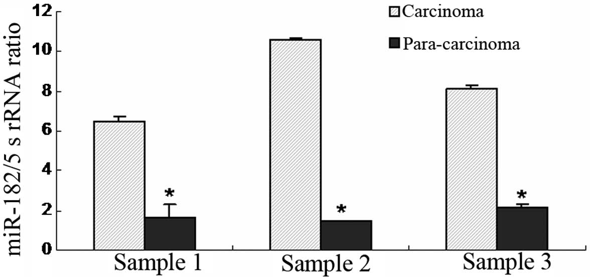
Higher expression of miR-182 in BC
tissues
miR-182, as an oncogene, is important for the
development of BC (20,21). To further demonstrate the role of
miR-182 in BC, its expression was detected in the BC tissues. The
results showed that miR-182 expression was markedly increased
(>4-fold higher) in BC tissues (n=3) compared with paracancerous
tissues (n=3) (Fig. 1), which is
consistent with the oncogenic role of miR-182.
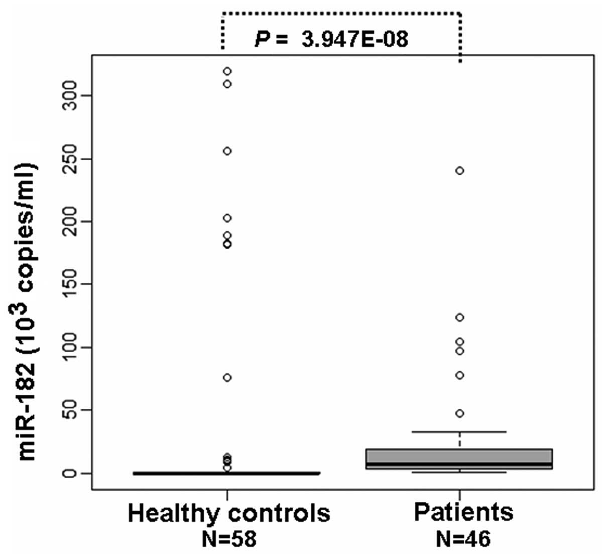
Higher levels of miR-182 in the serum of
patients with BC
Furthermore, the serum levels of miR-182 were
detected by qPCR to investigate the role of miR-182 in the
diagnosis of BC. It was found that the serum miR-182 levels in BC
patients were 7.075×10³ copies/ml (n=46), which were
significantly higher compared with the serum of healthy controls
(0.003×10³ copies/ml) (P<0.01; n=58; Table I; Fig.
2). The results demonstrated that the serum levels of miR-182
were higher in BC, indicating that miR-182 may also be an important
factor for the pathogenesis of BC.
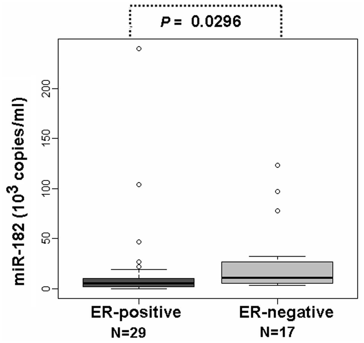
Correlation of ER/PR with circulating
miR-182 in the serum of BC patients
ER and PR are important factors associated with the
etiology and therapy of BC (22,23).
To study the correlation between ER and PR with the serum levels of
miR-182, the serum levels of miR-182 were detected in ER- and
PR-positive patients and compared with ER- and PR-negative
patients. The results showed that the serum levels of miR-182 in
the ER-positive patients (n=29) were 5.41×10³ copies/ml,
considerably lower compared with the ER-negative patients (n=17)
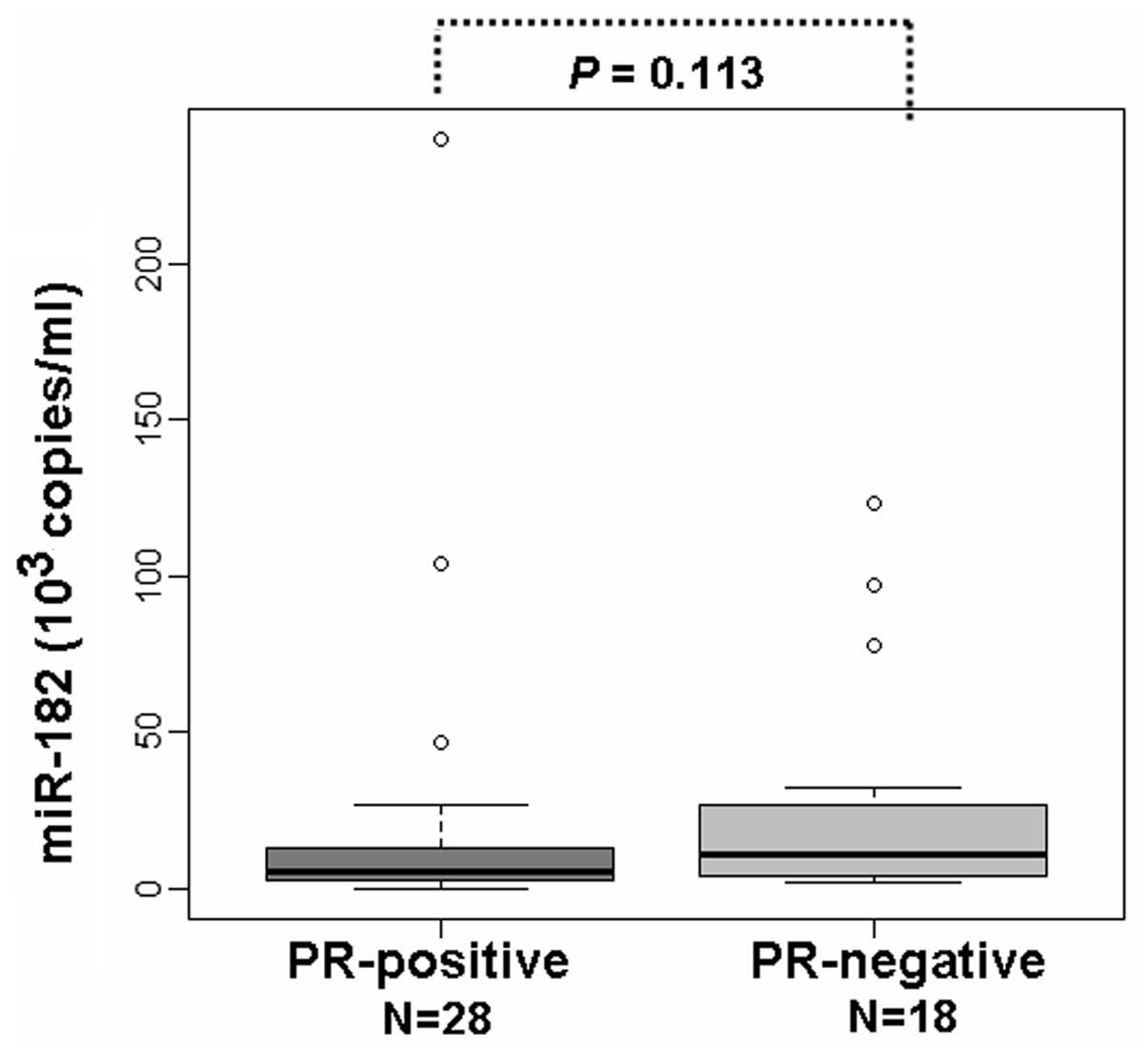
(P<0.05; Table II; Fig. 3). The serum levels of miR-182 in the
PR-positive patients (n=28) were also found to be lower compared
with the PR-negative patients (n=18) (Table II; Fig.
4).
| Table IICorrelation between ER- and
PR-positive samples with miR-182. |
Table II
Correlation between ER- and
PR-positive samples with miR-182.
| Receptor | n | Median miR-182 | P-valuea |
|---|
| ER |
| Positive | 29 | 5.409b | 0.0296 |
| Negative | 17 | 10.648b | |
| PR |
| Positive | 28 | 5.395b | 0.1130 |
| Negative | 18 | 10.643b | |
Discussion
The ideal biomarkers for BC diagnosis should be
easily accessible in order that they may be sampled relatively
non-invasively. In addition, biomarkers must be sensitive enough to
be detected in early stage tumors in almost all patients, while
absent or minimal in healthy control individuals (24). miRNAs are markedly stable molecules,
preserved well in formalin-fixed and fresh snap frozen specimens
(25, 26). Their expression profiles are
pathognomonic or tissue-specific in tumors (27), which establishes them as an ideal
class of biomarker for BC diagnosis. In the current study, miR-182
was isolated from the tissues of BC patients and healthy controls
and it was found that miR-182 expression was considerably higher in
the BC tissues compared with the control tissues. This result is
consistent with the oncogenic role of miR-182 in various types of
cancer. Furthermore, miR-182 was isolated from the serum of BC
patients and controls. The results demonstrated that miR-182 levels
in the serum of BC patients were also markedly higher than those of
the controls, indicating miR-182 is a useful biomarker for BC
diagnosis.
Previous miRNA expression studies in BC have
indicated the importance and potential roles of miRNA as disease
classifiers and prognostic tools. Iorio et al(28) previously identified that 29 miRNAs
were differentially expressed in BC tissues compared with control
tissues. In addition, Mattie et al reported unique sets of
miRNAs associated with BCs, which were defined by their roles of
HER2/neu or ER/PR status (29). It
has been previously reported that the pre-miR-27a rs895819
polymorphism may be associated with BC susceptibility or cancer
development in Caucasians (30).
The C allele of hsa-miR-146a (31)
and hsa-miR-196a2 rs11614913 SNP (32), associated with BC risk, were also
demonstrated to be important in familial breast/ovarian tumor
development. These studies indicated that miRNAs are crucial in the
development and diagnosis of BC.
Recently, several studies support that miR-182 acts
as an oncogene in the development of BC (20,21,33,34).
miR-182 is overexpressed in human BC tissues and cell lines (MB-231
cells) and β-catenin binds to the promoter to increase the
expression of miR-182 (20). Highly
expressed miR-182 functions as a potential oncomir in BC (21), which disrupts the homologous
recombination pathway in BC tissues. Mechanistically, the
overexpression of miR-182 decreases BRCA1 protein levels and
impedes DNA repair, while antagonizing miR-182 enhances BRCA1
levels and induces resistance to the poly (ADP-ribose) polymerase 1
inhibitor (33). FOXO1, a putative
tumor suppressor, is also a target of miR-182 and an antisense
inhibitor specific to miR-182 which leads to a significant increase
in endogenous FOXO1 expression (34). Similarly, the current study
demonstrated that miR-182 was upregulated in BC tissues compared
with control tissues, consistent with the important role of miR-182
in the tumorigenesis of BC.
miRNAs have been previously demonstrated to be
present in the serum in a stable and reproducible manner. In
addition, the unique expression patterns of serum miRNAs may be
used as biomarkers for various diseases (13,35).
Using qPCR, miR-205 was demonstrated to be downregulated, while
miR-155 was upregulated in BC patient serum (36). The plasma levels of circulating
miR-10b and miR-373 were found to be significantly higher in BC
patients with lymph node metastasis compared with normal controls
(37). In contrast to increased
miR-21 levels, circulating miR-92a levels were decreased in the BC
patients compared with the controls (38). Although miR-182 has been reported to
be important for BC tumorigenesis, no previous studies have
analyzed the role of circulating miR-182 in the diagnosis of BC. To
explore the diagnostic role of circulating miR-182 in BC, the
present study isolated miRNAs from the serum of BC patients and
healthy control individuals. The results revealed that the miR-182
levels in BC were higher compared with the healthy controls, which
confirmed the diagnostic role of miR-182 in BC.
The prognostic and therapeutic roles of ER or PR in
BC have been studied extensively and are well established (22,23,39).
Significant associations have been found between ER- and
PR-positive rates with menopausal status, tumor size or the
presence of distant metastases in BCs (40). In total, >75% of primary BC
patients express ER and ~50% of these tumors are stained positively
with PR4. The results of the present study also showed that 64.4%
of primary BC patients express ER and 62.2% of these patients PR.
The effects of ER- and PR-positive expression on the serum levels
of miR-182 were further investigated and it was found that serum
levels of miR-182 were lower in the ER- and PR-positive patients
compared with the ER- and PR-negative subjects. The results
indicated that there is a close correlation between serum levels of
miR-182 and ER- and PR-positive expression in BC patients. Although
alcohol and passive smoking increases BC risk (17–19),
in the present study, no significant differences were identified
between the BC patients and their controls. The relatively small
number of available previous studies may lead to this
limitation.
In summary, the results of the present study showed
that the levels of miR-182 in the serum of BC patients were
upregulated compared with healthy controls. Notably, the levels of
miR-182 in the serum of ER-positive patients was considerably lower
compared with the ER-negative patients. Overall, the present study
highlights miR-182 as a novel diagnostic marker for BC.
Acknowledgements
The present study was supported by the NCET-10-0919,
the National Natural Science Foundation (no. 31371321, 81141114 and
81200601) and the Foundation of Shandong Educational Committee of
China (no. J10LC60 and J11LC01).
References
|
1
|
Sariego J: Breast cancer in the young
patient. Am Surg. 76:1397–1400. 2010.
|
|
2
|
Anders CK and Carey LA: Biology,
metastatic patterns, and treatment of patients with triple-negative
breast cancer. Clin Breast Cancer. 9(Suppl 2): S73–S81. 2009.
View Article : Google Scholar : PubMed/NCBI
|
|
3
|
Kloosterman WP and Plasterk RH: The
diverse functions of microRNAs in animal development and disease.
Dev Cell. 11:441–450. 2006. View Article : Google Scholar : PubMed/NCBI
|
|
4
|
Stefani G and Slack FJ: Small non-coding
RNAs in animal development. Nat Rev Mol Cell Biol. 9:219–230. 2008.
View Article : Google Scholar : PubMed/NCBI
|
|
5
|
Di Leva G, Piovan C, Gasparini P, et al:
Estrogen mediated-activation of miR-191/425 cluster modulates
tumorigenicity of breast cancer cells depending on estrogen
receptor status. PLoS Genet. 9:e10033112013.PubMed/NCBI
|
|
6
|
Ozgün A, Karagoz B, Bilgi O, Tuncel T,
Baloglu H and Kandemir EG: MicroRNA-21 as an indicator of
aggressive phenotype in breast cancer. Onkologie. 36:115–118.
2013.PubMed/NCBI
|
|
7
|
Han M, Liu M, Wang Y, et al: Re-expression
of miR-21 contributes to migration and invasion by inducing
epithelial-mesenchymal transition consistent with cancer stem cell
characteristics in MCF-7 cells. Mol Cell Biochem. 363:427–436.
2012. View Article : Google Scholar : PubMed/NCBI
|
|
8
|
Yu SJ, Hu JY, Kuang XY, et al:
MicroRNA-200a promotes anoikis resistance and metastasis by
targeting YAP1 in human breast cancer. Clin Cancer Res.
19:1389–1399. 2013. View Article : Google Scholar : PubMed/NCBI
|
|
9
|
Castilla MÁ, Díaz-Martín J, Sarrió D, et
al: MicroRNA-200 family modulation in distinct breast cancer
phenotypes. PLoS One. 7:e477092012.PubMed/NCBI
|
|
10
|
Sun X, Qin S, Fan C, Xu C, Du N and Ren H:
Let-7: a regulator of the ERα signaling pathway in human breast
tumors and breast cancer stem cells. Oncol Rep. 29:2079–2087.
2013.PubMed/NCBI
|
|
11
|
Taylor DD and Gercel-Taylor C: MicroRNA
signatures of tumor-derived exosomes as diagnostic biomarkers of
ovarian cancer. Gynecol Oncol. 110:13–21. 2008. View Article : Google Scholar : PubMed/NCBI
|
|
12
|
Hunter MP, Ismail N, Zhang X, et al:
Detection of microRNA expression in human peripheral blood
microvesicles. PLoS One. 3:e36942008. View Article : Google Scholar : PubMed/NCBI
|
|
13
|
Mitchell PS, Parkin RK, Kroh EM, et al:
Circulating microRNAs as stable blood-based markers for cancer
detection. Proc Natl Acad Sci USA. 105:10513–10518. 2008.
View Article : Google Scholar : PubMed/NCBI
|
|
14
|
Zhu W, Qin W, Atasoy U and Sauter ER:
Circulating microRNAs in breast cancer and healthy subjects. BMC
Res Notes. 2:892009. View Article : Google Scholar : PubMed/NCBI
|
|
15
|
Heneghan HM, Miller N, Lowery AJ, Sweeney
KJ, Newell J and Kerin MJ: Circulating microRNAs as novel minimally
invasive biomarkers for breast cancer. Ann Surg. 251:499–505. 2010.
View Article : Google Scholar : PubMed/NCBI
|
|
16
|
Schrauder MG, Strick R, Schulz-Wendtland
R, et al: Circulating micro-RNAs as potential blood-based markers
for early stage breast cancer detection. PLoS One. 7:e297702012.
View Article : Google Scholar : PubMed/NCBI
|
|
17
|
Wu AH, Vigen C, Razavi P, Tseng CC and
Stancyzk FZ: Alcohol and breast cancer risk among Asian-American
women in Los Angeles County. Breast Cancer Res. 14:R1512012.
View Article : Google Scholar : PubMed/NCBI
|
|
18
|
Shrubsole MJ, Gao YT, Dai Q, et al:
Passive smoking and breast cancer risk among non-smoking Chinese
women. Int J Cancer. 110:605–609. 2004. View Article : Google Scholar : PubMed/NCBI
|
|
19
|
Gammon MD, Eng SM, Teitelbaum SL, et al:
Environmental tobacco smoke and breast cancer incidence. Environ
Res. 96:176–185. 2004. View Article : Google Scholar : PubMed/NCBI
|
|
20
|
Chiang CH, Hou MF and Hung WC:
Up-regulation of miR-182 by β-catenin in breast cancer increases
tumorigenicity and invasiveness by targeting the matrix
metalloproteinase inhibitor RECK. Biochim Biophys Acta.
1830:3067–3076. 2013.
|
|
21
|
Krishnan K, Steptoe AL, Martin HC, et al:
MicroRNA-182-5p targets a network of genes involved in DNA repair.
RNA. 19:230–242. 2013. View Article : Google Scholar : PubMed/NCBI
|
|
22
|
Cui X, Schiff R, Arpino G, Osborne CK and
Lee AV: Biology of progesterone receptor loss in breast cancer and
its implications for endocrine therapy. J Clin Oncol. 23:7721–7735.
2005. View Article : Google Scholar : PubMed/NCBI
|
|
23
|
Diaz NM: Laboratory testing for HER2/neu
in breast carcinoma: an evolving strategy to predict response to
targeted therapy. Cancer Control. 8:415–418. 2001.PubMed/NCBI
|
|
24
|
Heneghan HM, Miller N, Lowery AJ, Sweeney
KJ and Kerin MJ: MicroRNAs as Novel Biomarkers for Breast Cancer. J
Oncol. 2009:9502012009.PubMed/NCBI
|
|
25
|
Xi Y, Nakajima G, Gavin E, et al:
Systematic analysis of microRNA expression of RNA extracted from
fresh frozen and formalin-fixed paraffin-embedded samples. RNA.
13:1668–1674. 2007. View Article : Google Scholar : PubMed/NCBI
|
|
26
|
Li J, Smyth P, Flavin R, et al: Comparison
of miRNA expression patterns using total RNA extracted from matched
samples of formalin-fixed paraffin-embedded (FFPE) cells and snap
frozen cells. BMC Biotechnol. 7:362007. View Article : Google Scholar
|
|
27
|
Lu J, Getz G, Miska EA, et al: MicroRNA
expression profiles classify human cancers. Nature. 435:834–838.
2005. View Article : Google Scholar : PubMed/NCBI
|
|
28
|
Iorio MV, Ferracin M, Liu CG, et al:
MicroRNA gene expression deregulation in human breast cancer.
Cancer Res. 65:7065–7070. 2005. View Article : Google Scholar : PubMed/NCBI
|
|
29
|
Mattie MD, Benz CC, Bowers J, et al:
Optimized high-throughput microRNA expression profiling provides
novel biomarker assessment of clinical prostate and breast cancer
biopsies. Mol Cancer. 5:242006. View Article : Google Scholar
|
|
30
|
Zhong S, Chen Z, Xu J, Li W and Zhao J:
Pre-mir-27a rs895819 polymorphism and cancer risk: a meta-analysis.
Mol Biol Rep. 40:3181–3186. 2013. View Article : Google Scholar : PubMed/NCBI
|
|
31
|
Pastrello C, Polesel J, Della Puppa L,
Viel A and Maestro R: Association between hsa-mir-146a genotype and
tumor age-of-onset in BRCA1/BRCA2-negative familial breast and
ovarian cancer patients. Carcinogenesis. 31:2124–2126. 2010.
View Article : Google Scholar : PubMed/NCBI
|
|
32
|
Linhares JJ, Azevedo M Jr, Siufi AA, et
al: Evaluation of single nucleotide polymorphisms in microRNAs
(hsa-miR-196a2 rs11614913 C/T) from Brazilian women with breast
cancer. BMC Med Genet. 13:1192012. View Article : Google Scholar : PubMed/NCBI
|
|
33
|
Moskwa P, Buffa FM, Pan Y, et al:
miR-182-mediated downregulation of BRCA1 impacts DNA repair and
sensitivity to PARP inhibitors. Mol Cell. 41:210–220. 2011.
View Article : Google Scholar : PubMed/NCBI
|
|
34
|
Guttilla IK and White BA: Coordinate
regulation of FOXO1 by miR-27a, miR-96, and miR-182 in breast
cancer cells. J Biol Chem. 284:23204–23216. 2009. View Article : Google Scholar : PubMed/NCBI
|
|
35
|
Chen X, Ba Y, Ma L, et al:
Characterization of microRNAs in serum: a novel class of biomarkers
for diagnosis of cancer and other diseases. Cell Res. 18:997–1006.
2008. View Article : Google Scholar : PubMed/NCBI
|
|
36
|
Liu J, Mao Q, Liu Y, Hao X, Zhang S and
Zhang J: Analysis of miR-205 and miR-155 expression in the blood of
breast cancer patients. Chin J Cancer Res. 25:46–54.
2013.PubMed/NCBI
|
|
37
|
Chen W, Cai F, Zhang B, Barekati Z and
Zhong XY: The level of circulating miRNA-10b and miRNA-373 in
detecting lymph node metastasis of breast cancer: potential
biomarkers. Tumour Biol. 34:455–462. 2013. View Article : Google Scholar : PubMed/NCBI
|
|
38
|
Si H, Sun X, Chen Y, et al: Circulating
microRNA-92a and microRNA-21 as novel minimally invasive biomarkers
for primary breast cancer. J Cancer Res Clin Oncol. 139:223–229.
2013. View Article : Google Scholar : PubMed/NCBI
|
|
39
|
Osborne CK, Schiff R, Arpino G, Lee AS and
Hilsenbeck VG: Endocrine responsiveness: understanding how
progesterone receptor can be used to select endocrine therapy.
Breast. 14:458–465. 2005. View Article : Google Scholar
|
|
40
|
Faheem M, Mahmood H, Khurram M, Qasim U
and Irfan J: Estrogen receptor, progesterone receptor, and Her 2
Neu positivity and its association with tumour characteristics and
menopausal status in a breast cancer cohort from northern Pakistan.
Ecancermedicalscience. 6:2832012.
|