Introduction
Malignant gliomas are the most common type of
primary tumor in the central nervous system. Not only are they very
difficult to treat, their invasive nature makes recurrence likely
(1). Gliomas are typical
vascular-dependent solid tumors that induce an abundance of new
capillaries, which provides the structural basis for rapid tumor
cell proliferation, invasion and recurrence (2). The process of angiogenesis is tightly
regulated by multiple factors; thus, understanding the molecular
underpinnings of angiogenesis and how to effectively inhibit this
process is critical for the treatment of malignant gliomas
(3,4).
Thrombospondin-1 (TSP-1) is an extracellular matrix
glycoprotein that contains multiple functional domains. It is
involved in the proliferation and adhesion of tumor and epithelial
cells. TSP-1 also strongly inhibits tumor angiogenesis, thus
inhibiting malignant tumor growth and metastasis (5,6).
Transforming growth factor-β (TGF-β) is important in
promoting tumor progression and is expressed in most cell and
tissue types. Previous studies have demonstrated that TSP-1
activates TGF-β precursor, thus increasing TGF-β expression levels
in tumor tissues, which in turn promotes tumor angiogenesis, growth
and invasion (1–4).
Peroxisome proliferator-activated receptor-γ
(PPAR-γ) is the most notable factor associated with TGF-β. It is a
member of the nuclear receptor superfamily that is able to inhibit
malignant tumor progression following activation via its ligands.
Previous studies have identified that PPAR-γ agonists are capable
of inhibiting TGF-β-induced metastasis in lung cancer (7). Furthermore, PPAR-γ is involved in
adipocyte differentiation, insulin resistance, glucose metabolism,
inflammation, the immune response and tumorigenesis (8,9).
The relationships between TSP-1, TGF-β and PPAR-γ
expression levels and microvascular density (MVD) in gliomas are
unknown. The present study examined the expression levels of these
three proteins in different grades of glioma using
immunohistochemical staining, and investigated their relationships
with MVD.
Materials and methods
Clinical data and reagents
From June, 2011 to July, 2012, a series of 99
patients with pathologically confirmed gliomas, who were seen in
the Department of Neurosurgery, Provincial Hospital Affiliated to
Shandong University (Jinan, China), were studied. All patients were
undergoing a first surgery and had not received pre-operative
radiation or chemotherapy. The subject pool was comprised of 54
males and 45 females aged 6–71 years (median age, 47±1.2). The
tumor tissues were fixed in 10% neutral formalin prior to being
embedded in paraffin. Grading of the tumors according to the 2007
WHO classification of tumors of the central nervous system
(10), indicated that there were 21
grade I astrocytoma cases, 30 cases of grade II astrocytoma, 29
cases of grade III anaplastic astrocytoma and 19 cases of grade IV
anaplastic astrocytoma and glioblastoma. Fifty-one cases were in
the low-grade group (I–II) and 48 cases were in the high-grade
group (III–IV; Table I). Reagents
used in this study included; rabbit anti-human TSP-1 polyclonal
antibody and PPAR-γ rabbit anti-human polyclonal antibody (Beijing
Biosynthesis Biotechnology Co., Ltd., China); rabbit anti-human
TGF-β polyclonal antibody (Abcam, Cambridge, MA, USA); CD34 rabbit
anti-human polyclonal antibody (Wuhan Boster Biotechnology Co.,
Ltd., China); and secondary antibodies from an immunohistochemistry
kit for rabbit antibodies (Beijing Zhongshan Golden Bridge
Biotechnology Co., Ltd., China). All experiments were approved by
the Ethics Committee of Shandong University School of Medicine
(Jinan, China). The specimens used in this study were approved by
the Provincial Hospital Affiliated to Shandong University (Jinan,
China), and all patients provided written informed consent prior to
enrollment.
 | Table IPatient clinical data. |
Table I
Patient clinical data.
| Variable | Low-grade group
(n) | High-grade group
(n) |
|---|
| Cases | 51 | 48 |
| Age |
| ≥40 years | 20 | 25 |
| <40 years | 31 | 23 |
| Gender |
| Male | 30 | 24 |
| Female | 21 | 24 |
| Tumor site |
| Supratentorial | 44 | 32 |
| Subtentorial | 7 | 16 |
| Operative method |
| Total resection | 40 | 34 |
| Subtotal
resection | 11 | 14 |
Methods
Immunohistochemistry
[streptavidin-biotin complex (SABC) method]
Paraffin-embedded glioma specimens were sliced into
4-μm thick sections, which were deparaffinized in turpentine,
rehydrated in an ascending ethanol series, and incubated in a
citrate buffer (0.01 mol/l, pH 6.0) with water bath heating (~98ºC)
for 15 min for antigen recovery. Subsequently, the sections were
rinsed with phosphate-buffered saline (PBS) for 5 min prior to the
dropwise addition of normal goat serum blocking solution. Following
incubation at room temperature for 20 min, the sections were
treated with the primary antibody dropwise, incubated at 37ºC for 1
h, and then rinsed with PBS buffer three times (2 min each).
Thereafter, the sections were treated with anti-rabbit biotinylated
secondary antibody, incubated at 20–37ºC for 20 min, and then
rinsed with PBS buffer three times (2 min each). Subsequently, the
sections were treated by the dropwise addition of SABC, incubated
at 20–37ºC for 20 min, and rinsed with PBS buffer four times (5 min
each). Following a general SABC immunohistochemical protocol,
immunoreactivity was visualized with 3,3′-diaminobenzidine and the
nuclei were counterstained with hematoxylin. Sections were
subsequently dehydrated in a series of graded alcohols, cleared in
xylene and coverslipped. Breast cancer samples from the Department
of Pathology, Provincial Hospital Affiliated to Shandong University
were used as the positive control and slides incubated with PBS
without antibody were used as the negative control.
Immunohistochemical scores
The presence of brown or yellow-brown granular
cytoplasmic or intracellular staining was considered as positive
staining. Cells were counted in five randomly selected fields
(magnification, ×200) and the percentage of positive cells was
calculated. According to semi-quantitative immunohistochemical
methods, the staining density and range were evaluated by
integrated scoring. The staining density of the cells and the
number of positive cells were scored as 0–3, and the staining
density was determined according to the degree of coloration in the
majority of cells. The staining density of cells was scored as
follows: Cytoplasm or intercellular substances with a light brown
or light yellow color, 1; brown or yellow color, 2; deep brown or
deep yellow color, 3; and non-stained, 0. The number of positive
cells (percentage) was scored as follows: <25%, 1; 26–50%, 2;
>50%, 3; and no cell staining, 0. According to the integration
of the above two indicators, four levels were graded as follows: 0,
negative (−); 2 or 3, mild positive (+); 4, positive (++); and 5 or
6, intense positive (+++).
MVD evaluation
CD34 was used as a marker of vascular endothelial
cells to indicate blood vessels. The three most vascularized areas
within each tumor were viewed at low magnification (×100), and
intratumoral vessels were imaged and counted in five fields at ×400
magnification by a blinded investigator. Any single cell or cell
cluster stained by the antibody, regardless of its formation of
lumen, was taken as a countable microvessel if it formed a clear
separation with the surrounding vessels, tumor cells and other
tissues. The mean value of the quantified images from the three
regions was considered as the MVD (Table II).
| Table IIDetection results of MVD in low- and
high-grade glioma. |
Table II
Detection results of MVD in low- and
high-grade glioma.
| Groups | Number of cases | MVD
(vessels/field) | t |
|---|
|
|---|
| Minimum | Maximum | Mean |
|---|
| Low-grade | 51 | 15 | 40 | 28±7.2 | 2.17 |
| High-grade | 48 | 28 | 53 | 45±6.2 | |
| Total | 99 | 15 | 53 | 33±9.5 | |
Statistical analysis
Statistical analysis was performed with SPSS,
version 18.0 software (SPSS, Inc., Chicago, IL, USA). Data are
expressed as the means ± SD. Between-group comparisons of low- and
high-grade gliomas were performed with χ2 tests.
Within-group changes of low- and high-grade gliomas were analyzed
using Student’s t-tests. The correlations between TSP-1, TGF-β and
PPAR-γ expression levels and MVD were analyzed with Pearson’s
χ2 difference test. P<0.05 was considered to indicate
a statistically significant difference.
Results
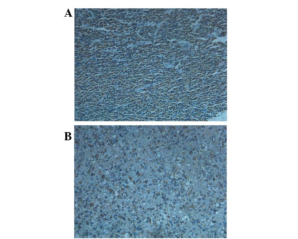
TSP-1 was mainly expressed in the cytoplasm or
intracellular space of the glioma cells and its expression was much
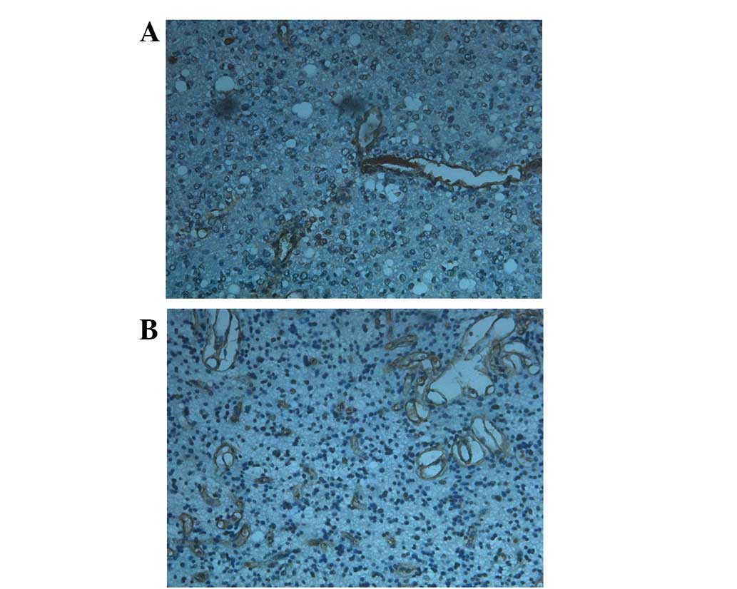
higher in low-grade than in high-grade tumors (Fig. 1). TGF-β was predominantly expressed
in the cytoplasm of vascular endothelial cells and the positive
staining was greater in high-grade gliomas than in low-grade tumors
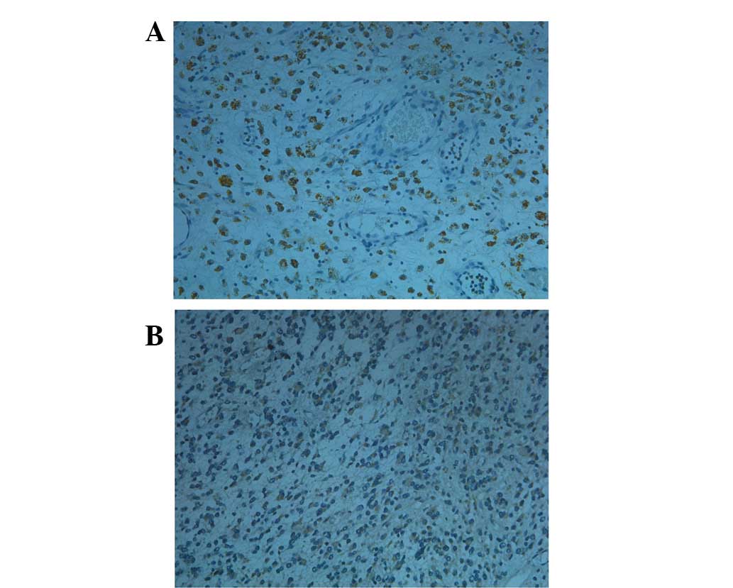
(Fig. 2). PPAR-γ was largely
cytoplasmic or nuclear and the expression was higher in low-grade
gliomas than in high-grade tumors (Fig.
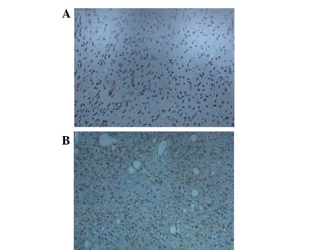
3). CD34 was mainly expressed in the cytoplasm or membrane of
vascular endothelial cells and was often tubular, streak-like,
comma-like and lumpy in staining shape, and it was much greater in
high-grade gliomas than in low-grade tumors (Fig. 4). As presented in Table III, there were significant
differences for the intense positive rate (χ2=16.4,
13.8, and 29.7, respectively) and total positive rate
(χ2=33.6, 11.2, and 29.0, respectively) of PPAR-γ, TSP-1
and TGF-β, between low- and high-grade gliomas. The MVD in the
high-grade glioma group (45±6.2 vessels/field) was significantly
higher than that in the low-grade glioma group (28±7.2
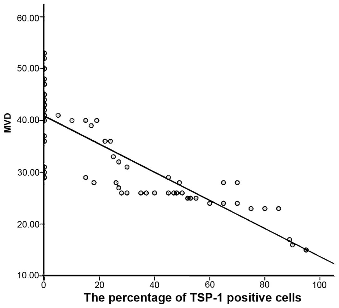
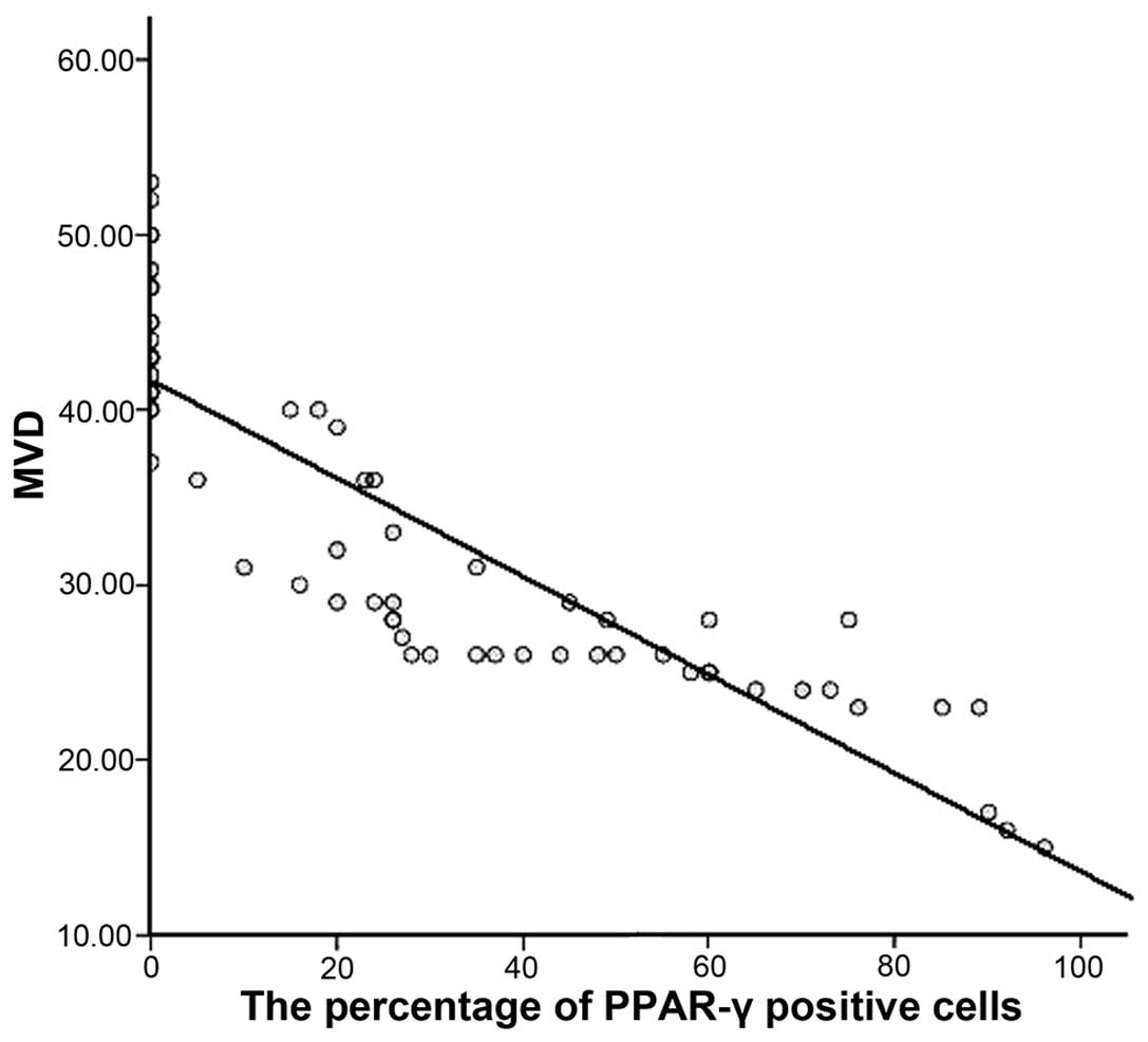
vessels/field) (t=2.17). Immunoreactivity for TSP-1 (r=−0.61;
Fig. 5) and PPAR-γ (r=−0.82;
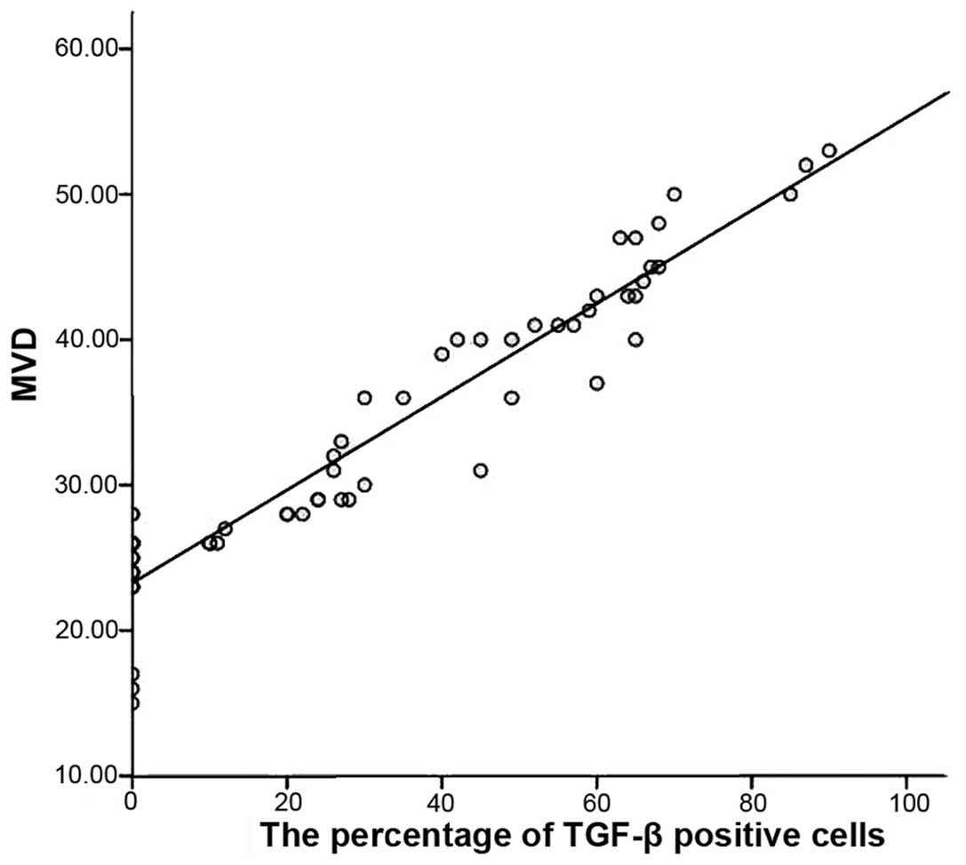
Fig. 6) was negatively correlated
with the MVD, whilst TGF-β expression levels were positively
correlated with the MVD (r=0.95; Fig.
7).
| Table IIITSP-1, TGF-β and PPAR-γ expression
levels by glioma grade. |
Table III
TSP-1, TGF-β and PPAR-γ expression
levels by glioma grade.
| Indicators | Groups | Cases (n) | Negative (−) | Mild positive
(+) | Positive (++) | Intense positive
(+++) | Total positive rate
(%) | P1-value
(χ2) | Intense positive rate
(%) | P2-value
(χ2) |
|---|
| TSP-1 | Low-grade | 51 | 11 | 3 | 18 | 19 | 78.4 | 0.00a (11.2) | 37.3 | 0.00a (13.8) |
| High-grade | 48 | 26 | 13 | 6 | 3 | 45.8 | | 6.3 | |
| TGF-β | Low-grade | 51 | 29 | 11 | 8 | 3 | 43.1 | 0.00a (29.0) | 5.9 | 0.00a (29.7) |
| High-grade | 48 | 3 | 3 | 15 | 27 | 93.8 | | 56.3 | |
| PPAR-γ | Low-grade | 51 | 3 | 8 | 19 | 21 | 94.1 | 0.00a (33.6) | 41.2 | 0.00a (16.4) |
| High-grade | 48 | 29 | 8 | 8 | 3 | 39.6 | | 6.3 | |
Discussion
TSP-1 is reportedly involved in tumor angiogenesis
by mediating endothelial cell migration and apoptosis, and
regulating vascular endothelial growth factor (VEGF) expression
(11–14), but these functions are
controversial. Previous studies have demonstrated that TSP-1
inhibits tumor angiogenesis and invasion in melanoma and breast,
prostate, and pancreatic cancers (3–6,15–17).
However, Elpek et al(18)
observed that TSP-1 promotes angiogenesis during chronic liver
injury and did not identify a correlation between TSP-1 expression
and MVD in pituitary tumors (19).
In the present study, it was demonstrated that TSP-1 is negatively
correlated with the MVD in the gliomas, and TSP-1 immunoreactivity
decreases with increasing tumor grade, suggesting that TSP-1 may
inhibit tumor angiogenesis and progression in gliomas. Fontana
et al reported that TSP-1 inhibits tumor angiogenesis in the
early stages of tumor growth and induces local hypoxia to produce
greater quantities of VEGF, which promotes angiogenesis and
inhibits TSP-1 expression (20).
TSP-1 activates latent TGF-β, thus increasing TGF-β
expression to affect the biological behavior of tumors. In
malignant tumors, TGF-β promotes tumor angiogenesis, immune
escaping and metastasis, but it has opposite effects in normal
epithelial cells during early tumor stages (1,12,13,21).
The effect that TGF-β exerts on tumor progression may ultimately
depend on the tumor microenvironment (13,14,22–24).
The results of the present study demonstrated that TGF-β is mainly
expressed in the surrounding blood vessels, and its expression is
positively correlated with the MVD, particularly in the high-grade
gliomas. These findings suggest that TGF-β may promote angiogenesis
in gliomas. The comprehensive effect of TSP-1 and TGF-β in tumors
may be influenced by the balance between anti-angiogenic and
invasive factors. Further studies are required to investigate the
interaction of TSP-1 and TGF-β in regulating glioma
angiogenesis.
The possible tumor-suppressive effect of PPAR-γ
remains controversial. PPAR-γ agonists applied in vitro are
able to inhibit tumor cell proliferation and decrease the
expression of extracellular matrix proteins, such as type I
collagen and fibronectin (8,9).
PPAR-γ agonists inhibit tumor angiogenesis through different
mechanisms. For example, they may induce and activate hepatocyte
growth factor, which activates C-methionine receptors and
upregulates Smad transcriptional repressor expression, thus
blocking the Smad pathway required for TGF-β nuclear translocation
(25). PPAR-γ agonists have also
been shown to block the epithelial-mesenchymal transition (EMT),
which inhibits tumor metastasis by inhibiting the Smad pathway
(7). In addition, PPAR-γ agonists
upregulate the expression of CD36, a TSP-1 receptor, which in turn
promotes TSP-1 expression and inhibits tumor angiogenesis (26). The present study identified that
PPAR-γ expression was significantly different between the low- and
high-grade gliomas and was negatively correlated with the MVD,
suggesting that PPAR-γ may inhibit angiogenesis in gliomas. In
addition, PPAR-γ expression in the gliomas was negatively
correlated with TGF-β (r=−0.38, P=0.002), but positively correlated
with TSP-1 (r=0.37, P=0.003) (data not shown), suggesting that
PPAR-γ inhibits angiogenesis by regulating TSP-1 and TGF-β
expression.
In the present study, we observed that the
expression levels of TSP-1, PPAR-γ, and TGF-β correlated with the
glioma grades. Furthermore, TSP-1 and PPAR-γ expression levels
negatively correlated with MVD, while TGF-β expression levels
positively correlated with MVD. Collectively, these results suggest
that TSP-1 and PPAR-γ expression levels are closely correlated with
angiogenesis in gliomas and may exert a synergistic effect, which
may provide potential therapeutic targets for glioma therapy. TSP-1
and PPAR-γ expression in gliomas may also serve as indicators for
tumor malignancy and prognosis, whereas TGF-β promotes angiogenesis
during glioma progression and its expression is correlated with the
degree of malignancy. Therefore, TGF-β expression in gliomas may
serve as an indicator for tumor malignancy.
Acknowledgements
This study was supported by the Shandong Province
Science and Technology Development Program (2010GSF10225), the
Young Doctor Foundation of Shandong Province Science and Technology
Department (2007BSB02028) and the Natural Science Foundation of
China (Y2008C64).
References
|
1
|
Crawford SE, Stellmach V, Murphy-Ullrich
JE, et al: Thrombospondin-1 is a major activator of TGF-beta1 in
vivo. Cell. 93:1159–1170. 1998. View Article : Google Scholar : PubMed/NCBI
|
|
2
|
Presser LD, Haskett A and Waris G:
Hepatitis C virus-induced furin and thrombospondin-1 activate
TGF-β1: role of TGF-β1 in HCV replication. Virology. 412:284–296.
2011.PubMed/NCBI
|
|
3
|
Recouvreux MV, Camilletti MA, Rifkin DB,
Becu-Villalobos D and Diaz-Torga G: Thrombospondin-1 (TSP-1)
analogs ABT-510 and ABT-898 inhibit prolactinoma growth and recover
active pituitary transforming growth factor-β1 (TGF-β1).
Endocrinology. 153:3861–3871. 2012.PubMed/NCBI
|
|
4
|
Hayashi H, Sakai K, Baba H and Sakai T:
Thrombospondin-1 is a novel negative regulator of liver
regeneration after partial hepatectomy through transforming growth
factor-beta1 activation in mice. Hepatology. 55:1562–1573. 2012.
View Article : Google Scholar
|
|
5
|
Nakao T, Kurita N, Komatsu M, et al:
Expression of thrombospondin-1 and Ski are prognostic factors in
advanced gastric cancer. Int J Clin Oncol. 16:145–152. 2011.
View Article : Google Scholar
|
|
6
|
Punekar S, Zak S, Kalter VG, et al:
Thrombospondin 1 and its mimetic peptide ABT-510 decrease
angiogenesis and inflammation in a murine model of inflammatory
bowel disease. Pathobiology. 75:9–21. 2008. View Article : Google Scholar : PubMed/NCBI
|
|
7
|
Reka AK, Kurapati H, Narala VR, et al:
Peroxisome proliferator-activated receptor-gamma activation
inhibits tumor metastasis by antagonizing Smad3-mediated
epithelial-mesenchymal transition. Mol Cancer Ther. 9:3221–3232.
2010. View Article : Google Scholar
|
|
8
|
Dong YW, Wang XP and Wu K: Suppression of
pancreatic carcinoma growth by activating peroxisome
proliferator-activated receptor gamma involves angiogenesis
inhibition. World J Gastroenterol. 15:441–448. 2009. View Article : Google Scholar
|
|
9
|
Shigeto T, Yokoyama Y, Xin B and Mizunuma
H: Peroxisome proliferator-activated receptor alpha and gamma
ligands inhibit the growth of human ovarian cancer. Oncol Rep.
18:833–840. 2007.
|
|
10
|
Louis DN, Ohgaki H, Wiestler OD, Cavenee
WK, Burger PC, Jouvet A, Scheithauer BW and Kleihues P: The 2007
WHO classification of tumours of the central nervous system. Acta
Neuropathol. 114:97–109. 2007. View Article : Google Scholar
|
|
11
|
Wang-Rodriguez J, Urquidi V, Rivard A and
Goodison S: Elevated osteopontin and thrombospondin expression
identifies malignant human breast carcinoma but is not indicative
of metastatic status. Breast Cancer Res. 5:R136–R143. 2003.
View Article : Google Scholar
|
|
12
|
Aspiotis M, Tsanou E, Gorezis S, et al:
Angiogenesis in pterygium: study of microvessel density, vascular
endothelial growth factor, and thrombospondin-1. Eye (Lond).
21:1095–1101. 2007. View Article : Google Scholar
|
|
13
|
Karavasilis V, Malamou-Mitsi V, Briasoulis
E, Tsanou E, Kitsou E and Pavlidis N: Clinicopathologic study of
vascular endothelial growth factor, thrombospondin-1, and
microvessel density assessed by CD34 in patients with stage III
ovarian carcinoma. Int J Gynecol Cancer. 16(Suppl 1): 241–246.
2006. View Article : Google Scholar
|
|
14
|
Baltaci S, Orhan D, Göğüs C, Filiz E,
Tulunay O and Göğüs O: Thrombospondin-1, vascular endothelial
growth factor expression and microvessel density in renal cell
carcinoma and their relationship with multifocality. Eur Urol.
44:76–81. 2003. View Article : Google Scholar
|
|
15
|
Hyder SM, Liang Y and Wu J: Estrogen
regulation of thrombospondin-1 in human breast cancer cells. Int J
Cancer. 125:1045–1053. 2009. View Article : Google Scholar : PubMed/NCBI
|
|
16
|
Fitchev PP, Wcislak SM, Lee C, et al:
Thrombospondin-1 regulates the normal prostate in vivo through
angiogenesis and TGF-beta activation. Lab Invest. 90:1078–1090.
2010. View Article : Google Scholar : PubMed/NCBI
|
|
17
|
Byrne GJ, Hayden KE, McDowell G, et al:
Angiogenic characteristics of circulating and tumoural
thrombospondin-1 in breast cancer. Int J Oncol. 31:1127–1132.
2007.
|
|
18
|
Elpek GO, Gokhan GA and Bozova S:
Thrombospondin-1 expression correlates with angiogenesis in
experimental cirrhosis. World J Gastroenterol. 14:2213–2217. 2008.
View Article : Google Scholar
|
|
19
|
Jiang M, Mou CZ, Han T, Wang M and Yang W:
Thrombospondin-1 and transforming growth factor-β1 levels in
prolactinoma and their clinical significance. J Int Med Res.
40:1284–1294. 2012.
|
|
20
|
Fontana A, Filleur S, Guglielmi J, et al:
Human breast tumors override the antiangiogenic effect of stromal
thrombospondin-1 in vivo. Int J Cancer. 116:686–691. 2005.
View Article : Google Scholar : PubMed/NCBI
|
|
21
|
Kaygusuz G, Tulunay O, Baltaci S and Gogus
O: Microvessel density and regulators of angiogenesis in malignant
and nonmalignant prostate tissue. Int Urol Nephrol. 39:841–850.
2007. View Article : Google Scholar : PubMed/NCBI
|
|
22
|
Pardali K and Moustakas A: Actions of
TGF-beta as tumor suppressor and pro-metastatic factor in human
cancer. Biochim Biophys Acta. 1775:21–62. 2007.PubMed/NCBI
|
|
23
|
Karavasilis V, Malamou-Mitsi V, Briasoulis
E, et al: Angiogenesis in cancer of unknown primary:
clinicopathological study of CD34, VEGF and TSP-1. BMC Cancer.
5:252005. View Article : Google Scholar : PubMed/NCBI
|
|
24
|
Kasper HU, Ebert M, Malfertheiner P,
Roessner A, Kirkpatrick CJ and Wolf HK: Expression of
thrombospondin-1 in pancreatic carcinoma: correlation with
microvessel density. Virchows Arch. 438:116–120. 2001. View Article : Google Scholar : PubMed/NCBI
|
|
25
|
Li Y, Wen X, Spataro BC, Hu K, Dai C and
Liu Y: Hepatocyte growth factor is a downstream effector that
mediates the antifibrotic action of peroxisome
proliferator-activated receptor-gamma agonists. J Am Soc Nephrol.
17:54–65. 2006. View Article : Google Scholar
|
|
26
|
Boyer JF, Balard P, Authier H, et al:
Tumor necrosis factor alpha and adalimumab differentially regulate
CD36 expression in human monocytes. Arthritis Res Ther. 9:R222007.
View Article : Google Scholar : PubMed/NCBI
|