Atypical imaging observations of branchial cleft cysts

  • Authors:
    • Su Hu
    • Chun‑Hong Hu
    • Ling Yang
    • Jian‑Ming Xing
    • Jian‑Hua Chen
    • Zi‑Li Ge
    • Ji‑Sheng Liu
  • View Affiliations

  • Published online on: November 4, 2013     https://doi.org/10.3892/ol.2013.1656
  • Pages: 219-222
Metrics: Total Views: 0 (Spandidos Publications: | PMC Statistics: )
Total PDF Downloads: 0 (Spandidos Publications: | PMC Statistics: )


Abstract

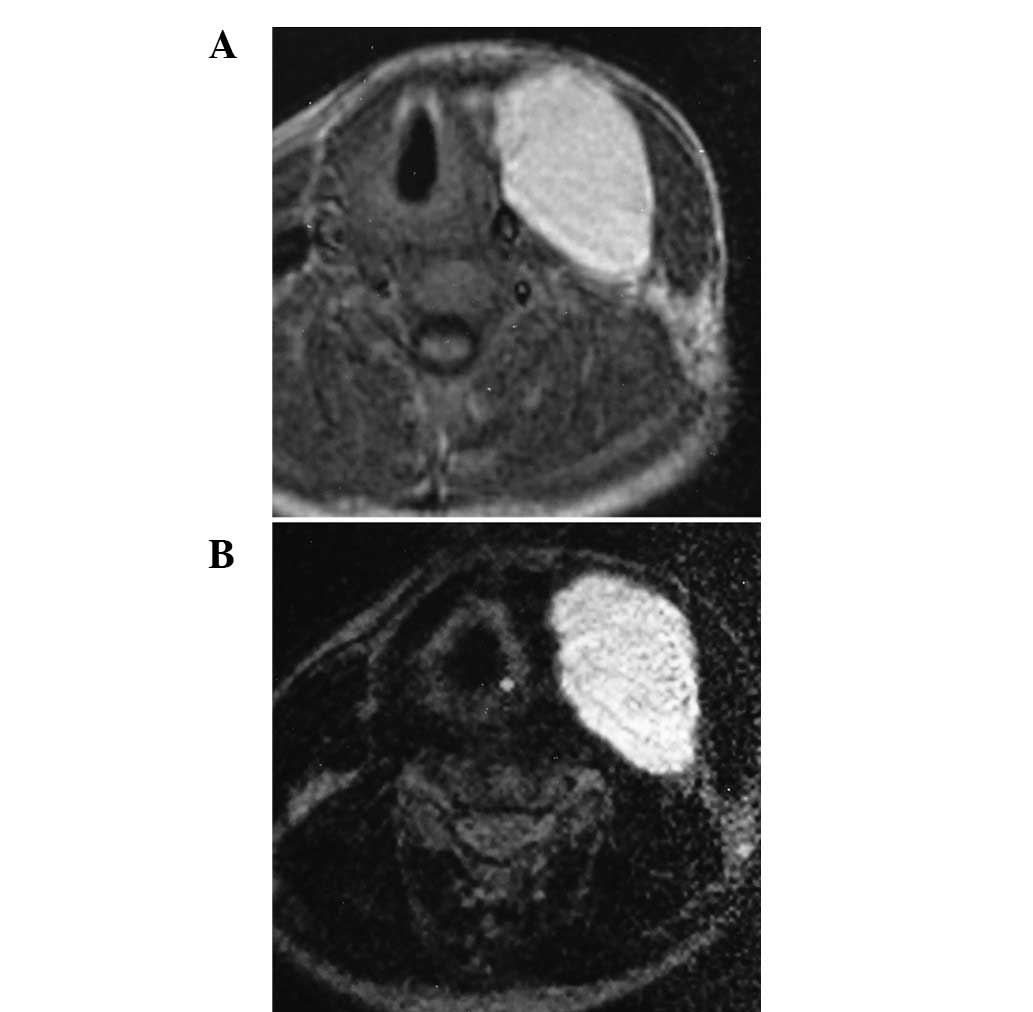
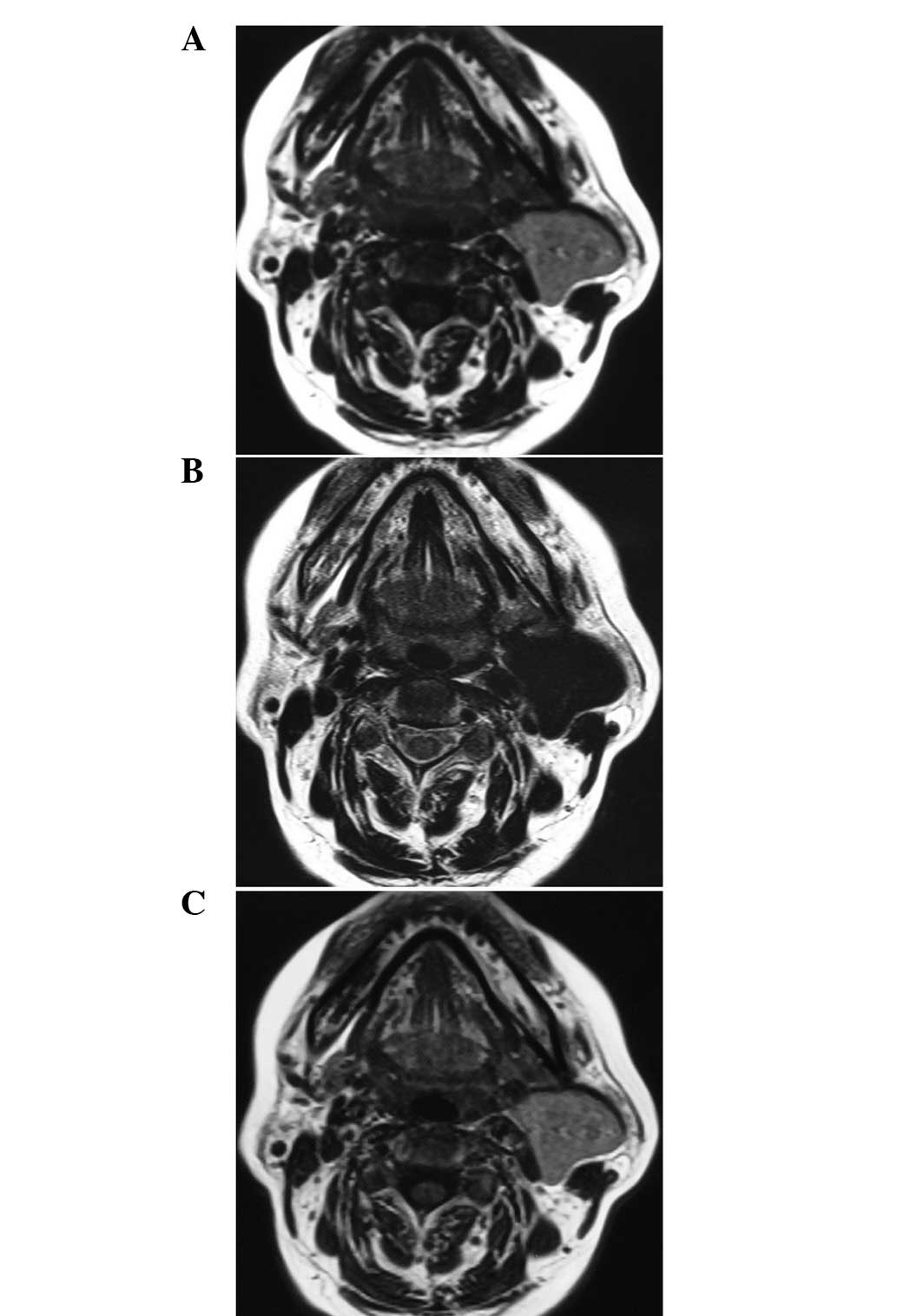
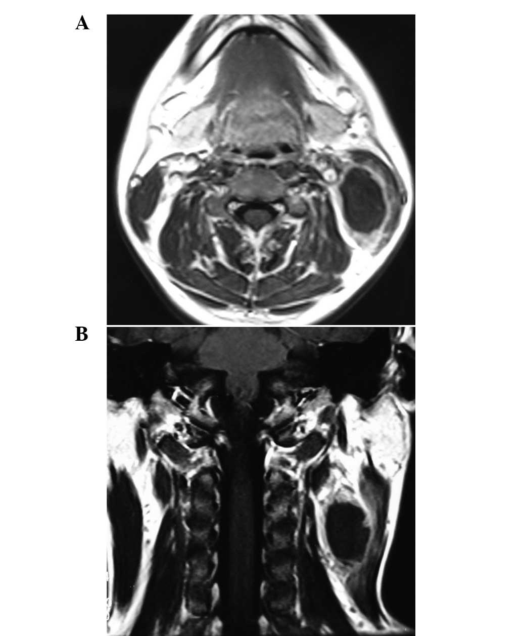
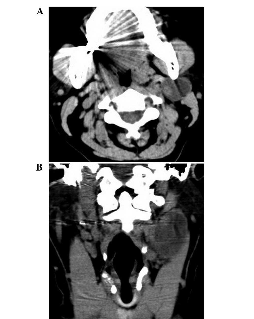
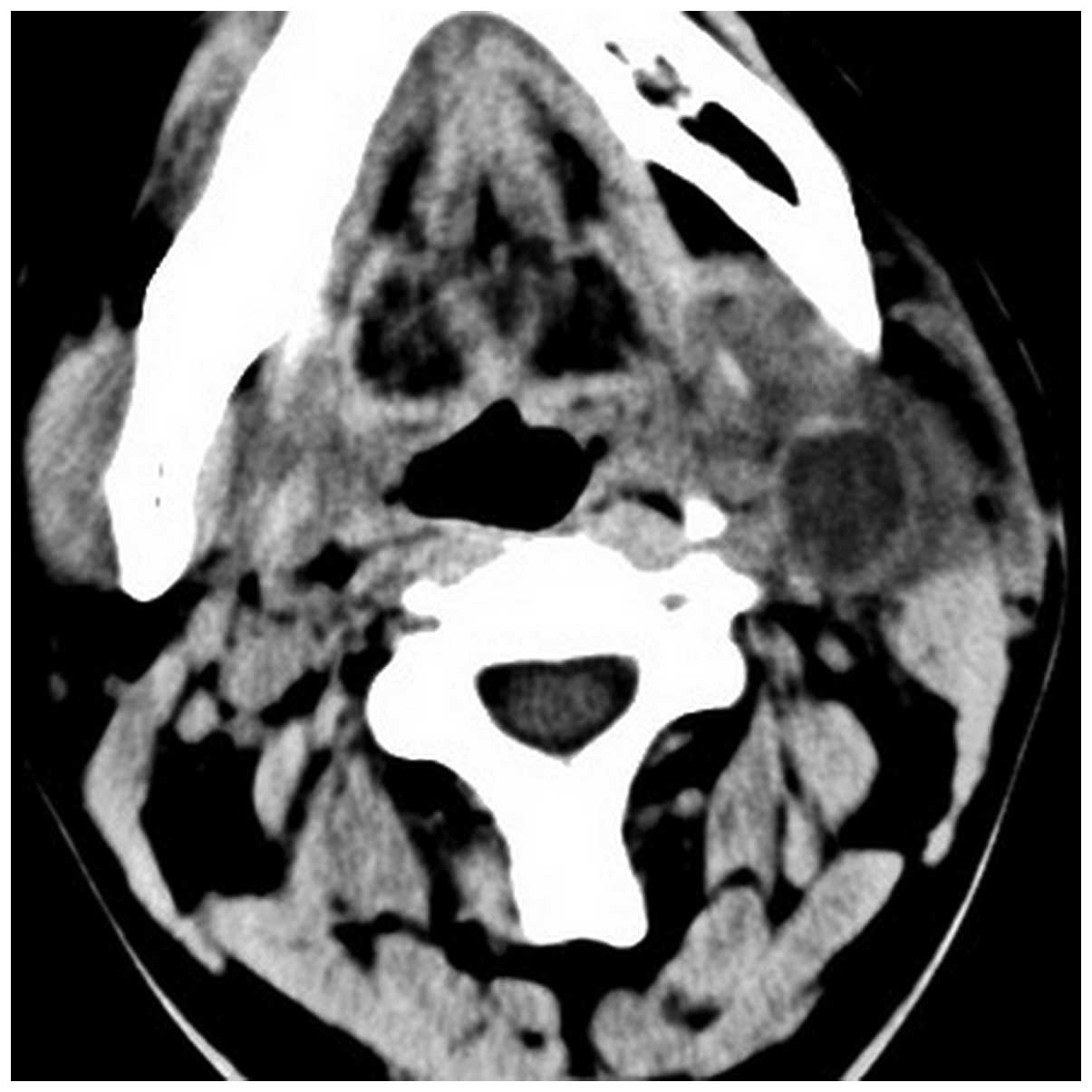
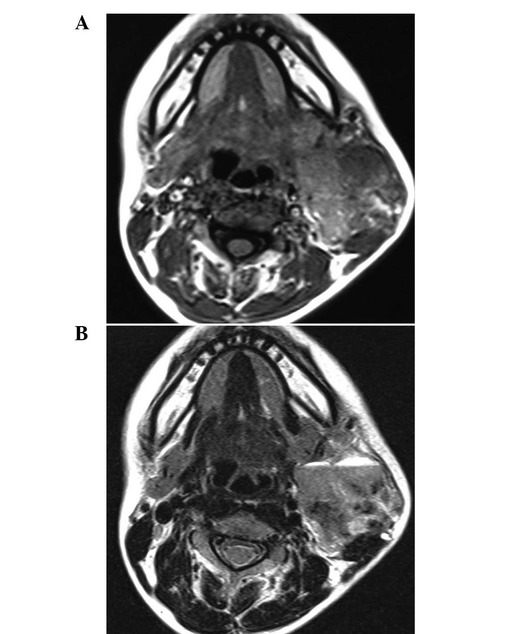
The aim of the present study was to assess the atypical imaging manifestations of branchial cleft cysts (BCCs) confirmed by pathology. Computerized tomography (CT) or magnetic resonance imaging (MRI) of 17 BCC cases were reviewed. The imaging features, including laterality, location, border, attenuation and internal architecture, were evaluated. All 17 cases were second BCCs, including 5 cases of Bailey type Ⅰ classification cysts and 12 cases of type Ⅱ classification cysts. The atypical imaging features included signal and morphological abnormalities. The abnormal signal intensities were caused by intracapsular bleeding (n=2) or solidification of cystic fluid (n=2). Intracystic hemorrhaging revealed homogeneous hyperintensity on T1‑weighted image (T1WI) and T2‑weighted image (T2WI). Solidification of cystic fluid revealed slightly homogeneous hyperintensity compared with muscle on T1WI and homogeneous hypointensity on T2WI without enhancement. The aberrant morphology mainly presented as thickening of the cystic wall (n=13). Thickened walls of BCCs with ill‑ (n=5) or well‑ (n=8) defined borders were observed in 13 patients. In 3 patients, significant enhancement was identified following intravenous gadolinium administration (n=4). When with atypical CT or MRI features are presented, the typical location of BCCs can help in the diagnosis, as it is located at the lateral portion of the neck adjacent to the anterior border of the mandibular angle or sternocleidomastoid muscle. The atypical observations, including variable signals, imply that the cystic content has changed. Thickened walls indicate inflammation or cancerous tendency and patients with ill‑defined margins, vascular involvement or lymphadenopathy atelectasis indicate malignant conversion.
View Figures
View References

Related Articles

Journal Cover

2014-January
Volume 7 Issue 1

Print ISSN: 1792-1074
Online ISSN:1792-1082

Sign up for eToc alerts

Recommend to Library

Copy and paste a formatted citation
x
Spandidos Publications style
Hu S, Hu CH, Yang L, Xing JM, Chen JH, Ge ZL and Liu JS: Atypical imaging observations of branchial cleft cysts. Oncol Lett 7: 219-222, 2014
APA
Hu, S., Hu, C., Yang, L., Xing, J., Chen, J., Ge, Z., & Liu, J. (2014). Atypical imaging observations of branchial cleft cysts. Oncology Letters, 7, 219-222. https://doi.org/10.3892/ol.2013.1656
MLA
Hu, S., Hu, C., Yang, L., Xing, J., Chen, J., Ge, Z., Liu, J."Atypical imaging observations of branchial cleft cysts". Oncology Letters 7.1 (2014): 219-222.
Chicago
Hu, S., Hu, C., Yang, L., Xing, J., Chen, J., Ge, Z., Liu, J."Atypical imaging observations of branchial cleft cysts". Oncology Letters 7, no. 1 (2014): 219-222. https://doi.org/10.3892/ol.2013.1656