Introduction
Despite a global decline in the incidence and
mortality of gastric cancer in the last 60 years, it remains the
fourth most common and second most frequent cause of cancer-related
mortality. Gastric cancer continues to be a major health concern
due to the slow decrease in incidence in Asia and high mortality
from diagnosed gastric carcinomas in the West, despite the advanced
diagnostic and operative techniques that are commonly used in
clinical practice (1,2). An increased understanding of the
changes that occur in gene expression in gastric cancer,
particularly the identification of novel biomarkers for cancer
diagnosis and novel targets for treatment, is required for the
improvement of diagnosis, treatment and prevention.
Paxillin is a focal adhesion-associated,
phosphotyrosine-containing 68-kDa adaptor protein discovered in
1990 by Turner et al(3).
Paxillin contains a number of motifs that mediate protein-protein
interactions, including C-terminal LIM domains resembling a double
zinc-finger domain, N-terminal LD motifs, SH3 and SH2
domain-binding sites, whose motifs serve as docking sites for
cytoskeletal proteins, tyrosine kinases, serine/threonine kinases,
GTPase activating proteins and other adaptor proteins that recruit
additional enzymes into complex with paxillin (4). Multiple tyrosine, serine and threonine
phosphorylation sites exist throughout the paxillin molecule, and
are targeted by a diverse array of kinases that are activated in
response to various adhesion stimuli and growth factors (PDGF, EGF
and IL-3). These include p21-activated kinase, FAK-Src, receptor
for activated C kinase 1, c-Jun N-terminal kinase,
extracellular-signal-regulated kinase, p38 mitogen-activated
protein kinase, cyclin-dependent kinase 5 and c-Abl. Paxillin is
tyrosine-phosphorylated upon integrin engagement or growth factor
stimulation, creating binding sites for the Crk adapter protein
(5,6). Thus, paxillin may be involved in
signal transduction, regulation of cell morphology and the
recruitment of structural and signaling molecules to focal
adhesions to control cell spread and migration (7,8).
Previous studies have demonstrated that paxillin was
overexpressed in esophageal squamous cell carcinoma, lung
carcinoma, breast cancer and prostate cancer (9–13). In
breast cancer, it has been found that the overexpression of
paxillin may represent a useful prognosticator and be employed to
predict the clinical response to chemotherapy (12,14).
To better understand the clinicopathological and prognostic
significance of paxillin, we observed its expression in gastric
non-neoplastic mucosa, adenoma and carcinoma using a combination of
tissue microarray and immunohistochemistry. Paxillin expression was
compared with the clinicopathological and prognostic features of
gastric cancer.
Materials and methods
Patients
This retrospective study was carried out using
curatively resected specimens of gastric cancer (n=392) and
adjacent non-neoplastic mucosa (n=197) collected at Toyama
University Hospital (Toyama, Japan) from 1993 to 2006. The adenoma
samples were resected from endoscopic biopsy at Toyama University
Hospital from 1997 to 2008. The patients with gastric carcinomas
were 120 males and 272 females (38–88 years; mean, 66.7 years).
Archival materials were obtained from the Department of Pathology
of Toyama University Hospital. In 151 cases, tumor development was
accompanied by lymph node metastasis. None of the patients
underwent chemotherapy, radiotherapy and adjuvant treatment prior
to surgery. All patients were followed up by consulting their case
documents and by telephone.
Pathology
All tissues were fixed in 10% neutralized formalin,
embedded in paraffin, cut into 4-μm sections and stained with
hematoxylin and eosin (H&E) in order to confirm the
histological diagnosis and microscopic characteristics of the
specimens. The staging for each gastric carcinoma was evaluated
according to the Union for International Cancer Control system,
which indicates the extent of tumor spread (15). Histological architecture was defined
using the Lauren classification (16,17).
The tumor size, depth of invasion, lymphatic and venous invasion,
and lymph node metastasis of tumors were also determined.
Tissue microarray (TMA)
From H&E-stained sections of the tumor cases,
representative areas of solid tumor were selected for sampling and
2-mm diameter tissue cores per donor block were punched out and
transferred to a recipient block with a maximum of 48 cores using a
tissue microarrayer (KIN-1; Azumaya, Tokyo, Japan). Sections (4-μm)
were consecutively cut from the microarrays and transferred to
poly-lysine-coated glass slides.
Immunohistochemistry
Serial sections of TMA were deparaffinized with
xylene, rehydrated with alcohol, and subjected to
immunohistochemical staining with intermittent microwave radiation,
as previously described (18).
Rabbit anti-human paxillin antibody (Epitomics, Inc., Burlingame,
CA, USA) was used at 1:100 dilution to detect the respective
proteins, with anti-rabbit Envison-PO (Dako, Carpinteria, CA, USA)
as the secondary antibody. Binding was visualized with
3,3′-diaminobenzidine and counterstaining with Mayer’s hematoxylin
was performed to aid orientation. Omission of the primary antibody
was used as a negative control.
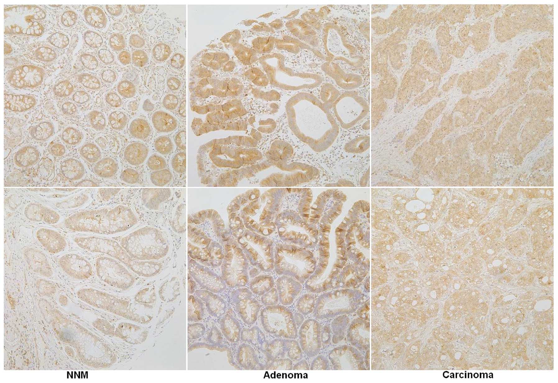
Immunoreactivity for paxillin showed a cytoplasmic
pattern (Fig. 1). One hundred cells
were randomly selected and counted from five representative fields
of each section, blindly, by three independent observers (L.J. Xiao
and H.C. Zheng). The inconsistent data were confirmed by both
observers until final agreements were reached. The expression
positivity was graded and counted as follows: 0 = 0%; 1 = 1–49%; 2
= 50–74%; and 3≥ 75%. The staining intensity score was graded as
follows: 1 = weak; 2 = intermediate; and 3 = strong. The scores for
paxillin positivity and staining intensity were multiplied to
obtain a final score, which determined their expression as − = 0; +
= 1–2; ++ = 3–4; or +++ = 6–9.
Statistical analysis
Statistical evaluation was performed using
Spearman’s rank correlation test. Kaplan-Meier survival plots were
generated and comparisons between survival curves were made with
the log-rank test. Cox proportional hazards model was employed for
multivariate analysis. SPSS 17.0 software (SPSS, Inc., Chicago, IL,
USA) was applied to analyze all data, and P<0.05 was considered
to indicate a statistically significant difference.
Results
As indicated in Fig.
1, paxillin was positively expressed in the cytoplasm of
gastric superficial epithelium, intestinal metaplasia, adenoma and
carcinoma. The levels of paxillin expression was detected in
gastric non-neoplastic mucosa (64.5%, 127/197), adenoma (92.3%,
60/67) and carcinoma (66.8%, 262/392), respectively. The expression
of paxillin was significantly more highly expressed in gastric
adenoma than in non-neoplastic mucosa and carcinoma (P<0.05,
Table I). As shown in Table II, paxillin expression was
negatively correlated with tumor size, depth of invasion, and lymph
node metastasis, but not with gender, lymphatic or venous invasion,
or TNM staging (P>0.05). Paxillin expression was higher in the
elder carcinoma patients than in the younger carcinoma patients
(P<0.05). There was higher paxillin expression in intestinal-
compared with diffuse-type carcinoma (P<0.05).
 | Table IPaxillin expression in gastric
carcinomas. |
Table I
Paxillin expression in gastric
carcinomas.
| | Paxillin
expression | |
|---|
| |
| |
|---|
| Groups | n | − | + | ++ | +++ | PR (%) |
|---|
| Non-cancerous
mucosa | 197 | 70 | 91 | 26 | 10 | 64.5 |
| Adenoma | 67 | 7 | 21 | 28 | 11 | 92.3a |
| Carcinoma | 392 | 130 | 164 | 59 | 39 | 66.8 |
| Table IICorrelation between paxillin
expression and clinicopathological features of gastric
carcinomas. |
Table II
Correlation between paxillin
expression and clinicopathological features of gastric
carcinomas.
| | Paxillin
expression | | |
|---|
| |
| | |
|---|
| Clinicopathological
features | n | − | + | ++ | +++ | PR (%) | P-value |
|---|
| Age (years) | | | | | | | 0.027 |
| <65 | 156 | 56 | 68 | 19 | 13 | 64.1 | |
| ≥65 | 236 | 74 | 96 | 40 | 26 | 68.6 | |
| Gender | | | | | | | 0.060 |
| Male | 272 | 89 | 105 | 46 | 32 | 67.3 | |
| Female | 120 | 41 | 59 | 13 | 7 | 65.8 | |
| Tumor size (cm) | | | | | | | 0.001 |
| <4 | 204 | 58 | 80 | 40 | 26 | 71.6 | |
| ≥4 | 188 | 72 | 84 | 19 | 13 | 61.7 | |
| Depth of
invasion | | | | | | | <0.001 |
|
Tis-1 | 200 | 59 | 85 | 35 | 21 | 70.5 | |
| T2–4 | 192 | 71 | 79 | 24 | 18 | 63.0 | |
| Lymphatic
invasion | | | | | | | 0.799 |
| − | 250 | 80 | 111 | 32 | 27 | 68.0 | |
| + | 142 | 50 | 53 | 27 | 12 | 64.8 | |
| Venous
invasion | | | | | | | |
| − | 335 | 113 | 142 | 48 | 32 | 66.3 | 0.287 |
| + | 57 | 17 | 22 | 11 | 7 | 70.2 | |
| Lymph node
metastasis | | | | | | | |
| − | 241 | 70 | 102 | 39 | 30 | 71.0 | 0.006 |
| + | 151 | 60 | 62 | 20 | 9 | 60.3 | |
| UICC staging | | | | | | | 0.352 |
| 0–I | 215 | 68 | 89 | 36 | 22 | 68.4 | |
| II–IV | 177 | 62 | 75 | 23 | 17 | 65.0 | |
| Lauren
classification | | | | | | | <0.001 |
| Intestinal
type | 210 | 46 | 98 | 36 | 30 | 78.1 | |
| Diffuse type | 172 | 80 | 62 | 22 | 8 | 53.5 | |
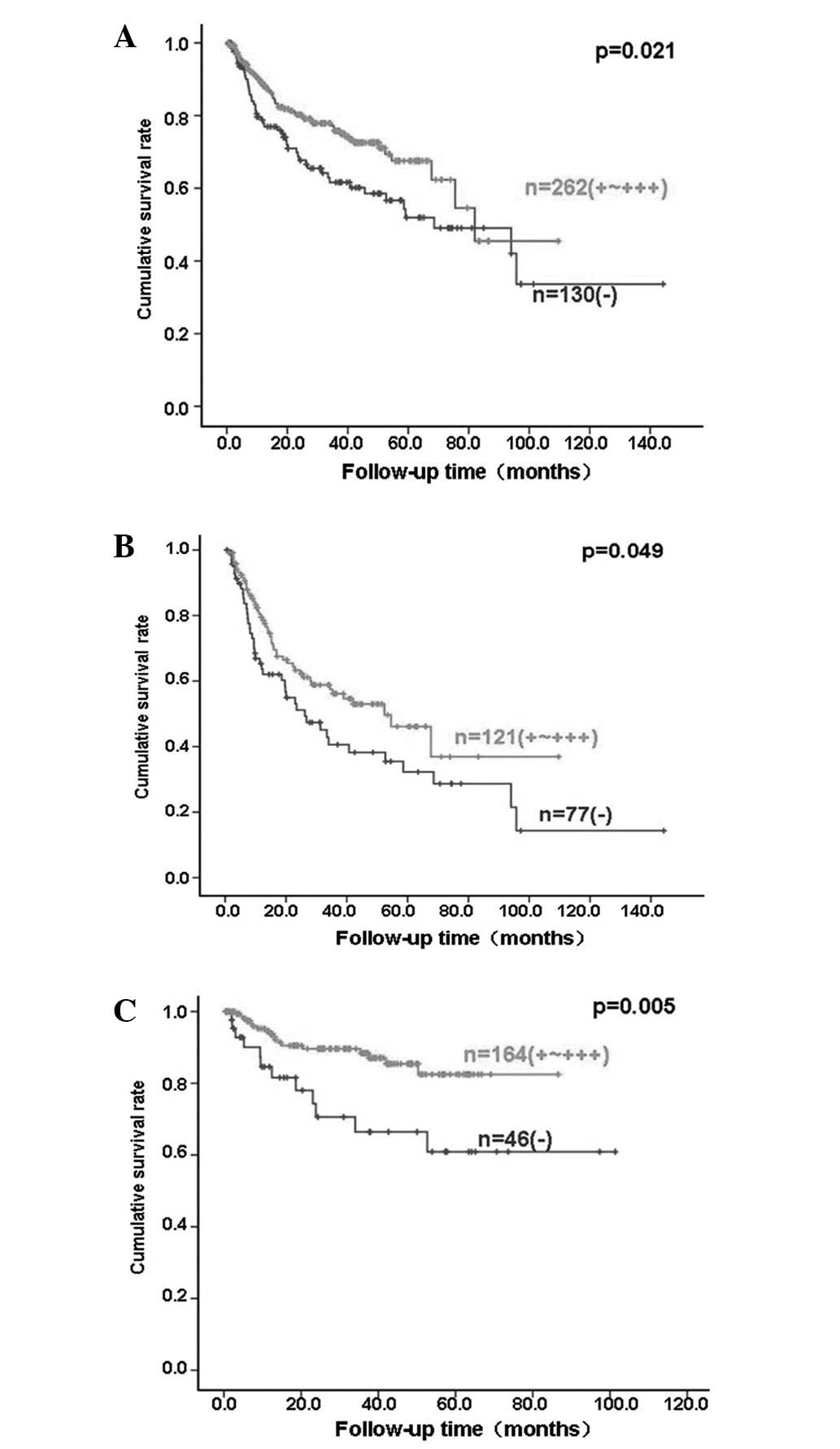
Follow-up information was available on 392 of the
gastric carcinoma patients for periods ranging from 0.2 months to
121 months (mean, 70.4 months). Fig.
2 shows survival curves stratified according to paxillin
expression. Univariate analyses using the Kaplan-Meier method
indicated a higher cumulative survival rate in all, advanced and
intestinal-type carcinoma patients with weak, moderate and strong
paxillin expression than in those without paxillin expression
(P<0.05). Multivariate analysis using the Cox proportional
hazards model indicated that patient age, depth of invasion,
lymphatic invasion, lymph node metastasis, TNM staging and Lauren
classification (P<0.05), but not patient gender, tumor size,
venous invasion or paxillin expression (P>0.05), were
independent prognostic factors for all gastric carcinomas (Table III).
| Table IIIMultivariate analysis of
clinicopathological variables for survival with gastric
carcinomas. |
Table III
Multivariate analysis of
clinicopathological variables for survival with gastric
carcinomas.
| Clinicopathological
parameters | Relative risk (95%
CI) | P-value |
|---|
| Age (≥65
years) | 1.902
(1.254–2.883) | 0.002 |
| Gender (male) | 1.212
(0.750–1.959) | 0.432 |
| Tumor size (≥4
cm) | 1.285
(0.771–2.141) | 0.336 |
| Depth of invasion
(T2–4) | 5.979
(2.084–17.152) | 0.001 |
| Lymphatic invasion
(+) | 1.995
(1.201–3.313) | 0.008 |
| Venous invasion
(+) | 1.202
(0.751–1.922) | 0.444 |
| Lymph node
metastasis (+) | 2.932
(1.535–5.602) | 0.001 |
| TNM staging
(III–IV) | 0.341
(0.119–0.974) | 0.045 |
| Lauren
classification (diffuse type) | 2.235
(1.396–3.577) | 0.001 |
| Paxillin expression
(+~+++) | 0.714
(0.475–1.073) | 0.105 |
Discussion
Paxillin is a cytoskeletal protein that was recently
identified as a component of focal adhesions and links between
F-actin and integrin (19). In the
present study, the cytoplasmic expression pattern of paxillin was
observed in the gastric non-neoplastic epithelial cells, adenomas
and adenocarcinomas. Statistically, paxillin expression was
increased in gastric adenoma in comparison with that in the
non-neoplastic mucosa and carcinoma. The adenoma can progress into
and be incorporated with gastric well-differentiated carcinoma when
it grows larger and de novo carcinogenesis is well
understood, particularly in diffuse-type gastric carcinomas
(20). These findings suggested
that aberrant paxillin expression may be involved in the
progression from gastric adenoma to adenocarcinoma. Higher paxillin
expression in adenoma and intestinal-type carcinoma indicated that
paxillin overexpression may be closely linked to the intestinal
carcinogenic pathway of gastric cancer.
Cai et al(21) found that paxillin mRNA expression
levels were significantly correlated with the differentiation
degree, depth of invasion and lymph node metastasis of esophageal
carcinoma. A previous study indicated that paxillin expression was
correlated with distant metastasis and clinical stage of salivary
adenoid cystic carcinoma (22). Li
et al(23) documented that
positive paxillin expression was significantly associated with low
differentiation, with the presence of portal vein thrombosis, and
with extra-hepatic metastasis of hepatocellular cell carcinoma. Li
et al(24) found that
paxillin positivity in human gastric cancer was associated with
tumor stage, and siRNA targeting paxillin decreased the
phosphorylation of paxillin (tyr118) and the invasiveness of AGS
cells significantly as compared with controls. Previously, it was
identified that overexpression of wild-type paxillin plasmids
promoted cell proliferation and also enhanced migration, invasive
capacity and metastasis of the colorectal cancer cells (25). However, paxillin expression was
negatively correlated with tumor size, depth of invasion and lymph
node metastasis of gastric cancer in the present study. The
contradictory phenomena should be further investigated in the
future.
Although all types of gastric cancer are malignant
tumors that originate from the same gastric epithelium, the
morphological features of the cancers vary substantially in
individual patients. According to Lauren classification, gastric
intestinal-type carcinoma is characterized by cohesive carcinoma
cells that form gland-like tubular structures, such as well- and
moderately differentiated carcinoma; while cell cohesion is less
apparent or absent in diffuse-type carcinoma, such as poorly
differentiated or signet ring cell carcinoma (16,17).
Our findings demonstrated that paxillin was more highly expressed
with a higher incidence in intestinal-type gastric cancer, which is
presumed to arise from preceding dysplastic lesions, than
diffuse-type ones, which evolve without any precedent dysplastic
changes, indicating that distinct paxillin expression underlies the
molecular mechanisms for the differentiation of intestinal- and
diffuse-type carcinomas.
To date, there have been several studies describing
the prognostic significance of paxillin expression in malignancies
(11,12,14,26).
In the present study, for the first time, we analyzed the
correlation between paxillin expression and the survival rate of
392 patients with gastric carcinoma. The results revealed a close
association between its overexpression and favorable survival. When
stratified according to the depth of invasion, the significant
correlation disappeared in the early gastric cancers, but not in
the advanced ones, indicating that the association between paxillin
expression and prognosis depends on the depth of invasion. The
multivariate analysis demonstrated that patient age, depth of
invasion, lymphatic invasion, lymph node metastasis, TNM staging
and Lauren classification, but not patient gender, tumor size,
venous invasion or paxillin expression, were independent prognostic
factors for all gastric carcinomas. These findings suggested that
paxillin expression is a good indicator for the favorable prognosis
of gastric carcinoma patients, albeit it is not independent. By
contrast, Li et al(11)
found that no correlation occurred between expression of paxillin
and patient survival of these patients with esophageal cancer. Zuo
et al(26) found that
paxillin expression was closely correlated with the prognosis of
non-small cell lung carcinoma.
In conclusion, aberrant paxillin expression may be
important in the malignant transformation of gastric epithelial
cells. Its reduced expression was closely correlated with growth,
invasion, metastasis and a worse prognosis of gastric carcinomas.
Its expression may be employed to differentiate between the
intestinal- and diffuse-type carcinomas. It was considered as a
promising marker to indicate the pathobiological behaviors and
prognosis of gastric carcinomas.
Acknowledgements
This study was supported by Shenyang Science and
Technology Grant (F11-264-1-10; F12-277-1-01); Liaoning Science and
Technology Grant; Natural Scientific Foundation of China (81172371)
and Grant-in aid for Scientific Research from the Ministry of
Education, Culture, Sports and Technology of Japan (23659958).
References
|
1
|
Rivera F, Vega-Villegas ME and López-Brea
MF: Chemotherapy of advanced gastric cancer. Cancer Treat Rev.
33:315–324. 2007. View Article : Google Scholar
|
|
2
|
Kelley JR and Duggan JM: Gastric cancer
epidemiology and risk factors. J Clin Epidemiol. 56:1–9. 2003.
View Article : Google Scholar : PubMed/NCBI
|
|
3
|
Turner CE, Glenney JR Jr and Burridge K:
Paxillin: a new vinculin-binding protein present in focal
adhesions. J Cell Biol. 111:1059–1068. 1990. View Article : Google Scholar : PubMed/NCBI
|
|
4
|
Deakin NO and Turner CE: Paxillin comes of
age. J Cell Sci. 121:2435–2444. 2008. View Article : Google Scholar : PubMed/NCBI
|
|
5
|
Schaller MD: Paxillin: a focal
adhesion-associated adaptor protein. Oncogene. 20:6459–6472. 2001.
View Article : Google Scholar : PubMed/NCBI
|
|
6
|
Brown MC and Turner CE: Paxillin: adapting
to change. Physiol Rev. 84:1315–1339. 2004. View Article : Google Scholar : PubMed/NCBI
|
|
7
|
Sattler M, Pisick E, Morrison PT and
Salgia R: Role of the cytoskeletal protein paxillin in oncogenesis.
Crit Rev Oncog. 11:63–76. 2000. View Article : Google Scholar : PubMed/NCBI
|
|
8
|
Huang C, Jacobson K and Schaller MD: A
role for JNK-paxillin signaling in cell migration. Cell Cycle.
3:4–6. 2004. View Article : Google Scholar : PubMed/NCBI
|
|
9
|
Tremblay L, Hauck W, Aprikian AG, Begin
LR, Chapdelaine A and Chevalier S: Focal adhesion kinase (pp125FAK)
expression, activation and association with paxillin and p50CSK in
human metastatic prostate carcinoma. Int J Cancer. 68:164–171.
1996. View Article : Google Scholar : PubMed/NCBI
|
|
10
|
Jagadeeswaran R, Surawska H, Krishnaswamy
S, Janamanchi V, Mackinnon AC, Seiwert TY, Loganathan S, Kanteti R,
Reichman T, Nallasura V, et al: Paxillin is a target for somatic
mutations in lung cancer: implications for cell growth and
invasion. Cancer Res. 68:132–142. 2008. View Article : Google Scholar : PubMed/NCBI
|
|
11
|
Li BZ, Lei W, Zhang CY, Zhou F, Li N, Shi
SS, Feng XL, Chen ZL, Hang J, Qiu B, et al: Increased expression of
paxillin is found in human oesophageal squamous cell carcinoma: a
tissue microarray study. J Int Med Res. 36:273–278. 2008.
View Article : Google Scholar
|
|
12
|
Short SM, Yoder BJ, Tarr SM, Prescott NL,
Laniauskas S, Coleman KA, Downs-Kelly E, Pettay JD, Choueiri TK,
Crowe JP, et al: The expression of the cytoskeletal focal adhesion
protein paxillin in breast cancer correlates with HER2
overexpression and may help predict response to chemotherapy: a
retrospective immunohistochemical study. Breast J. 13:130–139.
2007. View Article : Google Scholar
|
|
13
|
Mackinnon AC, Tretiakova M, Henderson L,
Mehta RG, Yan BC, Joseph L, Krausz T, Husain AN, Reid ME and Salgia
R: Paxillin expression and amplification in early lung lesions of
high-risk patients, lung adenocarcinoma and metastatic disease. J
Clin Pathol. 64:16–24. 2011. View Article : Google Scholar : PubMed/NCBI
|
|
14
|
Scibelli A, d’Angelo D, Pelagalli A,
Tafuri S, Avallone L, Della Morte R and Staiano N: Expression
levels of the focal adhesion-associated proteins paxillin and
p130CAS in canine and feline mammary tumors. Vet Res. 34:193–202.
2003. View Article : Google Scholar : PubMed/NCBI
|
|
15
|
Sobin LH and Wittekind CH: TNM
Classification of malignant tumors. 6th edn. New Jersey: John Wiley
& Sons; Hoboken: 2002
|
|
16
|
Zheng H, Takahashi H, Murai Y, Cui Z,
Nomoto K, Miwa S, Tsuneyama K and Takano Y: Pathobiological
characteristics of intestinal and diffuse-type gastric carcinoma in
Japan: an immunostaining study on the tissue microarray. J Clin
Pathol. 60:273–277. 2007. View Article : Google Scholar
|
|
17
|
Zheng HC, Li XH, Hara T, Masuda S, Yang
XH, Guan YF and Takano Y: Mixed-type gastric carcinomas exhibit
more aggressive features and indicate the histogenesis of
carcinomas. Virchows Arch. 452:525–534. 2008. View Article : Google Scholar
|
|
18
|
Kumada T, Tsuneyama K, Hatta H, Ishizawa S
and Takano Y: Improved 1-h rapid immunostaining method using
intermittent microwave irradiation: practicability based on 5 years
application in Toyama Medical and Pharmaceutical University
Hospital. Mod Pathol. 17:1141–1149. 2004.
|
|
19
|
Chen J and Gallo KA: MLK3 regulates
paxillin phosphorylation in chemokine- mediated breast cancer cell
migration and invasion to drive metastasis. Cancer Res.
72:4130–4140. 2012. View Article : Google Scholar : PubMed/NCBI
|
|
20
|
Zheng HC, Tsuneyama K, Takahashi H, Miwa
S, Sugiyama T, Popivanova BK, Fujii C, Nomoto K, Mukaida N and
Takano Y: Aberrant Pim-3 expression is involved in gastric
adenoma-adenocarcinoma sequence and cancer progression. J Cancer
Res Clin Oncol. 134:481–488. 2008. View Article : Google Scholar : PubMed/NCBI
|
|
21
|
Cai HX, Yang LC, Song XH, Liu ZR, Chen YB
and Dong GK: Expression of paxillin and FAK mRNA and the related
clinical significance in esophageal carcinoma. Mol Med Rep.
5:469–472. 2012.PubMed/NCBI
|
|
22
|
Shi J, Wang S, Zhao E, Shi L, Xu X and
Fang M: Paxillin expression levels are correlated with clinical
stage and metastasis in salivary adenoid cystic carcinoma. J Oral
Pathol Med. 39:548–551. 2010.PubMed/NCBI
|
|
23
|
Li HG, Xie DR, Shen XM, Li HH, Zeng H and
Zeng YJ: Clinicopathological significance of expression of
paxillin, syndecan-1 and EMMPRIN in hepatocellular carcinoma. World
J Gastroenterol. 11:1445–1451. 2005. View Article : Google Scholar : PubMed/NCBI
|
|
24
|
Li D, Ding J, Wang X, Wang C and Wu T:
Fibronectin promotes tyrosine phosphorylation of paxillin and cell
invasiveness in the gastric cancer cell line AGS. Tumori.
95:769–779. 2009.PubMed/NCBI
|
|
25
|
Jun Q, Zhiwei W, Lilin M, Jing K and
Qichao N: Effects of paxillin on HCT-8 human colorectal cancer
cells. Hepatogastroenterology. 58:1951–1955. 2011.PubMed/NCBI
|
|
26
|
Zuo W and Li H: Relationship of the
expression of CD44v6 and paxillin to the prognosis of non-small
cell lung carcinoma. Sichuan Da Xue Xue Bao Yi Xue Ban. 34:484–485.
2003.(In Chinese).
|